NIKOLAUS FLOIMAYR2, PETER KOCH1 1-3
1
Giant cell tumors of the bone are characterized by aggressive growth. Joint involvement often ends in arthroplasty. Especially for young patients, transplantation of an osteochondral allograft might delay joint replacement and can provide good functional outcome in selected cases. NIKOLAUS FLOIMAYR 2 , PETER KOCH 1 ,BRUNO FUCHS 1-3 Sarcoma Service, Cantonal Hospitals Winterthur 1 & Luzern 2 , University Hospital 3 www.SARCOMA.SURGERY / www.SWISS-SARCOMA.NET INTRODUCTION CONCLUSION PATIENT & METHODS RESULTS HIGHLIGHTS Transplantation of a fresh osteochondral allograft provides a joint preserving alternative to arthroplasty after intraarticular tumor resection. SALVAGE OF MASSIVE GIANT CELL TUMOR OF THE PROXIMAL TIBIA USING A FRESH OSTEOCHONDRAL ALLOGRAFT Figure 2: (A) CT-guided biopsy. (B) Haematoxylin-Eosin (HE) staining of the left proximal tibia showing giant cell containing lesions (arrows). (C) Intraoperative pictures showing the left proximal tibia with tumor mass in situ and (D) after curettage (the blue star flags proximal, the red arrow distal and the green cross medial in each case). (E) and (F) depict resected tumor tissue. Figure 4: (A) Example of a fresh osteochondral allograft with viable donor cartilage (red arrow) and bone (green cross) which after transplantation undergoes creeping substitution. (B) Illustration of the creeping susbtitution process. Cytokines and growth factors become activated by hematoma, thus fibrovascular tissue is built. Vascular ingrowth occurs by development of Haversian canals. Osteoconduction leads to new bone formation on the graft surface. The graft itself becomes reabsorbed by osteoclasts. 1 Figure 3: (A) AP and (B) lateral standard postoperative radiographs showing the osteochondral allograft secured with a Tomofix plate. (C) and (D) corresponding coronal and sagittal CT slices. The transplantation of osteochondral allografts is a joint preserving procedure for large combined defects of the subchondral bone and cartilage that furthermore facilitates the reestablishment of hyaline cartilage. Indications include posttraumatic defects, osteonecrosis, osteochondritis dissecans and tumors, mainly of the ankle and knee. Here we present a case of a patient with a massive Giant Cell Tumor (GCT) of the proximal tibia, treated with resection and reconstruction with fresh osteochondral allograft. A 35 year old male patient presented with left medial knee pain for 4 months. Imaging revealed a 64x57x55 mm expansive, heterogenous, partially liquid lesion with cortical arrosions of the left proximal tibia. Biopsy showed multinucleated giant cells of the osteoblastic type. Curettage of the meta- and diaphysis including the subchondral bone but preserving the joint cartilage as well as cementation was performed. Postoperatively denosumab treatment was used. During the following 24 months, progressive destruction of the articular surface occurred. To postpone arthroplasty the decision was taken to transplant a fresh osteochondral allograft and medial meniscus. The medial collateral ligament was separated and kept in the soft tissue sleeve en bloc. The cement was completely removed and the cartilage of the medial tibial plateau together with the meniscus resected. The defect of the proximal tibia was prepared and the allograft adjusted so that a proper axis in extension and medial stability was achieved. As the allograft was 3-4 mm underdesigned the intercondylar region was slightly widened. To correct the slope a small wedge was placed in the dorsal diaphysis. The osteochondral allograft was reduced, the meniscus secured to the capsule with sutures, the medial collateral ligament fixed and a Tomofix plate placed. At 2 years follow- up, the patient showed a remarkable functional outcome being able to walk without crutches, impaired only by residual medial instability, and was fully reintegrated to his standing profession. A B D A B C D F E B C D E A Figure 1: (A) Clinical finding of a soft palpable mass at the left proximal medial tibia. (B) AP and (C) lateral views of a standard X-ray imaging showing a corresponding large osseodestructive lesion (D). Coronal and (E) axial MRI T2 fatt saturated slices of a giant cell tumor of the left proximal tibia. The chondral plateau appears intakt. A B C articular capsula 1 Sheikh, Z., Sima, C. & Glogauer M. Bone Replacement Materials and Techniques Used for Achieving Vertical Alveolar Bone Augmentation. Materials 2015, 8
Transcript of NIKOLAUS FLOIMAYR2, PETER KOCH1 1-3
SWISSSARCOMANETWORK
Giant cell tumors of the bone are characterized by aggressive growth. Joint
involvement often ends in arthroplasty. Especially for young patients,
transplantation of an osteochondral allograft might delay joint replacement
and can provide good functional outcome in selected cases.
NIKOLAUS FLOIMAYR2, PETER KOCH1,BRUNO FUCHS1-3
Sarcoma Service, Cantonal Hospitals Winterthur1 & Luzern2 , University Hospital3
www.SARCOMA.SURGERY / www.SWISS-SARCOMA.NET
INTRODUCTION
CONCLUSION
PATIENT & METHODS
RESULTS
HIGHLIGHTSTransplantation of a fresh osteochondral allograft provides a joint
preserving alternative to arthroplasty after intraarticular tumor
resection.
SALVAGE OF MASSIVE GIANT CELL TUMOR OF THE PROXIMAL TIBIA USING A FRESH
OSTEOCHONDRAL ALLOGRAFT
Figure 2: (A) CT-guided biopsy. (B) Haematoxylin-Eosin (HE) staining of the left proximal tibia showing giant
cell containing lesions (arrows). (C) Intraoperative pictures showing the left proximal tibia with tumor mass in
situ and (D) after curettage (the blue star flags proximal, the red arrow distal and the green cross medial in
each case). (E) and (F) depict resected tumor tissue.
Figure 4: (A) Example of a fresh osteochondral allograft with viable donor cartilage (red arrow) and bone
(green cross) which after transplantation undergoes creeping substitution. (B) Illustration of the creeping
susbtitution process. Cytokines and growth factors become activated by hematoma, thus fibrovascular tissue
is built. Vascular ingrowth occurs by development of Haversian canals. Osteoconduction leads to new bone
formation on the graft surface. The graft itself becomes reabsorbed by osteoclasts. 1
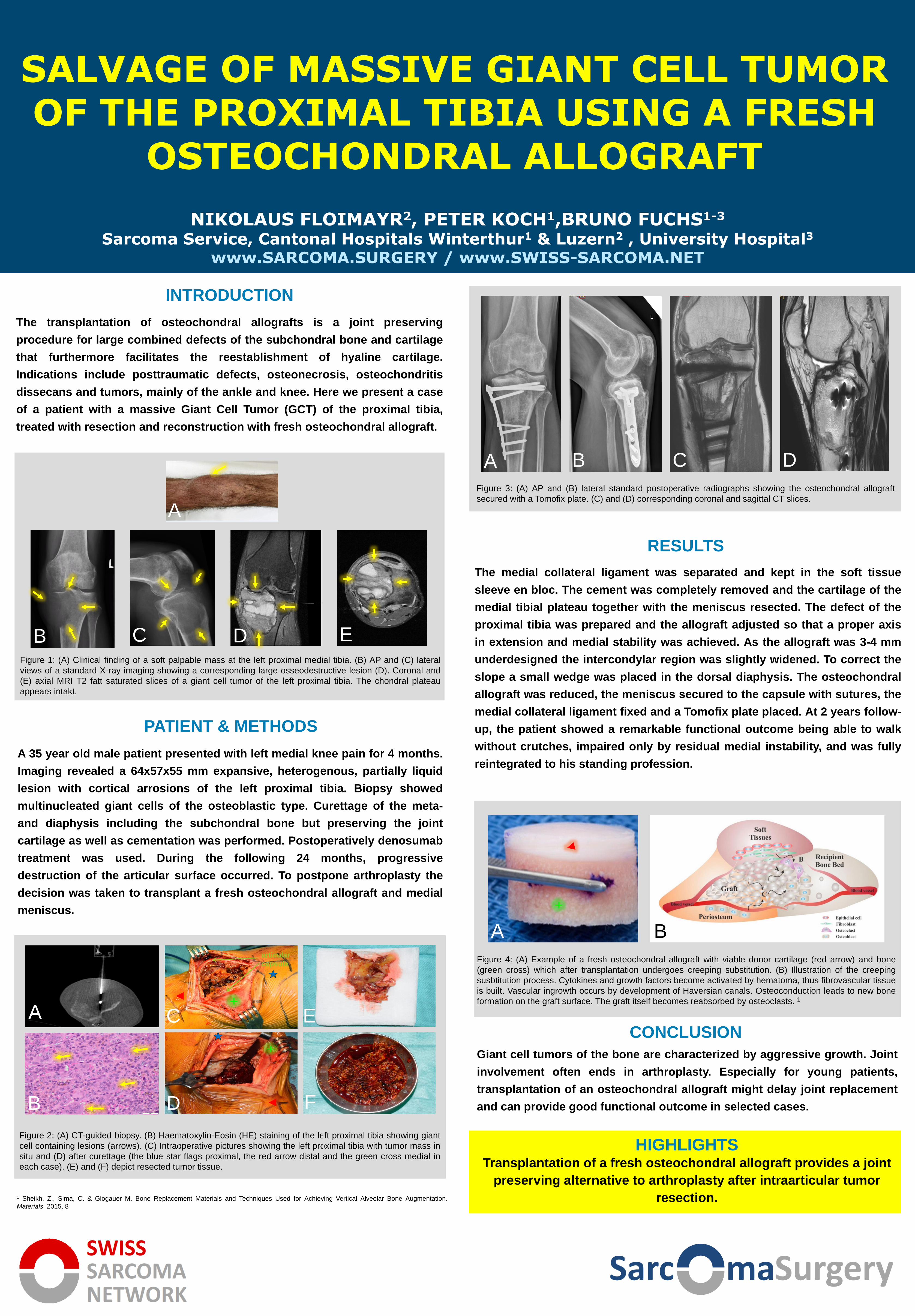
Figure 3: (A) AP and (B) lateral standard postoperative radiographs showing the osteochondral allograft
secured with a Tomofix plate. (C) and (D) corresponding coronal and sagittal CT slices.
The transplantation of osteochondral allografts is a joint preserving
procedure for large combined defects of the subchondral bone and cartilage
that furthermore facilitates the reestablishment of hyaline cartilage.
Indications include posttraumatic defects, osteonecrosis, osteochondritis
dissecans and tumors, mainly of the ankle and knee. Here we present a case
of a patient with a massive Giant Cell Tumor (GCT) of the proximal tibia,
treated with resection and reconstruction with fresh osteochondral allograft.
A 35 year old male patient presented with left medial knee pain for 4 months.
Imaging revealed a 64x57x55 mm expansive, heterogenous, partially liquid
lesion with cortical arrosions of the left proximal tibia. Biopsy showed
multinucleated giant cells of the osteoblastic type. Curettage of the meta-
and diaphysis including the subchondral bone but preserving the joint
cartilage as well as cementation was performed. Postoperatively denosumab
treatment was used. During the following 24 months, progressive
destruction of the articular surface occurred. To postpone arthroplasty the
decision was taken to transplant a fresh osteochondral allograft and medial
meniscus.
The medial collateral ligament was separated and kept in the soft tissue
sleeve en bloc. The cement was completely removed and the cartilage of the
medial tibial plateau together with the meniscus resected. The defect of the
proximal tibia was prepared and the allograft adjusted so that a proper axis
in extension and medial stability was achieved. As the allograft was 3-4 mm
underdesigned the intercondylar region was slightly widened. To correct the
slope a small wedge was placed in the dorsal diaphysis. The osteochondral
allograft was reduced, the meniscus secured to the capsule with sutures, the
medial collateral ligament fixed and a Tomofix plate placed. At 2 years follow-
up, the patient showed a remarkable functional outcome being able to walk
without crutches, impaired only by residual medial instability, and was fully
reintegrated to his standing profession.
A
B D
A B C D
F
E
B C D E
A
Figure 1: (A) Clinical finding of a soft palpable mass at the left proximal medial tibia. (B) AP and (C) lateral
views of a standard X-ray imaging showing a corresponding large osseodestructive lesion (D). Coronal and
(E) axial MRI T2 fatt saturated slices of a giant cell tumor of the left proximal tibia. The chondral plateau
appears intakt.
A B
C
articular
capsula
1 Sheikh, Z., Sima, C. & Glogauer M. Bone Replacement Materials and Techniques Used for Achieving Vertical Alveolar Bone Augmentation.
Materials 2015, 8


![Nikolaus Goldmann’s architectural rods Extended Version · Nikolaus Goldmann Nikolaus Goldmann was a mathematician who had been working in Leiden [12]. With his Elementa architecturae](https://static.fdocuments.in/doc/165x107/61167cea7b224e6e2e64f118/nikolaus-goldmannas-architectural-rods-extended-nikolaus-goldmann-nikolaus-goldmann.jpg)