NIH Public Access Ji-Hye Paik Emily Thomas Shan Jiang ... · Mariela Jaskelioff1, Florian L....
15
Telomerase reactivation reverses tissue degeneration in aged telomerase deficient mice Mariela Jaskelioff 1 , Florian L. Muller 1 , Ji-Hye Paik 1 , Emily Thomas 1 , Shan Jiang 1 , Andrew Adams 2 , Ergun Sahin 1 , Maria Kost-Alimova 1 , Alexei Protopopov 1 , Juan Cadiñanos 1 , James W. Horner 1 , Eleftheria Maratos-Flier 2 , and Ronald A. DePinho 1 1 Belfer Institute for Applied Cancer Science and Departments of Medical Oncology, Medicine and Genetics, Dana-Farber Cancer Institute, Harvard Medical School, Boston, MA 02115 2 Division of Endocrinology, Diabetes & Metabolism, Beth Israel Deaconess Medical Center, Harvard Medical School, Boston, MA 02215 Abstract An ageing world population has fueled interest in regenerative remedies that may stem declining organ function and maintain fitness. Unanswered is whether elimination of intrinsic instigators driving age-associated degeneration can reverse, as opposed to simply arrest, various afflictions of the aged. Such instigators include progressively damaged genomes. Telomerase deficient mice have served as a model system to study the adverse cellular and organismal consequences of wide- spread endogenous DNA damage signaling activation in vivo 1 . Telomere loss and uncapping provokes progressive tissue atrophy, stem cell depletion, organ system failure, and impaired tissue injury responses 1 . Here, we sought to determine whether entrenched multi-system degeneration in adult mice with severe telomere dysfunction can be halted or possibly reversed by reactivation of endogenous telomerase activity. To this end, we engineered a knock-in allele encoding a 4- hydroxytamoxifen (4-OHT)-inducible telomerase reverse transcriptase-Estrogen Receptor (TERT- ER) under transcriptional control of the endogenous TERT promoter. Homozygous TERT-ER mice display short dysfunctional telomeres and sustain increased DNA damage signaling and classical degenerative phenotypes upon successive generational matings and advancing age. Telomerase reactivation in such late generation TERT-ER mice extends telomeres, reduces DNA damage signaling and associated cellular checkpoint responses, allows resumption of proliferation in quiescent cultures, and eliminates degenerative phenotypes across multiple organs including testes, spleens and intestines. Notably, somatic telomerase reactivation reversed neurodegeneration with restoration of proliferating Sox2+ neural progenitors, DCX+ newborn neurons, and Olig2+ oligodendrocyte populations. Consistent with the integral role of SVZ neural progenitors in generation and maintenance of olfactory bulb interneurons 2 , this wave of telomerase-dependent neurogenesis resulted in alleviation of hyposmia and recovery of innate olfactory avoidance responses. Accumulating evidence implicating telomere damage as a driver of age-associated organ decline and disease risk 1,3 and the dramatic reversal of systemic Users may view, print, copy, download and text and data- mine the content in such documents, for the purposes of academic research, subject always to the full Conditions of use: http://www.nature.com/authors/editorial_policies/license.html#terms Correspondence and requests for materials should be addressed to [email protected]. Supplementary information is linked to the online version of the paper at www.nature.com/nature Authors contributions: M.J. and R.A.D. designed and guided the research; M.J., F.L.M., J-H.P., E.S., E.T., S.J. and M.A. performed research. J.C. and J.W.H. generated the TERT-ER mouse. M.J., F.L.M., A.A., A.P., E.M-F. and R.A.D. analyzed data. M.J. and R.A.D. wrote the manuscript. Reprints and permissions information is available at www.nature.com/reprints The authors declare no competing financial interests. NIH Public Access Author Manuscript Nature. Author manuscript; available in PMC 2011 July 6. Published in final edited form as: Nature. 2011 January 6; 469(7328): 102–106. doi:10.1038/nature09603. NIH-PA Author Manuscript NIH-PA Author Manuscript NIH-PA Author Manuscript
Transcript of NIH Public Access Ji-Hye Paik Emily Thomas Shan Jiang ... · Mariela Jaskelioff1, Florian L....

Telomerase reactivation reverses tissue degeneration in agedtelomerase deficient mice
Mariela Jaskelioff1, Florian L. Muller1, Ji-Hye Paik1, Emily Thomas1, Shan Jiang1, AndrewAdams2, Ergun Sahin1, Maria Kost-Alimova1, Alexei Protopopov1, Juan Cadiñanos1,James W. Horner1, Eleftheria Maratos-Flier2, and Ronald A. DePinho1
1 Belfer Institute for Applied Cancer Science and Departments of Medical Oncology, Medicineand Genetics, Dana-Farber Cancer Institute, Harvard Medical School, Boston, MA 021152 Division of Endocrinology, Diabetes & Metabolism, Beth Israel Deaconess Medical Center,Harvard Medical School, Boston, MA 02215
AbstractAn ageing world population has fueled interest in regenerative remedies that may stem decliningorgan function and maintain fitness. Unanswered is whether elimination of intrinsic instigatorsdriving age-associated degeneration can reverse, as opposed to simply arrest, various afflictions ofthe aged. Such instigators include progressively damaged genomes. Telomerase deficient micehave served as a model system to study the adverse cellular and organismal consequences of wide-spread endogenous DNA damage signaling activation in vivo1. Telomere loss and uncappingprovokes progressive tissue atrophy, stem cell depletion, organ system failure, and impaired tissueinjury responses1. Here, we sought to determine whether entrenched multi-system degeneration inadult mice with severe telomere dysfunction can be halted or possibly reversed by reactivation ofendogenous telomerase activity. To this end, we engineered a knock-in allele encoding a 4-hydroxytamoxifen (4-OHT)-inducible telomerase reverse transcriptase-Estrogen Receptor (TERT-ER) under transcriptional control of the endogenous TERT promoter. Homozygous TERT-ERmice display short dysfunctional telomeres and sustain increased DNA damage signaling andclassical degenerative phenotypes upon successive generational matings and advancing age.Telomerase reactivation in such late generation TERT-ER mice extends telomeres, reduces DNAdamage signaling and associated cellular checkpoint responses, allows resumption of proliferationin quiescent cultures, and eliminates degenerative phenotypes across multiple organs includingtestes, spleens and intestines. Notably, somatic telomerase reactivation reversedneurodegeneration with restoration of proliferating Sox2+ neural progenitors, DCX+ newbornneurons, and Olig2+ oligodendrocyte populations. Consistent with the integral role of SVZ neuralprogenitors in generation and maintenance of olfactory bulb interneurons2, this wave oftelomerase-dependent neurogenesis resulted in alleviation of hyposmia and recovery of innateolfactory avoidance responses. Accumulating evidence implicating telomere damage as a driver ofage-associated organ decline and disease risk1,3 and the dramatic reversal of systemic
Users may view, print, copy, download and text and data- mine the content in such documents, for the purposes of academic research,subject always to the full Conditions of use: http://www.nature.com/authors/editorial_policies/license.html#terms
Correspondence and requests for materials should be addressed to [email protected].
Supplementary information is linked to the online version of the paper at www.nature.com/nature
Authors contributions: M.J. and R.A.D. designed and guided the research; M.J., F.L.M., J-H.P., E.S., E.T., S.J. and M.A. performedresearch. J.C. and J.W.H. generated the TERT-ER mouse. M.J., F.L.M., A.A., A.P., E.M-F. and R.A.D. analyzed data. M.J. andR.A.D. wrote the manuscript.
Reprints and permissions information is available at www.nature.com/reprints
The authors declare no competing financial interests.
NIH Public AccessAuthor ManuscriptNature. Author manuscript; available in PMC 2011 July 6.
Published in final edited form as:Nature. 2011 January 6; 469(7328): 102–106. doi:10.1038/nature09603.
NIH
-PA Author Manuscript
NIH
-PA Author Manuscript
NIH
-PA Author Manuscript
degenerative phenotypes in adult mice observed here support the development of regenerativestrategies designed to restore telomere integrity.
Accelerating structural and functional decline across diverse organ systems is observed inthe aged1,3,4. The loss of genome integrity and associated DNA damage signaling andcellular checkpoint responses are well-established intrinsic instigators that drive tissuedegeneration during aging5. Of particular relevance to this study, age-progressive loss oftelomere function in mice has been shown to provoke widespread p53 activation resulting inactivation of cellular checkpoints of apoptosis, impaired proliferation and senescence,compromised tissue stem cell and progenitor function, marked tissue atrophy andphysiological impairment in many organ systems1,6.
Mounting evidence in humans has also provided strong association of limiting telomereswith increased risk of age-associated disease7 and with onset of tissue atrophy and organsystem failure in degenerative diseases such as Ataxia-Telangiectasia (A-T), WernerSyndrome (WS), Dyskeratosis Congenita, Liver Cirrhosis, among others1,3. In cell-basedmodels of A-T and WS, enforced TERT can restore normal cellular proliferative potential8.These findings build on seminal cell culture studies showing that enforced TERT expressioncan endow primary human cells with unlimited replicative potential9. Importantly, TERToverexpression in epithelial tissues of cancer-resistant mice leads to extended medianlifespan10. In addition, intercrossing wildtype and late generation mTerc−/− mice with severedegenerative phenotypes results in healthy offspring11, suggesting that viable late generationmTerc−/− germ cells can be restored to normal telomere function upon introduction of awildtype mTerc allele at the time of fertilization. However, to our knowledge, there are nogenetic or pharmacological studies showing somatic reversal of age-related degenerativephenotypes driven by endogenous genotoxic stresses in adult mammals. Here, in telomerasedeficient mice experiencing severe tissue degeneration, we asked whether endogenoustelomerase-mediated restoration of telomere function throughout the organism would quellDNA damage signaling and either arrest, or possibly reverse, cellular checkpoint responsesand associated tissue atrophy and dysfunction. Notably, the mice enlisted into this study areadults exhibiting significant progeroid phenotypes.
Construction and functional validation of the germline TERT-ER knock-in allele aredetailed in Figure S1. In the absence of 4-OHT, ER fusion proteins remain in an inactivemisfolded state12 and thus we first sought to verify whether mice homozygous for TERT-ERrecapitulated the classical premature aging phenotypes of mice null for mTerc or mTert. Tothat end, mice heterozygous for TERT-ER (hereafter G0TERT-ER) were intercrossed toproduce first generation mice homozygous for TERT-ER (G1TERT-ER) which were thenintercrossed to produce successive G2, G3 and G4TERT-ER cohorts. G1-G4TERT-ER cellshave no detectable telomerase activity (Figure 1a). Accordingly, G4TERT-ER primarysplenocytes exhibited hallmark features of short dysfunctional telomeres includingdecreased telomere-specific FISH signal and Robertsonian fusions (Figures 1b,e,f).Moreover, G4TERT-ER fibroblasts failed to divide after 5–6 passages and adopted a flat,senescence-like morphology (Figure 1c,d). Adult G4TERT-ER mice showed widespreadtissue atrophy, particularly in highly proliferative organs including extreme testicularatrophy and reduced testes size due to apoptotic elimination of germ cells, resulting indecreased fecundity (Figures 2a,d, S2a), marked splenic atrophy with accompanyingincreased 53BP1 foci consistent with DNA damage (Figures 2b,e,h) and intestinal cryptdepletion and villus atrophy in conjunction with numerous apoptotic crypt cells andincreased 53BP1 foci (Figures 2c,f,i, S2b). Finally, median survival of G4TERT-ER mice issignificantly decreased relative to that of telomere intact mice (43.5 vs. 86.8 weeks,
Jaskelioff et al. Page 2
Nature. Author manuscript; available in PMC 2011 July 6.
NIH
-PA Author Manuscript
NIH
-PA Author Manuscript
NIH
-PA Author Manuscript

***p<0.0001, Figure S2f). Thus, G4TERT-ER mice phenocopy late generation mTert−/− andmTerc−/− animals13,14, indicating that TERT-ER is inactive in the absence of 4-OHT.
Next, we assessed the impact of telomerase reactivation on telomere dysfunction-inducedproliferative arrest. Upon passage of adult G4TERT-ER fibroblast cultures, cells adopted flatsenescent-like morphology at approximately 5 population doublings (Figure 1d, upper).These quiescent cultures showed prominent G0/G1 accumulation in the cell cycle by FACSanalysis and rare cell division events by time-lapse video microscopy (not shown).However, upon replating these cells in media containing 100nM 4-OHT, telomerasereactivation led to elongated telomeres, prompt resumption of proliferation over >8additional passages tested, and reduction in the G0/G1 phase fraction (Figure 1c; data notshown). Coincidently, high levels of cyclin-dependent kinase inhibitor, p21CIP1, declinedupon 4-OHT treatment of the G4TERT-ER cultures, allowing cell cycle re-entry (Figure S2e).This pattern of p21CIP1 regulation aligns with previous work documenting its role as a keymediator of cell cycle arrest induced by telomere dysfunction in mouse tissues15. Parallel G0or G4TERT-ER fibroblasts maintained in 4-OHT at initial isolation did not undergo passage-induced senescence and instead showed sustained proliferation (>20 passages; Figure 1c,d).
These cell-based studies prompted systemic analyses of the impact of 4-OHT-mediatedtelomerase reactivation in the setting of entrenched tissue degeneration. At the end of 4weeks of continuous 4-OHT exposure, documentation of telomerase-mediated telomererestoration and function in G4TERT-ER tissues included increased telomere-FISH signal inprimary splenocytes (Figures 1b,e), decreased p53 activation and expression of p21CIP1 inliver (Figure S2d,e) and marked decrease in 53BP1 foci in splenocytes (Figures 2b,e) andintestinal crypt cells (Figures 2c,f). These molecular changes paralleled striking tissuerejuvenation including reduced apoptosis of testes germ cells (data not shown) and intestinalcrypt cells (Figures S2b, 2i), reduced tissue atrophy with restoration in normal testes andspleen size (Figures 2d,h, S2a) and, most strikingly, increased fecundity (Figure 2g).Moreover, median survival increased in G4TERT-ER mice treated with a 4-week course of 4-OHT (**p<0.005, Figure S2f). Sustained 4-OHT treatment had no effect on G0TERT-ER age-and gender-matched controls which were included in all experiments. Together, these dataindicate that despite an entrenched degenerative state, endogenous telomerase reactivationresults in dramatic extinction of DNA damage signaling, alleviation of cellular checkpointresponses and reversal of tissue atrophy in highly proliferative organ systems of the lategeneration TERT-ER mice.
While the dramatic impact of telomerase reactivation on highly proliferative organs isencouraging, we sought to more intensively assess the potential benefits on brain healthwhich is a prime determinant of age-progressive declining health in humans. Along theselines, it is worth noting that the aging mammalian brain shows accumulating DNA damage16
and a progressive restriction of neurogenesis and impaired re-myelination due to a decline inneural stem and progenitor cell proliferation and differentiation17. As neural stem/progenitorcells (hereafter NSCs) support neurogenesis, particularly in the subventricular zone (SVZ),we first examined the properties of NSCs derived from adult G0 and G4TERT-ER mice. Asreported previously for late generation mTerc−/− mice6,14,18, vehicle-treated G4TERT-ER
NSC cultures showed decreased self-renewal activity relative to G0TERT-ER controls and thisdefect was partially corrected with 4-OHT treatment (Figure 3a,d). G4TERT-ER neurosphereswere not only rarer but also smaller in diameter than G0TERT-ER controls, and their averagediameter was restored to normal by 4-OHT treatment (Figures 3a and S2c). These self-renewal profiles tracked with activated p53-mediated DNA damage signaling in vehicle-treated G4TERT-ER NSC cultures, which was extinguished with 4-OHT treatment and absentin the G0TERT-ER controls (Figure 3b,e). Examination of NSC differentiation capacityrevealed significant (2-fold) reduction in G4TERT-ER NSC capacity to generate neurons
Jaskelioff et al. Page 3
Nature. Author manuscript; available in PMC 2011 July 6.
NIH
-PA Author Manuscript
NIH
-PA Author Manuscript
NIH
-PA Author Manuscript
relative to 4-OHT-treated G4TERT-ER cultures and 4-OHT or vehicle-treated G0TERT-ER
controls (Figures 3c,f). Consistent with previous work14,18, there was no impact on astrocytedifferentiation (data not shown).
On the basis of these cell culture observations, we examined the SVZ, a region where NSCsreside and play an active role in adult brain physiology. In adult mice, NSCs give rise totransit-amplifying progenitor cells which rapidly divide and contribute to generation ofneuroblasts, astrocytes and myelinating oligodendrocytes. Consistent with previous reportsof an SVZ proliferation defect in mTerc−/− mice6,14,18 and wildtype aged mice19, vehicle-treated G4TERT-ER mice show a profound decrease in proliferating (Ki67+) cells in the SVZrelative to G0TERT-ER controls. Notably, 4-OHT-treated G4TERT-ER mice show a striking,albeit partial, restoration of proliferation following only 4 weeks of treatment (Figure 4, firstrow). This resumed SVZ proliferation mirrors well restoration of Sox2+ cells, a marker ofNSCs (Figure 4, second row), and doublecortin (DCX)+ cells, an early neuronal lineagemarker, together demonstrating preservation of neural stem/progenitor reserves and theirneurogenic capacity in vivo (Figure 4, third row). Finally, quantitative FISH analysis showstelomere elongation in the SVZ after 4 weeks of 4-OHT treatment (Figure S3). Thus, themarkedly constrained neural progenitor proliferation and neurogenesis profile associatedwith telomere dysfunction can be ameliorated by reactivation of endogenous telomeraseactivity.
In order to test the hypothesis that telomerase reactivation leads to tissue rejuvenation, weconducted detailed morphological and functional fitness analyses of different brainstructures upon telomerase reactivation. First, we examined the white matter of the corpuscallosum (CC) and observed that aged G4TERT-ER mice have far fewer Olig2+ matureoligodendrocytes (Figure 4, fourth row). This cellular deficiency is associated with reducedbrain weight (Figures 5a,b) and significantly thinner myelin sheathing of neurons with gratios (numerical ratio between the diameter of the axon proper and the outer diameter of themyelinated fiber) of 0.7756±0.0054 for G4TERT-ER mice vs. 0.7032±0.0049 for G0TERT-ER
(mean±SEM, ***p<0.0001) (Figures 5c,d). Remarkably, endogenous telomerasereactivation reinstates normal numbers of mature oligodendrocytes (Figure 4) and reversesthe hypomyelination phenotype at the level of mean myelin sheath diameters (with g ratiosof 0.7058±0.0006 and 0.7164±0.0063 for 4-OHT-treated G4 and G0TERT-ER mice,respectively) (Figures 5c,d). Furthermore, a 4-OHT treatment course of only 4 weeks issufficient to cause significant partial reversion of the brain size defect, with G4TERT-ER
brain weights increasing from 77.3±3.3% of G0TERT-ER brain weights in the vehicle groupto 89.7±4.0% in the 4-OHT group (Figures 5a,b). Importantly, telomere elongation can bedetected in the CC after 4 weeks of telomerase reactivation (Figure S3c). Thus, endogenoustelomerase reactivation exerts a swift impact on oligodendrocyte proliferation anddifferentiation, and promotes repopulation of white matter structures with matureoligodendrocytes and active myelin deposition.
Lastly, we investigated the physiological effect of telomere dysfunction and telomerasereactivation on olfactory function. Age-associated hyposmia, as evidenced by an increasedolfactory threshold and a reduced ability in odor identification and discrimination, is a wellestablished phenomenon in aged humans20. In rodents, aging is associated with diminishedolfactory neurogenesis and deficits in fine olfactory discrimination19,21. Olfactoryinterneurons in the olfactory bulb (OB) that receive and process information from theolfactory sensory neurons in the olfactory epithelium (OE) derive from SVZ stem cells2.Rodents demonstrate avoidance responses towards predators’ odorants as well as spoiledsmells like aliphatic acids, aliphatic aldehydes and alkyl amines, which are processed in theOB22. Given the dramatic decrease in SVZ neurogenesis of G4TERT-ER mice and the factthat the OB retains high telomerase activity in adult wildtype mouse brains23, we sought to
Jaskelioff et al. Page 4
Nature. Author manuscript; available in PMC 2011 July 6.
NIH
-PA Author Manuscript
NIH
-PA Author Manuscript
NIH
-PA Author Manuscript

determine whether telomere dysfunction results in a functional deficit of these mice to detectand process odorants for elicitation of instinctive avoidance/defensive behaviors.
Pathology within the olfactory epithelium which may be considered a basis of age-relatedolfactory dysfunction, was ruled out by confirmation of grossly normal histology of the OEin both cohorts (Figure S4). Next, we ruled out alterations in exploration behavior andoverall locomotion by monitoring total distance traveled by the animals in the absence ofodorants, which was similar for all experimental groups (Table S1; Figure 5e). Next, weperformed innate avoidance tests using serially diluted 2-methylbutyric acid (2-MB), anodorant that rouses innate aversive responses in mice. Whereas G0TERT-ER micedemonstrated avoidance responses at all 2-MB concentrations tested (1.87×10⁻⁴M through1.87×10−6M), G4 mice showed attraction/neutral behaviors at concentrations lower than1.87×10−4M (Figure 5e,f). Strikingly, following only 4 weeks of 4-OHT treatment, theperformance of G4TERT-ER mice was markedly improved, with avoidance behaviors beingapparent at all 2-MB concentrations (Figure 5e,g). Accordingly, the frequency of entry intothe odor zone was higher for vehicle-treated G4TERT-ER mice than for the other threeexperimental groups (Table S2). These findings are consistent with significant alleviation ofthe olfactory defect stemming from the documented wave of telomerase-mediated SVZneurogenesis and oligodendrocyte maturation which would promote repopulation ofolfactory bulbs with functional interneurons and improve olfactory neuron function viaremyelination.
Here, we report the generation of a novel mouse model to explore the impact ofphysiological telomerase reactivation across diverse adult cell types and organ systems. InG4TERT-ER mice with advanced degenerative phenotypes, short-term telomerase reactivationrestored telomere reserves, quelled DNA damage signaling, and alleviated cellularcheckpoint responses in multiple high-turnover organ systems with significant functionalimpact including increased fecundity. From this, we speculate that some tissue stem/progenitor cells are retained in a quiescent and intact state yet can be enlisted to resumenormal repopulating function upon elimination of genotoxic stress at telomeres. Despitechromosomal instability, the brief course of telomerase reactivation was not sufficient topromote carcinogenesis (data not shown), a finding consistent with a role for telomerase inpromoting progression of established neoplasms24. However, it remains possible that moreprolonged telomerase reactivation schedules or applications in later life may provokecarcinogenesis.
As noted, age-associated compromise in mammalian brain function is associated withextensive accumulation of DNA damage and progressive reduction in neurogenesis andmyelination. Indeed, many aspects of this CNS decline are accelerated and worsened in thesetting of telomere dysfunction (25,26, this study). Our data establish that telomerasereactivation in adult mice with telomere dysfunction can restore SVZ neurogenesis and,consistent with its role in sustaining new OB neurons, can ameliorate odor detection withimproved performance in innate odor avoidance tests. These results are consistent withprevious studies showing that prolonged inhibition of neurogenesis in the SVZ has anegative effect on odor detection thresholds27. In conclusion, this unprecedented reversal ofage-related decline in the CNS and other organs vital to adult mammalian health justifyexploration of telomere rejuvenation strategies for age-associated diseases, particularly thosedriven by accumulating genotoxic stress.
Methods SummaryTERT-ER mice were generated with traditional knock-in methods and following standardbreeding protocol of successive generations of telomerase deficient mice13. All studies were
Jaskelioff et al. Page 5
Nature. Author manuscript; available in PMC 2011 July 6.
NIH
-PA Author Manuscript
NIH
-PA Author Manuscript
NIH
-PA Author Manuscript
performed on adult males. 2.5mg 4-OHT time-release pellets (Innovative Research ofAmerica) were inserted subcutaneously to reach steady state blood levels of 1 ng/mL 4-OHT. For neurosphere assays, SVZs were dissected, dispersed into a single-cell suspensionand plated in neurobasal media supplemented with EGF, bFGF and 100nM 4-OHT orvehicle. For multipotentiality assays, neurospheres were transferred to differentiation media(1% FBS). For histological studies, mice were perfused with 10% formalin; equivalentcoronal sections were stained with indicated antibodies following standard IHC protocol.Laser scanning cytometric quantification was performed by iCys Research ImagingCytometer (Compucyte). For innate olfactory avoidance tests, mice were fasted for 20 h andhabituated for 20 min to the test cage where their responses were recorded on a videocamera mounted above the test chamber. A filter paper scented with water, or progressivelyhigher concentrations of 2-methylbutyric acid was placed in the cage and mouse behaviorwas recorded for 3 minutes. NoldusEthovision v3.1 behavioral analysis software was used todetermine innate avoidance behavior (time spent in the third of the cage containing thescented filter paper).
MethodsGeneration of TERT-ER mice
A knock-in targeting vector containing the ERT2-LBD domain upstream and in frame withthe mTert genomic sequence (exon 1 through intron 2) and a Lox-pgk-Neo-Lox fragmentwas introduced into ES cells. Neomycin-resistant clones yielded five independent lines, twoof which were injected into C57BL/6 blastocysts and implanted into surrogate mothers,yielding 10 high-percentage chimeras. Germline transmission was confirmed by crossing thechimeras to C57BL/6 females. Heterozygous TERT-ERneo animals were crossed to EIIa-Cre animals to delete the NeoR cassette and further intercrossed to homozygosity. The EIIa-Cre allele was then bred out of the line and heterozygous animals were backcrossed toC57BL\6 at least 3 times. From this point, standard breeding protocol of successivegenerations of telomerase deficient mice was followed28. All studies were performed onadult (30–35 week old) males, heterozygous (G0TERT-ER) or homozygous (G4TERT-ER) forthis allele, unless otherwise noted. 2.5mg 4-OHT time-release pellets (Innovative Researchof America) were inserted subcutaneously to reach steady state blood levels of 1 ng/mL 4-OHT.
Mice were maintained in specific pathogen-free (SPF) conditions at Dana-Farber CancerInstitute. All manipulations were performed with IACUC approval.
Histology and Electron MicroscopyBrains from animals perfused with 10% formalin were further fixed for 24 hours andcoronally sectioned using a brain matrix (Electron Microscopy Sciences). Equivalentsections were used for chromogenic immunohistochemistry, which was performedaccording to standard procedures. Antibodies used include Ki67 (Dako), 53BP1 (BethylLabs), Sox-2 and DCX (Santa Cruz Biotechnology) and Olig-2 (Chemicon). Forimmunofluorescence studies, cells were fixed in 4% paraformaldehyde (PFA) in phosphate-buffered saline for 10 min, permeabilized (50 mM NaCl, 3 mM MgCl2, 200 mM sucrose, 10mM HEPES [pH 7.9], 0.5% TX-100) for 5 min, and then stained with primary antibodiesagainst 53BP1 (Bethyl Labs), TUJ1 (Chemicon), GFAP (Dako) and secondary antibodiesconjugated to Alexa Fluor-488 or Alexa Fluor-568 (Molecular Probes). Cells were mountedin DAPI-containing antifade solution (Vector). Foci were scored by eye from a minimum of300 randomly chosen nuclei by using a 40× objective, and scoring was performed in ablinded manner with respect to genotype. Immunofluorescence images were captured ingrayscale for each fluorophore and were merged by compilation in respective red-green-blue
Jaskelioff et al. Page 6
Nature. Author manuscript; available in PMC 2011 July 6.
NIH
-PA Author Manuscript
NIH
-PA Author Manuscript
NIH
-PA Author Manuscript

(RGB) channels using Adobe Photoshop CS 8.0. For apoptosis assays, sections fromparaffin-embedded testes were deparaffinized and processed for apoptotic staining (terminaldeoxynucleotidyltransferase-mediated dUTP-biotin nick end labeling [TUNEL]) accordingto the manufacturer’s instructions (Chemicon).
For EM studies, animals were perfused for 30 min in Karnovsky’s solution B, brains werefurther fixed for 24 hours and delivered to the Harvard Medical School EM Facility forembedding, sectioning and staining. Electron micrographs were generated using a JEOL1200EX microscope and analyzed with ImageJ software29. Inner and outer diameters wereanalyzed as per ref30.
Assessment of telomerase activityTelomeric repeats amplification protocol (TRAP) was combined with real-time detection ofamplification products to determine telomerase activity with a Quantitative TelomeraseDetection kit (US Biomax) following the manufacturer’s recommendations. 0.5 μg totalprotein extract was used in each reaction. End products were resolved by PAGE in a 12.5%non-denaturing gel, stained with Sybr Green Nucleic Acid gel stain (Invitrogen) andvisualized with a Bio-Rad Molecular Imager ChemiDoc System.
Cell culture and cytogenetic analysisEar skin fibroblasts were isolated as described previously31. Proliferation assays werecarried in triplicate on 6-well plates. Cells were grown in RPMI-10% fetal calf serum-50μM β-mercaptoethanol with the addition of 100nM 4-OHT or vehicle (ethanol). Cells werecounted and replated at a density of 1,000 cells/well every 4 days. Splenocytes were isolatedby generating single cell suspensions from whole spleen, stimulated for 48 hours with 2.5μg/ml concavalin A and 20 μg/ml LPS (Sigma) and treated with KaryoMAX Colcemidsolution (Invitrogen) for 2 hours before harvesting. Telomere fluorescence in situhybridization (FISH) was performed on metaphase nuclei as described previously32. At least15 metaphases from harvested cell cultures were analyzed for telomere integrity by telomereFISH. Telomere signal was normalized using a Pacific Blue centromeric PNA probe. Fortelomere-tissue-FISH, frozen tissue sections (8 μm thick) were fixed in 2% PFA for 15minutes and permeabilized in 0.5% Triton X-100 for 10 minutes. Two PNA probes,telomere-specific FITC-00-T2AG3 and Pacific Blue-centromere-specific, were hybridizedafter 4 minutes denaturing at 83 °C under the following conditions: 70% formamide, 0.06XSSC, 0.2% BSA, 0.5 ng/μL tRNA, 0.5 ng/μL PNA probe; overnight at 25°C. To achieveuniform hybridization we used MAUI Mixer (BioMicro) with 40μL chamber. Nuclei werestained with TOTO3 (Invitrogen) far red stain. Telomere signal was normalized using thecentromeric PNA probe. For neurosphere assays, subventricular region of brain from 3.6week-old mice was dissected, dispersed into a single-cell suspension and plated inneurobasal media (StemCellTechnologies) supplemented with EGF and bFGF (20ng/mleach) for 4 days, in the presence of 100nM 4-OHT or vehicle (ethanol). Primaryneurospheres were dissociated and seeded at 2 cells/μl density in multiwell plates. After 7–10 days, cultures were monitored for the formation of neurospheres. Alternatively, singlecells were sorted into individual well on 384-well plate at a density of 10 cells/well on DakoMoFlo high-speed cell sorter and cultured for 3 weeks. Neurospheres were transferred toculture wells coated with poly-L-ornithine (15 μg/ml) and fibronectin (1 μg/ml) anddifferentiated in 1% FBS in neurobasal media to measure their multipotentiality.Quantification of neurosphere numbers and diameters were performed by bright-fieldmicroscopy coupled with an in-house semiautomated segmentation algorithm generated withMATLAB software (The Mathworks, Natick, MA). For multipotentiality assays, cells werefixed, stained with GFAP and TUJ1 antibodies and quantified as previously described33.
Jaskelioff et al. Page 7
Nature. Author manuscript; available in PMC 2011 July 6.
NIH
-PA Author Manuscript
NIH
-PA Author Manuscript
NIH
-PA Author Manuscript
Laser Scanning Analysis for the Quantitation of IHC and FISHLaser scanning cytometry quantification was performed by iCys Research ImagingCytometer (Compucyte) as described earlier34,35 with a few modifications. Counts ofKi67+, DCX+ or Sox2+ (DAB positive) were carried out within the subventricular zonesthat were predefined by a certified pathologist with the H&E-stained brain architecture. Thetarget number for each sample was approximately 500 cells counted. Olig2+ cell within thecorpus callosum were counted in a similar manner.
RT-PCR, Southern Blotting and Western BlottingDNase-treated total RNA extracted from fresh liver samples with the RNeasy kit (Qiagen)was used to prepare oligo-dT complementary DNA with Superscript III (Invitrogen). RT-PCR PCR primers are described in Supplemental Table S3. Southern and Western blotswere performed following standard techniques. For Western blots, 40 μg of protein wereloaded per lane. Antibodies used include phospho-p53 (Ser15, Cell Signaling Technologies),p21 (Santa Cruz Biotechnology), Actin (Biolegend) and HRP-conjugated secondaryantibodies (Pierce/ThermoScientifics).
Innate olfactory avoidance testAnimals were kept in a 12-hour light/dark cycle and tested in the second half of the lightcycle. 30–35 week old male mice (n=4/experimental condition) were fasted for 20 h prior totesting. To avoid confounding of data owing to learning, mice were used only once. Tohabituate to the experimental environment, mice were placed individually in a cage that wasidentical to the test cage (259mm × 476mm × 209mm) for 20 minutes prior to the onset oftesting. Following acclimation, mice were placed in an identical test chamber where theirresponses were recorded on a video camera (30 frames per second, 640 × 480) mountedabove the test chamber. Confounding environmental/spatial cue effects were ruled out bymonitoring total time spent in different zones of the chamber in the absence of odorants. Forinnate avoidance tests, circular filter paper (2.5cm diameter) was scented with 40 μl ofeither water or progressively higher concentrations of 2-MB (1.74 × 10⁻⁶ M through 1.74 ×10⁻⁴M) and mouse behavior was recorded for 3 minutes. Videos were transferred to PC forsubsequent analysis using NoldusEthovision v3.1 behavioral analysis software36. First thecage was divided into thirds and then time spent in each third for the duration of therecording was determined using the software. Total distance traveled, frequency of entryinto the third containing the filter paper treated with the odorants (zone 3) as well as timespent investigating in this zone (to determine innate avoidance behavior) was collated foreach treatment and genotype.
Statistical AnalysisAll the data were analyzed by one way ANOVA with Bonferroni’s post test (significantlydifferent at p<0.05). Survival curves were analyzed with Mantel-Cox test.
Supplementary MaterialRefer to Web version on PubMed Central for supplementary material.
AcknowledgmentsThe authors would like to thank Dr. R. Segal for critical comments, Drs. R. Bronson, K. Ligon and C. Maire forhistological advice, Dr. S.S. Chae for assistance with neurosphere measurement studies and Dr. L. Cameron fortime-lapse microscopy studies. M.J. was supported in part by a Susan G. Komen for the Cure fellowship(PDF060881). F.L.M. was supported by ACS fellowship PF-08-261-01-TBE. This work and R.A.D. was supportedby R01CA84628 and U01CA141508 grants from the NIH National Cancer Institute and the Belfer Foundation.R.A.D. was supported by an American Cancer Society Research Professorship.
Jaskelioff et al. Page 8
Nature. Author manuscript; available in PMC 2011 July 6.
NIH
-PA Author Manuscript
NIH
-PA Author Manuscript
NIH
-PA Author Manuscript
References1. Sahin E, Depinho RA. Linking functional decline of telomeres, mitochondria and stem cells during
ageing. Nature. 7288; 464:520–528. [PubMed: 20336134]
2. Whitman MC, Greer CA. Adult neurogenesis and the olfactory system. Progress in neurobiology.2009; 89(2):162–175. [PubMed: 19615423]
3. Sharpless NE, DePinho RA. How stem cells age and why this makes us grow old. Nature reviews.2007; 8(9):703–713.
4. Ju Z, Lenhard Rudolph K. Telomere dysfunction and stem cell ageing. Biochimie. 2008; 90(1):24–32. [PubMed: 18029082]
5. Hoeijmakers JH. DNA damage, aging, and cancer. The New England journal of medicine. 2009;361(15):1475–1485. [PubMed: 19812404]
6. Ferron, S.; Mira, H.; Franco, S., et al. Development. Vol. 131. Cambridge, England: 2004. Telomereshortening and chromosomal instability abrogates proliferation of adult but not embryonic neuralstem cells; p. 4059-4070.
7. Cawthon RM, Smith KR, O’Brien E, et al. Association between telomere length in blood andmortality in people aged 60 years or older. Lancet. 2003; 361(9355):393–395. [PubMed: 12573379]
8. Wyllie FS, Jones CJ, Skinner JW, et al. Telomerase prevents the accelerated cell ageing of Wernersyndrome fibroblasts. Nature genetics. 2000; 24(1):16–17. [PubMed: 10615119]
9. Bodnar, AG.; Ouellette, M.; Frolkis, M., et al. Science. Vol. 279. New York, N.Y: 1998. Extensionof life-span by introduction of telomerase into normal human cells; p. 349-352.
10. Tomas-Loba A, Flores I, Fernandez-Marcos PJ, et al. Telomerase reverse transcriptase delaysaging in cancer-resistant mice. Cell. 2008; 135(4):609–622. [PubMed: 19013273]
11. Samper E, Flores JM, Blasco MA. Restoration of telomerase activity rescues chromosomalinstability and premature aging in Terc−/− mice with short telomeres. EMBO reports. 2001; 2(9):800–807. [PubMed: 11520856]
12. Metzger D, Clifford J, Chiba H, et al. Conditional site-specific recombination in mammalian cellsusing a ligand-dependent chimeric Cre recombinase. Proc Natl Acad Sci U S A. 1995; 92(15):6991–6995. [PubMed: 7624356]
13. Blasco MA, Lee HW, Hande MP, et al. Telomere shortening and tumor formation by mouse cellslacking telomerase RNA. Cell. 1997; 91(1):25–34. [PubMed: 9335332]
14. Wong KK, Maser RS, Bachoo RM, et al. Telomere dysfunction and Atm deficiency compromisesorgan homeostasis and accelerates ageing. Nature. 2003; 421(6923):643–648. [PubMed:12540856]
15. Choudhury AR, Ju Z, Djojosubroto MW, et al. Cdkn1a deletion improves stem cell function andlifespan of mice with dysfunctional telomeres without accelerating cancer formation. Naturegenetics. 2007; 39(1):99–105. [PubMed: 17143283]
16. Best BP. Nuclear DNA damage as a direct cause of aging. Rejuvenation research. 2009; 12(3):199–208. [PubMed: 19594328]
17. Drapeau E, Nora Abrous D. Stem cell review series: role of neurogenesis in age-related memorydisorders. Aging cell. 2008; 7(4):569–589. [PubMed: 18221417]
18. Ferron SR, Marques-Torrejon MA, Mira H, et al. Telomere shortening in neural stem cells disruptsneuronal differentiation and neuritogenesis. J Neurosci. 2009; 29(46):14394–14407. [PubMed:19923274]
19. Enwere E, Shingo T, Gregg C, et al. Aging results in reduced epidermal growth factor receptorsignaling, diminished olfactory neurogenesis, and deficits in fine olfactory discrimination. JNeurosci. 2004; 24(38):8354–8365. [PubMed: 15385618]
20. Lafreniere D, Mann N. Anosmia: loss of smell in the elderly. Otolaryngologic clinics of NorthAmerica. 2009; 42(1):123–131. [PubMed: 19134495]
21. Ma DK, Kim WR, Ming GL, et al. Activity-dependent extrinsic regulation of adult olfactory bulband hippocampal neurogenesis. Annals of the New York Academy of Sciences. 2009; 1170:664–673. [PubMed: 19686209]
22. Kobayakawa K, Kobayakawa R, Matsumoto H, et al. Innate versus learned odour processing in themouse olfactory bulb. Nature. 2007; 450(7169):503–508. [PubMed: 17989651]
Jaskelioff et al. Page 9
Nature. Author manuscript; available in PMC 2011 July 6.
NIH
-PA Author Manuscript
NIH
-PA Author Manuscript
NIH
-PA Author Manuscript

23. Caporaso GL, Lim DA, Alvarez-Buylla A, et al. Telomerase activity in the subventricular zone ofadult mice. Molecular and cellular neurosciences. 2003; 23(4):693–702. [PubMed: 12932448]
24. Artandi SE, DePinho RA. Telomeres and telomerase in cancer. Carcinogenesis. 31(1):9–18.[PubMed: 19887512]
25. Zhang P, Dilley C, Mattson MP. DNA damage responses in neural cells: Focus on the telomere.Neuroscience. 2007; 145(4):1439–1448. [PubMed: 17207936]
26. Lee J, Jo YS, Sung YH, et al. Telomerase deficiency affects normal brain functions in mice.Neurochemical research. 2010; 35(2):211–218. [PubMed: 19685288]
27. Breton-Provencher V, Lemasson M, Peralta MR 3rd, et al. Interneurons produced in adulthood arerequired for the normal functioning of the olfactory bulb network and for the execution of selectedolfactory behaviors. J Neurosci. 2009; 29(48):15245–15257. [PubMed: 19955377]
28. Maser RS, Wong KK, Sahin E, et al. DNA-dependent protein kinase catalytic subunit is notrequired for dysfunctional telomere fusion and checkpoint response in the telomerase-deficientmouse. Molecular and cellular biology. 2007; 27(6):2253–2265. [PubMed: 17145779]
29. Abramoff MD, Magelhaes PJ, Ram SJ. Image Processing with ImageJ. Biophotonics International.2001; 11(7):36–42.
30. Potzner MR, Griffel C, Lutjen-Drecoll E, et al. Prolonged Sox4 expression in oligodendrocytesinterferes with normal myelination in the central nervous system. Molecular and cellular biology.2007; 27(15):5316–5326. [PubMed: 17515609]
31. Shao C, Deng L, Henegariu O, et al. Mitotic recombination produces the majority of recessivefibroblast variants in heterozygous mice. Proceedings of the National Academy of Sciences of theUnited States of America. 1999; 96(16):9230–9235. [PubMed: 10430925]
32. Paik JH, Ding Z, Narurkar R, et al. FoxOs cooperatively regulate diverse pathways governingneural stem cell homeostasis. Cell stem cell. 2009; 5(5):540–553. [PubMed: 19896444]
33. Mahoney JE, Sahin E, Jaskelioff M, et al. Quantification of telomere length by FISH and laserscanning cytometry. Proc SPIE. 2008; 6859(1V):1–9.
34. Gorczyca W, Deptala A, Bedner E, et al. Analysis of human tumors by laser scanning cytometry.Methods in cell biology. 2001; 64:421–443. [PubMed: 11070850]
35. Spink AJ, Tegelenbosch RA, Buma MO, et al. The EthoVision video tracking system--a tool forbehavioral phenotyping of transgenic mice. Physiology & behavior. 2001; 73(5):731–744.[PubMed: 11566207]
Jaskelioff et al. Page 10
Nature. Author manuscript; available in PMC 2011 July 6.
NIH
-PA Author Manuscript
NIH
-PA Author Manuscript
NIH
-PA Author Manuscript

Figure 1. 4-OHT-dependent induction of telomerase activity in TERT-ER cells(a, top) Telomerase activity in eNSCs (* telomerase products); (a, bottom) real-timequantitation of reactions above; (b) representative G4TERT-ER splenocyte metaphases; (c)proliferation of adult G4TERT-ER fibroblasts (n=3) in media with vehicle (black) or 4-OHT(red); (d) representative image of G4TERT-ER fibroblasts (passage 6) in media with 4-OHT(bottom) or vehicle (top) ; (e) signal-free ends in primary splenocyte metaphases, 15metaphases/sample, n=2 (*p<0.05); (f) mean telomere-FISH signal in primary splenocyteinterphases, normalized to centromeric signal, n=3 (***p<0.0001). Open bars correspond tovehicle-treated and filled bars to 4-OHT-treated, error bars represent s.d.
Jaskelioff et al. Page 11
Nature. Author manuscript; available in PMC 2011 July 6.
NIH
-PA Author Manuscript
NIH
-PA Author Manuscript
NIH
-PA Author Manuscript

Figure 2. Telomerase activation in adult TERT-ER miceRepresentative tissue images from age-matched experimental and control mice (a) H&Estained sections of testes; (b) primary splenocytes stained for 53BP1; (c) small intestinesections stained for 53BP1; (d) testes weight of adult males (30–50 w.o., n≥10); (e) 53BP1nuclear foci/100 nuclei (n=3); (f) 53BP1 nuclear foci/100 crypts (n=4†); (g) litter sizes(n=3); (h) spleen weights (n≥6); (i) apoptotic (TUNEL+) cells/100 intestinal crypts (n≥20).***p=0.0001, **p<0.005, *p<0.05. Open bars correspond to vehicle-treated and filled barsto 4-OHT-treated groups, error bars represent s.d.
Jaskelioff et al. Page 12
Nature. Author manuscript; available in PMC 2011 July 6.
NIH
-PA Author Manuscript
NIH
-PA Author Manuscript
NIH
-PA Author Manuscript
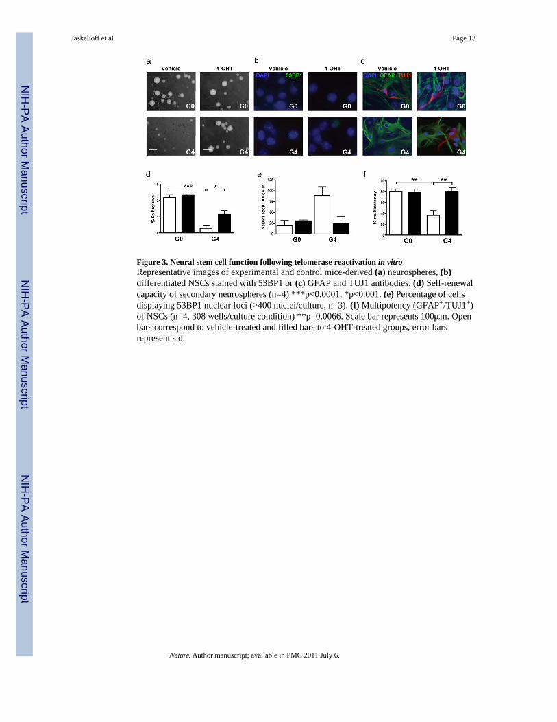
Figure 3. Neural stem cell function following telomerase reactivation in vitroRepresentative images of experimental and control mice-derived (a) neurospheres, (b)differentiated NSCs stained with 53BP1 or (c) GFAP and TUJ1 antibodies. (d) Self-renewalcapacity of secondary neurospheres (n=4) ***p<0.0001, *p<0.001. (e) Percentage of cellsdisplaying 53BP1 nuclear foci (>400 nuclei/culture, n=3). (f) Multipotency (GFAP+/TUJ1+)of NSCs (n=4, 308 wells/culture condition) **p=0.0066. Scale bar represents 100μm. Openbars correspond to vehicle-treated and filled bars to 4-OHT-treated groups, error barsrepresent s.d.
Jaskelioff et al. Page 13
Nature. Author manuscript; available in PMC 2011 July 6.
NIH
-PA Author Manuscript
NIH
-PA Author Manuscript
NIH
-PA Author Manuscript
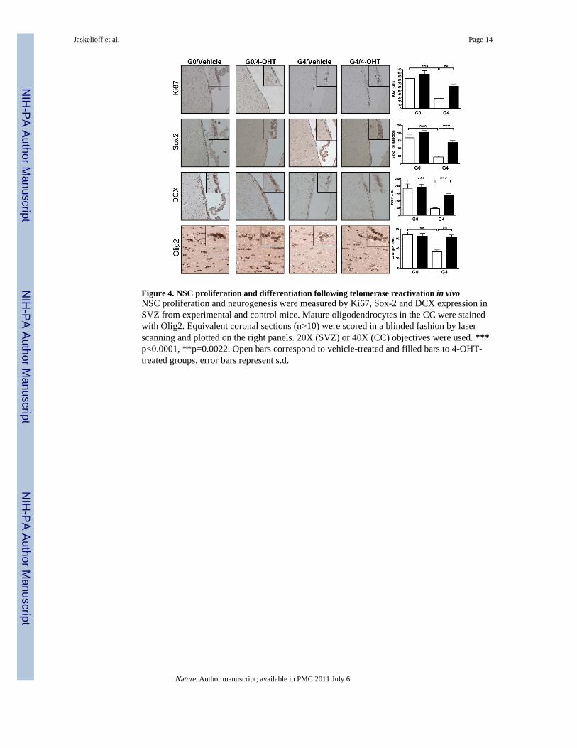
Figure 4. NSC proliferation and differentiation following telomerase reactivation in vivoNSC proliferation and neurogenesis were measured by Ki67, Sox-2 and DCX expression inSVZ from experimental and control mice. Mature oligodendrocytes in the CC were stainedwith Olig2. Equivalent coronal sections (n>10) were scored in a blinded fashion by laserscanning and plotted on the right panels. 20X (SVZ) or 40X (CC) objectives were used. ***p<0.0001, **p=0.0022. Open bars correspond to vehicle-treated and filled bars to 4-OHT-treated groups, error bars represent s.d.
Jaskelioff et al. Page 14
Nature. Author manuscript; available in PMC 2011 July 6.
NIH
-PA Author Manuscript
NIH
-PA Author Manuscript
NIH
-PA Author Manuscript
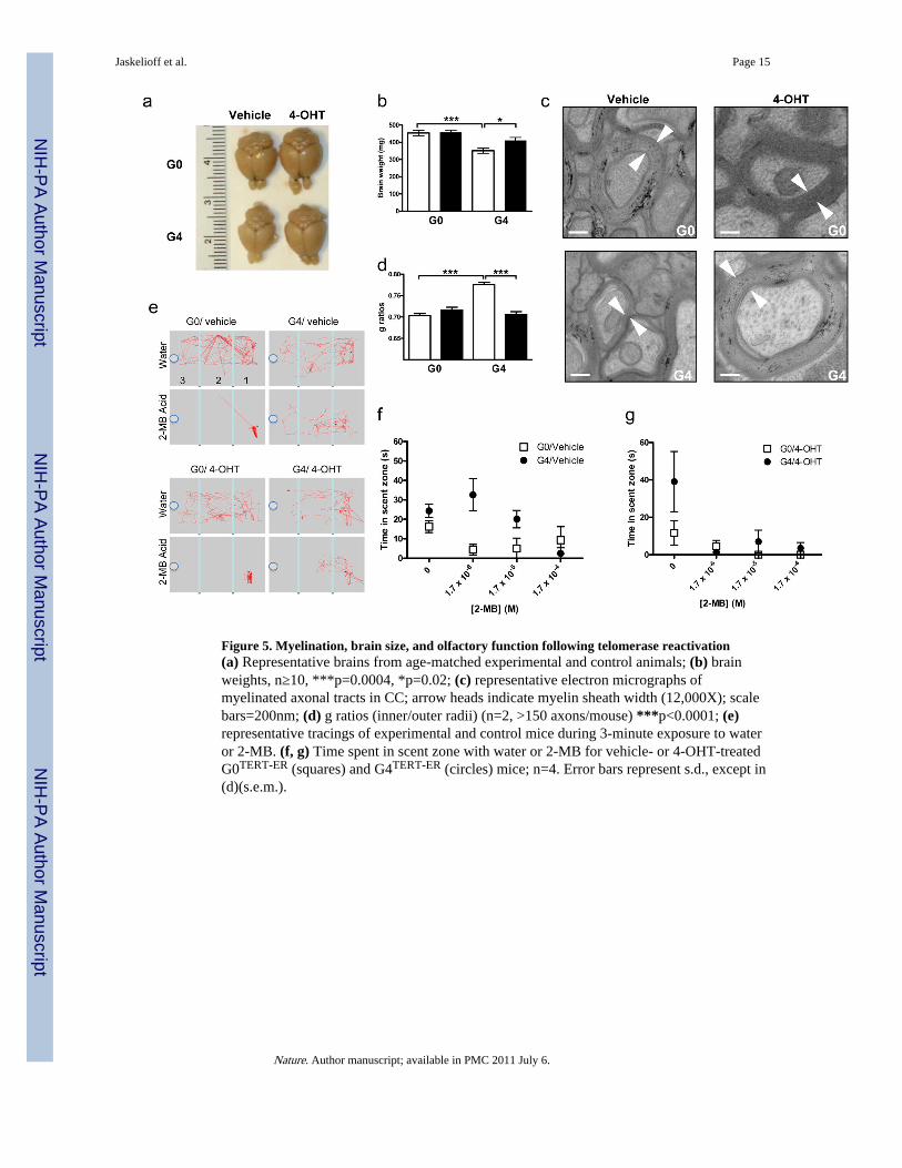
Figure 5. Myelination, brain size, and olfactory function following telomerase reactivation(a) Representative brains from age-matched experimental and control animals; (b) brainweights, n≥10, ***p=0.0004, *p=0.02; (c) representative electron micrographs ofmyelinated axonal tracts in CC; arrow heads indicate myelin sheath width (12,000X); scalebars=200nm; (d) g ratios (inner/outer radii) (n=2, >150 axons/mouse) ***p<0.0001; (e)representative tracings of experimental and control mice during 3-minute exposure to wateror 2-MB. (f, g) Time spent in scent zone with water or 2-MB for vehicle- or 4-OHT-treatedG0TERT-ER (squares) and G4TERT-ER (circles) mice; n=4. Error bars represent s.d., except in(d)(s.e.m.).
Jaskelioff et al. Page 15
Nature. Author manuscript; available in PMC 2011 July 6.
NIH
-PA Author Manuscript
NIH
-PA Author Manuscript
NIH
-PA Author Manuscript