NGRTCI Endocrine System Anatomy
51
JOFRED M. MARTINEZ
-
Upload
jofred-martinez -
Category
Documents
-
view
227 -
download
1
description
Endocrine System Anatomy
Transcript of NGRTCI Endocrine System Anatomy

JOFRED M. MARTINEZ

God the Father
Creator of all things
living and no-living
true source of light and wisdom
enlighten our heart and mind
to follow your will today
help us to avoid confusions
and lead us to clarification
let your sublime wisdom
penetrate our humanity…

and your light
shine our dark parts
give us the source of inspiration
that we may become also
an inspiration to others.
we ask this through Christ our Lord
AMEN.


The cells, tissues, and organs are called
endocrine glands
• They are ductless
• They use the bloodstream
• They secrete hormones
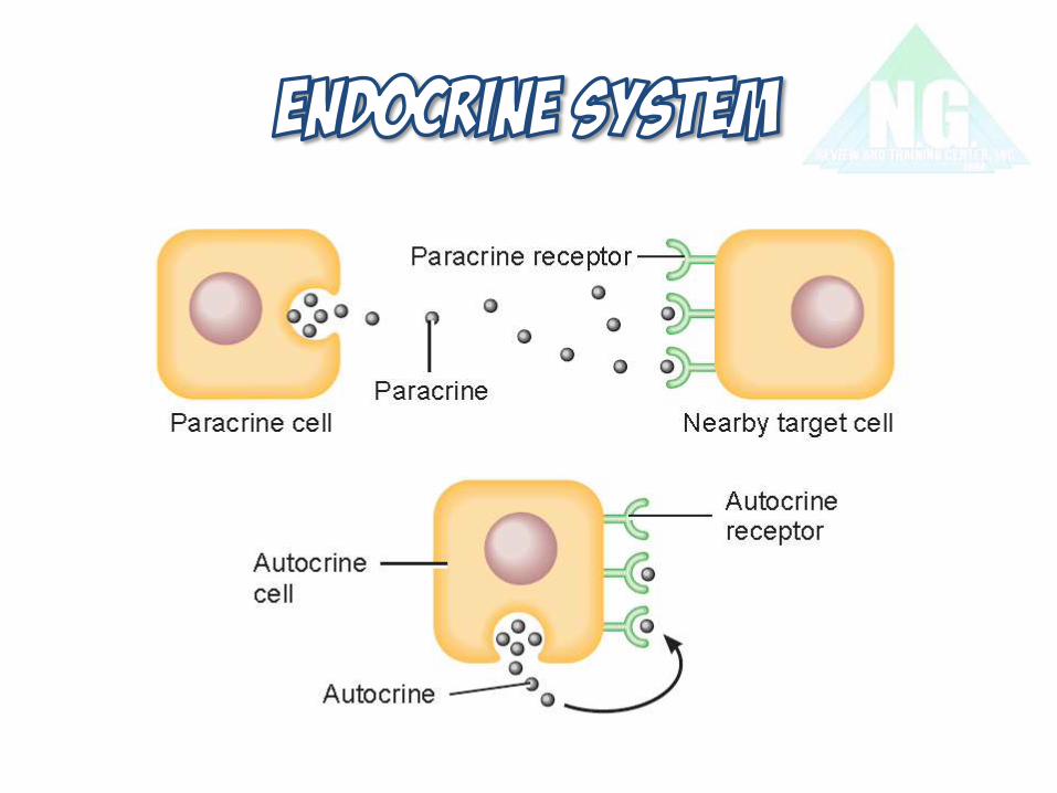
• There are also similar glands called
paracrine and autocrine glands that
are quasi-endocrine
Glands that secrete substances are the
exocrine glands
• They have ducts
• They deliver their products directly to
a specific site
Hormone secretion
(a)
(b)
Blood flow
Skin
Duct
Thyroid
gland
Endocrine
gland
Endocrine
cell
Exocrine gland
(sweat gland)
Exocrine
cells

Nerve impulse
Bloodstream
Neuron
transmits
nerve
impulse
Glandular
cells secrete
hormone into
bloodstream
Neurotransmitter
released into
synapse
Post-
synaptic
cell responds
Target cells
(cells with hormone
receptors) respond
to hormone
Hormones have no
effect on other cells
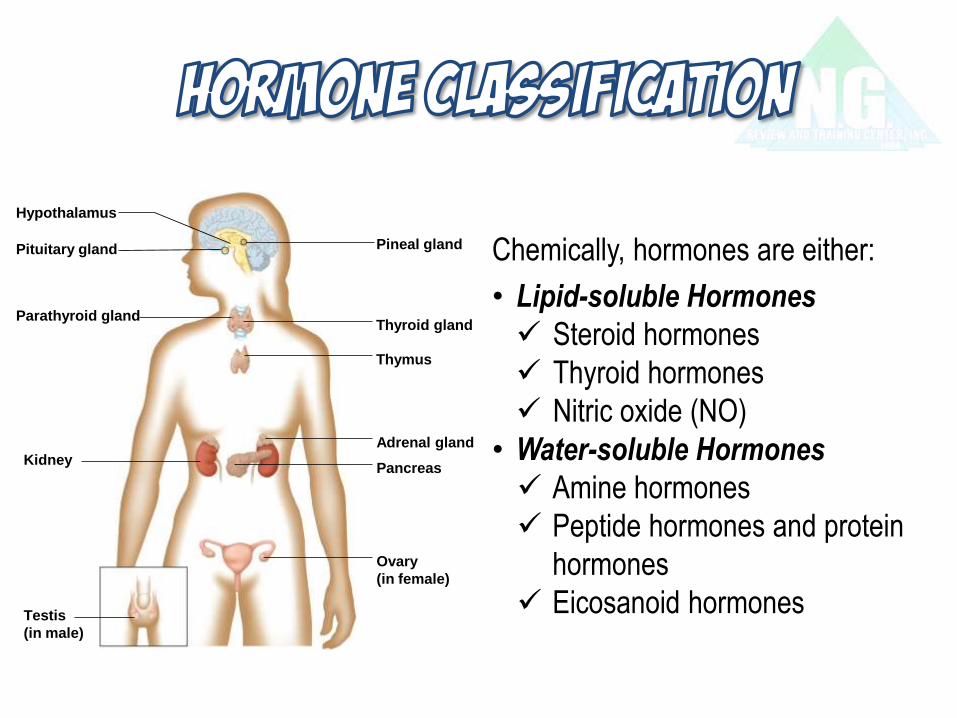
Hypothalamus
Pituitary gland
Thyroid gland
Thymus
Adrenal gland
Pancreas
Parathyroid gland
Pineal gland
Kidney
Testis
(in male)
Ovary
(in female)
Chemically, hormones are either:
• Lipid-soluble Hormones
Steroid hormones
Thyroid hormones
Nitric oxide (NO)
• Water-soluble Hormones
Amine hormones
Peptide hormones and protein
hormones
Eicosanoid hormones

1. Hormonal mechanism: for example, the hypothalamus secretes
hormones that stimulate the anterior pituitary gland to secrete
hormones that stimulate other endocrine glands to secrete
hormones.
2. Humoral mechanism: for example, capillary blood contains a low
concentration of calcium that stimulates secretion of parathyroid
hormone. Parathyroid hormone makes serum calcium go up.
3. Neural mechanism: preganglionic SNS fiber stimulates the
adrenal medulla cells to secrete catecholamines.

Nervous system
Target cells
Action
Target cells
Action
–
– – –
Anterior pituitary gland
Action
Hypothalamus
Peripheral
endocrine
gland
Target cells
Endocrine
gland
Changing level
of substance
in plasma
Endocrine
gland


ORTICOTROPIN-RELEASING HORMONE
HYROTROPIN-RELEASING HORMONE
ROWTH HORMONE-RELEASING HORMONE
ONADOTROPIN-RELEASING HORMONE
ROLACTIN - INHIBITING HORMONE
ROWTH HORMONE- INHIBITORY HORMONE
ELANOCYTE - STIMULATING HORMONE
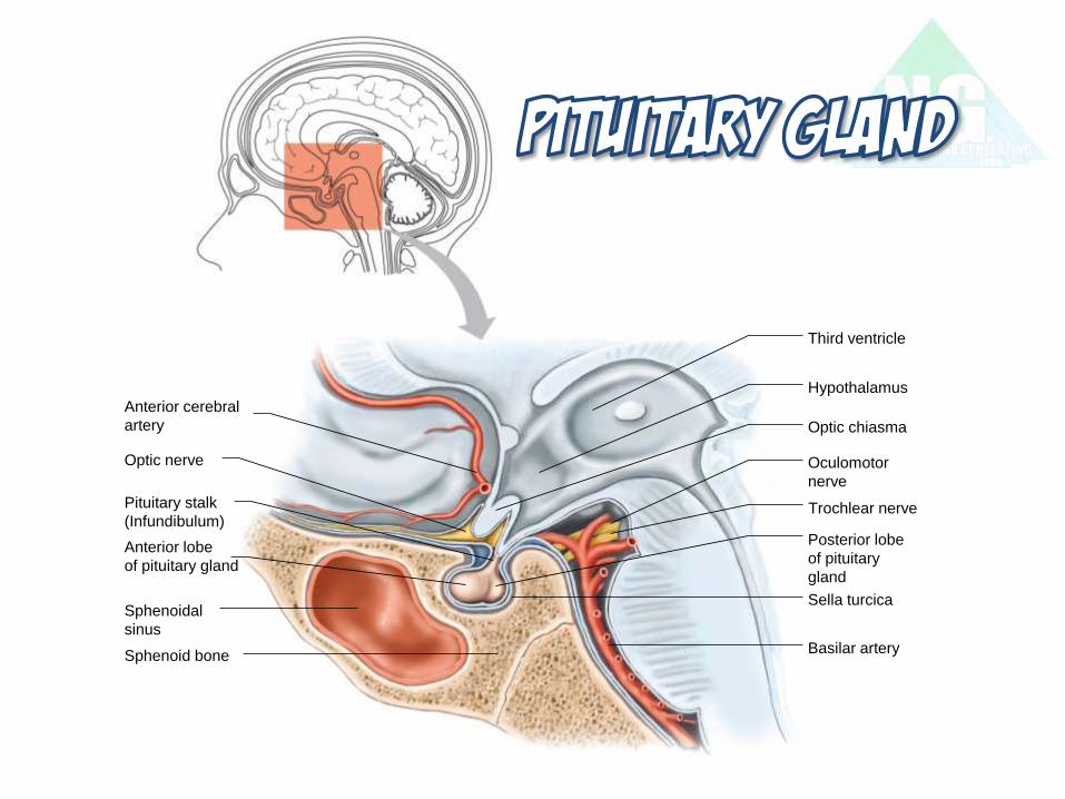
Optic nerve
Sphenoid bone
Hypothalamus
Optic chiasma
Sella turcica
Third ventricle
Trochlear nerve
Basilar artery
Anterior cerebral
artery
Pituitary stalk
(Infundibulum)
Anterior lobe
of pituitary gland
Sphenoidal
sinus
Posterior lobe
of pituitary
gland
Oculomotor
nerve

DRENOCORTICOTROPIC HORMONE
UTEINIZING HORMONE
OLLICLE-STIMULATING HORMONE
ROWTH / SOMATOTROPIC HORMONE
HYROID-STIMULATING HORMONE
ROLACTIN / LACTOGENIC HORMONE
ELANOCYTE-STIMULATING HORMONE
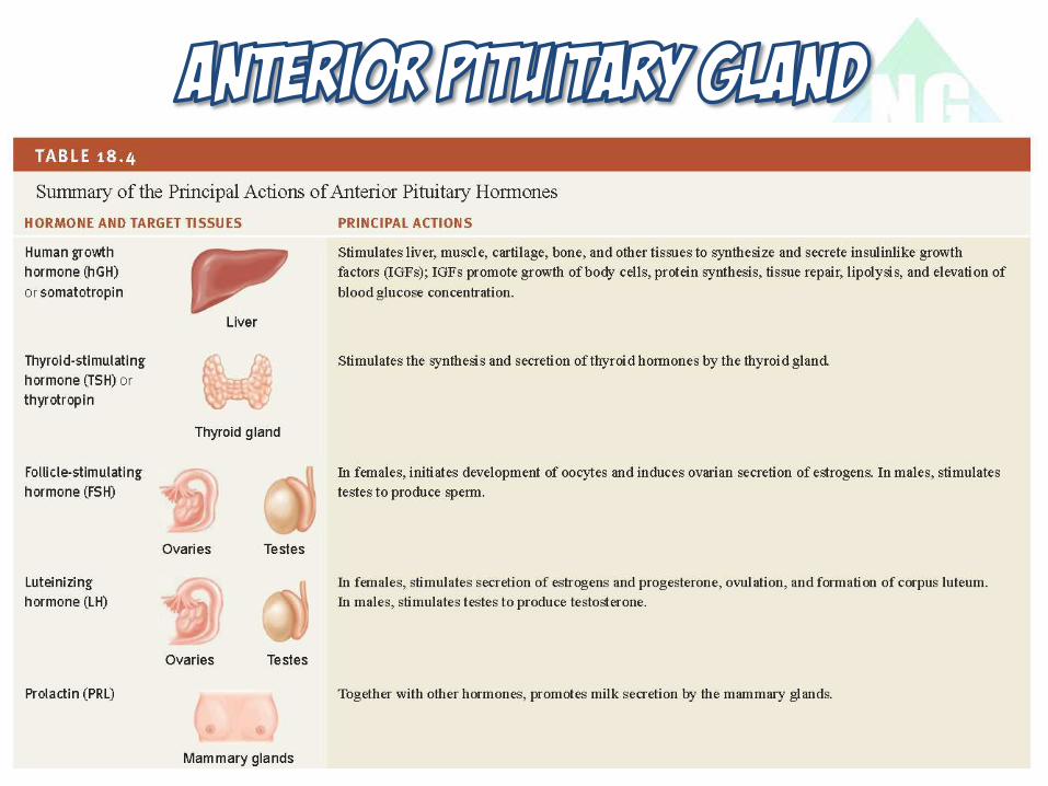

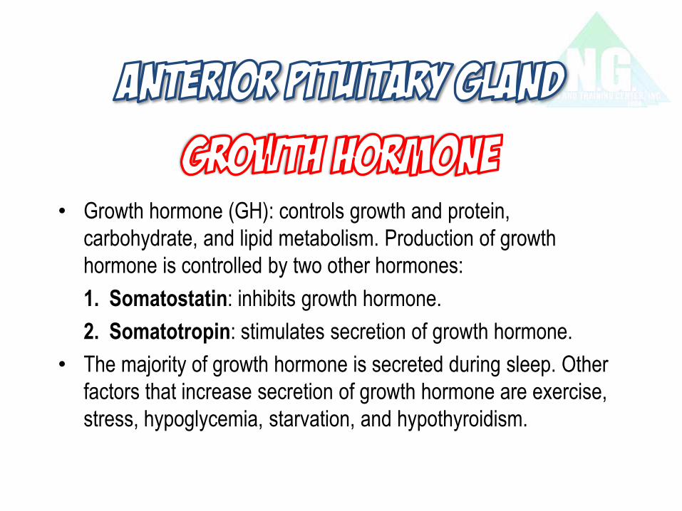
• Growth hormone (GH): controls growth and protein,
carbohydrate, and lipid metabolism. Production of growth
hormone is controlled by two other hormones:
1. Somatostatin: inhibits growth hormone.
2. Somatotropin: stimulates secretion of growth hormone.
• The majority of growth hormone is secreted during sleep. Other
factors that increase secretion of growth hormone are exercise,
stress, hypoglycemia, starvation, and hypothyroidism.
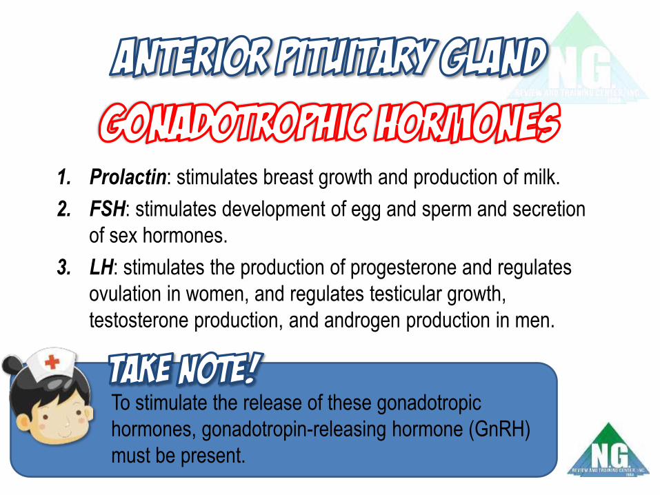
1. Prolactin: stimulates breast growth and production of milk.
2. FSH: stimulates development of egg and sperm and secretion
of sex hormones.
3. LH: stimulates the production of progesterone and regulates
ovulation in women, and regulates testicular growth,
testosterone production, and androgen production in men.
To stimulate the release of these gonadotropic
hormones, gonadotropin-releasing hormone (GnRH)
must be present.

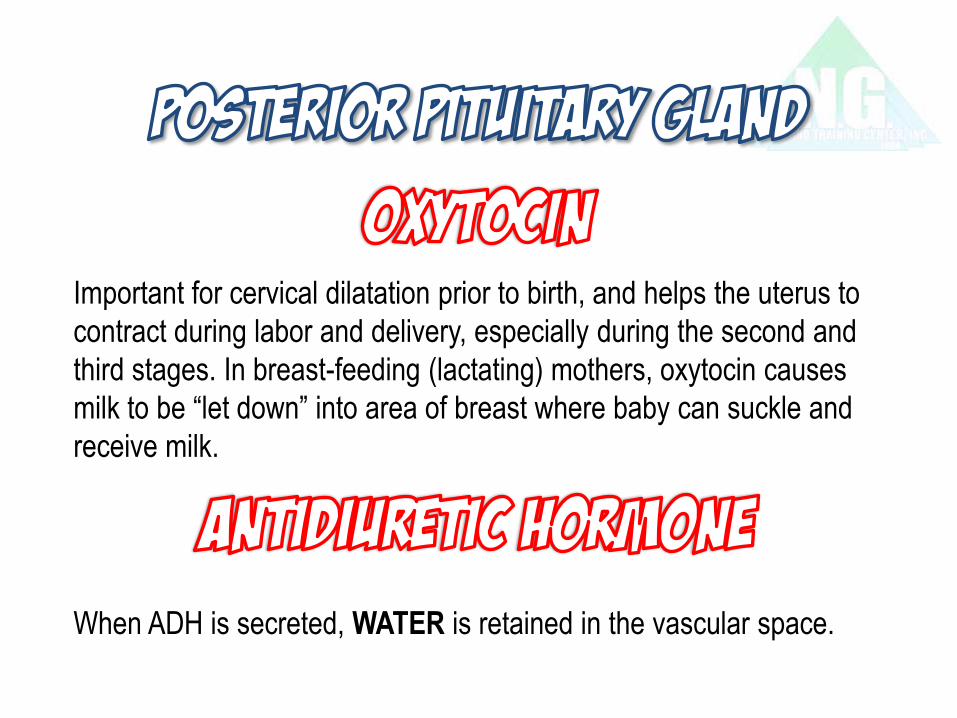
Important for cervical dilatation prior to birth, and helps the uterus to
contract during labor and delivery, especially during the second and
third stages. In breast-feeding (lactating) mothers, oxytocin causes
milk to be “let down” into area of breast where baby can suckle and
receive milk.
When ADH is secreted, WATER is retained in the vascular space.


Copyright © The McGraw-Hill Companies, Inc. Permission required for reproduction or display.
Follicular cells
Colloid
Extrafollicular
cells
© Fred Hossler/Visuals Unlimited
Larynx Colloid
Isthmus
(a)
(b)
Thyroid
gland Follicular
cell
Extrafollicular
cell
Copyright © The McGraw-Hill Companies, Inc. Permission required for reproduction or display.
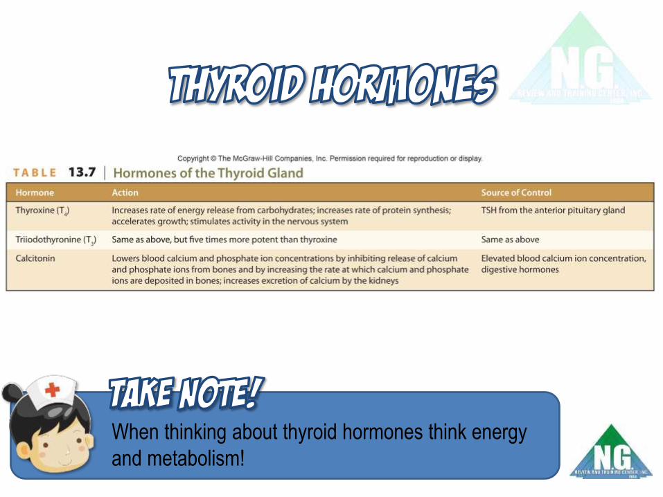
When thinking about thyroid hormones think energy
and metabolism!
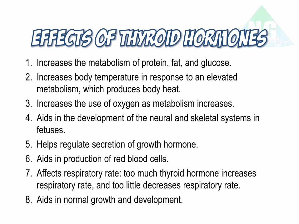
1. Increases the metabolism of protein, fat, and glucose.
2. Increases body temperature in response to an elevated
metabolism, which produces body heat.
3. Increases the use of oxygen as metabolism increases.
4. Aids in the development of the neural and skeletal systems in
fetuses.
5. Helps regulate secretion of growth hormone.
6. Aids in production of red blood cells.
7. Affects respiratory rate: too much thyroid hormone increases
respiratory rate, and too little decreases respiratory rate.
8. Aids in normal growth and development.

• Both T3 and T4 increase metabolic rate of cells and tissues.
• T4 is the precursor to T3.
• T3 regulates the metabolic rate of all cells and all processes of cell
growth and tissue differentiation.
• T3 and T4 indirectly increase blood glucose levels.
• Being cold increases the conversion of T4 to T3.
• Things such as stress, starvation, certain dyes, and certain drugs
like steroids, beta-blockers, PTU (propylthiouracil), and
amiodarone decrease the conversion of T4 to T3.

• Targets the bones, kidneys, and epithelial cells of the intestines.
• Decreases blood/serum calcium in three ways:
1. Decreases intestines’ ability to absorb calcium.
2. Decreases osteoclast activity in the bones.
3. Decreases calcium resorption from the kidney tubules.
Bone resorption occurs when osteoclasts break down
bone and release calcium from the bone into the
blood.

Posterior view
Esophagus
Pharynx
Thyroid
gland
Parathyroid
glands
Trachea
Copyright © The McGraw-Hill Companies, Inc. Permission required for reproduction or display.
Secretory cells
Capillaries
Copyright © The McGraw-Hill Companies, Inc. Permission required for reproduction or display.
© R. Calentine/Visuals Unlimited

Liver
Intestinal enzymes
Ultraviolet light in skin
Kidney Stimulated by PTH
Hydroxycholecalciferol
Foods
Ca+2
Ca+2
Ca+2
Cholesterol
Provitamin D
Vitamin D
(Cholecalciferol)
Also obtained directly
from foods
Dihydroxycholecalciferol
(active form of vitamin D)
Controls absorption of
calcium in intestine
• Parathyroid hormone (PTH) makes
the serum calcium level go up!
• Too much PTH
hyperparathyroidism
hypercalcemia,
hypophosphatemia, bone damage,
and renal damage.
• Too little PTH hypoparathyroidism,
hypocalcemia,
hyperphosphatemia, hyperreflexia,
and cognitive changes (altered
sensorium).

Copyright © The McGraw-Hill Companies, Inc. Permission required for reproduction or display.
PTH Ca+2
+ PTH
PTH
Ca+2
+ Ca+2
Bloodstream
– Stimulation
Inhibition
Release into
bloodstream
Parathyroid glands (on
posterior of thyroid gland)
Decreased blood calcium
stimulates parathyroid
hormone secretion
Increased blood
calcium inhibits
PTH secretion
Kidneys
conserve Ca+2 and
activate Vitamin D
Bone
releases Ca+2
Intestine
absorbs Ca+2
Active
Vitamin D

Co
rte
x
Me
du
lla
Capsule
Zona
glomerulosa
Zona
fasciculata
Zona
reticularis
Chromaffin
cells
Copyright © The McGraw-Hill Companies, Inc. Permission required for reproduction or display.
© Ed Reschke
Adrenal gland
Kidney
Adrenal cortex
(a)
Zona
lomerulosa
Connective
tissue capsule
(b)
Zona
fasciculata
Zona
reticularis
Adrenal
medulla
Adrenal
cortex
Adrenal
medulla
Surface of
adrenal gland
Copyright © The McGraw-Hill Companies, Inc. Permission required for reproduction or display.


Many heart attacks occur during the early morning hours when
people are coming out of REM sleep, as this is a very stressful
time for the body.
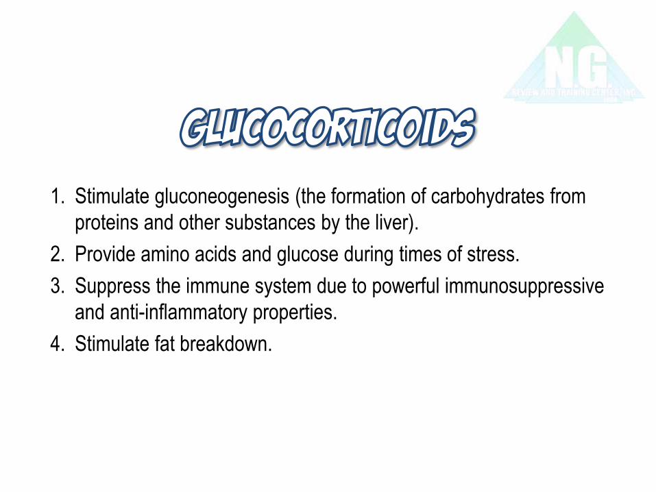
1. Stimulate gluconeogenesis (the formation of carbohydrates from
proteins and other substances by the liver).
2. Provide amino acids and glucose during times of stress.
3. Suppress the immune system due to powerful immunosuppressive
and anti-inflammatory properties.
4. Stimulate fat breakdown.

An increase in aldosterone secretion is also caused by:
• Low fluid volume levels in the vascular space as in shock or
hypovolemia.
• High blood levels of potassium.
The illnesses associated with aldosterone are:
• Hyperaldosteronism (Conn’s syndrome)
• Hypoaldosteronism

Sex hormones are usually broken down into three categories:
1. Androgens, testosterone being the main one.
2. Estrogens, estradiol being the main one.
3. Progestagens, progesterone being the main one.
Synthetic androgens (sex hormones) are referred to
as anabolic steroids.

Copyright © The McGraw-Hill Companies, Inc. Permission required for reproduction or display.
Pancreatic islet (Islet of Langerhans)
From Kent M. Van De Graaff and Stuart Ira Fox, Concepts of Human Anatomy and
Physiology, 2nd ed. ©1989 Wm. C. Brown Publishers, Dubuque, Iowa. All Rights
Reserved. Reprinted with permission
Copyright © The McGraw-Hill Companies, Inc. Permission required for reproduction or display.
Gallbladder Common bile duct
Pancreatic duct
Pancreas Duct
Capillary
Small
intestine
Digestive enzyme-
secreting cells
Pancreatic islet
(Islet of Langerhans)
Hormone-secreting
islet cells

too high
too low
Control center
Beta cells secrete
insulin
Receptors
Beta cells detect a rise
in blood glucose
Effectors
Insulin
• Promotes movement of glucose
into certain cells
• Stimulates formation of glycogen
from glucose
Stimulus
Rise in blood glucose
Response
Blood glucose drops toward
normal (and inhibits insulin
secretion)
Normal
blood glucose
concentration
Stimulus
Drop in blood glucose
Response
Blood glucose rises toward
normal (and inhibits glucagon
secretion)
Receptors
Alpha cells detect a drop
in blood glucose
Effectors
Glucagon
• Stimulates cells to break down glycogen
into glucose
• Stimulates cells to convert
noncarbohydrates into glucose
Control center
Alpha cells secrete
glucagon

Pineal Gland
• Secretes melatonin
• Regulates circadian rhythms
Thymus Gland
• Secretes thymosins
• Promotes development of certain lymphocytes
• Important in role of immunity
Reproductive Organs
• Ovaries produce estrogens and progesterone
• Testes produce testosterone
• Placenta produces estrogens, progesterone, and
gonadotropin
Other organs: digestive glands, heart, and kidney
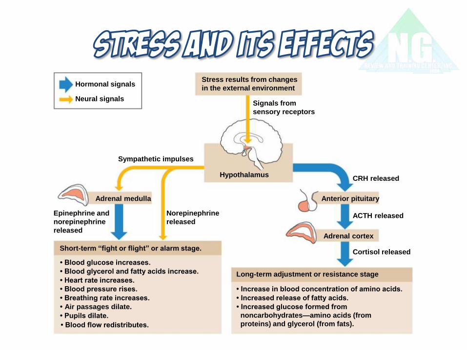
Sympathetic impulses
CRH released
ACTH released
Cortisol released
Long-term adjustment or resistance stage
• Increase in blood concentration of amino acids.
• Increased release of fatty acids.
• Blood glucose increases.
• Blood glycerol and fatty acids increase.
• Heart rate increases.
• Blood pressure rises.
• Breathing rate increases.
• Air passages dilate.
• Pupils dilate.
Anterior pituitary
Hypothalamus
Adrenal cortex
Adrenal medulla
Neural signals
Hormonal signals Stress results from changes
in the external environment
Signals from
sensory receptors
Epinephrine and
norepinephrine
released
Norepinephrine
released
Short-term “fight or flight” or alarm stage.
• Blood flow redistributes.
• Increased glucose formed from
noncarbohydrates—amino acids (from
proteins) and glycerol (from fats).


• Endocrine glands decrease in size
• Muscular strength decreases as GH levels decrease
• ADH levels increase due to slower break down in liver and
kidneys
• Calcitonin levels decrease; increase risk of osteoporosis
• PTH level changes contribute to risk of osteoporosis
• Insulin resistance may develop
• Changes in melatonin secretion affect the body clock
• Thymosin production declines increasing risk of infections

test Normal values significance Thyroid-stimulating
hormone
0.5–5.0 U/mL ↑ in primary hypothyroidism
↓in primary hyperthyroidism
Triiodothyronine (T3) 80–200 ng/100 mL ↓ in hypothyroidism
↑ in hyperthyroidism
Thyroxine (T4 ) 4–12 g/100 mL ↓ in hypothyroidism
↑ in hyperthyroidism

test Normal values significance Parathyroid
hormone
25 pg/mL ↑ in primary hyperparathyroidism
↓ in primary hypoparathyroidism,
parathyroid trauma during thyroid
surgery
Calcium 8.5–10.5 mg/100 mL ↑ in some cancers,
hyperparathyroidism
↓ in hypothyroidism
Phosphorus 2.4–4.7 mg/dL ↑ in hypoparathyroidism
↓ in hyperparathyroidism

test Normal values significance Growth hormone 5 ng/mL ↑ in acromegaly
↓ in small stature
Antidiuretic
hormone
2.3–3.1 pg/mL ↑ in SIADH
↓ in diabetes insipidus
Urine specific
gravity
1.010–1.025 ↓ in diabetes insipidus
Adrenocorticotropic
hormone
120 pg/mL at 6–8 a.m. ↑ in Addison’s disease
↓ in Cushing’s syndrome, long-
term corticosteroid therapy

test Normal values significance Cortisol 5–25 g/100 mL ↑ in Cushing’s syndrome, stress
↓ in Addison’s disease, steroid
withdrawal
Vanillylmandelic
acid (VMA)
0.7–6.8 mg/24 h ↑ in pheochromocytoma

test Normal values significance Fasting plasma
glucose (FPG)
70–100 mg/dL in stress, Cushing’s syndrome
FPG 101–125 pre-diabetes
FPG 126 diabetes mellitus
↓ in hypoglycemia, Addison’s
disease
Oral glucose
tolerance test
Blood glucose less
than 140 mg/dL at 2
hours
BG 140–199 at 2 hours pre-
diabetes; BG 200 at 2 hours
diabetes mellitus
Glycosylated
hemoglobin
4%–7%
↑ in poor diabetes control
• A thyroid scan may be done to determine the presence of
tumors or nodules.
• A computed tomographic (CT) scan or magnetic resonance
imaging (MRI) may be done to locate a tumor or identify
hypertrophy of a gland.
• Ultrasound may be done of the thyroid or parathyroid glands to
determine if they are enlarged or to find masses.
• Biopsy is done to obtain tissue to examine for possible
cancerous cells.