Newborn Screening for Severe Combined Immunodeficiency ... · Newborn Screening for Severe Combined...
38
Newborn Screening for Severe Combined Immunodeficiency (SCID) by Quantifying T-cell Receptor Excision Circles (TREC) Patricia R. Slev, PhD, D(ABCC) Medical Director, Serologic Hepatitis and Retrovirus Laboratory, ARUP Assistant Professor, Department of Pathology, University of Utah School of Medicine [email protected] July, 2013
Transcript of Newborn Screening for Severe Combined Immunodeficiency ... · Newborn Screening for Severe Combined...
Newborn Screening for Severe Combined
Immunodeficiency (SCID) by Quantifying T-cell
Receptor Excision Circles (TREC)
Patricia R. Slev, PhD, D(ABCC)
Medical Director, Serologic Hepatitis and Retrovirus Laboratory, ARUP
Assistant Professor, Department of Pathology,
University of Utah School of Medicine
July, 2013

Disclosures
None
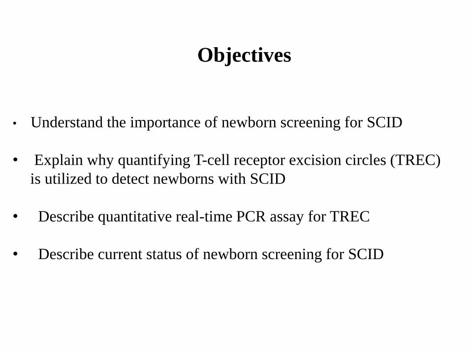
Objectives
• Understand the importance of newborn screening for SCID
• Explain why quantifying T-cell receptor excision circles (TREC)
is utilized to detect newborns with SCID
• Describe quantitative real-time PCR assay for TREC
• Describe current status of newborn screening for SCID
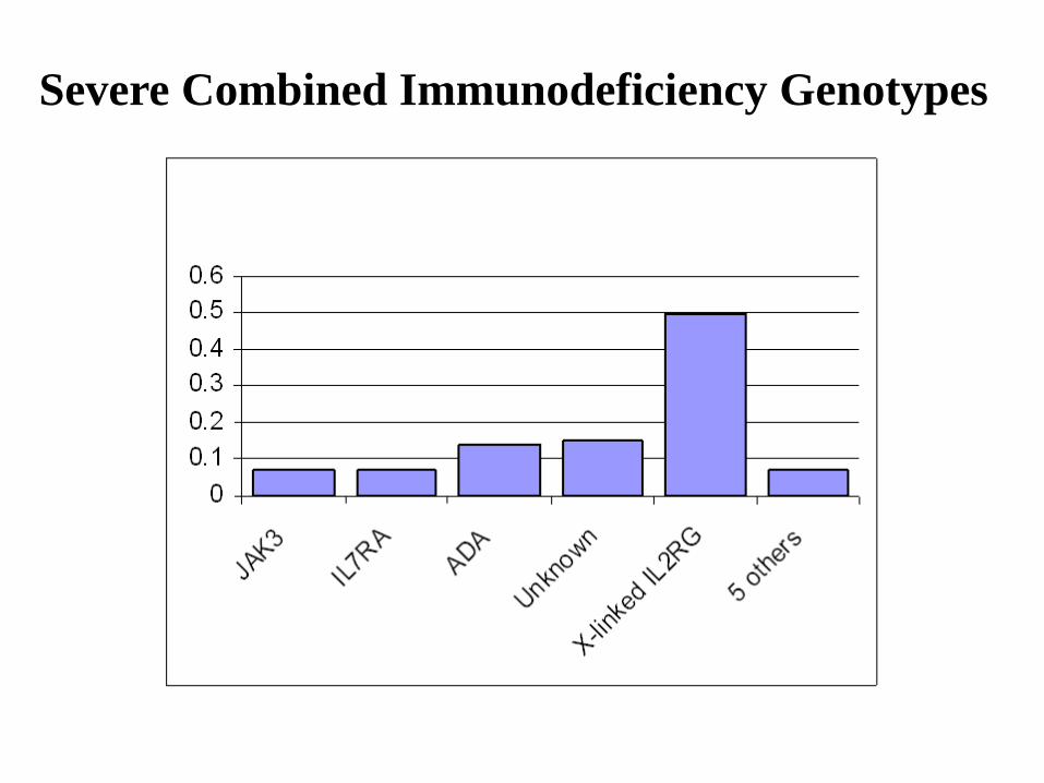
Severe Combined Immunodeficiency Genotypes
Adapted from IL2RG database, Puck et al.
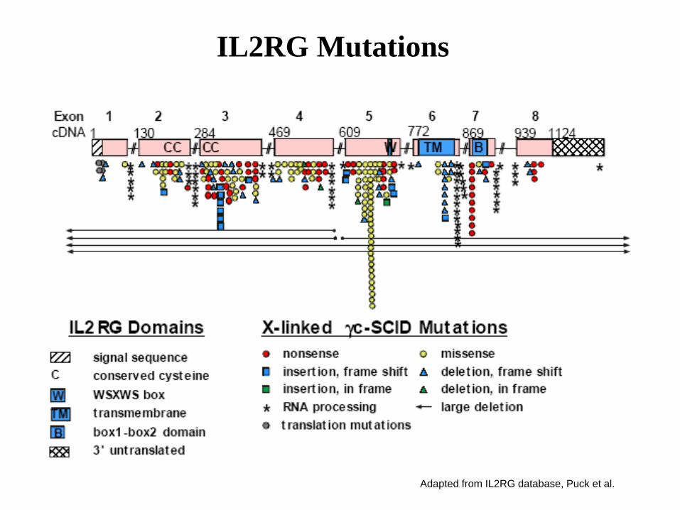
IL2RG Mutations
Adapted from SCID website (Duke University)
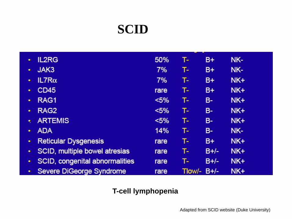
T-cell lymphopenia
SCID
SCID Newborn Screening
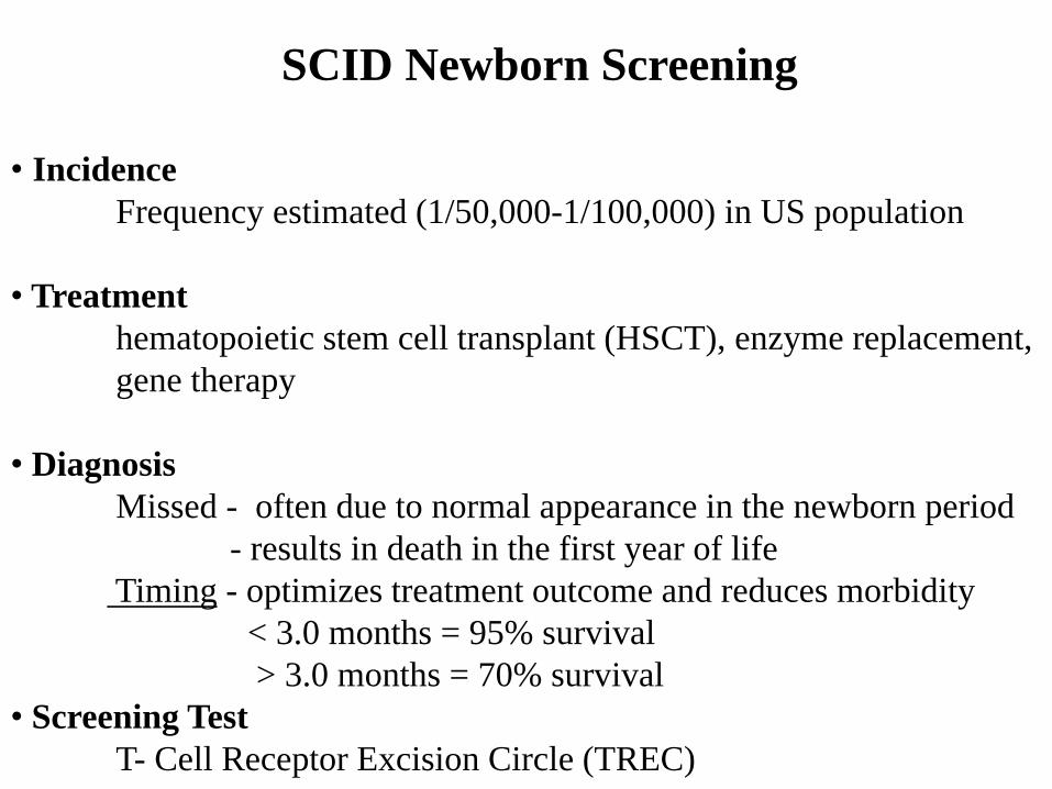
• Incidence
Frequency estimated (1/50,000-1/100,000) in US population
• Treatment
hematopoietic stem cell transplant (HSCT), enzyme replacement,
gene therapy
• Diagnosis
Missed - often due to normal appearance in the newborn period
- results in death in the first year of life
Timing - optimizes treatment outcome and reduces morbidity
< 3.0 months = 95% survival
> 3.0 months = 70% survival
• Screening Test
T- Cell Receptor Excision Circle (TREC)
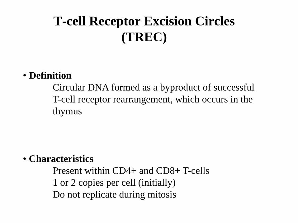
• Definition
Circular DNA formed as a byproduct of successful
T-cell receptor rearrangement, which occurs in the
thymus
• Characteristics
Present within CD4+ and CD8+ T-cells
1 or 2 copies per cell (initially)
Do not replicate during mitosis
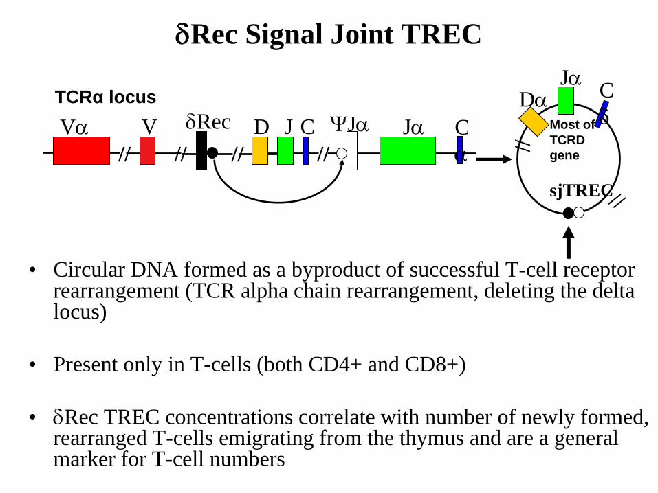
T-cell Receptor Excision Circles
(TREC)
// // // //
V V Rec J D C J J C
J D C
TCRα locus
Most of
TCRD
gene
sjTREC
Rec Signal Joint TREC
• Circular DNA formed as a byproduct of successful T-cell receptor rearrangement (TCR alpha chain rearrangement, deleting the delta locus)
• Present only in T-cells (both CD4+ and CD8+)
• Rec TREC concentrations correlate with number of newly formed, rearranged T-cells emigrating from the thymus and are a general marker for T-cell numbers
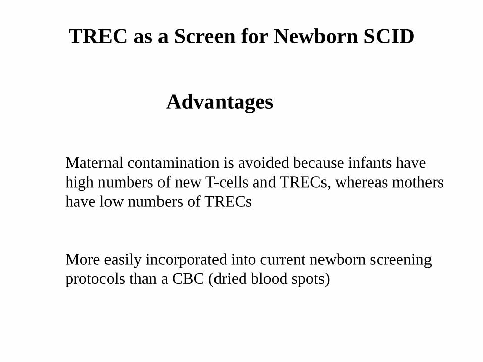
TREC as a Screen for Newborn SCID
Advantages
Maternal contamination is avoided because infants have
high numbers of new T-cells and TRECs, whereas mothers
have low numbers of TRECs
More easily incorporated into current newborn screening
protocols than a CBC (dried blood spots)
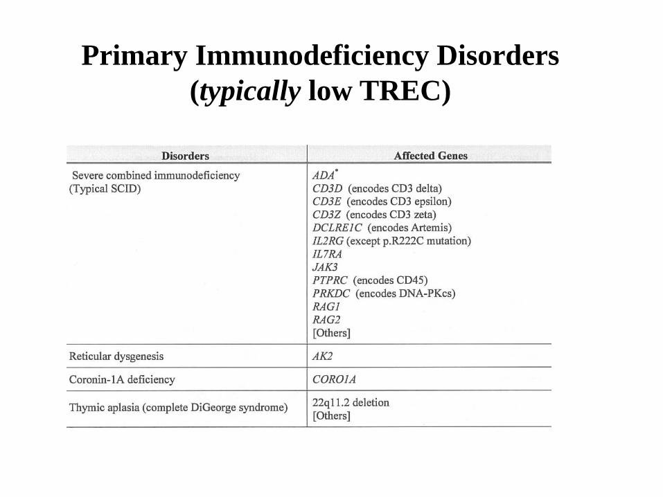
Primary Immunodeficiency Disorders
(typically low TREC)
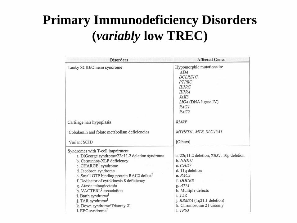
Primary Immunodeficiency Disorders
(variably low TREC)
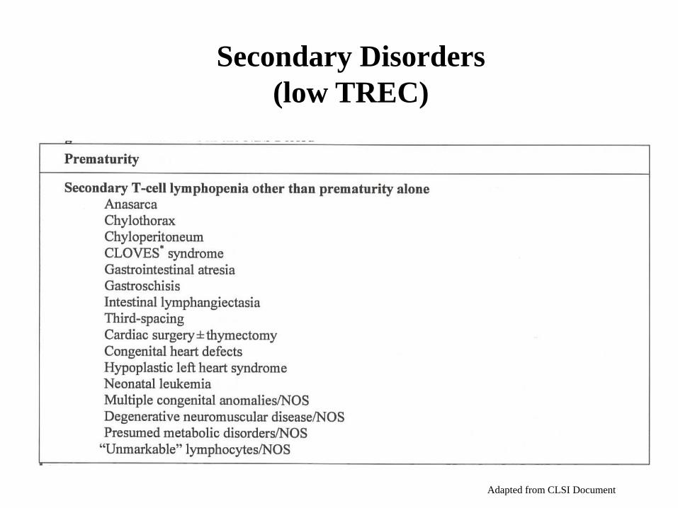
Secondary Disorders
(low TREC)
Adapted from CLSI Document
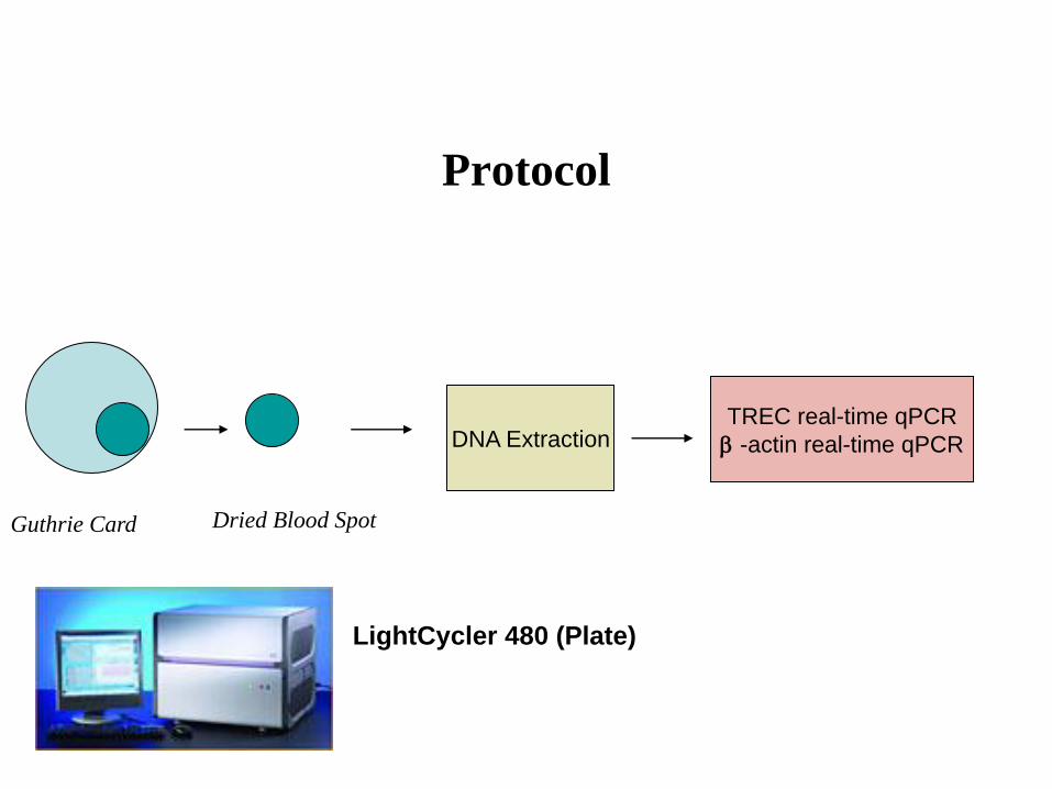
Protocol
Guthrie Card Dried Blood Spot
DNA Extraction TREC real-time qPCR
-actin real-time qPCR
LightCycler 480 (Plate)
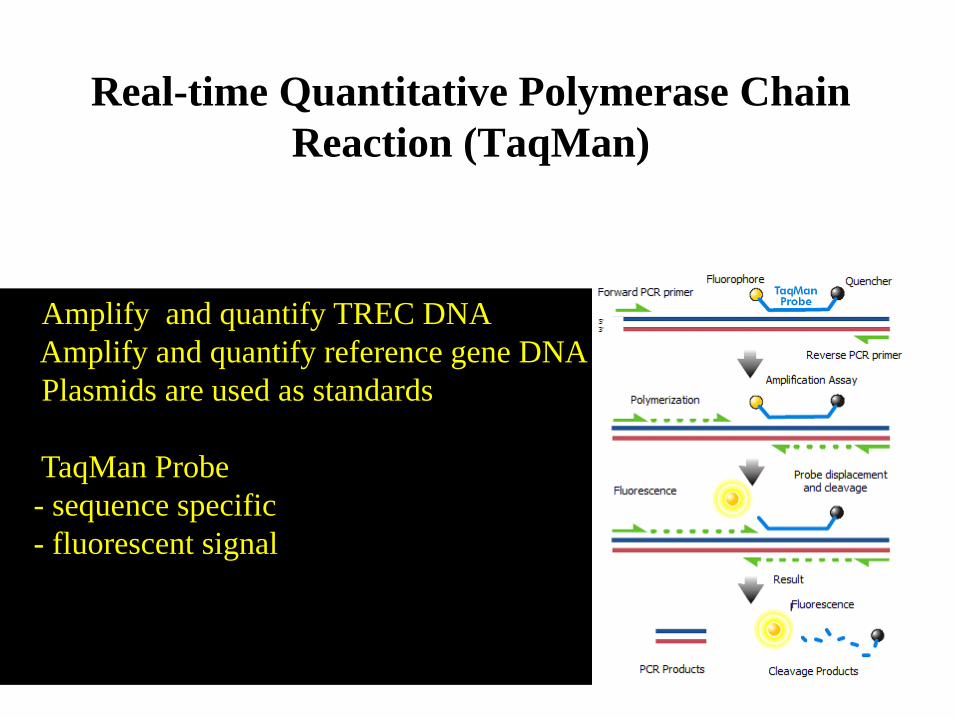
Amplify and quantify TREC DNA
Amplify and quantify reference gene DNA
Plasmids are used as standards
TaqMan Probe
- sequence specific
- fluorescent signal
Real-time Quantitative Polymerase Chain
Reaction (TaqMan)
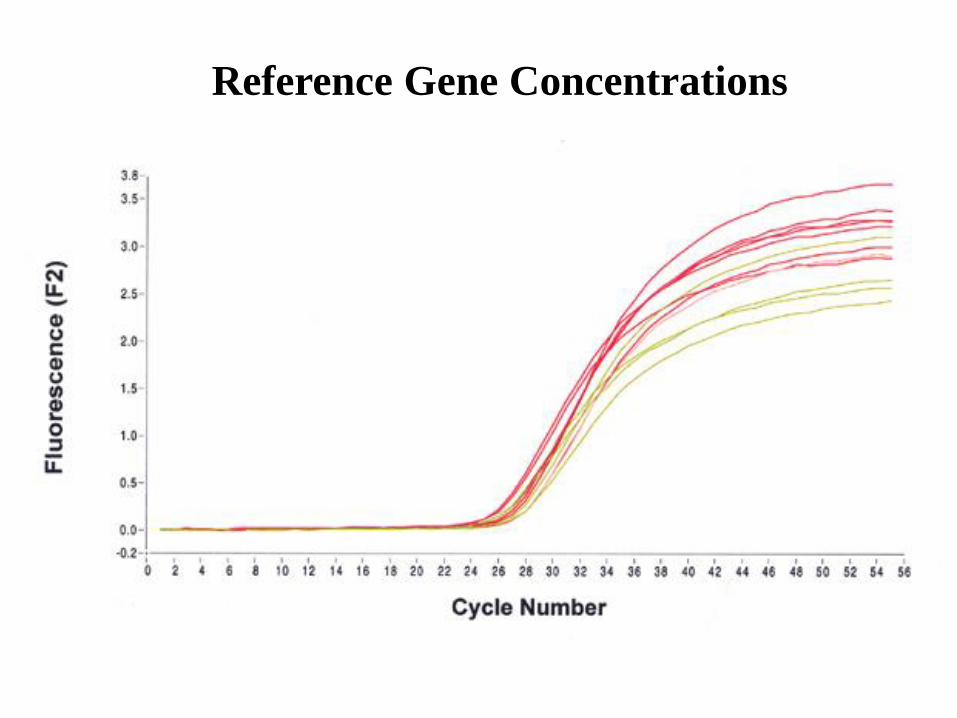
Reference Gene Concentrations
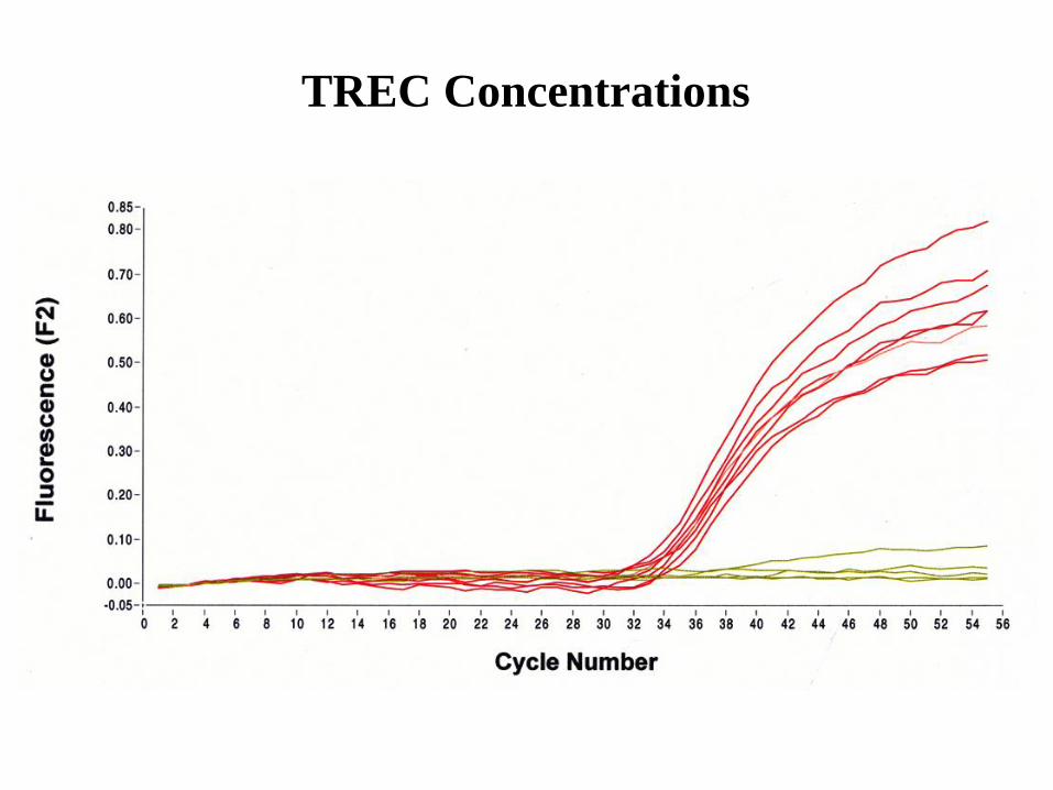
TREC Concentrations
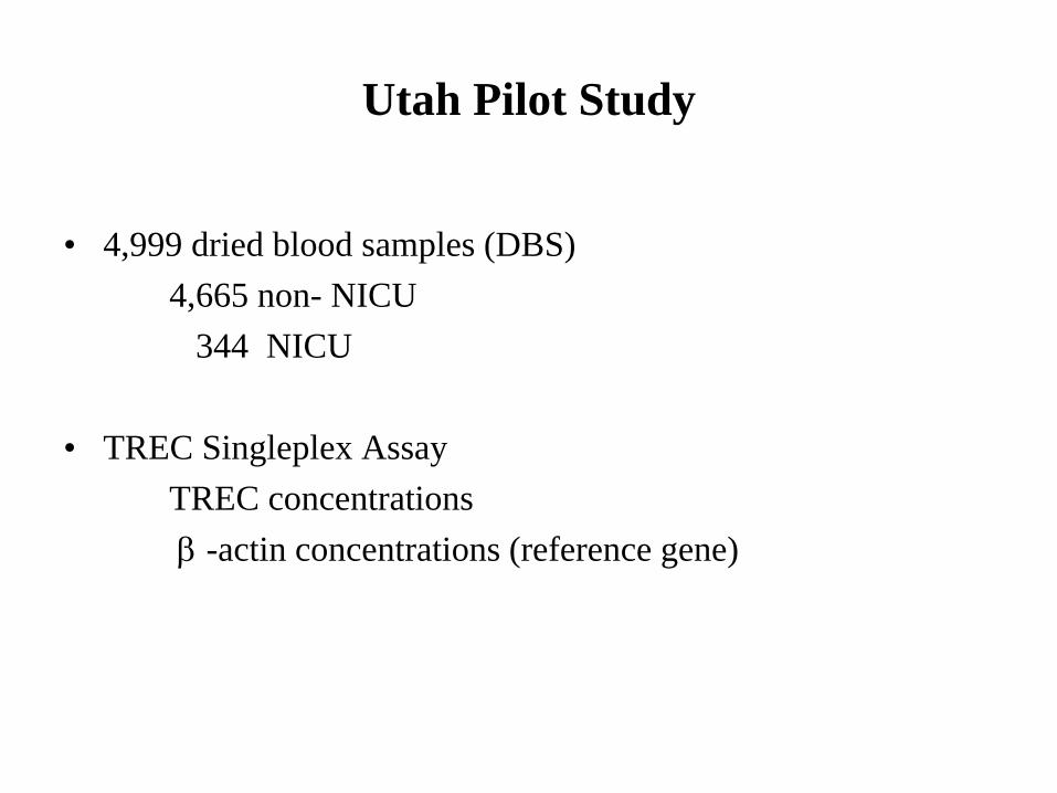
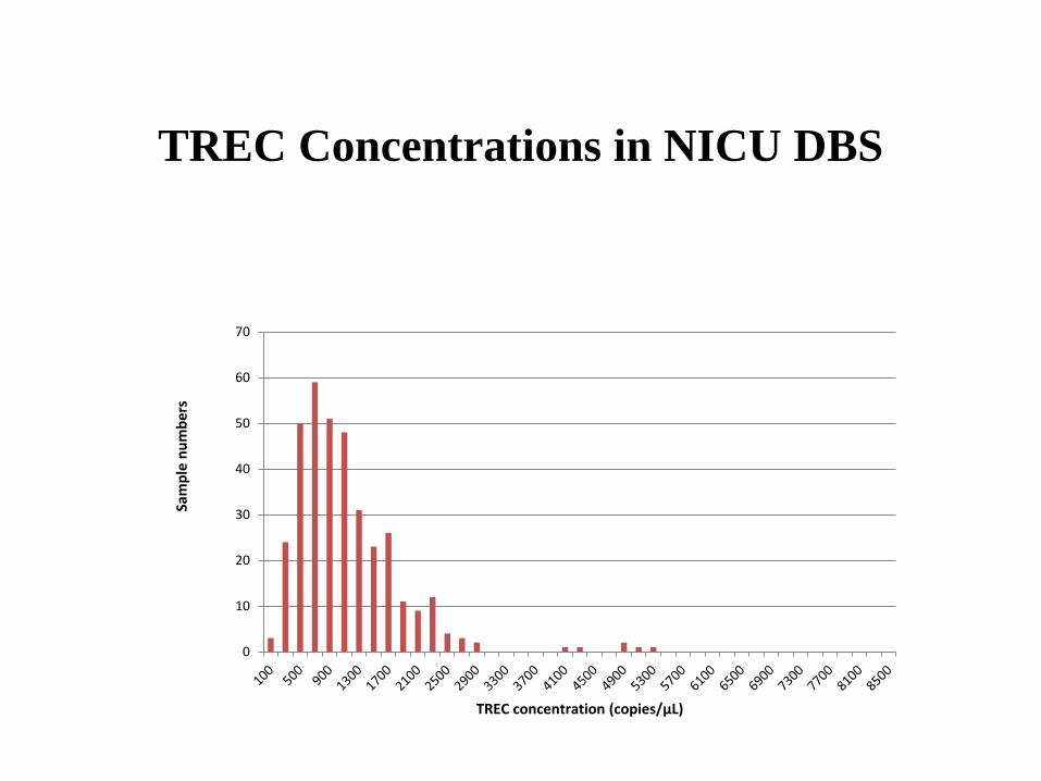
Utah Pilot Study
• 4,999 dried blood samples (DBS)
4,665 non- NICU
344 NICU
• TREC Singleplex Assay
TREC concentrations
-actin concentrations (reference gene)
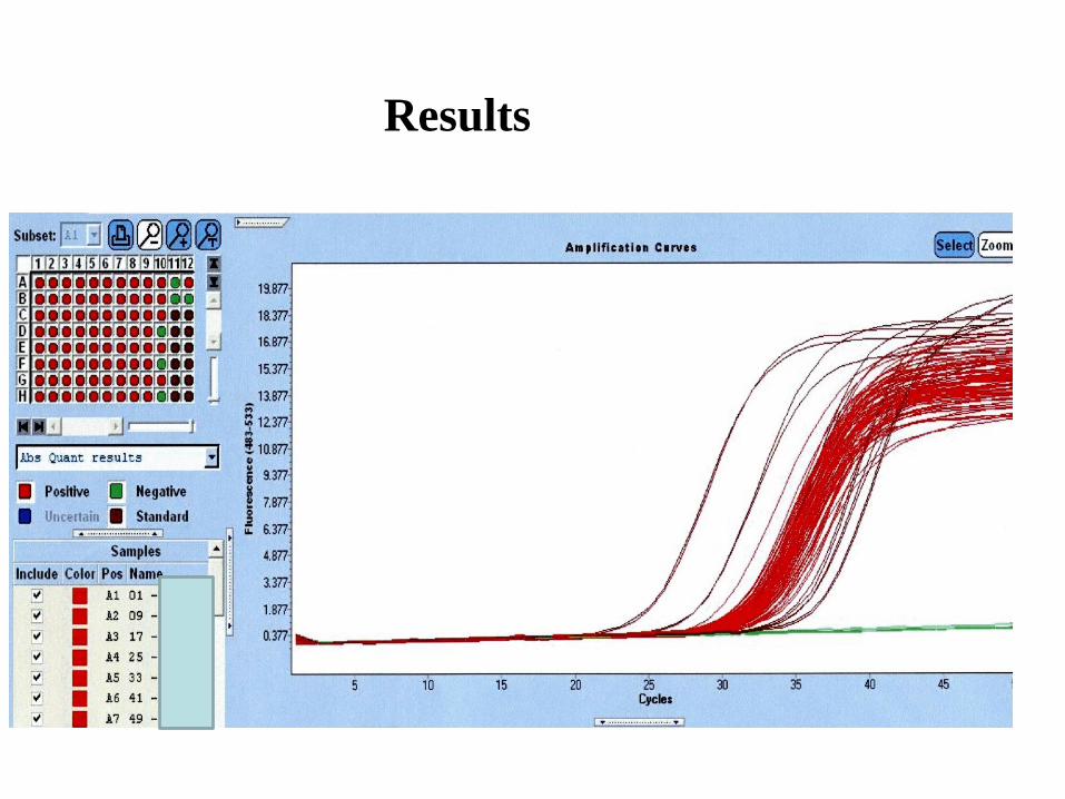
Results
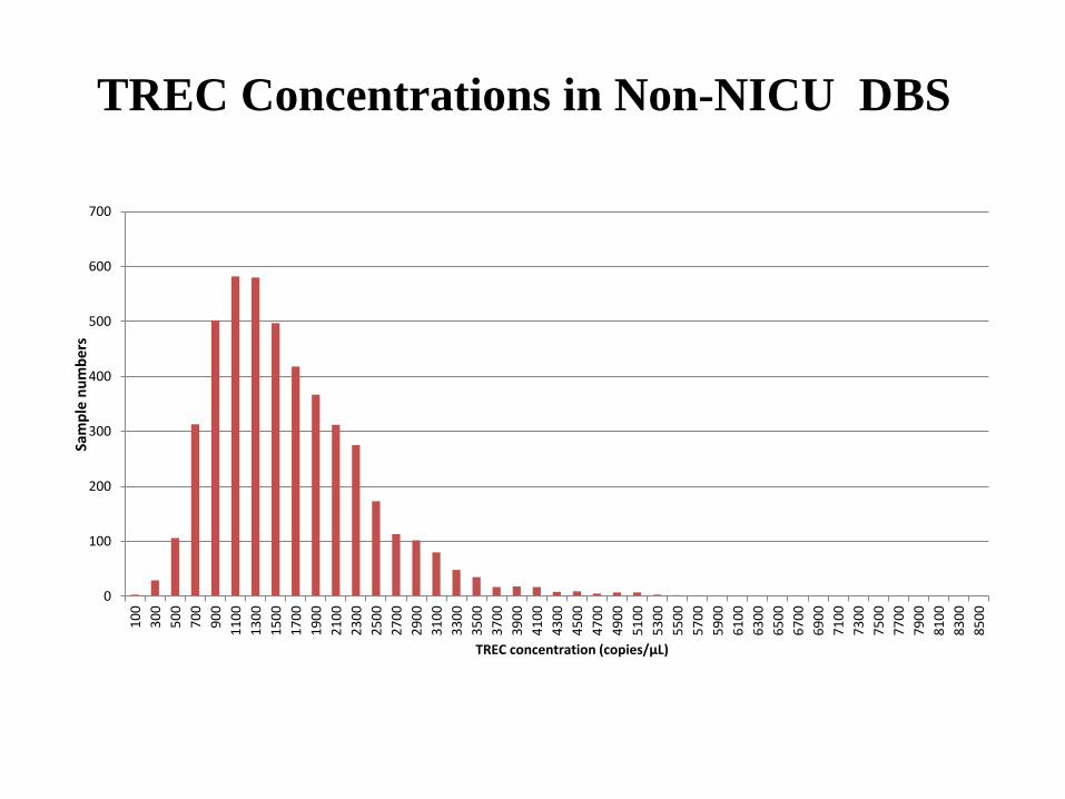
TREC Concentrations in Non-NICU DBS
0
100
200
300
400
500
600
700
10
0
30
0
50
0
70
0
90
0
11
00
13
00
15
00
17
00
19
00
21
00
23
00
25
00
27
00
29
00
31
00
33
00
35
00
37
00
39
00
41
00
43
00
45
00
47
00
49
00
51
00
53
00
55
00
57
00
59
00
61
00
63
00
65
00
67
00
69
00
71
00
73
00
75
00
77
00
79
00
81
00
83
00
85
00
Sam
ple
nu
mb
ers
TREC concentration (copies/µL)
0
10
20
30
40
50
60
70
Sam
ple
nu
mb
ers
TREC concentration (copies/µL)
TREC Concentrations in NICU DBS
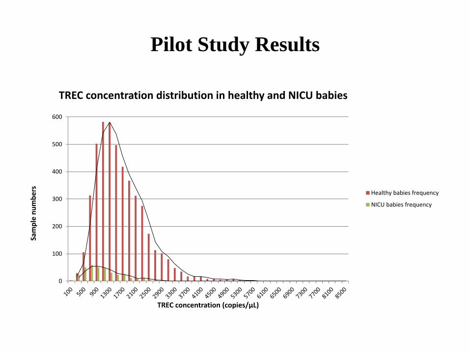
Pilot Study Results
0
100
200
300
400
500
600
Sam
ple
nu
mb
ers
TREC concentration (copies/µL)
TREC concentration distribution in healthy and NICU babies
Healthy babies frequency
NICU babies frequency
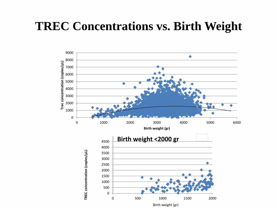
TREC Concentrations vs. Birth Weight
0
1000
2000
3000
4000
5000
6000
7000
8000
9000
0 1000 2000 3000 4000 5000 6000
Tre
c co
nce
ntr
atio
n (
cop
ies/
µL)
Birth weight (gr)
0
500
1000
1500
2000
2500
3000
3500
4000
4500
0 500 1000 1500 2000TREC
co
nce
ntr
atio
n (
cop
ies/μ
L)
Birth weight <2000 gr
Birth weight (gr)
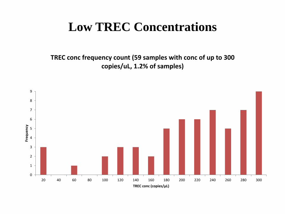
Low TREC Concentrations
0
1
2
3
4
5
6
7
8
9
20 40 60 80 100 120 140 160 180 200 220 240 260 280 300
Fre
qu
en
cy
TREC conc (copies/µL)
TREC conc frequency count (59 samples with conc of up to 300 copies/uL, 1.2% of samples)
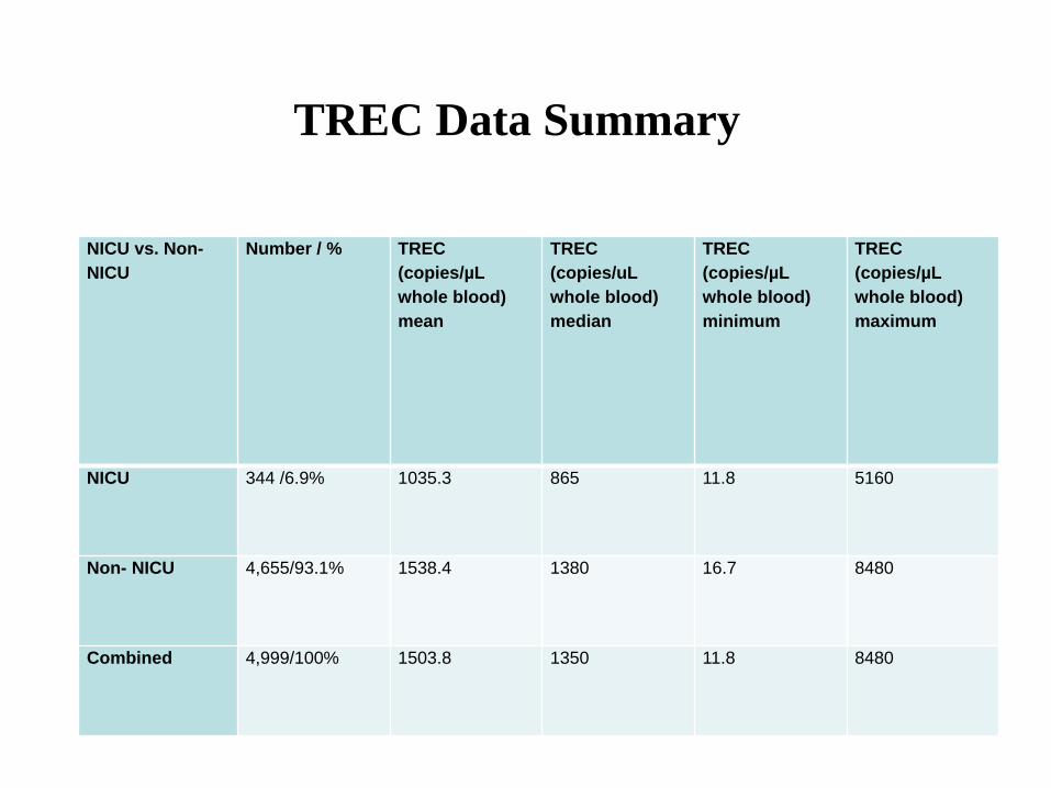
TREC Data Summary
NICU vs. Non-
NICU
Number / % TREC
(copies/µL
whole blood)
mean
TREC
(copies/uL
whole blood)
median
TREC
(copies/µL
whole blood)
minimum
TREC
(copies/µL
whole blood)
maximum
NICU 344 /6.9% 1035.3 865 11.8 5160
Non- NICU 4,655/93.1% 1538.4 1380 16.7 8480
Combined 4,999/100% 1503.8 1350 11.8 8480
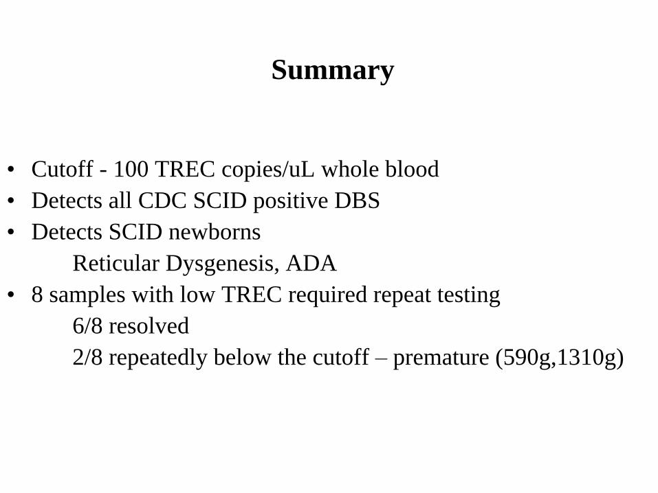
Summary
• Cutoff - 100 TREC copies/uL whole blood
• Detects all CDC SCID positive DBS
• Detects SCID newborns
Reticular Dysgenesis, ADA
• 8 samples with low TREC required repeat testing
6/8 resolved
2/8 repeatedly below the cutoff – premature (590g,1310g)
8 abnormal flow cytometry
3 normal flow cytometry
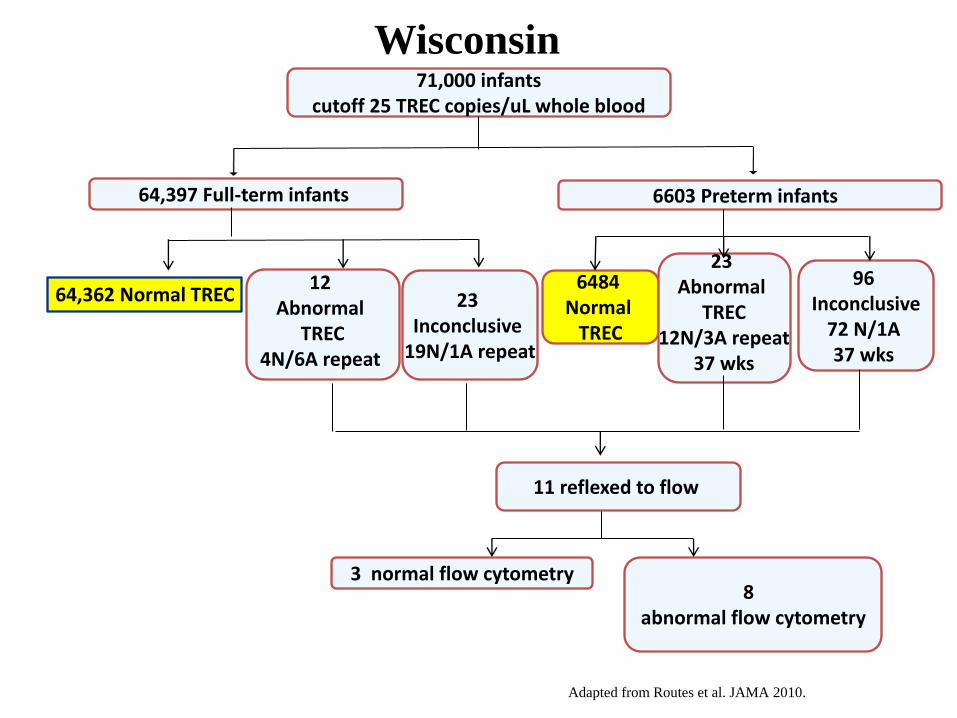
6484 Normal
TREC
11 reflexed to flow
64,362 Normal TREC
23 Abnormal
TREC 12N/3A repeat
37 wks
96 Inconclusive
72 N/1A 37 wks
23 Inconclusive
19N/1A repeat
64,397 Full-term infants
12 Abnormal
TREC 4N/6A repeat
6603 Preterm infants
71,000 infants cutoff 25 TREC copies/uL whole blood
Adapted from Routes et al. JAMA 2010.
Wisconsin
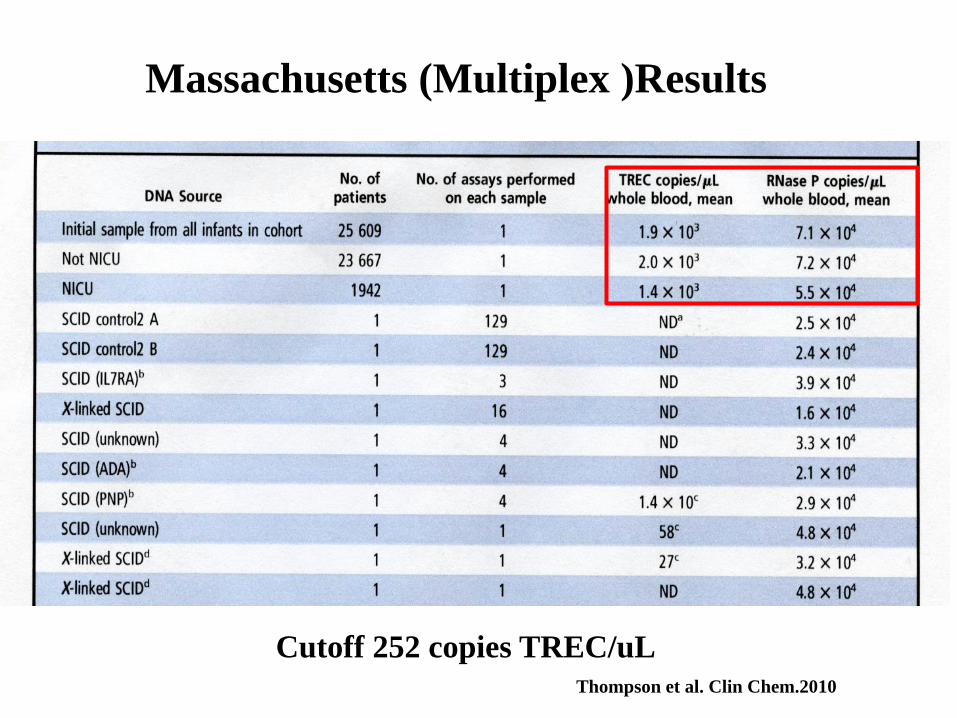
Massachusetts (Multiplex )Results
Thompson et al. Clin Chem.2010
Cutoff 252 copies TREC/uL
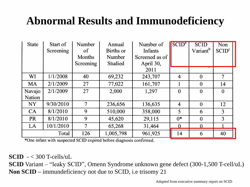
SCID - < 300 T-cells/uL
SCID Variant – “leaky SCID”, Omenn Syndrome unknown gene defect (300-1,500 T-cell/uL)
Non SCID – immundeficiency not due to SCID, i.e trisomy 21
Abnormal Results and Immunodeficiency
Adapted from executive summary report on SCID
Adapted from executive summary report on SCID
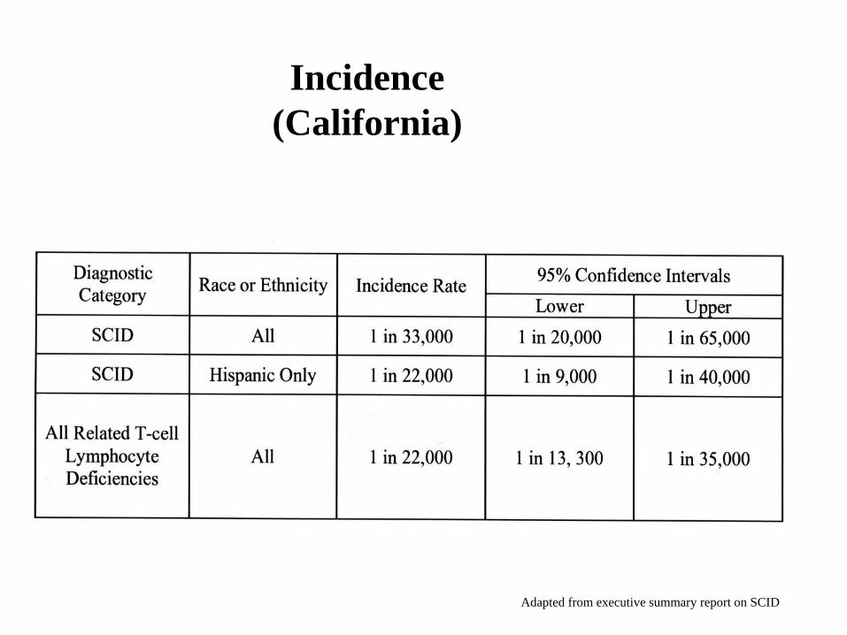
Incidence
(California)
Adapted from executive summary report on SCID
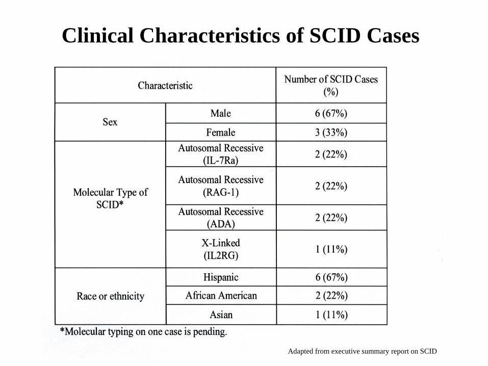
Clinical Characteristics of SCID Cases
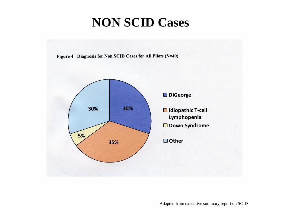
NON SCID Cases
Adapted from executive summary report on SCID
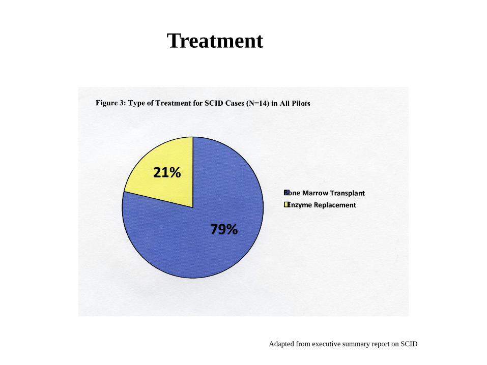
Treatment
Adapted from executive summary report on SCID
Take Home Points
• SCID is fatal unless treated
• Prompt treatment for SCID infants significantly increases survival
• SCID infants do not have any symptoms at birth, making prompt diagnosis difficult
• T-cell receptor excision circles (TREC) assays are currently being used to screen
newborns for SCID
• TRECs are not specific for SCID, but markers for T-cell lymphopenia, recent thymic
emigrant T-cells
• Cut-off for TREC concentrations vary from state to state and are method dependent
• Screening for SCID with TREC – 100% sensitivity 98% specificity
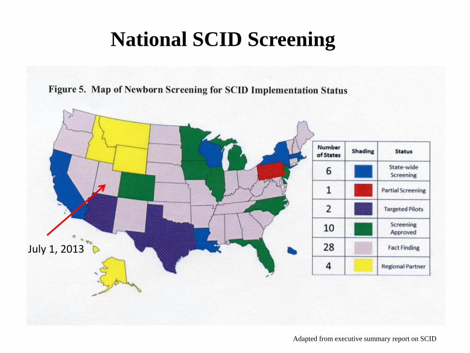
National SCID Screening
Adapted from executive summary report on SCID
July 1, 2013
Acknowledgements
• Utah Department of Health
(UDOH)
Dr. Harper Randall
Kim Hart
• University of Utah/ARUP
Dr. Harry Hill
Dr. Marzia Pasquali
Dr. Noriko Kusukawa
Dr.Carl Wittwer
Dr. Orly Ardon
Jorja Warren
Wei Xie
Mike Graczyk
Andy Lorance
• Wisconsin State Laboratory of Hygiene
Dr. Mei Baker
• Primary Children”s Medical Center
Dr. Karin Chen
Thank you!
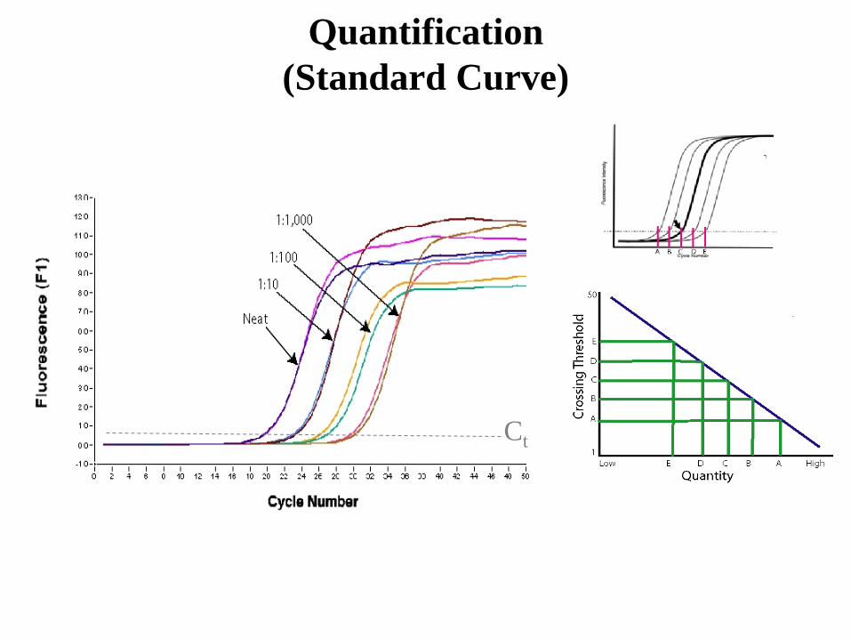
Quantification
(Standard Curve)
Ct