New therapeutic modalities to modulate orthodontic tooth ... · PDF file2014 123 2014 19612333...
11
© 2014 Dental Press Journal of Orthodontics Dental Press J Orthod. 2014 Nov-Dec;19(6):123-33 123 special article New therapeutic modalities to modulate orthodontic tooth movement Ildeu Andrade Jr 1 , Ana Beatriz dos Santos Sousa 2 , Gabriela Gonçalves da Silva 2 How to cite this article: Andrade JR I, Sousa ABS, Silva GG. New thera- peutic modalities to modulate orthodontic tooth movement. Dental Press J Orthod. 2014 Nov-Dec;19(6):123-33. DOI: http://dx.doi.org/10.1590/2176- 9451.19.6.123-133.sar Submitted: September 30, 2014 - Revised and accepted: October 14, 2014 » Patients displayed in this article previously approved the use of their facial and intraoral photographs. Contact address: Ildeu Andrade Jr Departamento de Ortodontia da Faculdade de Odontologia da PUC Minas Av. Dom José Gaspar, 500 - CEP: 31.270-901 - Belo Horizonte/MG — Brazil. E-mail: [email protected] » The author reports no commercial, proprietary or financial interest in the prod- ucts or companies described in this article. 1 Adjunct professor, Masters program in Orthodontics, School of Dentistry, Catholic University of Minas Gerais (PUC-MG). 2 Undergraduate student, School of Dentistry, Catholic University of Minas Gerais (PUC-MG). Modulation of orthodontic tooth movement (OTM) is desirable not only to patients because it shortens treatment time, but also to orthodontists, since treatment duration is associated with increased risk of gingival inflammation, decalcification, dental caries, and root resorption. The increased focus on the biological basis of tooth movement has rendered Orthodontics a more comprehensive specialty that incorporates facets of all fields of medicine. Current knowledge raises the possibility of using new therapeutic modalities for modulation of OTM, such as corticotomy, laser therapy, vibration (low-intensity pulsed ultrasound), local injections of biomodulators and gene therapy; with the latter being applicable in the near future. They are intended to enhance or inhibit recruitment, differentiation and/or activation of bone cells, accelerate or reduce OTM, increase stability of orthodontic results, as well as assist with the prevention of root resorption. This article summarizes recent studies on each one of these therapeutic mo- dalities, provides readers with information about how they affect OTM and points out future clinical perspectives. Keywords: Orthodontic tooth movement. Corticotomy surgery. Gene therapy. Ultrasound. DOI: http://dx.doi.org/10.1590/2176-9451.19.6.123-133.sar A modulação do movimento dentário ortodôntico (MDO) é desejável para os pacientes, pois reduz o tempo de tratamento, e também para ortodontistas, uma vez que a duração do tratamento tem sido associada a um aumento do risco de inflamação gengival, descalcificação, cárie dentária e reabsorção radicular. O crescente foco sobre os mecanismos biológicos da movimentação dentária levou a Ortodontia a ser uma especialidade mais abrangente, que hoje incorpora aspectos de todas as áreas da Medicina. Com o conhecimento atual, o uso de novas modalidades te- rapêuticas que visam a modulação da MDO, como a corticotomia, terapia a laser de baixa intensidade e vibração (ul- trassom pulsátil de baixa intensidade) já são uma realidade clínica. Outras, como injeções locais de biomoduladores e a terapia genética, serão utilizadas em breve. Elas destinam-se a aumentar ou inibir o recrutamento, à diferenciação e/ou ativação das células ósseas, a acelerar ou reduzir a MDO, a aumentar a estabilidade dos resultados ortodônticos, bem como auxiliar na prevenção da reabsorção radicular. Esse artigo resume os estudos mais recentes sobre cada uma dessas novas modalidades terapêuticas, fornecendo informações aos leitores a respeito de como afetam a MDO e aponta futuras perspectivas clínicas. Palavras-chave: Movimento dentário ortodôntico. Cirurgia de corticotomia. Terapia genética. Ultrassom.
Transcript of New therapeutic modalities to modulate orthodontic tooth ... · PDF file2014 123 2014 19612333...
© 2014 Dental Press Journal of Orthodontics Dental Press J Orthod. 2014 Nov-Dec;19(6):123-33123
special article
New therapeutic modalities to modulate orthodontic
tooth movement
Ildeu Andrade Jr1, Ana Beatriz dos Santos Sousa2, Gabriela Gonçalves da Silva2
How to cite this article: Andrade JR I, Sousa ABS, Silva GG. New thera-peutic modalities to modulate orthodontic tooth movement. Dental Press J Orthod. 2014 Nov-Dec;19(6):123-33. DOI: http://dx.doi.org/10.1590/2176-9451.19.6.123-133.sar
Submitted: September 30, 2014 - Revised and accepted: October 14, 2014
» Patients displayed in this article previously approved the use of their facial and intraoral photographs.
Contact address: Ildeu Andrade JrDepartamento de Ortodontia da Faculdade de Odontologia da PUC MinasAv. Dom José Gaspar, 500 - CEP: 31.270-901 - Belo Horizonte/MG — Brazil. E-mail: [email protected]
» The author reports no commercial, proprietary or financial interest in the prod-ucts or companies described in this article.
1 Adjunct professor, Masters program in Orthodontics, School of Dentistry, Catholic University of Minas Gerais (PUC-MG).
2 Undergraduate student, School of Dentistry, Catholic University of Minas Gerais (PUC-MG).
Modulation of orthodontic tooth movement (OTM) is desirable not only to patients because it shortens treatment time, but also to orthodontists, since treatment duration is associated with increased risk of gingival inflammation, decalcification, dental caries, and root resorption. The increased focus on the biological basis of tooth movement has rendered Orthodontics a more comprehensive specialty that incorporates facets of all fields of medicine. Current knowledge raises the possibility of using new therapeutic modalities for modulation of OTM, such as corticotomy, laser therapy, vibration (low-intensity pulsed ultrasound), local injections of biomodulators and gene therapy; with the latter being applicable in the near future. They are intended to enhance or inhibit recruitment, differentiation and/or activation of bone cells, accelerate or reduce OTM, increase stability of orthodontic results, as well as assist with the prevention of root resorption. This article summarizes recent studies on each one of these therapeutic mo-dalities, provides readers with information about how they affect OTM and points out future clinical perspectives.
Keywords: Orthodontic tooth movement. Corticotomy surgery. Gene therapy. Ultrasound.
DOI: http://dx.doi.org/10.1590/2176-9451.19.6.123-133.sar
A modulação do movimento dentário ortodôntico (MDO) é desejável para os pacientes, pois reduz o tempo de tratamento, e também para ortodontistas, uma vez que a duração do tratamento tem sido associada a um aumento do risco de inflamação gengival, descalcificação, cárie dentária e reabsorção radicular. O crescente foco sobre os mecanismos biológicos da movimentação dentária levou a Ortodontia a ser uma especialidade mais abrangente, que hoje incorpora aspectos de todas as áreas da Medicina. Com o conhecimento atual, o uso de novas modalidades te-rapêuticas que visam a modulação da MDO, como a corticotomia, terapia a laser de baixa intensidade e vibração (ul-trassom pulsátil de baixa intensidade) já são uma realidade clínica. Outras, como injeções locais de biomoduladores e a terapia genética, serão utilizadas em breve. Elas destinam-se a aumentar ou inibir o recrutamento, à diferenciação e/ou ativação das células ósseas, a acelerar ou reduzir a MDO, a aumentar a estabilidade dos resultados ortodônticos, bem como auxiliar na prevenção da reabsorção radicular. Esse artigo resume os estudos mais recentes sobre cada uma dessas novas modalidades terapêuticas, fornecendo informações aos leitores a respeito de como afetam a MDO e aponta futuras perspectivas clínicas.
Palavras-chave: Movimento dentário ortodôntico. Cirurgia de corticotomia. Terapia genética. Ultrassom.

© 2014 Dental Press Journal of Orthodontics Dental Press J Orthod. 2014 Nov-Dec;19(6):123-33124
New therapeutic modalities to modulate orthodontic tooth movementspecial article
INTRODUCTIONResearch in the field of orthodontic tooth move-
ment (OTM) has evolved rapidly and changed con-siderably since the work of Reitan et al in the 1950s.1 Moreover, the importance of all tissues, be it alveo-lar bone, periodontal ligament (PDL), root cemen-tum, and associated vascular and neural networks, has been investigated to delineate the role played by them.2 This growing attention given to the biologi-cal basis of Orthodontics expands current knowledge and augments understanding of the effects produced by mechanical loading over living tissues. Ortho-dontics, which for a long time was considered a technique-oriented profession, has steadily evolved to a comprehensive specialty that incorporates as-pects of all fields of medicine, emphasizing that live human beings are being treated instead of dental ty-podonts, only. Moreover, a sound biological back-ground is critical for the well-educated clinician to ensure optimal evidence-based treatment plan and to promote clinical excellence.
OTM is a biological process characterized by PDL and alveolar bone remodeling in response to an orth-odontic force which will promote extensive cellular and molecular changes in the periodontium. Orth-odontic treatment time ranges between 21-27 and 25-35 months for nonextraction and extraction ther-apies, respectively.3,4
Accelerating the rate of tooth movement is desirable to orthodontists because treatment duration has been associated with an increased risk of gingival inflam-mation, decalcification,5 dental caries, and, especially, root resorption.6 Shorter treatment duration with con-sequent lower costs are also important to all patients, particularly to adults who have been increasingly seek-ing treatment.7 However, adult patients typically re-quire longer treatment time due to having slower me-tabolism in comparison to younger patients.8
It has been estimated that teeth move 0.8-1.2 mm/month when continuous forces are applied.9 Since the best way to shorten treatment time is to speed up tooth movement, new therapeutic modalities have been re-ported to this end.10 Tooth movement has been acceler-ated by local injection of biomodulators, application of laser therapy, mechanical vibration and gene therapy, as well as by corticotomies. Some of these approaches cannot yet be applied clinically; but others, such as
corticotomy, laser therapy and vibration are somewhat already part of the therapeutic arsenal. Nevertheless, a question remains. How can these procedures acceler-ate or inhibit tooth movement? Since OTM is a bio-logical process, any procedure used to modulate OTM is direct or indirectly related to the cellular and mo-lecular mechanisms involved in the biology of tooth movement. The aim of the present review is to sum-marize recent studies on each of these therapeutic mo-dalities and to provide readers with information about how they affect OTM.
BIOLOGY OF TOOTH MOVEMENTOrthodontists work in a unique biological envi-
ronment wherein applied forces engender remodeling of both mineralized (alveolar bone) and nonmineral-ized (PDL and gingiva) paradental tissues, includ-ing associated blood vessels and neural elements. Bone remodeling processes begin when an orthodontic force is applied over the periodontium which, in turn, generates aseptic inflammatory response. This inflam-mation alters homeostasis and microcirculation of PDL, thereby creating areas of ischemia and vasodi-latation, which results in the release of several biologi-cal mediators, such as cytokines, chemokines, growth factors, neurotransmitters, metabolites of arachidonic acid and hormones. These molecules trigger a number of cellular responses that will promote bone resorption by osteoclasts in the pressure sites and bone formation by osteoblasts in the tension sites.2
Osteoclasts are multinucleated cells derived from precursors in the myeloid/monocyte lineage that cir-culate in the blood after being formed in the bone marrow. They are the only cells in nature that can degrade mineralized bone tissue and are important for physiological remodeling and modeling pro-cesses, calcium homeostasis, tooth eruption, and OTM. Mature osteoclasts attach to bone surface by a sealing zone. In this area, proton pumps and chlo-ride channels are expressed. They are important for extracellular acidification and demineralization of bone. Proteolytic enzymes are then released and de-grade the extracellular matrix proteins.11 Therefore, when alveolar bone is stimulated to resorb by means of an orthodontic force, a sequence of events is ini-tiated and ultimately result in recruitment, differ-entiation, activation and maintenance of osteoclasts

© 2014 Dental Press Journal of Orthodontics Dental Press J Orthod. 2014 Nov-Dec;19(6):123-33125
Andrade JR I, Sousa ABS, Silva GG special article
playing a role in bone remodeling induced by orth-odontic forces are: tumor necrosis factor (TNF)-α, interleukin (IL)-1β, IL-2, IL-3, IL-4, IL-5, IL-6, IL-10, interferon-γ (IFN-y), tissue biomarkers (ma-trix metalloproteinases (MMP)-1, MMP-2, MMP-9, tissue inhibitors of MMPs (TIMP)-1 and 2), and chemokines (CCL2, CCL3, CCL5, CCL7, CCL9, CXCL-8, CXCL9, CXCL10, CXCL12 and CXCL-13), all of which play a central role in trafficking and homing of leukocytes, immune cells and stromal cells.13,15 Mechanical loading also stimulates local ex-pression of many growth factors (GFs) (i.e: vascular endothelial GF (VEGF), transforming GF (TGF)-β, bone morphogenetic proteins (BMPs), insulin-like GF (IGFs) and fibroblast GF (FGF) involved in bone and PDL remodeling in the early stages of OTM in both tensile and compressive sites.13
Taken all together, chemokines, cytokines and growth factors (Gfs) are the main molecules involved in bone cell recruitment, activation, proliferation, differentiation and survival. These molecules stim-ulate PDL and bone cells to orchestrate an inflam-matory response followed by osteoclastogenesis and bone resorption in compression sites, and bone neo-formation by osteoblasts at PDL tension sites. Re-search trend is now directed toward elucidating the molecular mechanisms involved in the aforemen-tioned events. Current knowledge raises the possibil-ity of using therapeutic modalities (local injections of biomodulators, laser therapy, mechanical vibration, gene therapy, and corticotomy) capable of acting on or increasing the expression of specific cytokines, chemokines and GFs. These molecules can modulate the outcomes of orthodontic force application, ac-celerating OTM, enhancing biological anchorage at specific sites, possibly decreasing the rebound effect, and assisting with the prevention of root resorption.
CORTICOTOMYOver the past 10 years, corticotomies have become
a popular means of increasing the rate of tooth move-ment. In corticotomy, the cortical layer is cut or per-forated to the depth of the medullary bone which is preserved (Fig 1). During bone healing process, a re-gional acceleratory phenomenon (RAP) takes place in the periodontium. RAP is a natural localized reaction of soft and hard tissues in response to an injury, and
in bone remodeling sites. Osteoclastogenesis begins with stem cell division and proliferation of osteoclast precursors cells in hematopoietic tissues (bone mar-row, spleen, liver and peripheral blood). The second step is the migration of cells to bone resorption sites where they will be differentiated and activated. Tooth movement efficiency is directly linked, quantitatively and qualitatively, to recruitment, differentiation, ac-tivation and maintenance of these cells in these sites.12
Since osteoclasts are bone specific cells, they are recruited from blood stream by chemotactic factors released from components of bone matrix and osteo-blasts.2 After proliferation and migration of osteoclast precursors to bone remodeling sites, these progenitors will differentiate when their receptor c-Fms interacts with the ligand macrophage colony-stimulating fac-tor (M-CSF), which is also important for osteoclast survival. Specific differentiation of osteoclasts is due to activation of RANK (receptor activator of nuclear factor-kB) by RANKL (receptor activator of nuclear factor-kB ligand) expressed by stromal cells in bone marrow and osteoblasts.12
Osteoblasts are of mesenchymal origin and are re-sponsible for bone formation during embryonic de-velopment, growth, bone remodeling and fracture healing. In Orthodontics, bone formation begins 40-48 hours after force application in PDL tension sites.2 Osteocytes, which are osteoblasts that become embedded in their own bone matrix, participate in the process of osteogenesis, being acutely sensitive and responsive to applied tensile orthodontic forces. Their cellular projections favor communication with neigh-boring osteocytes, as well as with alveolar bone sur-face-lining cells and bone marrow cavity cells. Osteo-blasts, which maintain direct contact with osteocytes, respond to these signals and initiate bone apposition.13 Moreover, stretched PDL fiber bundles stimulate cell replication.2 Stem-cells (pericytes) which migrate from blood vessel walls, and mesenchymal stem-cells differentiate into pre osteoblastic cells 10 hours af-ter force application.2 Chemokines, cytokines, and growth factors are directly involved in this process.13,14 Osteoblasts also positively regulate osteoclast activity by expressing cytokines such as RANKL, a key ac-tivator of osteoclast differentiation, and negatively by expression of osteoprotegerin (OPG), a soluble decoy receptor that inhibits RANKL.12,13 Other cytokines

© 2014 Dental Press Journal of Orthodontics Dental Press J Orthod. 2014 Nov-Dec;19(6):123-33126
New therapeutic modalities to modulate orthodontic tooth movementspecial article
Figure 1 - Corticotomy. Mucoperiosteal flaps are raised and corticotomy carried out on buccal and palatal surfaces. Monocortical perforations are performed in areas of intended tooth movement.
is associated with increased perfusion, bone turnover and decreased bone density.16,17 It is similar to the pro-cesses associated with normal fracture healing which include a reactive phase, a reparative phase, and a re-modeling phase. The reactive phase lasts 7 days and it is characterized by immediate constriction of blood vessels to mitigate bleeding, followed by hematoma within a few hours.16 The cells within the hematoma will die and a loose aggregate of fibroblasts, inter-cellular materials and other supporting cells is then formed. This granulation tissue is formed within ap-proximately two weeks.18 A few days later, periosteal cells surrounding the injury site and the granulation-tissue fibroblasts will be transformed into chondro-blasts and form hyaline cartilage. Periosteal cells distal to the injury site develop into osteoblasts which form woven bone.19 The association of the mass of hyaline cartilage and woven bone is called callus and will be
replaced by lamellar bone in the subsequent phase. In fractures, the time between callus formation and mineralization is of 1-4 months;16 corticotomies are expected to heal faster than fractures (2-3 months). The last phase of healing takes 1-4 years and it is characterized by complete remodeling of the bone into functionally mature lamellar bone.
Tooth movement should be faster in less dense al-veolar bone which is rapidly remodeled for the same reasons tooth movement is faster in growing children than in adults.20 Moreover, animal studies showed that corticotomies provide three times as many osteoclasts, three times greater bone apposition rate and a twofold decrease in calcified trabecular bone.20 Moreover, an-other study demonstrated that perforations in the cor-tical bone increase the expression of 37 inflammatory cytokines, which leads to more osteoclasts and, conse-quently, greater bone remodeling process.21

© 2014 Dental Press Journal of Orthodontics Dental Press J Orthod. 2014 Nov-Dec;19(6):123-33127
Andrade JR I, Sousa ABS, Silva GG special article
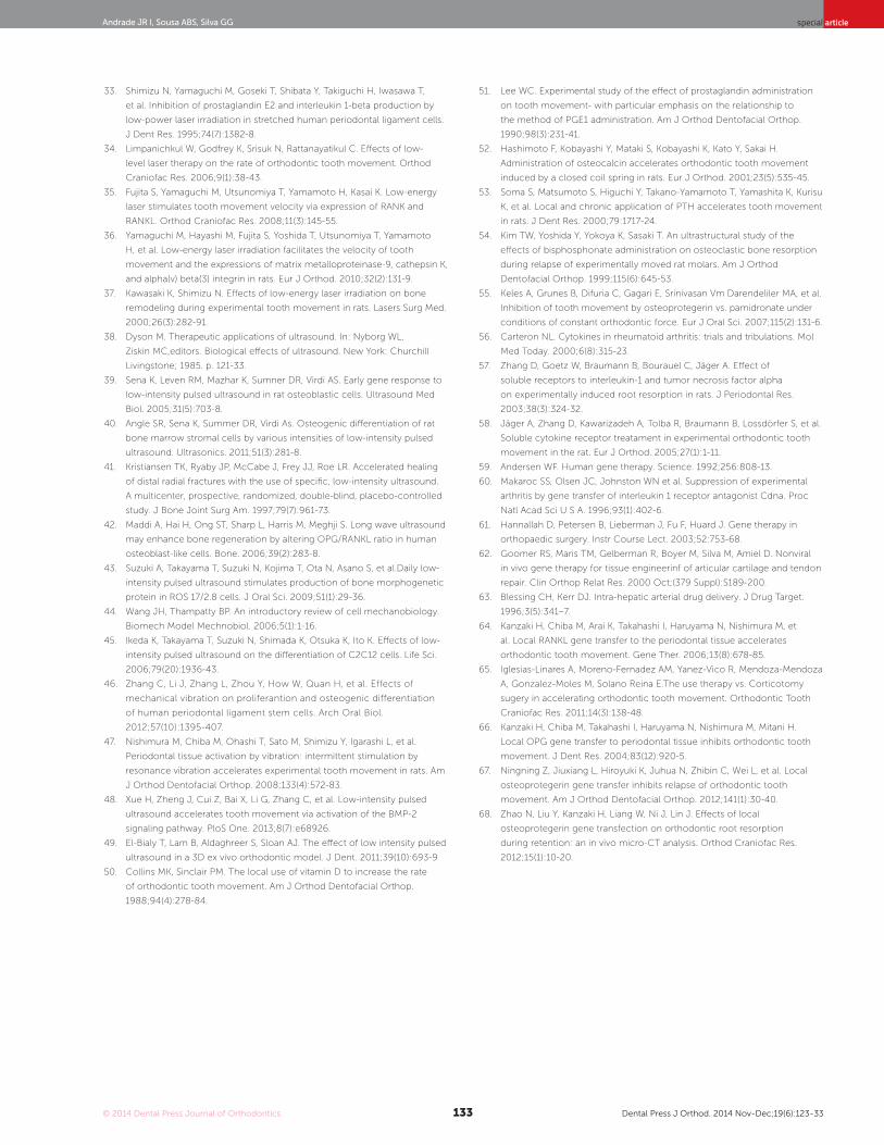
Figure 2 - Laser irradiation. Application of LLLT in areas of intended tooth movement.
Although effective and highly predictable, corticotomy-assisted orthodontic treatment is quite invasive as it requires extensive flap elevation and bone surgery. A previous study proposed the use of a piezoelectric knife instead of a high-speed surgical bur to decrease surgical trauma and still achieve rapid tooth movement. Due to its micrometric and selec-tive cut, piezoelectric devices have been claimed to produce safe and precise osteotomies without osteo-necrotic damage.22
Taken all together, there is twice as much tooth movement with than without corticotomies. How-ever, this window of opportunity used to accelerate tooth movement is limited to 2-3 months, in which 4-6 mm of tooth movement might be expected (twice as much the normal rate).20 Nevertheless, fur-ther controlled clinical trials are needed to determine the actual effects of corticotomies.
LASER THERAPYThe term "laser" originated as an acronym for "light
amplification by stimulated emission of radiation". It is a device that emits light through a process of optical amplification based on the stimulated emission of elec-tromagnetic radiation.23 Lasers differ from other light sources by their coherence which allows them to be focused to a limited spot, to stay narrow over long dis-tances or to have a very narrow spectrum (emitting a single color of light). In medicine, lasers have many important applications: bloodless surgery, laser healing, surgical treatment, kidney stone treatment, eye treat-ment and many others. The laser technique has also been widely applied in Dentistry; in orthodontic treat-ment, it has proved to have many benefits. They can be used to perform gingivectomy, frenectomy, surgi-cal exposure of tooth (with less bleeding and swelling, improved precision, reduced pain and improved heal-ing), enamel etching, bonding, bracket debonding, pain control, treatment of traumatic ulcers in the oral mucosa and to accelerate tooth movement24,25 (Fig 2).
Lasers can be classified as low and high-intensity lasers of which main differences are their potency and mechanism of action.42 High-intensity lasers, such as the CO2 laser, Nd laser: Yttrium aluminum garnet (Nd:YAG), argon laser, Er:YAG laser, and the excimer laser act by increasing the temperature, showing a destructive potential, and are usually used
in surgical procedures. Meanwhile, the low-intensi-ty laser (also known as soft laser, cold laser or laser therapy) does not have a destructive potential. Its photobiomodulation mechanism of action penetrates tissues and stimulates cellular metabolism, bone re-modeling and tooth movement which is of greatest interest in Orthodontics.24,25 Different low-energy laser modalities have been used in different doses and in various treatment protocols, including heli-um-neon (632.8 nm wavelength) and semiconductor lasers (emitting light in the range of 780–950 nm), gallium-aluminum-arsenide (GaAlAs) (805 ± 25 nm wavelength) and gallium-arsenide (904 nm wave-length).26 GaAlAs diode laser has been repeatedly used in the past years and has proved to have higher depth of tissue penetration in comparison to other modalities, therefore, providing the clinicians with a suitable penetrative instrument with great efficiency in orthodontic treatment.27
The exact mechanism of laser–cell interaction is still to be investigated. The stimulation of pho-toreceptors in the mitochondrial respiratory chain, changes in cellular ATP levels and cell membrane stabilization have been discussed.28 It is generally accepted that laser effects on cells are wavelength and dose-dependent. The existence of a "window of spec-ificity" at certain wavelengths and energy dosages has been postulated.29 Molecular absorption of laser light is a prerequisite for any cellular effect.
© 2014 Dental Press Journal of Orthodontics Dental Press J Orthod. 2014 Nov-Dec;19(6):123-33128
New therapeutic modalities to modulate orthodontic tooth movementspecial article
A previous study30 demonstrated that low-level laser therapy (LLLT) stimulates cellular prolifera-tion and differentiation of osteoblast lineage nodule-forming cells, especially in committed precursors, resulting in an increase in the number of differenti-ated osteoblastic cells as well as in bone formation. Meanwhile, another study found that low-energy la-ser irradiation stimulated the amount of tooth move-ment and formation of osteoclasts on the side of pres-sure during experimental tooth movement in vivo. As bone remodeling is a physiological process that involves osteoclastic bone resorption and osteoblastic bone formation,2 those findings are not surprising. Furthermore, recent studies showed that low-energy laser irradiation accelerated orthodontic movement of human teeth.31,32
However, the effect of LLLT on tooth move-ment is reportedly controversial, as different stim-ulatory, inhibitory and irrelevant effects have been shown in the literature. A previous study33 reported that low-energy laser irradiation significantly in-hibited the production of prostaglandin E (PGE2), and that interleukin (IL-1β) was increased by me-chanical stress in vitro. If low-energy laser irradia-tion functions to inhibit these pro-inflammatory cytokines, OTM might be slow. Another LLLT study34 demonstrated low stimulatory or inhibi-tory effect on the rate of orthodontic tooth move-ment. Conversely, other studies35,36 reported that IL-1, RANKL, M-CSF, MMP-9, cathepsin K, and α(v)β3 integrin were stimulated via their respective pathways during the differentiation of bone cells, and the amount of tooth movement was increased by low-energy laser irradiation.37 Moreover, an in vitro study35 showed that the gene expression of RANK in osteoclast precursor cells increased when cells were irradiated with low-energy laser. On the basis of the findings of this review, it is possible to assert that LLLT speed up tooth movement via RANK ⁄ RANKL expression.
Although further studies are necessary to evaluate the effects of different irradiation dosages, the pro-longed use of laser irradiation on tooth movement or bone remodeling, or both, and the introduction of laser therapy at an early stage of tooth movement in orthodontic treatment seem feasible and may be of great therapeutic benefit to abbreviate treatment time.
VIBRATIONTooth movement is closely related to response to
applied orthodontic forces that cause remodeling of periodontal tissues, especially the alveolar bone. Bone is a highly specialized form of connective tissue and consists of a cortical bone that overlies the softer in-ner structure named cancellous or trabecular bone. Its formation and regeneration involve chemotaxis, cell proliferation, differentiation and synthesis of ex-tracellular matrix; a result of interaction established amongst biochemical, biomechanical, cellular and hormonal signals.2
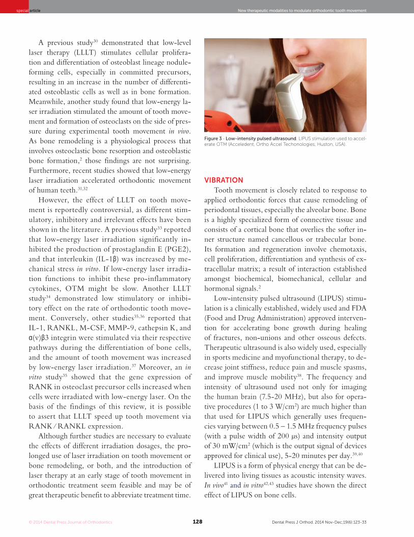
Low-intensity pulsed ultrasound (LIPUS) stimu-lation is a clinically established, widely used and FDA (Food and Drug Administration) approved interven-tion for accelerating bone growth during healing of fractures, non-unions and other osseous defects. Therapeutic ultrasound is also widely used, especially in sports medicine and myofunctional therapy, to de-crease joint stiffness, reduce pain and muscle spasms, and improve muscle mobility38. The frequency and intensity of ultrasound used not only for imaging the human brain (7.5-20 MHz), but also for opera-tive procedures (1 to 3 W/cm2) are much higher than that used for LIPUS which generally uses frequen-cies varying between 0.5 – 1.5 MHz frequency pulses (with a pulse width of 200 μs) and intensity output of 30 mW/cm2 (which is the output signal of devices approved for clinical use), 5-20 minutes per day.39,40
LIPUS is a form of physical energy that can be de-livered into living tissues as acoustic intensity waves. In vivo41 and in vitro42,43 studies have shown the direct effect of LIPUS on bone cells.
Figure 3 - Low-intensity pulsed ultrasound. LIPUS stimulation used to accel-erate OTM (Acceledent, Ortho Accel Techonologies, Huston, USA).

© 2014 Dental Press Journal of Orthodontics Dental Press J Orthod. 2014 Nov-Dec;19(6):123-33129
Andrade JR I, Sousa ABS, Silva GG special article
Although the mechanism by which LIPUS in-creases the rate of fracture healing is unclear, it is known that the mechanical strains received by cells are translated into biochemical events.44 LIPUS, in essence a wave of alternating pressure, is translated into an extracellular mechanical force at the cell membrane where it is transduced into intracellular electrical and/or biochemical signals. Previous stud-ies indicate that LIPUS accelerates the differentiation pathway of mesenchymal stem cells in the osteogenic lineage via activated phosphorylation of MAPK (mi-togen-activated protein kinase) pathways,45 up-regu-lation of cyclo-oxygenase-2 (COX-2), prostaglandin E2 (PGE2),40 altering the OPG/RANKL ratio in the microenvironment.42 and stimulating the production of bone morphogenetic proteins.43
As bone, the PDL is also a dynamic tissue which is constantly being remodelled to adapt to mechani-cal loading. Therefore, it is expected that an appro-priate level of mechanical stress be able to induce an anabolic response of the periodontium. The PDL is both the medium of force transfer and the means by which alveolar bone remodels itself in response to ap-plied forces. Moreover, PDL cells (PDLCs) play an important role not only in the maintenance of the periodontium, but also in promoting periodontal re-generation during and after the OTM.2 They are a heterogeneous cell population, including cells at dif-ferent stages of differentiation and lineage commit-ment. Mechanical vibration can affect osteogenesis by increasing the commitment of PDLSCs to the osteogenic lineage. A previous study has shown that the protein levels of RUNX2 and OSX (transcrip-tion factors that play a role in the differentiation and activation of osteoblasts) were both prominently en-hanced under ultrasound stimulation.46
It has also been shown that LIPUS (Fig 3) stim-ulation accelerates OTM by increasing osteoclast number and activity, probably by enhancing the ex-pression of RANKL on the pressure sites.47,48 These same studies have hypothesized that resonance vi-bration might prevent blood flow obstruction and hyalinization at the compression sites. Furthermore, LIPUS minimizes orthodontically induced tooth root resorption by enhancing dentine and cemen-tum deposition, thereby forming a preventive layer against root resorption.49
In short, LIPUS has many clinical advantages, in-cluding the fact that it is a biological stimulus, easy to use and noninvasive, in addition to being widely used in clinical medicine.
LOCAL INJECTION OF BIOMODULATORSOrthodontic forces create areas of tension and com-
pression in the PDL, which affects remodelling of the periodontium. Following mechanical stress, changes to vascularity and blood flow within the PDL are induced by signalling molecules. The signalling cascade initiates with arachadonic acid metabolites (eicosanoids), neu-rotransmitters, (substance P and calcitonin gene-related peptide) and second messengers, such as cyclic AMP, phosphoinositol phosphate and diacyl-glycerol.2 These molecules trigger the release of cytokines, growth fac-tors and colony stimulating factors, which affect bio-logical mediators such as RANKL, OPG, MMPs and TIMPs.13 Recent research advances have suggested that these biological modulators, which enhance or inhibit recruitment, differentiation or activation of osteoclasts, could be used to provide new adjunctive approaches to orthodontic treatment. In other words, local injections of biomodulators could be used to accelerate OTM, re-duce root resorption, enhance anchorage and improve stability of orthodontic results (Fig 4).
Numerous reports have described the pharmaco-logical acceleration of OTM through activation of osteoclasts. A previous study50 reported that vitamin D3 activated osteoclasts and accelerated OTM. Lo-cal administration of prostaglandins (PGs),51 osteo-calcin,52 or PTH53 also induced OTM. However, be-cause these drugs are rapidly flushed by blood flow, daily systemic administration or daily local injection are needed. In addition, frequent injections of this substances in local regions may evoke fear in patients and cause problems in medical treatment.
The undesired movement of anchor teeth and the relapse of previously moved teeth are major clinical problems in Orthodontics. Recent research advances suggest that biological modulators which inhibit os-teoclasts could be used to address these problems and provide new adjunctive approaches to orthodontic treatment. Several inhibitors have been examined, in-cluding bisphosphonates and osteoprotegerin (OPG), and their efficiency in preventing tooth movement has been proved in animal models.54,55

© 2014 Dental Press Journal of Orthodontics Dental Press J Orthod. 2014 Nov-Dec;19(6):123-33130
New therapeutic modalities to modulate orthodontic tooth movementspecial article
Moreover, advances in understanding cytokine-mediated development and progression of rheuma-toid arthritis have led to efforts to neutralize these cytokines by using antibody or soluble receptor tech-niques.56 Soluble receptors are able to bind their li-gands with specificity and affinity, and effectively neutralize cytokine activity.57 It has been shown in animal models that systemic application of soluble receptors to IL-1 (sIL-RII) or TNF-α (sTNF-α-RI) leads to reduction or even prevention of root resorp-tion.57 The concentration of these soluble receptors in the local microenvirement of the target periodon-tium was also sufficient to interfere in the remodeling processes induced in the periodontal tissues, reducing the number of osteoclasts and, consequently, the amount of OTM.58
Nevertheless, routine clinical use of these biomod-ulators in orthodontics still requires further investiga-tions, to determine the correct dosage, frequency of administration and, especially, the possible local and systemic side effects of its long term use.
GENE THERAPYThe original premise behind gene therapy (GT) in
the 90s was the believe that if a defective gene result-ing in a specific disease could be replaced by a healthy gene, then the disease could be cured.59 However, the
potential role of GT as a clinical tool has expanded and it is no longer limited to replacement of defec-tive genes, but rather has become a tool for producing individual proteins to specific tissues and cells (Fig 5). Although all cells contain the genes for all proteins, cells derived from a particular tissue express only a limited selection of these proteins. With GT, it is pos-sible to deliver a gene to a given cell, which allows the inserted gene product to be expressed constitutively.
Modern technology has allowed the manufacture of these proteins (human recombinant proteins) for therapeutic use. However, their life spam is short after injection into the human body. As GT provides the gene for protein production rather than just replacing degradable protein, it achieves higher and more con-stant levels of protein expression. For this reason, it has became an effective method used to deliver these proteins to specific tissues.60
Once protein and location of protein delivery have been chosen, the next step is to choose the vector to deliver the protein. The objective is to get the DNA that encodes the specific protein into the target cell and force it to express the desired protein. The most common delivery vector is by means of a virus, a pro-cess also known as “transduction.” Nonviral vectors are also used, in which case the process is referred to as “transfection”. It is carried out by means of sev-eral methods, including liposome and gene gun.61 The easiest way to implement local GT is by inject-ing the vector into a specific tissue. The vector may be delivered systemically to all cells in the body (as in treatment of metastatic diseases) or locally to the tar-get tissue, only (as desired in Orthodontics). Direct GT has been effectively used in knee and ankle joints, skeletal muscle, bone and ligaments.61,62 Nevertheless, in indirect GT, target cells are harvested from the pa-tient and then reinserted. It is advantageous for being able to accurately select a particular cell as the pro-tein delivery vehicle. The indirect method has been effectively used to target articular cartilage, spine and human metacarpophalangeal joints.61
Numerous reports have described the pharmaco-logical acceleration of OTM through activation of os-teoclasts. However, due to their rapid flush out by blood circulation, daily systemic administration or daily local injection is needed. Local gene transfer has two advan-tages.63 First, it maintains local effective concentration
Figure 4 - Injection of biomodulators. Injection of inflammatory mediators in the periodontium.

© 2014 Dental Press Journal of Orthodontics Dental Press J Orthod. 2014 Nov-Dec;19(6):123-33131
Andrade JR I, Sousa ABS, Silva GG special article
and prolonged protein expression, regardless of blood circulation. Second, protein expression occurs at a lo-cal site, thereby avoiding systemic effects.
A previous animal study demonstrated that trans-fer of RANKL gene to periodontal tissue activated osteoclastogenesis and accelerated OTM without producing any systemic effects.64 When comparing
corticotomy surgery and RANKL gene transfer to periodontal tissue as two methods that might substan-tially reduce orthodontic treatment time, RANKL GT demonstrated higher efficacy than standard surgi-cal methods.65 Local GT has also been used to inhibit OTM, which might be, in the near future, an impor-tant tool to enforce the anchorage unit or increase sta-bility of orthodontic results. Local OPG gene transfer significantly inhibited RANKL-mediated osteoclas-togenesis in the periodontium caused by experimen-tal tooth movement.66 Moreover, local OPG gene transfer might be a biologic method employed to pre-vent or inhibit relapse after orthodontic treatment.67 Other local or systemic pharmacological agents, such as bisphosphonates and simvastatin, also decrease the extent of initial relapse, but they are rapidly distrib-
uted by blood circulation and, for this reason, require daily systemic administration.
Local OPG gene transfer is also clinically relevant for enhancing external root resorption (ERR) repair during retention.68 However, the precise biological mechanism behind this finding has not yet been fully elucidated and further studies are required to assess the role of RANK ⁄ RANKL ⁄ OPG axis in ERR repair.
In short, GT is a pioneering new therapeutic mo-dality based on complex biological systems occur-ring at the leading edge of biomedical knowledge. It offers an alternative method to deliver proteins to a given target tissue, which, in turn, can enhance or inhibit osteoclast recruitment and lead to a more or less OTM. Nonetheless, further research is needed to determine the safety and efficacy of these techniques.
CONCLUSIONUnderstanding the biology of tooth movement
and treatment outcomes individually is a complex process that requires knowledge in many different areas of biomedicine. The rapid development of mo-lecular biology along with translational studies in hu-mans and experimental systems are likely to provide us with a much more thorough insight into the cel-lular and molecular mechanisms involved in the bone remodeling processes induced by orthodontic forces. This is a prerequisite to understand the responses in different individuals and to develop new mecha-nisms by which tooth movement could be regulated not only by mechanical forces, but also by biological agents, if needed.
Basic researchers continue, at an increasing pace, to contribute to the advancement of clinical Or-thodontics. Publications on the outcomes of well-planned investigations in every field of medicine in-spire researchers who have selected the areas that may be helpful in addressing orthodontic clinical issues faced by the clinician on a daily basis. The biological uniqueness of each patient dictates the need for con-tinuous acquisition of knowledge. Current researches tend to focus on areas such as monitoring patient's reaction to mechanical forces by searching bone re-modeling markers in the GCF, saliva, and blood se-rum. Special attention is given to the speed of tooth movement enhanced by adding certain physical and chemical agents to mechanical orthodontic force.
Figure 5 - Gene therapy. Delivering a gene to a given cell allows the inserted gene product to be expressed constitutively.

© 2014 Dental Press Journal of Orthodontics Dental Press J Orthod. 2014 Nov-Dec;19(6):123-33132
New therapeutic modalities to modulate orthodontic tooth movementspecial article
1. Reitan K. Tissue behavior during orthodontic tooth movement. Am J
Orthod. 1960;46(12):881-90.
2. Krishnan V, Davidovitch Z. On a path to unfolding the biological
mechanisms of orthodontic tooth movement. J Dent Res.
2009;88(7):597-608.
3. Skidmore KJ, Brook KJ, Thomson WM, Harding WJ. Factors influencing
treatment time in orthodontic patients. Am J Orthod Dentofacial Orthop.
2006;129(2):230-8.
4. Vu CQ, Roberts WE, Hartsfield JK Jr, Ofner S. Treatment complexity
index for assessing the relationship of treatment duration and outcomes
in a graduate orthodontics clinic. Am J Orthod Dentofacial Orthop.
2008;133(1):9.e1-13.
5. Ristic M, Vlahovic Svabic M, Sasic M, Zelic O. Clinical and microbiological
effects of fixed orthodontic appliances on periodontal tissues. Orthod
Craniofac Res. 2007;10(4):187-95.
6. Segal GR, Schiffman PH, Tuncay OC. Meta analysis of the treatment-
related factors of external apical root resorption. Orthod Craniofac Res.
2004;7(2):71-8.
7. McKiernan EX, McKiernan F, Jones ML. Psychological profiles and motives
of adults seeking orthodontic treatment. Int J Adult Orthodon Orthognath
Surg. 1992;7(3):187-98.
8. Ong MM, Wang HL. Periodontic and orthodontic treatment in adults. Am J
Orthod Dentofacial Orthop. 2002;122(4):420-8.
9. Barlow M, Kula K. Factors influencing efficiency of sliding mechanics
to close extraction space: A systematic review. Orthod Craniofac Res.
2008;11(2):65-73.
10. Long H, Pyakurel U, Wang Y, Liao L, Zhou Y, Lai W. Interventions for
accelerating orthodontic tooth movement. A systematic review. Angle
Orthod. 2013;83(1):164-71.
11. Boyle WJ, Simonet WS, Lacey DL. Osteoclast differentiation and activation.
Nature. 2003;423:337-42.
12. Yamaguchi M. RANK/RANKL/OPG during orthodontic tooth movement.
Orthod Craniofac Res. 2009;12:113-9.
13. Andrade Jr I, Taddei SRA, Souza PEA. Inflammation and tooth movement:
the role of cytokines, chemokines, and growth factors. Semin Orthod.
2012;18(4):257-69.
14. Andrade I Jr, Silva TA, Siva GA, Teixeira AL, Teixeira MM. The role of tumor
necrosis factor receptor type 1 in orthodontic tooth movement. J Dent
Res. 2007;86(11):1089-94.
15. Taddei SR, Andrade I Jr, Queiroz-Junior CM, Garlet TP, Garlet GP, Cunha
Fde Q, et al. Role of CCR2 in orthodontic tooth movement. Am J Orthod
Dentofacial Orthop. 2012;141(2):153-60.
16. Frost HM. The biology of fracture healing. An overview for clinicians. Part I.
Clin Orthop Relat Res. 1989;(248):283-93.
REFERENCES
17. Gantes B, Rathibun E, Anholm M. Effects on the periodontium following
corticotomy-facilitated orthodontics. Case Reports. J Periodontol.
1990;61(4):234-7.
18. Ham AW, Harris WR. Repair and transplantation of bone. In: The
Biochemistry and Physiology of Bone. New York: Academic Press; 1972. p.
337-99.
19. Brighton CT, Hunt RM. Early histologic and ultrastructuralchanges in
microvessels of periosteal callus. J Orthop Trauma. 1997;11(4):244-53.
20. Buschang PH, Campbell PM, Ruso S. Accelerating tooth movement
with corticotomies: Is It Possible and Desirable? Semin Orthod.
2012;18(4):286-94.
21. Teixeira CC, Khoo E, Tran J, Chartres I, Liu Y, Thant LM, et al.
Cytokine expression and accelerated tooth movement. J Dent Res.
2010;89(10):1135-41.
22. Kotrikova B, Wirtz R, Krempien R, Blank J, Eggers G, Samiotis A, et al.
Piezosugery: a new safe technique in cranial osteoplasty? Int J Oral
Maxillofac Surg. 2006;35(5):461-5.
23. Gould RG. The LASER, Light amplification by stimulated emission of
radiation. In: Franken PA, Sands RH, editors. The Ann Arbor Conference
on Optical Pumping, the University of Michigan, 15 June through 18 June
1959. p. 128.
24. Walsh LJ. The current status of low level laser therapy in dentisty. Part 1.
Soft tissue applications. Aust Dent J. 1997;42(4):247-54.
25. Walsh LJ. The current status of low level laser therapy in dentistry. Part 2.
Hard tissue applications. Aust Dent J. 1997;42(5):302-6.
26. Basford JR. Low intensity laser therapy: still not an established clinical tool.
Lasers Surg Med. 1995;16(4):331-42.
27. Khadra M, Kasem N, Haanaes HR, Ellingsen JE, Lyngstadaas SP.
Enhancement of bone formation in rat calvarial bone defects using low-
level laser therapy. Oral Surg Oral Med Oral Pathol Oral Radiol Endod.
2004;97(6):693-700.
28. Conlan MJ, Rapley JW, Cobb CM. Biostimulation of wound healing by low-
energy laser irradiation. A review. J Clin Periodontol. 1996;23(5):492-6.
29. Karu TI. Yearly review: effects of visible radiation on cultured cells.
Photochem Photobiol. 1990;52(6):1089-98.
30. Ozawa Y, Shimzu n, kariya G, Abiko Y. Low energy laser irradiation
stimulates bone nodule formation at early stages of cell culture in rat
calvarial cells. Bone. 1998;22(4):347-54.
31. Cruz DR, Kohara EK, Ribeiro MS, Wetter NU. Effects of low- intensity
laser therapy on the orthodontic movement velocity of human teeth: a
preliminary study. Lasers Surg Med. 2004;35(2):117-20.
32. Doshi-Mehta G, Brad-Patil WA. Efficacy of low-intensity laser therapy in
reducing treatment time and orthodontic pain: a clinical investigation. Am
J Orthod Dentofacial Orthop. 2012;141(3):289-97.
Moreover, current knowledge raises the possibility of enhancing biological anchorage at specific sites, thereby decreasing the rebound effect and assisting with prevention of root resorption.
These new therapeutic modalities have yielded ma-jor accomplishments, but new challenges have arisen, which requires continuous investigative efforts in both the research laboratory and the associated clinic.
© 2014 Dental Press Journal of Orthodontics Dental Press J Orthod. 2014 Nov-Dec;19(6):123-33133
Andrade JR I, Sousa ABS, Silva GG special article
33. Shimizu N, Yamaguchi M, Goseki T, Shibata Y, Takiguchi H, Iwasawa T,
et al. Inhibition of prostaglandin E2 and interleukin 1-beta production by
low-power laser irradiation in stretched human periodontal ligament cells.
J Dent Res. 1995;74(7):1382-8.
34. Limpanichkul W, Godfrey K, Srisuk N, Rattanayatikul C. Effects of low-
level laser therapy on the rate of orthodontic tooth movement. Orthod
Craniofac Res. 2006;9(1):38-43.
35. Fujita S, Yamaguchi M, Utsunomiya T, Yamamoto H, Kasai K. Low-energy
laser stimulates tooth movement velocity via expression of RANK and
RANKL. Orthod Craniofac Res. 2008;11(3):145-55.
36. Yamaguchi M, Hayashi M, Fujita S, Yoshida T, Utsunomiya T, Yamamoto
H, et al. Low-energy laser irradiation facilitates the velocity of tooth
movement and the expressions of matrix metalloproteinase-9, cathepsin K,
and alpha(v) beta(3) integrin in rats. Eur J Orthod. 2010;32(2):131-9.
37. Kawasaki K, Shimizu N. Effects of low-energy laser irradiation on bone
remodeling during experimental tooth movement in rats. Lasers Surg Med.
2000;26(3):282-91.
38. Dyson M. Therapeutic applications of ultrasound. In: Nyborg WL,
Ziskin MC,editors. Biological effects of ultrasound. New York: Churchill
Livingstone; 1985. p. 121-33.
39. Sena K, Leven RM, Mazhar K, Sumner DR, Virdi AS. Early gene response to
low-intensity pulsed ultrasound in rat osteoblastic cells. Ultrasound Med
Biol. 2005;31(5):703-8.
40. Angle SR, Sena K, Summer DR, Virdi As. Osteogenic differentiation of rat
bone marrow stromal cells by various intensities of low-intensity pulsed
ultrasound. Ultrasonics. 2011;51(3):281-8.
41. Kristiansen TK, Ryaby JP, McCabe J, Frey JJ, Roe LR. Accelerated healing
of distal radial fractures with the use of specific, low-intensity ultrasound.
A multicenter, prospective, randomized, double-blind, placebo-controlled
study. J Bone Joint Surg Am. 1997;79(7):961-73.
42. Maddi A, Hai H, Ong ST, Sharp L, Harris M, Meghji S. Long wave ultrasound
may enhance bone regeneration by altering OPG/RANKL ratio in human
osteoblast-like cells. Bone. 2006;39(2):283-8.
43. Suzuki A, Takayama T, Suzuki N, Kojima T, Ota N, Asano S, et al.Daily low-
intensity pulsed ultrasound stimulates production of bone morphogenetic
protein in ROS 17/2.8 cells. J Oral Sci. 2009;51(1):29-36.
44. Wang JH, Thampatty BP. An introductory review of cell mechanobiology.
Biomech Model Mechnobiol. 2006;5(1):1-16.
45. Ikeda K, Takayama T, Suzuki N, Shimada K, Otsuka K, Ito K. Effects of low-
intensity pulsed ultrasound on the differentiation of C2C12 cells. Life Sci.
2006;79(20):1936-43.
46. Zhang C, Li J, Zhang L, Zhou Y, How W, Quan H, et al. Effects of
mechanical vibration on proliferantion and osteogenic differentiation
of human periodontal ligament stem cells. Arch Oral Biol.
2012;57(10):1395-407.
47. Nishimura M, Chiba M, Ohashi T, Sato M, Shimizu Y, Igarashi L, et al.
Periodontal tissue activation by vibration: intermittent stimulation by
resonance vibration accelerates experimental tooth movement in rats. Am
J Orthod Dentofacial Orthop. 2008;133(4):572-83.
48. Xue H, Zheng J, Cui Z, Bai X, Li G, Zhang C, et al. Low-intensity pulsed
ultrasound accelerates tooth movement via activation of the BMP-2
signaling pathway. PloS One. 2013;8(7):e68926.
49. El-Bialy T, Lam B, Aldaghreer S, Sloan AJ. The effect of low intensity pulsed
ultrasound in a 3D ex vivo orthodontic model. J Dent. 2011;39(10):693-9
50. Collins MK, Sinclair PM. The local use of vitamin D to increase the rate
of orthodontic tooth movement. Am J Orthod Dentofacial Orthop.
1988;94(4):278-84.
51. Lee WC. Experimental study of the effect of prostaglandin administration
on tooth movement- with particular emphasis on the relationship to
the method of PGE1 administration. Am J Orthod Dentofacial Orthop.
1990;98(3):231-41.
52. Hashimoto F, Kobayashi Y, Mataki S, Kobayashi K, Kato Y, Sakai H.
Administration of osteocalcin accelerates orthodontic tooth movement
induced by a closed coil spring in rats. Eur J Orthod. 2001;23(5):535-45.
53. Soma S, Matsumoto S, Higuchi Y, Takano-Yamamoto T, Yamashita K, Kurisu
K, et al. Local and chronic application of PTH accelerates tooth movement
in rats. J Dent Res. 2000;79:1717-24.
54. Kim TW, Yoshida Y, Yokoya K, Sasaki T. An ultrastructural study of the
effects of bisphosphonate administration on osteoclastic bone resorption
during relapse of experimentally moved rat molars. Am J Orthod
Dentofacial Orthop. 1999;115(6):645-53.
55. Keles A, Grunes B, Difuria C, Gagari E, Srinivasan Vm Darendeliler MA, et al.
Inhibition of tooth movement by osteoprotegerin vs. pamidronate under
conditions of constant orthodontic force. Eur J Oral Sci. 2007;115(2):131-6.
56. Carteron NL. Cytokines in rheumatoid arthritis: trials and tribulations. Mol
Med Today. 2000;6(8):315-23.
57. Zhang D, Goetz W, Braumann B, Bourauel C, Jäger A. Effect of
soluble receptors to interleukin-1 and tumor necrosis factor alpha
on experimentally induced root resorption in rats. J Periodontal Res.
2003;38(3):324-32.
58. Jäger A, Zhang D, Kawarizadeh A, Tolba R, Braumann B, Lossdörfer S, et al.
Soluble cytokine receptor treatament in experimental orthodontic tooth
movement in the rat. Eur J Orthod. 2005;27(1):1-11.
59. Andersen WF. Human gene therapy. Science. 1992;256:808-13.
60. Makaroc SS, Olsen JC, Johnston WN et al. Suppression of experimental
arthritis by gene transfer of interleukin 1 receptor antagonist Cdna. Proc
Natl Acad Sci U S A. 1996;93(1):402-6.
61. Hannallah D, Petersen B, Lieberman J, Fu F, Huard J. Gene therapy in
orthopaedic surgery. Instr Course Lect. 2003;52:753-68.
62. Goomer RS, Maris TM, Gelberman R, Boyer M, Silva M, Amiel D. Nonviral
in vivo gene therapy for tissue engineerinf of articular cartilage and tendon
repair. Clin Orthop Relat Res. 2000 Oct;(379 Suppl):S189-200.
63. Blessing CH, Kerr DJ. Intra-hepatic arterial drug delivery. J Drug Target.
1996;3(5):341–7.
64. Kanzaki H, Chiba M, Arai K, Takahashi I, Haruyama N, Nishimura M, et
al. Local RANKL gene transfer to the periodontal tissue accelerates
orthodontic tooth movement. Gene Ther. 2006;13(8):678-85.
65. Iglesias-Linares A, Moreno-Fernadez AM, Yanez-Vico R, Mendoza-Mendoza
A, Gonzalez-Moles M, Solano Reina E.The use therapy vs. Corticotomy
sugery in accelerating orthodontic tooth movement. Orthodontic Tooth
Craniofac Res. 2011;14(3):138-48.
66. Kanzaki H, Chiba M, Takahashi I, Haruyama N, Nishimura M, Mitani H.
Local OPG gene transfer to periodontal tissue inhibits orthodontic tooth
movement. J Dent Res. 2004;83(12):920-5.
67. Ningning Z, Jiuxiang L, Hiroyuki K, Juhua N, Zhibin C, Wei L, et al. Local
osteoprotegerin gene transfer inhibits relapse of orthodontic tooth
movement. Am J Orthod Dentofacial Orthop. 2012;141(1):30-40.
68. Zhao N, Liu Y, Kanzaki H, Liang W, Ni J, Lin J. Effects of local
osteoprotegerin gene transfection on orthodontic root resorption
during retention: an in vivo micro-CT analysis. Orthod Craniofac Res.
2012;15(1):10-20.