New techniques for computer-based simulation in surgical...
14
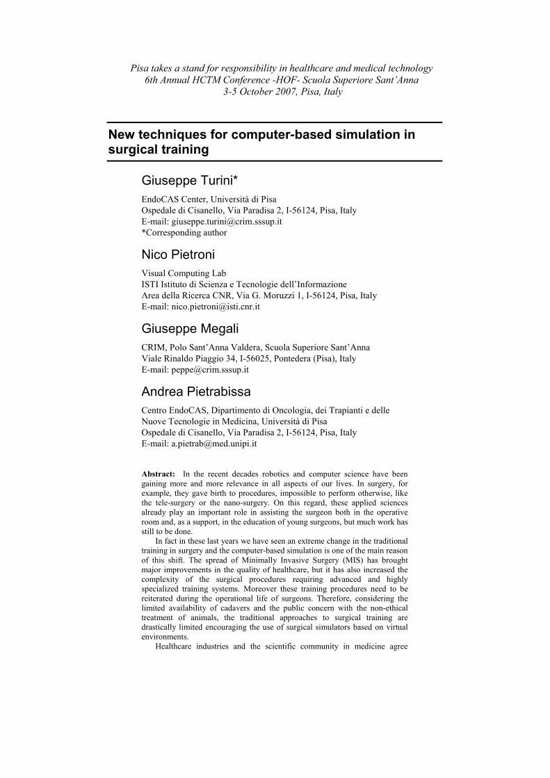
Pisa takes a stand for responsibility in healthcare and medical technology 6th Annual HCTM Conference -HOF- Scuola Superiore Sant’Anna 3-5 October 2007, Pisa, Italy New techniques for computer-based simulation in surgical training Giuseppe Turini* EndoCAS Center, Università di Pisa Ospedale di Cisanello, Via Paradisa 2, I-56124, Pisa, Italy E-mail: [email protected] *Corresponding author Nico Pietroni Visual Computing Lab ISTI Istituto di Scienza e Tecnologie dell’Informazione Area della Ricerca CNR, Via G. Moruzzi 1, I-56124, Pisa, Italy E-mail: [email protected] Giuseppe Megali CRIM, Polo Sant’Anna Valdera, Scuola Superiore Sant’Anna Viale Rinaldo Piaggio 34, I-56025, Pontedera (Pisa), Italy E-mail: [email protected] Andrea Pietrabissa Centro EndoCAS, Dipartimento di Oncologia, dei Trapianti e delle Nuove Tecnologie in Medicina, Università di Pisa Ospedale di Cisanello, Via Paradisa 2, I-56124, Pisa, Italy E-mail: [email protected] Abstract: In the recent decades robotics and computer science have been gaining more and more relevance in all aspects of our lives. In surgery, for example, they gave birth to procedures, impossible to perform otherwise, like the tele-surgery or the nano-surgery. On this regard, these applied sciences already play an important role in assisting the surgeon both in the operative room and, as a support, in the education of young surgeons, but much work has still to be done. In fact in these last years we have seen an extreme change in the traditional training in surgery and the computer-based simulation is one of the main reason of this shift. The spread of Minimally Invasive Surgery (MIS) has brought major improvements in the quality of healthcare, but it has also increased the complexity of the surgical procedures requiring advanced and highly specialized training systems. Moreover these training procedures need to be reiterated during the operational life of surgeons. Therefore, considering the limited availability of cadavers and the public concern with the non-ethical treatment of animals, the traditional approaches to surgical training are drastically limited encouraging the use of surgical simulators based on virtual environments. Healthcare industries and the scientific community in medicine agree
Transcript of New techniques for computer-based simulation in surgical...
Pisa takes a stand for responsibility in healthcare and medical technology
6th Annual HCTM Conference -HOF- Scuola Superiore Sant’Anna
3-5 October 2007, Pisa, Italy
New techniques for computer-based simulation in surgical training
Giuseppe Turini*
EndoCAS Center, Università di Pisa
Ospedale di Cisanello, Via Paradisa 2, I-56124, Pisa, Italy
E-mail: [email protected]
*Corresponding author
Nico Pietroni
Visual Computing Lab
ISTI Istituto di Scienza e Tecnologie dell’Informazione
Area della Ricerca CNR, Via G. Moruzzi 1, I-56124, Pisa, Italy
E-mail: [email protected]
Giuseppe Megali
CRIM, Polo Sant’Anna Valdera, Scuola Superiore Sant’Anna
Viale Rinaldo Piaggio 34, I-56025, Pontedera (Pisa), Italy
E-mail: [email protected]
Andrea Pietrabissa
Centro EndoCAS, Dipartimento di Oncologia, dei Trapianti e delle
Nuove Tecnologie in Medicina, Università di Pisa
Ospedale di Cisanello, Via Paradisa 2, I-56124, Pisa, Italy
E-mail: [email protected]
Abstract: In the recent decades robotics and computer science have been
gaining more and more relevance in all aspects of our lives. In surgery, for
example, they gave birth to procedures, impossible to perform otherwise, like
the tele-surgery or the nano-surgery. On this regard, these applied sciences
already play an important role in assisting the surgeon both in the operative
room and, as a support, in the education of young surgeons, but much work has
still to be done.
In fact in these last years we have seen an extreme change in the traditional
training in surgery and the computer-based simulation is one of the main reason
of this shift. The spread of Minimally Invasive Surgery (MIS) has brought
major improvements in the quality of healthcare, but it has also increased the
complexity of the surgical procedures requiring advanced and highly
specialized training systems. Moreover these training procedures need to be
reiterated during the operational life of surgeons. Therefore, considering the
limited availability of cadavers and the public concern with the non-ethical
treatment of animals, the traditional approaches to surgical training are
drastically limited encouraging the use of surgical simulators based on virtual
environments.
Healthcare industries and the scientific community in medicine agree

Pisa takes a stand for responsibility in healthcare and medical technology
6th Annual HCTM Conference -HOF- Scuola Superiore Sant’Anna
3-5 October 2007, Pisa, Italy
2
indicating the disruptive potential of the application of Virtual Reality (VR) to
the training in the medical field. Therefore the next step is the development of
surgical simulators with an high level of realism in order to practice complex
procedures in a safe environment. Moreover it is decisive that this evolution is
done integrating advanced medical imaging and processing, allowing surgeons
to practice simulated interventions on patient specific dataset.
The increasing importance of MIS techniques will cause a drastic change in
pre-operation planning and basic surgical training. In fact, the features of this
kind of surgical approach (the workspace limitation, the 2D vision through a
laparoscopic camera and the indirect physical interaction with the patient body)
make it possible to use a surgical simulator to train, plan or simulate an
intervention, reproducing the visual and tactile feedback of the real surgical
procedure on a real patient.
This paper presents some research and applicative results on Computer
Assisted Surgery (CAS) achieved in the framework of EndoCAS, a newly
founded Center of Excellence in Pisa. The research has involved: the
development of segmentation algorithms for volumetric datasets, the simulation
of bone drilling procedures, the modeling of deformable object cuts and
deformations and the simulation of rope interactions during a suture procedure
in MIS. All these projects were been developed using a new open source library
to support the implementation of techniques for simulating deformable objects.
Our purpose is to enhance the surgical training with new improved
techniques applied both to the medical imaging and to the computer-based
simulation in order to carry the surgical training to a next level of realism.
Keywords: surgical simulation; computer aided surgery.
Biographical notes: Giuseppe Turini was born in 1975 in Pescia (Pistoia,
Italy). He received the master degree in Computer Science from the University
of Pisa in 2004, and in the same year he joined to the Visual Computing Lab of
the CNR-ISTI in Pisa. Since 2005 he is a research fellow at the EndoCAS
Centre, and his main research interests are in the field of computer graphics
visualization and physical simulation. At the present time his research is carried
out in the context of computer-assisted surgery.
Nico Pietroni was born in 1978 in Siena (Italy). He received the Laurea degree
in Computer Science from the University of Pisa in 2004, and in the same year
he joined to the Visual Computing Lab of the National Research Council in
Pisa. He start his Ph.D. activity in 2005. His main research interests are in the
field of Computer graphics, deformable models simulation and interaction.
Giuseppe Megali was born in 1972 in Reggio Calabria (Italy). He received the
Laurea degree in Computer Science from the University of Pisa in 1998, and in
the same year he joined to the MiTech Lab (now CRIM Lab) of the Scuola
Superiore Sant’Anna in Pisa. He received his Ph.D. degree in Robotics from
the University of Genova in 2002. From 2002 to 2004 he had a Post-Doctoral
fellowship in Bioengineering at the Scuola Superiore Sant’Anna in Pisa. Since
2004 he is assistant professor of Biomedical Robotics at the Scuola Superiore
Sant’Anna and coordinator of EndoCAS. His main research interests are in the
field of biomedical robotics, computer-assisted surgery and analysis of surgical
gesture. His research is carried out in the context of National and European
projects in the field of biomedical engineering.
Andrea Pietrabissa was born in 1959 in Pisa. He graduated with honors in
Medicine and Surgery at the University of Pisa and he specialized in General

Pisa takes a stand for responsibility in healthcare and medical technology
6th Annual HCTM Conference -HOF- Scuola Superiore Sant’Anna
3-5 October 2007, Pisa, Italy
3
Surgery. From 1986 to 1988 he was Research Fellow in Hepatic
Transplantation at the University of Chicago, and from the 1992 to the 1993 he
was Senior Registrar at the Ninewells Hospital in Dundee (Scotland) and
Lecturer in Surgery at the University of Dundee. Since 1999 he is Associate
Professor of General Surgery at the University of Pisa. He is responsible of the
Minimally Invasive Section of the Department of General Surgery and
Transplantation at the University of Pisa, and, until now, he performed over
than 3000 surgical interventions. He is member of several scientific societies
(EAES European Association of Endoscopic Surgeons, SAGES Society of
American Gastrointestinal Surgeons), and he is in the international Editorial
Board of the Surgical Endoscopy journal. He was involved in a lot of research
projects at national and international level and co-inventor of some patents.
1 Introduction
The quality of life is strictly related to the progress in various fields of applied sciences.
Usually these advancements are carried on by researchers with the same skills and in
these situations the results are directly available. Using applied sciences in medicine is
different, because there is a wide difference between the background of the developers of
technology and the background of the surgeons, so the researchers can hardly
communicate their needs correctly. Therefore to develop a simulator for non-invasive
surgery we need the guide of trained surgeons as well as to develop a flight simulator we
need the help of trained pilots.
EndoCAS is a Center for Endoscopic Computer Assisted Surgery located inside the
Ospedale di Cisanello, in Pisa. The center was born to put together researchers from
different scientific backgrounds that would not meet otherwise. It is the result of a joint
proposal of the University of Pisa, of Scuola Superiore Sant’Anna (CRIM Lab) and the
Visual Computing Laboratory (ISTI-CNR) in the framework of MIUR funding for
centers of excellence. Its goals are to address key knowledge, technology, and systems
design barriers that must be overcome in the development of CAS systems.
In this paper we overview some of the results obtained in the first years of activity of
the center. In Section 2 we show a novel scheme for extracting anatomical organs from
noisy Computed Tomography (CT) data based on active contours and marching cubes
techniques. This approach was successfully used to reconstruct the vena cava from CT
data where standard methods failed. In Section 3 a technique for simulating bone drilling
is shown. The approach is very general and it can be adopted by all the approaches using
tetrahedral meshes for representing the solid, with limited influence on the performance
of the rest of the simulation. In Section 4 we briefly describe a fast and robust technique
to perform interactive virtual cutting on deformable objects. Our algorithm is highly
general and does not use any assumption neither on how the deformation function is
computed nor on how the boundary of the object is encoded. Then in Section 5 we
present a physically based model for real-time simulation of thread dynamics. Our
approach leads to a stable real time simulation. Section 6 is dedicated to an open source
library called IdoLib, created to develop simulation-oriented applications. Conclusions
are exposed in Section 7 that completes the paper.
Pisa takes a stand for responsibility in healthcare and medical technology
6th Annual HCTM Conference -HOF- Scuola Superiore Sant’Anna
3-5 October 2007, Pisa, Italy
4
2 Segmentation of noisy data
The reconstruction of the external surface of an organ is an important task for 3D
visualization of medical datasets and it can also be used to support quantitative diagnostic
measurements. However, this assignment may be challenging even in the seemingly easy
cases of visually uniform regions. In fact segmentation methods should provide a closed
smooth surface avoiding leakages but not missing bifurcations with smaller
vascularizations. For these reasons these algorithms need topology control, robustness
against noise and an appropriate smoothing method for the surface generation in order to
preserve small structures or local pathologies that characterize the organ.
Many surface reconstruction methods can be found in literature, each with specific
advantages and drawbacks. Simple isosurface extraction (Lorensen and Cline, 1987) does
not guarantee topology control and it is quite sensitive to noise. Holes and leakages are
also a relevant problem for more advanced level sets or geodesic surfaces methods
(Mallardi and Sethian, 1995; Caselles and Kimmel, 1995). To prevent irregularities, holes
and leakages the best approach is to use a parametric deformable surface instead of an
implicit curve, with the same approach as the classic balloon snakes (Cohen and Cohen,
1993). These methods (see McInerney and Terzopoulos, 1996; for a review of the first
medical applications) are efficient and robust. The elastic forces avoid the surface block
near a noisy pixel and prevent leakages.
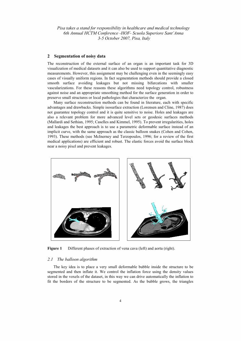
Figure 1 Different phases of extraction of vena cava (left) and aorta (right).
2.1 The balloon algorithm
The key idea is to place a very small deformable bubble inside the structure to be
segmented and then inflate it. We control the inflation force using the density values
stored in the voxels of the dataset, in this way we can drive automatically the inflation to
fit the borders of the structure to be segmented. As the bubble grows, the triangles
Pisa takes a stand for responsibility in healthcare and medical technology
6th Annual HCTM Conference -HOF- Scuola Superiore Sant’Anna
3-5 October 2007, Pisa, Italy
5
become bigger, but since the final goal of the process is to fit the bubble over the surface
of the structure, it is self evident that the size of the triangles must be of the order of the
size of the voxel. As a triangle grows more than a fixed threshold, we perform a simple
mesh refinement, splitting the longest edge and adding two new triangles.
2.2 Self intersection management
During the inflation, the bubble mesh can intersect itself, because the anatomical
structure to be segmented is not always topologically equivalent to a sphere. The
detection of the intersections is needed to stop the bubble inflation. Therefore, to restore a
non self-intersecting surface our algorithm performs a periodical collision detection in
order to find intersecting portions of the surface and blocking the vertices belonging to
intersecting faces. Then we build a distance map assigning to each voxel of the volume
the signed distance from the closest non intersected face, which is negative if the point is
inside the volume enclosed by the surface and positive otherwise. Due to the use of the
part of the surface that is not self-intersecting , the result is that the isosurface of value 0
of the map is a surface defining the limits of the region of our interest with no self-
intersections and the correct topology.
Figure 2 Example of self intersection management: the bubble expansion generate a
self intersection (in red on the left) that is managed obtaining the final
connected mesh (in cyan on the right).
2.3 Results
We have tested our algorithm on synthetic images of structures to validate the self
collision detection/marching cubes method (see Figure 2). In all the tests, we have
obtained a closed smooth surface defining an internal region close to the voxelized
volume of interest.
The algorithm was applied to CT scans of the abdomen in order to segment the aorta
and the vena cava. Figure 1 shows the results of these test cases. The segmentation is
accurate and fast and no manual corrections are required.
3 Towards surgical simulator: bone drilling
The bone drilling procedure is an important task in many surgical interventions as:
mastoidectomy, cochlear implantation or orbital surgery. The procedure mainly consists
in the removal of part of the bone in contact with the surgical drill when the surgeon

Pisa takes a stand for responsibility in healthcare and medical technology
6th Annual HCTM Conference -HOF- Scuola Superiore Sant’Anna
3-5 October 2007, Pisa, Italy
6
exerts sufficient pressure. The fact that the bone is an almost rigid material make the
virtual bone object suitable for a regular discretization of the space, and therefore most of
the approaches proposed in literature use voxel-based techniques that allow an easy bone
material removal playing with the density in the voxels. Some examples of these
techniques are: the voxmap-pointshell methods (McNeely and Puterbaugh, 1999; Petersik
and Pflesser, 2002; Morris and Sewell, 2004), or the Graphics Processing Unit (GPU)
voxel-based techniques (Agus and Giachetti, 2002). Mesh based approaches are also
widely used to perform cuts (Bielser and Maiwald, 1999; Ganovelli and Cignoni, 2000;
Niehuys and van der Stappen, 2000) and to render material removal (Agus and Gobbetti,
2006; Cotin and Ayache, 1999) but their accuracy depends on the size of the mesh
elements. However, there are cases in which the bone drilling is only a part of the
surgical procedure, and the same bone object can also be cut or, worse, slightly deformed
and therefore voxel-based techniques do not work well.
We have developed a novel drilling method to overtake these drawbacks (Figure 3
left). Our algorithm is based on objects represented explicitly by means of tetrahedral
meshes that can be easily generated from CT (or Magnetic Resonance) dataset. These
volumetric meshes can be used to perform physical simulation of solids, they can be
easily visualized and can also be used to simulate material with non uniform density.
Unfortunately, since the number of mesh elements (tetrahedra) strongly influences the
performance of the physical simulation, we have to face two opposite constraints: a high
number of tetrahedra to achieve a realistic visualization of drilling and a low number of
elements to execute the physical simulation in real-time.
Figure 3 An example of drilling simulation (left) and the tetrahedral subdivision
scheme (right).
3.1 Our drilling approach
Develop a drilling method that is independent from the physical simulation, means to
decouple the drilling from the physical system. To do this we have decided to represent
the drilling at sub-tetrahedral level defining a hierarchical tetrahedral decomposition
scheme recursively splitting tetrahedra in 8 new tetrahedra (see Figure 3 right).
Moreover the algorithm manages the collisions between the tool and the bone mesh,
so when a tetrahedron come in contact with the tool, the decomposition scheme is applied
to adapt the mesh detail to the tool shape, in this way we can easily perform the bone
Pisa takes a stand for responsibility in healthcare and medical technology
6th Annual HCTM Conference -HOF- Scuola Superiore Sant’Anna
3-5 October 2007, Pisa, Italy
7
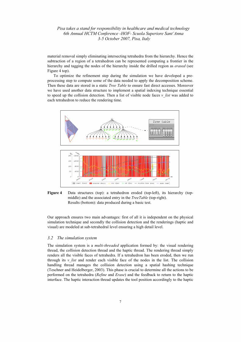
material removal simply eliminating intersecting tetrahedra from the hierarchy. Hence the
subtraction of a region of a tetrahedron can be represented computing a frontier in the
hierarchy and tagging the nodes of the hierarchy inside the drilled region as erased (see
Figure 4 top).
To optimize the refinement step during the simulation we have developed a pre-
processing step to compute some of the data needed to apply the decomposition scheme.
Then these data are stored in a static Tree Table to ensure fast direct accesses. Moreover
we have used another data structure to implement a spatial indexing technique essential
to speed up the collision detection. Then a list of visible node faces v_list was added to
each tetrahedron to reduce the rendering time.
Figure 4 Data structures (top): a tetrahedron eroded (top-left), its hierarchy (top-
middle) and the associated entry in the TreeTable (top-right).
Results (bottom): data produced during a basic test.
Our approach ensures two main advantages: first of all it is independent on the physical
simulation technique and secondly the collision detection and the renderings (haptic and
visual) are modeled at sub-tetrahedral level ensuring a high detail level.
3.2 The simulation system
The simulation system is a multi-threaded application formed by: the visual rendering
thread, the collision detection thread and the haptic thread. The rendering thread simply
renders all the visible faces of tetrahedra. If a tetrahedron has been eroded, then we run
through its v_list and render each visible face of the nodes in the list. The collision
handling thread manages the collision detection using a spatial hashing technique
(Teschner and Heidelberger, 2003). This phase is crucial to determine all the actions to be
performed on the tetrahedra (Refine and Erase) and the feedback to return to the haptic
interface. The haptic interaction thread updates the tool position accordingly to the haptic
Pisa takes a stand for responsibility in healthcare and medical technology
6th Annual HCTM Conference -HOF- Scuola Superiore Sant’Anna
3-5 October 2007, Pisa, Italy
8
interface movements and ensure high rendering frequency of force feedback that is
essential to simulate a realistic haptic interaction.
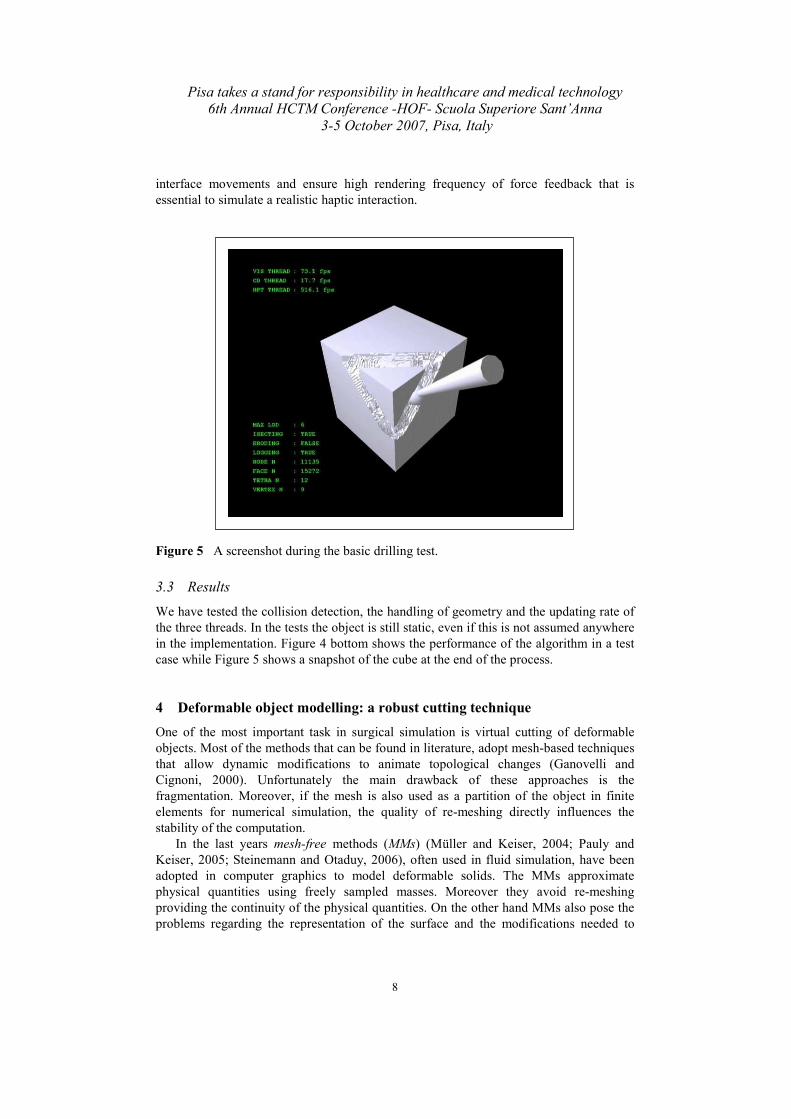
Figure 5 A screenshot during the basic drilling test.
3.3 Results
We have tested the collision detection, the handling of geometry and the updating rate of
the three threads. In the tests the object is still static, even if this is not assumed anywhere
in the implementation. Figure 4 bottom shows the performance of the algorithm in a test
case while Figure 5 shows a snapshot of the cube at the end of the process.
4 Deformable object modelling: a robust cutting technique
One of the most important task in surgical simulation is virtual cutting of deformable
objects. Most of the methods that can be found in literature, adopt mesh-based techniques
that allow dynamic modifications to animate topological changes (Ganovelli and
Cignoni, 2000). Unfortunately the main drawback of these approaches is the
fragmentation. Moreover, if the mesh is also used as a partition of the object in finite
elements for numerical simulation, the quality of re-meshing directly influences the
stability of the computation.
In the last years mesh-free methods (MMs) (Müller and Keiser, 2004; Pauly and
Keiser, 2005; Steinemann and Otaduy, 2006), often used in fluid simulation, have been
adopted in computer graphics to model deformable solids. The MMs approximate
physical quantities using freely sampled masses. Moreover they avoid re-meshing
providing the continuity of the physical quantities. On the other hand MMs also pose the
problems regarding the representation of the surface and the modifications needed to
Pisa takes a stand for responsibility in healthcare and medical technology
6th Annual HCTM Conference -HOF- Scuola Superiore Sant’Anna
3-5 October 2007, Pisa, Italy
9
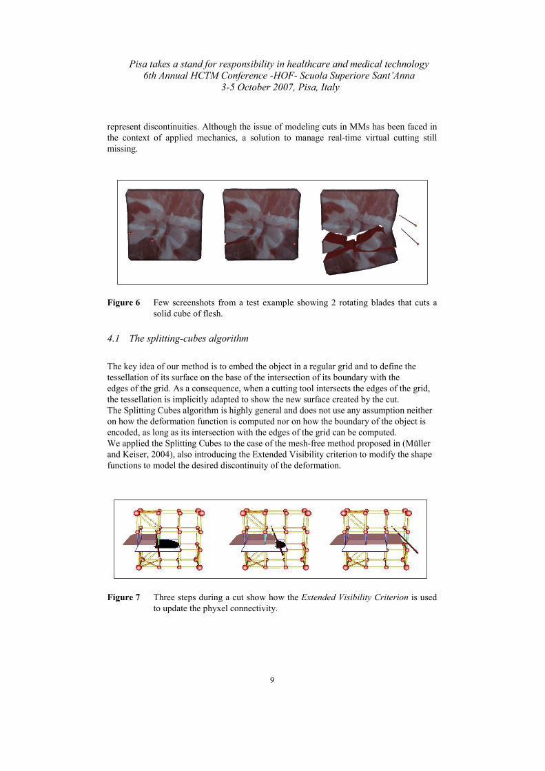
represent discontinuities. Although the issue of modeling cuts in MMs has been faced in
the context of applied mechanics, a solution to manage real-time virtual cutting still
missing.
Figure 6 Few screenshots from a test example showing 2 rotating blades that cuts a
solid cube of flesh.
4.1 The splitting-cubes algorithm
The key idea of our method is to embed the object in a regular grid and to define the
tessellation of its surface on the base of the intersection of its boundary with the
edges of the grid. As a consequence, when a cutting tool intersects the edges of the grid,
the tessellation is implicitly adapted to show the new surface created by the cut.
The Splitting Cubes algorithm is highly general and does not use any assumption neither
on how the deformation function is computed nor on how the boundary of the object is
encoded, as long as its intersection with the edges of the grid can be computed.
We applied the Splitting Cubes to the case of the mesh-free method proposed in (Müller
and Keiser, 2004), also introducing the Extended Visibility criterion to modify the shape
functions to model the desired discontinuity of the deformation.
Figure 7 Three steps during a cut show how the Extended Visibility Criterion is used
to update the phyxel connectivity.

Pisa takes a stand for responsibility in healthcare and medical technology
6th Annual HCTM Conference -HOF- Scuola Superiore Sant’Anna
3-5 October 2007, Pisa, Italy
10
4.2 The extended visibility criterion
Once the object is being cut and deformed we need to change the deformation function to
reflect the discontinuity introduced. This mainly means to check the phyxel connectivity
in an appropriate manner. Our approach relies on a visibility criterion that ensures smooth
cuts and that can be implemented on the GPU optimizing the performance of the
simulation.
4.2 Results
We have introduced the Splitting Cubes, a new algorithm for dynamic tesselation of an
implicit surface interactively generated in response to cuts (Figure 6). Moreover we have
also implemented an Extended Visibility Criterion (Figure 7), a novel GPU friendly
solution to handle discontinuities in MMs taking advantage of the graphics hardware.
It is important to evidence that the Splitting Cubes algorithm only relies on a generic
deformation function and on a description of the object’s surface.
5 Robust interactive rope simulation
The main use of a thread simulator is certainly in endoscopic surgical simulation.
Handling the surgical thread to make knots is one of the most difficult tasks for a surgeon
because it requires ambidexterity with the endoscopic forceps.
Although the rope simulation can seem simpler than the simulation of more complex
3D structures, the interaction with a thread involves self-collision detection and contact
handling. Furthermore, the thread is almost inextensible, and from the point of view of
the simulation, this means that the methods modeling elasticity and using explicit time-
integration schemes are poorly conditioned.
Most of the approaches in literature are energy-based (Wang and Burdet, 2006) and
model the rope as a 1-dimensional chain of mass points connected by springs and
physically simulated. However there are also non energy-based models (Brown and
Latombe, 2004) that allow the making of complex knots in real-time even if the
perception of the lack of physical simulation is clear.
Our method for simulating surgical thread is based on Position Based Dynamics (Müller
and Heidelberger, 2007), the collision detection is carried out by spatial hashing.
5.1 Our approach
We simulate rope dynamic by defining a set of physical constraints. The simulation
involves three steps: (1) moving the mass points according to their velocity and external
action, (2) moving the mass points to satisfy the constraints and then (3) performing time
integration. Position Based Dynamics is particularly useful to handle collision and
contact, while it is hard to achieve stability using energy based methods, and this is the
main reason for our choice.
The collision detection for the rope simulation is a crucial process. For this reason we
have chosen to use a regular partition of the space cause the thread is divided in segments
equally sized, so we have used a Spatial Hashing technique (Teschner and Heidelberger,
2003) with temporal marks.
Pisa takes a stand for responsibility in healthcare and medical technology
6th Annual HCTM Conference -HOF- Scuola Superiore Sant’Anna
3-5 October 2007, Pisa, Italy
11
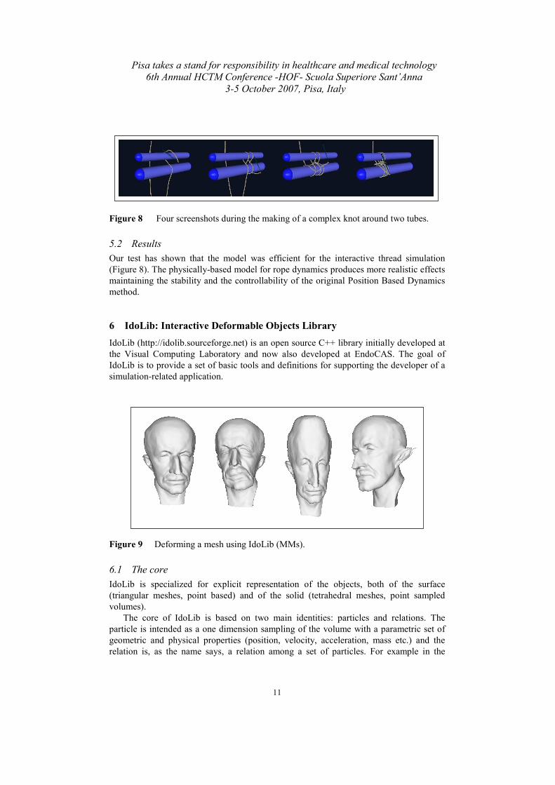
Figure 8 Four screenshots during the making of a complex knot around two tubes.
5.2 Results
Our test has shown that the model was efficient for the interactive thread simulation
(Figure 8). The physically-based model for rope dynamics produces more realistic effects
maintaining the stability and the controllability of the original Position Based Dynamics
method.
6 IdoLib: Interactive Deformable Objects Library
IdoLib (http://idolib.sourceforge.net) is an open source C++ library initially developed at
the Visual Computing Laboratory and now also developed at EndoCAS. The goal of
IdoLib is to provide a set of basic tools and definitions for supporting the developer of a
simulation-related application.
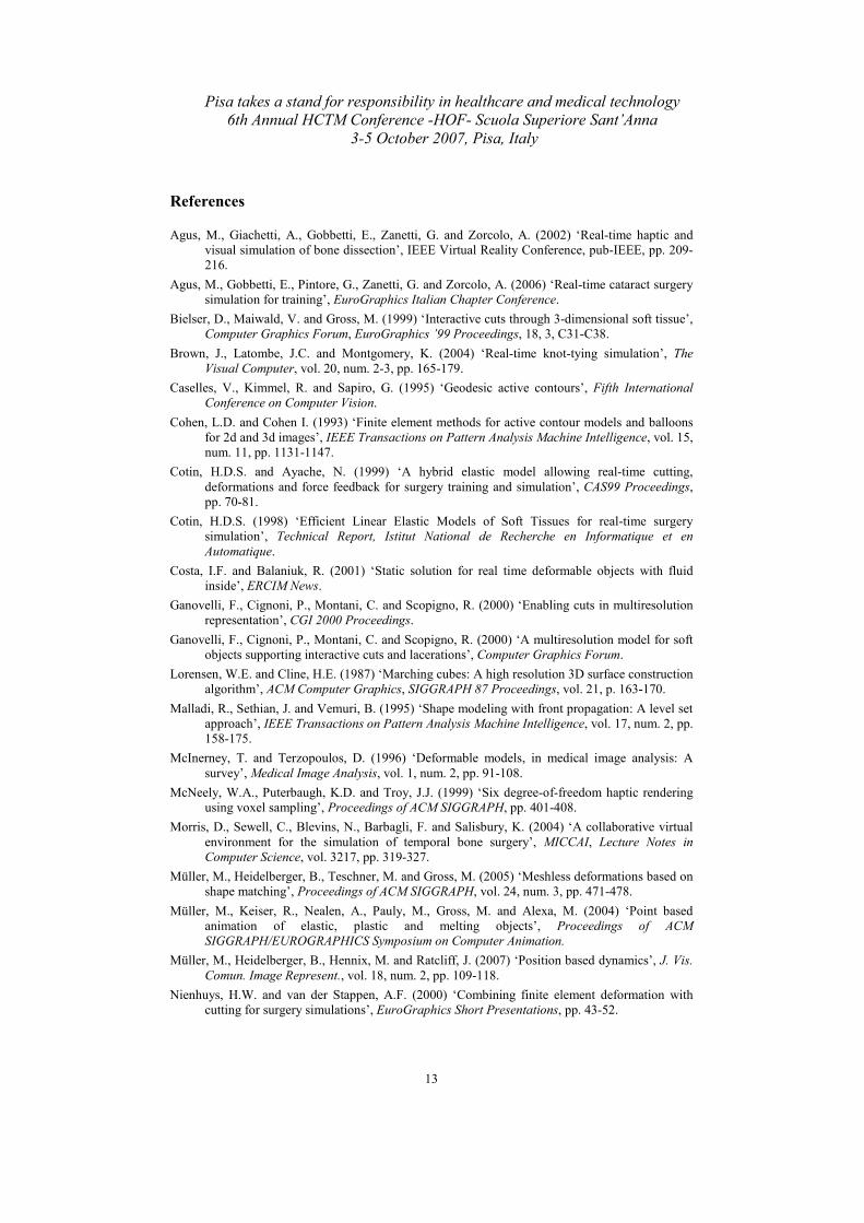
Figure 9 Deforming a mesh using IdoLib (MMs).
6.1 The core
IdoLib is specialized for explicit representation of the objects, both of the surface
(triangular meshes, point based) and of the solid (tetrahedral meshes, point sampled
volumes).
The core of IdoLib is based on two main identities: particles and relations. The
particle is intended as a one dimension sampling of the volume with a parametric set of
geometric and physical properties (position, velocity, acceleration, mass etc.) and the
relation is, as the name says, a relation among a set of particles. For example in the

Pisa takes a stand for responsibility in healthcare and medical technology
6th Annual HCTM Conference -HOF- Scuola Superiore Sant’Anna
3-5 October 2007, Pisa, Italy
12
simplest case of the mass spring system the particle is the mass and the relation is the
spring. The relation does not have to correspond to a geometric figure as for spring or
triangles of tetrahedra: for example in the implementation of a point based method, the
relation binds together all the particles closer than a given radius to each other. Figure 9
and Figure 10 show few examples of an application of the methods described above.
Moreover IdoLib provides a number of common integration schemes and a callback-
based manner to add new schemes, so the user can switch among different integration
schemes during the simulation. Similarly, the library is extendible and new methods can
be easily added to those already implemented in IdoLib (refer to: Cotin and Ayache,
1999; O’Brien and Hodgins, 1999; Müller and Heidelberger, 2005; Costa and Balaniuk,
2001; Müller and Keiser, 2004; for the details). Note that IdoLib does not take control of
the simulation loop and the main call only performs a single step of simulation.
Dependencies: IdoLib is written in C++ with STL library and relies only on another
library, called VCG (Visualization and Computer Graphics Library,
http://vcg.sourceforge.net), for the basic geometry structures and linear algebra.
6.2 Further work
IdoLib is still at a early stage. Although the structure is quite stable, further work is
needed to provide a simple way to visualize the state of the simulation which is always
essential in the debugging phase. Similarly, a complete documentation and examples is
still to be done.
Figure 10 Deforming a virtual liver using IdoLib (mass-spring).
7 Conclusions
This paper reports some recent results obtained in the framework of the center for
endoscopic computer-assisted surgery EndoCAS. We have presented some applications
of physical simulation to medicine: a method for automatic segmentation and
reconstruction of shapes from noisy 3D datasets, a technique to simulate drilling over
quasi rigid objects, an approach for the interactive simulation of deformable objects
allowing cuts and deformations and a method to simulate surgical threads. The paper also
present the library for physical simulation which is being developed and over which the
applications described above were built.
Pisa takes a stand for responsibility in healthcare and medical technology
6th Annual HCTM Conference -HOF- Scuola Superiore Sant’Anna
3-5 October 2007, Pisa, Italy
13
References
Agus, M., Giachetti, A., Gobbetti, E., Zanetti, G. and Zorcolo, A. (2002) ‘Real-time haptic and
visual simulation of bone dissection’, IEEE Virtual Reality Conference, pub-IEEE, pp. 209-
216.
Agus, M., Gobbetti, E., Pintore, G., Zanetti, G. and Zorcolo, A. (2006) ‘Real-time cataract surgery
simulation for training’, EuroGraphics Italian Chapter Conference.
Bielser, D., Maiwald, V. and Gross, M. (1999) ‘Interactive cuts through 3-dimensional soft tissue’,
Computer Graphics Forum, EuroGraphics ’99 Proceedings, 18, 3, C31-C38.
Brown, J., Latombe, J.C. and Montgomery, K. (2004) ‘Real-time knot-tying simulation’, The
Visual Computer, vol. 20, num. 2-3, pp. 165-179.
Caselles, V., Kimmel, R. and Sapiro, G. (1995) ‘Geodesic active contours’, Fifth International
Conference on Computer Vision.
Cohen, L.D. and Cohen I. (1993) ‘Finite element methods for active contour models and balloons
for 2d and 3d images’, IEEE Transactions on Pattern Analysis Machine Intelligence, vol. 15,
num. 11, pp. 1131-1147.
Cotin, H.D.S. and Ayache, N. (1999) ‘A hybrid elastic model allowing real-time cutting,
deformations and force feedback for surgery training and simulation’, CAS99 Proceedings,
pp. 70-81.
Cotin, H.D.S. (1998) ‘Efficient Linear Elastic Models of Soft Tissues for real-time surgery
simulation’, Technical Report, Istitut National de Recherche en Informatique et en
Automatique.
Costa, I.F. and Balaniuk, R. (2001) ‘Static solution for real time deformable objects with fluid
inside’, ERCIM News.
Ganovelli, F., Cignoni, P., Montani, C. and Scopigno, R. (2000) ‘Enabling cuts in multiresolution
representation’, CGI 2000 Proceedings.
Ganovelli, F., Cignoni, P., Montani, C. and Scopigno, R. (2000) ‘A multiresolution model for soft
objects supporting interactive cuts and lacerations’, Computer Graphics Forum.
Lorensen, W.E. and Cline, H.E. (1987) ‘Marching cubes: A high resolution 3D surface construction
algorithm’, ACM Computer Graphics, SIGGRAPH 87 Proceedings, vol. 21, p. 163-170.
Malladi, R., Sethian, J. and Vemuri, B. (1995) ‘Shape modeling with front propagation: A level set
approach’, IEEE Transactions on Pattern Analysis Machine Intelligence, vol. 17, num. 2, pp.
158-175.
McInerney, T. and Terzopoulos, D. (1996) ‘Deformable models, in medical image analysis: A
survey’, Medical Image Analysis, vol. 1, num. 2, pp. 91-108.
McNeely, W.A., Puterbaugh, K.D. and Troy, J.J. (1999) ‘Six degree-of-freedom haptic rendering
using voxel sampling’, Proceedings of ACM SIGGRAPH, pp. 401-408.
Morris, D., Sewell, C., Blevins, N., Barbagli, F. and Salisbury, K. (2004) ‘A collaborative virtual
environment for the simulation of temporal bone surgery’, MICCAI, Lecture Notes in
Computer Science, vol. 3217, pp. 319-327.
Müller, M., Heidelberger, B., Teschner, M. and Gross, M. (2005) ‘Meshless deformations based on
shape matching’, Proceedings of ACM SIGGRAPH, vol. 24, num. 3, pp. 471-478.
Müller, M., Keiser, R., Nealen, A., Pauly, M., Gross, M. and Alexa, M. (2004) ‘Point based
animation of elastic, plastic and melting objects’, Proceedings of ACM
SIGGRAPH/EUROGRAPHICS Symposium on Computer Animation.
Müller, M., Heidelberger, B., Hennix, M. and Ratcliff, J. (2007) ‘Position based dynamics’, J. Vis.
Comun. Image Represent., vol. 18, num. 2, pp. 109-118.
Nienhuys, H.W. and van der Stappen, A.F. (2000) ‘Combining finite element deformation with
cutting for surgery simulations’, EuroGraphics Short Presentations, pp. 43-52.

Pisa takes a stand for responsibility in healthcare and medical technology
6th Annual HCTM Conference -HOF- Scuola Superiore Sant’Anna
3-5 October 2007, Pisa, Italy
14
O’Brien, J.F. and Hodgins, J.K. (1999) ‘Graphical modeling and animation of brittle fracture’,
SIGGRAPH, pp. 137-146.
Pauly, M., Keiser, R., Adams, B., Dutr, P., Gross, M. and Guibas, L.J. (2005) ‘Meshless animation
of fracturing solids’, ACM Transactions on Graphics, vol. 24, num. 3, pp. 957-964.
Petersik, A., Pflesser, B., Tiede, U., Höhne, K. and Leuwer, R. (2002) ‘Haptic volume interaction
with anatomic models at sub-voxel resolution’, Symposium on Haptic Interfaces for Virtual
Environment and Teleoperator Systems, pp. 66-72.
Steinemann, D., Otaduy, M.A., Gross, M. (2006) ‘Fast arbitrary splitting of deforming objects’,
EuroGraphics/SIGGRAPH Symposium on Computer Animation.
Teschner, M., Heidelberger, B., Müller, M., Pomeranets, D. and Gross, M. (2003) ‘Optimized
spatial hashing for collision detection of deformable objects’, Proceedings of the Conference
on Vision, Modeling and Visualization 2003 (VMV-03), pp. 47-54.
Wang, F., Burdet, E., Vuillemin, R. and Bleuler, H. (2006) ‘Knot-tying with visual and force
feedback for VR laparoscopic training’, IEEE Computer Society, pp. 5778-5781.