
New species of the harvestmen Hutamaia (Laniatores ... · New species of the harvestmen Hutamaia...
12
ZOOLOGIA 31 (5): 463–474, October, 2014 http://dx.doi.org/10.1590/S1984-46702014000500006 2014 Sociedade Brasileira de Zoologia | www.sbzoologia.org.br | www.scielo.br/zool All content of the journal, except where identified, is licensed under a Creative Commons attribution-type BY-NC. Ampycinae Kury, 2003 was erected under Gonyleptidae by KURY (2003), and originally included three species in two gen- era (Ampycus Simon, 1879 and Hexabunus Roewer, 1913). Later, the author acknowledged that, although his initial intention had been to widen the definition of the subfamily to include nine more genera in it, he decided not do to do it, which resulted in some inconsistencies in his 2003 publication (see KURY & ALONSO- ZARAZAGA 2011: 52). KURY & ALONSO-ZARAZAGA (2011) officially trans- ferred those nine genera (Ampycella Roewer, 1929, Glysterus Roewer, 1931, Hernandariodes Pickard-Cambridge, 1905, Hutamaia Soares & Soares, 1977, Neopachyloides Roewer, 1913, Nesopachylus Chamberlin, 1925, Parahernandria Goodnight & Goodnight, 1947, Sibollus, Roewer, 1929, and Thaumatopachylus Roewer, 1929) to Ampycinae. More recently, two genera have been included in Ampycinae, Pirunipygus Roewer, 1936 (PINTO-DA-ROCHA et al. 2012) and Licornus Roewer, 1932 (VILLARREAL & KURY 2012). Currently, Ampycinae comprises 25 species grouped into 13 genera. Most of the genera were originally placed in Pachylinae, some in Hernandariinae or Gonyleptinae, and Cranainae. As it is usually the case with Neotropical harvestmen, almost half of the genera of Ampycinae were monotypic – six out of 13, until this paper. Ampycinae was recovered as a clade, which is closely related to Cranainae, in the most recently published molecular phylogeny of Gonyleptidae (PINTO-DA-ROCHA et al. 2013). “ProVárzea” was a large project on the diversity and po- tential biogeographic regions of the Amazon flooded forests – várzea – (Brazil), conducted by the staff of the Instituto Nacional de Pesquisas da Amazônia (INPA) and funded by the Instituto Brasileiro do Meio Ambiente e dos Recursos Naturais Renováveis (IBAMA). This project resulted in numerous har- vestmen samples, including new species of some genera pre- viously considered monotypic, or with only one species recorded for the Brazilian Amazon (TOURINHO & PÉREZ 2006). In this paper we describe three new species of Hutamaia, formerly monotypic, collected from the flooded forests (várzea) of the Amazon River. We discuss the affinities of Hutamaia with Licornus and their biogeographic distribution patterns. Addi- tionally, we provide a map and key for the genus, and new diagnoses for Hutamaia and H. caramaschii, including infor- mation on the new species. MATERIAL AND METHODS Photographs were taken at different focus distances with a Sony Cybershot DSC-V1 attached to an Olympus dissecting mi- croscope. Later, photographs were aligned – command Macro > Align and Balance Used Frames (Thorough) – and stacked – com- mand Macro > Pyramoid Maximum Contrast – to obtain an image with greater depth of field, using the software CombineZ, version Combine ZP (HADLEY 2014). We made schematic illustrations from photographs or using a camera lucida, with the help of the soft- ware CorelDRAW X5 and a Wacom Bamboo Connect tablet. New species of the harvestmen Hutamaia (Laniatores: Gonyleptidae: Ampycinae) and generic diagnosis Ana Lúcia Tourinho 1 & Amanda Cruz Mendes 2 1 Museum of Comparative Zoology and Department of Organismic and Evolutionary Biology, Harvard University. 26 Oxford Street, Cambridge, MA 02138, USA. E-mail: [email protected] 2 Departamento de Zoologia, Instituto de Biologia, Universidade do Estado do Rio de Janeiro. Rua São Francisco Xavier 524, Maracanã, 20550-900 Rio de Janeiro, RJ, Brazil. E-mail: [email protected] ABSTRACT. We add three new species to the formerly monotypic Amazonian Hutamaia, Hutamaia maceta sp. nov., Hutamaia plei sp. nov. and Hutamaia trompsonica sp. nov. and compare them with the type species, Hutamaia caramaschii Soares & Soares, 1977. Hutamaia was known only from two localities: Humaitá, Amazonas, Brazil (type locality of the type species), and Madre de Dios, Peru. Herein we record species from the following additional localities, all in Brazil: Coari, Codajás, Juruá, Jutaí, Manacapuru, Tefé (state of Amazonas) and Gurupá (state of Pará), indicating that the genus has a widespread distribution in the Brazilian and Peruvian Amazon. Hutamaia is newly diagnosed by having yellowish granules on dorsal scutum, armature of coxa IV of males, metatarsi with yellow rings, ventral plate of penis trapezoid with V-shaped cleft, bearing two pairs of longitudinal rows of setae, and glans without dorsal or ventral processes. The genus is likely closely related to Licornus Roewer, 1932, from which it differs by the shape of the ventral plate of the penis and lack of dorsal process of glans. KEY WORDS. Amazonas-Solimões River; floodplains; Neotropics; Opiliones; taxonomy.
Transcript of New species of the harvestmen Hutamaia (Laniatores ... · New species of the harvestmen Hutamaia...

ZOOLOGIA 31 (5): 463–474, October, 2014http://dx.doi.org/10.1590/S1984-46702014000500006
2014 Sociedade Brasileira de Zoologia | www.sbzoologia.org.br | www.scielo.br/zoolAll content of the journal, except where identified, is licensed under a Creative Commons attribution-type BY-NC.
Ampycinae Kury, 2003 was erected under Gonyleptidaeby KURY (2003), and originally included three species in two gen-era (Ampycus Simon, 1879 and Hexabunus Roewer, 1913). Later,the author acknowledged that, although his initial intention hadbeen to widen the definition of the subfamily to include ninemore genera in it, he decided not do to do it, which resulted insome inconsistencies in his 2003 publication (see KURY & ALONSO-ZARAZAGA 2011: 52). KURY & ALONSO-ZARAZAGA (2011) officially trans-ferred those nine genera (Ampycella Roewer, 1929, GlysterusRoewer, 1931, Hernandariodes Pickard-Cambridge, 1905, HutamaiaSoares & Soares, 1977, Neopachyloides Roewer, 1913, NesopachylusChamberlin, 1925, Parahernandria Goodnight & Goodnight,1947, Sibollus, Roewer, 1929, and Thaumatopachylus Roewer, 1929)to Ampycinae. More recently, two genera have been included inAmpycinae, Pirunipygus Roewer, 1936 (PINTO-DA-ROCHA et al. 2012)and Licornus Roewer, 1932 (VILLARREAL & KURY 2012). Currently,Ampycinae comprises 25 species grouped into 13 genera. Mostof the genera were originally placed in Pachylinae, some inHernandariinae or Gonyleptinae, and Cranainae. As it is usuallythe case with Neotropical harvestmen, almost half of the generaof Ampycinae were monotypic – six out of 13, until this paper.Ampycinae was recovered as a clade, which is closely related toCranainae, in the most recently published molecular phylogenyof Gonyleptidae (PINTO-DA-ROCHA et al. 2013).
“ProVárzea” was a large project on the diversity and po-tential biogeographic regions of the Amazon flooded forests –
várzea – (Brazil), conducted by the staff of the InstitutoNacional de Pesquisas da Amazônia (INPA) and funded by theInstituto Brasileiro do Meio Ambiente e dos Recursos NaturaisRenováveis (IBAMA). This project resulted in numerous har-vestmen samples, including new species of some genera pre-viously considered monotypic, or with only one speciesrecorded for the Brazilian Amazon (TOURINHO & PÉREZ 2006). Inthis paper we describe three new species of Hutamaia, formerlymonotypic, collected from the flooded forests (várzea) of theAmazon River. We discuss the affinities of Hutamaia withLicornus and their biogeographic distribution patterns. Addi-tionally, we provide a map and key for the genus, and newdiagnoses for Hutamaia and H. caramaschii, including infor-mation on the new species.
MATERIAL AND METHODS
Photographs were taken at different focus distances with aSony Cybershot DSC-V1 attached to an Olympus dissecting mi-croscope. Later, photographs were aligned – command Macro >Align and Balance Used Frames (Thorough) – and stacked – com-mand Macro > Pyramoid Maximum Contrast – to obtain an imagewith greater depth of field, using the software CombineZ, versionCombine ZP (HADLEY 2014). We made schematic illustrations fromphotographs or using a camera lucida, with the help of the soft-ware CorelDRAW X5 and a Wacom Bamboo Connect tablet.
New species of the harvestmen Hutamaia (Laniatores: Gonyleptidae:Ampycinae) and generic diagnosis
Ana Lúcia Tourinho1 & Amanda Cruz Mendes2
1 Museum of Comparative Zoology and Department of Organismic and Evolutionary Biology, Harvard University. 26 OxfordStreet, Cambridge, MA 02138, USA. E-mail: [email protected] Departamento de Zoologia, Instituto de Biologia, Universidade do Estado do Rio de Janeiro. Rua São Francisco Xavier 524,Maracanã, 20550-900 Rio de Janeiro, RJ, Brazil. E-mail: [email protected]
ABSTRACT. We add three new species to the formerly monotypic Amazonian Hutamaia, Hutamaia maceta sp. nov.,Hutamaia plei sp. nov. and Hutamaia trompsonica sp. nov. and compare them with the type species, Hutamaia caramaschii
Soares & Soares, 1977. Hutamaia was known only from two localities: Humaitá, Amazonas, Brazil (type locality of the
type species), and Madre de Dios, Peru. Herein we record species from the following additional localities, all in Brazil:
Coari, Codajás, Juruá, Jutaí, Manacapuru, Tefé (state of Amazonas) and Gurupá (state of Pará), indicating that the
genus has a widespread distribution in the Brazilian and Peruvian Amazon. Hutamaia is newly diagnosed by having
yellowish granules on dorsal scutum, armature of coxa IV of males, metatarsi with yellow rings, ventral plate of penis
trapezoid with V-shaped cleft, bearing two pairs of longitudinal rows of setae, and glans without dorsal or ventral
processes. The genus is likely closely related to Licornus Roewer, 1932, from which it differs by the shape of the ventral
plate of the penis and lack of dorsal process of glans.
KEY WORDS. Amazonas-Solimões River; floodplains; Neotropics; Opiliones; taxonomy.

464 A.L. Tourinho & A.C. Mendes
ZOOLOGIA 31 (5): 463–474, October, 2014
All measurements are given in millimeters. Abbreviationsused in the descriptions: (CL) carapace length, (CW) carapacemaximal width, (AL) abdominal scutum length, (AW) abdomi-nal scutum maximal width, (Pe) pedipalpus, (Tr) trochanter,(Fe) femur, (Pa) patella, (Ti) tibia, (Mt) metatarsus, (Ta) tarsus,(Cl) claw.
Repositories of specimens are as follows: Instituto Nacio-nal de Pesquisas da Amazonia, Amazonas, Brazil (INPA) andMuseu Nacional, Universidade Federal do Rio de Janeiro, Riode Janeiro, Brazil (MNRJ). We made the map using the soft-ware ArcGIS Desktop 10.
TAXONOMY
Ampycinae Kury, 2003Ampycinae Kury, 2003: 106. Villarreal & Kury, 2012: 72. Type
genus: Ampycus Simon, 1879.
Hutamaia Soares & Soares, 1977Hutamaia Soares & Soares, 1977: 217; Kury & Alonso-Zarazaga,
2011: 53; Villarreal & Kury, 2012: 71.
Placement. Hutamaia originally in Pachylinae. Trans-ferred to the Ampycinae by Kury & Alonso-Zarazaga, 2011.
Type species. Hutamaia caramaschii Soares & Soares, 1977,by original designation.
New diagnosis. Mesotergum divided into four areas, areaI divided into left and right halves by longitudinal groove. Yel-lowish granules scattered on carapace and at least one row oneach mesotergal area and free tergite. Mesotergum with pairedarmature in areas – inconspicuous in areas I and II, and incon-spicuous or acuminate tubercles in area III. Lateral areas withat least one conspicuous row of flattened yellowish white tu-bercles each. Coxa IV of males with spiniform curved prolateraldistal apophysis, posteriorly projected; much reduced in fe-males. Metatarsi with yellow rings. Ventral plate of penis trap-ezoid, with V-shaped cleft. Without dorsal or ventral processes.Two pairs of longitudinal rows of setae on ventral plate, onemore dorsal and other more ventral, dorsal row with at leastfive pairs of macrosetae. Tarsal counts: 4-6(2-3)/8(3)/6-7/6-7.
Distribution. BRAZIL, Amazonas: Coari, Codajás, Humaitá,Juruá, Jutaí, Manacapuru, Tefé. Pará: Gurupá. PERU: Madre de Dios.
Species included. Hutamaia caramaschii Soares & Soares,1977 (type species), Hutamaia maceta sp. nov., Hutamaia pleisp. nov., Hutamaia trompsonica sp. nov.
Remarks. KURY (2003) proposed the subfamily Ampycinae,composed of three species transferred from Pachylinae. He includedHutamaia in the Gonyleptidae as incertae sedis and also as memberof the Ampycinae in the general list of species by region, and inthe list of nomenclatorial changes. However, he did not includethe genus in the specific section treating the Ampycinae generaand species. The problem as later pointed out by KURY & ALONSO-ZARAZAGA (2011), who formally transferred Hutamaia to Ampycinae.
Key to the species of Hutamaia1. Areas of mesotergum divided (Figs 25, 29). Free tergites I
and II armed with a median apophysis each (Figs 25, 27-30) ...................................... Hutamaia trompsonica sp. nov.
1’. Only area I completely divided by a longitudinal groove(Figs 5, 13, 17). Areas II and III entire (Figs 13, 17) or partiallydivided in half longitudinally (Figs 1, 5). Area IV entire.Free tergites I-II unarmed (Fig. 1) ..................................... 2
2. Areas II and III of mesotergum partially divided in half byincomplete longitudinal groove (Figs 1, 5). Tibia IV of maleselongate, more than four times the length of patella IV(Figs 9, 10) ............................................. H. maceta sp. nov.
2’. Areas II and III entire (Figs 13, 17). Tibia IV less than fourtimes the length of patella IV (Figs 21, 22) ..................... 3
3. Stigmatic area unarmed (Fig. 14). Trochanter II with smallretrolateral distal tubercle (Figs 13, 17). Trochanter IV withsmall retrolateral distal tubercle and a large spiniformventro-retrolateral distal apophysis (Figs 13, 18). Ventro-basal spiniform apophysis on femur IV (Figs 14, 15, 18).Metatarsus IV with ventral row of spines larger than dorsaland lateral tubercles of the podomere ....................................................................................... Hutamaia plei sp. nov.
3’. Stigmatic area with two posterolateral blunt projections.Trochanter II with large curved retrolateral distal apophysis.Trochanter IV with retrolateral distal curved spiniformapophysis. Femur IV without ventro-basal apophysis.Metatarsus IV with rows of sub equally sized tuberclesHutamaia caramaschii Soares & Soares, 1977
Hutamaia caramaschii Soares & Soares, 1977Fig. 37
Hutamaia caramaschii Soares & Soares, 1977: 217 (MNRJ-HS 614,male holotype, examined).
Type locality. BRAZIL, Amazonas: Humaitá (Igarapé Banheiro,left margin of Madeira River).
New diagnosis. Only area I of mesotergum divided by alongitudinal groove, areas II-IV entire. Area III with a paramed-ian pair of small tubercles. Free tergites I and II unarmed, freetergite III with apophysis stout on well-developed males (alphamales), reduced on beta males and females. Stigmatic area with2 posterolateral blunt projections. Trochanter II with large curvedretrolateral distal apophysis. Legs of males not elongate, tibia IVtwice the length of patella IV, with rows of subequal granules.Ventral plate of penis pyriform, with two longitudinal rows ofsetae, one pair more dorsal and the other more ventral in theventral plate. Dorsal row composed by five pairs of alignedmacrosetae: three more distal and two more basal; and onesmaller mesodorsal setae between those two groups. Ventral rowcomposed by three pairs of distal setae. Glans complex quitelong – much longer than ventral plate, stylus entirely above ven-tral plate (vp) border and bent over the ventral surface of the vp.
465New species of the harvestmen Hutamaia and generic diagnosis
ZOOLOGIA 31 (5): 463–474, October, 2014
Material examined. BRAZIL, Amazonas: Humaitá (IgarapéBanheiro), 1 male holotype, 04.IV.1975, Caramaschi, U. leg.,MNRJ-HS 614. Same locality, 1 male paratype, 24.II.1976,Caramaschi, U. leg., MNRJ-HS 633. Same locality, 2 males, 1female, 10-11.III.1976, Caramaschi, U. leg., MNRJ 4469.
Records. PERU, Madre de Dios: Puerto Maldonado (Lodge“Cuzco Amazónico”, 15 Km E Puerto Maldonado, 200 m) (KURY
2003: 105).
Hutamaia maceta sp. nov.Figs 1-12, 37
Type locality. BRAZIL, Amazonas: Juruá (Médio Juruá,Margem direita RDS Uacari, -5.43917, -67.2734).
Diagnosis. Only area I of mesotergum totally divided bya longitudinal groove, areas II-III with incomplete longitudi-nal groove, area IV entire. Area III with a paramedian pair of
acuminate tubercles. Free tergites I and II unarmed, free tergiteIII with apophysis stout on well-developed males (alpha males),reduced on beta males and females. Stigmatic area unarmed.Legs of males elongate, tibia IV of males 4X the length of pa-tella IV, with rows of granules, and a ventral row of subequalspines on the median third. Ventral plate of penis pyriformattenuated, with two pairs of lateral longitudinal rows of se-tae, one pair more dorsal and the other more ventral in theventral plate. Dorsal row composed by five pairs of alignedmacrosetae: three most distal strongly curved, two most basalslightly curved; and one small mesodorsal setae between thosetwo groups. Ventral row composed by three pairs of distal smallsetae and two pairs of basal macrosetae slightly curved. Glanscomplex long – but only slightly longer than vp, stylus en-tirely above ventral plate border, curved in lateral view andpointing dorsally, not bent over vp.
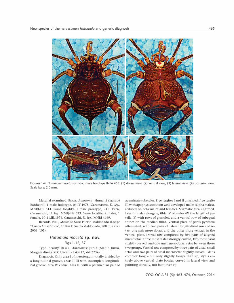
Figures 1-4. Hutamaia maceta sp. nov., male holotype INPA 453: (1) dorsal view; (2) ventral view; (3) lateral view; (4) posterior view.Scale bars: 2.0 mm.
1 2
3 4

466 A.L. Tourinho & A.C. Mendes
ZOOLOGIA 31 (5): 463–474, October, 2014
Description of male (holotype). Measurements: CL: 2.8;CW: 3.5; AL: 4.5; AW: 6.2. Legs. Pp: Tr: 0.8; Fe 1.9; Pa 0.8; Ti1.0; Ta: 0.9; Cl: 0.8; Leg I: Tr: 0.7; Fe: 4.1; Pa: 1.2; Tb: 2.6; Mt:4.6; Ta: 2.5. Leg II: Tr: 0.9; Fe: 8.7; Pa: 1.6; Ti: 6.4; Mt: 8.5; Ta:5.1. Leg III: Tr: 0.9; Fe: 8.6; Pa: 1.6; Ti: 6.5; Mt: 8.5; Ta: 2.2. LegIV: Tr: 1.5 Fe: 9.8 Pa: 2.2 Ti: 11.2 Mt: 9.9 Ta: 2.1. Dorsum (Figs1, 3-6): outline of dorsal scutum in dorsal view pyriform.Mesotergum divided into 4 areas. Ocularium low, armed witha dorsal pair of small median tubercles and several small dis-persed. Carapace with small granules in anterior margin andbehind the ocularium, smaller anteriorly and larger lateral toareas II and III. Lateral margin of dorsal scutum with a row ofyellowish white tubercles extending from carapace to area IV.Scutal area I with a posterior row of 8 yellowish white gran-
ules; area II with 1 row of yellowish white granules; area IIIwith 2 large spines (spiniform tubercles) and some yellowishwhite granules in lateral; area IV with a row of about 10 yel-lowish white granules. Free tergites I and II each with a trans-verse row of granules; free tergite III with a large spiniformposterior medial projection with apex bending. Dorsal analoperculum with a pair of granules. Venter (Figs 2, 4): stigmaticarea unarmed. Free sternites each with a transverse row of gran-ules. Ventral anal operculum with a posterior row of granules.Chelicerae (Figs 1-3): chelicera neither swollen nor elongate,with well-marked bulla with a little ectoproximal tubercle.Pedipalps (Figs 1, 5, 7-8): coxa with a ventral setiferous tu-bercle. Trochanter with 1 dorsal and 1 ventral setiferous tu-bercles. Femur with 1 ventroproximal setiferous tubercle and a
Figures 5-12. Hutamaia maceta sp. nov., male holotype INPA 453: (5-6) habitus, dorsal and lateral views, respectively; (7-8) pedipalps:(7) right, ectal view; (8) left, mesal view; (9-10) tibiae IV: (9) right, dorsal view; (10) left, retrolateral view; (11-12) penis dorsal andlateral views, respectively. Scale bars: 5, 6, 9, 10 = 2.0 mm; 7, 8 = 1.0 mm; 11, 12 = 0.2 mm.
5
7
8
9
11
10
6 12

467New species of the harvestmen Hutamaia and generic diagnosis
ZOOLOGIA 31 (5): 463–474, October, 2014
row of 4 minute ventroectal tubercles. Patella smooth andunarmed, with only minute scattered setiferous granules. Tibia:ectal (IiIi/IIi) and mesal (IiIi). Tarsus: ectal (IiIi); mesal (IiIi).Legs (Figs 1, 2, 5, 6, 9, 10): legs elongate, II and IV twice thelength of body. Coxa I with 2 dorsal tubercles; II with 1 ante-rior and 2 posterior dorsal tubercle, 1 retrolateral distalspiniform tubercle; coxa IV, in dorsal view, surpassing dorsalscutum, with numerous granules and 1 large spiniform curvedprolateral distal apophysis, posteriorly directed. Trochanters I-III with retrolateral setiferous tubercles, larger on III; trochanterIV with some small tubercles on both sides and 1 largeretrolateral distal apophysis, ventral scattered tubercles. FemoraI-III with several longitudinal rows of granules, each one witha larger retrolateral basal granule; femur IV straight in dorsalview, with rows of tubercles, more concentrated in distal por-tion, two ventral rows with larger tubercles. Patella IV withscattered tubercles. Tibia IV straight in lateral view, incrassatesub distally, with rows of granules, and a ventral row of subequalspines on the median third. Tarsal segmentation. 6(3)/8(3)/7/7. Penis (Figs 11, 12): ventral plate pyriform attenuated: long,with lateral margins subparallel, convex at base, tapering atdistal part. Distal border with small V-shaped cleft. Two pairsof lateral longitudinal rows of setae, one pair more dorsal andthe other more ventral in the ventral plate. Dorsal row com-posed by five pairs of aligned macrosetae: three most distalstrongly curved, two most basal slightly curved; and one smallmesodorsal setae between those two groups. Ventral row com-posed by three pairs of distal small setae and two pairs of basalmacrosetae slightly curved. Glans complex long – but onlyslightly longer than vp, stylus entirely above ventral plate bor-der, sinuous and pointing dorsally in lateral view, not bentover vp. Color (in ethanol) (Figs 1-4): Dorsal scutum darkbrown, with reticulated carapace and lateral groove, with yel-low spots on lateral and dorsal areas. Chelicerae brown, pedi-palps and trochanters I-III yellow; rest of the legs dark withfew light spots but tarsi yellow. All metatarsi with yellow rings.
Female. Measurements (female paratype, INPA 453): CL2.5; CW 3.3, AL 4.5, AW 6.2. Pp. Tr: 0.6; Fe: 1.6; Pa: 0.9; Ti: 1.1;Ta: 0.8; Cl: 0.7. Leg I. Tr: 0.7; Fe: 2.9; Pa: 1.1; Ti: 1.9; Mt: 3.7; Ta:2.0. Leg II. Tr: 0.9; Fm: 5.9; Pa: 1.3; Ti: 4.1; Mt: 5.2; Ta: 4.0. LegIII. Tr: 0.9; Fe: 4.5; Pa: 1.4; Ti: 2.6; Mt: 4.5; Ta: 2.0. Leg IV. Tr:0.9 Fe: 5.9; Pa: 1.6; Ti: 3.8; Mt: 6.3; Ta: 1.9. Description: ingeneral aspect, similar to the male, but dorsal tubercles of areaIII smaller. Free tergite III with very small apophysis. Distaltubercles of coxa IV and retrolateral spiniform apophysis oftrochanter IV smaller than in males. Ventral and conspicuoustubercles on tibia IV.
Type material. BRAZIL, Amazonas: Juruá (Médio Juruá,Margem direita RDS Uacari, -5.43917, -67.2734), 1 male holo-type + 1 male paratype, VIII.2005, Rohe, F. leg., INPA 453; Otherparatypes: (Paraná Teiú, -2.679, -65.64), 2 males + 1 female,26.IX.2003, nocturnal search, Venticinque, E. leg., MNRJ 4280.Tefé (Lago do Jacaré, -3.26, -64.628), 1 male, 21.IX.2003, noc-
turnal search, Venticinque, E. leg., INPA 160; (Guariba Solimões,-3.664, -64.17), 1 female, 22.IX.2003, nocturnal search,Venticinque, E. leg., INPA 166; 2 females, 19.IX.2003, noctur-nal search, Rego, F. leg., INPA 186. Codajás (Lago Cuxuará, -3.972, -61.96), 1 male, 26.IX.2003, nocturnal search,Venticinque, E. leg., INPA 211.
Distribution (Fig. 37). BRAZIL, Amazonas: Juruá, Tefé, Codajás.Etymology. The word “maceta” is used by the local people
of the Amazon basin to refer to extremely big things. Noun inapposition.
Hutamaia plei sp. nov.Figs 13-24, 37
Type locality. BRAZIL, Amazonas: Jutaí (Restinga do Cevalho).Diagnosis. Only area I of mesotergum divided by a longi-
tudinal groove, areas II-IV entire. Area III with paramedian pairof small tubercles. Free tergites I and II unarmed, free tergite IIIwith median spiniform apophysis, with a pair of small tubercleson basis, and one pair of blunt paralateral apophysis on the freetergite. Stigmatic area unarmed. Trochanter IV with smallretrolateral distal tubercle and one large spiniform ventro-retrolateral distal apophysis. Femur IV with ventrobasalspiniform apophysis. Legs not elongate, tibia IV of males twicethe size of patella IV, with rows of granules, ventral rows of acumi-nate tubercles. Ventral plate of penis, with basal portion nar-row, widening towards the apex, and abruptly forming laterallobes on medial portion. Insertion of ventral plate very basal inthe truncus of the penis. Two pairs of lateral longitudinal rowsof setae, one more dorsal and the other ventral in the ventralplate. Dorsal row composed by five aligned macrosetae: threepairs most distal straight, two pairs most basal straight and onthe lateral lobes; and one small mesodorsal setae between thosetwo groups. Ventral row composed by a distal group of two verysmall setae and a basal group of one macrosetae. Complex glansonly slightly longer than vp. Distal half of stylus surpass distalborder of ventral plate. Stylus slightly curved, pointing ventrally.Apex of stylus with small scales.
Description of male (holotype). Measurements: CL 1.9;CW 2.6, AL 3.0, AW. Pp: Tr: 0.5; Fe: 1.2; Pa: 0.7; Ti: 0.8; Ta: 0.8;Cl: 0.5. Leg I. Tr: 0.7; Fe: 2.0; Pa: 0.8; Ti: 1.3; Mt: 2.2; Ta: 1.4.Leg II. Tr: 0.6; Fe: 3.4; Pa: 0.8; Ti: 2.7; Mt: 2.9; Ta: 3.0. Leg III. Tr:0.8; Fe: 2.9; Pa: 0.8; Ti: 1.8; Mt: 2.8; Ta: 1.4. Leg IV. Tr: 1.0; Fe:3.4; Pa: 1.4; Ti: 2.9; Mt: 4.1; Ta: 1.6. Dorsum (Figs 13, 15, 16,17, 18): outline of dorsal scutum in dorsal view pyriform.Mesotergum divided into 4 very well-marked areas, areas II-IV.Ocularium low, armed with a dorsal pair of small median tu-bercles. Carapace with small granules in anterior margin andbehind the ocularium. Lateral margin of dorsal scutum withtwo rows of small yellowish white tubercles: the innermostcomposed by minute granules, extending from lateral ofocularium to of the area IV, the outermost extending from lat-eral of area I to area II, starting small and incrementing in size.Scutal area I with a posterior row of 6 yellowish white gran-

468 A.L. Tourinho & A.C. Mendes
ZOOLOGIA 31 (5): 463–474, October, 2014
ules, the median pair slightly larger than others; II-IV with 1row of small yellowish white granules each, II and IV withmedian pair slightly larger than others and area III with me-dian pair of acuminate tubercles; posterior margin with a rowof small granules. The holotype has a teratologic extra left halfof the posterior margin, before the entire posterior margin.Free tergites I and II unarmed, each with a row of small gran-ules; III with median spiniform apophysis, ventrally directed,with a pair of small tubercles on basis, and one pair of bluntparalateral apophysis on the free tergite. Dorsal anal opercu-lum with a pair of rounded tubercles, and smaller tuberclesscattered. Venter (Figs 14, 18): stigmatic area unarmed. Freesternites each with a transverse row of granules. Anal opercu-lum ventral with one posterior row of rounded tubercles, largerlaterally. Chelicerae (Figs 13, 14, 17, 18): neither swollen norelongate, with well-marked bulla with a little ectoproximaltubercle. Pedipalps (Figs 13, 14, 17, 19, 20): coxa with a ven-
tral setiferous tubercle. Trochanter with 1 ventral setiferoustubercle. Femur with 1 ventroproximal setiferous tubercle anda row of 3 minute ventroectal tubercles. Patella smooth andunarmed, with few scattered minute setiferous granules. Tibia:ectal (IIi) and mesal (II). Tarsus: ectal (IiIi); mesal (IiIi/IIi). Legs(Figs 13-15, 17, 18, 21, 22): coxa I with 2 dorsal tubercles; IIwith 1 dorsal anterior conspicuous tubercle and lower poste-rior tubercles, 1 retrolateral distal tubercle; coxa IV, in dorsalview, surpassing dorsal scutum, with numerous yellowish gran-ules and 1 large spiniform curved prolateral distal apophysis,posteriorly directed. Trochanters I-III with scattered granules;trochanter IV with small retrolateral distal tubercle and onelarge spiniform ventro-retrolateral distal apophysis. Femora I-III with several longitudinal rows of granules, II and III with alarger retrolateral basal granule; femur IV curved in dorsal view,with several rows of tubercles, two ventral with larger onesand with a ventrobasal spiniform apophysis. Patella IV with
Figures 13-16. Hutamaia plei sp. nov., male holotype INPA 014: (13) dorsal view; (14) ventral view; (15) lateral view; (16) posteriorview. Scale bars: 1.0 mm.
13
15
14
16

469New species of the harvestmen Hutamaia and generic diagnosis
ZOOLOGIA 31 (5): 463–474, October, 2014
scattered tubercles, the ventral larger and spiniform. Tibia IVstraight in lateral view, with rows of acuminate tubercles, theones of the ventro-retrolateral row much larger. Metatarsus andbasitarsus I slightly incrassate. Metatarsus IV with rows of tu-bercles, ventral row with larger spiniform tubercles, larger onbasal portion of podomere, decreasing in size towards apex.Tarsal segmentation. 4(2)/8(3)/6/6. Penis (Figs 23, 24): Ventralplate of penis, with basal portion narrow, widening towardsthe apex, and abruptly forming lateral lobes on medial por-tion. Insertion of ventral plate very basal in the truncus of thepenis. Distal border with small V-shaped cleft. Two pairs oflateral longitudinal rows of setae, one more dorsal and the other
ventral in the ventral plate. Dorsal row composed by fivealigned macrosetae (on the right half, the left half with sixpossibly due to teratologic multiplication on the distal groupof macrosetae): three most distal straight (on the right half,the left half with four), two pairs most basal straight and onthe lateral lobes; and one small mesodorsal setae between thosetwo groups. Ventral row composed by a distal group of twovery small setae and a basal group of one macrosetae. Com-plex glans only slightly longer than vp. Distal half of stylussurpass distal border of ventral plate. Stylus slightly curved,pointing ventrally. Apex of stylus with small scales. Color (inethanol) (Figs 13-16): dorsal scutum orangey brown, with re-
Figures 17-24. Hutamaia plei sp. nov., male holotype INPA 014: (17-18) habitus, dorsal and lateral views, respectively; (19-20) pedi-palps: (19) left, ectal view; (20) left, mesal view; (21-22) tibiae IV; (21) left, dorsal view; (22) right, prolateral view; (23-24) penis dorsaland lateral views, respectively. Scale bars: 17-22 = 1.0 mm, 23-24: 0.2 mm.
17
21 22
18
24
23
2019

470 A.L. Tourinho & A.C. Mendes
ZOOLOGIA 31 (5): 463–474, October, 2014
ticulated carapace and lateral groove, with yellow spots in lat-eral and dorsal areas, and region around mesotergal granulesseeming depigmented. Chelicerae dark yellow, pedipalps andtrochanters I-III pale yellow; rest of the legs dark yellow buttarsi pale yellow. Leg IV orangey brown. All metatarsi withyellow rings.
Female unknown.Type material. BRAZIL, Amazonas: Jutaí (Restinga do
Cevalho, -2.731, -66.916, 528 m), 1 male holotype, 16.IX.2003,nocturnal search, Venticinque, E. leg., INPA 14.
Distribution (Fig. 37). Known only from the type locality.Etymology. Latinization of the English word “play”,
which is used by the Brazilian opilionologist Adriano B. Kury,our friend and former tutor, in reference to anything he per-ceives to be extremely good. The name is to honor him forerecting Ampycinae. Noun in apposition.
Hutamaia trompsonica sp. nov.Figs 25-37
Type locality. BRAZIL, Pará: Gurupá (Furinho).Diagnosis. Areas I-IV divided, areas II-IV narrow, elevated
ridges. Area III with row of tubercles subequally sized. Free terg-ites I-III with a median large apophysis, I acuminate, II blunt,conic, dorsally directed, III blunt and conic ventrally directed.Stigmatic area unarmed. Legs of males not elongate, tibia IVless than 3X the length of patella IV, with rows of tubercles, aventral row of blunt tubercles on basal half, largest at the middleof row. Ventral plate of penis attenuated pyriform with twopairs of lateral longitudinal rows of setae, one pair more dorsaland the other more ventral in the ventral plate. Dorsal rowcomposed by five pairs of aligned macrosetae: three pairs mostdistal strongly curved, two pairs most distal slightly curved;and one small mesodorsal setae between those two groups.Ventral row composed by four pairs of distal setae: the threemost distal smaller and the most basal larger. Glans long,curved. Stylus does not surpass distal border of ventral plate,curved, pointing dorsalwards.
Description of male (holotype). Measurements: CL 2.0;CW 2.6, AL 3.2, AW 5.2. Pp. Tr: 0.5; Fe: 1.0; Pa: 0.7; Ti 0.8; Ta:0.8; Cl: 0.5. Leg I. Tr: 0.6; Fe: 2.2; Pa: 0.8; Ti: 1.4; Mt: 2.6; Ta: 1.4.Leg II. Tr: 0.7; Fe: 4.0; Pa: 1.0; Ti: 2.9; Mt: 3.6; Ta: 3.0. Leg III. Tr:0.7; Fe: 3.3; Pa: 0.9; Ti: 1.9; Mt: 2.7; Ta: 1.5. Leg IV. Tr: 0.6; Fe:4.0; Pa: 1.3; Ti: 3.6; Mt: 5.0; Ta: 1.6. Dorsum (Figs 25, 27, 28, 29-30): outline of dorsal scutum in dorsal view pyriform.Mesotergum divided into 4 not very well-marked areas, area II-IV as elevated ridges in posterior margin. Ocularium low, armedwith a dorsal pair of small median tubercles and several smalldispersed. Carapace with several small granules on anteriormargin, lateral and behind the ocularium. Lateral margin ofdorsal scutum with two rows of small yellowish white tubercles,the innermost extending from carapace to lateral of area III,outermost from carapace to posterior margin, tubercles largerlateral to areas II and III. Scutal area I with a posterior transver-
sal row of 9 yellowish white granules and several additional dis-persed; II-IV with 1 row of small yellowish white granules onelevated ridges; posterior margin with a row of small granules.Free tergite I with a row of small granules, the median largerand acuminate, II with a row of granules, bearing a median bluntand conic apophysis, dorsally directed, III with a row of smallgranules, with a median blunt and conic apophysis ventrallydirected. Dorsal anal operculum with a pair of small roundedtubercles. Venter (Figs 26, 28, 30): stigmatic area unarmed. Freesternites each with a transverse row of granules. Ventral analoperculum with posterior row of tubercles, larger laterally. Che-licerae (Figs 25, 26, 29): neither swollen nor elongate, with well-marked bulla with a little ectoproximal tubercle. Pedipalps (Figs31, 32): Coxa with a ventral setiferous tubercle. Trochanter with1 ventral setiferous tubercle. Femur with 1 ventroproximalsetiferous tubercle and a row of 2 very small ventroectal tubercles.Patella smooth and unarmed. Tibia: ectal (IIi/II) and mesal (IiIi/IiiIi). Tarsus: ectal (IiIi); mesal (IiIi). Legs (Figs 25-27, 29, 30, 33,34): legs of male not elongate. Coxa I with 2 dorsal tubercles; IIwith 1 anterior dorsal and two fused posterior tubercles, 1 smallretrolateral distal tubercle; coxa IV, in dorsal view, surpassingdorsal scutum, with numerous yellowish granules and 1 largespiniform curved prolateral distal apophysis, posteriorly directed.Trochanters I-III with scattered granules; IV with some smalltubercles in both sides and 1 prolateral distal apophysis. FemurI-III with several longitudinal rows of granules; femur IV curvedin dorsal view, with several rows of tubercles, the two ventralrows with larger ones. Patella IV with scattered tubercles,ventrodistal ones acuminate. Tibia IV curved in lateral view, lessthan 3X the length of patella IV, incrassate sub basally, withrows of tubercles, the ventral composed of blunt tubercles onbasal half, largest at the middle of row. Tarsal segmentation.6(3)/8(3)/7/7. Penis (Figs 35, 36): ventral plate pyriform attenu-ated: long, with lateral margins subparallel, convex at base, ta-pering at distal part. Distal border with small V-shaped cleft.Two pairs of lateral longitudinal rows of setae, one pair moredorsal and the other more ventral in the ventral plate. Dorsalrow composed by five aligned macrosetae (on the left half, theright half with six possibly due to teratologic multiplication onthe distal group of macrosetae): three most distal strongly curved(on the left half, the right half with four), two pairs most distalslightly curved; and one small mesodorsal setae between thosetwo groups. Ventral row composed by four pairs of distal setae:the three most distal smaller and the most basal larger. Glanslong, curved. Stylus does not surpass distal border of ventralplate, curved, pointing dorsalwards. Color (in ethanol) (Figs 25-28): dorsal scutum dark brown, with reticulated carapace andlateral groove. Chelicerae reticulated of brown, pedipalps andtrochanters I-III yellow; rest of the legs dark but tarsi yellow. Allmetatarsi with yellow rings.
Female. Measurements (female paratype, INPA 261): CL1.7; CW 2.4, AL 3.0, AW 4.8. Pp. Tr: 0.5; Fe: 0.9; Pa: 0.6; Ti: 0.7;Ta: 0.6; Cl: 0.5. Leg I. Tr: 0.5; Fe: 1.9; Pa: 0.8; Ti: 1.3; Mt: 1.8; Ta:
471New species of the harvestmen Hutamaia and generic diagnosis
ZOOLOGIA 31 (5): 463–474, October, 2014
1.2. Leg II. Tr: 0.7; Fm: 3.6; Pa: 0.9; Ti: 2.5; Mt: 3.4; Ta: 2.7. LegIII. Tr: 0.8; Fe: 3.1; Pa: 1.0; Ti: 1.6; Mt: 2.7; Ta: 1.5. Leg IV. Tr:0.9 Fe: 3.6; Pa: 1.0; Ti: 2.5; Mt: 3.9; Ta: 1.5. Description: ingeneral aspect, similar to the male, but free tergite Iwith a rowof granules equally sized, lacking a median larger and acumi-nate. Free tergites II and III with median larger and acuminatearmature, but relatively smaller than in males.
Type material. BRAZIL, Pará: Gurupá (Furinho, -1.204, -51.818), 1 male holotype, 18.IX.2003, Rheims, C. leg., INPA 118.Paratypes: same locality of holotype, 1 male, 25.IX.2003, Rego,F. leg., INPA 137. Amazonas: Manacapuru (Lago do Piranha, -3.403, -60.96354), 1 female, without collection date, Tourinho,A.L. leg, INPA 261. Codajás (Lago Cuxuará, -3.972, -61.996), 1male, 26.IX.2003, Rego, F. leg., INPA 200. Coari (Tocari-SolimõesII, -3.894, -62.855), 1 male, 25.IX.2003, Venticinque, E. leg., MNRJ4281. All from nocturnal collecting.
Distribution (Fig. 37). BRAZIL, Amazonas: Coari (Tocari-Solimões), Codajás (Lago Cuxuará), Manacapuru (Lago do Pi-ranha); Pará: Gurupá (Furinho).
Etymology. “trompsonica” is a word made up by AdrianoKury to express all kinds of ordinary things. Following his us-age as an adjective in Portuguese, latinization as follows: f.trompsonica, m. trompsonicus, n. trompsonicum.
DISCUSSION
Ampycinae comprises 13 genera and 28 species, includ-ing the three new species herein described. Its monophyly issupported by two morphological characters, the presence of adeep cleft on the distal margin of the ventral plate of the penisand a free tergite III armed with spine (KURY 2003). The mono-phyly of Ampycinae was also recovered by molecular analysis
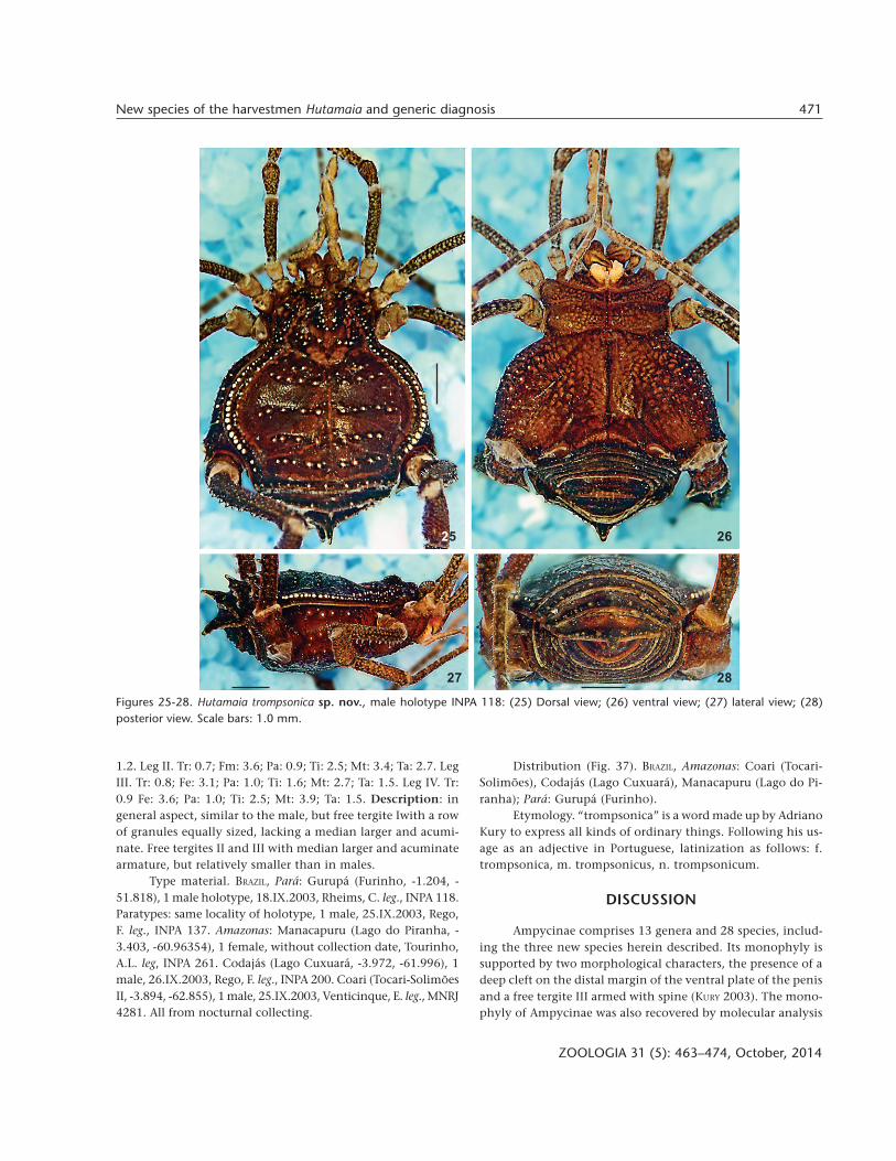
Figures 25-28. Hutamaia trompsonica sp. nov., male holotype INPA 118: (25) Dorsal view; (26) ventral view; (27) lateral view; (28)posterior view. Scale bars: 1.0 mm.
25 26
27 28
472 A.L. Tourinho & A.C. Mendes
ZOOLOGIA 31 (5): 463–474, October, 2014
(PINTO-DA-ROCHA et al. 2013). Species of this subfamily are dis-tributed in the Brazilian Amazon, Ecuador and Peru (Ampycus,Hexabunus, Hutamaia, Sibollus, and Thaumatopachylus), Pacificforest of Ecuador (Ampycella), Central America (Glysterus,Hernandarioides, Nesopachylus, and Parahernandria) and Ecua-dorian/Peruvian montane forest of the Andes (Neopachyloidesand Pirunipygus).
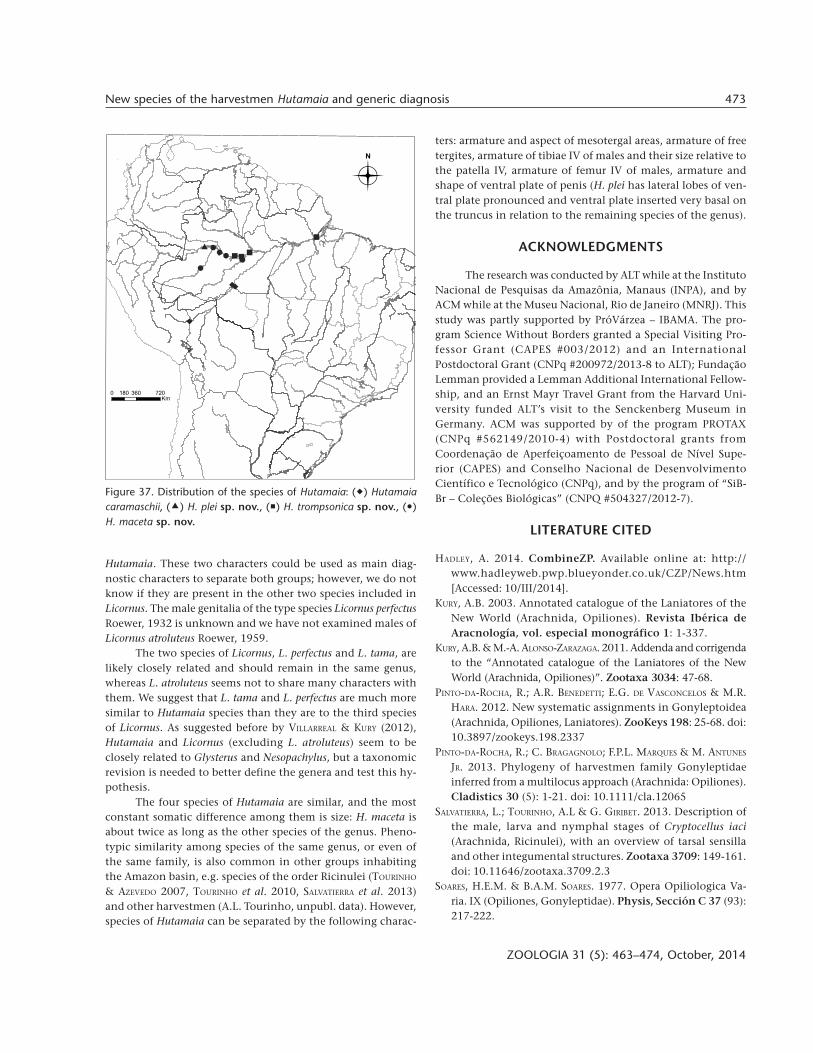
SOARES & SOARES (1977) described Hutamaia for Hutamaiacaramaschii Soares & Soares, 1977, which was only known fromthe type locality, Humaitá. KURY (2003) added a record fromMadre de Dios, Peru. In this paper, we expand the distributionof the genus to the following localities in the Northern por-tion of Brazil: Coari, Codajás, Juruá, Jutaí, Manacapuru, Tefé(state of Amazonas) and Gurupá (state of Pará). This expanded
distribution indicates that Hutamaia is widespread in the Bra-zilian and Peruvian Amazon.
The species included in Ampycinae, however, have neverbeen subjected to taxonomic revisions. The diagnoses of mostspecies and genera are inadequate according to current stan-dards, being composed of characters that are mostly useless atthe generic and suprageneric levels, as it is the case withHutamaia. The Amazonian Hutamaia and the Andean Licornusspecies are morphologically very similar; there is not a combi-nation of non-genital characters that may be used to supportthem as separate genera. On the other hand, the ventral plateof the penis of Licornus tama Villarreal & Kury, 2012 is muchnarrower than the ventral plate of Hutamaia species; in L. tamathe penis also has a dorsal process that is not present in
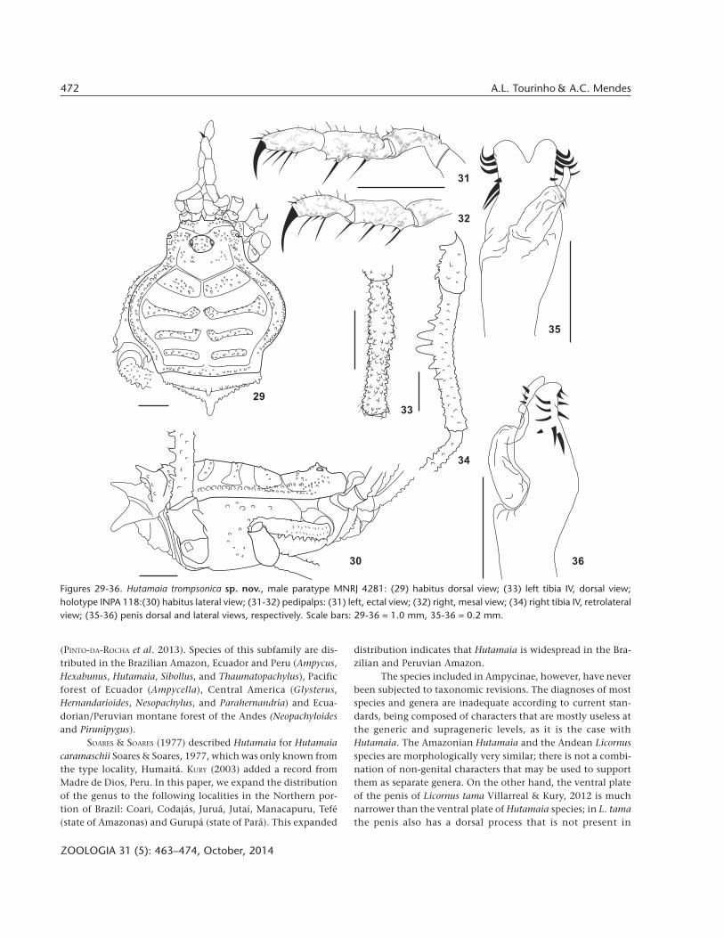
Figures 29-36. Hutamaia trompsonica sp. nov., male paratype MNRJ 4281: (29) habitus dorsal view; (33) left tibia IV, dorsal view;holotype INPA 118:(30) habitus lateral view; (31-32) pedipalps: (31) left, ectal view; (32) right, mesal view; (34) right tibia IV, retrolateralview; (35-36) penis dorsal and lateral views, respectively. Scale bars: 29-36 = 1.0 mm, 35-36 = 0.2 mm.
29
30
33
34
36
35
32
31
473New species of the harvestmen Hutamaia and generic diagnosis
ZOOLOGIA 31 (5): 463–474, October, 2014
Hutamaia. These two characters could be used as main diag-nostic characters to separate both groups; however, we do notknow if they are present in the other two species included inLicornus. The male genitalia of the type species Licornus perfectusRoewer, 1932 is unknown and we have not examined males ofLicornus atroluteus Roewer, 1959.
The two species of Licornus, L. perfectus and L. tama, arelikely closely related and should remain in the same genus,whereas L. atroluteus seems not to share many characters withthem. We suggest that L. tama and L. perfectus are much moresimilar to Hutamaia species than they are to the third speciesof Licornus. As suggested before by VILLARREAL & KURY (2012),Hutamaia and Licornus (excluding L. atroluteus) seem to beclosely related to Glysterus and Nesopachylus, but a taxonomicrevision is needed to better define the genera and test this hy-pothesis.
The four species of Hutamaia are similar, and the mostconstant somatic difference among them is size: H. maceta isabout twice as long as the other species of the genus. Pheno-typic similarity among species of the same genus, or even ofthe same family, is also common in other groups inhabitingthe Amazon basin, e.g. species of the order Ricinulei (TOURINHO
& AZEVEDO 2007, TOURINHO et al. 2010, SALVATIERRA et al. 2013)and other harvestmen (A.L. Tourinho, unpubl. data). However,species of Hutamaia can be separated by the following charac-
ters: armature and aspect of mesotergal areas, armature of freetergites, armature of tibiae IV of males and their size relative tothe patella IV, armature of femur IV of males, armature andshape of ventral plate of penis (H. plei has lateral lobes of ven-tral plate pronounced and ventral plate inserted very basal onthe truncus in relation to the remaining species of the genus).
ACKNOWLEDGMENTS
The research was conducted by ALT while at the InstitutoNacional de Pesquisas da Amazônia, Manaus (INPA), and byACM while at the Museu Nacional, Rio de Janeiro (MNRJ). Thisstudy was partly supported by PróVárzea – IBAMA. The pro-gram Science Without Borders granted a Special Visiting Pro-fessor Grant (CAPES #003/2012) and an InternationalPostdoctoral Grant (CNPq #200972/2013-8 to ALT); FundaçãoLemman provided a Lemman Additional International Fellow-ship, and an Ernst Mayr Travel Grant from the Harvard Uni-versity funded ALT’s visit to the Senckenberg Museum inGermany. ACM was supported by of the program PROTAX(CNPq #562149/2010-4) with Postdoctoral grants fromCoordenação de Aperfeiçoamento de Pessoal de Nível Supe-rior (CAPES) and Conselho Nacional de DesenvolvimentoCientífico e Tecnológico (CNPq), and by the program of “SiB-Br – Coleções Biológicas” (CNPQ #504327/2012-7).
LITERATURE CITED
HADLEY, A. 2014. CombineZP. Available online at: http://www.hadleyweb.pwp.blueyonder.co.uk/CZP/News.htm[Accessed: 10/III/2014].
KURY, A.B. 2003. Annotated catalogue of the Laniatores of theNew World (Arachnida, Opiliones). Revista Ibérica deAracnología, vol. especial monográfico 1: 1-337.
KURY, A.B. & M.-A. ALONSO-ZARAZAGA. 2011. Addenda and corrigendato the “Annotated catalogue of the Laniatores of the NewWorld (Arachnida, Opiliones)”. Zootaxa 3034: 47-68.
PINTO-DA-ROCHA, R.; A.R. BENEDETTI; E.G. DE VASCONCELOS & M.R.HARA. 2012. New systematic assignments in Gonyleptoidea(Arachnida, Opiliones, Laniatores). ZooKeys 198: 25-68. doi:10.3897/zookeys.198.2337
PINTO-DA-ROCHA, R.; C. BRAGAGNOLO; F.P.L. MARQUES & M. ANTUNES
JR. 2013. Phylogeny of harvestmen family Gonyleptidaeinferred from a multilocus approach (Arachnida: Opiliones).Cladistics 30 (5): 1-21. doi: 10.1111/cla.12065
SALVATIERRA, L.; TOURINHO, A.L & G. GIRIBET. 2013. Description ofthe male, larva and nymphal stages of Cryptocellus iaci(Arachnida, Ricinulei), with an overview of tarsal sensillaand other integumental structures. Zootaxa 3709: 149-161.doi: 10.11646/zootaxa.3709.2.3
SOARES, H.E.M. & B.A.M. SOARES. 1977. Opera Opiliologica Va-ria. IX (Opiliones, Gonyleptidae). Physis, Sección C 37 (93):217-222.
Figure 37. Distribution of the species of Hutamaia: ( ) Hutamaiacaramaschii, ( ) H. plei sp. nov., ( ) H. trompsonica sp. nov., ( )H. maceta sp. nov.

474 A.L. Tourinho & A.C. Mendes
ZOOLOGIA 31 (5): 463–474, October, 2014
TOURINHO, A.L. & A. PÉREZ. 2006. On the family FissiphalliidaeMartens, 1988, with description of two new Amazonianspecies. Zootaxa 1325: 235-254.
TOURINHO, A.L & C.S. AZEVEDO. 2007. A new Amazonian CryptocellusWestwood (Arachnida, Ricinulei). Zootaxa 1540: 55-60.
TOURINHO, A.L.; N.F.L. MAN-HUNG & A.B. BONALDO. 2010. A new
Submitted: 19.III.2014; Accepted: 16.VIII.2014.Editorial responsibility: Antonio D. Brescovit
species of Ricinulei of the genus Cryptocellus Westwood(Arachnida) from northern Brazil. Zootaxa 2684: 63-68.
VILLARREAL, O. & A.B. KURY. 2012. Licornus Roewer, 1932: newlytransferred to Ampycinae and first record of the familyGonyleptidae (Opiliones: Laniatores) from Venezuela.Zootaxa 3544: 71-78.


