New species of Mycosphaerella from Myrtaceae in ...
14
New species of Mycosphaerella from Myrtaceae in plantations and native forests in eastern Australia Angus J. Carnegie 1 Forest Resources Research, NSW Department of Primary Industries, PO Box 100, Beecroft, New South Wales 2119, Australia Treena I. Burgess School of Biological Sciences and Biotechnology, Murdoch University, Perth, WA 6150, Australia Vyrna Beilharz Primary Industries Research Victoria, Department of Primary Industries, Knoxfield, Victoria 3180, Australia Michael J. Wingfield Forestry and Agricultural Biotechnology Institute, Faculty of Natural and Agricultural Sciences, University of Pretoria, Pretoria, South Africa Abstract: The majority of Mycosphaerella species from eucalypts (Eucalyptus, Corymbia and Angophora) in Australia have been recorded only from trees growing in plantations. This illustrates a bias in research in the past two decades toward commercial enterprise, and it emphasises a lack of understanding of the occurrence of these important fungi under natural conditions. Surveys of foliar fungi in native forests in eastern Australia, as well as adjacent plantations, thus have been initiated in recent years. In this study we describe four new species of Mycosphaerella from Eucalyptus spp. as well as other Myrtaceae. Mycosphaerella tumulosa sp. nov. (ana- morph: Pseudocercospora sp.) was found on more than seven species of Eucalyptus and Corymbia in native forests and plantations in northeastern New South Wales and southeastern Queensland and appears to be relatively common, although not damaging to these trees. Mycosphaerella multiseptata sp. nov. was recorded from several locations on species of Angophora in native forests and amenity plantings. Mycosphaerella pseudovespa sp. nov. was found in one location in native forest on E. biturbinata. The first species of Mycosphaerella to be described from Syncarpia, M. syncarpiae sp. nov., was found in native forests in numerous locations from Sydney through to northeastern New South Wales and appears to be relatively common. Key words: Angophora, Corymbia, Eucalyptus, ITS, Mycosphaerella, Pseudocercospora, Syncarpia, taxonomy INTRODUCTION The family Myrtaceae incorporates a large number of tree genera, including Eucalyptus, Corymbia, Ango- phora and Syncarpia. More than 700 species are accommodated in these four genera, the majority endemic to Australia (Pryor and Johnson 1971, Boland et al 1992), and they are the dominant tree species over most of the Australian landscape. Many species of Eucalyptus and Corymbia are grown widely in Australia and elsewhere for commercial purposes and as amenity trees, while Syncarpia is commercially important in eastern Australia. More than 500 species of leaf-infecting fungi have been recorded from eucalypts (including the genera Eucalyptus and Corymbia) (Sankaran et al 1995). Aulographina eucalypti (Cooke & Massee) Arx & E. Mu ¨ll. and species of Cylindrocladium Morgan, Myco- sphaerella Johansen and its associated anamorphs are considered to be the most common and numerous (Park et al 2000). Some of these fungi are serious pathogens, particularly of plantation-grown Eucalyp- tus spp. In this regard these fungi threaten the economic viability of paper and pulp industries based on fiber from these trees. More than 60 species of Mycosphaerella have been described from eucalypts in Australia and elsewhere (Park and Keane 1982, Carnegie and Keane 1994, 1998, Crous 1998, Maxwell et al 2003, Crous et al 2004, 2006, 2007, Hunter et al 2004, Burgess et al 2006). Most of these have been described from plantations but have not been found in native forests. This is not surprising because most studies on Mycosphaerella in Australia in the past two decades have concentrated on eucalypt plantations (e.g. Park 1984, Carnegie 1991, 2000, Maxwell 2004, Barber 2005, Milgate 2005). Only three species are common- ly found in native forests in Australia: M. cryptica, which occurs on many hosts, the anamorph of M. swartii (Sonderhenia eucalyptorum [Hansf.] H.J. Swart & J. Walker), which occurs on several hosts, and M. nubilosa on E. globulus (Park et al 2000, Carnegie pers obs). More than 20 species of Pseudocercospora Deighton also have been described from Eucalyptus spp. (Crous et al 1989, Crous 1998, Yuan et al 2000, Braun and Accepted for publication 2 February 2007. 1 Corresponding author. E-mail: [email protected] Mycologia, 99(3), 2007, pp. 461–474. # 2007 by The Mycological Society of America, Lawrence, KS 66044-8897 461
Transcript of New species of Mycosphaerella from Myrtaceae in ...

New species of Mycosphaerella from Myrtaceae in plantations and native forestsin eastern Australia
Angus J. Carnegie1
Forest Resources Research, NSW Department of PrimaryIndustries, PO Box 100, Beecroft, New South Wales2119, Australia
Treena I. BurgessSchool of Biological Sciences and Biotechnology,Murdoch University, Perth, WA 6150, Australia
Vyrna BeilharzPrimary Industries Research Victoria, Department ofPrimary Industries, Knoxfield, Victoria 3180, Australia
Michael J. WingfieldForestry and Agricultural Biotechnology Institute,Faculty of Natural and Agricultural Sciences,University of Pretoria, Pretoria, South Africa
Abstract: The majority of Mycosphaerella speciesfrom eucalypts (Eucalyptus, Corymbia and Angophora)in Australia have been recorded only from treesgrowing in plantations. This illustrates a bias inresearch in the past two decades toward commercialenterprise, and it emphasises a lack of understandingof the occurrence of these important fungi undernatural conditions. Surveys of foliar fungi in nativeforests in eastern Australia, as well as adjacentplantations, thus have been initiated in recent years.In this study we describe four new species ofMycosphaerella from Eucalyptus spp. as well as otherMyrtaceae. Mycosphaerella tumulosa sp. nov. (ana-morph: Pseudocercospora sp.) was found on more thanseven species of Eucalyptus and Corymbia in nativeforests and plantations in northeastern New SouthWales and southeastern Queensland and appears tobe relatively common, although not damaging tothese trees. Mycosphaerella multiseptata sp. nov. wasrecorded from several locations on species ofAngophora in native forests and amenity plantings.Mycosphaerella pseudovespa sp. nov. was found in onelocation in native forest on E. biturbinata. The firstspecies of Mycosphaerella to be described fromSyncarpia, M. syncarpiae sp. nov., was found in nativeforests in numerous locations from Sydney through tonortheastern New South Wales and appears to berelatively common.
Key words: Angophora, Corymbia, Eucalyptus,
ITS, Mycosphaerella, Pseudocercospora, Syncarpia,taxonomy
INTRODUCTION
The family Myrtaceae incorporates a large number oftree genera, including Eucalyptus, Corymbia, Ango-phora and Syncarpia. More than 700 species areaccommodated in these four genera, the majorityendemic to Australia (Pryor and Johnson 1971,Boland et al 1992), and they are the dominant treespecies over most of the Australian landscape. Manyspecies of Eucalyptus and Corymbia are grown widelyin Australia and elsewhere for commercial purposesand as amenity trees, while Syncarpia is commerciallyimportant in eastern Australia.
More than 500 species of leaf-infecting fungi havebeen recorded from eucalypts (including the generaEucalyptus and Corymbia) (Sankaran et al 1995).Aulographina eucalypti (Cooke & Massee) Arx & E.Mull. and species of Cylindrocladium Morgan, Myco-sphaerella Johansen and its associated anamorphs areconsidered to be the most common and numerous(Park et al 2000). Some of these fungi are seriouspathogens, particularly of plantation-grown Eucalyp-tus spp. In this regard these fungi threaten theeconomic viability of paper and pulp industries basedon fiber from these trees.
More than 60 species of Mycosphaerella have beendescribed from eucalypts in Australia and elsewhere(Park and Keane 1982, Carnegie and Keane 1994,1998, Crous 1998, Maxwell et al 2003, Crous et al2004, 2006, 2007, Hunter et al 2004, Burgess et al2006). Most of these have been described fromplantations but have not been found in native forests.This is not surprising because most studies onMycosphaerella in Australia in the past two decadeshave concentrated on eucalypt plantations (e.g. Park1984, Carnegie 1991, 2000, Maxwell 2004, Barber2005, Milgate 2005). Only three species are common-ly found in native forests in Australia: M. cryptica,which occurs on many hosts, the anamorph of M.swartii (Sonderhenia eucalyptorum [Hansf.] H.J. Swart& J. Walker), which occurs on several hosts, and M.nubilosa on E. globulus (Park et al 2000, Carnegie persobs).
More than 20 species of Pseudocercospora Deightonalso have been described from Eucalyptus spp. (Crouset al 1989, Crous 1998, Yuan et al 2000, Braun and
Accepted for publication 2 February 2007.1 Corresponding author. E-mail: [email protected]
Mycologia, 99(3), 2007, pp. 461–474.# 2007 by The Mycological Society of America, Lawrence, KS 66044-8897
461
Dick 2002, Crous et al 2004, 2007, Hunter et al 2006),the majority of which are known only from countriesother than Australia. Many of these have been linkedto Mycosphaerella teleomorphs (e.g. Crous 1998,Burgess et al 2007) and others are known to belinked to this genus based on phylogenetic inference(Crous et al 2001, 2004). A number of other species ofPseudocercospora currently are being described asa result of detailed studies of this genus in Australia(V. Beilharz unpubl). Of the numerous species ofPseudocercospora detected on Eucalyptus in countriesoutside Australia, only two species, found in NewZealand, are known to occur in Australia: P. subulataZ.Q. Yuan, D. de Little & C. Mohammed (as P.pseudobasitruncata U. Braun & M. Dick) and P. crousiiU. Braun & M. Dick. Pseudocercospora subulata occurson E. nitens in both countries, while P. crousii, foundon a number of Eucalyptus spp. including E. regnansin New Zealand, has been identified on E. regnansin eastern Australia. Only P. eucalyptorum howeveris considered a significant pathogen of eucalypts.It occurs mostly in Eucalyptus plantations (Crouset al 1989, Park et al 2000) and is not known inAustralia.
Twelve foliar fungi have been identified fromSyncarpia, most from single collections, and lodgedin Australian herbaria (www.planthealthaustralia.com.au). Few species of foliar fungi have beendescribed from Angophora (Park et al 2000) and only10 are lodged in Australian herbaria (www.planthealthaustralia.com.au). In a detailed study ofthe then seven species of Mycosphaerella and relatedanamorphs from Myrtaceae, excluding Eucalyptus,Crous (1999) did not report any species fromSyncarpia and only one species, M. angophorae Hansf.,was described from Angophora. One other species hasbeen reported from Myrtaceae, namely M. melaleu-coides Sivan. & R.G. Shivas, from native Melaleuca inQueensland (Sivanesan and Shivas 2002).
Because of the bias of surveys toward Eucalyptusspp. in plantations, and thus the large number ofidentified species from this artificially establishedenvironment, the first author has made a purposefuleffort to survey the species of Mycosphaerella in nativeforests of eastern Australia. This has revealed severalundescribed species of Mycosphaerella and theiranamorphs that are found in both native forests andplantations. The aim of this study is to provide namesfor these fungi and to discuss their relative signifi-cance.
MATERIALS AND METHODS
Sample collection.—Samples of diseased leaves were collect-ed from various eucalypt plantations and native forests
throughout eastern Australia over several years. These wereplaced in paper bags, pressed and kept cool duringtransportation back to the laboratory, where they werestored at 4 C.
Examination of samples and isolation from samples.—Lesioncharacteristics, including size and color, were recorded foreach fungal species on each host. Pseudothecia wereremoved from diseased leaves and mounted in ammoniacalCongo red, lactic acid or acid fuchsin, squashed undera cover slip and examined with a compound microscope.Thin sections through pseudothecia were examined ina similar manner. Ascospore germination patterns wereobserved by excising mature pseudothecia from lesions onfresh leaves and attaching these to the undersides of 90 mmPetri dish lids with double-sided tape. The lesions weremoistened for up to 2 h, blotted dry, then inverted over 2%
malt-extract agar (MEA) in the Petri dish base and placedon a laboratory bench at room temperature for 24 h. Slabsof agar were cut from the dishes after 24 h, placed ona microscope slide, stained with ammoniacal Congo red,covered with a cover slip and examined with a compoundmicroscope. Ascospore germination Type (A–N) was de-scribed following the definitions of Crous (1998). Furtheragar slabs were observed after 12 h and 36 h so thatvariation in germination patterns could be observed overtime.
Germinating ascospores were removed from MEA plateswith a sterile needle, transferred to fresh Petri dishescontaining 2% MEA and incubated in the dark at 25 C(single-spore cultures). Plates were examined at weeklyintervals and linear growth determined after 1 mo. Culturecolor was rated with the aid of color charts (Kornerup andWanscher 1983). Mycelium was removed with a sterileneedle from the edges of single spore cultures, replatedon carnation leaf agar (CLA) and incubated undercontinuous near-ultraviolet light at room temperature.Plates were examined for the presence of anamorphs atweekly intervals or until 3 mo. Specimens and cultures usedin this study are kept at herbaria DAR (Orange AgriculturalInstitute, Orange, NSW, Australia), VPRI (Victorian De-partment of Primary Industries, Knoxfield, Australia) andNSWF (State Forests of NSW, Sydney, Australia), with ex-typecultures also lodged at Centraalbureau voor Schimmelcul-tures (CBS).
Molecular phylogenetic characterization.—For each isolateapproximately 50 mg of fungal mycelium was scraped fromthe surface of 21 d old cultures, ground with a glass rod,suspended in 200 mL of DNA extraction buffer (200 mMTris-HCL pH 8.0, 150 mM NaCl, 25 mM EDTA, 0.5% SDS)and incubated 1 h at 70 C. DNA was purified with theUltrabindH DNA purification kit according to the manu-facture’s instructions (MO BIO Laboratories, Solana Beach,California). A part of the internal transcribed spacer (ITS)region of the ribosomal DNA operon was amplified withthe primers ITS-1F (59CTTGGTCATTTAGAGGAAGTAA)(Gardes and Bruns 1993) and ITS4 (59TCCTCCGCTTATT-GATATGC) (White et al 1990). The PCR reaction mixture(25 mL), PCR conditions and visualization of products wereas described by Burgess (et al 2001) except that 1 U of Taq
462 MYCOLOGIA

polymerase (Biotech International, Needville, Texas) wasused in each reaction. PCR products were cleaned withUltrabindH DNA purification kit (MO BIO Laboratories).Products were sequenced with the BigDye terminator cyclesequencing kit (PE Applied Biosystems, Foster City,California) with the same primers that were used in theinitial amplification. The products were separated with anABI 3730?48 capillary sequencer (Applied Biosystems) usinga BioRad Biofocus 2000 capillary gel electrophoresis system.
To compare Mycosphaerella isolates in this study withother Mycosphaerella spp., ITS rDNA sequences obtainedfrom GenBank, including isolates used in recent studies(Burgess et al 2006, Cortinas et al 2006, Crous et al 2006,Crous et al 2004, Crous et al 2001, Hunter et al 2004,Maxwell et al 2003), were used in the phylogenetic analysis(culture number, identity and GenBank number are avail-able at TreeBASE SN3238). ITS trees were rooted withNeofusicoccum ribis as outgroup taxon.
Sequence data were analysed with Sequence Navigatorversion 1.0.1TM (Perkin Elmer Corp. Foster City, California)and manually aligned by inserting gaps. Gaps were treatedas a fifth character; all ambiguous characters and parsimonyuninformative characters were excluded before analysis.The most parsimonious trees were obtained with heuristicsearches with random stepwise addition in 100 replicates,with the tree bisection-reconnection branch-swapping op-tion on and the steepest-descent option off. MAXTREES wereunlimited, branches of zero length were collapsed and allmultiple equally parsimonious trees were saved. Estimatedlevels of homoplasy and phylogenetic signal (retention andconsistency indices) were determined (Hillis and Huelsen-beck 1992). Branch and branch node supports weredetermined with 1000 bootstrap replicates (Felsenstein1985).
RESULTS
Phylogenetic analyses.—A BLAST search was firstconducted on GenBank to compare the ITS se-quences for the Mycosphaerella spp. collected in thisstudy (TABLE I) with those for existing species thathave been lodged in GenBank. The four purportednew species were closely related to but different from
currently described Mycosphaerella spp. The sequencefor these species then was compared with otherMycosphaerella spp. isolated from eucalypts. Due tothe large number of species, two separate analyseswere conducted, both containing representative iso-lates from the different phylogenetic lineages inMycosphaerella (Burgess et al 2006, Crous et al2006). The first aligned dataset, concentrating onthe ‘nubilosa clade’, consisted of 572 characters ofwhich 191 were parsimony informative. The datasetcontained significant phylogenetic signal comparedto 1000 random trees (P , 0.01, g1 5 21.33).Heuristic searches in PAUP resulted in one mostparsimonious tree of 582 steps (CI 5 0.59, RI 5 0.79)(FIG. 1, TreeBASE SN3238-13881). Two of the newMycosphaerella spp. treated in this study were found instrongly supported (100% bootstrap) terminal cladesand appeared to be distinct from all known relatedMycosphaerella spp. Mycosphaerella sp. from Syncarpiaand Mycosphaerella sp. from Angophora are mostsimilar primarily to each other, although bootstrapanalysis does not support subgroupings within the‘nubilosa clade’ (FIG. 1)
The second aligned dataset, concentrating onspecies with Pseudocercospora anamorphs, consistedof 586 characters of which 172 were parsimonyinformative. The dataset contained significant phylo-genetic signal compared to 1000 random trees (P ,
0.01, g1 5 20.57). Initial heuristic searches ofunweighted characters in PAUP resulted in 1000most parsimonious trees of 676 steps (CI 5 0.51, RI 5
0.73). Characters were weighted according to the CI(32 characters had a weight of 1, 140 a weight of ,1)and subsequent heuristic searches resulted in 30 mostparsimonious trees of 348 steps (CI 5 0.60, RI 5
0.78). (FIG. 2, TreeBASE SN3238-13882). Mycosphaer-ella sp. from E. moluccana resided in a stronglysupported (100% bootstrap) terminal clade. Com-pared to other Pseudocercospora anamorphs, which areclosely related to each other, this species differed by
TABLE I. Isolates considered in the study
Culture no. teleomorph anamorph Host Location CollectorGenBank
accession No.
DAR 77433 M. syncarpiae Syncarpia glomulifera New South Wales AJ Carnegie DQ530219NSWF 005320 M. syncarpiae S. glomulifera New South Wales AJ Carnegie DQ530220DAR 77439 M. multiseptata Angophora subvelutina New South Wales AJ Carnegie DQ530223DAR 77438 M. multiseptata A. subvelutina New South Wales AJ Carnegie DQ530224DAR 77440 M. multiseptata A. costata New South Wales AJ Carnegie DQ530225DAR 77432 M. pseudovespa E. biturbinata New South Wales AJ Carnegie DQ530216DAR 77424 M. tumulosa P. tumulosa E. moluccana Queensland AJ Carnegie DQ530217NSWF 005313 M. tumulosa P. tumulosa E. moluccana Queensland AJ Carnegie DQ530218
CARNEGIE ET AL: NEW MYCOSPHAERELLA SPECIES 463

FIG. 1. A phylogram of the most parsimonious tree of 582 steps obtained from ITS sequence data, indicating the placementof two new Mycosphaerella spp. in bold. Bootstrap support is given above the branches. The trees were rooted toNeofusicoccum ribis.
464 MYCOLOGIA
at least 12 steps (FIG. 2). Mycosphaerella spp. associat-ed with wasp galls on E. biturbinata was related to butdifferent from M. citri, which is not known to bea pathogen of Eucalyptus, M. obscures, which wasisolated from eucalypts in southeastern Asia, and anundescribed Mycosphaerella sp. (FIG. 2).
Based on DNA sequence comparisons the fourMycosphaerella spp. found in this study clearlyrepresent discrete taxa. These fungi also could bediscriminated from species to which they are mostclosely related with morphological characteristics. Wetherefore describe them as new in the followingtaxonomic treatments.
TAXONOMY
Mycosphaerella tumulosa Carnegie & Beilharz sp.nov. FIGS. 3–8
MycoBank MB500742Anamorph: Pseudocercospora sp.
Etymology. Referring to the mounded shape of theculture (tumulosus, mounded).
Mycosphaerella marksii subsimilis, differt ascosporis grandi-oribus 15–19 3 4.5–5 mm (M. marksii 11–16 3 2–3 mm) etcum cellvla apicali asymmetrica rariore, etcoloniis in agarotardioribus crescentibus (M. tumulosa 9–12 mm, M. marksii24–29 mm, post mensam unam).
Pseudocercosporae crousii subsimilis sed conidiis minusobclavatis, conidiophoris simplicibus plerumque irregular-ibus et brevioribus (conidiophora in P. tumulosa 26–60 mmlonga, in P. crousii 5–100 mm longa) differt.
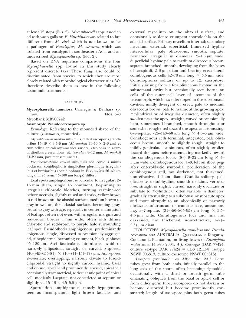
Leaf spots amphigenous, subcircular to irregular, 2–15 mm diam, single to confluent, beginning asirregular chlorotic blotches, turning carmine-redbefore necrosis, slightly raised and corky, yellow-brownto red-brown on the abaxial surface, medium brown togray-brown on the adaxial surface, becoming gray-brown to gray with age, especially in center, maturationof leaf spot often not even, with irregular margins andred-brown border 1 mm wide, often with diffusechlorotic and red-brown to purple halo surroundingleaf spot. Pseudothecia amphigenous, predominantlyepigenous, single, dispersed to occasionally aggregat-ed, subepidermal becoming erumpent, black, globose,95–120 mm. Asci fasciculate, bitunicate, ovoid tonarrowly ellipsoidal, straight or curved, 8-spored,(40–)45–65(–81) 3 (10–)11–15(–17) mm. Ascospores2–3-seriate, overlapping, narrowly clavate to fusoid-ellipsoidal, straight to slightly curved, hyaline, basalend obtuse, apical end prominently tapered, apical celloccasionally asymmetrical, widest at midpoint of apicalcell, medianly 1-septate, not constricted at septum orslightly so, 15–19 3 4.5–5.5 mm.
Sporulation amphigenous, mostly hypogenous,seen as inconspicuous light brown fascicles and
external mycelium on the abaxial surface, andoccasionally as dense erumpent sporodochia on theadaxial surface. Primary mycelium internal; secondarymycelium external, superficial. Immersed hyphaeintercellular, pale olivaceous, smooth, septate,branched, irregular in diameter, 2–4.5 mm wide.Superficial hyphae pale to medium olivaceous brown,septate, branched, smooth, developing from the basesof caespituli, 2–3 mm diam and bearing erect lateralconidiogenous cells 42–70 mm long 3 5.5 mm wide.Conidiophores solitary or up to 12, caespitose,initially arising from a few olivaceous hyphae in thesubstomatal cavity but occasionally seen borne oncells of the outer cell layer of ascomata of theteleomorph, which have developed in the substomatalcavities, mildly divergent or erect, pale to mediumolivaceous brown, pale to hyaline at the growing apex,6cylindrical or of irregular diameter, often slightlyswollen near the apex, straight, curved or occasionallybent, sometimes 1-branched, smooth throughout orsomewhat roughened toward the apex, anastomosing,0–8-septate, (26–)40–60 mm long 3 4.5–6 mm wide.Conidiogenous cells terminal, integrated, pale oliva-ceous brown, smooth to slightly rough, straight tomildly geniculate or sinuous, often slightly swollentoward the apex before attenuating markedly towardthe conidiogenous locus, (8–)19–32 mm long 3 4–5 mm wide. Conidiogenous loci 1–3, left on short pegsafter enteroblastic sympodial proliferation of theconidiogenous cell, not darkened, not thickened,nonrefractive, 1–2 mm diam. Conidia solitary, paleolivaceous to subhyaline, smooth to faintly verrucu-lose, straight or slightly curved, narrowly obclavate orsubulate to 6cylindrical, often variable in diameter,gradually attenuating to a subobtuse or subacute apexand more abruptly to an obconically or narrowlyobclavate, subtruncate or truncate base, anastomos-ing, 3–7-septate, (31–)50–80(–95) mm long 3 3.5–4.5 mm wide. Conidiogenous loci and hila notdarkened, not thickened, nonrefractive, 1–2(–2.5) mm diam.
HOLOTYPES: Mycosphaerella tumulosa and Pseudo-cercospora sp.: AUSTRALIA: QUEENSLAND: Kingaroy,Coolabunia Plantation, on living leaves of Eucalyptusmoluccana, 14 Feb 2004, A.J. Carnegie (DAR 77424,culture ex-type DAR 77424 5 CBS 121158; isotypeNSWF 005313, culture ex-isotype NSWF 005313).
Ascospore germination on MEA after 24 h. Germtubes grow from both ends, initially parallel to thelong axis of the spore, often becoming sigmoidal,occasionally with a third or fourth germ tubeemanating obliquely from the basal or apical cell orfrom either germ tube; ascospores do not darken orbecome distorted but become prominently con-stricted; length of ascospore plus both germ tubes
CARNEGIE ET AL: NEW MYCOSPHAERELLA SPECIES 465

FIG. 2. A phylogram of one of the 30 most parsimonious tree of 348 steps obtained from ITS sequence data, indicating theplacement of two new Mycosphaerella spp. in bold. Bootstrap support is given above the branches. The trees were rooted toNeofusicoccum ribis.
466 MYCOLOGIA

after 24 h (96–)130–150(–165) mm. This germinationpattern most closely resembles Type C and Type D ofCrous (1998).
Variation in germination patterns of M. tumulosawas observed between 12 h, 24 h and 36 h periods. At12 h germ tubes grew parallel to the long axis of thespore, often becoming sigmoidal, with no secondarygerm tubes; the length of spore plus two germ tubeswas (51–)64–102(–124) mm. At 24 h most germ tubeswere obviously sigmoidal and the length of spore plusgerm tubes was (96–)130–150(–165) mm. A smallproportion of ascospores also had started to produceup to two secondary germ tubes, which emanatedobliquely from the basal or apical cell or from eithergerm tube. At 36 h most ascospores had producedtwo or three secondary germ tubes and weredistorted, and the length of the ascospore plusoriginal germ tubes was mostly greater than 178 mm.
Cultures. Single ascospore colonies grew slowly onMEA, reaching 9–12 mm diam after 1 mo, witha mound of light gray (2D1) aerial hyphae and anouter edge (1–2 mm wide) of olive-gray (2F2) aerialhyphae; submerged hyphae dark gray to black; readilyproducing conidiophores of the Pseudocercosporaanamorph in culture after 1 mo on CLA. Singleconidia formed in single ascospore cultures, and singleconidia produced in Pseudocercospora colonies growingin situ in close association with the teleomorph on theleaf surface, were germinated and grown on PDA 3 wkat 25 C in 12 h dark/12 h fluorescent light. Thecultures were compared based on DNA sequenceswith the methods of Beilharz and Cunnington (2003)and were found to be of the same fungus, thusconfirming the anamorph-teleomorph connection.
Hosts. On living leaves of E. moluccana, E. tereti-
cornis, E. amplifolia, Eucalyptus sp., Corymbia variegata,E. acmenoides, E. seeana.
Known distribution. Australia, eucalypt plantationsand native forests in New South Wales and southeast-ern Queensland; relatively common but not damaging.
Notes. Mycosphaerella tumulosa was found on severalEucalyptus and Corymbia species in plantations as wellas on species of Eucalyptus in native forests in NewSouth Wales and southeastern Queensland. It is notconsidered a significant pathogen in plantations ornative forests, although individual trees can be heavilyinfected. Although distinguishable from other Myco-sphaerella species on eucalypts it most closely resem-bles M. marksii Carnegie & Keane in having an apicalcell that is occasionally asymmetrical (Carnegie andKeane 1994). However ascospores of M. marksii aresmaller (11–16 3 2–3 mm) than those of M. tumulosa(15–19 3 4.5–5.5 mm), and cultures of M. marksii arefaster growing (24–29 mm diam) than those of M.tumulosa (9–12 mm). Also M. marksii more commonlyhas ascospores with an asymmetrical apical cell whilein M. tumulosa this is less common.
There are several species of Mycosphaerella fromEucalyptus with Pseudocercospora anamorphs (Crous1998, Park et al 2000, Burgess et al 2006), includingM. colombiensis Crous & M.J. Wingf., M. crystallinaCrous & M.J. Wingf., M. gracilis Crous & Alfenas, M.heimii Crous, M. irregulariramosa Crous & M.J. Wingf.and M. obscuris Barber & T.I. Burgess. These can bedistinguished from M. tumulosa based on differencesin morphology of the teleomorph and anamorph, aswell as the ascospore germination patterns. Several
FIGS. 3–6. Mycosphaerella tumulosa. 3. Leaf of E.moluccana showing symptoms. 4. Asci (bar 5 30 mm). 5.Ascospores (bar 5 10 mm). 6. Ascospore germination after24 h on 2% MEA (bar 5 20 mm).
FIG. 7. Mycosphaerella tumulosa. a. Ascus (bar 5 20 mm).b. Ascospores (bar 5 10 mm). c. Ascospore germinationafter 24 h on 2% MEA (bar 5 20 mm).
CARNEGIE ET AL: NEW MYCOSPHAERELLA SPECIES 467

species of Pseudocercospora from Eucalyptus spp. donot have a known teleomorph (Crous 1998, Braunand Dick 2002, Hunter et al 2006), including P.basiramifera Crous, P. basitruncata Crous, P. irregu-laris Crous, P. eucalyptorum Crous, M.J. Wingf.,Marasas & B. Sutton, P acerosa U. Braun & M. Dick,P crousii U. Braun & M. Dick and P. flavomarginataG.C. Hunter, Crous & M.J. Wingf. These can bedistinguished from the Pseudocercospora anamorph ofM. tumulosa based on conidial morphology and theassociation with a sexual state.
Additional specimens examined. Mycosphaerella tumulosaand Pseudocercospora sp.: AUSTRALIA: NEW SOUTH WALES:Richmond, University of Western Sydney, Koala FoodPlantation, on living leaves of E. moluccana, 15 Feb 2002,A.J. Carnegie (DAR 77425; NSWF 005010); Richmond,University of Western Sydney, Koala Food Plantation, onliving leaves of E. tereticornis, 15 Feb 2002, A.J. Carnegie(DAR 77426); Whiporie, Whiporie State Forest, nativeforest, on living leaves of Eucalyptus sp., 21 Jan 2005, A.J.Carnegie (DAR 77429); Bungawalbin, Robinson Plantation,on living leaves of C. variegata, 23 Jan 2005, A.J. Carnegie(DAR 77427); Lawrence, Maunders Plantation, native forestregeneration within plantation boundary, on living leaves of
E. amplifolia, 21 Jan 2005, A.J. Carnegie (DAR 77428);Ewingar, roadside native regeneration, on living leaves of E.seeana, 15 Apr 2005, A.J. Carnegie (DAR 77430); Urbenville,Smith Plantation, on living leaves of E. acmenoides, 9 Feb2006, A.J. Carnegie (DAR 77431); Richmond, University ofWestern Sydney, Koala Food Plantation, on living leaves ofE. moluccana, 7 May 2002, A.J. Carnegie (VPRI 24973).
Mycosphaerella pseudovespa Carnegie sp. nov.FIGS. 9–13
MycoBank MB500743Anamorph: Not seen
Etymology. Morphologically similar to M. vespaCarnegie & Keane.
Mycosphaerellae vespa subsimilis sed tubis germinationi-bus duobus ascosporae extremis orentibus et cum tubisgerminationibus secondariis usque ad quatuor (in M. vespatubis germinationibus ascosporis duobus, rarior tribus)differt.
Leaf spots amphigenous, subcircular, 3–7 mmdiam, single, yellow-brown with thin red-brown topurple margin, associated with wasp gall, often withraised center and empty gall, wasp pupae rarely
FIG. 8. Pseudocercospora sp. anamorph of M. tumulosa on E. moluccana. a. Leaf symptoms. b. Conidiophores (fascicle andanastomosing conidiophores VPRI 24973, longer conidiophores VPRI 24949). c. Conidia (two on left VPRI 24949, others VPRI24973). d. Conidioma (VPRI 24973. e. External conidiophores (VPRI 24949). Bars 5 1 cm (a), 20 mm (b–e).
468 MYCOLOGIA
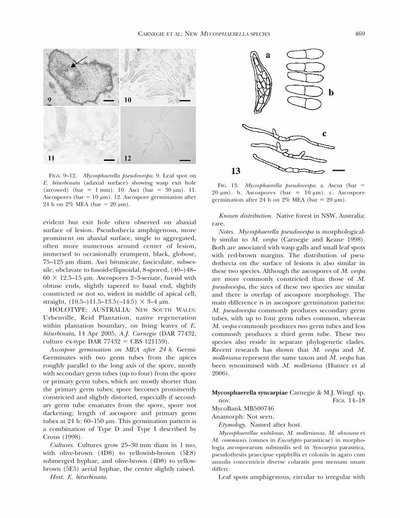
evident but exit hole often observed on abaxialsurface of lesion. Pseudothecia amphigenous, moreprominent on abaxial surface, single to aggregated,often more numerous around center of lesion,immersed to occasionally erumpent, black, globose,75–125 mm diam. Asci bitunicate, fasciculate, subses-sile, obclavate to fusoid-ellipsoidal, 8-spored, (40–)48–60 3 12.5–15 mm. Ascospores 2–3-seriate, fusoid withobtuse ends, slightly tapered to basal end, slightlyconstricted or not so, widest in middle of apical cell,straight, (10.5–)11.5–13.5(–14.5) 3 3–4 mm.
HOLOTYPE: AUSTRALIA: NEW SOUTH WALES:Urbenville, Reid Plantation, native regenerationwithin plantation boundary, on living leaves of E.biturbinata, 14 Apr 2005, A.J. Carnegie (DAR 77432,culture ex-type DAR 77432 5 CBS 121159).
Ascospore germination on MEA after 24 h. Germi-Germinates with two germ tubes from the apicesroughly parallel to the long axis of the spore, mostlywith secondary germ tubes (up to four) from the sporeor primary germ tubes, which are mostly shorter thanthe primary germ tubes; spore becomes prominentlyconstricted and slightly distorted, especially if second-ary germ tube emanates from the spore, spore notdarkening; length of ascospore and primary germtubes at 24 h: 60–150 mm. This germination pattern isa combination of Type D and Type I described byCrous (1998).
Cultures. Cultures grow 25–30 mm diam in 1 mo,with olive-brown (4D8) to yellowish-brown (5E8)submerged hyphae, and olive-brown (4D8) to yellow-brown (5E5) aerial hyphae, the center slightly raised.
Host. E. biturbinata.
Known distribution. Native forest in NSW, Australia;rare.
Notes. Mycosphaerella pseudovespa is morphological-ly similar to M. vespa (Carnegie and Keane 1998).Both are associated with wasp galls and small leaf spotswith red-brown margins. The distribution of pseu-dothecia on the surface of lesions is also similar inthese two species. Although the ascospores of M. vespaare more commonly constricted than those of M.pseudovespa, the sizes of these two species are similarand there is overlap of ascospore morphology. Themain difference is in ascospore germination patterns:M. pseudovespa commonly produces secondary germtubes, with up to four germ tubes common, whereasM. vespa commonly produces two germ tubes and lesscommonly produces a third germ tube. These twospecies also reside in separate phylogenetic clades.Recent research has shown that M. vespa and M.molleriana represent the same taxon and M. vespa hasbeen synonimised with M. molleriana (Hunter et al2006).
Mycosphaerella syncarpiae Carnegie & M.J. Wingf. sp.nov. FIGS. 14–18
MycoBank MB500746Anamorph: Not seen.
Etymology. Named after host.Mycosphaerellae nubilosae, M. mollerianae, M. ohnowae et
M. communis (omnes in Eucalypto parasiticae) in morpho-logia ascosporarum subsimilis sed in Syncarpia parasitica,pseudothesiis praecipue epiphyllis et coloniis in agaro cumannulis concentricis diverse colaratis post mensam unamdiffert.
Leaf spots amphigenous, circular to irregular with
FIGS. 9–12. Mycosphaerella pseudovespa. 9. Leaf spot onE. biturbinata (adaxial surface) showing wasp exit hole(arrowed) (bar 5 1 mm). 10. Asci (bar 5 30 mm). 11.Ascospores (bar 5 10 mm). 12. Ascospore germination after24 h on 2% MEA (bar 5 20 mm).
FIG. 13. Mycosphaerella pseudovespa. a. Ascus (bar 5
20 mm). b. Ascospores (bar 5 10 mm). c. Ascosporegermination after 24 h on 2% MEA (bar 5 20 mm).
CARNEGIE ET AL: NEW MYCOSPHAERELLA SPECIES 469

an irregular margin, 4–12 mm diam, red-brown onthe adaxial and yellow-brown on the abaxial surface,often associated with leaf distortion and oftencracking. Pseudothecia amphigenous, predominantlyepiphyllous, immersed to erumpent, scattered, black,globose, 55–88 mm. Asci bitunicate, fasciculate, ovoidto ellipsoidal, straight or curved, 8-spored, 48–59 3
10–13 mm. Ascospores bi- to multiseriate, overlapping,narrowly clavate, with obtuse ends, tapering towardbasal end, not constricted at median septum or onlyslightly so, widest in middle of apical cell, hyaline, 15–18 3 3.5–5 mm.
HOLOTYPE: AUSTRALIA: NEW SOUTH WALES:Nana Glen, Orara State Forest, native forest, on livingleaves of Syncarpia glomulifera, 23 Aug 2003, A.J.Carnegie & M.J. Wingfield (DAR 77433, culture ex-type DAR 77433, 5 CBS 121160; isotype NSWF005320, culture ex-isotype NSWF 005320).
Ascospore germination on MEA after 24 hr. Germtubes grow from both ends parallel to the long axis ofthe spore, spore not darkening or distorting butbecoming prominently constricted; length of asco-spore plus both germ tubes after 24 h (120–)165–216(–239) mm. Germination Type C.
Cultures. Colonies 20–25 mm after 1 mo, in con-centric rings of color: submerged hyphae olive-gray(2F2) in center to olive (2F8) with outermost ring(s)sparse and olive (2F8) to olive-yellow (2D8); aerialhyphae in center brownish-gray (6C2) and raised, withseveral thin rings of gray (6B1) out from center;reverse homogenous dark gray to black.
Host. Syncarpia glomulifera.Known distribution. New South Wales, Australia;
common.
Notes. Mycosphaerella syncarpiae was found at nu-merous locations in native forests of Syncarpiaglomulifera in northeastern NSW and also from speci-mens in herbarium DAR collected mostly from theSydney region. These latter specimens had beenexamined by R.F. Park in 1983, who noted that theyrepresented a distinct, undescribed species. Like ourobservations R.F. Park (in 1983) and J. Walker (in1962) noted that leaf spots were similar to M. cryptica(especially in pseudothecial distribution) and asco-spores were similar to M. nubilosa. During this studyM. marksii was observed commonly on S. glomuliferathroughout eastern NSW; however the asymmetricalapical cell of M. marksii ascospores (Carnegie andKeane 1994) clearly distinguishes this species from M.syncarpiae. No other species of Mycosphaerella havebeen reported from Syncarpia. Based on ascosporemorphology and germination M. syncarpiae resemblesseveral species of Mycosphaerella from Eucalyptus,including M. nubilosa, M. molleriana, M. crystallina,M. communis and M. ohnowa (Park and Keane 1982,Crous 1998, Crous et al 2004). However M. syncarpiaecan be distinguished from these latter species by itsdifferent pseudothecial distribution and distinctivecultural characteristics.
Crous (1999) reviewed the species of Mycosphaerellafrom Myrtaceae (other than Eucalyptus) and reportedseven species. Only one of these, M. angophorae, hasbeen recorded in Australia where it occurs onAngophora bakeri in NSW. Mycosphaerella angophoraehas slightly corky lesions, predominantly hypogenouspseudothecia and small, broadly ellipsoidal asco-spores (Hansford 1957, Crous 1999), features thatdistinguish it from M. syncarpiae. Based on lesiontype, pseudothecial distribution and ascospore mor-phology, M. syncarpiae is also distinct from the other
FIG. 18. Mycosphaerella syncarpiae. a. Ascus (bar 5
20 mm). b. Ascospores (bar 5 10 mm). c. Ascosporegermination after 24 h on 2% MEA (bar 5 15 mm).FIGS. 14–17. Mycosphaerella syncarpiae. 14. Asci (bar 5
30 mm). 15. Ascospores (bar 5 10 mm). 16. Ascosporegermination after 24 h on 2% MEA (bar 5 20 mm). 17.Four single-spore cultures after 1 mo on 2% MEA.
470 MYCOLOGIA

species dealt with by Crous (1999). In their examina-tion of herbarium specimens from Queensland,Sivanesan and Shivas (2002) described 12 new speciesof Mycosphaerella from a range of hosts, including onefrom Myrtaceae, given the name M. melaleucoidesfrom Melaleuca quinquenervia. Although ascosporesof M. melaleucoides and M. syncarpiae are similar, theepigenous leaf spots and ascomata of M. melaleucoideshelp distinguish it from M. syncarpiae.
Additional specimens examined. AUSTRALIA: NEW
SOUTH WALES: Urunga, Newry State Forest, native forest,on living leaves of S. glomulifera, 20 Feb 2004, A.J. Carnegie(DAR 77434); Morrisett, Olney State Forest, native forest,on living leaves of S. glomulifera, 21 Nov 1996, J.A. Simpson& A.J. Carnegie (DAR 77436; NSWF 005525); Nana Glen,Wedding Bells State Forest, native forest, on living leaves ofS. glomulifera, 11 Apr 2005, A.J. Carnegie (DAR 77437); asMycosphaerella sp., Baulkham Hills, on S. glomulifera, Jun1957, J. Walker (DAR 5200); Pittwater, on S. glomulifera, Mar1948, L. Fraser (DAR 4787); Mountain Lagoon, on S.glomulifera, 28 Mar 1982, M. Priest (DAR 45633); as M.nubilosa, Baulkham Hills, on S. glomulifera, 1 Jan 1957, J.Walker (DAR 4986); Kurrajong Heights, on S. glomulifera,27 Apr 1949, L. Fraser (DAR 3870); Palm Beach, on S.glomulifera, Apr 1949, L. Fraser (DAR 3869).
Mycosphaerella multiseptata Carnegie sp. nov.FIGS. 19–25
MycoBank MB500744Anamorph: not seen.
Etymology. Forming multiple septa in the sporebody and germ tubes after 24 h germination.
Mycosphaerellae mexicanae in Eucalypto ascosporarummorphologia et modo germinationis subsimilis sed Ango-phora parasitica, coloniis in agaro lente crescentibus 5–6 mm post mensam unam et septis transverses numerosis intubis germinationibus post 24 h.
Leaf spots on A. subvelutina: amphigenous, circularto irregular, single to confluent, 2–7 mm diam,yellow-brown becoming gray-brown on adaxial sur-face, yellow-brown to red-brown on abaxial surface,with prominent red-brown border; on A. costata:amphigenous, subcircular with irregular margins,bordered by veins, single to confluent, 3–12 mmdiam, yellow-brown becoming gray-brown on adaxialsurface, yellow-brown to red-brown on abaxial surface,with prominent red-brown border, often with antho-cyanin pigmentation surrounding border. Pseudothe-cia on A. subvelutina: hypophyllous, scattered singly,immersed, black, globose, 75–100 mm; on A. costata:hypophyllous, in numerous clusters, immersed be-coming erumpent, black, globose, 85–110 mm. Asciaparaphystae, fasiculate, bitunicate, subsessile, ob-ovoid to broadly ellipsoid, straight to slightly in-curved, 8-spored, 43–56 3 10–15 mm. Ascospores 2–3-seriate, overlapping, hyaline, straight, rarely curved,narrowly obovoid to fusiform with obtuse ends,medianly or unequally 1-septate, widest in middle ofapical cell, mostly not constricted at septum, taperingto basal end, (12.5–)13.5–17(–19) 3 (3–)3.5–4.5(–5) mm.
HOLOTYPE: AUSTRALIA: NEW SOUTH WALES:Whiporie, Pintexan Property, native forest, on livingleaves of Angophora subvelutina, 18 Jan 2005, A. J.Carnegie (DAR 77438; culture ex-type DAR 77438 5
CBS 121312).Ascospore germination on MEA after 24 h. Asco-
spores germinating from both ends, with germ tubes
FIGS. 19–24. Mycosphaerella multiseptata. 19. Pseudothe-cial distribution on abaxial surface of leaf spot on A.subvelutina (bar 5 1 mm). 20. Pseudothecial distributionon abaxial surface of leaf spot on A. costata (bar 5 1 mm).21. Asci (bar 5 30 mm). 22. Ascospores (bar 5 20 mm). 23–24. Ascospore germination after 24 h on 2% MEA (bar 5
15 mm).
FIG. 25. Mycosphaerella multiseptata. a. Ascus (bar 5
20 mm). b. Ascospores (bar 5 8 mm). c. Ascospore germi-nation after 24 h on 2% MEA (bar 5 8 mm).
CARNEGIE ET AL: NEW MYCOSPHAERELLA SPECIES 471

parallel to the long axis of the spore, spore bodybecoming 2–3-septate, spore darkening and becomingdistorted with a prominent constriction at the asco-spore septum; even though relatively short, germ tubeshave many septa, occasionally producing secondarygerm tubes; length of ascospore and germ tubes after24 h (38–)58–71(–81) mm. This germination patternmost closely resembles Type E and Type H.
Cultures. Slow growing, reaching 5–6 mm diam in1 mo; submerged hyphae olive (1F4), aerial hyphaeolive-brown (4E5) with white patches.
Hosts. A. subvelutina, A. costataKnown distribution. Native forests and amenity
plantings in NSW, Australia; rare.Notes. Based on symptoms and pseudothecia, this
species on the two different host species initially wasthought to represent two different taxa. On A.subvelutina leaf spots are mostly circular, while on A.costata they are subcircular to irregular. Pseudotheciaon A. subvelutina are mostly scattered and immersed,and on A. costata they often are clustered andimmersed to erumpent. However on both hostsascospore morphology and germination patterns arethe same and DNA sequence comparisons have shownthat they are the same species of Mycosphaerella.
Mycosphaerella multiseptata differs from otherspecies of Mycosphaerella from Eucalyptus based on acombination of host (Angophora), symptoms, asco-spore morphology and ascospore germination pat-tern. It is most similar in ascospore morphology toM. mexicana; however ascospores of M. multiseptatabecome more constricted on germination and pro-duce multiple germ tubes within the spore and germtubes. Mycosphaerella marksii has been found onAngophora, as well as other Myrtaceae, but hasdifferent ascospore morphology and germinationpatterns (Carnegie and Keane 1994) to M. multi-septata. The only other species of Mycosphaerellafound from Angophora, M. angophorae Hansf., can bedistinguished from M. multiseptata by having smallerascospores.
Additional specimens examined. AUSTRALIA: NEW
SOUTH WALES: Baryugil, Yuligibar Property, native forestwithin plantation boundary, on living leaves of A. sub-velutina, 20 Jan 2005, A. J. Carnegie (DAR 77439, cultureDAR 77439 5 CBS 121161); Sydney, Greenwich, FrenchsRoad, amenity planting, on living leaves of A. costata, 22Dec 2004, A.J. & G. F. Carnegie (DAR 77440, culture DAR77440).
DISCUSSION
We have described four new species of Mycosphaerellafrom Myrtaceous trees growing in native forests as wellas in plantations. Mycosphaerella tumulosa was found
from many locations in both native forests andplantations in northern NSW and southeasternQueensland and on at least eight hosts in Eucalyptusand Corymbia. Although individual trees can beheavily infected this pathogen did not cause signifi-cant damage at a stand or plantation level. Myco-sphaerella syncarpiae was identified from manylocalities in native forests and amenity plantingsonly from Syncarpia glomulifera. It was observedcausing minor damage, mostly on individual trees ina stand. Mycosphaerella multiseptata was observedcausing minor damage on species of Angophora inseveral native stands and an amenity planting. Myco-sphaerella pseudovespa was recorded from only onelocality and was not associated with significantdamage.
A specific focus of this study was to survey forMycosphaerella spp. on Myrtaceae in native forestsituations or on trees in plantations adjacent to nativeforests, where the plantations might have acted as‘‘sinks’’ for these fungi. Ultimately such collectionsmight assist us in understanding why only six of themore than 40 species of Mycosphaerella first describedfrom outside Australia have ever been found in thiscountry. These include M. suberosa Crous, F.A.Ferreira, Alfenas & M.J. Wingf. (Carnegie et al1997), M. lateralis Crous & M.J. Wingf. (Maxwell etal 2000), M. mexicana Crous (Maxwell et al 2003), M.fori Hunter et al (Jackson et al 2005), M. heimii Crous(Whyte et al 2005) and M. ohnowa Crous & M.J.Wingf. (Crous et al 2007). It is most likely that thesespecies originated in Australia but were detectedoverseas first. This is because these species cause littledamage in native ecosystems in Australia but, whenintroduced to susceptible, even aged exotic planta-tions, their impact is greater, disease symptoms areobvious and they are collected, isolated and de-scribed.
Recent surveys in native forests in NSW, includingthe current work, have resulted in the discovery ofnumerous new species of Mycosphaerella and otherfoliar fungi from eucalypts (Summerell et al 2006,Crous et al 2007). It seems likely that future surveyswill reveal additional species of these fungi, includingspecies currently known only from other countries.Although it might seem unusual that there are somany species of Mycosphaerella on Eucalyptus, wesupport the view of Crous et al (2006) that therecould be at least as many species of Mycosphaerella onthese trees as the number of species of the treesthemselves. This would imply that ultimately there willmore than 700 species of Mycosphaerella on Eucalyp-tus and that the majority of species have not yet beendiscovered.
472 MYCOLOGIA

ACKNOWLEDGMENTS
We thank John Walker for assistance with the Latin and forreviewing the manuscript, Dianne White for technicalsupport associated with the DNA comparisons presentedin this study and Pedro Crous for reviewing the manuscript.
LITERATURE CITED
Barber PA. 2005. A study of foliar diseases of eucalypts inplantations and native forests of south-eastern Australia[Doctoral dissertation]. Australia: La Trobe University.p 1–200.
Boland DJ, Brooker MIH, Chippendale GM, Hall N, HylandBPM, Johnston RD, Kleinig DA, Turner JD. 1992.Forest trees of Australia. Melbourne: Australian Gov-ernment Publishing Service. 687 p.
Braun U, Dick MA. 2002. Leaf spot diseases of eucalypts inNew Zealand caused by Pseudocercospora species. NZ JFor Sci 32:221–234.
Burgess T, Wingfield MJ, Wingfield BD. 2001. Simplesequence repeat (SSR) markers distinguish betweenmorphotypes of Sphaeropsis sapinea. Appl EnvironMicrobiol 67:354–362.
———, Barber PA, Sufaati S, Xu D, Hardy GESJ, Dell B.2006. Mycosphaerella spp. on eucalypts in Asia; newspecies, new hosts and new records. Fungal Divers 24:135–157.
Carnegie AJ. 1991. The susceptibility of certain Eucalyptusspecies and provenances to infection by Mycosphaerellaspp. and other leaf parasites (Honors dissertation).Australia: La Trobe University. p 1–160.
———. 2000. A study of the species of Mycosphaerella oneucalypts in Australia and the impact of Mycosphaerellaleaf diseases on Eucalyptus globulus Labill. [Doctoraldissertation]. Australia: University of Melbourne. p 1–203.
———, Keane PJ. 1994. Further Mycosphaerella speciesassociated with leaf diseases of Eucalyptus. Mycol Res98:413–418.
———, ———. 1998. Mycosphaerella vespa sp. nov. fromdiseased Eucalyptus leaves in Australia. Mycol Res 102:1274–1276.
———, ———, Podger FD. 1997. The impact of threespecies of Mycosphaerella newly recorded on Eucalyptusin Western Australia. Aust Plant Path 26:71–77.
Cortinas M.-N, Burgess TI, Dell B, Xu D, Wingfield MJ,Wingfield BD. 2006. First record of Colletogloeopsiszuluense comb. nov., causing a stem canker ofEucalyptus spp. in China. Mycol Res 110:229–236.
Crous PW. 1998. Mycosphaerella spp. and their anamorphsassociated with leaf spot diseases of Eucalyptus. MycolMem 21:1–170.
———. 1999. Species of Mycosphaerella and related ana-morphs occurring on Myrtaceae (excluding Eucalyp-tus). Mycol Res 103:607–621.
———, Groenewald JZ, Mansilla JP, Hunter GC, WingfieldMW. 2004. Phylogenetic reassessment of Mycosphaerellaspp. and their anamorphs occurring on Eucalyptus.Stud Mycol 50:195–214.
———, Hong L, Wingfield BD, Wingfield MJ. 2001. ITSrDNA phylogeny of selected Mycosphaerella species andtheir anamorphs occurring on Myrtaceae. Mycol Res105:425–431.
———, Knox-Davies PS, Wingfield MJ. 1989. A summary offungal leaf pathogens of Eucalyptus and the diseasesthey cause in South Africa. SA For J 149:9–16.
———, Summerell BA, Carnegie AJ, Mohammed C, Hima-man W, Groenewald JZ. 2007. Foliicolous Mycosphaer-ella spp. and their anamorphs on Corymbia andEucalyptus. Fungal Divers 26. (In press).
———, Wingfield MJ, Mansilla JP, Alfenas AC, GroenewaldJZ. 2006. Phylogenetic reassessment of Mycosphaerellaspp. and their anamorphs occurring on Eucalyptus. II.Stud Mycol 55:99–131.
Dick MA, Dobbie K. 2001. Mycosphaerella suberosa and M.intermedia sp. nov. on Eucalyptus in New Zealand. NZ JBot 39:269–279.
Felsenstein J. 1985. Confidence intervals on phylogenetics:an approach using bootstrap. Evolution 39:783–791.
Gardes M, Bruns T. 1993. ITS primers with enhancedspecificity for basidiomycetes—application to the iden-tification of mycorrhiza and rusts. Mol Ecol 2:113–118.
Hansford CG. 1957. Australian fungi IV. New species andrevisions (continued). Proc Linn Soc NSW 82:209–229.
Hillis DM, Huelsenbeck JP. 1992. Signal, noise and re-liability in molecular phylogenetic analysis. J Hered 83:189–195.
Hunter GC, Roux J, Wingfield BD, Crous PW, Wingfield MJ.2004. Mycosphaerella species causing leaf disease inSouth African Eucalyptus plantations. Mycol Res 108:672–681.
———, Wingfield BD, Crous PW, Wingfield MJ. 2006a. Amulti-gene phylogeny for species of Mycosphaerellaoccurring on Eucalyptus leaves. Stud Mycol 55:147–161.
———, Crous PW, Wingfield BD, Pongpanich K, WingfieldMJ. 2006b. Pseudocercospora flavomarginata sp. nov.,from Eucalyptus leaves in Thailand. Fungal Divers 22:71–90.
Jackson SL, Maxwell A, Collins S, Dell B, Hardy GEStJ. 2005.Mycosphaerella species present on Eucalyptus globulusin Western Australia, new records for WA and Australia.In: 15th Biennial Australasian Plant Pathology SocietyConference Handbook. 185 p.
Kornerup A, Wanscher JH. 1978. Methuen Handbook ofColour. London: Eyre Methuen. p 1–252.
Maxwell A. 2004. The taxonomy, phylogeny and impact ofMycosphaerella species on eucalypts in south-westernAustralia [Doctoral dissertation]. Western Australia:Murdoch University. p 1–225.
———, Dell B, Neumeister-Kemp HG, Hardy GEStJ. 2003.Mycosphaerella species associated with Eucalyptus insouth-western Australia: new species, new records anda key. Mycol Res 107:351–359.
———, Hardy GEStJ, Wingfield MJ, Dell B. 2000. Firstrecord of Mycosphaerella latreralis on Eucalyptus inAustralia. Aust Plant Path 29:279.
Milgate A. 2005. Mycosphaerella species of Tasmania andtheir interactions with Eucalyptus plantations [Doctoral
CARNEGIE ET AL: NEW MYCOSPHAERELLA SPECIES 473

dissertation]. Tasmania: University of Tasmania. p 1–199.
Park RF. 1984. The taxonomy, pathology and epidemiologyof Mycosphaerella species associated with leaf diseases ofEucalyptus in Australia [Doctoral dissertation]. Austra-lia: La Trobe University. p 1–205.
———, Keane PJ. 1982. Three Mycosphaerella species fromleaf diseases of Eucalyptus. Trans Brit Mycol Soc 79:95–100.
———, ———, Wingfield MJ, Crous PW. 2000. Fungaldiseases of Eucalyptus foliage. In: Keane PJ, Kile GA,Podger FD, Brown BN, eds. Diseases and pathogens ofeucalypts. Melborne: CSIRO. p 153–240.
Pryor LD, Johnson LAS. 1971. A classification of theeucalypts. Canberra: Australian National University.p 1–102.
Sankaran KV, Sutton BC, Minter D. 1995. A checklist offungi recorded on Eucalyptus. United Kingdom: CABInternational. p 1–376.
Sivanesan A, Shivas RG. 2002. Studies on Mycosphaerellaspecies in Queensland, Australia. Myco Res 106:355–364.
Slippers B, Stenlid J, Wingfield MJ. 2005. Emergingpathogens: fungal host jumps following anthropogenicintroduction. Trends Ecol Evol 20:420–421.
Summerell BA, Groenewald JZ, Carnegie AJ, SummerbellRC, Crous PW. 2006. Eucalyptus microfungi knownfrom culture 2. Alysidiella, Fusculina and Phlogicylin-drium genera nova, with notes on some other poorlyknown taxa. Fungal Divers 23:323–350.
White TJ, Bruns T, Lee S, Taylor J. 1990. Amplificationand direct sequencing of fungal ribosomal RNAgenes for phylogenetics. In: Innis MA, Gelfand DH,Snisky JJ, White TJ, eds. PCR protocols: a guide tomethods and applications. San Diego: Academic Press.p 315–322.
Whyte G, Burgess TI, Barber PA, Hardy GEStJ. 2005. Firstrecord of Mycosphaerella heimii in Australia. Aust PlantPath 34:605–606.
Yuan Z.-Q, de Little DAR, Mohammed C. 2000. A newspecies of Pseudocercospora (Hyphomycetes) describedon Eucalyptus nitens from Tasmania, Australia. NovHedwig 71:415–419.
474 MYCOLOGIA