New interventional MRI deep brain stimulator surgery ... · New interventional MRI deep brain...
8
New interventional MRI deep brain stimulator surgery program offered Artist’s rendition of the remote control unit for the SMARTFrame. The control knobs on the trajectory guide are connected via semirigid rods to this remote actuator, permitting the clinician to manipulate the device while the patient is in the center of the magnet without reaching into the bore. by R. Mark Richardson, MD, PhD; Douglas Kondziolka, MD I n January, our department became one of the first few centers in the world to offer interventional-MRI guided placement of deep brain stimulators (iMRI-DBS) for patients with movement disorders. We are the first in Pennsylvania to use this new ClearPoint® system. Traditional frame-based DBS surgery is a gold-standard procedure that has worked extremely well for over 15 years. We are continuing to explore developing technologies for our patients. The interventional MRI platform allows visualization of electrode placement into brain targets in real-time with patients under general anesthesia in the MRI scanner. Thus, severely anxious, dyskinetic, or pediatric patients who would rather not undergo frame placement and awake brain mapping in the operating room now have another option for DBS implantation under general anesthesia. Frame-based procedures rely on imaging obtained before surgery. In the operating room, the surgeon must perform a registration process to make “imaging space” match physical space as closely as possible. In frame-based surgery, this involves setting up the frame coordinates. Once the operation begins and the dura is opened, loss of cerebrospinal fluid and entry of air may combine to cause unpredictable and varying degrees of brain shift, even in deep brain structures. For this reason, intraoperative awake brain mapping with microelectrode recording during frame-based cases has been the gold standard for verification of electrode placement in the desired brain nucleus. The innovation allowed by the interventional MRI procedure is to select the location for lead placement by visualizing the target nucleus (subthalamic nucleus or globus pallidus internus) on an MRI scan in real-time during the procedure, and then to verify that the electrode is placed at the intended target by an immediate post-implantation scan, rather than by microelectrode recording. The ClearPoint implantation system is based on a burr-hole-mounted aiming de- vice and trajectory guide (SMARTFrame) with control software. A head fixation device is integrated with radiofrequency receiving coils for imaging of the brain and SMARTFrame. An integrated software system that communicates with the host computer of the MR sys- tem via a network link exists as a standalone workstation to provide the surgeon with the necessary feedback. The general workflow is divided into three different sections: burr-hole planning (entry), target selection and trajectory visualization (target), and alignment of trajectory guide and insertion monitoring (navigate). The software is designed for either unilateral or bilateral procedures. Thus far, the average error of the ClearPoint system ap- pears to be as small as that of the frame-based method. Of note, several published studies have shown previously that MR imaging of implanted DBS electrodes is extremely safe, if basic guidelines are followed. DBS in patients with Parkinson disease and dystonia can reduce motor symptoms significantly and can allow for reduction of medications that have adverse side effects. Parkinson and dystonia patients typically obtain clinically meaningful motor function improvements of greater than 50% following DBS. We have used the iMRI-DBS platform to implant DBS electrodes in both of these types of patients. Additionally, the iMRI-DBS platform is an attractive option for performing DBS surgery in pediatric movement disor- ders, since children are typically not good candidates for awake surgery in the operating room. Frame-based awake brain mapping, however, is still the gold standard for thalamic electrode placement to treat essential tremor, as the desired target in the thalamus is not well visualized on MRI. In our movement disorder program, appropriate surgical candidates have the option to undergo traditional, frame-based surgery in the operating room, or iMRI-DBS using the ClearPoint system in the MRI scanner. We hope that offering this new option to our move- ment disorder patients will allow more patients who are good candidates for DBS surgery, but would not otherwise undergo the procedure, to receive the benefits of this treatment. • Artist’s impression of the pitch and roll adjustment during the “navigate” step of the procedure. The targeting cannula of the SMARTFrame is aimed along the red trajectory; the desired trajectory to the target is in yellow. The inset is a screenshot from the ClearPoint software, which informs the surgeon of the adjustments needed using the pitch and roll knobs to make the targeting cannula collinear with the desired trajectory. 1 2
Transcript of New interventional MRI deep brain stimulator surgery ... · New interventional MRI deep brain...
New interventional MRI deep brain stimulator surgery program offered
Artist’s rendition of the remote control unit for the SMARTFrame. The control knobs on the trajectory guide are connected via semirigid rods to this remote actuator, permitting the clinician to manipulate the device while the patient is in the center of the magnet without reaching into the bore.
by R. Mark Richardson, MD, PhD; Douglas Kondziolka, MD
In January, our department became one of the first few centers in the world to offer interventional-MRI guided placement of deep brain stimulators (iMRI-DBS) for patients with movement disorders. We are the first in Pennsylvania to use this new ClearPoint®
system. Traditional frame-based DBS surgery is a gold-standard procedure that has worked extremely well for over 15 years. We are continuing to explore developing technologies for our patients. The interventional MRI platform allows visualization of electrode placement into brain targets in real-time with patients under general anesthesia in the MRI scanner. Thus, severely anxious, dyskinetic, or pediatric patients who would rather not undergo frame placement and awake brain mapping in the operating room now have another option for DBS implantation under general anesthesia. Frame-based procedures rely on imaging obtained before surgery. In the operating room, the surgeon must perform a registration process to make “imaging space” match physical space as closely as possible. In frame-based surgery, this involves setting up the frame coordinates. Once the operation begins and the dura is opened, loss of cerebrospinal fluid and entry of air may combine to cause unpredictable and varying degrees of brain shift, even in deep brain structures. For this reason, intraoperative awake brain mapping with microelectrode recording during frame-based cases has been the gold standard for verification of electrode placement in the desired brain nucleus. The innovation allowed by the interventional MRI procedure is to select the location for lead placement by visualizing the target nucleus (subthalamic nucleus or globus pallidus internus) on an MRI scan in real-time during the procedure, and then to verify that the electrode is placed at the intended target by an immediate post-implantation scan, rather than by microelectrode recording. The ClearPoint implantation system is based on a burr-hole-mounted aiming de-vice and trajectory guide (SMARTFrame) with control software. A head fixation device is integrated with radiofrequency receiving coils for imaging of the brain and SMARTFrame. An integrated software system that communicates with the host computer of the MR sys-tem via a network link exists as a standalone workstation to provide the surgeon with the necessary feedback. The general workflow is divided into three different sections: burr-hole planning (entry), target selection and trajectory visualization (target), and alignment of trajectory guide and insertion monitoring (navigate). The software is designed for either unilateral or bilateral procedures. Thus far, the average error of the ClearPoint system ap-pears to be as small as that of the frame-based method. Of note, several published studies have shown previously that MR imaging of implanted DBS electrodes is extremely safe, if basic guidelines are followed. DBS in patients with Parkinson disease and dystonia can reduce motor symptoms significantly and can allow for reduction of medications that have adverse side effects. Parkinson and dystonia patients typically obtain clinically meaningful motor function improvements of greater than 50% following DBS. We have used the iMRI-DBS platform to implant DBS electrodes in both of these types of patients. Additionally, the iMRI-DBS platform is an attractive option for performing DBS surgery in pediatric movement disor-ders, since children are typically not good candidates for awake surgery in the operating room. Frame-based awake brain mapping, however, is still the gold standard for thalamic electrode placement to treat essential tremor, as the desired target in the thalamus is not well visualized on MRI. In our movement disorder program, appropriate surgical candidates have the option to undergo traditional, frame-based surgery in the operating room, or iMRI-DBS using the ClearPoint system in the MRI scanner. We hope that offering this new option to our move-ment disorder patients will allow more patients who are good candidates for DBS surgery, but would not otherwise undergo the procedure, to receive the benefits of this treatment. •
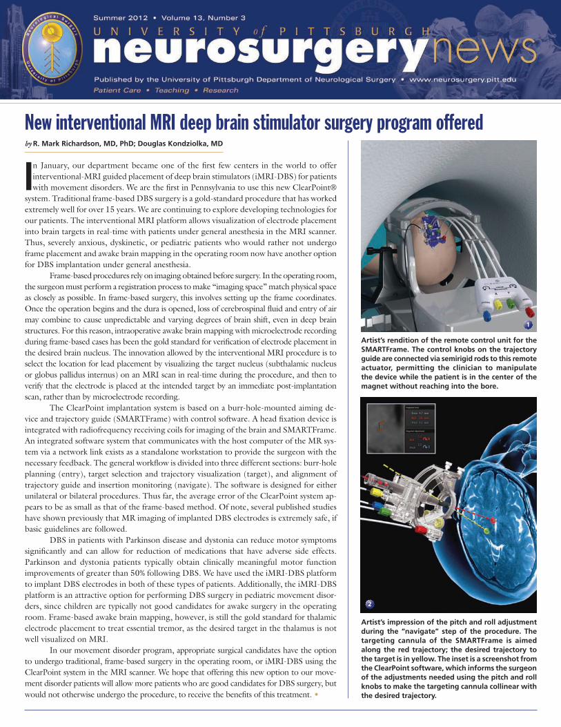
Artist’s impression of the pitch and roll adjustment during the “navigate” step of the procedure. The targeting cannula of the SMARTFrame is aimed along the red trajectory; the desired trajectory to the target is in yellow. The inset is a screenshot from the ClearPoint software, which informs the surgeon of the adjustments needed using the pitch and roll knobs to make the targeting cannula collinear with the desired trajectory.
1
2

environment brought change to the traditional brain tumor management process by developing bold and innovative ideas and strategies. These strategies include radiosurgery for patients with oligometastatic disease, skull base lesions, and other conditions; and the endoscopic endonasal approach to resect very challenging anterior skull base lesions.
High-definition fiber tractography, also, provides thorough clinical informa-tion on that residual and displaced normal functional tracts, enabling neurosurgeons to more accurately, and safely, remove le-sions in extremely eloquent brain. Change has occurred and will continue to occur at the University of Pittsburgh Department of Neurological Surgery. We are proud of our fruitful and supporting environment, which enables
us to look forward and continue to change neu-rosurgery in ways that we can only imagine (and not imagine yet)!
Robert M. Friedlander, MD, MAChairman, Department of Neurological Surgery
UPMC Endowed Professor of Neurosurgery & NeurobiologyUniversity of Pittsburgh School of Medicine
University of Pittsburgh Medical Center
facultyneurosurgery
ofd e p a r t m e n t
All University of Pittsburgh Neurosurgery News content is copyrighted and is meant solely for the educational purpose of the reader. Please consult your physician before taking any medical actions, or contact the University of Pittsburgh Department of Neurological Surgery at (412) 647-3685.
Moving forwardChairmanRobert M. Friedlander, MD, MA
ProfessorsC. Edward Dixon, PhD (Vice Chairman, Research)Robert Ferrante, PhDPeter C. Gerszten, MD, MPH Michael B. Horowitz, MDLarry W. Jenkins, PhDDouglas S. Kondziolka, MD, MSc (Vice Chairman, Education) L. Dade Lunsford, MDJohn J. Moossy, MD Hideho Okada, MD, PhD Ian F. Pollack, MD (Vice Chairman, Academic Affairs) Mingui Sun, PhD
Associate Professors Jeffrey Balzer, PhD Ajay Niranjan, MDDavid O. Okonkwo, MD, PhD
Assistant Professors David J. Bissonette, PA-C, MBA (Executive Director)Donald J. Crammond, PhDJohnathan Engh, MD Juan C. Fernandez-Miranda, MDPaul A. Gardner, MDAvniel Ghuman, PhDPaola Grandi, PhD Stephanie Greene, MDMiguel Habeych, MD, PhDBrian Jankowitz, MDAdam S. Kanter, MDAva Puccio, PhD, RNShengjun Ren, PhDR. Mark Richardson, MD, PhDRichard M. Spiro, MDMandeep Tamber, MD Elizabeth C. Tyler-Kabara, MD, PhDYu Zhang, PhD
Clinical Professors Adnan A. Abla, MDMatt El-Kadi, MD, PhD (Vice Chair, Passavant Neurosurgery)Joseph C. Maroon, MD Daniel A. Wecht, MD, MSc
Clinical Associate ProfessorMichael J. Rutigliano, MD, MBA
Clinical Assistant ProfessorsPedro J. Aguilar, MDEric M. Altschuler, MDOren Berkowitz, MDJ. William Bookwalter, MDDaniel M. Bursick, MDDavid J. Engle, MDDavid L. Kaufmann, MDParthasarathy D. Thirumala, MDMatthew M. Wetzel, MD
Research Assistant ProfessorsJonathan Bellotte, PhD Diane L. Carlisle, PhD Yue-Fang Chang, PhDWendy Fellows-Mayle, PhDEsther Jane, PhDWenyan Jia, PhDHideyuki Kano, MD, PhDRekha Pal, MDDaniel Premkumar, PhDHong Qu Yan, MD, PhD
Clinical InstructorsJeff Bost, PA-C Maria Koutourousiou, MD
U N I V E R S I T Y of P I T T S B U R G H N E U R O S U R G E R Y N E W S
C H A I R M A N ’ S M E S S A G E
P A G E 2
What makes neurosurgery change and move forward? Is change a random process, or does change result from a carefully orches-
trated set of events and circumstances, leading to a real difference in the approach in which we treat a specific disease entity? I suspect is a little of both. To alter the manner in which we evaluate and treat neurosurgical conditions, the change must not only be a meaning-ful change, but more importantly, the change must be a step forward. I propose that luck, awareness, perseverance, intel-lect and environment are all important factors in enabling effective and produc-tive ‘change.’ At the University of Pittsburgh, the ingredients required to both develop and enact change must be part of our environment. Take, for example, the management of a brain tumor. In the not too distant past, a paucity of options existed for patients with brain tumors. Craniotomies and radical skull base approaches were the most prevalent options for many of these patients. However, at the University of Pitts-burgh, forward thinking in a receptive and fertile
Congratulations to 2012 graduating chief residents Pawel G. Ochalski, MD (front row, left), Matthew Maserati, MD (front row, second left) and Nestor D. Tomycz, MD (front row, right) seen here at graduation reception at Fox Chapel Golf Club on June 22 with department chairman Robert Friedlander, MD, and residency director L. Dade Lunsford, MD, (front row) and other residents. (See more in ‘News & Notes’ on page 7.)

P A G E 3P A G E 3
by L. Dade Lunsford, MD; Douglas Kondziolka, MD; John C. Flickinger, MDYoshio Arai, MD; Jagdish Bhatnagar, ScD;Andy Xu, PhD
In August 2012, UPMC Presbyterian will celebrate the 25th anniversary of the in-stallation of the first dedicated 201-source
cobalt 60 Gamma Knife in the United States. Since that time, the field of radiosurgery has grown dramatically. The field of radiosurgery was estab-lished by Lars Leksell in 1951 as an adjunct to the treatment of deep brain lesions oth-erwise not accessible to more conventional neurosurgery. His primary interest was in the creation of brain lesions for the manage-ment of trigeminal neuralgia and behavioral neurosurgery in an era when pharmacological management was almost nonexistent. Since its inception, the application of stereotactic radiosurgery (SRS) has expanded and trans-formed. The Gamma Knife is most commonly used now for metastatic tumors of the brain and treatment of benign neoplastic or vascular lesions such as AVMs. Since the year 2000, SRS has achieved an additional role in the management of benign and malignant spinal tumors. In 1987, the field of stereotactic radiosurgery was in its infancy with less than 2,000 patients having been treated world-wide. Since that time, Elekta, the manufactur-er of Gamma Knife radiosurgery technology estimates that more than 700,000 patients have undergone SRS worldwide during the last 33 years. The total number of LINAC-based SRS procedures is not known. The market research firm IMV has provided data that 32,335 US patients underwent LINAC or Gamma Knife SRS in 2009 and that the average increase in volume is approximately 10% per year. The Center for Medicare and Medic-aid services reports that the primary Gamma Knife hospital billing code 77371 was billed 5929 times in 2010. The primary LINAC SRS billing code G0339 was billed 9,337 times in 2010. It is estimated that the Gamma Knife procedure is performed in more than 15,000 patients annually in the United States. As part of a process of evaluating the impact of SRS in United States neurosurgical train-ing programs, we asked Katie Orrico, JD, of the AANS/CNS Washington Committee to review the Center for Medicaid and Medicare
S U M M E R 2 0 1 2 • V O L U M E 1 3 , N U M B E R 3
Twenty-five years of Gamma Knife radiosurgery at the University of Pittsburghservices (CMS) SRS CPT codes. We asked her to compare those numbers to the current numbers of CPT codes submitted for crani-otomy for tumor, not meningioma, during the same interval (1993-2011). As shown in figure 1 below, in 2003, the number of submitted CPT codes for radiosurgery (then code number 61793) exceeded the number of codes submitted for craniotomy for tumor (not meningioma). Annually, our center performs approx-imately 650-700 frame-based stereotactic radiosurgical procedures using the Gamma Knife. Currently approximately 40% of these cases represent single or multiple metastatic tumors. The application of SRS for the man-agement of brain cancer has changed the
paradigm completely in the management of such cases. (See L. Dade Lunsford, MD, talk about why the Gamma Knife has caused a shift in the paradigm of how neurosurgeons and oncologists care for patients with brain metastases in a Discovery Channel.com video at discoveryhealthcme.discovery.com/physi-cianed/physicianed.html) We know that radiosurgery results in significantly less toxicity to the brain; can be repeated as needed in the face of the devel-opment of new disease; does not impact the management of patients’ ongoing chemo-therapy for systemic disease; and, finally, is
1
2
(continued on back cover)
Journal Articles by Year

P A G E 4
U N I V E R S I T Y of P I T T S B U R G H N E U R O S U R G E R Y N E W S
by R. Mark Richardson, MD, PhD
Epilepsy is often called the most common serious neurological disorder because at any given time 1% of the world’s popula-
tion has active epilepsy. The only potential cure for a patient’s epilepsy is the surgical removal of the seizure focus, if it can be identified. Chances for seizure freedom can be as high as 90% in some cases of seizures that originate in the temporal lobe. In 2003, the American Association of Neurology (AAN) recognized that the ben-efits of temporal lobe resection for disabling seizures is greater than continued treatment with antiepileptic drugs, and issued a practice parameter recommending that patients with temporal lobe epilepsy be referred to a surgi-cal epilepsy center. In addition, patients with extra-temporal epilepsy who are experiencing difficult seizures or troubling medication side effects may also benefit from talking to an epilepsy surgeon, especial those with a brain lesion such as a tumor or vascular malforma-tion. Tragically, it takes an average of 20 years for patients with drug-resistant epilepsy to be referred to an epilepsy surgeon. For this reason, the University of Pittsburgh Adult Epilepsy Surgery Program has implemented a process for patients and their families to meet with the epilepsy surgeon earlier in the course of their disease treatment.
Surgical alternatives for epilepsy (SAFE) offers counseling, choices for patients
Surgical Alternatives For Epilepsy (SAFE) counseling is a process that allows epilepsy patients, and their families, to talk to a neurosurgeon about the role of brain surgery in the treatment of epilepsy, even if surgery has not yet been recommended. In this program, neurologists and general practitioners are referring epilepsy patients as soon as surgical candidacy is a possibility, recognizing that surgery for epilepsy is not a “last resort” but a potential cure. SAFE counseling is an appropriate step even if patients are not ready to undergo brain surgery, as meeting with the neurosurgeon does not represent a commitment to surgery. The philosophy of our comprehensive epi-lepsy program is that early education about surgery gives patients more control over the treatment of their disease. Also, surgical treatment earlier in the course of epilepsy is more effective. Some facts that are discussed include: •Upto40%ofpeoplewithepilepsycannot control their seizures with medication. •The chanceof becoming seizure-free after failing two medications is less than
Myths
• There are always ‘serious complications’ from
epilepsy surgery.
• All approved anti-seizure medications should
fail, or
• a vagal nerve stimulator (VNS) should be at-
tempted and fail, before surgery is considered.
• A seizure focus near the language area of the
brain cannot be removed.
• A seizure focus near the movement area of the
brain cannot be removed
• Surgery on the head leaves a huge scar where
hair doesn’t grow and is disfiguring.
Facts
Epilepsy surgery is relatively safe:
• the rate of permanent neurologic deficits is
about 3%
• the rate of cognitive deficits is about 6%,
although half of these resolve in two months
• complications are well below the danger of
continued seizures.
• Some forms of temporal lobe epilepsy are
progressive and seizure outcome is better when
surgical intervention is early.
• Early surgery helps to avoid the adverse conse-
quences of continued seizures (increased risk of
death, physical injuries, cognitive problems and
lower quality of life.
• Resection surgery should be considered before
vagal nerve stimulator placement.
• Language and movement areas of the brain can
be preserved by carefully mapping these func-
tions with electrical stimulation.
• Cosmetic changes are often only noticed by the
patient, and hair does grow back over the incision.
10% and drops to less than 3% after failing three medications. •70-90%ofpatientsareseizurefreeone year after temporal lobe surgery (see figure 1 at left). •100,000peoplewithdrug-resistanttemporal lobe epilepsy are eligible for surgery every year in the U.S., but less than 3% get surgical treatment. Why are more patients who would benefit from epilepsy surgery not referred and treated? Myths and lack of education about epilepsy surgery probably play a large role (see table above). As part of a neurosurgical consultation at UPMC, epilepsy patients also have the op-portunity to talk to a representative from the Epilepsy Foundation of Western Pennsylvania (EFWP) to learn about available resources for people with epilepsy. Additionally, in conjunc-tion with the EFWP, our department hosts an Epilepsy Surgery Discussion Group every third Friday of the month, where anyone who has had, or is considering having, surgery for epilepsy is invited to come and share their experiences or ask questions. •
Post-operative MRI demonstrating a right an-terior temporal lobectomy (red dashed line) in an epilepsy patient who has been seizure-free after surgery, without any cognitive changes.
1

P A G E 5P A G E 5
S U M M E R 2 0 1 2 • V O L U M E 1 3 , N U M B E R 3
by Matt El-Kadi, MD, PhD
Lumbar fusion has been used to treat patients with a variety of conditions that include trauma, tumor, infection, instability and spondylolisthesis. For patients undergoing elective surgery,
often they have failed a series of conservative treatment options, including medications, physical therapy, chiropractic care and pain management. When patients present with continued pain that affects the quality of their life, it seems clear that surgery might be a good option for them. However, there has been discussion about ad-ditional factors that may affect patient outcome. Factors such as patient age, obesity, preoperative diagnosis, tobacco use and medi-cal co-morbidities may play a role in the long-term outcome for patients following lumbar fusion. Postoperative course may also differ within these groups. The effect of radiographic fusion on clinical outcome has also been postulated. Bone grafting can be used alone or as an adjunct to instrumentation, and there is debate regarding the selection of graft product. Although autologus iliac crest graft is associated with a high rate of fusion, it may be argued that it will increase the length of procedure and blood loss and may increase short and long term complications at the donor site. The use of allograft has eliminated this subgroup of complications, but some question remains about the long term outcome and if radiographic fusion is associated with good clinical outcome. A retrospective review of 500 cases using instrumented lumbar fusion and allograft bone was performed. Clinical outcome was examined during the initial postoperative period and followed over three months postoperative and up to two years. X-rays were reviewed postoperatively to assess fusion mass. In the early postoperative period, younger patients had more postoperative pain than elderly, however, long term outcome is similar. There was no difference in the rate of intraoperative complications between first-time surgical patients and redo surgery patients. Additionally to note, no intraoperative blood transfusion was required in any of the first time patients or redo surgery patients. There was no correlation between the rate of fusion and clinical outcome. There was no clinical difference between the fusion for smokers versus non-smokers. Good decortication of transverse processes and facet joints and removal of all soft tissue plays a significant role in the fusion rate. Patients with congenital pars defect and anterolisthesis had better clinical outcome than patients with mechanical lower back pain and degenerative disc disease. Patients with postlaminectomy syndrome including mechanical lower back pain and radiculapathy with collapse of the disc space and lateral recess had good outcome. Patients with higher body mass index had more postoperative pain and less functional improvement. Obesity was the most common risk in patients who develop postoperative infection Clinical outcome after lumbar fusion is less influenced by rate of radiographic fusion and more likely determined by indication for the surgery and patient expectations. Understanding the factors that affect postoperative course and long-term clinical outcome is crucial for the surgeon to provide the patient with appropriate preoperative counseling. •
Understanding all factors crucial in lumbar fusion counseling/outcomes
Type of surgery Total Excellent/Good Percentage
Pars defect 141 107 76%
Redo Surgery 214 156 73%
Mechanical LBP 113 72 64%
Adjacent Level 12 7 58%
Age Total Excellent/Good Percentage
18-40 36 24 67%
41-60 202 140 69%
61-84 262 212 81%

U N I V E R S I T Y of P I T T S B U R G H N E U R O S U R G E R Y N E W S
by Brian Jankowitz, MD
Virginia Kristoff isn’t your average 92-year-old. She enjoys weekly visits to Pitts-burgh’s new casino and recently traveled
by airplane for the first time in years to visit her son in Phoenix. She also made a remark-able recovery from one of the most severe forms of an ischemic stroke. In July of 2011, Virginia collapsed in her home. She experienced the sudden onset of left arm and leg weakness. Her daughter immediately recognized the tell-tale signs of a stroke. Within minutes, SouthBridge EMS had arrived to the scene and within an hour she was in the UPMC Mercy emergency department. Vascular neurologist Maxim Hammer, MD, recognized the symptoms of an intracranial large vessel occlusion. Virginia had an NIHSS of 17, result-ing in complete paralysis of her left side, loss of sensation to the left side, right gaze preference and hemineglect. A CT scan of the brain, in combination with a CT angiogram and CT perfusion, diagnosed a right carotid occlusion with a profound hemispheric perfu-sion deficit. Although she presented within three hours of her stroke, she was not a candidate for IV-tPA because she takes Coumadin for atrial fibrillation. She was taken directly to the angiography suite where a clot was removed by literally sucking it out of the blood vessel using a long hollow tube or catheter.
92-year-old stroke victim living normal life thanks to innovative procedure
Within three hours from presenta-tion, the blood clot had been removed and Virginia was on the road to recovery. In fact, within minutes of removing the blood clot, she started to regain strength on her left side. By the following morning, she was back to normal and ultimately walked out of the hospital and returned home to her everyday life, three days after the procedure. Stroke is the third leading cause of death in the U.S with an estimated 134,000 deaths annually and 892,000 hospitaliza-tions each year. It is the number one cause of adult disability with 6.4 million survivors in the U.S. alone, costing an estimated
Ramesh Grandhi, MD, (left photo) gets ready for pitch during the 9th Annual Neurosurgery Charity Softball Tournament held recently in New York’s Central Park. The event included teams from 28 institutions from across the country and was held to benefit brain tumor research. The University of Pittsburgh squad finished third and included (front row, kneeling), Chris Bonfield, Ramesh Grandhi, Dan Wecht; (middle row), Adam Kanter, Ali Kooshkabadi, Matt Tormenti, Robert Friedlander, Phillip Parry, Will Ares, Nate Zwagerman, Jim Burke; and (back row), Stephen Johnson, Rob Miller.
73.7 billion dollars a year for care of these patients. The mortality rate for an untreated internal carotid artery occlusion like Vir-ginia’s is over 50%. The advent of new devices, catheters, and training now allows surgeons unprec-edented access to blood vessels within the brain. Catheters have become extremely flexible, allowing long hollow tubes to access intracranial blood clots. Once the catheter is engaged in the clot, removing the clot can be as simple as manually suctioning the tube with a syringe, a technique known as manual aspiration thrombectomy (MAT). Along with Tudor Jovin, MD, director of the UPMC Stroke Institute, we recently published a study of 191 patients treated with MAT in the May edition of Stroke, the leading scientific publication to discuss cerebrovascu-lar disease. The study found recanalization rates of over 90% with favorable outcomes in 54% of patients. Considering that the natural history of these occlusions results in a good outcome in less than 25% of patients, MAT treatment effectively doubled the chances of living independently. It has been 11 months since Virginia’s stroke. Her daughter relates that she is better than ever, with a renewed zest for life. She even visited SouthBridge EMS to show off her good health. Last time they saw her, she was a partially paralyzed 91-year-old fighting for her life. Now, they saw a vivacious 92-year-old eager to show that age doesn’t always matter. •
Pitt neurosurgery team finishes third in national softball charity tournament
Virginia Kristoff (left) with UPMC Mercy Stroke Program coordinator Kathy Seiler

• Joseph C. Maroon, MD, is appearing in a nationally syndicated Public Broadcast Station special this summer and fall entitled Secrets of Longevity, based on his 2009 book, The Longevity Factor, published by Simon & Schuster. Airing on over 60 stations, the special explores healthy aging and provides the viewer choices to avoid disease and improve quality of life as we get older. •Douglas Kondziolka, MD, was quoted in a May 1 Pitts-burgh Post-Gazette article discussing how researchers at UPMC and Stanford hope they can enhance stroke recovery by infusing millions of stem cells directly into patients’ brains
Congratulations •Gia Zagacki RN, was promoted to OP Nurse Coordinator II on April 23. •Hideho Okada, MD, PhD, was promoted to full professor of neurological surgery, surgery and immunology on May 1. •Dr. Kondziolka, has assumed the position of chair of the editorial board of the Journal of Neurosurgery, (2012-13). •Maria Koutourousiou, was awarded the Karl Storz Fel-lowship Awards for her abstracts at the Sixth Annual International Congress of Skull Base Societies held in Brighton, England, May 14-19. Koutourousiou was also appointed clinical instructor in the
department as of May 1. •Oren Berkowitz, PhD, was appointed to clinical assistant professor in the spring. •James Bales MD, PhD, a University of Pittsburgh med student mentored by C. Edward Dixon, PhD, graduated from Pitt in May 2012 and received the Department of Neurological Surgery’s Theodore Kurze award, given to a senior University of Pittsburgh medical student exemplifying excel-lence in clinical neuroscience. Dr. Bales is entering the neurosurgery residency program at the Univer-sity of Washington in Seattle.
Prominent Lectures and Appearances •Peter Gerszten, MD, gave the Brian D.
Silber Lecture at Massachusetts General Cancer Center on May 17.
Personal Notes •Avniel Ghuman, PhD, and wife Sapna, had a baby girl, Anisha Harshada Kaur, on April 11. •Pawel G. Ochalski, MD, and wife Melani, had a baby boy, Nicholas on June 3.
Condolences John Y. Moossy, MD, father of department neurosurgeon John J. Moossy, MD, passed away June 12. The department extends its deepest sympathy to Dr. Moossy and the Moossy family. A memo-rial service is planned for July 27 at Scaife Hall beginning at 6:00 p.m. For more information, please call (412) 647-0980.
Welcome •Incomingneurosurgeryresidents:William J. Ares, MD (University of Vermont), Stephen A. Johnson, MD (University of Pennsylvania), W. Christopher Newman, MD (Harvard Univer-sity), and Christian B. Ricks, MD (Brigham Young University). •Beth Kerr, office coordinator for Dr. Engh, MD; Jamie Noel, administrative assistant associate for Dr. Engh; Laura Wake-field, PhD, laboratory assistant to Diane Carlisle, PhD; Miatta Nyanforh, clinical assistant for Dr. Lunsford. •
P A G E 7
Department Neurosurgeons Named to Top Docs List Fourteen University of Pittsburgh neurosurgeons have been named among this area’s top doctors in their field in a national survey published locally in the May issue of Pittsburgh Magazine. The list includes: Daniel M. Bursick, MD; Matt El-Kadi, MD, PhD; Johnathan Engh, MD; Robert M. Friedlander, MD; Paul A. Gardner, MD; Peter C. Gerszten, MD; Adam S. Kanter, MD; Douglas Kondziolka, MD; L. Dade Lunsford, MD; Joseph C. Maroon, MD; John J. Moossy, MD; David O. Okonkwo, MD, PhD; Ian Pollack, MD; Richard M. Spiro, MD. The annual survey was conducted by BestDoctors, Inc, a 23-year-old company founded by doctors affiliated with Harvard Medical School.
Department Honors Graduating Residents A special black-tie graduation reception and dinner was held Friday, June 22 at the Fox Chapel Golf Club honoring 2012 chief residents Matthew Maserati, MD, Pawel G. Ochalski, MD, and Nestor D. Tomycz, MD, on their successful completion of the University of Pittsburgh’s seven-year neurological surgery residency program. The event was attended by more than 100 faculty mem-bers, colleagues, family and friends. Dr. Maserati is headed to Altoona Regional Hospital, while Dr. Ochalski is headed to WellSpan Health Neurosurgery in eastern Pennsylvania. Dr. Tomycz is set to join Allegheny General Hospital in Pittsburgh. During the evening, department chairman Robert M. Friedlander, MD, paid special tribute to Peter Jannetta, MD, chairman of the depart-ment from 1971-1997 and universally known for his work in cranial nerve disorders. Dr. Jannetta is hailed for his development of a microvascular decompression procedure—widely recognized as the ‘Jannetta Procedure’—offering trigeminal neuralgia patients an effective therapeutic alterna-tive when medications fail. Annual teaching awards were also announced at the dinner with Dr. Ochalski selected as best resident teacher by the staff and John J. Moossy, MD, chosen as best faculty teacher by the residents.
Pollack to Receive Mahaley Clinical Research Award The Congress of Neurological Surgeons has announced that Ian F. Pollack, MD, Walter Dandy Professor of Neurological Sur-gery and chief of pediatric neurosurgery at Children’s Hospital’s of Pittsburgh, will receive the National Brain Tumor Society’s Mahaley Clinical Research Award for his paper, “Peptide Vaccine Therapy for Childhood Gliomas: Interim Results of a Pilot Study.” The award will be presented at the 2012 CNS annual meeting in Chicago, IL, October 6-10.
In the Media •Adam Kanter, MD, and David Okonkwo, MD, PhD, were featured in Pittsburgh Magazine’s annual ‘Best Docs in Pitts-burgh’ issue in a special article about how a minimally invasive spine procedure helped prevent paralysis and promoted full recovery in a crushed spine patient. • Johnathan Engh, MD, was interviewed in a May 15 WTAE-TV Evening News story about a 22-year-old stroke patient and the warning signs that can help spot—or prevent—strokes.
S U M M E R 2 0 1 2 • V O L U M E 1 3 , N U M B E R 3
� � � � � � � � � � � �������������
��
��
��
��
��
��
��
��
��
��
��
��
&

Department of Neurological SurgeryUniversity of Pittsburgh Medical CenterUPMC Presbyterian/Suite B-400200 Lothrop StreetPittsburgh, PA 15213(412) [email protected]
www.neurosurgery.pitt.edu
Non-ProfitOrganizationU.S. Postage
PAIDPermit #4166Pittsburgh, PA
newsneurosurgeryP I T T S B U R G Ho fU N I V E R S I T Y
(412) 647-3685Patient Referrals
S U M M E R 2 0 1 2 • V O L U M E 1 3 , N U M B E R 3
Recognized as an honor roll member of U.S.News & World Report’s ‘America’s Best Hospitals’ 2012
(continued from page 3)
Twenty-five years of Gamma Knife radiosurgery at the University of Pittsburgh
associated with improved survivals and, for many patients, reduced costs in the management of intracranial disease. In the training of future neurological surgeons, there are currently no clear program re-quirements regarding SRS. Unlike our program, many neurosurgical training programs do not have a dedicated “hands-on” experience in SRS. There is no mandated number of SRS cases, but the ACGME ADS software can be used to assess the current US national resident experience in radiosurgery. In 2011, the average number of cases performed by neurosurgical residents was three arteriovenous malformations, 25 tumors, five functional cases, three spine cases, and one pediatric SRS case. We believe that it is necessary to provide for adequate SRS training for residents in order for our specialty to remain equal partners with other neurosurgical approaches. As with all other areas of neurosurgical experience, using SRS allows the residents to learn the advantages and limitations of various SRS technologies. At UPMC, residents have the opportunity to participate in Gamma Knife radiosurgery, Synergy spinal radiosurgery, CyberKnife radiosurgery, and possibly Varian trilogy observation cases. The resident needs to understand current indications, outcomes, and risks of the procedure, the options, as well as the technologies. At UPMC, we will celebrate our 25th anniversary of the installation of the Leksell Gamma Knife on August 14, 1987. Our goal is to reach a case load of 12,000 patients by December 12, 2012. SRS represents a unique collaboration between medical professionals including radiation oncologists, medical physicists, and neurosurgeons. We believe it is critical for our discipline to ensure that the next generation of neurological surgeons are trained and prepared to participate in this evolutionary and revolutionary health-care paradigm. •
The University of Pittsburgh has become a leading training ground for Gamma Knife radiosurgery, hosting seven courses annually for physicians from around the globe.