New insights on the painting “Portrait of Mario Nuzzi”: a ...
14
Eur. Phys. J. Plus (2020) 135:616 https://doi.org/10.1140/epjp/s13360-020-00607-1 Regular Article New insights on the painting “Portrait of Mario Nuzzi”: a preliminary analytical study of Mario Nuzzi’s pictorial production and of his artistic collaborations Lucilla Pronti 1 , Martina Romani 1,a , Ombretta Tarquini 2 , Gianluca Verona-Rinati 3 , Francesco Petrucci 4 , Marcello Colapietro 2 , Augusto Pifferi 2 , Marco Marinelli 3 , Mariangela Cestelli-Guidi 1 1 INFN-Laboratori Nazionali di Frascati, Via Enrico Fermi 54, 00044 Frascati, RM, Italy 2 C.N.R. Istituto di Cristallografia–Montelibretti, Via Salaria Km 29300, 00015 Monterotondo, RM, Italy 3 INFN-Dipartimento di Ingegneria Industriale, Università degli Studi di Roma Tor Vergata, Via del Politecnico 1, 00133 Rome, Italy 4 Palazzo Chigi, Piazza di Corte, 14, 00040 Ariccia, RM, Italy Received: 2 March 2020 / Accepted: 13 July 2020 © Società Italiana di Fisica and Springer-Verlag GmbH Germany, part of Springer Nature 2020 Abstract The study and characterization of an art work carried out by two or more artists is always an interesting challenge since each painter, although belonging to the same artistic current, can introduce specific pictorial materials or peculiar aesthetic effects, which consti- tute the artist fingerprint. This work presents a preliminary analytical study of the “Portrait of Mario Nuzzi”, painted by Giovanni Maria Morandi and Mario Nuzzi. In particular, non- invasive investigations were conducted using UV–VIS–NIR–SWIR multispectral imaging, radiography and X-ray spectroscopy (XRF). Multivariate analyses (PCA) were performed on VIS–NIR multispectral imaging in order to highlight some details not clearly visible using a traditional univariate analysis. Such an approach could be useful for authentication pro- cesses. In particular, in this paper we propose the use of the score density plot to determine the artist’s “chiaroscuro” fingerprint. Finally, the artist’s palette was compared with that of the “Primavera” painting, painted by Mario Nuzzi and Filippo Lauri, by using XRF analy- ses. We discovered that “Portrait of Mario Nuzzi” has not been significantly changed since the final version. Furthermore, the elemental composition of the pigments revealed that the palette used in the two paintings does not completely overlap. This aspect encourages us to perform a more detailed analysis of the pigments with the aim of better discriminating the parts painted by Mario Nuzzi from those of each collaborator. 1 Introduction It is known that paintings were often made with the contribution of several painters, who usually left their artistic “signature” in various details around the surface of the painting. This has been noted above all in artworks associated with a specific artist (i.e. Verrocchio’s workshop, Michelangelo’s workshop) [1] and/or in the retouching carried out for possible his- a e-mail: [email protected] (corresponding author) 0123456789().: V,-vol 123
Transcript of New insights on the painting “Portrait of Mario Nuzzi”: a ...

Eur. Phys. J. Plus (2020) 135:616 https://doi.org/10.1140/epjp/s13360-020-00607-1
Regular Art icle
New insights on the painting “Portrait of Mario Nuzzi”:a preliminary analytical study of Mario Nuzzi’s pictorialproduction and of his artistic collaborations
Lucilla Pronti1, Martina Romani1,a , Ombretta Tarquini2, Gianluca Verona-Rinati3,Francesco Petrucci4, Marcello Colapietro2, Augusto Pifferi2, Marco Marinelli3,Mariangela Cestelli-Guidi1
1 INFN-Laboratori Nazionali di Frascati, Via Enrico Fermi 54, 00044 Frascati, RM, Italy2 C.N.R. Istituto di Cristallografia–Montelibretti, Via Salaria Km 29300, 00015 Monterotondo, RM, Italy3 INFN-Dipartimento di Ingegneria Industriale, Università degli Studi di Roma Tor Vergata, Via del Politecnico
1, 00133 Rome, Italy4 Palazzo Chigi, Piazza di Corte, 14, 00040 Ariccia, RM, Italy
Received: 2 March 2020 / Accepted: 13 July 2020© Società Italiana di Fisica and Springer-Verlag GmbH Germany, part of Springer Nature 2020
Abstract The study and characterization of an art work carried out by two or more artistsis always an interesting challenge since each painter, although belonging to the same artisticcurrent, can introduce specific pictorial materials or peculiar aesthetic effects, which consti-tute the artist fingerprint. This work presents a preliminary analytical study of the “Portraitof Mario Nuzzi”, painted by Giovanni Maria Morandi and Mario Nuzzi. In particular, non-invasive investigations were conducted using UV–VIS–NIR–SWIR multispectral imaging,radiography and X-ray spectroscopy (XRF). Multivariate analyses (PCA) were performed onVIS–NIR multispectral imaging in order to highlight some details not clearly visible usinga traditional univariate analysis. Such an approach could be useful for authentication pro-cesses. In particular, in this paper we propose the use of the score density plot to determinethe artist’s “chiaroscuro” fingerprint. Finally, the artist’s palette was compared with that ofthe “Primavera” painting, painted by Mario Nuzzi and Filippo Lauri, by using XRF analy-ses. We discovered that “Portrait of Mario Nuzzi” has not been significantly changed sincethe final version. Furthermore, the elemental composition of the pigments revealed that thepalette used in the two paintings does not completely overlap. This aspect encourages us toperform a more detailed analysis of the pigments with the aim of better discriminating theparts painted by Mario Nuzzi from those of each collaborator.
1 Introduction
It is known that paintings were often made with the contribution of several painters, whousually left their artistic “signature” in various details around the surface of the painting.This has been noted above all in artworks associated with a specific artist (i.e. Verrocchio’sworkshop, Michelangelo’s workshop) [1] and/or in the retouching carried out for possible his-
a e-mail: [email protected] (corresponding author)
0123456789().: V,-vol 123

616 Page 2 of 14 Eur. Phys. J. Plus (2020) 135:616
torical–political purposes (for example the repainting of Siviero on Sironi’s fascist artworks)[2].
It must be said that it can be very difficult to establish which parts could have been paintedby one artist rather than another, even after performing scientific analyses, since the materialsand execution techniques can be very close and the artistic notions can be approximately thesame. However, archaeometric investigations can be very useful for reconstructing the areasof intervention of an artist or for tracing the historical events of a work of art; in fact,some scientific research aims to develop databases of the artistic materials used by someartists [3–6] or to improve analytical techniques and data post-processing methods to supportstylistic studies and support the authentication process [7].
Collaborations between artists can arise for several reasons. One of these may be the strongability of a specific artist to make detailed motifs or figures, as the case of Mario Nuzzi, alsocalled Mario de’ Fiori, who was the main specialist in the genre of flower still life in theXVII century [8].
Mario Nuzzi was a roman painter of Baroque style and made several paintings for CardinalFlavio Chigi, one of the most important collectors of Nuzzi’s paintings. Among these, thereare four paintings depicting the seasons and portrait of Mario Nuzzi which were painted incollaboration with other important artists of the time and are kept in Palazzo Chigi (Ariccia,Rome). In particular, it is known that Filippo Lauri painted the figures in “Primavera”, CarloMaratta in “Summer”, Giacinto Brandi in “Autumn” and Bernardino Mei in “Winter”, whileGiovanni Maria Morandi posed Nuzzi intent on portraying a vase of flowers [9].
Mario Nuzzi is a painter not very known from the point of view of archaeometry, as wellas other artists who worked at Palazzo Chigi. Therefore, the study of the artist’s palettes andthe execution techniques of these artworks could be extremely important for reconstructingthe operational phases of the artistic collaboration. In this paper, we report the results ofnon-destructive analyses performed on the portrait of Mario Nuzzi, which he performed incollaboration with Giovanni Maria Morandi.
The presence of underdrawings and “pentimenti” was detected by NIR-SWIR reflectog-raphy and radiography.
Furthermore, the VIS–NIR multispectral images were acquired and processed throughprincipal component analysis (PCA) in order to highlight some details that are not clearlyvisible using a univariate approach (analysing single images). This approach has been welldemonstrated on other scientific works, for both the identification of pictorial materials ordegradation products [10–12] and to identify “pentimenti” [13, 14].
Although hyperspectral imaging associated with chemometric analysis is well-establishedapproach in the field of cultural heritage due to the intrinsic need to “reduce” a large amountof data, its application on linked multispectral images is not yet widespread, especially forthe identification of “pentimenti” and pictorial details useful for the authentication of thepaintings.
In the latter field, the potential value of multispectral imaging coupled with chemometricanalysis has not been yet studied extensively. For example, this approach could be suitablefor defining the “chiaroscuro” technique for each artist that could represent a method ofauthenticating the artworks [7]. PCA is useful for emphasizing the “chiaroscuro” method theconsidering not only the RGB spectral range (i.e. the photographic image) but also VIS–NIRmultispectral ones. Furthermore, this approach shows even more advantages if we considerthat white and brown/black pigments could show a specific spectral behaviour in the visible(i.e. zinc white, titanium white and lead white) [15] and infrared ranges (i.e. carbon- andiron-based pigments) [16].
123

Eur. Phys. J. Plus (2020) 135:616 Page 3 of 14 616
Fig. 1 The painting of “Portrait of Mario Nuzzi” (a) and the painting “Primavera” (b)
To support the results obtained from UV–VIS–NIR–SWIR imaging analyses, the use ofcomplementary techniques with a high identification capacity of pigments and mixtures isoften considered, such as X-ray fluorescence spectroscopy (XRF) [17, 18].
In this work, we present the analysis of inorganic pigments performed by X-ray fluores-cence spectroscopy on the portrait of Mario Nuzzi and on the “Primavera”, a painting createdby Mario Nuzzi and Filippo Lauri, in order to compare the palettes.
2 Materials and methods
2.1 Materials
2.1.1 The “four seasons” paintings at Palazzo Chigi, Ariccia (Rome)
The painting represents one of the most emblematic works of the Roman Baroque style, bothfor the pictorial quality and for the fusion of two pictorial genres: still life and the portrait. Thecanvas belongs to a celebratory circle: “four seasons”, painted by Mario de’ Fiori himselfin collaboration with Giacinto Brandi, Filippo Lauri, Carlo Maratti and Bernardino Mei,inaugurating the decorative genre in which figurative artists and still life painters collaboratefor the same project, or “mixed painting”. The series was commissioned by the CardinalFlavio Chigi between April 1658 and December 1659.
2.1.2 The “Portrait of Mario Nuzzi” at Palazzo Chigi, Ariccia (Rome)
The “Portrait of Mario Nuzzi” is an oil painting on canvas, 191×262 cm (Fig. 1a). It is a typ-ical example of a Baroque portrait in which the Caravaggio style joins Bernini’s theatricality:the painter turns suddenly as if he were called by an external interlocutor, interrupting thepainting of the floral composition; a large curtain introduces us to the scene reproducing theeffect of the theatrical act. The large canvas comes from the stitching of two different identicalcanvases, as revealed by the vertical trace in the centre of the painting; in this way, the twoartists had the opportunity to work separately, even if the still life on the left, painted by MarioNuzzi, was painted before the other one painted by Giovanni Maria Morandi, otherwise itwould not have been possible to paint the floral composition on the painted canvas.
123

616 Page 4 of 14 Eur. Phys. J. Plus (2020) 135:616
2.1.3 The “Primavera” of Palazzo Chigi, Ariccia (Rome
“Primavera” is an oil painting on canvas, 150×250 cm (Fig. 1b). In this painting, the figurerepresents “Flora” and was painted by Filippo Lauri, while the floral composition was paintedby Mario Nuzzi. Caravaggio’s style also emerges in this painting and the baroque and festiveleaves/fruits make the scene rich in details.
2.2 Methods
2.2.1 VIS–NIR–SWIR multispectral imaging
VIS and NIR multispectral imaging were taken with a converted NIR camera (Nikon D7100).The camera is equipped with three band-pass filters centred at 370 nm, 440 nm, 526 nm(Edmund Optics) and five long pass filters above 600 nm, 695 nm, 780 nm, 830 nm and1000 nm. The SWIR camera (XENICS “Xeva—1.7-640”) has an InGaAs sensor with thespectral range at 900–1700 nm.
All multispectral images were suitably calibrated radiometrically with a reflectance stan-dard (Spectralon, Edmund Optics). To illuminate, we used two halogen bulbs (Uniflood300 W, 3350 K, Cosmolight) positioned at 45° with respect to the normal of the paintedsurface. The working distance was about 2 m.
The images were recorded using ImageJ (an open-source image processing software)and post-processed using MATLAB 2019b integrated by Hypertools (Free Graphical UserInterface for Hyperspectral Image Analysis) [19].
We performed principal component analysis (PCA) on the hypercube, originated fromthe VIS–NIR multispectral images linked using MATLAB functions. PCA is a technique forreducing redundancy in multispectral images and for emphasizing in a few channels, calledthe “principal components”, the information contained in an original set of multispectralimages [20, 21]. Each component explains a certain percentage of the variance: the firstcomponent (PC1) explains the maximum variance, the second component (PC2) anotherconsistent part and so on, until reaching 100% of the explained variance. The algorithmchosen is the singular value decomposition (SVD) with mean centring as data pre-treatment.
2.2.2 UV fluorescence
The UV fluorescence image was acquired with a converted NIR camera (Nikon D7100)equipped with RGB filters. The lighting source used is two UV lamps with an excitationwavelength centred at 370 nm.
2.2.3 X-ray fluorescence spectroscopy
Energy-dispersive X-ray fluorescence (ED-XRF) is a non-invasive technique for identifyingthe elements present in a sample. The analyses were performed with a portable instrument withtungsten (W) X-ray source, a Peltier-cooled silicon drift detector complete with its amplifier-feeder and multi-channel (Amptek MCA 8000A). The detector resolution is 140 eV at 5.9 keV(Mn Kα). This apparatus is equipped with an optical triangulation system consisting of adigital microscope (PCE MM200) and a red laser pointer allowing to document the analysedpoint. The analyses were carried out by supplying the X generator with a voltage of 38 kVand a current of 350 μA. With this instrumentation, all the K lines for the elements with12 < Z<52 and the L lines of the elements with Z > 35 are revealed. A qualitative analysis
123
Eur. Phys. J. Plus (2020) 135:616 Page 5 of 14 616
was carried out in order to identify the elements in the points examined. The collected datawere analysed with the PyMCA programme.
2.2.4 Radiography
The radiography was performed with a tungsten X-ray source and an image plate detector (IP)with reading system (Kodak CR7400) which allows to obtain digital image with dimensionof 18×24 cm, grey scale at 16 bit and a 600 dpi resolution. The operating conditions were38 kV and 1 MAS.
3 Results and discussion
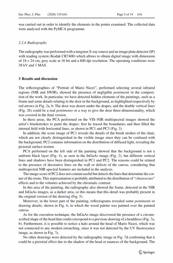
The reflectographies of “Portrait of Mario Nuzzi”, performed selecting several infraredregions (NIR and SWIR), showed the presence of negligible pentimenti in the composi-tion of the work. In particular, we have detected hidden elements of the paintings, such as aframe and some details relating to the door in the background, as highlighted respectively byred arrows in Fig. 2a, b. The door was drawn under the drapes, and the double vertical lines(Fig. 2b) could be a real pentimento or a way to give the door three-dimensionality, whichwas covered in the final version.
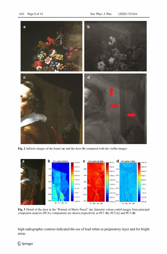
In these areas, the PCA performed on the VIS–NIR multispectral images showed theartist’s brushstrokes to paint the drapes: first he traced the boundaries and then filled theinternal field with horizontal lines, as shown in PC1 and PC3 (Fig. 3).
In addition, the score image of PC1 reveals the details of the brush strokes of the chair,which are not clearly distinguished in the visible image since they can be confused withthe background. PC2 contains information on the distribution of diffused light, revealing thepictorial surface texture.
PCA performed on the left side of the painting showed that the background is not auniform black layer (Fig. 4), as seen in the InGaAs image (Fig. 2), but different verticallines and shadows have been distinguished in PC1 and PC2. The reasons could be relatedto the presence of decorative lines on the wall or defects of the canvas, considering thatmultispectral NIR spectral features are included in the analysis.
The image score of PC2 does not contain useful but detects the lines that determine the cor-ner of the room. This representation is probably attributed to the distribution of “chiaroscuro”effects and to the volumes achieved by the chromatic contrast.
In this area of the painting, the radiography also showed the frame, detected in the NIRand InGaAs images, as a darker area, so this means that this detail was probably present inthe original version of the drawing (Fig. 5).
Moreover, in the lower part of the painting, reflectograms revealed some pentimenti ordrawing details, shown in Fig. 6, in which the wood palette was painted over the paintedcanvas.
As for the execution technique, the InGaAs image discovered the presence of a circum-scribed shape of the head that could correspond to a previous drawing of a headdress (Fig. 7a,b). Furthermore, it is possible to notice a halo around the head of Mario Nuzzi, which wasnot connected to any modern retouching, since it was not detected by the UV fluorescenceimage, as shown in Fig. 7c.
No other drawings were detected by the radiographic image in Fig. 7d confirming that itcould be a pictorial effect due to the shadow of the head or nuances of the background. The
123
616 Page 6 of 14 Eur. Phys. J. Plus (2020) 135:616
Fig. 2 InGaAs images of the frame (a) and the door (b) compared with the visible images
Fig. 3 Detail of the door in the “Portrait of Mario Nuzzi” (a). Intensity colour-coded images form principalcomponent analysis (PCA); components are shown respectively as PC1 (b), PC2 (c) and PC3 (d)
high radiographic contrast indicated the use of lead white as preparatory layer and for brightareas.
123

Eur. Phys. J. Plus (2020) 135:616 Page 7 of 14 616
Fig. 4 Detail of the frame in the “Portrait of Mario Nuzzi” (a). Intensity colour-coded images form principalcomponent analysis (PCA); components are shown respectively as PC1 (b), PC2 (c) and PC3 (d)
Fig. 5 Details of the frame in the “Portrait of Mario Nuzzi” (a) and radiography (b)
Fig. 6 Detail of the “Portrait of Mario Nuzzi” (a) and corresponding InGaAs image (b)
123

616 Page 8 of 14 Eur. Phys. J. Plus (2020) 135:616
Fig. 7 Details of the Mario Nuzzi’s face (a), InGaAs camera (b), UV fluorescence image (c), radiography (d)
In this area, the PCA performed on the VIS–NIR multispectral images, shown in Fig. 8,highlighted the distribution of the volumes given by light and shadows. In particular, PC1showed the distribution of the high contrasted areas while the halftones are well definedin PC2 and PC3. Furthermore, the image score of PC2 describes the distribution of thescattered light revealing in particular the pictorial surface texture. The spectral behaviour ofthe pictorial surface texture, achieved by using PCA calculated by VIS–NIR multispectralimages, depends on the preparatory layer, which could partially or completely cover thetexture of the canvas and on the spectral response of the pigments used for the pictoriallayers. These latter two characteristics could be attributed to a specific way of painting.
The possibility to scientifically define the use of “chiaroscuro” for each artist could beconsidered a useful step during the authentication process and represents an interesting chal-lenge for the researchers. Therefore, the representation of the “scores density plot”, obtained
123

Eur. Phys. J. Plus (2020) 135:616 Page 9 of 14 616
Fig. 8 Detail of the frame in the “Portrait of Mario Nuzzi” (a). Intensity colour-coded images form principalcomponent analysis (PCA); components are shown respectively as PC1 (b), PC2 (c) and PC3 (d)
Fig. 9 Score density plot (PC1 vs PC2) and pixel distribution (in red) of selected scores values (1, 2, 3, 4, 5areas of the score density plot)
from the combination of PCA and VIS–NIR multispectral images, here discussed, could beuseful to solve this problem.
The scores density plot describes how the score values are distributed graphically in thenew coordinate system (described, for example, by PC1 versus PC2). The scores density plotof PC1 versus PC2 is reported in Fig. 9. It shows the distribution of the scores values for eachpixel of the spectral hypercube in the space of the components PC1 and PC2. False coloursrepresent a density scale: from red (high amount of pixels with the same score values) toblue (few pixels with the same score values). For the portrait of Mario, most of the scores aredistributed around the “zero” value of PC2, which means that PC2 does not explain a highpercentage of variance and all the differences, in terms of “chiaroscuro”, could be observedas distribution of PC1 values. Indeed, a large quantity of “dark” pixels (Fig. 9, area 1) arelocalized at negative values of PC1, while the white pixels are correlated to high PC1 values(area 3). The areas 2, 4 and 5 are related to the half tones.
As for the artist’s palette, the VIS–NIR multispectral acquisitions revealed that the blueand purple hues are characterized by an intense reflection in the infrared range. This aspect
123

616 Page 10 of 14 Eur. Phys. J. Plus (2020) 135:616
Fig. 10 Detail of the “Portrait of Mario Nuzzi” (a) and False colour image (b)
Fig. 11 Detail of the flowers in the “Portrait of Mario Nuzzi” (a). Intensity colour-coded images from principalcomponent analysis (PCA); components are shown respectively as PC1 (b), PC2 (c) and PC3 (d)
is best highlighted by the false colour image in which these shades appear red, as shown inFig. 10.
Since the portrait area was realized by Giovanni Maria Morandi and the flowers by MarioNuzzi, we performed a PCA on the VIS–NIR multispectral images in order to find anydifferences in the pigments used for the “real flowers” and the “painted flowers”. The PCA,in this case, was unable to find significant variations, as shown in Fig. 11. This could berelated to a limitation of the technique used or to selection of filters or to the presence ofmixtures.
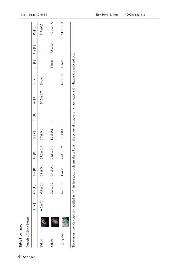
For this reason, we performed XRF spectroscopy in several areas of the painting and theresults were compared with those obtained on the “Primavera” painting. A selection of theresults was reported in Table 1.
As for the two paintings, the presence of lead (Pb) and calcium (Ca) suggests the use ofstandard preparatory layers: a gypsum-based layer to cover the heterogeneities of the support(canvas) and a white lead pigment as a preparatory layer. In addition, all areas analysed werecharacterized by low copper (Cu) counts, indicating the use of a copper-based pigment asthe background layer for both paintings.
123

Eur. Phys. J. Plus (2020) 135:616 Page 11 of 14 616
Table1
Som
ere
pres
enta
tive
XR
Fda
ta.T
heX
RF
coun
tsar
ere
port
edas
perc
enta
ges
afte
rbe
ing
norm
aliz
edto
Wco
unts
,whi
chis
intr
oduc
edby
the
inst
rum
enta
lapp
arat
usus
ed
Prim
aver
a
Ana
lyse
dpo
int
K[K
]C
a[K
]T
i[K
]C
r[K
]M
n[K
]Fe
[K]
Cu
[K]
Sr[K
]Sn
[L]
Hg
[L]
Pb[L
]
Whi
te0.
2±0
.10.
9±0
.2–
––
2.4±0
.32.
1±0
.3–
––
94.3±1
.9
Bro
wn
0.3±0
.13.
3±0
.3–
–2.
3±0
.337
.4±1
.01.
1±0
.2–
––
55.5±1
.2
Yel
low
–2.
5±0
.4–
––
19.2±0
.93.
5±0
.4–
1.2±0
.4–
74.5±1
.8
Red
0.5±0
.24.
0±0
.5–
––
40.8±1
.45.
4±0
.6–
–21
.1±1
.128
.3±1
.2
Lig
htgr
een
0.4±0
.11.
2±0
.20.
8±0
.20.
5±0
.20.
5±0
.229
.7±0
.81.
3±0
.2–
––
65.6±1
.2
Red
0.7±0
.11.
7±0
.2–
––
33.8±0
.812
.3±0
.5–
––
51.5±1
.0
Port
rait
ofM
ario
Nuz
zi
K[K
]C
a[K
]M
n[K
]Fe
[K]
Cu
[K]
Zn
[K]
As
[K]
Sr[K
]Sb
[L]
Hg
[L]
Pb[L
]
Red
–2.
3±0
.4–
5.9±0
.62.
7±0
.5–
––
–72
.0±2
.117
.0±1
.1
Bro
wn
–3.
6±0
.31.
7±0
.338
.2±1
.12.
7±0
.3–
–0.
7±0
.2–
–53
.2±1
.3
Red
–1.
8±0
.211
±0.2
30.8±0
.92.
8±0
.3–
–1.
8±0
.2–
1.7±0
.359
.9±1
.2
Gre
en0.
3±0
.10.
6±0
.10.
2±0
.125
.1±0
.545
.0±0
.73.
7±0
.2–
0.3±0
.1–
1.1±0
.223
.7±0
.5
123
616 Page 12 of 14 Eur. Phys. J. Plus (2020) 135:616
Table1
cont
inue
d
Port
rait
ofM
ario
Nuz
zi
K[K
]C
a[K
]M
n[K
]Fe
[K]
Cu
[K]
Zn
[K]
As
[K]
Sr[K
]Sb
[L]
Hg
[L]
Pb[L
]
Yel
low
0.2±0
.10.
8±0
.10.
8±0
.135
.5±0
.50.
7±0
.1–
59.2±0
.7T
race
s–
–2.
7±0
.2
Yel
low
–3.
0±0
.30.
4±0
.138
.5±0
.81.
3±0
.2–
––
Tra
ces
7.5±0
.449
.1±1
.0
Lig
htgr
een
–6.
9±0
.4T
race
s26
.8±0
.93.
3±0
.3–
–1.
3±0
.2T
race
s–
61.4±1
.3
The
elem
ents
notd
etec
ted
are
labe
lled
as“–
”.In
the
seco
ndco
lum
n,th
ere
ddo
tin
the
cent
reof
imag
esis
the
lase
rtr
ace
and
indi
cate
sth
ean
alys
edpo
int
123

Eur. Phys. J. Plus (2020) 135:616 Page 13 of 14 616
The white areas of the paintings are characterized by the presence of lead, probablyattributed to lead white pigment, while the red parts were constituted by mercury (Hg) andiron (Fe), indicating a mixture of cinnabar and iron-based pigment, such as earth pigments.
Other similarities in the elemental composition are relative to shadows, or generally tothe brown areas, made up of manganese (Mn). This element is a marker of umber earth (ironand manganese oxides and hydroxides) widely used in paintings [22].
For yellow pigments, a high iron count has been detected which indicates the use ofiron-based compounds; however, we also detected antimony (Sb) or arsenic (As) in someareas: arsenic was found in the darker areas, suggesting the use of orpiment, while antimony(detected in traces) could be related to lead antimonate yellow, called Naples yellow. Thesetwo elements were not found in the painting “Primavera”, which instead showed the presenceof tin (detected both in the figure and the flowers), generally linked to the use of lead tinoxide.
Yellow pigments based on lead, tin and antimony are still an exciting field of researchbecause these pigments seem to be used differently by the different pictorial circles of anartist or his workshop [23]. The presence of antimony could be very interesting due to thefact that during XVII century lead tin yellows, widely used between XIV and XVII centuries,gradually turned into lead antimonate (Naples) yellows [24].
From these preliminary results, it appears that Morandi preferred arsenic or antimonyyellow pigments, while Nuzzi and Lauri used tin yellow pigments.
Furthermore, the green areas of the “Portrait of Mario Nuzzi”, painted by Morandi, arecharacterized by the presence of copper and iron-based pigments. In some cases, we havefound the presence of zinc which could suggest the use of verdigris [25] or rosasite [26].
In contrast, the “Primavera” painting did not show a significant amount of copper. Inparticular, in this painting the light green areas (i.e. Flora’s dress) showed the presence ofchrome and iron. This aspect could suggest the use of Verona earth pigment [27].
4 Conclusions
A non-invasive approach has been adopted to analyse the painting of “Portrait of Mario Nuzzi”in order to gather information on the presence of regrets and the artist’s palettes. Some detailsof the execution technique have been revealed by conventional infrared reflectography andradiography, while others (i.e. the brush strokes directions and the effects of “chiaroscuro”)were discovered by PCA performed on VIS–NIR multispectral images.
We have shown that this approach could represent a powerful method to improve the studyof an artist’s “chiaroscuro”, through the use of different spectral ranges from the visible tothe near infrared, making it possible to represent this stylistic element through measurablequantities. The graphical representation of the score density diagram from the PCA couldbe used as a painter’s fingerprint. Furthermore, since Mario Nuzzi has collaborated withother contemporary artists, the reconstruction of the areas he painted with respect to thosemade by his “collaborators” could be important information for art historians to support theirstylistic observations. To this end, we have analysed the palette of two paintings (“Portraitof Mario Nuzzi” and “Primavera”) using XRF measurements. We found a similar elementalcomposition, except for the yellow and green hues.
These differences suggest that it could be extremely important to carry out other analyseson Mario Nuzzi’s paintings located in Palazzo Chigi, in order to better understand this artist’spalettes and the execution techniques and to bring new insight on their collaboration.
123
616 Page 14 of 14 Eur. Phys. J. Plus (2020) 135:616
Acknowledgements We acknowledge the funding of Regione Lazio under the ADAMO Project No.B86C18001220002 of the Centre of Excellence at the Technological District for Cultural Heritage of Lazio(DTC). The authors want to acknowledge all the staff of Palazzo Chigi for their support during the mea-surement campaigns. We are grateful to A. Raco, G. Viviani, A. Grilli and M. Pietropaoli for the technicalsupport. Finally, we acknowledge Dr. Roberta Fantoni responsible of the ADAMO project, and ProfessorMauro Missori, WP leader.
References
1. S. Laureti, C. Colantonio, P. Burrascano, M. Melis, G. Calabrò, H. Malekmohammadi, S. Sfarra, M. Ricci,C. Pelosi, J. Cult. Herit. 40, 1–16 (2019)
2. N. Carter, S. Martin, J. Mod. Ital. Stud. 22, 338–364 (2017)3. T. Cavaleri, P. Buscaglia, S. Migliorini, M. Nervo, G. Piccablotto, A. Piccirillo, M. Pisani, D. Puglisi, D.
Vaudan, M. Zucco, Appl. Phys. A Mater. Sci. Process. 123, 419 (2017)4. B.A. Driel, K.J. van den Berg, J. Gerretzen, J. Dik, Herit. Sci. 6, 16 (2018)5. D. Burchette-Lere, A. Zebala, Sam Francis: the Artist’s Materials (Getty Conservation Institute, Los
Angeles, 2019, 168)6. M. Geldof, A.N. Proaño Gaibor, F. Ligterink, E. Hendriks, E. Kirchner, Herit. Sci. 6, 1–20 (2018)7. J. Asmus, V. Parfenov, J. Cult. Herit. 38, 167–173 (2019)8. V. Golzio, L’Urbe 1, 1–19 (1965)9. F. Petrucci, Dipinti del barocco romano da Palazzo Chigi in Ariccia (Gangemi, Roma, 2013, 112)
10. G. Bonifazi, G. Capobianco, C. Pelosi, Serranti. J. Imaging 5(1), 8 (2019)11. A. Hayem-Ghez, E. Ravaud, C. Boust, G. Bastian, M. Menu, N. Brodie-Linder, Appl. Phys. A Mater.
Sci. Process. 121, 939–947 (2015)12. G. Capobianco, M.P. Bracciale, D. Sali, F. Sbardella, P. Belloni, G. Bonifazi, S. Serranti, M.L. Santarelli,
M. Cestelli Guidi, Microchem. J. 132, 69–76 (2017)13. L. Fragasso, N. Masini, Int. J. Geophys. (2011). https://doi.org/10.1155/2011/73827914. L. Pronti, M. Romani, G. Verona-Rinati, O. Tarquini, F. Colao, M. Colapietro, A. Pifferi, M. Cestelli-
Guidi, M. Marinelli, Heritage 2, 2275–2286 (2019)15. L. Pronti, A.C. Felici, M. Ménager, C. Vieillescazes, M. Piacentini, Appl. Spectrosc. 71, 2616–2625
(2017)16. J.R. Mansfield, M. Attas, C. Majzels, E. Cloutis, C. Collins, H.H. Mantsch, Vib. Spectrosc. 28, 59 (2002)17. L. De Viguerie, S. Rochut, M. Alfeld, P. Walter, S. Astier, V. Gontero, F. Boulch, Herit. Sci. 6, 11 (2018)18. C. Cucci, J.K. Delaney, M. Picollo, Acc. Chem. Res. 49, 2070–2079 (2016)19. N. Mobaraki, J.M. Amigo, Chemom. Intell. Lab. Syst. 172, 174–187 (2018)20. R. Bro, A.K. Smilde, Anal. Methods 6, , 2812–2831 (2014)21. M.E. Wall, A. Rechtsteiner, M.L. Rocha, A Practical Approach to Microarray Data Analysis (Springer,
Boston, 2003, 368)22. K. Trentelman, K. Janssens, G. van der Snickt, Y. Szafran, A.T. Woollett, J. Dik, Appl. Phys. A Mater.
Sci. Process. 121, 801–811 (2015)23. G. Agresti, P. Baraldi, C. Pelosi, U. Santamaria, Color Res. Appl. 41, 226–231 (2016)24. D. Hradil, T. Grygar, J. Hradilová, P. Bezdicka, V. Grunwaldová, I. Fogaš, C. Miliani, J. Cult. Herit. 8,
377 (2007)25. J. Buse, V. Otero, M. Melo, Heritage 2, 1614–1629 (2019)26. E. Martin, M. Eveno, In: BetaGamma (ed.) III Confererenza Internazionale sulle prove non distruttive,
motodi microanalitici e indagini ambientali per lo studio e la conservazione delle opera d’arte, Viterbo,1992, edited by M. Marabelli and P. Santopadre (1992), 837–856
27. M. Sbroscia, M. Cestelli-Guidi, F. Colao, S. Falzone, C. Gioia, P. Gioia, C. Marconi, D. Mirabile Gattia,E.M. Loreti, M. Marinelli, M. Missori, F. Persia, L. Pronti, M. Romani, A. Sodo, G. Verona-Rinati, M.A.Ricci, R. Fantoni, Microchem. J. 153, 104450 (2020)
123