New insights into the thioredoxindependent redox ...
232
New insights into the thioredoxin-dependent redox regulation in the cyanobacterium Synechocystis sp. PCC 6803 Trabajo presentado para optar al grado de Doctor en Ciencias Biológicas por el Licenciado Alejandro Mata Cabana Sevilla, septiembre 2010 Directores: Dr. Francisco Javier Florencio Bellido Catedrático de Bioquímica y Biología Molecular Dra. Anna Marika Lindahl Científico Titular del Consejo Superior de Investigaciones Científicas
Transcript of New insights into the thioredoxindependent redox ...

Newinsightsintothethioredoxindependentredox
regulationinthecyanobacteriumSynechocystissp.PCC6803
Trabajopresentadoparaoptaralgradode
DoctorenCienciasBiológicasporelLicenciado
AlejandroMataCabana
Sevilla,septiembre2010
Directores:
Dr.FranciscoJavierFlorencioBellidoCatedráticodeBioquímicayBiología
Molecular
Dra.AnnaMarikaLindahlCientíficoTitulardelConsejoSuperiorde
InvestigacionesCientíficas


3
“Eltiempoeselladróndelamemoria"LatorreoscuraI.Elpistolero
StephenKing
“Laúnicarazónporlaqueunapersonaescribeunahistoria,esporqueatravésdeellapuede
entenderelpasado”StephenKing
"Time'sthethiefofmemory"TheDarkTowerI.TheGunslinger
StephenKing
“Theonlyreasonapersonwritesastory,becausethroughityoucanunderstandthepast”
StephenKing


5
INDEX


Index
7
INDEX
INDEX 7
FIGUREINDEX 11
TABLEINDEX 12
ABREVIATIONS 13
INTRODUCTION 17
1.Cyanobacteria 19
1.1Synechocystissp.PCC6803 22
1.1.1 StructureofSynechocystissp.PCC6803 22
2.Photosynthesisandoxidativestress 26
2.1Photosynthesis 26
2.2Productionofreactiveoxygenspeciesandtheconceptofoxidativestress 29
3.Redoxsignallingandredoxregulationincyanobacteria 33
3.1Cysteinereactivity 33
3.2Cyanobacterialthioredoxinsystems 36
4.Disulphideproteomes 40
4.1Methodology 41
4.2Analysisofdisulphideproteomeinprokaryotes(Exceptcyanobacteria) 45
4.3ThedisulphideproteomeinPhotosyntheticorganisms(PlantsandAlgae) 51
4.4ThedisulphideproteomeinCyanobacteria 55
OBJECTIVES 61
RESULTS 65
IMEMBRANEPROTEINSFROMTHECYANOBACTERIUMSynechocystissp.PCC6803
INTERACTINGWITHTHETHIOREDOXIN 67
Introduction 69
Resultsanddiscussion 70
1.SubcellularlocalisationofTrxA 70
2.Isolationofmembraneproteinsinteractingwiththioredoxin 72
3.Resolutionandidentificationofmembrane‐associatedthioredoxintargetproteins 76
4.Interactionsof1‐Cys‐and2‐CysperoxiredoxinswithTrxAinSynechocystis 88

AlejandroMataCabana
8
5.TheUniversalStressProtein(USP)anditsinteractionwithTrx 94
6.TheFtsHprotease 99
IITHIOREDOXINTARGETSINTHETHYLAKOIDLUMEN 105
Introduction 107
Resultsanddiscussion 109
1.ProteomicidentificationofputativeTrx‐targetsfromtheArabidopsischloroplastlumen109
2.ThePrxQ2interactionwithTrxAinSynechocystis 114
IIITHIOREDOXINMEDIATEDREDOXREGULATIONOFAEUKARYOTETYPESer/ThrKINASE
INTHECYANOBACTERIUMSynechocystissp.PCC6803 119
Introduction 121
Resultsanddiscussion 124
1.RedoxdependentproteinphosphorylationinSynechocystis 124
2.SpkBredoxregulation 130
3.IdentificationofatargetforSpkBphosphorylation 137
4.SpkBCys‐motifinvolvementinredoxregulation 140
DISCUSSION 145
I‐MembraneproteinsfromthecyanobacteriumSynchocystissp.PCC6803interactingwiththe
thioredoxin 147
II–Thioredoxintargetsinthethylakoidlumen 154
III–Thioredoxin‐mediatedredoxregulationofaeukaryotetypeSer/Thrkinaseinthe
cyanobacteriumSynechocystissp.PCC6803 157
MATERIALSANDMETHODS 163
1. Organismsandcultureconditions 165
1.1. Cyanobacteria 165
1.1.1. Cyanobacterialstrains 165
1.1.2 Cyanobacterialculturemediumandconditions 166
1.2. Escherichiacoli 167
1.2.1. StrainsofE.coli 167
1.2.2. E.coliculturemediumandconditions 167
1.3. Harvestingofcells 168
2. DNAanalysisandmanipulation 168
2.1. Plasmidsandprimers 168
2.2. DNAisolation 172
2.2.1. PlasmidDNAisolationfromE.coli 172
2.2.2. CyanobacterialgenomeDNAisolation 173
2.3. DNAanalysisandquantification 173

Index
9
2.3.1. DNAelectrophoresisinagarosegels 173
2.3.2. DNAquantification 174
2.3.3. DNAextractionfromagarosegels 174
2.3.4. EnzymaticmanipulationofDNA 174
2.4. PolymeraseChainReaction(PCR) 174
2.5. DNAsequencing 175
2.6. IntroductionofDNAintodifferentorganismsbytransformation 175
2.6.1. E.colitransformation 175
2.6.2. Synechocystistransformation 176
2.7. Sitedirectedmutagenesis 177
3. Proteinsynthesis,purificationandanalysis 178
3.1. ProteinexpressioninE.coli 178
3.2. Preparationofcellextracts 178
3.2.1. Celllysisusingglassbeads 178
3.2.2. Celllysisbysonication 179
3.2.3. PreparationofSynechocystistotalmembranes 179
3.3. Proteinquantification 179
3.4. Proteinelectrophoresis 180
3.4.1. 1‐DSDS‐PAGE 180
3.4.2. Twodimensionalproteinelectrophoresis 181
3.4.2.1. Non‐reducing/reducing2‐DSDS‐PAGE 181
3.4.2.2. 2‐DIEF/SDS‐PAGE 181
3.5. Proteinstaining 182
3.6. Proteinidentificationbypeptidemassfingerprinting(PMF) 182
3.7. Proteininmunodetection(Westernblot) 183
3.8. Proteingeldrying 184
3.9. Detectionofradioactivity 184
3.10. Proteinpurification 184
3.10.1. Metalaffinitychromatography 184
3.10.2. Gelfiltration 185
3.10.3. Proteinspurifiedinthiswork 185
3.10.4. Trx‐targetisolation 186
3.10.4.1. Isolationofmembrane‐boundTrx‐targetproteincomplexes 186
3.10.4.2. IsolationoflumenalTrx‐targetproteinscomplexes 187
3.11. Proteinconcentration 187
3.12. Productionofpolyclonalantibodies 187
4. Enzymaticassays 188
4.1. Peroxidaseassay 188
4.2. ProteinphosphorylationincellextractsofSynechocystis 188
4.3. Assaysforproteinkinaseactivity 189

AlejandroMataCabana
10
5. Bioinformaticmethods 189
5.1. DNAandProteinsequencesanalysis 189
6. Othermethods 190
6.1. Determinationofchlorophyllconcentration 190
6.2. Spectrophotometricmeasurements 191
6.3. pHmeasurements 191
CONCLUSIONS 193
BIBLIOGRAPHY 197

Index
11
FIGUREINDEX
Figure1.MorphologicaldiversityofCyanobacteria 20
Figure2.Synechocystiscellstructure 25
Figure3.Photosyntheticelectrontransfer 27
Figure4.Phycobiliome 29
Figure5.Reactivecysteinemodifications 35
Figure6.Trxreductionsystems 40
Figure7.MechanismofTrx‐targetreduction 43
Figure8.IsolationofTrx‐targetsbyTrxaffinitychromatography 44
Figure9.DistributionoftheSynechocystissp.PCC6803thioredoxinTrxAbetweensolubleand
membranefractions 71
Figure10.Schematicrepresentationoftheprocedureforisolationofmembraneproteins
interactingwiththioredoxin 73
Figure11.Analysisof1‐DEproteinprofilesfromtheprocessofisolationofSynechocystissp.PCC
6803membraneproteinsinteractingwiththioredoxin 75
Figure12.Resolutionofmembrane‐associatedthioredoxintargetproteinsby2‐DSDS‐PAGEunder
non‐reducing/reducingconditions 77
Figure13.Trx‐targetsdistributioninSynechocystis 84
Figure14.CysteinelocalisationwithintheaminoacidsequencesoftheAAA+‐protein 86
Figure15.2‐CysPrxlocation 90
Figure16.Electrophoreticmigrationof2‐CysPrxand1‐CysPrx 91
Figure17.Analysisoftheinteractionbetweentheperoxiredoxins2‐CysPrxand1‐CysPrxwiththe
TrxA 92
Figure18.TrxA‐dependentperoxidaseactivityof2‐CysPrxand1‐CysPrx 93
Figure19.PositionsofUsp1andUsp2inthe2‐DSDS‐PAGEundernon‐reducing/reducing
conditions 95
Figure20.ReductionofUsp1(slr0244)byDTTandTrxA 96
Figure21.Re‐oxidationofUsp1(slr0244)usingH2O2andCu2+ 97
Figure22.AutophosphorylationofUsp1(slr0244)andtheeffectofthethiolredoxstate 98
Figure23.PresenceofFtsHintheeluateofFractionIII 101
Figure24.TrxA35interactionwiththeGST‐FtsH2fusion 103
Figure25.AgrowthoftheSynechocystisFtsHmutantstrainsuponHighLightconditions 104
Figure26.ThylakoidTrx‐targetsisolation 111
Figure27.ElectrophoreticPrxQ2gelmigration 115
Figure26.CyanobacterialPrxQaminoacidsequencescomparison 117
Figure27.AnalysisofthePrxQ2‐TrxAinteraction 118
Figure30.TrxA‐dependentperoxidaseactivityofPrxQ2 118

AlejandroMataCabana
12
Figure31.PhosphorylationofSynechocystisproteinsinseparatemembranes/solublefraction 125
Figure32.PhosphorylationsensibilitytooxidantagentsofSynechocystiscytosolicproteins 126
Figure33.Reactivationofthe90kDaproteinphosphorylationbyreductantagents 127
Figure34.Pkn2kinasesofSynechocystis 129
Figure35.AlignmentofSpkBhomologues 131
Figure36.ActivityofSpkBkinaseexpressedinE.coli 133
Figure37.Kinaseactivityofpartially‐purifiedSpkBkinaseexpressedinE.coli 135
Figure38.ΔSpkBmutantcomplementationwithadditionofexogenouspurifiedSpkB 136
Figure39.SpkBsubstrateidentification 138
Figure40.SpkBphosphorylatesGlySinvitro 140
Figure41.KinaseactivityoftheβversionofSpkB 141
Figure42.Kinaseactivityreactivationofβ‐SpkB 143
TABLEINDEX
Table1.ThioredoxintargetproteinsidentifiedinSynechocystissp.PCC6803. 55Table2.ProteinsindicatedinFigure11AidentifiedbyPMF 74Table3.Synechocystissp.PCC6803membraneproteinsinteractingwiththioredoxin 78Table4.LocalisationofSynechocystissp.PCC6803membraneproteinspresentinourstudy 82Table5.TrxtargetsdetectedinthelumenalchloroplastofArabidopsis 111Table6.SynechocystisproteinshomologoustotheTrxlumenaltargetsidentifiedinArabidopsis113Table7.Synechocystismutantstrainsusedinthethesis 165Table8.StainsofE.coliusedinthiswork 167Table9.Commercialplasmidsandgifts 169Table10.Plasmidsconstructedinthiswork 169Table11.Syntheticprimersusedinthiswork 171Table12.Proteinspurifiedduringthecourseofthiswork 185

Index
13
ABREVIATIONS
1‐DE Onedimensionelectrophoresis
2‐DE Twodimensionelectrophoresis
AAA+ ATPasesAssociatedwithavarietyofcellularActivities
ADP Adenosinediphosphate
Ap Ampicillin
ATP Adenosinetriphosphate
ATS Activatedthiol‐Sepharose
BSA Bovineserumalbumin
CBB Coomassiebrilliantblue
Ci Curie
DNA Deoxyribonucleicacid
dNTPs Deoxyribonucleotidetriphosphatemix
DTT Dithiothreitol
E Einstein
EDTA Ethylenediaminetetraaceticacid
FAD FlavinAdenineDinucleotide
FPLC FastProteinLiquidChromatography
FTR FerredoxindependentThioredoxinReductase
g Gravity(acceleration)
gDNA GenomicDNA
Grx Glutaredoxin
GS Glutaminesynthetase
GSH Glutathione
GSSG Oxidisedglutathione
GST GlutathioneS‐transferase
GTP Guanosinetriphosphate
HEPES 4‐(2‐hydroxyethyl)‐1‐piperazineethanesulfonicacid
IAM Iodoacetamide
ICAT Isotope‐CodedAffinityTag
IEF Isoelectricfocusing
IPTG Isopropyl‐β‐ThioGalactopyranoside
kDa Kilodalton
Km Kanamycin
LC‐MS Liquidchromatography‐massspectrometry

AlejandroMataCabana
14
m/z Mass/charge
MALDI‐TOF Matrix‐assistedlaserdesorption/ionization–Timeofflight
Mb Megabase
MS Massspectrometry
mBBr Monobromobimane
NADH Nicotinamideadeninedinucleotide
NADPH Nicotinamideadeninedinucleotidephosphate
NCBI NationalCenterforBiotechnologyInformation
NEM N‐Ethylmaleimide
NTR NADPHdependentThioredoxinReductase
OD Opticaldensity
ORF OpenReadingFrame
PCC PasteurCultureColection
PCR Polymerasechainreaction
Pi Inorganicphosphate
PM Plasmamembrane
PMF Peptidemassfingerprinting
PMSF Phenylmethylsulfonylfluoride
Prx Peroxiredoxin
PSI PhotosystemI
PSII PhotosystemII
RNA Ribonucleicacid
RNS Reactivenitrogenspecies
ROS Reactiveoxygenspecies
rpm Revolutionsperminute
SDS Sodiumdodecylsulfate
SDS‐PAGE Sodiumdodecylsulfatepolyacrylamidegelelectrophoresis
Sp Spectinomycin
St Streptomycin
TM Thylakoidmembrane
tRNA TransferRNA
Trx Thioredoxin
USP Universalstressprotein
v/v Volume/volume
w/v Weight/volume
WT Wildtype

Index
15
X‐gal Bromo‐chloro‐indolyl‐galactopyranoside
NITROGENOUSBASES
A adenine
C cytosine
G guanine
T thymine
AMINOACIDS
A Ala alanine L Leu leucine
R Arg arginine K Lys lysine
N Asn asparagine M Met methionine
D Asp asparticacid F Phe phenilalanine
C Cys cysteine P Pro proline
E Glu glutamicacid S Ser serine
Q Gln glutamine T Thr threonine
G Gly glycine W Trp tryptophan
H His histidine Y Tyr tyrosine
I Ile isoleucine V Val valine


17
INTRODUCTION


Introduction
19
INTRODUCTION
1.Cyanobacteria
Cyanobacteria formadiversegroupofoxygenicphotosyntheticprokaryotes,
which are major contributors to global photosynthetic productivity (Ting et al.,
2002). Cyanobacteria arewidely distributed:many are aquatic, but some are land‐
living, andtheirhabitats includeextremeenvironment, suchashot springs,deserts
and polar regions. Some species are even found in symbiosis with fungi, ferns or
cycads. Cyanobacteria play a central ecological role as primary producers in the
Earth’s carbon cycle. The nitrogen‐fixing species also figure prominently in the
nitrogen cycle. Moreover, as the only organisms ever to have evolved coupled
photosystems,whichharvest electrons fromwaterandproducedioxygen, theyalso
standoutinourplanet’sredoxhistory(Knoll,2008).
Life on Earth originated at least 3500 My ago and possibly much earlier.
Between 2450‐2320 My ago cyanobacteria should have been important primary
producers in the world’s oceans, and they could have emerged even earlier. The
cyanobacterial production of molecular oxygen transformed the environment in a
way that made novel physiologies and morphologies, including animals, possible
(Knoll, 2008). The modern chloroplast is believed to have evolved from
cyanobacteria,throughaprimaryendosymbioticeventbetweenaeukaryoticcelland
an ancestral oxygenic photosynthetic prokaryote. Hence, the photoautotrophy was
introduced to eukaryotes and, then, cyanobacteria contributed further to the
environmentaltransformationoflandaswellasthesea(Knoll,2008).
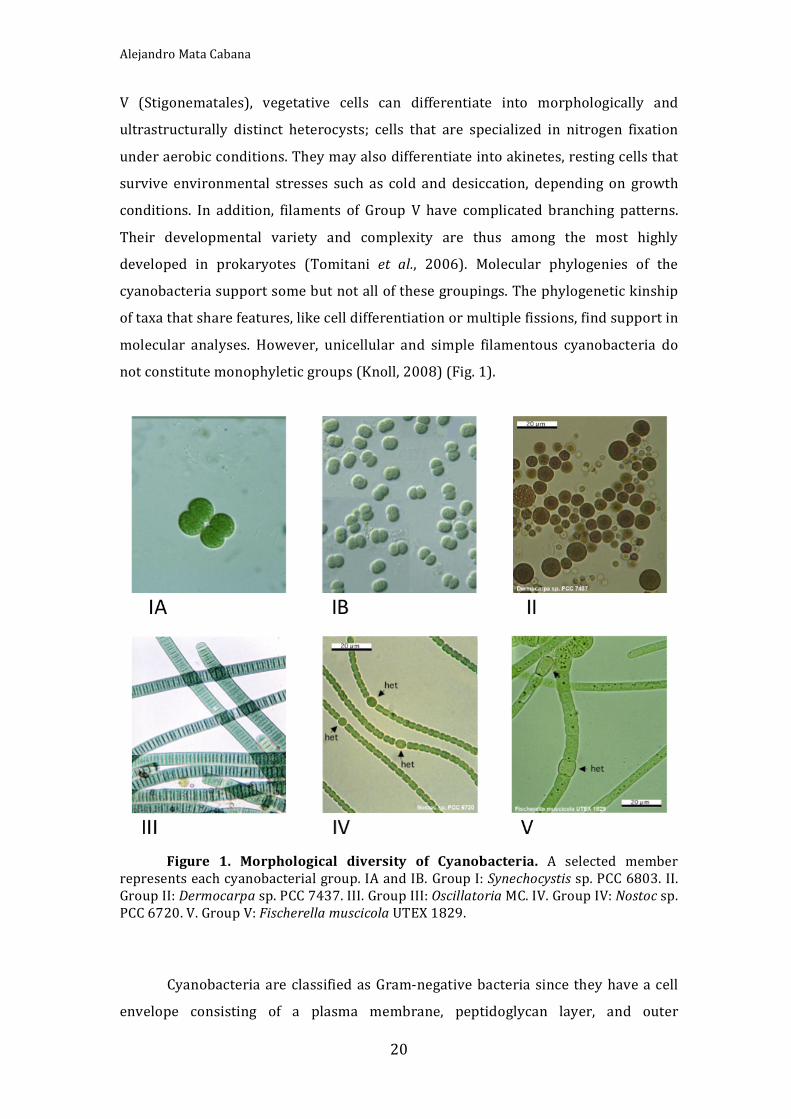
Cyanobacteriaaremonophyleticbutmorphologicallydiverse.Thetraditional
taxonomic classifications focused on morphology and development, and divided
these bacteria into five principal groups (Rippka et al., 1979). Group I (formerly
Chroococcales) and II (Pleurocapsales) are unicellular coccoids. Cells of Group I
dividebybinaryfission,whereasthoseofGroupIIcanalsoundergomultiplefission
toproducesmall,easilydispersedcellscalledbaeocytes.CyanobacteriaofGroupsIII‐
Vformfilamentsofvaryingmorphologicalcomplexity.Filamentouscyanobacteriaof
GroupIII(Oscillatoriales)haveonlyvegetativecells,butinGroupIV(Nostocales)and
AlejandroMataCabana
20
V (Stigonematales), vegetative cells can differentiate into morphologically and
ultrastructurally distinct heterocysts; cells that are specialized in nitrogen fixation
underaerobicconditions.Theymayalsodifferentiateintoakinetes,restingcellsthat
survive environmental stresses such as cold and desiccation, depending on growth
conditions. In addition, filaments of Group V have complicated branching patterns.
Their developmental variety and complexity are thus among the most highly
developed in prokaryotes (Tomitani et al., 2006). Molecular phylogenies of the
cyanobacteriasupportsomebutnotallofthesegroupings.Thephylogenetickinship
oftaxathatsharefeatures,likecelldifferentiationormultiplefissions,findsupportin
molecular analyses. However, unicellular and simple filamentous cyanobacteria do
notconstitutemonophyleticgroups(Knoll,2008)(Fig.1).
Figure 1. Morphological diversity of Cyanobacteria. A selected member
representseachcyanobacterialgroup.IAandIB.GroupI:Synechocystissp.PCC6803.II.GroupII:Dermocarpasp.PCC7437.III.GroupIII:OscillatoriaMC.IV.GroupIV:Nostocsp.PCC6720.V.GroupV:FischerellamuscicolaUTEX1829.
CyanobacteriaareclassifiedasGram‐negativebacteria sincetheyhaveacell
envelope consisting of a plasma membrane, peptidoglycan layer, and outer

Introduction
21
membrane(HoiczykandHansel2000).However,theseorganismsareuniqueamong
Gram‐negative bacteria, because they also have an internal system of thylakoid
membranes,wherethelargeproteincomplexesofthephotosyntheticandrespiratory
electron transfer chains reside. Therefore, the cyanobacteria are considered more
complex than typical Gram‐negative bacteria (Liberton et al., 2006). An ongoing
controversy regarding these organisms has concerned the exactmorphology of the
thylakoid membranes, its biogenesis and its physical relationship with the plasma
membrane(Libertonetal.,2006;vandeMeeneetal.,2006).
From a metabolic view, all cyanobacteria are capable to grow
photoautotrophicallyandsomeofthemcanuseareducedcarbonsourcetodevelopa
heterotrophic ormixotrophic growth. As in plants, the atmospheric CO2 fixation is
performed by the Calvin cycle. However, cyanobacteria have an incomplete Krebs
cycle because of the lack of the 2‐oxoglutarate dehydrogenase enzymatic complex.
This feature gives an anabolic role to the Krebs cycle owing to the fact that 2‐
oxogutarate provides carbon skeletons for nitrogen assimilation. The oxidative
pentosephosphatecycle is themainmetabolicpathwayofcarbohydratecatabolism
(Stanier and Cohen‐Bazire, 1977). Moreover, cyanobacteria can use nitrate, nitrite
and ammonium as nitrogen source (Guerrero and Lara, 1987). However, some
cyanobacterial strains are also able to use urea, certain aminoacids, or molecular
atmosphericnitrogen(N2)(Stewart,1980;FloresandHerrero,1994).TheN2‐fixing
strains need to separate the nitrogen fixation and photosynthesis to avoid the
irreversible inactivation of the nitrogenase enzyme by molecular oxygen. This
separation can be achieved temporally (Fay, 1992) or spatially, by differentiating
somevegetativecellsintoheterocystswheretheN2assimilationisperformed(Wolk,
1982).
From 1996 until now several full genome sequences of different
cyanobacterial species have been made public (Cyano‐Base,
www.kazusa.or.jp/cyano/cyano.html).Thegenomeoftheunicellularcyanobacterium
Synechocystissp.PCC6803(Kanekoetal.,1996)wasthefirsttobesequencedfroma
photosynthetic organism. The G+C content is very variable and ranges from 32 to
71%. Most of the studied strains are highly polyploid, having around 12 copies of
chromosomepercell(Labarreetal.,1989).Thesizesofthecyanobacterialgenomes

AlejandroMataCabana
22
rangefrom9.20Mb(Nostocpunctiforme)to1.66Mb(ProchlorococcusMED4),which
isconsideredtheminimumgenomesizerequiredforencodingthecomponentsofthe
oxygenic photosynthetic apparatus in addition to the enzymes of basicmetabolism
(Dufresneetal.,2005).Thegenomesequenceof thefilamentousheterocyst‐forming
nitrogen‐fixing cyanobacterium Anabaena sp. PCC 7120 (CyanoBase) has provided
valuable information about the genes related to heterocyst differentiation and the
processofnitrogenfixation(Kanekoetal.2001).Manycyanobacteriaarecapableof
integrating exogenous DNA stably into their genome (transformation) bymeans of
homologous recombination processes (Porter, 1986). In addition, some genetic
transfer techniques have been developed, such as conjugation (Wolk et al., 1984;
Flores and Wolk, 1985) or electroporation (Thiel and Poo, 1989). These features
makecertaincyanobacterialstrainsadequatemodelorganismsformolecularstudies.
The cyanobacterium used to carry out the present research was the
unicellularSynechocystissp.PCC6803,whichbelongstothecyanobacterialGroupI.
1.1Synechocystissp.PCC6803
Synechocystis sp. PCC 6803 (hereafter Synechocystis) is a model organism,
which displays a unique combination of highly desirable molecular genetic,
physiological, and morphological characteristics. This species is spontaneously
transformable, incorporates foreign DNA into its genome by double‐homologous
recombination,growsundermanydifferentphysiologicalconditions(i.e.,photoauto‐,
mixo‐ and heterotrophycally), and has a short duplication time. These features
combined with the fact that its entire genome has been sequenced (Kaneko et al.,
1996) makes this species an ideal experimental system to answer questions
regarding photosynthesis and the biogenesis and maintenance of thylakoid
membranes,thesitesofsolarenergycaptureandenergytransduction(vandeMeene
etal.,2006).
1.1.1 StructureofSynechocystissp.PCC6803
Synechocystis cells are spherical immediately after cell division. During

Introduction
23
growth, the cells elongate and an incipient septum invaginates into the cytoplasm.
The septum continues to furrow inwards until the cytoplasm is divided producing
two daughter cells. Themature cellwall is comprisedof the periplasmic space, the
peptidoglycan layer, the outer membrane, and the S‐layer. Frequently, a second
division,inaperpendicularplanetothefirst,beginsbeforethetwodaughtercellsare
separated.At all stages of the cell cycles, the pairsof thylakoidmembranesappear
mainly to progress around the cell as concentric layers close to the plasma
membrane. Inaddition, layersof thylakoidmembranesformlarge loopsortraverse
the central cytoplasm. Thylakoidmembranes converge at various sites close to the
cytoplasmicmembrane creating contoured arrays of thesemembranes. During cell
divisionthethylakoidmembranesexpandtoaccommodatetheinvaginatingseptum.
The arrangement of the thylakoid membrane pairs optimizes the surface area for
membrane‐associated processes, such as respiration and photosynthesis, and
accommodatesthelargenumberofproteinscomplexesinvolvedintheseprocesses,
includingphycobilisomes.Themechanisms that regulate thylakoidorganizationare
unknown. However, it is possible that unidentified anchoring and/or cytoskeletal
proteinsareinvolved.(vandeMeeneetal.,2006).(Fig.2)
Thylakoid membrane biogenesis remains an outstanding question in
cyanobacteria. These membranes do not seem to be simple invaginations of the
cytoplasmic membrane, it has been argued that there are not even any physical
connectionsbetween thylakoidandcytoplasmicmembranes (Libertonet al., 2006).
The thylakoid center is adistinctive cylindrical structurewith thylakoidmembrane
sheets associated along its length. These centers are observed in association with
thylakoidmembranepairs intheperipheralregionsofthecell.Inaddition, theyare
closely associated with the cytoplasmic membrane and their cores appear to be
continouswith the periplamic space.The functionof thylakoid centers isunknown,
but likely play a role in the thylakoidmembranes biogenesis (van deMeene et al.,
2006).LipidbodiesareabundantinSynechocystisandtheirdistributionisrestricted
tolocationsbetweenthethylakoidmembranespairsandadjacenttothecytoplasmic
membrane. This location suggests a role in thylakoid maintenance and biogenesis
(vandeMeene,etal.,2006).Itispossiblethatthereareoccasionaltransientdynamic
connections between the twomembrane systems. Another possibility is that lipids
andproteinsmay beexchangedbetween cytoplasmic and thylakoidmembranesby

AlejandroMataCabana
24
vesicletrafficking,butvesicleshavenotbeenobserved inSynechocystis.Howeverin
the cyanobacterium Microcoleus sp. small membrane bound vesicles have been
reported.Nevertheless, theSynechocystis plasmamembrane harbours a homologue
of the Arabidopsis thaliana protein Vipp1 (Vesicle Inducing Protein in Plastids 1),
which is essential for thylakoid biogenesis in plants as well as in Synechocystis
(Mullineux,2008).
The interior of the Synechocystis cell contains components that are readily
observed in most cyanobacterial cells. These include polygonal carboxysomes that
contain the RuBP‐carboxylase enzyme tightly packed in crystalline arrays (Price et
al., 2008). Cyanophycin granules, a nitrogen reserve composed of nonribosomally‐
synthesized arginine and aspartic acid polypeptides (Simon, 1987) are also found.
Other storage structures include polyhydroxyalkanoate (PHA) granules, glycogen
granulesandpolyphosphatebodies,whichfunctioninthestorageofreducedcarbon
andphosphorus,respectively.InthecytoplasmtherearetheDNA‐containingregions
anda largenumberof ribosomes.Ribosomesare inthemajorityexcluded fromthe
areas between the thylakoid membranes, appearing instead in the central
cytoplasmic regionof thecellandbetweenthylakoidandplasmamembranes.They
havealsobeenobservedtobeassociatedwithsheet‐likestructuresintheinteriorof
the cell, which appear to be continuous with the inner thylakoid membranes. This
suggests a role in direct co‐translational protein insertion into the thylakoid
membranes.(Libertonetal.,2006;vandeMeeneetal.,2006).(Fig.2)

Introduction
25
Figure 2. Synechocystis cell structure. A‐B. Standard transmission electronmicroscopy of a non dividing cell (A) and a cell in early division (B). The whitearrowheads indicate the peripheral arrays of thylakoid membrane pairs. The blackasterisk points the convergence of arrays of thylakoid membranes adjacent tocytoplasmic membranes. The cytoplasmic inclusions are marked as: black arrowheads,the carboxysomes; small black arrows, the polyphosphate bodies;white asterisk, a PHAgranule; and large white arrow, a lipid body. In B the small white arrows indicate thephycobilisomes and the large black arrow the invaginating septum of dividing cell. C.Tomographicslicefromadividingcell.Themarkedstructuresare:thylakoidmembranespairs(whitearrowheads);thylakoidmembranesconvergence(blackasterisk);athylakoidcenter(blackarrow);carboxysome(whiteasterisk);ribosomes(blackarrowheads); lipidbodies (white arrows). D. 3‐D Model of C. The structures are coloured differently:thylakoid membranes pairs (green); a thylakoid center (blue); lipid bodies (pink);ribosomes(white);thecarboxysome(yellow),andthecytoplasmaticmembrane(brown).TakenfromvandeMeeneetal.,2006.

AlejandroMataCabana
26
2.Photosynthesisandoxidativestress
2.1Photosynthesis
All higher life on Earth depends on the oxygenic photosynthesis, since this
processproducesalltheoxygenintheatmosphere.Thephotosynthesisconvertsthe
lightenergyfromthesunintochemicalenergy.Theorganismsthatperformoxygenic
photosynthesis are plants, green algae and cyanobacteria. All of them contain two
photosystems,photosystemI(PSI)andphotosystemII(PSII),whichoperateinseries
inthephotosyntheticelectrontransportchain.(Fig.3)
The energy conversions occurring in photosynthetic organisms are divided
into the light reactions, which convert the light energy into an electrochemical
gradient, which is used to synthesise the high‐energy substrates ATP and NADPH,
and the dark reactions that consume ATP and NADPH for the production of
carbohydrates by CO2 fixation. The light reactions take place in the thylakoid
membranes and are catalysed by four large multiprotein complexes: PSI, PSII,
cytochromeb6fandATPsynthase.(Fig.3)
PSIandPSIIcontainthereactioncentres,inwhichthechargeseparationtakes
place. PSII catalyses the water spliting and the electron transfer from water to
plastoquinone(PQ)(VassilievandBruce,2008).Plastoquinoneisasmallliposoluble
molecule that can move freely through the membrane. In each charge separation
event, one electron is extracted from theMn‐cluster, and oxygen is evolved after 4
positivechargeshavebeenaccumulated.During thisprocess,4H+arereleased into
the lumenof thethylakoids.Theelectron is transferredfromthechlorophyllP680 in
PSIIbyachainofelectroncarrierstoPQ. Aftertwochargeseparationevents,PQis
completely reduced, takes up two protons from the cytoplasm and leaves PSII as
plastoquinol(PQH2).ThePQH2servesasmobileelectronandprotoncarrierandisin
constantequilibriumwiththePQpoolinthemembrane.Itdonatestwoelectronsand
two protons to the cytochromeb6f complex,which releases2protons insideof the
thylakoid,andsubsequentlyreduces2moleculesofplastocyanin(PC).Plastocyaninis
a soluble protein located in the lumen of the thylakoids. In some cyanobacteria,
cytochrome c6 replaces PC (De la Rosa et al., 2002). The two additional protons

Introduction
27
pumped across the membrane by the cytochrome b6f complex contribute to the
establishmentofaprotongradientacrossthethylakoidmembrane.ThereducedPC/
cytochromec6serveasmobileelectroncarriersbetweenthecytochromeb6fcomplex
andthePSI.
PSI catalyses the electron transfer across the membrane between the PC/
cytochrome c6 and ferredoxin. When the light energy reaches and excites the
chlorophyll P700 in PSI, an electron is ejected and transferred to a terminal Fe‐S
cluster,locatedatthecytoplasmicsideofPSI.Thereafter,theelectronistransferred
from the Fe‐S cluster to ferredoxin. Under iron deficiency, flavodoxin can replace
ferredoxin as a soluble electron carrier (Nield et al., 2003). The electron transfer
process is completed by the reduction of the P700+ al the lumenal side by the
PC/cytochrome c6. Finally, ferredoxin or flavodoxin transfers the electron to the
ferredoxinNADP+ reductase (FNR),which reducesNADP+ toNADPH. (Frommeand
Grotjohann,2008a;DeRuyterandFromme,2008).
Figure 3. Photosynthetic electron transfer. The electron is transferred fromH2OtoNADPHinthelinealnoncyclicfluxacrossdifferentelements:PSII,photosystemII;PQ/PQH2, oxidised/reduced plastoquinone; Cyt b6f, cytochrome b6f complex; PC/c6,plastocyanin/cytochrome c6; PSI, photosystem I; Fd/Fld, ferredoxin/flavodoxin; FNR,ferredoxinNADP+reductase.Inthecyclicflux(dashline)theelectronmaybetransferredfromFd/FlddirectlytoCytb6fortoPQbymeansoftheFQR,ferredoxin‐quinolreductase.

AlejandroMataCabana
28
The electrochemical proton gradient generated by the electron transfer
reactions isusedbytheATPsynthasetoproduceATPfromADPandPi. In thedark
reactions the ATP and NADPH are used to fix CO2 in the Calvin cycle, and other
anabolicreactions.(FrommeandGrotjohann,2008a;DeRuyterandFromme,2008).
The electron transfer performed by the threemembrane protein complexes,
PSI,PSIIandcytochromeb6f,isalinearnon‐cyclicflux.However,thereisalsoacyclic
alternativepathway, inwhichtheelectron returnstoPQ from ferredoxin througha
ferredoxin‐quinol reductase (FQR) or by direct transfer. Therefore, the cytochrome
b6fpumpsthetwoprotonsintothelumen,therebygeneratingATP,butNADPHisnot
produced. Inthismanner,thecellisthoughttoregulatetheATP/NADPHbalance in
responsetocertainstress(BandallandManasse,1995).
Cyanobacteria contain large membrane attached but extrinsic antenna
complexes, the phycobilisomes.Thephycobilisomepigmentsabsorb strongly in the
550‐660 nm region, thereby complementing the spectral region covered by
chlorophyll a in the blue and red regions. Therefore, the phycobilisome allows
cyanobacteria toabsorbgreen lightand to use the full spectrumofvisible light for
photosynthesis. The phycobilisomesmainly serve as antenna for PSII, but can also
move to PSI in a process of state transitions balancing the excitation between the
photosystems, adapting to variable light conditions. The structure of the
phycobilisomecanbedescribedasasetofrodlikestacksofdisksthatradiatefroma
central core of tightly packed disks. The pigment composition is formed by:
allophycocyanin, preferentially located in the core; phycocyanin, which forms the
innerpartoftherods;andphycoerythrin,locatedintheperipheryofrods.(Fromme
and Grotjohann, 2008b). The phycobilisome pigments are responsible for the cyan
colourofcyanobacteria.

Introduction
29
Figure4.Phycobiliome. Thephycobilisome is formedbyrodsandcore. In the
rodsarethephycocianinandthephycoerythrin.Inthecoreistheallophycocyanin.
Photosynthesisconstitutestheprimordialenergeticprocessincyanobacteria,
but some strains are capable of growing in darkness through respiration using a
reduced carbon source. Cyanobacteriaperform the reactionsof photosynthesis and
respiration in the same compartment and both processes also share some
components of the electron transfer, such as PC and the cytochrome b6f complex
(Paumannetal.,2005).However,thecyanobacteriacanseparatetheseprocessesdue
to the development of an internal membrane system. Hence, the photosynthetic
componentsarelocatedinthethylakoidmembraneandtherespiratoryonesinboth
thylakoid and cytoplasmic membranes (Gantt, 1994). Furthermore, the
cyanobacterial respiration is inhibited by light (Brown and Webster, 1953), and,
hence,photosynthesisandrespirationaretemporarilyseparatedaswell.
2.2Production of reactive oxygen species and the concept of oxidative
stress
Theevolutionofaerobiclifehashadtheconsequencethatorganismshaveto
cope with the damaging effects of oxygen on the metabolic pathways, which had
originally evolved in an anoxic environment. Reactive oxygen species (ROS) are

AlejandroMataCabana
30
unavoidablygeneratedasintermediatesofO2reduction,orbyenergisationofground
state molecular oxygen. ROS, including singlet oxygen (1O2), the superoxide anion
(O2−), hydrogen peroxide (H2O2) and the hydroxyl radical (OH) are powerful
oxidisingagents. Singletoxygen (1O2) isproducedbyenergy inputtooxygenand is
highlyreactive,hasashorthalflifeincellsandreactswithtargetmolecules(proteins,
pigments, and lipids) in the immediateneighbourhood.The three oxygen reduction
intermediates(O2−,H2O2,and OH)havedifferent intrinsicproperties,andtherefore
possess different reactivities, toxicity levels and targets (D'Autréaux and Toledano,
2007).BothO2−andOHhaveanunpairedelectronthatrendersthemhighlyreactive
with biomolecules. Because O2− is negatively charged, it does not diffuse through
membranes. It oxidizes the [4Fe–4S]2+ clusters to [3Fe–4S]1+ releasing iron (Fe2+).
Thehydroxylradicalissoreactivethatreactionratesbecomediffusionlimited.Even
ifH2O2islessreactivethantheotherROSitcanbereducedtohydroxylradicalviathe
Fenton reaction(Fe2++H2O2OH−+FeO2++H+ Fe3++OH−+ OH)and, thus, is
potentially highly damaging. Even though DNA is not the direct target of H2O2 and
O2−, in contrast to OH, these are nevertheless considered as potential mutagens,
becausetheycanengenderthereleaseoftheFenton‐activeferrousiron,thusleading
to the production of hydroxyl radicals, which can cause extensive DNA lesions.
Organisms have developed various enzymatic and non‐enzymatic defence
mechanisms against ROS‐induced damage. When the balance between oxidant
production and antioxidant levels is perturbed, the organisms have to face an
oxidativestressthatgeneratesdifferentdamagesleadingtocelldeathinbacteriaand
diversepathologiesinhigherorganisms(Imlay,2003;Latifietal.,2009).
Inaerobicorganisms respiration produces intracellular ROS inside the cells.
Molecular oxygen is able to diffuse passively into the cell and is reduced to
superoxide anion and H2O2 via the oxidation of flavoproteins, such as the NADH
dehydrogenase II (NdhII) in Escherichia coli (Imlay, 2003). The oxygenic
phototrophic organisms do not only need tomanage the oxidative stress resulting
from respiration, as heterotrophic organisms, but also that produced during
photosynthetic electron transfer. Singlet oxygen (1O2) is formed by the transfer of
excitation energy from excited chlorophylls to oxygen. This oxygen species was
consideredtheprimarycauseofphotodamagetoPSII,butnowis thoughtto inhibit
therepairofPSIIinactivatedbylight.Whenthelightintensity,andconsequentlythe

Introduction
31
excitation pressure, exceeds the rate of utilisation not only the 1O2 production
increases, but also other ROS can be formed. In this case, the oxygen is reduced,
instead of ferredoxin, on the acceptor side of PSI resulting in the formation of the
superoxide radical, which can be further converted to H2O2 and hydroxyl radicals
(Nishiyamaetal.,2006;Latifietal.,2009).
Exposure of PSII to strong light severely inactivates this photosystem. This
phenomenonhasbeencalled‘photoinhibition’.PSIIphotoinhibitionhasbeenshown
to involve damage of the oxygen‐evolving complex by strong blue light or UV light
and release of manganese ions. Photosynthetic organisms are able to overcome
photodamagebytherapidandefficientrepairofPSII.Aprerequisiteforrepairisthe
degradation of the D1 protein, which together with the D2 protein constitutes the
reaction centre of PSII. Recently, it has been demonstrated that photoinhibition is
exclusively a light‐dependent process and that the target of ROS is the de novo
synthesis of D1 in the repair step. Some cyanobacteria are able to replace the
constitutiveformofD1withanalternativeandmoreresistantisoformofthisprotein,
allowingthebacteriatoovercomephotoinhibitorydamagetoPSII(Nishiyamaetal.,
2006; Latifi et al., 2009). Phycobilisomes are another possible target of ROS. H2O2
induces the interruption of energy transfer between the core and the terminal
emitter of phycobilisomes in Synechocystis PCC 6803, which suggests that the
phycobilisomes core is disassembled under oxidising conditions (Liu et al., 2005;
Latifietal.,2009).
Cyanobacteria have developed diverse strategies to avoid the production of
ROSortoenhancetheirdisposal.Therearethreemechanismsofenergydissipation
as prevention strategies. The first is a blue light‐induced non‐photochemical
quenching that requires the interaction between the orange caratenoid protein
(OCP), a soluble caratenoid containing protein widely distributed among
cyanobacterial species, and the phycobilisome core (Kirilovsky, 2007). The second
oneisrelatedtothehighlight‐inducibleproteins(HLIPs),alsodesignatedsmallCAB‐
like proteins (SCPs), and their association with PSII dissipating the excess energy
absorbed(Xuetal.,2004).Athirdmechanismofenergydissipationiscarriedoutby
the iron stress‐inducedproteinIsiA (CP43’)under iron starvationconditions(Latifi
etal.,2009). Inaddition,somecomponentsoftheelectrontransferchainhavebeen

AlejandroMataCabana
32
shown to be important for tolerating oxidative stress. Cytochrome oxidases are
thought to help removing excess electrons (Schubert et al., 1995). Another way of
avoiding excessive excitation of PSI is the use of alternative electron transfer
pathways to get rid of electrons in excess downstream PSI, for example the
plastoquinolterminaloxidase(PTOX)inthecyanobacteriumSynechococcusWH8102
(Bailey et al., 2008). The stromal (or cytosolic) extrinsic PSI subunit PsaE was
recentlysuggestedtoplayaregulatoryroleinpreventingelectronleakage fromPSI
tooxygen,therebyavoidingphoto‐oxidativedamage(Jeanjean,etal.,2008).
Once, ROS have been produced, as a second strategy, nonenzymatic
antioxidantsmay prevent their accumulation. α‐tocopherol andcarotenoidsare the
most importantnonenzymaticantioxidants inphototrophs.Cyanobacteriapossessa
wide variety of carotenoids like myxoxanthophyll, β‐carotene, and its derivatives
(zeaxanthin,echinenone).Thesepigmentsabsorbenergyfromexcitedchlorophyllor
from 1O2 (Edge et al., 1997). Cyanobacteria also possess antioxidant enzymes for
detoxificationofROS. Superoxidedismutase (SOD) catalyses thedisproportionation
ofO2−toH2O2andoxygen.ThedecompositionofH2O2tomolecularoxygenandwater
iscatalysedbycatalases.Peroxidases reducehydrogenperoxide towater.Catalases
exclusively decompose H2O2, whereas peroxidases may use a broad range of
peroxides as substrates. Several studies have reported the implication of SODs in
protectiveprocesses incyanobacteria(Lafitietal.,2009).Catalase‐orthologueshave
been found in 20 cyanobacterial genomes. A catalase‐peroxidase activity has been
purifiedandcharacterisedinSynechococcusPCC7942andA.nidulans(Mutsudaetal.,
1996;Obingeretal.,1997)Theactivityofthebifunctionalcatalase‐peroxidaseKatG
hasbeenstudiedinSynechocystissp.PCC6803(TichyandVermaas,1999;Smulevich
etal.,2006). InSynechocystis sp.PCC6803twoglutathioneperoxidase‐likeproteins
have also been characterised (Gaber et al., 2004). Peroxiredoxins were recently
identified and characterised in cyanobacteria. They constitute a family of thiol‐
specific antioxidant proteins that can catalyse the reduction of H2O2, alkyl
hydroperoxides and peroxynitrite (Wood et al., 2003). (Reviewed by Latifi et al.,
2009;Bernroitneretal.,2009).
Introduction
33
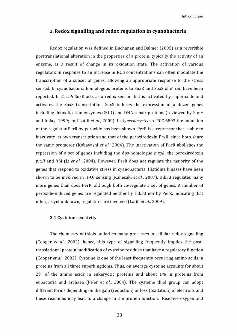
3.Redoxsignallingandredoxregulationincyanobacteria
RedoxregulationwasdefinedinBuchananandBalmer(2005)asareversible
posttranslationalalteration inthepropertiesofaprotein,typicallytheactivityofan
enzyme, as a result of change in its oxidation state. The activation of various
regulators inresponsetoan increase inROSconcentrationscanoftenmodulate the
transcription of a subset of genes, allowing an appropriate response to the stress
sensed. IncyanobacteriahomologousproteinstoSoxRandSoxSofE.colihavebeen
reported. InE. coli SoxRactsasa redox sensor that is activatedby superoxideand
activates the SoxS transcription. SoxS induces the expression of a dozen genes
includingdetoxificationenzymes(SOD)andDNArepairproteins(reviewedbyStorz
andImlay,1999;andLatifietal.,2009).InSynechocystissp.PCC6803theinduction
oftheregulatorPerRbyperoxidehasbeenshown.PerRisarepressorthatisableto
inactivateitsowntranscriptionandthatoftheperoxiredoxinPrxII,sincebothshare
the samepromotor (Kobayashiet al., 2004).The inactivation ofPerR abolishes the
repression of a set of genes including the dps‐homologuemrgA, the peroxiredoxin
prxII and isiA (Li etal., 2004).However,PerR does not regulate themajorityof the
genesthatrespondtooxidativestress incyanobacteria.Histidinekinaseshavebeen
showntobe involved inH2O2sensing(Kanesakietal.,2007).Hik33regulatesmany
more genes than does PerR, although both co‐regulate a set of genes. A number of
peroxide‐inducedgenesare regulatedneitherbyHik33norbyPerR, indicatingthat
other,asyetunknown,regulatorsareinvolved(Latifietal.,2009).
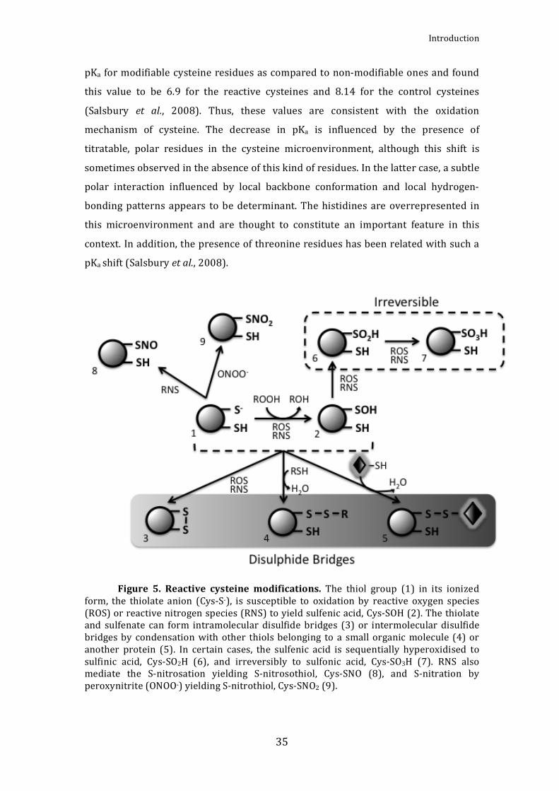
3.1Cysteinereactivity
Thechemistryof thiolsunderliesmanyprocesses incellularredoxsignalling
(Cooper et al., 2002), hence, this type of signalling frequently implies the post‐
translationalproteinmodificationofcysteineresiduesthathavearegulatoryfunction
(Cooperetal.,2002).Cysteineisoneoftheleastfrequentlyoccurringaminoacidsin
proteinsfromallthreesuperkingdoms.Thus,onaveragecysteineaccountsforabout
2% of the amino acids in eukaryotic proteins and about 1% in proteins from
eubacteria and archaea (Pe’er et al., 2004). The cysteine thiol group can adopt
differentformsdependingonthegain(reduction)orloss(oxidation)ofelectronsand
these reactionsmay lead toa change in theprotein function. Reactive oxygen and

AlejandroMataCabana
34
nitrogen species (ROSandRNS, respectively) can induce redox signalsbymeansof
themodificationofcysteineresidues.Anindividualenzymemayresponddifferently
todiversestimulibydifferentialmodificationofasinglecysteine.Thus,thisresidue
canbeconsideredasignaltransducer(Cooperetal.,2002).
Typically, the reactive cysteine thiol (Cys‐SH) is present in its thiolate form
(Cys‐S‐). This ionised form is susceptible to oxidation by compounds, such asH2O2,
organic hydroperoxides and hypochlorous acid. A product of such oxidation is the
sulfenic acid (Cys‐SOH). In many cases, the cysteine thiolate and sulfenate forms
undergo rapid condensationwith other thiols to formdisulfide bonds (Fig. 5). The
resulting disulphides can be intramolecular, with another cysteine from the same
protein,orintermolecular,withacysteinefromanotherproteinorasmallmolecule,
such as glutathione or free cysteine (Poole and Nelson, 2008). The covalent
attachment of glutathione to protein cysteines through a mixed disulfide bond is
known asglutathionylation (Poole et al., 2004). The disulfide bonds resulting from
cysteineoxidationplay important roles inprotein structureandoligomerization,as
well as enzyme activity regulation. The RNS, including nitric oxide (NO) and
peroxynitrite (ONOOH), canmediate S‐nitrosationyieldingS‐nitrosothiol (Cys‐SNO)
and S‐nitration yielding S‐nitrothiol (Cys‐SNO2), respectively (Fig. 5). Nearly all of
theseoxidativemodificationsarereversiblethroughreductioncatalysedbyenzymes,
such as thioredoxins or glutaredoxins. However, in certain proteins, the
microenvironmentmaystabilizethecysteinesulfenicacidandthisformcouldinturn
besensitivetohyperoxidationyielding sulfinic (R‐SO2H)andsulfonicacid (R‐SO3H)
(Cooper et al., 2002: Poole and Nelson, 2008). While cysteine sulfinic acid can be
revertedto thethiol formbytheenzymesulfiredoxin (Pooleetal., 2004:Pooleand
Nelson,2008),hyperoxidationtosulfonicacidisbelievedtobeirreversible.
Protein cysteines differ in reactivity and not all cysteines are susceptible to
modification. The properties of the protein microenvironment, which control
cysteinereactivityandstability,arenotcompletelyunderstood.However,itseemsto
be clear that the cysteine pKa plays an important role. Any cysteine modification
involves an initial thiol deprotonation, suggesting a lowered sulfhydryl pKa for the
modifiablecysteines(Salsburyetal.,2008).Salsburyetal.havecalculatedthemean
Introduction
35
pKa formodifiablecysteineresiduesascomparedtonon‐modifiableonesandfound
this value to be 6.9 for the reactive cysteines and 8.14 for the control cysteines
(Salsbury et al., 2008). Thus, these values are consistent with the oxidation
mechanism of cysteine. The decrease in pKa is influenced by the presence of
titratable, polar residues in the cysteine microenvironment, although this shift is
sometimesobservedintheabsenceofthiskindofresidues.Inthelattercase,asubtle
polar interaction influenced by local backbone conformation and local hydrogen‐
bondingpatternsappears tobedeterminant.Thehistidinesare overrepresented in
this microenvironment and are thought to constitute an important feature in this
context.Inaddition,thepresenceofthreonineresidueshasbeenrelatedwithsucha
pKashift(Salsburyetal.,2008).
Figure 5. Reactive cysteinemodifications. The thiol group (1) in its ionized
form, the thiolate anion (Cys‐S‐), is susceptible to oxidation by reactive oxygen species(ROS)orreactivenitrogenspecies(RNS)toyieldsulfenicacid,Cys‐SOH(2).Thethiolateand sulfenate can form intramolecular disulfide bridges (3) or intermolecular disulfidebridgesby condensationwith other thiolsbelonging to a smallorganicmolecule (4)oranother protein (5). In certain cases, the sulfenic acid is sequentially hyperoxidised tosulfinic acid, Cys‐SO2H (6), and irreversibly to sulfonic acid, Cys‐SO3H (7). RNS alsomediate the S‐nitrosation yielding S‐nitrosothiol, Cys‐SNO (8), and S‐nitration byperoxynitrite(ONOO‐)yieldingS‐nitrothiol,Cys‐SNO2(9).

AlejandroMataCabana
36
The cysteine sulfenic acid (Cys‐SOH) plays an important role in redox
signalling for many proteins. In these cases, the protein structure may ensure the
stability of this cysteine form to maintain its reactivity and, hence, its signalling
capacity.Thus,theCys‐SOHmicroenvironmentischaracterisedbythelackofnearby
reduced cysteines and the presence of local hydrogen‐bonding residues, which
stabilisethesulfenateform(Pooleetal.,2004;Salsburyetal.,2008).
3.2Cyanobacterialthioredoxinsystems
In photosynthetic organisms, several enzymes involved in CO2‐fixation are
activatedby the reductionof a cystine‐bridge formedby twovicinal cysteines.This
reduction is mediated by a thioredoxin (Trx), which receives reducing equivalents
from the photosynthetic electron transport. Thioredoxins are present in nearly all
living organisms, though their abundance and diversity are particularly striking in
plantsandphotosyntheticbacteria(BaumannandJuttner,2002;Meyeretal.,2005).
Theyarecharacterisedbytheirlowmolecularmass(around12kDa)andconserved
compact architecture. The Trx active site contains a conserved sequence motif (–
WCGPC–), which constitutes a highly reactive dithiol/disulphide. TheTrxs convert
disulphides todithiols in their respective target enzymes, therebymodulating theiractivities.
Trx was initially identified inE. coli as a hydrogen donor to ribonucleotide
reductase (RNR)(Laurentetal.,1964).The firstevidence forTrx inphotosynthetic
organismscamefromstudiesoftheferredoxin‐dependentactivationoffructose‐1,6‐
bisphosphatase, which belongs to the Calvin‐Benson cycle of CO2 assimilation in
chloroplasts(Buchanan,1980;Buchananetal.,2002;Florencioetal.,2006).Inplants
two chloroplast Trxs named Trx f and Trx m were identified. Both are light‐
dependent regulators of several carbon metabolism enzymes (Schürmann and
Jacquot, 2000; Lemaire et al., 2007). Trx f was found to be responsible for the
fructose‐1,6‐biphosphatase regulation, and Trx m activates the NADP malate
dehydrogenase(Buchanan,1980;Buchananetal.,2002).Later,plantsandalgaewere
foundtopossesscytosolic(Florencioetal.,1988)aswellasmitochondrialTrxs(Laoi
etal.,2001).ThefullgenomesequencesofArabidopsisthalianaandotherplantshave
revealed severalmore types ofTrxsandmultiplegenes for nearlyeach typeofTrx
(BalmerandBuchanan2002;Meyeretal.,2005).Thus,plastidscontainthef‐,m‐,x‐,

Introduction
37
y‐typeTrxs.Inaddition,thereareTrx‐likeproteinssuchasCDSP32andLilium1to5
(Meyeretal.,2005).H‐typeTrxsarepresentinthecytosolandmitochondria;ando‐
typeTrxsarepresentonlyinmitochondria(Jacquotetal.,2009).
The first evidence for Trx in cyanobacteria was found inAnabaena sp. PCC
7119,inwhichtwoTrx‐likeactivitiesweredescribed(Yeeetal.,1981).Oneofthemcorrespondedtoam‐typeTrx,whichwas thefirst tobeclonedfromcyanobacteria
(Gleason and Holmgren, 1981). The increasing number of complete cyanobacterialgenome sequences has allowed the identification, cloning and characterisation ofseveral others Trxs in cyanobacteria. The phylogenetic analysis of amino acid
sequenceshasrevealedfourdistinctgroupsofTrxincyanobacteria.Threegroupsareshared between cyanobacteria and photosynthetic eukaryotes and includem‐type
(also knownas TrxA),x–type (TrxB)andy–type (TrxQ). The fourthgroup, TrxC, isuniqueto cyanobacteria.Asa rule, largecyanobacerialgenomes, forexamplethatofAnabaena sp. PCC 7120, contain more Trxs than smaller ones, for example the
Prochlorococcus marinus MED4 genome. Them‐type is the only one present in allcyanobacteria, and in themarine speciesProchlorococcus marinus MED4,MIT9313
andSS120therearenoadditionalTrxs.AlltheTrxmfromcyanobacteriahaveahigh
degreeof sequence identityaround theactive site. The x‐type Trx (TrxB) is absent
fromthemarinespeciesandisalsomissinginthegenomesofThermosynechococcus
elongatusBP‐1andGloeobacterviolaceus.ThecyanobacterialTrxxsequencesdisplay
somevariation,buttheconsensusactivesite(WCGPC)isthesameasforthem‐type.
The y‐type Trx has the typical active site, too. The most prominent feature of the
cyanobacterial Trx y sequences is the presence of numerous glutamine residues,
particularly in the C‐terminal half of the protein. For this reason the Trx y from
Synechocystis sp. PCC 6803 was named TrxQ (Pérez‐Pérez et al., 2006). TrxC is
characterised by a different active site sequence (WCGLC) and is present in all
examined genomes except in those of the marine cyanobacteria. No activity or
function has so far been ascribed to this class of Trx (Florencio et al., 2006). The
cellularproteinlevelsforeachtypeofTrxhavebeendeterminedinSynechocystissp.
PCC 6803. The four Trxs are expressed simultaneously under standard
photoautotrophicgrowthconditions.Thevalues forTrxA,B,CandQwere2,5±0,4,
0,16±0,01,2,4±0,1and0,046±0,003ngperμgtotalprotein,respectively.Hence,
TrxAandCarequiteabundantproteins,withcellularconcentrationssimilartothose
ofE. coli Trx1. TrxB is 16 times less abundant and present in amounts compatible
withthoseofE.coliTrx2.TrxQismorethan50timeslessabundantthanTrxsAandC

AlejandroMataCabana
38
(Florencioetal.,2006).
There are two types of Trx reduction system, the NADPH dependent
thioredoxinreductase(NTR)andferredoxindependentthioredoxinreductase(FTR)
systems.TheNTRispresentinheterotrophicaswellasinphotosyntheticorganisms,
butFTRisonlyinchloroplastsofphotosyntheticeukaryotesaswellincyanobacteria
(Florencioetal.,2006).FTRisa20to25kDaheterodimercomposedofonevariable
and one catalytic subunit. The primary structure of the catalytic subunit is highly
conserved including six cysteines. Four of them ligate a Fe‐S cluster and the
remainingtwoformtheadjacentredox‐activedisulfidebridgeoftheactivesite(Sun
etal.,2001;Jacquotetal.,2009).Theprimarystructureofthevariablesubunitdiffers
significantly indifferent species.Themain functionof this subunit seems tobe the
stabilisationofthecatalyticsubunit,particularlyofitsactivesiteregion.Thecatalytic
subunitreceiveselectronsfromthephotosyntheticelectrontransportviaferredoxin
and transforms this signal into a dithiol signal reducing Trxs. Given that Trx
reductionrequirestwoelectronsandferredoxin isaone‐electrondonor,a two‐step
mechanism has been proposed involving a one‐electron reduced intermediate: a
transientcovalentcomplexbetweenFTRandTrx(Waltersetal.,2005;Jacquotetal.,
2009).ThisreactionmechanismsuggestssimultaneousinteractionofFTRwithboth
ferredoxin and Trx (Jacquot et al., 2009). All oxygenic photosynthetic organisms
except for the cyanobacteria Gloeobacter violaceus and the three species of
Prochlorococcus marinus possess the FTR reduction system. Gloeobacter violaceus,
which lacks thylakoid membranes, adapts poorly to the environment and is
extremelysensitivetochangesinlightintensity.TheProchlorococcusspeciesliveina
stablehabitat.Therefore, there isno need for light‐dependentmetabolic regulation
and,consequently,no requirement fora linkbetween lightand theTrx redox state.
Among the remaining species of cyanobacteria the catalytic subunit is well
conserved, particularly the six cysteines residues involved in the function of the
protein. The gene encoding this subunit inSynechocystis sp. PCC 6803 seems to be
essential. The sequences of the variable subunit are less conserved among
cyanobacteria(Florencioetal.,2006).
NTR is a homodimerwith subunits of approximately 320 residues in plants
that contain a redox‐active CxxC motif. The subunit is divided into two similar

Introduction
39
domains, one that binds the FAD and one that binds NADPH. The redox‐active
disulfide is located in the NADPH domain and is in close contact with the
isoalloxazinemoietyofFAD(Daietal.,1996;Jacquotetal.,2009).Themechanismof
reductioninvolvesconformationalchanges,whichenableNADPHtoreduceFAD.FAD
thenreducesthedisulfide,followedbyalargedomainrotationinordertoenablethe
reduction of Trx by a disulphide‐dithiol exchange reaction (Lennon et al., 2000;
Jacquotetal.,2009).ANTRfusedtoaTrxdomain,analogoustotheonedescribedin
Mycobacteriumtuberculosis(Wielesetal.,1995),was identifiedinthechloroplastof
rice and Arabidopsis thaliana (Serrato et al., 2004). This kind of NTR is known as
NTRC.TheNTRsystemincyanobacteriaismuchmorediversethaninphotosynthetic
eukaryotes.AtleastthreedifferentgroupsofNTRcanbediscernedincyanobacteria
based on sequence comparison. These groups are clearly separated from the
cytosolic/mitochondrial NTRs of plants and algae (Florencio et al., 2006). In some
cyanobacteria NTRC is also present, for example in Anabaena sp. PCC 7120.
Interestingly, thiscyanobacteriumpossesses in itsgenomeanothergeneencodinga
NTR located immediately upstreamof agenewhich codes foranunusualTrx. This
suggeststhattheNTRChasarisenfromthepre‐endosymbioticfusionbetweenaNTR
gene and a neighbouring Trx gene of an ancestral photosynthetic prokaryote
(Florencioetal.,2006).
Recently, a comparative study on the NTR and FTR reduction systems has
beencarriedoutinthecyanobacteriumSynechocystissp.PCC6803.Inthisstudythe
authors conclude that the NTR system is linked to antioxidant reactions while the
FTR systemcontrols the cell growth (Hishiyaet al., 2008).These findings raise the
questionof theoriginofFTRandtheevolutionofredoxregulation(Schürmannand
Buchanan,2008).

AlejandroMataCabana
40
Figure 6. Trx reduction systems.Trx can be reduced by the FTR (ferredoxin
dependentthioredoxinreductase)ortheNTR(NADPHdependentthioredoxinreductase)system. FTR receives the electrons from the photosynthetic electron transport viaferredoxinandreducesthedisulfideboundoftheTrx.NADPHreducestheNTRthatactsreducingtheTrx.ThereducedTrxpossessestwothiolgroupsinitsactivesitethatallowittoreduceddifferentproteintargets.
Another thiol‐based oxidoreductase enzyme is the glutaredoxin (Grx). Grxs
are small proteins,whichbelong to theTrx superfamily exhibitinga similaroverall
3D structure. Two molecules of glutathione (GSH) are necessary to reduce a Grx
forming a molecule of oxidased glutathione (GSSG). GSH is regenerated by the
glutathione reductase enzyme (Lemaire et al., 2007). Grxs are able to reduce other
proteinsandmixeddisulfidesbetweenproteinsandGSH.Theseoxidoreductasesare
conservedinmosteukaryotesandprokaryotes,exceptinsomebacterialorarchaeal
phyla (Couturier et al., 2009). Two families of glutaredoxins have been described:
dithiolic glutaredoxins (which have two cysteines in their catalytic site) and
monothiolic glutaredoxins (which only have one cysteine in their catalytic site).
Synechocystis possesses two dithiolic glutaredoxins (GrxA and GrxB) and one
monothiolic (GrxC) (López‐Maury et al., 2009; Pérez‐Pérez et al., 2009). In some
casestheTrxandGrxfunctionmightberedundant.
4.Disulphideproteomes
Previous to the proteomics studies several cyanobacterial functions were
suggested to undergo redox regulation (Florencio et al., 2006). A thiol reductant‐
dependentactivitywas reported forcyanophycinsynthetase inAnabaenacylindrica

Introduction
41
(Simon,1976)and forRNApolymerase inAnabaenasp.PCC7120(Schneideretal.,
1987). The glucose‐6‐phosphate dehydrogenase of Anabaena sp. PCC 7120 is
inhibited in vitro by the Trx m (Gleason, 1996). A possible role of Trxs as signal
transducersbetweenthephotosystemsandatranscriptionfactorinthetranscription
of psbAII and psbAIII genes in Synechococcus sp. PCC 7942 has been suggested
(Sippola and Aro, 1999). The reduction of the peroxiredoxin 2‐Cys Prx from
SynechocystisbyanE.coliTrxwasshowninvitro(Yamamotoetal.,1999).However,
thebiochemicalapproachestowardstheTrxfunctionhavehadalimitedcontribution
to the globalknowledgeon the redox regulation in cyanobacteria.The comparative
studies of Trx targets in other organisms might be informative, but there are
important metabolic differences and evolutionary divergence that make the
comparison with the cyanobacterial proteins difficult. Thus, it was necessary to
develop new approaches to extend the knowledge on enzymatic activities, which
undergoredoxregulationincyanobacteria.
Disulphide proteomes may be defined widely as sets of proteins, which
contain cysteines thatexhibit changes in their redox state (Lindahl andKieselbach,
2009). Several proteomics approaches have been developed to search for proteins
withreactive,accessiblecysteinesinordertorevealnewtargetsofredoxregulation
and signal transducers. These methods were originally designed for analyses of
soluble Trx targets. Methods have also been developed to study the disulphides
proteomewithouttheaidof redoxenzymes (LindahlandKieselbach,2009).This is
the case of studies based on the mobility shifts on SDS‐PAGE under non‐reducing
versus reducing conditions (Ströher andDietz, 2008)or theanalysisof the protein
cysteine sensitivity to S‐thiolation (Hochgräfe et al., 2007). Disulphide proteomics
analyses have provided useful insights into different redox regulated cellular
processes. However, it should be kept in mind that complementary studies are
necessarytoconfirmthephysiologicalrelevanceoftheresults.
4.1Methodology
The twomainmethods that use Trxs as tools for analysis of the disulphide
proteome are the thiol labelling procedure and the thioredoxin affinity
chromatography(LindahlandKieselbach,2009).

AlejandroMataCabana
42
The first method involves the labelling of thiols with the fluorescent
compound monobromobimane (mBBr) (Yano et al., 2001; 2002), radioactive
iodoacetamide (14C‐IAM) (Marchandetal., 2004)or reagentswithdifferentmasses
suchasIAMandDMA(Schillingetal.,2004).Inthismethod,thefreethiolsarefirstly
blocked using alkylation reagents such as IAM or NEM. Thereafter, disulphide
reductionby Trxgives rise to new free thiols,which are susceptible to labellingby
the dye mBBr, that binds covalently to thiols, radioactive alkylation by 14C‐IAM or
differential alkylation by IAM or DMA. Proteins with labelled thiols are separated
usingIEF/SDS‐PAGE,detectedandidentifiedbymassspectrometry.Targetslabelled
withmBBrmaybedetectedbyananalysisoftheUVimagescomparingTrx‐reduced
andunreducedsamples.Theradioactivityofthe14C‐IAMlabelledthiolsisdetectedby
autoradiography. Inthedifferential alkylationapproach the2Delectrophoresisgels
are analysed using MALDI‐TOF‐MS and the reduced cysteines are detected by the
massshiftsduetothedifferentalkylationagentsused for thiol labelling.ThemBBr
methodhasrelativelylowsensitivity,whichlimitsitsapplicationtothedetectionof
abundant Trx targets. The sensitivity of the other two approaches is higher and
similarbetweenthem.Anadvantageofdifferentialalkylationisthatitcouldprevent
false positives due to proteins comigrating in the same 2‐DE gel spot, by direct
identificationofthepeptidesthatcontainthereactivecysteine.Inaddition,itallows
assignment of redox active cysteine within the amino acid sequence of a target
protein(LindahlandKieselbach,2009).Thelabellingapproacheshavetheadvantage
that they may be adapted to monitor the in vivo redox state of proteins and to
quantifythefluctuations.However,theypotentiallyyield falsepositivesinvitro,due
tothehighconcentrationofTrxused,whichmayreduceoxidisedthiolsbelongingto
non‐targetproteins(Montrichardetal.,2009).
Trx affinity chromatography is based on the stabilisation of the mixed
disulfide bond intermediate formed during the Trx reduction of its protein targets
(Motohashi et al., 2001; Balmer et al., 2003; Lindahl and Florencio, 2003). This
mechanism involves a two‐step oxidation of the cysteine residues belonging to the
Trx active site. In the first step theTrxN‐terminal catalytic cysteine formsamixed
disulphide bond intermediate with the target (Fig. 7). Consecutively, the Trx C‐
terminalcysteinebreaksthismixedbondbymeansofasecondnucleophilicattack.

Introduction
43
Finally, the products of the reaction are the reduced target and the oxidised Trx.
Under normal conditions the mixed disulphide intermediate is labile, but if the
secondcysteineisreplacedwithanotheraminoacid,e.g.serineoralanine,themixed
intermediate canbe stabilised.Thus themonocysteinicTrx is auseful tool to study
the proteins interacting with Trx (Buchanan and Balmer, 2005; Lindahl and
Kieselbach,2009)
Figure7.MechanismofTrxtargetreduction. In theWTTrx(A) thecatalytic
cystein of the active site (‐WCGPC‐) reduces a oxidised cystein of the protein target anformsthemixeddisulphidebondintermediate,thenthesecondcysteinofTrxperformsanucleophilic attack that breaks the mixed bond resulting the reduced target and theoxidised thioredoxin. In the monocysteinic Trx (B) the labile mixed intermediate isstabilised because of the replacement of the second cysteine with a serine in the Trxactive.
Thisstrategywasfirstdescribedforascreeninginvivoinyeast(Verdoucqet
al., 1999) and was later adapted for column‐affinity in vitro by immobilising the
monocysteinic Trx on a Sepharose matrix (Motohashi et al., 2001; Balmer et al.,
2003). A variant of this technique was developed in our laboratory (Lindahl and
Florencio,2003;LindahlandFlorencio,2004),inwhichthemonocysteinicTrxisnot
permanently bound to the matrix. Instead it is bound to a nickel‐affinity
chromatography matrix through a histidine tag. Therefore, the mixed Trx‐target

AlejandroMataCabana
44
complexes are purified by nickel affinity and eluted using imidazole, without
breakingthedisulphidebonds(Fig.8).Separationofelutedcomplexesisperformed
on 2‐D SDS‐PAGE under non‐reducing/reducing conditions. Themixed disulphides
remain intact in the first dimension but, before the second dimension, this bond is
brokenbyDTTtreatment.Thus,inthissteptheTrxanditstargetsareseparatedand
migrate independently in the second dimension SDS‐PAGE. Hence, the thioredoxin
targetsmigrateslowerinthefirstdimensionthaninthesecondduetotheextramass
of the Trx. Contaminants and non‐target subunits can be discriminated since they
migrateequallyinthefirstandtheseconddimensionandlineuponthediagonalin
thestainedgel.Thetargetsarelocatedbelowthediagonal(Fig.8).Sometimesappear
spots above the diagonal, which correspond to proteins with an intramolecular
disulphidebond.Thisapproachhasalimitationintheidentificationofproteintargets
with a molecular weight similar to that of Trx, because of the high amount of this
protein used in the analyses. Therefore, the risk of contamination of these protein
spots by Trx is elevated. The Trx affinity chromatography methods have the
advantageoftheenrichmentoftargetsthatallowsthevisualizationoftheseproteins
inCoomassiestainedgels.
Figure 8. Isolation of Trxtargets by Trx affinity chromatography. The
complexes formed by the monocysteinic his‐tagged thioredoxin and its targets are

Introduction
45
purified by means of Nickel‐affinity chromatography. The complexes migrate in a firstnon‐reducing dimension, after that the mixed disulphide bonds are broken using thereductive agent DTT. The broken complexes migrate in a second reducing dimensionwhere the targets of Trx are separated. The non‐target protein form a diagonal line(dashed line), the targetsare locatedbelowthis diagonal (markedwith a red triangle),proteinswithanintramoleculardisulphidebondmigrateabovethediagonal(bluecircle)andthefreethioredoxinsformaline(markedwithawhitestar)inthe12kDaregioninthegel.
4.2 Analysis of disulphide proteome in prokaryotes (Except
cyanobacteria)
Apartfromstudiesincyanobacteria,mostofthedisulphideproteomicstudies
in prokaryotes have been performed in the gram‐negative model bacterium
Escherichia coli and in the gram‐positive model bacteriumBacillus subtilis. Despite
thefactthatTrxwasoriginallyidentifiedinE.coliin1964(Laurentetal.,1964),Trx‐
dependent disulphide proteomic approaches have been developed and applied
principallyinphotosyntheticorganisms(seeBuchananandBalmer,2005;Florencio
et al., 2006; Lindahl and Kieselbach, 2009). The E. coli genome encodes two Trxs,
threeGrxsandtwoGrx‐likeproteins(Grx4andNrdH).Outofthese,theonlyessential
protein is Grx4, which has a unique function, possibly related to Fe‐S cluster
assembly. Although Trxs and Grxs play fundamental roles as electron donors inE.
coli,theyarelargelyredundant(Meyeretal.,2009).ThetrxAandtrxCgenesinE.coli
encode theTrx1andTrx2, respectively.While noneof the trxgenes is required for
viability in E. coli, Trx1 is essential in several other bacteria, e.g., Rhodobacter
sphaeroides, Bacillus subtilis, Anacystis nidulans and Synechocystis sp. PCC 6803
(ZellerandKlug,2006).
Long before the proteomics era, biochemical and genetic studies have
described some targets of redoxins in E. coli. Initially, Trx was identified as an
electrondonorforribonucleotidereductase,akeyenzymeinDNAsynthesis(Laurent
etal.,1964).Since,variousother functionshavebeenattributedtoprokaryoticTrxs
and Grxs as electron donors to enzymes reducing methionine sulfoxide or
participating in reductive sulphate assimilation (Gleason and Holmgren, 1988). For
example, Trx1, Trx2 and Grx1 donate reducing equivalents to the E. coli PAPS
reductase, the enzyme responsible for reduction of 3’‐phosphoadenylylsulfate to

AlejandroMataCabana
46
sulfite (Lillig et al. 1999), whereas Grx2 is an efficient electron donor for arsenate
reductase (ArsC), which catalyses the reduction of arsenate to arsenite (Shi et al.,
1999).
An interesting example of redox regulation discovered in E. coli before
disulphideproteomicsistheoxidativeactivationofthemolecularchaperoneHsp33,
which contains four conserved cysteines prone to formation of disulphide bridges
(Jakobetal.,1999).AnotherexampleisthebacterialtranscriptionfactorOxyR,which
servesasaperoxide‐sensitivethiol‐based redox sensorandcontrols theexpression
of several genes involved intheantioxidant response (Zhengetal.,1998;Åslundet
al., 1999). Among the genes controlled by OxyR are a peroxidase, AhpC, and its
reductase,AhpF, firstdescribed inSalmonellatyphimurium (Christmanetal.,1985).
AhpCbelongstothe familyofperoxiredoxins,whichcatalysereductionofH2O2and
alkylhydroperoxidesthroughreversibledisulphideformation(Poole,2005).
ThefirstglobalstudyofproteinsinteractingwithTrxinE.coliwasbasedona
TandemAffinityPurification(TAP)strategy,expressingaTAP‐taggedversionofTrx1
in E. coli cells with a trxA‐ knockout genetic background (Kumar et al., 2004).
However, it shouldbenotedthat theTAP‐taggedTrx1used in this studypossessed
thewildtypeactive site, includingboth cysteines,andwouldhencenot formstable
mixed disulphides. Therefore, the target proteins isolated and identified did not
necessarily interact with the Trx through thiol chemistry, but rather through
electrostatic and/or hydrophobic interactions, forming multiprotein complexes. In
this context, it isworthmentioning thatE. coliTrx1was previously found to bean
essentialstructuralsubunitofthebacteriophageT7DNApolymerase,independently
oftheTrxactivesitecysteines(Huberetal.,1986)Furthermore,Trx1isrequiredfor
theassemblyof several filamentousphages(RusselandModel1985).Nevertheless,
some of the targets identified in the study using a TAP‐tagged Trx (Kumar et al.,
2004) were found later in studies of E. coli using different disulphide proteome
approaches(LeichertandJakob,2004;Brandesetal.,2007;Leichertetal.,2008).
The principal method used to analyse the disulphide proteome in E .coli
involves a differential thiol‐trapping techniquecombinedwith2‐DEgel analysis, in
which the cysteines that were oxidised in the cell are reduced and thereafter

Introduction
47
carbamidomethylated by radioactively labelled 14C‐IAM (iodoacetamide) (Leichert
and Jakob, 2004; Brandes et al., 2007). A quantitative thiol proteome study was
carriedoutcombiningthismethodwithICATchemistry(Leichertetal.,2008).Hence,
these approaches were used to monitor the thiol status of cellular protein under
normalgrowthconditionsaswellasunderdifferentoxidativestressconditions.The
redox‐sensitive proteins identified in E. coli can be classified into three categories
belonging to antioxidant mechanisms, intermediary metabolism or regulation
processes.Most of these proteins have a role in the intermediarymetabolism. The
glyceraldehyde‐3‐phosphatedehydrogenase(GapA)hasbeendetected reproducibly
inthedifferentstudies.TheredoxregulationofGapAhasbeensuggestedto involve
glutathionylationofitsactivesitecysteine149(Cotgreaveetal.,2002).Notably,the
metabolism of amino acids and related molecules is highly represented by, for
example, MetE (cobalamin‐independent methionine synthase), IlvC (ketol‐acid
reductoisomerase,plays a central role in thebiosynthesisof isoleucineandvaline),
AlaS(alanyl‐tRNAsynthetase),PheT(phenylalanyl‐tRNAsynthetasebeta‐subunit)or
GlyA(serinehydroxymethyltransferase).ThereversiblethioloxidationofMetEafter
oxidative stress has been shown to be responsible for inhibition of methionine
biosynthesis (HondorpandMatthews,2004).TheperoxiredoxinsAhpCandTpx, as
well as the reductase of AhpC (AhpF), are reproducibly shown in the studies and
belongtotheantioxidantenzymes.Severalregulatoryproteinshavebeenshownto
beredoxthiol‐sensitive,suchas,TufA(elongationfactorTu),ProQ(regulatorofProP,
involvedinosmoregulation)orYhiF(transcriptionalregulator).Otherthiol‐modified
proteinsshowninE.coliaretheaconitaseB(AcnB)anddifferentribosomalsubunits.
Recently, a study was published in which thiol‐ and disulphide‐containing
proteins from E. coli were selected by using an Activated Thiol‐Sepharose (ATS)
chromatography (Hu et al., 2010). ATS is a thiol‐specific resin that possesses an
activated disulphide structure, which is able to react and to form covalent mixed
disulphides with thiolic groups. The procedure involves a denaturation step with
urea prior to purification, thus the thiols detected under normal conditions do not
necessarily belong to reactive cysteines. More interesting are the proteins that
contain disulphide bonds or thiol groups under oxidative stress generated by
menadione treatment. Amongst the identified proteins, ribosomal proteins,
amynoacyl‐tRNA synthetases, and metabolic and antioxidant enzymes were

AlejandroMataCabana
48
prominent(Huetal.,2010).
An approach similar to that used in E. coli (Leichert and Jakob, 2004) was
developed,butinsteadofradioactivelabelling,afluorescentreagentwasintroduced
(Hochgräfeetal., 2005).Thismethodwasapplied foranalysisof thecysteine redox
state of cytoplasmicB. subtilis proteins under normal growth conditions and upon
oxidativestressinducedbydiamide,H2O2orO2‐(Hochgräfeetal.,2005).Theredox‐
sensitve proteins identified under normal growth were, among others, the
antioxidantsystemmadeupoftheperoxirredoxinAhpCanditsreductase(AhpF),the
thioredoxin TrxA and the phosphoadenosine phosphosulfate reductase (CysH). The
proteinswith thiolmodificationwere found to be specific for each oxidative stress
condition, but five proteins were identified under all conditions. These were the
peroxiredoxins AhpC and Tpx, and the proteins belonging to the amino acid
metabolism LeuC (large subuit of 3‐isopropylmalate dehydratase), ArgC (N‐
acetylgutamate gamma‐semialdehyde dehydrogenase) and MtnA
(methylthionucleoside‐1‐phosphate isomerase). Other proteins with thiol
modification were detected under several but not all of the oxidative conditions
tested. Someof theseplayarole intheaminoacidmetabolism, suchasMetE,AroA
(3‐deoxy‐D‐arobino‐heptulosonate7‐phosphatesynthase)andLeuD(largesubuitof
3‐isopropylmalatedehydratase).OtherproteinsdetectedreproduciblywerethePfkA
(6‐phosphofructokinase) and some enzymes of the nucleotide metabolism, such as
GuaB (inosine‐monophosphate dehydrogenase) and PurA (adenylosuccinate
synthase). There were also proteins modified in only one of the oxidative stress
treatments,forexampleIlvCandGlyA,whichbelongtotheaminoacidmetabolismor
theelongationfactorTu(TufA).Theseproteinscoincidedwiththose identified inE.
coli.Thesameapproachhasbeenusedtotesttheoxidativeeffectofthequinonesin
B. subtilis. However, the only redox‐sensitive protein identified was the
glyceraldehyde‐3‐phosphatedehydrogenase(GapDH)(Liebekeetal.,2008).
A different technique was developed in B. subtilis to analyse the protein S‐
thiolation by cysteine (called S‐cysteinylation) under condition of disulfide stress
using diamide treatment. By in vivo [35S]cysteine labelling in the presence of
chloramphenicol and 2‐DE, six proteins showed S‐cysteinylation in response to
diamide stress (Hochgräfe et al., 2007). Four of these, GuaB, MetE, PpaC and the

Introduction
49
proteinYwaAwithsimilaritytobranched‐chainaminoacidaminotransferases,were
alreadyreportedinthepreviouslymentionedstudy.
ThesamefluorescencelabellingapproachdescribedforB.subtillis(Hochgräfe
etal.,2005)wasusedtostudythereversiblyoxidisedthiolsofcytoplasmicproteins
inStaphylococcusaureus during normalgrowth andunderoxidative stress induced
bydiamideandH2O2treatments(Wolfetal.,2008).Amongsttheproteins identified
under normal growth condition were the peroxiredoxin system AhpC/AhpF,
dihydrolipoyl dehydrogenase PdhD of the pyruvate dehydrogenase complex, and a
probablecysteinedesulfuraseCsd.Diamidetreatmentresultsinaglobaloxidationof
manycysteineresiduesinintracellularproteinsofS.aureus.Manyofthesehavebeen
previouslydetected inB. subtilisaswell as inE. coli,e.g.,AhpC/AhpFsystem,MetE,
GlyA, IlvD, LeuD, LeuD, GuaB and GapA. Eighteen different proteins with thiol
oxidation after H2O2 stress were identified. The largest number belongs to the
intermediary metabolism, for example, AldA (aldehyde dehydrogenase), MetE and
the CTP synthase PyrG. Moreover, proteins implicated in oxidative stress and
peroxidedetoxification,suchasthethioredoxinsystemcomposedbythethioredoxin
TrxAand its reductaseTrxB, theTpxperoxiredoxinand the glutathione peroxidase
homologueBsaA.TheregulatoryproteinSpxwasalsoobservedtoberedox‐sensitive.
A study of S‐thiolationhasbeencarried out inS.aureuswhere twoproteins
were identified that are modified by S‐cysteinylation (Pöther et al., 2009); the
acetoine reductase and a hypothetical lipoprotein. Results with lower confidence
levels suggest S‐cysteinylation of a glycyl‐tRNA synthetase, a
phosphoribosylaminoidazole‐succinocarboxamide synthase, a pyruvate kinase, a
triosephosphate isomeraseandSarA. In the samestudy,Pötheretal., 2009analyse
the intramolecular and intermolecular disulfide bond formation as inB. subtilis as
wellasinS.aureus.SomeproteinswithintramoleculardisulphidebondinB.subtilis
areMetEGuaB,ArgFandTpx.Proteinswithintermoleculardisulphidebondsinclude
IlvC,HisI,HisBandAhpCinB.subtilisandAhpC,MetN‐2,PurHandtheglycyl‐t‐RNA
synthetaseGlyQSinS.aureus.
Mycobacterium tuberculosis and the obligate anaerobicphotosynthetic green
sulfurbacteriumChlorobaculumtepidumareotherprokaryotes,wherethedisulphide

AlejandroMataCabana
50
proteomes have been studied. In M. tuberculosis proteins with S‐nitrosylation
sensitive cysteines have been analysed (Rhee et al., 2005). The Trx‐interacting
proteome has been studied in C. tepidum (Hosoya‐Matsuda et al., 2009). In both
bacteria the majority of proteins identified are implicated in the intermediary
metabolism, especially in the amino acid metabolism, e.g., AsnB (asparagine
synthase),SerA(3‐phosphoglyceratedehydrogenase),GlnA1(glutaminesynthetase)
and GltB (glutamate synthase) in M. tuberculosis, and serine
hydroxymethyltransferase, S‐adenosylmethionine synthetase and tryptophan
synthase inC. tepidum. Antioxidantdefenseproteinsarebeen shown inboth cases,
for example, the catalaseKatG inMycobacterium and the peroxiredoxins 2‐Cys Prx
andPrxQinC.tepidum.
Many proteins are described, which possess reactive cysteines and, hence,
mightundergoaputativeredoxregulation,butnotably,thereisahighabundanceof
proteinimplicatedinthemetabolismofaminoacidsandrelatedmolecules.Bacteria
havelowmolecularweight(LMW)thiolsthatactasthiolredoxbufferandprotectthe
proteincysteinesagainstoveroxidationbyS‐thiolation.Gram‐negativebacteriahave
glutathione (GSH) as redox buffer, but many gram‐positive bacteria lack genes for
GSH biosynthesis. B. subtilis and S. aureus rely on different LMW thiols, such as
bacillithiol (BSH), or even the free cysteine (Pöther et al., 2009). Thus, it has been
suggested that under oxidative stress it is necessary increase the cysteine pool to
protectthecellfromdamage.AmetabolomestudycarriedoutinB.subtillisrevealed
significantmodificationsinthecysteineandbranched‐chainaminoacidbiosynthesis
pathwaysinresponsetodiamide(Pötheretal.,2009).Adrasticdepletionofcysteine
and its precursors supports the idea of cysteine generation and S‐thiolation of
protein thiols after exposure to diamide. Under these stress conditions, cysteine
biosynthesis is inducedbyderepressionoftheCymRregulon(Leichertetal.,2003).
The concentration of glycine, which is a precursor of serine, is reduced under
oxidative stress and the synthesis of the glycyl‐tRNA synthetase is also decreased
(Leichertetal.,2003;Pötheretal.,2009).Thismightindicateanenhancedprovision
of glycine for serine synthesis, a cysteine precursor. The CymR‐controlled MccA
(YrhA) and MccB (YrhB) proteins participate in the conversion of methionine to
cysteine and are induced under disulphide stress (Liechert et al., 2003). The
methioninesynthaseMetEisS‐cysteinylatedunderdiamidestressandinactivatedby

Introduction
51
S‐thiolation in E. coli (Hondorp and Matthews, 2009; Hochgräfe et al., 2007). The
methionine pool is decreased in diamide treated cells. All these findings indicate a
decreasedconversionofcysteinetomethionine(Pötheretal.,2009).
4.3 The disulphide proteome in Photosynthetic organisms (Plants and
Algae)
The thioredoxin redox control in alage and plants shows a high complexity
because of the large number of Trx present in theses organism. The green algae
Chlamydomonas reinhardtii contains 8 Trx genes, 5 plastidic Trx: 2 Trx f, 1 Trxm,
1Trxxand1Trxy;andthreenon‐plastidic,1Trxoand2Trxh.TheplantArabidopsis
thalianacontains20Trxgenes in itsgenome,9ofwhichencodeproteins located in
thechloroplast:2Trx f,4Trxm,1Trxxand2Trxy.Mitochondriaharbour2Trxo
and,inaddition,9Trxharedistributedbetweenseveralcellcompartments(cytosol,
mitochondria, endoplasmic reticulum) as well as outside the cell (Lemaire et al.,
2007). Arabidopsis thaliana contains in its genome 42 genes that encode Trx‐like
disulfide proteins (Meyer et al., 2005). Among these are the chloroplastic proteins
NTRC,CDSP32andHCF164.CDSP32containstwoTrxdomainsanditsexpressionis
inducedunderdroughtstressandisabletoreducetwochloroplasticperoxiredoxins.
HCF164 is anchored to thylakoid membranes and required for the biosynthesis of
cytochromeb6/f(Lemaireetal.,2007).AnewtypeofTrxcalledTrxsinvolvedinthe
symbiosisprocessandwasidentifiedinthelegumeMedicagotruncatula(Alkhalfioui
etal.,2008).
Beforethedevelopmentofproteomicsapproaches,therewereseveralplant
proteins known toundergo light‐dependent redox regulation catalysedby Trx.The
fructose‐1,6‐bisphosphatase(FBPase)wasthefirsttobeidentified.Thereafter,more
Trx‐linked proteins belonging to different processes were detected, such as four
additionalproteinsof theCalvin‐Bensoncycle, ‐namelyphosphoribulokinase(PRK),
sedoheptulose bisphosphatase (SBPase), NADP‐glyceraldehyde 3‐phosphate
dehydrogenase (GADPH) and the Rubisco activase‐. The NADP‐malate
dehydrogenase (NADP‐MDH), the ATP synthase γ‐subunit, the glucose 6‐phosphate
dehydrogenase, the acetyl‐CoA carboxylase and ADP‐glucose pyrophosphorylase
werealsofoundtobesubjecttoTrx‐dependentregulation(Montrichardetal.,2009).

AlejandroMataCabana
52
These findingslinkedthethioredoxinstothecontroloftheCalvin‐Bensoncycleand
theoxidativepentosephosphatepathway,fattyacidandstarchsynthesis.Somelater
biochemical studies provided evidence of regulation of germination, growth and
developmentbyTrxhinwheat(Montrichardetal.,2009).
Themainapproachesusedfordisulphideproteomicsinplantsarethesame
as explained previously in themethodology section: thiol labelling and thioredoxin
affinitychromatography,butsomenewstrategieshavealsobeendeveloped.Targets
of different subcellular compartments have been identified, including chloroplast,
cytosol,mitochondriaandnucleus.Differenttissuesofplanthavebeenusedinthese
studies from leafs (Lemaire et al., 2007) to embryos of germinating barley grain
(Marxetal.,2003).
Thedisulphideproteomicstudiesinthechloroplasthaveresultedinatotalof
136potentially redox‐regulatedTrx‐targetproteins(Lindahl andKieselbach,2009).
The target proteins reported participate in the major chloroplast metabolic
pathways,forexample,thephotosyntheticelectrontransport,ATPsynthesis,Calvin‐
Benson cycle and some biosynthetic pathways including starch synthesis, sulphur
andnitrogenassimilation,andthesynthesisofproteins,tetrapyrroles,chlorophillide
andheme(LindahlandKieselbach,2009).
Nineteen Trx‐targeted proteins are found in the photosynthetic apparatus,
suchastheD1protein(PsbA)of thePSIIreactioncentre(StröherandDietz,2008).
ChloroplasticTrxscouldalsobeinvolvedinthemechanismofstatetransition,which
is based on the phosphorylation of LHCII mobile antenna by a redox dependent
kinase.Thecloningof thekinasephosphorylatingLHCII inChlamydomonas(Depége
et al., 2003) andArabidopsis (Bellafiore et al., 2005) revealed the presence of two
conservedcysteinesintheN‐terminalpartoftheprotein,whichmightbeinvolvedin
a Trx‐dependent regulation mechanism (Lemaire et al., 2007). The activation of
chloroplastATPsynthasebyTrxviaFTRhasbeenknownforalongtime(Lemaireet
al., 2007) and there is evidence that it depends on theTrx‐mediated reductionof a
disulphide in the γ‐subunit (Wu et al., 2007). In addition, the results from
independentscreeningstudiesinArabidopsis,spinach,potatoandC.reinhardtiishow
thatalso the α‐,β‐, δ‐, andε‐subunitsof CF1arepotential targets for regulationby

Introduction
53
Trx(MotohashiandHisabori,2006;Balmeretal.,2006;Lemaireetal.,2004;Ströher
andDietz,2008;Reyetal.,2005;Balmeretal.,2004;LindahlandKieselbach,2009).
NineteeninvitrotargetsofTrxparticipatedirectlyorindirectlyintheCalvin‐
Bensoncycle,indicatingthatTrxregulatesallCalvin‐Bensoncycleenzymes.Thus,Trx
control might function as a main switch to activate this cycle upon illumination
(Schürmann and Buchanan, 2008; Lemaire et al., 2007; Lindahl and Kieselbach,
2009).
InthestarchsynthesispathwaytheADPglucosepyrophosphorylasehasbeen
identifiedinseveralindependentproteomicsstudies(LindahlandKieselbach,2009).
Redox regulation of α–glucan water dikinase controls starch degradation in both
photosynthetic and non‐photosynthetic tissues (Mikkelsen et al., 2005). The β‐
amylasewasalsodetectedbyproteomicsapproaches(Balmeretal.,2003).Thefirst
enzyme of the reductive assimilation of sulphate, the adenylylsulphate reductase
(APS), was identified as a Trx‐target in C. reinhardtii (Lemaire et al., 2004); the
chloroplastAPSreductaseshaveaC‐terminalTrx‐likedomain,whichisnecessaryfor
activity(Weberetal.,2000).Thebiosynthesisofcysteine linkssulphurassimilation
andaminoacidsynthesis.InplantstheO‐acetylserinelyase(OASB)andtheCyp20‐3
have been detected as Trx‐targets, suggesting a redox regulation of the cysteine
synthesis(Motohashietal.,2001;Motohashietal.,2003;Balmeretal.,2003).
Some proteins involved in the nitrogen assimilation have been decribed as
redoxregulated.ThechloroplastidicglutaminesynthetaseGLN2hasbeendetectedin
manyscreeningstudiesofTrxtargetedproteins(Motohashietal.,2003;Lemaireet
al., 2004;Marchand etal., 2004).The cytosolic glutamine synthetase (GLN1;3)was
also reported to be a target of Trx (Balmer et al., 2003; Balmer et al., 2004).
Activation of theChlamydomonas plastidic glutamine synthetase bym‐typeTrx has
been shown (Florencio et al., 1993). This enzyme is activated by reduction of an
intramolecular disulphide in C. lineata (Choi et al., 1999). The glutamate synthase
(GOGAT)wasalsofoundtoberegulatedbyTrx(LichterandHäberlein,1998).
The biosynthesis of tetrapyrroles is amulti‐branched pathway and there is
evidence that redox control via Trx is involved in several steps, for example, the
activityofCHL1subunitofMgchelataseisstimulateduponreductionofadisulphide

AlejandroMataCabana
54
bridgebyTrxf(Ikegamietal.,2007).
The protein metabolism is highly represented in the Trx‐target screenings.
Severalribosomalsubunitsandtranslationelongationfactors,suchasEF‐TuandEF‐
G,havebeen found. Inaddition, someproteinsrelatedtoprotein foldinghavebeen
identified,forexample,thechaperonesCPN60(Rubiscobindingprotein)andHsp70,
thechaperonine20ortheimmunophilinsFKBP12,FKBP13,FKBP20‐2,thatcatalyse
thecis‐transisomerisationofprolylpeptidebonds.OthertargetsofTrxarerelatedto
proteindegradation,e.g.,theproteasesClpCandFtsH(LindahlandKieselbach,2009).
Disulphide proteome studies in plants and algae have identified some enzymes
functioning in the oxidative stress response; and this is the case of several
peroxiredoxins, glutathione peroxidases and themethionine sulfoxide reductasesA
andB1(LindahlandKieselbach,2009).
The majority of the chloroplastic disulphide proteome studies have been
carried out by means of Trx affinity chromatography techniques. However, the
screeningperformedusingplantseedsandcerealgrainsinvolvedmainlyfluorescent
thiol labelling (Montrichard et al., 2009). The proteins found in these studies are
enzymesof differentkeyprocesses, suchas starchmetabolism,antioxidant defence
and protein metabolism, including degradation, assembly and folding. Moreover,
morespecifictargetshavebeenfound,includingadesiccation‐relatedprotein,aseed
maturationproteinandsomeproteinsinembryosofgerminatingbarleygrain(Marx
et al., 2003; Montrichard et al., 2009). A strategy based on the quantitative ICAT
methodwas employed to analyse the disulphide proteome linked to Trx in soluble
proteinsofbarley seedembryo (Hägglundetal., 2008).Ninety putativeTrx‐targets
wereidentified,amongwhichanotablenumberofribosomalproteinswerepresent.
Other targets related to protein metabolism were several proteins functional in
amino acid metabolism (e.g. the glycyl‐tRNA synthetase), and enzymes catalysing
protein folding and degradation. This work confirms the key role that redox
regulationplaysinproteinmetabolism(Montrichardetal.,2009).
TheTrx‐affinitychromatographyapproachwasusedtoanalysetheTrx‐linked
proteins in the plant mitochondria (Balmer et al., 2004b). Novel targets included
proteins functional in photorespiration, citric acid cycle, lipidmetabolism, electron

Introduction
55
transport,membranetransportandhormonesynthesis(Montrichardetal.,2009).
4.4ThedisulphideproteomeinCyanobacteria
The strategy used for analysis of the disulphide proteome in cyanobacteria
wasdevelopedinSynechocystissp.PCC6803andinvolvedthepurificationofmixed
disulphide complexes formed by a monocysteinic his‐tagged Trx and its putative
targets,followedby2‐DSDS‐PAGEundernon‐reducing/reducingconditions(Lindahl
andFlorencio,2003;Florencioetal.,2006).
TrxA,TrxBandTrxQhavebeenusedasbaitincomparativescreeningstudies
ofTrx‐targets (LindahlandFlorencio,2003;Pérez‐Pérezetal., 2006).There is little
variationbetweenthetargetproteomesusingdifferentTrx, indicatinga lowdegree
of target specificity in vitro. Thirty‐nine Trx‐targets in total were identified in
Synechocystis. These targets organised in groups according to their function or the
metabolicpathways,inwhichtheyparticipate,areshowninTable1,whichhasbeen
adaptedfromFlorencioetal.,2006.
Table1.ThioredoxintargetproteinsidentifiedinSynechocystissp.PCC6803.
ORF Proteinname
Carbondioxidefixation
slr0009 Rubiscolargesubunit(RbcL)
sll1031 Carboxysomalprotein(CcmM)
sll1525 Phosphoribulokinase
Glycolysisandpentosephosphatepathway
sll0018 Fructose‐1,6‐bisphosphatealdolase,classII
sll1342Glyceraldehyde‐3‐phosphatedehydrogenase(Gap2)
slr0394 Phosphoglyceratekinase
sll1841 PyruvatedehydrogenasesubunitE2
sll1070 Transketolase

AlejandroMataCabana
56
Glycogenmetabolism
sll0726 Phosphoglucomutase
slr1176 ADP‐glucosepyrophosphorylase
sll1393 Glycogensynthase(Glg2)
sll0158 Glucanbranchingenzyme(GlgB)
slr1367 Glycogenphosphorylase
Sugarnucleotidemetabolism
sll1212 GDP‐mannosedehydratase
sll0576 Sugar‐nucleotideepimerase
Sulphurmetabolism
slr1165 Sulfateadenylyltransferase
slr0963 Ferrredoxin‐sulfitereductase
Nitrogenmetabolism
sll1499 Ferredoxin‐GOGAT(GlsF)
sll1502 NADH‐GOGAT(GltB)
slr0585 Argininosuccinatesynthetase
slr1133 Argininosuccinatelyase
Tetrapyrrolebiosynthesis
sll1994 Porphobilinogensynthase
Oxidativestressresponse
slr1198 1‐Cysperoxiredoxin(1‐Cys‐Prx)
sll1621 YLR109‐homologue(TypeIIPrx)
sll1987 Catalase‐peroxidase(KatG)
Lightharvesting
ssr3383 Phycobilisomecorelinker(LC)
slr0335 Phycobilisomecore‐membranelinker(LCM)
sll1577 Phycocyaninβ‐subunit

Introduction
57
RNAmetabolism
sll1789 RNApolymeraseβ’‐subunit
sll1787 RNApolymeraseβ‐subunit
sll1043 Polyribonucleotidenucleotidyltransferase
Proteinsynthesisandfolding
slr0557 Valyl‐tRNAsynthetase
slr1550 Lysyl‐tRNAsynthetase
slr1463 TranslationelongationfactorEF‐G
sll1099 TranslationelongationfactorEF‐Tu
sll1804 30SribosomalproteinS3
slr2076 60‐kDachaperonin1(GroEL)
Redoxregulation
slr0623 Thioredoxin(TrxA)
Unknown
slr1855 Hypotheticalprotein
OnethirdoftheseSynechocystistargetproteinshavehomologues,whichhave
been found as targets for Trx or Grx, in other photosynthetic organisms. These
common targets emphasise the processes of carbon dioxide fixation, glycolysis,
glycogen synthesis, nitrogen metabolism, oxidative stress response and protein
synthesisandfoldingasprincipalsubjectstoredoxcontrol.Fiveenzymeswerefound
as universal Trx or Grx targets in all three classes of photosynthetic organisms
(plants, algae and cyanobacteria), namely Rubisco large subunit (RbcL),
phosphoribulokinase,TypeIIPrx,thetranslationelongationfactorEF‐Tuandthe60
kDa chaperonin GroEL, which is also known as Rubisco‐binding protein. The
conservationofcysteinesinthesefiveproteinsfromchloroplastsandcyanobacteria,
together with their presence in the target proteomes, suggests that they are early
redox‐regulatedproteins in theperspectiveofmolecularevolution (Florencioetal.,
2006).

AlejandroMataCabana
58
Acomparisonwiththedisulphideproteomeofotherbacteria(mainlyE. coli
andB.subtilis)showsalowdegreeofoverlapconcerningcellularprocesses.Common
processes include glycolysis, in which the glyceraldehyde‐3‐phosphate
dehydrogenaseisahighlyconservedredoxregulatedtargetinallbacteriaanalysed,
andoxidativestressresponse,wheretheperoxiredoxinsarereproduciblyidentified.
Protein synthesis and folding stand out for the detection of different t‐RNA
synthetases and the presence of the translation elongation factor EF‐Tu, which is
found as target of redox control in all the organism analysed. The type II
peroxiredoxin is the other protein found in all photosynthetic organisms and all
bacteria, in which this kind of studies have been performed. The glutamine
synthetase has also been found as Trx‐target in cyanobacteria in vivo, and in plant
andnon‐photosyntheticbacteriathishasbeenarecurrenttarget.
Thedifferencesbetweenthedisulphideproteomesofcyanobacteriaandthose
ofotherorganismsmayhavedifferentreasons.Firstly,cyanobacteriapossessspecific
cellularprocessesandstructuresabsentfromplantsandfromotherprokaryotes,for
example, the phycobilisomes and the carboxysomes. It seems obvious that the
photosynthesisestablishesagreatdifferencewiththenon‐phosyntheticprokaryotes
and this is reflected in theseproteomic studies. Therearealso someTrx‐targets in
plants thatarenotpresent incyanobacteria; this is thecaseof theenzymeRubisco
activase(Florencioetal.,2006).Otherreasonsmaybetheevolutionarydivergenceof
the target protein between prokaryotes and eukaryotes. For example, the glucan
branching enzyme (GlgB), phosphoglucomutase and sulphate adenylyl transferase
from Synechocystis belong to families of prokaryotic enzymes, whereas their
chloroplast counterparts have eukaryotic characteristics. The cysteines of the
cyanobacterial GlgB, phosphoglucomutase and sulphate adenylyl transferase are
amongstthemanyresiduesthatdifferbetweentheseproteinsandthecorresponding
chloroplast enzymes (Florencio et al., 2006). Furthermore, there are evolutionary
changes,suchastheinsertionofregulatoryCys‐containingmotifs inthechloroplast
fructose‐1,6‐bisphosphatase and the γ‐subunit of the CF1 ATP synthase, which are
missinginthecyanobacterialhomologues(Florencioetal.,2006).Onelastreasonis
derived from the differentmethodology used in each organism. For example,most
Trx proteomics studies on chloroplasts have employed isoelectric focusing for
separation of target proteins, which hampers the identification of high molecular

Introduction
59
massproteinssuchastheGOGATsandtheβ‐andβ´‐subunitsoftheRNApolymerase.
Ontheotherhand,themethodusedtoisolateSynechocystistargetproteins(Lindahl
andFlorencio,2004)impedesidentificationofproteinsintherangeof10to15kDa
molecularmass due to the presence of high amounts of recombinantSynechocystis
Trx in this region, which would exclude for example the small subunit of Rubisco
(Motohashietal.,2001;Florencioetal.,2006).
Thereporteddiscrepanciesbetweenthecyanobacterialdisulphideproteomes
with respect to chloroplast on one hand and non‐photosynthetic bacteria on the
othermight indicatethatthereismoretodiscoverbothincyanobacteriaand inthe
other organisms. Hence, it would be necessary to take further improved redox
proteomic studies to understand the complete network of redox regulation in
cyanobacteria. Since proteomic studies alone do not provide information on
regulation of activity biochemical analyses are also required. There are proteins of
low abundance, such as regulatory kinases and transcription factors, which elude
detection using conventional proteomic techniques. To study the possible
interactionsbetweenTrxandsuchproteinsnewapproachesmustbedeveloped.


61
OBJECTIVES

Objectives
63
OBJECTIVES
InordertoextendtheknowledgeaboutredoxsignallingandthefunctionofTrxin
cyanobacteria we aimed at identifying the putative Trx‐targets that, for different
reasons, had been overlooked in previous studies on cyanobacteria Therefore, the
mainobjectiveof thisthesiswastocompletetheefforttocharacterisetheTrx‐target
proteome of the cyanobacterium Synechocystis sp. PCC 6803. This general objective
maybedividedintothefollowingsubsidiaryobjectives:
I. Membrane proteins from the cyanobacterium Synechocystis sp. PCC
6803 interacting with Trx. We sought to develop a procedure in
SynechocystisthatwouldallowanalysisofTrx‐interactionswithmembrane
proteins, since the proteomic approaches previously used to analyse
putative Trx‐targets had not been compatible with membrane‐associated
proteins. The aim was to identify targets in both thylakoid and plasma
membranes.
II. Trxtargets in the thylakoid lumen. We set out to study the possible
targets for Trx‐mediated redox regulation in the soluble compartment
contained within thylakoids, the lumen. Since the isolation of
cyanobacterial thylakoid lumen is not possible, we decided to use
chloroplastthylakoidlumenpreparationsfromArabidopsisand,thereafter,
to search for proteins in Synechocystis homologous to the Trx‐targets
identifiedfromArabidopsis.
III. TrxmediatedredoxregulationofeukaryotictypeSer/Thrkinasesin
thecyanobacteriumSynechocystissp.PCC6803.Proteomicprocedures
used to identify Trx‐targets are not sensitive enough to detect most
componentsofsignallingpathways.Thus,wedecidedtotakeonthesearch
ofthiskindofproteinsusingadifferentapproachandfocusedonthestudy
oftheSer/ThrkinasesinSynechocystisandtheirpossibleredoxregulation.
Finally,wewantedtomakeanin‐depthanalysisofaspectsrelatedtotheredox
regulation in vitro and in vivo of some of theTrx‐targets found.Thus,wedecided to

AlejandroMataCabana
64
address further theactivityofTrx towardstheperoxiredoxins2CysPrx,1CysPrx
andPrxQ2, the universal stress proteinUsp1, the protease FtsH2 and the Ser/Thr
kinaseSpkB.
Partoftheworkexposedherehasbeenpublishedinthefollowingarticles:
Florencio FJ, Pérez‐Pérez ME, López‐Maury L, Mata‐Cabana A, Lindahl M(2006)Thediversityandcomplexityofthecyanobacterialthioredoxinsystems.PhotosynthRes89:157‐171
Mata‐CabanaA,FlorencioFJ,LindahlM(2007)Membraneproteinsfromthecyanobacterium Synechocystis sp. PCC 6803 interacting with thioredoxin.Proteomics7:3953‐3963
Pérez‐Pérez ME, Mata‐Cabana A, Sánchez‐Riego AM, Lindahl M, Florencio FJ(2009)Acomprehensive analysisof theperoxiredoxinreductionsystem in theCyanobacterium Synechocystis sp. strain PCC 6803 reveals that all five
peroxiredoxinsarethioredoxindependent.JBacteriol191:7477‐7489
Hall M, Mata‐Cabana A, Akerlund HE, Florencio FJ, Schröder WP, Lindahl M,KieselbachT(2010)Thioredoxintargetsoftheplantchloroplastlumenandtheirimplicationsforplastidfunction.Proteomics10:987‐1001

65
RESULTS


67
I‐MEMBRANEPROTEINSFROMTHECYANOBACTERIUMSynechocystissp.PCC6803INTERACTINGWITHTHE
THIOREDOXIN


Results
69
Introduction
Thedisulphideproteomicmethodsdevelopeduntilnowhavebeenveryuseful
for the identification of a large number of cytosolic putative Trx‐tagets in
cyanobacterialaswellasinplantcells.However,Meyeretal.(2005)pointedoutthata
limitation of the current strategies to identify thioredoxin target proteins is their
incompatibilitywithmembraneproteins.Consequently,aboutone‐thirdofallcellular
proteins (WallinandvonHeijne,1998)would bebeyond reachand perhaps several
putative targets for redox regulation of membrane‐associated processes, such as
respiration, photosynthesis and ion transport, would be left undiscovered. The
inherent difficulty of analysing membrane proteins lies in that they may either be
integraltothelipidbilayer,andthereforeincludeextendedhydrophobicstretches,or
strongly bound by electrostatic and/or hydrophobic interactions to integral
membraneproteins(Santonietal.,2000).Thus, chromatographicprocedurescannot
be performed unless the proteins are solubilised using detergents, which partially
replace the lipids, or extracted by organic solvents. Furthermore, a standard
separation technique in the protocols applied for resolution of thioredoxin target
proteins, IEF, isunsuitable formembraneproteins sincetheseareparticularlyprone
toprecipitationattheirisoelectricpoints(Santonietal.,2000;WuandYates,2003).
Morerecently,MotohashiandHisabori(2006)reportedamethodtoscreenfor
thylakoid‐boundtargetsoftheArabidopsisthalianathioredoxin‐likeenzymeHCF164,
the catalytic moiety of which faces the thylakoid lumen. Octylglucoside‐solubilised
thylakoid proteins were subjected to thioredoxin‐affinity chromatography using an
immobilisedmonocysteinic version of HCF164 and, as a result, nine putative target
proteins were identified on 1‐DE. However, it should be kept in mind that
solubilisation of membrane proteins prior to their contact with the monocysteinic
thioredoxinmightexposeburiedproteindisulphidesthatarenormallynotaccessible
toreactwiththioredoxinsorotherenzymes.Anothermethod,describedbyBalmeret
al. (2006), involved differential labelling of protein thiols with mBBr following
thioredoxin‐mediated reduction of Triton X‐100‐solubilised thylakoid proteins. This
strategyledtotheidentificationof14thylakoid‐boundthioredoxintargets,severalof
whichwerereportedforthefirsttime.However,sincethismethoddoesnotincludea
stepthatselectstargetproteins,theapplicationoftotalthylakoidproteinonIEF/SDS‐

AlejandroMataCabana
70
PAGE 2‐DE probably permits the identification only of the most abundant target
proteins.
In this study, we designed a new procedure to isolate membrane proteins
interacting with thioredoxin by binding in situ to a monocysteinic His‐tagged
thioredoxin added directly to the intact membranes. Solubilisation of membrane
proteins was performed after thioredoxin‐target disulphide formation, in order to
avoid the potential exposure of buried disulphides to the thioredoxin used as bait.
TargetsweresubsequentlyselectedbyNi‐affinitychromatographyandresolvedon2‐
D SDS‐PAGE under non‐reducing/reducing conditions, thus avoiding altogether the
application of IEF. This procedure was performed on total membranes, including
thylakoid and plasma membranes, from the cyanobacterium Synechocystis sp. PCC
6803and,hence,completestheefforttocharacterisethethioredoxintargetproteome
inthisorganism.
Resultsanddiscussion
1.SubcellularlocalisationofTrxA
The cyanobacterial m‐type thioredoxin, TrxA, was previously found to be
associated with purified thylakoid membranes from Synechocystis sp. PCC 6803
(Srivastava et al., 2005) and the thioredoxins m1, m2 and m4 were identified in a
studymappingtheA.thalianaperipheralthylakoidmembraneproteome(Peltieretal.,
2002). Since the Synechocystis thioredoxin TrxA is a highly abundant protein that
constitutesabout2.5ngpermgoftotalcellularprotein(Florencioetal.,2006),itmay
beassumedthatresidualamountscouldcontaminatethemembranefractions,despite
extensivepurification(Srivastavaetal.,2005).Inordertodeterminetheproportionof
TrxA associated with membranes in Synechocystis sp. PCC 6803, we performed
Western blot analysis on the soluble and the total membrane fraction, obtained by
centrifugationofbrokencells,usingantibodies raisedagainstTrxAand,asacontrol,
againstGSI,anotherabundantSynechocystisprotein(Fig.9A).Understandardgrowth
conditions about one‐third of all TrxA was found to be present in the membrane
fractionand,incontrast,onlyabout10%ofallGSwasfoundinthisfraction(Fig.9B),

Results
71
indicating that a subpopulation of TrxA indeed is associated with membranes in
Synechocystis.Thereafter,weaimedattestingthespecificityoftheTrxAassociationto
the two different membrane types in cyanobacteria. To this end, we analysed the
presence of TrxA in isolated thylakoid and plasma membranes, kindly provided by
BirgittaNorling’slaboratory(Seethemembranespreparationmethod:NorlingBetal.,
1998), by western blot (Fig. 9C). TrxA appeared to be equally distributed between
bothmembranetypes(Fig.9C).Itshouldbenotedthattheantibodiesusedarespecific
forTrxAanddonotrecognisetheotherSynechocystisTrxs(Florencioetal.,2006).
Figure 9. Distribution of the Synechocystis sp. PCC 6803 thioredoxin TrxA
between soluble and membrane fractions. (A) Western blot analysis. Equivalentamounts of soluble (S) and membrane proteins (M), corresponding to 9 and 12 µg ofprotein respectively, were separated by SDS‐PAGE and analysed by western blot usingantibodies raisedagainstTrxAandglutamine synthetase (GS).(B) Signals fromwesternblotswere quantified to determine the proportions of TrxA and GS associatedwith themembrane fraction, respectively. The data represent the means of the values obtainedfrom two independent experiments using different cell cultures and the standarddeviationsarepresentedaserrorbars.(C)Westernblotanalysis.Equivalentamountsofsoluble (S) total membrane (M), thylakoid membrane (TM) and plasma membraneproteins (PM) corresponding to 12µg of protein respectively, were separated by SDS‐PAGEandanalysedbywesternblotusingantibodiesraisedagainstTrxA.

AlejandroMataCabana
72
2.Isolationofmembraneproteinsinteractingwiththioredoxin
Since considerable amounts of TrxAmay be associatedwithmembranes and
little isknownaboutmembraneproteins interactingwith thioredoxins,weaimed at
developing a procedure for isolation and identification of membrane bound
thioredoxin target proteins. To this end, we used the recombinant His‐tagged
monocysteinicversionofTrxA,TrxA35,inwhichthesecondcysteineoftheactivesite
(WCGPC)hadbeenreplacedwithaserine(WCGPS)topermittheformationofstable
mixed disulphides with its putative target proteins. For sake of simplicity, proteins
formingmixed disulphideswith the thioredoxin are hereafter referred to plainly as
‘thioredoxin targets’. However, it should be understood that conclusions about the
physiological relevance of the redox state of the target protein cysteines and their
possible interaction with thioredoxins or other enzymes could only be drawn after
extensive experimental work implying, e.g. site‐directed mutagenesis of these
cysteinesandstudiesofenzymaticactivity.
Totalmembranes,includingthylakoidandplasmamembranes,preparedfrom
Synechocystissp.PCC6803culturesgrownunderstandardconditions,wereincubated
with TrxA35 in order to allow formation of disulphide bonds with the membrane‐
boundtargetproteinsinsitu(Fig.10).Afterthisstepitwouldbepossibletosolubilise
themembranesdirectlyusinganon‐ionicdetergentandto subject thesampletoNi‐
affinity chromatography for isolation of peripheral and integralmembrane proteins
bound to TrxA35. However, we choose to fractionate the membranes prior to the
chromatographic step in order to avoid overloading in subsequent separation steps
implying2‐DEand, hence, to improve the possibilities of identifying target proteins.
First, excess unbound TrxA35 and weakly associated membrane proteins were
removed by centrifugation and the resulting supernatant is hereafter referred to as
fractionI(Fig.10).TheeliminationofexcessTrxA35atthisstageoftheprocedurealso
preventsadditionaldisulphidebondformationwithtargetsduringsolubilisation.The
pelletedmembraneswerewashedwith a buffer containing 0.75MNaCl and 0.02%
Triton X‐100 to further remove peripheral membrane proteins associated by
electrostaticand/orhydrophobicinteractionsandthiswashsupernatantwasreferred
to as fraction II. Fraction III was obtained by solubilisation of the resuspended
membranepelletwith1.3%dodecylmaltoside(Fig.10)andfractionIVbyextractionof

Results
73
insolubleproteinsfromfractionIIIwith8Murea.
Figure 10. Schematic representation of the procedure for isolation ofmembrane proteins interacting with thioredoxin. The His6‐tagged recombinantthioredoxin,TrxA35,whichcarriesaCys‐to‐Sermutationinitsactivesite,wasincubatedfor16hourswithtotalSynechocystismembranesinordertoallowdisulphideformationinsitu with its target proteins. Membranes were collected by centrifugation and thesupernatant,whichcontainedweaklyassociatedmembraneproteinsandexcessTrxA35,wasdenoted Fraction I.Themembranepelletwaswashedwith 0.75MNaCland0.02%TritonX‐100andthewashsupernatant,whichcontainedmembrane‐associatedproteins,was termed Fraction II. Finally, the membranes, which contained integral and stronglybound peripheral proteins, were solubilised with 1.3% dodecylmaltoside, thus yieldingFraction III. TrxA35‐target protein complexes from all fractions were isolated by Ni‐affinitychromatography.
Each fractionwas subjected toNi‐affinity chromatography inorder to isolate
thetargetscovalentlyboundtotheHis‐taggedthioredoxinusedasbait,TrxA35.The1‐
DE protein profiles from this chromatographic procedure, including those of the
startingmaterial fromeachfraction(TPfortotalprotein)isshowninFig.11A.Large
amounts of phycobiliproteins are readily distinguished in the 16–20 kDa region of

AlejandroMataCabana
74
fractionsIandII,butareessentiallyremovedfromfractionIII (Fig.11A).Twoof the
mostabundantproteinsoffractionIIwereidentifiedbyPMFandfoundtobeRbcLand
the carboxysomal protein (CcmM), both indicating the presence of carboxysomes in
thisfraction(Fig.11A).ThetwomostabundantproteinsoffractionIIIwereidentified
as the β‐subunit of the ATP synthase and the subunit NrtA of the nitrate/nitrite
transporter (Fig. 11A). NrtA is known to be located in the Synechocystis plasma
membrane (Pisareva et al., 2007), whereas the ATP synthase β‐subunit is found in
both thylakoids (Srivastavaetal., 2005)and plasmamembranesofSynechocystissp.
PCC6803(Huangetal.,2006).Datasupportingtheidentificationoftheseproteinsby
PMFareprovidedinTable2.Finally,TrxA35‐targetcomplexesfromallfractionswere
elutedwithimidazoleandanalysedby1‐DEunderreducingconditions(Fig.11A,right
panel).Theproteinprofiles suggesteddifferences intargetcompositionbetweenthe
fractions, as judgedbyproteinmigrationand intensity of CBB‐staining.Asexpected,
the highest amounts of the recombinant TrxA35 were found in the eluate fromNi‐
affinity chromatography of fraction I,which should contain themajor partof excess
unboundTrxA35 (Fig. 11A).Western blot analysis using antibodies directed against
theknownthylakoidproteinsD1ofphotosystemIIandPsaCandPsaDofphotosystem
I showed that thesewere present only in fraction III (Fig. 11B), consistentwith the
assumption that this fraction should contain integral and strongly bound peripheral
membrane proteins. In contrast, RbcLwasmainly present in fractions I and II with
onlytraceamountsvisibleinfractionIII(Fig.11B).
Table2.ProteinsindicatedinFigure11AidentifiedbyPMF
Fraction Band a) Protein ORF b)Mass c)
exp./theor.(kDa)
Mowse score
Peptidesmatched
% seq. coverage
II A Rubisco large subunit (RbcL) slr0009 55/52.5 138 11 27
II B Carboxysomal protein CcmM sll1031 52/56.9 138 12 24
III C ATP synthase, β-subunit sll1329 58/51.7 151 14 44
III D NrtA sll1450 49/49.2 100 8 22
a) Band, protein band in Figure 11A, b) ORF, open reading frames according to theCyanobase,c)Massexp./theor.,experimentalandtheoreticalmassvalues

Results
75
Figure11.Analysisof1DEproteinprofiles from theprocessof isolationof
Synechocystis sp. PCC 6803 membrane proteins interacting with thioredoxin. (A)Proteins were separated by 1‐D SDS‐PAGE under reducing conditions using 12%acrylamidegelsandstainedwithCBB.ForallofthefractionsI,II,IIIandIV,volumesof20µl of total protein (TP) prior to chromatography, unbound protein (UB) from the Ni‐affinitychromatographyandproteinswashedofftheaffinitymatrixwith60mMimidazole(W) were applied. For the eluates obtained by elution with 1 M imidazole, 30µl wereappliedperlanefollowing30minincubationwith20mMDTTinordertoefficientlybreakthe thioredoxin‐targetmixeddisulphides.Therecombinantmutated thioredoxinused asbait, TrxA35, is indicated with a black arrowhead. Two of the most abundant proteinsfrom fraction II (A and B, white letters) and two of the most abundant proteins fromfraction III (C and D, white letters) were excised and identified by PMF. The identitieswere:A,Rubiscolargesubunit;B,carboxysomalproteinCcmM;C,ATPsynthaseβ‐subunit;D,Nitrate/nitritetransportersubunitNrtA.(B)ThepresenceofknownthylakoidproteinsinthefractionsI, II, IIIandIVwasexaminedbywesternblotagainstthephotosystemIID1‐proteinandagainstthephotosystemIPsaC‐andPsaD‐proteins.ForD1,4µgof totalprotein from each fraction was loaded per lane and the antibody was used at 1:7000dilution.ForPsaCandPsaD,6µgofproteinwasloadedperlaneandtheantibodieswereusedat1:3000dilution.Signalsweredetectedbyenhancedchemiluminescence.

AlejandroMataCabana
76
3. Resolution and identification of membraneassociated thioredoxin
targetproteins
Followingtheisolationprocedure,weproceededtoresolvetargetproteinson
2‐D SDS‐PAGE using non‐reducing conditions in the first dimension and reducing
conditions in the second dimension as described previously (Lindahl and Florencio,
2004). Thus, thioredoxin‐target mixed disulphides remained intact in the first
dimension andwere thereafter broken and the targetswere liberated prior to their
separationintheseconddimension.Consequently,targetproteinsweremoreretained
in the first dimension than in the second and, therefore, found below the diagonal
definedbythemolecularmassstandardon2‐DEgels(Fig.12). Inmostof the cases,
thedifferenceinmigrationbetweenfirstandseconddimensionscorrespondedtothe
mass of one TrxA35 molecule (15 kDa). However, in some cases target proteins
appeared in several spotsandthe respectivedifferences inmigration suggestedthat
these proteins were able to bind TrxA35 to more than one of their cysteines, as
discussedinPérez‐Pérezetal.(2006).CBB‐stainedtargetproteinsfromfractionsI,II
andIIIwereidentifiedbyPMFandarelistedinTable3.Asarule,wedecidedthatto
countas positively identified, a target should appear in the same fraction inat least
two of the three independent experiments of complete isolation procedures
performed. Only targets fulfilling this criterion are indicated in Fig. 12 and listed in
Table 3. In fact, most of these targets were identified in all three experiments and
several of them in more than one fraction. In Table 3, the highest MOWSE scores,
sequence coverage and numbers of matched peptides obtained for each target are
reported.ForfractionIV,fewtargetswereidentifiedandnoneofthemwereuniqueto
thisfraction(Fig.12andTable3).Atotalof50targetproteinswereidentified(Table
3), 38 of which have previously not been reported as thioredoxin targets in
Synechocystis.Theirmolecularmassesrange from107to15kDaandtheattemptsto
identify some targets with lower molecular masses than that of the recombinant
TrxA35itselfwereunsuccessful.TenofthenewlyidentifiedSynechocystistargetshave
homologues, which have earlier been reported as thioredoxin targets in other
organisms, and the references to the corresponding studies are listed in Table 3 as
well as the references corresponding to the identified Synechocystis target proteins
andtheirhomologuesinotherorganisms,asdiscussedinFlorencioetal.2006.
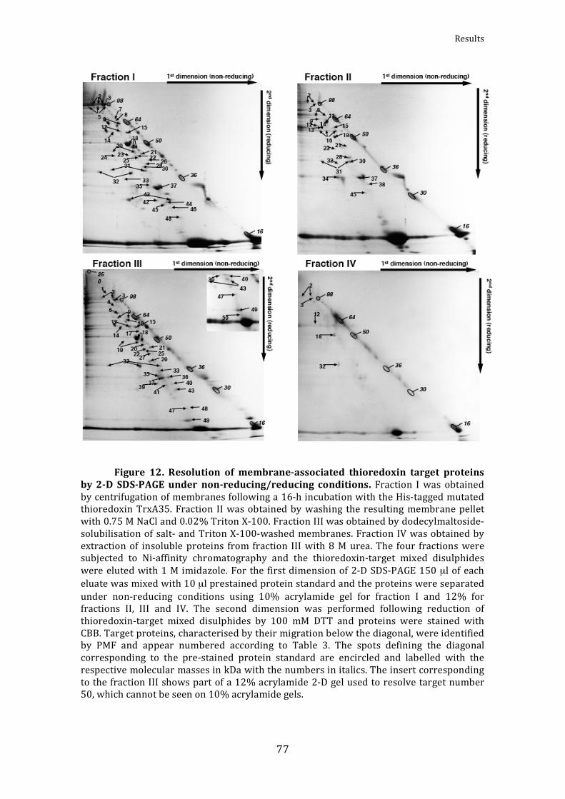
Results
77
Figure 12. Resolution of membraneassociated thioredoxin target proteins
by2D SDSPAGEunder nonreducing/reducing conditions.Fraction Iwas obtainedbycentrifugationofmembranesfollowinga16‐hincubationwiththeHis‐taggedmutatedthioredoxinTrxA35. Fraction IIwasobtainedbywashing theresultingmembranepelletwith0.75MNaCland0.02%TritonX‐100.FractionIIIwasobtainedbydodecylmaltoside‐solubilisationof salt‐andTritonX‐100‐washedmembranes.FractionIVwasobtainedbyextractionof insolubleproteins from fraction IIIwith8Murea.The four fractionsweresubjected to Ni‐affinity chromatography and the thioredoxin‐target mixed disulphideswereelutedwith1Mimidazole.For thefirstdimensionof2‐DSDS‐PAGE150µlofeacheluatewasmixedwith10µlprestainedproteinstandardandtheproteinswereseparatedunder non‐reducing conditions using 10% acrylamide gel for fraction I and 12% forfractions II, III and IV. The second dimension was performed following reduction ofthioredoxin‐target mixed disulphides by 100 mM DTT and proteins were stained withCBB.Targetproteins,characterisedbytheirmigrationbelowthediagonal,wereidentifiedby PMF and appear numbered according to Table 3. The spots defining the diagonalcorresponding to the pre‐stained protein standard are encircled and labelled with therespectivemolecularmassesinkDawiththenumbersinitalics.TheinsertcorrespondingtothefractionIIIshowspartofa12%acrylamide2‐Dgelusedtoresolvetargetnumber50,whichcannotbeseenon10%acrylamidegels.

AlejandroMataCabana
78
Table3.Synechocystissp.PCC6803membraneproteinsinteractingwiththioredoxin
Noa) Protein Cys b) ORF c) FractionMass d)
exp./theor. (kDa)
Mowse score
Peptides matched
% seq. coverage Ref.e)
1 Preprotein translocase SecA* 3 sll0616 III 114/106.9 183 30 33
2Phycobilisome core-
membrane linker polypeptide (LCM)
3 slr0335 I 101/100.2 173 32 37 A
II 160 22 37
III 163 26 36
IV 144 14 14
3 ClpC* 1 sll0020 I 93/91.1 129 15 21 1
II 93 12 23
III 161 23 37
IV 139 14 22
4 ClpB1* 1 slr1641 III 87/98.1 78 11 14 1
5
Polyribonucleotide nucleotidyl
transferase, α-subunit
3 sll1043 I 90/77.9 219 22 35 B
6 FtsH* 1 slr1604 III 70/67.2 99 12 18 7
7 Aspartyl-tRNA synthetase* 3 slr1720 I 69/67.4 81 7 15
8 Ferredoxin-sulfite reductase 7 slr0963 I 68/71.4 109 18 30 A
II 161 18 38
9 60-kDa chaperonin 1 (GroEL 1) 2 slr2076 I 62/57.5 183 26 44 A, 1,
2, 4
II 127 17 37
III 175 18 38
10Transcription
termination factor NusA*
1 slr0743 III 61/51.4 105 14 26
11Pyruvate
dehydrogenase component E2
1 sll1841 II 62/44.9 148 18 53 B

Results
79
12 ATP synthase, α-subunit* 2 sll1326 I 58/53.9 140 8 22 2, 4,
7
II 77 10 28
III 109 15 37
IV 141 8 22
13 Rubisco large subunit (RbcL) 11 slr0009 II 59/52.5 96 18 37 A, 4
14 ATPase of the AAA-family* 1 slr0374 I 57/56.4 140 11 22
II 188 26 46
III 170 19 40
15 ATP synthase, β-subunit* 1 sll1329 I 58/51.7 235 17 51 2, 3,
7
II 159 23 63
III 161 27 75
16 Carboxysomal protein CcmM 9 sll1031 II 56/56.9 192 24 38 A
17Type 2 NADH dehydrogenase,
NdbC*6 sll1484 III 55/56.8 91 13 37
18 Elongation factor EF-Tu 1 sll1099 I 50/43.8 244 31 72 A, 1,
2, 4
II 191 27 66
III 165 28 62
IV 89 8 24
19Oxyanion-
translocating ATPase, ArsA*
2 sll0086 III 49/44.6 124 15 59
20 Putative GTP-binding protein* 3 sll0245 I 46/39.3 144 8 31 6
III 152 15 42
21Urea transporter,
ATP-binding subunit (UrtD)*
1 sll0764 I 45/41.2 103 9 29
II 112 14 44
III 94 9 30

AlejandroMataCabana
80
22 RecA* 3 sll0569 I 44/37.8 119 10 36
III 88 8 28
23 RNA polymerase, α-subunit* 2 sll1818 I 44/35.1 131 6 26
II 137 13 40
24 Phosphoribulokinase 4 sll1525 I 44/37.9 89 13 31 B, 1, 4, 5
25 Twitching motility protein PilT* 3 slr0161 I 43/40.8 91 6 18
III 75 11 34
26
Glyceraldehyde-3-phosphate
dehydrogenase (GAPDH2)
5 sll1342 I 42/36.6 132 11 36 B, 1, 8
27 Hypothetical protein* 3 slr0658 III 42/37.7 114 14 55
28
Pyruvate dehydrogenase
component E1, α-subunit*
5 slr1934 I 38/38.1 231 32 64 2
II 132 14 34
29
Nitrate/nitrite transporter, ATP-binding subunit
(NrtD)*
3 sll1453 III 38/36.5 139 12 48
30
Pyruvate dehydrogenase
component E1, β-subunit*
3 sll1721 I 38/35.7 89 12 33 2
31 30S ribosomal protein S2* 3 sll1260 I 37/30.1 132 11 39
II 95 15 46
32 Universal stress protein Usp1* 4 slr0244 I 36/31.2 96 9 55
II 71 6 28
III 113 7 34
IV 88 7 33
33 Universal stress protein Usp2* 4 slr0670 I 33/32.9 75 4 19

Results
81
III 103 13 66
34 β-type carbonic anhydrase* 4 slr1347 II 32/30.7 98 15 52 1, 5,
6
35 Glutathione-S-transferase* 1 sll1545 I 31/29.7 82 6 28 1, 6
III 148 13 57
36 Serine-O-acetyl transferase* 3 slr1348 III 31/27.5 93 7 30
37 30S ribosomal protein S3 1 sll1804 I 30/27.0 165 17 58 A
II 108 10 41
III 119 16 68
38 50S ribosomal protein L3* 1 sll1799 II 30/22.7 86 10 60
39 Hypothetical protein* 1 slr1702 III 29/27.3 131 11 53
40 Heme oxygenase 1* 1 sll1184 III 29/27.0 79 11 45
41Phycobilisome rod-
core linker polypeptide (LRC)*
1 sll1471 III 28/28.5 108 12 50
42 Response regulator Rre1* 2 slr1783 I 27/31.5 129 7 35
43 1-Cys peroxiredoxin 3 slr1198 I 26/23.6 117 10 43 A, 8
III 75 8 31
44Transcription
antitermination protein NusG*
1 sll1742 I 25/23.5 110 10 51
45 Hypothetical protein ycf50* 1 slr2073 I 25/20.6 86 5 41
II 78 6 42
46 Precorrin isomerase* 2 slr1467 I 24/24.7 84 5 31
47Acetolactate
synthase, small subunit*
1 sll0065 III 21/18.9 121 9 61
48 Phycocyanin β-subunit 3 sll1577 I 21/18.1 88 6 47 B
III 103 9 76

AlejandroMataCabana
82
49 Hypothetical protein* 1 sll1106 III 18/18.0 80 8 46
50 Photosystem I protein PsaD* 1 slr0737 III 15/15.6 87 5 39
Proteinsmarkedwithanasterisk(*)arehereidentifiedasthioredoxintargetsforthefirsttimeinSynechocystissp.PCC6803.a)No.,spotnumbersinFigures12b)Cys,numberofcysteinesintheaminoacidsequencec)ORF,openreadingframesaccordingtotheCyanobased)Massexp./theor.,experimentalandtheoreticalmassvaluese)Referencestostudies,wherethethioredoxin‐interactingproteinhasbeenreportedasathioredoxin target incyanobacteria: (A) Lindahland Florencio, 2003; (B)Pérez‐Pérezetal.,2006.Orreportedinanotherorganism:(1)Balmeretal.,2003;(2)Balmeretal.,2004b(3)Balmeretal.,2006;(4)Lemaireetal.,2004;(5)Marchandetal.,2004;(6)Marchandetal.,2006;(7)MotohashiandHisabori,2006;(8)Wongetal.,2003.
The precise locations in Synechocystis thylakoid and/or plasma membranes
havepreviouslybeendetermined(Srivastavaetal.,2005;Pisarevaetal.,2007;Huang
etal.,2002;Huangetal.,2006)for18oftheproteinsidentifiedasputativethioredoxin
targets in the present study. These proteins, their locations and the corresponding
references are listed in Table 4. Among the newly identified proteins four were
predicted to be integral membrane proteins according to the TMHMM programme
(http://www.cbs.dtu.dk/services/TMHMM‐2.0). The FtsH protease encoded by the
ORF slr1604 (Fig. 12, spot no. 6)was predicted to have two transmembrane helices
between residues 10 and 29 and residues107 and 129.However, aspointedoutby
Pisarevaetal.2007,thefirstofthesehelicesmayactuallycorrespondtoanN‐terminal
signal peptide. The type 2 NADH dehydrogenase subunit NdbC, the nitrate/nitrite
transporter subunit NrtD and the hypothetical protein encoded by the ORF sll1106
(Fig.12,spotnos.17,29and49)werepredictedtopossessonetransmembranehelix
eachbetweenresidues487–504,293–315and53–75,respectively.
Table4.LocalisationofSynechocystissp.PCC6803membraneproteinspresentinourstudy
No. a) Protein ORF b) Location c) Reference d)
1 Preprotein translocase SecA* sll0616 TM Srivastava et al., 2005
2 Phycobilisome core-membrane linker polypeptide (LCM) slr0335 PM Pisareva et al., 2007

Results
83
3 ClpC* sll0020 TM, PMSrivastava et al., 2005
Huang et al., 2006
6 FtsH* slr1604 PM Pisareva et al., 2007
8 Ferredoxin-sulfite reductase slr0963 PM Pisareva et al., 2007
9 60-kDa chaperonin 1 (GroEL 1) slr2076 TM Srivastava et al., 2005
12 ATP synthase, α-subunit* sll1326 TM, PMSrivastava et al., 2005
Huang et al. 2002, 2006
13 Rubisco large subunit (RbcL) slr0009 PM Pisareva et al., 2007
15 ATP synthase, β-subunit* sll1329 TM, PMSrivastava et al., 2005
Huang et al., 2006
16 Carboxysomal protein CcmM sll1031 PM Pisareva et al., 2007
17 Type 2 NADH dehydrogenase, NdbC* sll1484 PM Pisareva et al., 2007
18 Elongation factor EF-Tu sll1099 TM Srivastava et al., 2005
24 Phosphoribulokinase sll1525 TM Srivastava et al., 2005
26 Glyceraldehyde-3-phosphate dehydrogenase (GAPDH2) sll1342 TM Srivastava et al., 2005
28 Pyruvate dehydrogenase component E1, α-subunit* slr1934 TM Srivastava et al., 2005
41 Phycobilisome rod-core linker polypeptide (LRC)* sll1471 TM Srivastava et al., 2005
48 Phycocyanin β-subunit sll1577 TM, PMSrivastava et al., 2005
Huang et al., 2006
50 Photosystem I protein PsaD* slr0737 TM, PMSrivastava et al., 2005
Huang et al., 2002
a) No., spot numbers in Figure 12, b) ORF, open reading frames according to theCyanobase,c)Location.PM,plasmamembrane;TM,thylakoidmembrane,d)References:Huang F,et al. (2002)Proteomics ofSynechocystis sp. strain PCC 6803. Identification ofplasmamembraneproteins.MolCellProteomics1:956‐966Huang F, et al. (2006) Proteomic screening of salt‐stress‐induced changes in plasmamembranesofSynechocystissp.strainPCC6803.Proteomics6:2733‐2745PisarevaT,etal.(2007)ProteomicsofSynechocystissp.PCC6803.Identificationofnovelintegralplasmamembraneproteins.FEBSJ274:791‐804SrivastavaR,etal. (2005)ProteomicstudiesofthethylakoidmembraneofSynechocystissp.PCC6803.Proteomics5:4905‐4916

AlejandroMataCabana
84
Aschematicrepresentationofthedistributionoftargetsbetweenthefractions
I, II and III (Fig. 13A and Table 3) shows that each fraction contains unique target
proteins.Thisunderlinestheconveniencetofractionatethemembranes,asdescribed,
prior to separation on 2‐DE in order to reduce the number of protein species to be
resolved on each gel. One fifth of the thioredoxin targets appeared in all three
fractions,generallythemostabundanttargetproteinssuchasthephycobilisomecore‐
membranelinker(LCM)andthetranslationelongationfactorEF‐Tu.Examinationofthe
distributionofallhithertoidentifiedthioredoxintargetsinSynechocystissp.PCC6803
(LindahlandFlorencio,2003;Pérez‐Pérezetal.,2006;andthepresentstudy)between
thesolublecytosolicfractionandthetotalmembranesreveals,somewhatsurprisingly,
thatthemajorityofthesetargetsareboundtomembranes(Fig.13B).Almosttwiceas
manyuniquetargetsareassociatedwiththemembranesascomparedtothosepresent
exclusivelyinthecytosol.Itshouldbenotedthatthreeoftheeighttargetsfoundinan
extract of peripheral membrane proteins reported in (Lindahl and Florencio, 2003)
werenotidentifiedinthepresentstudy,namelytheRNApolymerasesubunitsβandβ’
andsulphateadenylyltransferase.
Figure13.TrxtargetsdistributioninSynechocystis. (A)Distributionof the50
thioredoxin targets in the present studybetween thedifferent fractionsofSynechocystissp. PCC 6803 membrane proteins. (B) Distribution of all 77 Synechocystis thioredoxintargets identified to date (Lindahl and Florencio, 2003; Pérez‐Pérezet al., 2006 and thepresentstudy)betweencytosolandmembranes.

Results
85
ThepresenceoftheATP‐bindingsubunitsofseveraltransporters,suchasSecA,
ArsA,UrtDandNrtD(spotnos.1,19,21and29),inthethioredoxinmembranetarget
proteome is intriguing and suggests that translocation across membranes could be
regulated by means of redox‐dependent modulation of the ATPase activities of the
respectivetransporters,whichdeservesfurtherstudies.Acloserlookatthepositions
of the cysteines in the sequences of these proteins, which do not share homology,
shows that some cysteines are located precisely in the ATPase sequence signatures
and,hence,theirredoxstatesarelikelytoaffectATP‐bindingorATPaseactivity.The
secondof the three cysteinesof SecA is located in theATPaseWalkerBmotif (208‐
FCIIDEV‐213)andtwoofthethreecysteinesofNrtDappearintheWalkerAmotif(52‐
CIIGHSGCGKS‐62) (seeWalker et al., 1982 for a definition of thesemotifs). Another
interesting category ofmembrane associated thioredoxin target proteins belongs to
the AAA+‐ family (ATPases associated with a variety of cellular activities), which
constitutesasubfamilyoftheWalker‐typeNTPases(Neuwaldetal.,1999;Oguraand
Wilkinson,2001).ClpC,aregulatorysubunitoftheClp‐protease,andClpB1(spotnos.
3 and 4) share a single conserved cysteine, which is located in the so‐called AAA+
Sensor 1‐domain (Neuwald et al., 1999), also known as themotif C (Koonin, 1993).
The FtsH protease (Fig.12, spot no.6), encodedby theORFslr1604, isknown tobe
integral to theSynechocystissp.PCC 6803 plasmamembrane (Pisarevaetal., 2007).
ThesinglecysteineofthisAAAproteaseappearsintheWalkerBmotif(255‐CIVFIDEI‐
262)and this feature issharednotonlywiththeotherSynechocystis FtsHproteases,
whicharepresentinthylakoid‐orplasmamembranesinthisorganism(Srivastavaet
al., 2005; Pisareva et al., 2007), but also with the plant chloroplast homologues. In
some of our experiments, we could also detect tryptic fragments by PMF
corresponding to the Synechocystis thylakoid FtsH encoded by the ORF sll1463,
although the MOWSE scores obtained were not high enough to unambiguously
ascertainthe identity.TheFtsHproteasesofphotosyntheticorganismshaverecently
attracted much attention for their roles in the turnover of the photosystem II D1‐
proteinfollowinglightinduceddamageincyanobacteria(Silvaetal.,2003)aswellas
inplants(Lindahletal., 2000;Sakamoto,2006).Apossible redox regulationof their
activitiescouldhaveprofoundimplicationsforthemaintenanceofthephotosynthetic
apparatus and functioning of other membrane associated processes. Interestingly,
ClpA (Balmer et al., 2003) as well as two FtsH proteases (Motohashi and Hisabori,
2006) have previously been identified as thioredoxin targets in plant chloroplasts.

AlejandroMataCabana
86
Finally, anuncharacterisedmemberof theAAA+‐familyencodedby theORFslr0374,
whichispresentinastress‐responsiveoperon(SinghandSherman,2000;Singhand
Sherman,2002)wasidentifiedasathioredoxintarget(Fig.12,spotno.14).Theonly
cysteineofthisAAA+‐protein,whichisconservedamongthecyanobacterialspecies,is
present in the Walker A motif (262‐LILGVPGCGKS‐272). Common to all of these
members of the AAA+‐family is that they possess one single cysteine in their
sequences,whichimpliesthattheoxidisedstateinvolvesanintermoleculardisulphide
either with another protein or with a lowmolecular mass thiol compound such as
glutathione.Glutathionylationhasonlyrecentlybeenconsideredasareversiblepost‐
translationalmodificationinphotosyntheticorganisms(Masipetal.,2006;Micheletet
al., 2006), whereas this mechanism for regulation is better characterised in
mammalian systems. The localisation of the single cysteine within the amino acid
sequenceoftheseproteinshasbeenrepresentedintheFigure14.
Figure14.CysteinelocalisationwithintheaminoacidsequencesoftheAAA+
protein.TheAAA+‐proteinshavebeenrepresentedincludingtheATPbindingdomainandthe cysteine residues have been located inside of it. The protein domains have beenidentified using the engine PROSITE SCAN (http://npsa‐pbil.ibcp.fr/cgi‐bin/npsa_automat.pl?page=npsa_prosite.html).

Results
87
Twomembersof theuniversal stressprotein‐family(Usp)(Kvintetal.,2003)
were identified amongst the newmembrane‐associated thioredoxin targets (Fig. 12,
spotnos.32and33).TheexactfunctionoftheUsp‐domainisnotclear,althoughitis
known to possess autophosphorylating activity and can be found in many proteins
from archae, bacteria and plants, either alone, in tandem or as a fusion with other
domains (Kvint et al., 2003). In Escherichia coli, Usp‐expression is induced under
various stress conditions such as moderate heat shock, nutrient starvation and
exposure to uncouplers (Gustavsson et al., 2002) and deletion of Usp‐genes causes
hypersensitivity to stress (Nachin et al., 2005). TheSynechocystisUsp’s identified in
thisstudybothconsistoftwoUsp‐domainsintandem.
Concerningaminoacid‐andproteinmetabolism,aswasobservedpreviously
in other studies (Florencioetal., 2006;Montrichardetal., 2009), a largenumber of
proteins identified as thioredoxin targetswere involved in such pathways. Three of
themplaya role inaminoacidmetabolism;namelythesmallsubunitofacetolactate
synthase, serine‐O‐acetyl transferase and aspartyl‐tRNA synthetase, and have been
found to interact with Trx for the first time in the present study. However, several
tRNA syntheses have been shown previously to interact with Trx in Synechocystis,
valyl‐tRNAsynthase(LindahlandFlorencio,2004),lysyl‐tRNAsynthase(Pérez‐Pérez
etal.,2006); inplants,glycyl‐tRNAsynthaseandseryl‐tRNAsynthase(Häglundetal.,
2008);andinnon‐photosyntheticprokaryotes,phenylalanyl‐tRNAsynthase(Liechert
and Jakob, 2004), alanyl‐tRNA synthase (Liechert et al., 2008) and glutamyl‐tRNA
synthase (Hochgräfe et al., 2005). We have also found three ribosomal proteins,
whereasasmanyas30differentribosomalproteinshavebeenreportedinstudiesof
Trx‐target proteins in plants (Montrichard et al., 2009). Only the protein 60‐kDa
chaperonin 1 related with protein folding was identified here, however, diverse
chaperonesbelonging to chloroplast andmitochondria havebeen reported in plants
(Montrichard et al., 2009; Lindahl and Kieselbach, 2009). ClpB, ClpC and FtsH are
three proteins with protease activity, which have been defined as Trx target in the
presentstudyandpreviouslyinplants(Balmeretal.,2003;2004and2006).
Another thioredoxin targetworthmentioning is the haeme oxygenase1 (Fig.
12, spot no. 40), which functions in biosynthesis of open‐chain tetrapyrroles
(Sugishima et al., 2004), such as phycobilin, utilised for light harvesting and as the

AlejandroMataCabana
88
pigment of the phytochrome photoreceptors in cyanobacteria (Lamparter, 2004).
Finally, we identified the PsaD subunit of the photosystem I multiprotein complex
(Herranen et al., 2004), which participates in the docking of the soluble electron
carrierferredoxinofthephotosyntheticelectrontransport(Sétifetal.,2002).
Following the proteomics assisted screening for Trx‐target proteins in
membranes we choose to study further the peripheral peroxiredoxins and USP‐
proteinsandtheintegralFtsH.
4. Interactions of 1Cys and 2Cys peroxiredoxins with TrxA in
Synechocystis
Peroxiredoxins (Prx) are a ubiquitous group of thiol‐dependent peroxidases,
whichrequirereductionbyelectrondonorssuchasTrx,Grxorcyclophilins(Königet
al., 2002; Rouhier et al., 2001; Lee et al., 2001), though other enzymes have been
shown to function as reductants for Prx such as NTRC in plants (Pérez‐Ruiz et al.,
2006)andtheflavoproteinAhpFinnon‐photosyntheticbacteria(Poole,2005).Based
ontheirphylogenyaswell as theircatalyticmechanisms,Prxareclassified into four
groups; 2‐Cys Prx, 1‐Cys Prx, Type II Prx and PrxQ. The 2‐Cys Prx contains two
catalyticcysteinesinitsaminoacidsequence,referredtoasperoxidaticandresolving
cysteines, and is a homodimeric enzyme where the two subunits are linked via a
disulphidebondintheoxidisedform(BaierandDietz,1996;Königetal.,2002).Type
IIPrxandPrxQareatypical2‐CysPrx,sincetheypossesstwocatalyticcysteines,but
functions as a monomer. PrxQ, which is a homologue of the E. coli bacterioferritin
comigraing protein (BCP), carries the two cysteines spaced by only four aminoacids
and intype IIPrx thecysteinesareseparatedby24aminoacids(Storketal.,2009).
Thelastgroupisthe1‐CysPrxcontainingasingleconservedcatalyticcysteine(Stacy
etal.,1996;1999).
The genome of the cyanobacterium Synechocystis encodes five Prx, which
belongtoeachoftheestablishedgroups(Storketal.,2005):2‐CysPrx(slr0755),1‐Cys
Prx(sll1198),typeIIPrx(sll1621)andtwoPrxQ,PrxQ1(slr0242)andPrxQ2(sll0221).
Differentstudiesincyanobacteriashowthatthemutantstrainslackingsomeofthese

Results
89
enzymes are affected in the growth at elevated light intensities (Klughammer et al.,
2004;Perelmanetal.,2003;Hosoya‐Matsudaetal.,2005;Kobayashietal.,2004;Latifi
etal.,2007).
ThethioredoxintargetproteomicsstudiesinSynechocystishaveonlyreported
the1‐CysPrxandtypeIIPrxasputativeTrx‐interactingperoxiredoxins(Lindahland
Florencio, 2003; Pérez‐Pérez et al., 2006; Florencio et al., 2006). The 2‐Cys Prx has
neverbeenidentifiedamongstthetargetsofanythioredoxininSynechocystisandthe
explanation suggestedwas that thisproteinhas ahydrophobicdomain,which could
anchorittomembranes,andhenceitwouldbeabsentfromcytosolicextracts(Pérez‐
Pérez et al., 2006). However, in the proteomic approach here described, in which
membraneproteinswereanalysed,theonlyPrxfoundtointeractwithTrxAwasthe1‐
CysPrx(Fig.12andTable3).Therefore,aimingatconfirmingtherelationshipof1‐Cys
PrxwithTrxAandsearching forapossibleinteractionof the2‐CysPrxwiththisTrx,
bothperoxiredoxinswereexpressedinE.coliandpurifiedforfurtherstudies.
In order toanalyse the localisationof 2‐CysPrx inSynechocystis, the purified
proteinwasusedtoraisespecificantibodies.Hence,cytosolicandmembraneextracts
fromSynechocystiswerepreparedforwesternblotanalysesofthepossiblelocationin
membranesof2‐CysPrx.However,nosignalwasobtainedinthemembranefraction,
whereas a strong signalwasvisualised in the cytosolic fraction (Fig.15A).Thus,we
could discard the anchoring of this peroxiredoxin in membranes and confirm its
presenceinthecyanobacterialcytosol(Fig.15A).Sincewewereunabletodetectthis
proteinamongtheTrx‐targetsisolatedfromanycellularcompartmentresolvedingels
and stained with CBB we thought that the protein staining might not be sensitive
enough. Therefore, we decided to repeat the Trx‐affinity chromatography using
cytosolic extracts and to analyse the eluate resolved in non‐reducing/reducing 2‐D
SDS‐PAGEgelsbymeansofwesternblotusing theantibodyagainstSynechocystis 2‐
Cys Prx. As shown in Fig. 15B the 2‐Cys Prx was indeed present in the eluate and
migratedbelowthediagonal, indicating the formationof themixeddisulphideswith
the monocysteinic TrxA35. Moreover, the 2‐Cys Prx appeared distributed between
twobandscorrespondingtotwotypesofcomplexes;oneinwhichthePrxboundone
molecule of Trx and other in which it bound two molecules of Trx (Fig. 15B).
Therefore,wecouldconcludethat2‐CysPrxwasaputativeTrxtarget,althoughithad
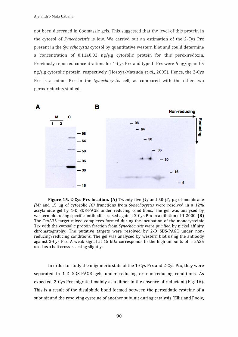
AlejandroMataCabana
90
notbeendiscerned inCoomassiegels.This suggestedthat the levelof thisprotein in
the cytosol of Synechocistis is low. We carried out an estimation of the 2‐Cys Prx
presentintheSynechocystiscytosolbyquantitativewesternblotandcoulddetermine
a concentration of 0.11±0.02 ng/µg cytosolic protein for this peroxiredoxin.
Previouslyreportedconcentrationsfor1‐CysPrxandtypeIIPrxwere6ng/µgand5
ng/µgcytosolicprotein,respectively(Hosoya‐Matsudaetal.,2005).Hence,the2‐Cys
Prx is a minor Prx in the Synechocystis cell, as compared with the other two
peroxiredoxinsstudied.
Figure15. 2CysPrx location. (A)Twenty‐five (1) and50 (2)µg ofmembrane
(M) and 15 µg of cytosolic (C) franctions from Synechocystis were resolved in a 12%acrylamide gel by 1‐D SDS‐PAGE under reducing conditions. The gel was analysed bywesternblotusingspecificantibodiesraisedagainst2‐CysPrxinadilutionof1:2000.(B)TheTrxA35‐targetmixed complexes formedduring the incubation of themonocysteinicTrxwiththecytosolicproteinfractionfromSynechocystiswerepurifiedbynickelaffinitychromatography. The putative targets were resolved by 2‐D SDS‐PAGE under non‐reducing/reducing conditions. The gelwas analysedbywestern blot using the antibodyagainst 2‐CysPrx. Aweak signal at 15 kDa corresponds to the high amounts of TrxA35usedasabaitcross‐reactingslightly.
Inordertostudytheoligomericstateofthe1‐CysPrxand2‐CysPrx,theywere
separated in 1‐D SDS‐PAGE gels under reducing or non‐reducing conditions. As
expected,2‐CysPrxmigratedmainlyasadimerintheabsenceofreductant(Fig.16).
This is a resultof the disulphidebond formed between theperoxidatic cysteineof a
subunitandtheresolvingcysteineofanothersubunitduringcatalysis(EllisandPoole,

Results
91
1997). However, various oligomeric forms with a superior molecular weight were
showed(Fig.16),whichmightbeexplainedasinteractionsbetweendifferentdimers
ofthisPrx.Highmolecularweightcomplexeshavebeenwellcharacterisedinseveral
organisms(Woodetal.,2003).Moreover,someofthesecomplexeshavebeenshown
tohaveachaperoneactivityinyeast(Jangetal.,2004).Underreducingconditionsthe
treatmentappliedwasperhapsnotstrongenoughtoreduceall thecomplexestothe
monomeric form.1‐CysPrxmigratedmainlyas adimerandother complexeswitha
highmolecularweightwere also shown (Fig. 16). The catalyticmechanism of 1‐Cys
Prx isstillunknown,but themigrationundernon‐reducingconditions indicatesthat
either of its two additional cysteines, apart from the peroxidatic cysteine, may
participateinintermoleculardisulphideformation(Hosoya‐Matsudaetal.,2005).
Figure16.Electrophoreticmigrationof2CysPrxand1CysPrx.Threeµgof
eachpurifiedperoxiredoxinwereresolvedina12%SDS‐PAGEgelunderreducing(Red),incubatingthesamplewithaloadingbuffercontainingfinalconcentrationof350µMof2‐mercapto‐ethanol,andnon‐reducing(NoRed)conditions,usingaloadingbufferwithoutanyreductant.
The ability of 1‐Cys Prx and 2‐Cys Prx to interact physically with TrxA was
analysed through incubation of the purified peroxiredoxin with the monocysteinic
TrxA35 followed by electrophoretic separation under non‐reducing conditions and
western blot using the antibodies against TrxA (Fig. 17). Since TrxA35 also forms
16
30
36
50 64 98
250
1-Cys Prx 2-Cys Prx
Red NoRed Red NoRed

AlejandroMataCabana
92
homodimers in theabsenceof reductant, itwas also incubatedalonewithoutPrx in
order to distinguish the TrxA dimers from the Trx‐Prx complexes. The result shows
mixeddisulphideformationbetweenTrxAandbothperoxiredoxins.Thesecomplexes
were not only dimers, butalso appearedasoligomerswitha highmolecularweight
that may correspond to Trx interacting with various Prx molecules linked to each
otherorthebondformationbetweentwoormoreTrxperPrx.Electrophoresisunder
reducingconditionsabolishedcompletelythecomplexes.Themainbandsobtainedin
thewesternblothavebeenreferredintheFigure17withaletter.Thebandssignalled
as c and f in the Figure 17 correspond to one molecule of Prx bound to one Trx
molecule. The bandsmarked as b and d correspond in the western blot to a cross‐
linkedreactionwiththehighlyabundant2‐CysPrxmonomeranddimerrespectively.
TheabandcouldbeexplainedasonemoleculeofTrxAboundtoa2‐CysPrxdimer,
although itcouldalsobetwomoleculesofTrxAboundtoasinglemoleculeof2‐Cys
Prx. Inthe caseof1‐CysPrxthebandnamedase probablycorrespondstoaunique
moleculeofPrxboundtotwomoleculesofTrxA,indicatingtheparticipationofsome
ofthenon‐peroxidaticcysteinespresentinitsaminoacidsequence.
Figure17.Analysisof the interactionbetweentheperoxiredoxins2CysPrx
and1CysPrxwiththeTrxA.(A)Twentyµgofeachperoxiredoxinwasincubatedwith8µgofthemonocysteinicTrxA35anda1/30dilutionoftheresultingreactionmixtureswasresolved on 12% SDS‐PAGE gel under non‐reducing (‐DTT) and reducing (+DTT)conditions. The thioredoxin was detected by western blotting using specific antibodiesraisedagainstTrxA.(B)Inparallel,thereactionmixtureswithoutdilutionwereappliedon12%acrylamidegelsandstainedwithCBB.Lanes1,TrxA35alone;lanes2,TrxA35and2‐CysPrx; lanes 3, TrxA35 and1‐CysPrx. The black arrowsmark themonomericTrxA35and the white arrows mark the TrxA35 homodimer. The main bands resulted in thewesternblothavebeennamedwithaletterfromatof.Themeaningsofthebandsmarkedwithlettersarediscussedinthetext.

Results
93
OnceconfirmedtheinteractionbetweentheseperoxiredoxinsandtheTrxA,we
decided to testwhether this interaction involves functional electron transfer.Hence,
weanalysedtheperoxidaseactivityofthePrxswithwildtypeTrxAaselectrondonor.
Tothisend,weincubated5µMofeachPrxwith4µMofTrxAreducedwithalow(0.2
mM) concentration of DTT, and the reduction of H2O2 was monitored by the FOX
(ferrous ionoxidation) colorimetric assay (Fig. 18).TheDTT aloneor togetherwith
TrxA did not produce appreciable rates of peroxidase activity (Fig. 18). Both Prx
showedperoxidaseactivity,butthe2‐CysPrxwasmoreefficient indecomposingthe
peroxide.Thus,thisPrxscavengedabout70%oftheinitialH2O2added(100µM)in10
min,whereasthe1‐CysPrxonlyremoved20%duringthesametime.After20min,the
2‐CysPrxhaddecomposedalltheperoxide,whereasthe1‐CysPrxhadscavengedonly
30%.Themeasuredactivitieswere1.7±0.1nmolH2O2/min⋅nmolPrxforthe2‐CysPrx
and0.4±0.05nmolH2O2/min⋅nmolPrxforthe1‐CysPrx.
Figure 18. TrxAdependent peroxidase activity of 2Cys Prx and 1Cys Prx.
Thedecompositionof100µMH2O2by5µMofeachpurifiedperoxiredoxinwasmeasureusing the FOX assay. The peroxidase activitywas analysed in presence of the completereductionsystem,0.2mMDTT+4µMTrxA+5µMPrx(2CPrx+Trxor1CPrx+Trx);orlacking the Trx (2CPrx or 1CPrx), the Prx (DTT+Trx) or both of them (DTT). Theremaining H2O2 was measured at 0, 10, 20 and 30 min. The error bars represent thestandarddeviation.

AlejandroMataCabana
94
5.TheUniversalStressProtein(USP)anditsinteractionwithTrx
Proteins of the Universal Stress Protein (USP) superfamily are present in
diverseorganismsrangingfromarcheaandbacteriatoplants,thoughtheyarerarein
fungi and absent from animals (Kvint et al., 2003). The UspA protein from E. coli
initially gave thename to the superfamily (Kvintetal., 2003) since the levelsof this
proteinbecomeelevatedinresponsetoalargevarietyofstressconditions,including
starvation for carbon, nitrogen,phosphate, sulfate, andamino acidsandexposure to
heat, oxidants, metals, uncouplers of the electron transport chain, polymixin,
cycloserine, ethanol and antibiotics (Gustavsson et al., 2002; Kvint et al., 2003;
Nyström and Neidhart, 1994; Nachin et al., 2005). Usp‐containing organisms are
usually equippedwith severalusp genes (Nachin et al., 2005). They encode either a
smallUspprotein(around14‐15kDa)harbouringoneUspdomain,oralargerversion
(around 30 kDa) consisting of two Usp domains in tandem. There are also large
proteins,inwhichtheUspdomainispresenttogetherwithanotherfunctionaldomain,
typicallyaniontransporteroraSer/Thrproteinkinase(Kvintetal.,2003).E.colihave
fivesmallUspProteinsandonetandem‐typeUsp(UspE).Theyhavebeendividedinto
fourdifferentclasses:UspA,UspCandUspDbelongtoclassI,UspFandUspGbelongto
classIIandthetwoUspdomainsofUspEseparateintoclassesIIIandIV(Kvintetal.,
2003).UspAisaserine/threoninephosphoproteinwithautokinaseactivity(Freestone
etal.,1997)anditsdeletionproducesaprematuredeathduringstasis(Nyströmand
Neidhart, 1994), whereas overproduction blocks the cell in a growth‐arrested state
(NyströmandNeidhart,1996).Moreover,deletionofclass Iorclass III/IVuspgenes
resultsinhypersensivitytoDNA‐damagingagents(Gustavssonetal.,2002).However,
cells lacking the class II Usp are sensitive to uncouplers but not to DNA‐damaging
agents(Bochkarevaetal.,2002),indicatingthatthefunctionsoftheclassIandIImay
be distinct (Nachin et al., 2005). Nevertheless, molecular mechanisms for the Usp
functioninthecellarestillundefined.
Wediscovered twoUsp‐likeproteinsamongst theputative targetsofTrxA in
Synechocystismembranes(Fig.12,Fig.19andTable3).BothUspproteinsmigratedin
the 2‐D SDS‐PAGE under non‐reducing/reducing conditions distributed between
several spots (Fig. 19). The different spots, which appeared below the diagonal
correspondedtoeachUspproteinboundtooneormoremoleculesofTrxA.Thespots

Results
95
resolved above the diagonal corresponded to intramolecular disulphide formation
(Fig. 19).These data indicated thatUsp’s areable to form intra‐ and intermolecular
disulphidebonds.TheUspproteinsincyanobacteriahavenotbeenstudied,however,
cyanobacterial genomes encode homologues of Usp domain‐containing proteins
(Kvintetal.,2003).ThetwoUspproteinshereidentified,namedUsp1andUsp2,are
encodedbytheORF’sslr0244andslr0670.BothproteinsconsistoftwoUspdomains
intandem,andhavefourcysteinesintheiraminoacidsequences.
Figure 19. Positions of Usp1 and Usp2 in the 2D SDSPAGE under non
reducing/reducing conditions. The Usp proteins identified in the Fraction I of themembraneproteinsinteractingwithTrxA(Fig.12andTable3)havebeen locatedin thegel. The spots corresponding toUsp1 (encodedby slr0244) are encircled in red and thespotscorrespondingtoUsp2(encodedbyslr0670)areencircledinorange.
InordertodeterminewhethertheTrxAisabletoreduceUspandwhetherthis
protein indeed undergoes redox regulation we expressed Usp1 in E. coli for
purification.
Firstly, we decided to analyse the redox state of the Usp1 cysteines by
observing the migration pattern in acrylamide gels in presence and absence of
reducing agents, such as DTT and TrxA (Fig. 20). Under oxidising conditions Usp1
migrated mainly as oligomers with a very high molecular weight, suggesting the
capacityofUsp1to formintermoleculardisulphidebonds(Fig.20).Intheabsenceof

AlejandroMataCabana
96
any reductant, a monomeric form that migrated with a lower apparent molecular
weight could also be seen, which may be the result of intramolecular disulphide
formation (Fig. 20).Whena reductantwasadded, theoligomeric forms disappeared
rapidlyandthebandcorrespondingtothemonomericformofUsp1increased.DTTat
2mMconcentrationwasenoughtoreducetheUsp1completely.Afixedconcentration
of0.1mMofDTTwasusedasanelectrondonorforTrxA(Fig.20B),sinceDTTatthis
concentrationdidnotreducemuchoftheUsp1.AgraphicrepresentationoftheTrxA‐
dependent increase of the monomeric form of Usp1 is shown (Fig. 20C), clearly
showing the positive correlation between TrxA concentration and the monomer of
Usp1. Therefore, we concluded that TrxA is a functional electron donor for Usp1 in
vitro.
Figure20.ReductionofUsp1(slr0244)byDTTandTrxA.Threeµgofpurified
Usp1(slr0244)was incubatedat25ºCduring20min inabsenceorpresenceofdifferentreductant(DTTandTrxA).Theresultofincubationwasresolvedon10%acrylamidegelsbySDS‐PAGEundernon‐reducingconditions.In(A)agradientconcentrationofDTTandTrxA was tested, from 0 to 4 mM to DTT and 1 to 8 µM of TrxA with a fixed DTTconcentrationof0.5mM.In(B)thegradientconcentrationofTrxAisrepeatedfrom0to10µM,butthefixedconcentrationofDTTwas0.1mMinthiscase.Acontrolwith2mMDTTwasused.TheblackarrowheadmarksthemonomericformofUsp,whereasthewhite

Results
97
arrowheadmarksanoxidisedformofthemonomericUsp,thegreyarrowheadmarksthedimericformoftheUspandtheasteriskindicatestheUspatotheroligomericforms.(C)Graphic representation of Usp1 monomer apparition by means of TrxA‐dependentreduction.
After verifying the capacity of TrxA to reduce the disulphides of Usp1, we
wanted to test the reversibility of this kind of reduction, using the thiol oxidising
reagents H2O2 and copper.Hence, the Usp1 proteinwas initially pre‐reducedwitha
ten‐fold saturating concentration (20mM) of DTT and after elimination of DTT, the
samplewas oxidised using different concentrations of the reagents. H2O2 aswell as
Cu2+ were able to re‐oxidise Usp1 (Fig. 21) as judged by the disappearance of the
monomericformandincreaseintheoligomericformsofUsp1(andtheintramolecular
disulphide). In the range of concentrations used, H2O2 oxidised more efficiently the
Usp1proteinthanCu2+.Therefore,theUsp1cysteinesundergoredoxmodificationand
aresensitivetotreatmentwiththiol‐oxidisingagents.
Figure21.ReoxidationofUsp1(slr0244)usingH2O2andCu2+.ThreeµgofUsp
were pre‐reduced by 30 min incubation on ice with 20 mM DTT and thereafterconcentratedanddilutedonVivaspin(cut‐off30kDa)in20mMTris‐HCl(pH8.0)/0.5MNaCluntiltheDTTconcentrationwasabout0.16mM.Therefore,thereducedsamplewasre‐oxidisedat25ºCduring20minusingH2O2andCuCl2attheindicatedconcentrationsandthe proteins we resolved by SDS‐PAGE in 10% acrylamide gels under non‐reducingconditions. The black arrowheadmarks themonomeric form ofUsp,whereas thewhitearrowheadmarksanoxidisedformofthemonomericUsp.ThegreyarrowheadmarksthedimericformoftheUspandtheasteriskindicatestheUspatotheroligomericforms.

AlejandroMataCabana
98
Finally,weaimedatfindingoutiftheredoxmodificationoftheUsp1cysteines
could alter the activity of this protein, though the precise function of this kind of
protein remains unknown. However, autophosphorylation of UspA (Freestone et al.,
1997)andUspG(WeberandJung,2006)fromE.coliusingradioactivelylabelledGTP
and ATP as phosphate donors has been reported. Hence, we tested whether
SynechocystisUsp1undergoesautophosphorylationand,ifso,whethertheautokinase
activity is altered by treatmentwitha reducing agent. Following incubation of Usp1
with[γ‐32P]ATPwithandwithoutDTT,thesampleswereresolvedonacrylamidegels
underreducingconditionstoavoidtheformationofoligomers,whichcouldmakethe
interpretation difficult. The CBB stained gel is shown as loading control. The
autoradiographyofUsp1sampleswithDTTat15and30mintimeofphosphorylation
revealedtheinhibitionofthisactivitybydisulphidereduction(Fig.22).Therefore,we
canaffirmthattheautophosphorylationoftheUsp1proteinisredox‐dependent.
Figure22.AutophosphorylationofUsp1(slr0244)andtheeffectofthethiol
redoxstate.TheUspsampleswereincubatedduring20minat25ºCwithandwithoutofDTTattheindicatedconcentrations.Afterthat, theautophosphorylationcapacityof3µgofpurifiedUspproteininpresenceof2µCi[γ‐32P]ATPwastestedat15and30mintimeofphosphorylationat25ºC.Theresultingsampleswereresolvedon10%acrylamidegelsbySDS‐PAGE under reducing conditions and analysed by autoradiography (upper picture)andCBBstain(lowerpicture).
Results
99
6.TheFtsHprotease
AthylakoidhomologueofthebacterialFtsHprotease(Lindahletal.,1996)was
suggested some years ago to play a role in the light‐induced turnover of the
Photosystem II (PSII) D1 protein, based on studies in vitro using isolated PS II
complexes and purified Arabidopsis thaliana FtsH1 (Lindahl et al., 2000). During
catalysisPSIIsplitswatertoprotonsandmolecularoxygen.Aninevitableconsequence
ofitsactivityassociatedwithelectrontransferandoxygenevolutionistheproduction
ofhighlyoxidising species, suchas tyrosine radicals, chlorophyll cations, and singlet
oxygen,whichcauseirreversibledamagetothePSIIreactioncentreproteins,resulting
inlossofphotosyntheticactivity(BarberandAndersson,1992;Aroetal.,1993).This
inactivation process is sometimes referred to as “photoinhibition”. As a rule, the
higherthelightintensity,themorefrequentisthephotoinhibition.OfallPSIIsubunits,
the reaction centreD1protein is themostprone toundergodamageandduring the
PSIIrepairprocessitisreplacedthroughpartialdisassembly,selectiveproteolysisand
insertionofanewly synthesisedD1subunit(Nishiyamaetal.,2006).FtsHproteases
constitutea subgroup of theAAA+ (forATPasesAssociatedwith avarietyof cellular
Activities) protein family (Sakamoto, 2006) and have been demonstrated in vivo to
participateinD1proteindegradationrelatedtoPSIIrepairbothinplantchloroplasts
(Bailey, et al., 2002) and cyanobacteria (Silva et. al., 2003; Komenda et al., 2006).
However,someaspectsofthisprocessarestillunclearandsofartherearenoreports
onregulationoftheFtsHactivity.
Fouropenreadingframes(ORF´s)havebeenfoundtoencodeFtsHhomologues
in the Synechocystis sp. PCC 6803 genome. Two of these homologues, encoded by
slr1390 (FtsH1) and slr1604 (FtsH3), are essential to the organism, whereas
elimination of the one encoded by sll1463 (FtsH4) does not lead to any phenotypic
change(Mannetal.,2000).Incontrast,theslr0228(FtsH2)insertionmutantdisplays
anextremelylight‐sensitivephenotypeanddiesevenatmoderatelightintensitiesasa
resultofimpairedD1proteindegradationandfailuretoperformPSIIrepair(Nixonet
al.,2005).Althoughthephenotypesdifferamongstthemutants,thefourFtsHarevery
similarandthepercentageofidentitybetweentheiraminoacidsequencesrangefrom
48%to66%andthepercentageofsimilarityfrom67%to80%.

AlejandroMataCabana
100
One of the putative membrane targets of TrxA identified here was the FtsH
encodedbytheORFslr1604.Moreover,ourdataalso indicatethat theFtsHencoded
by sll1463 is captured by Trx, though the MOWSE scores obtained were not high
enough tounambiguously ascertain identity. All fourSynechocystis FtsHhomologues
possess a single cysteine in their amino acid sequences. This cysteine,which is also
conserved in plant FtsH homologues, is located in the ATPase Walker B motif,
suggesting that its redox state could affect ATP‐bindingorATPaseactivity (Fig.14).
Modification of the redox state of a single regulatory cysteine may involve
intermoleculardisulphideformation,glutathionylationoroxidationtosulphenicacid,
tomentionafewexamples(Cooperetal.,2002).
Aiming at confirming the in vitro interaction between the TrxA and the FtsH
proteinsand to study theputative redox regulation of the FtsHactivityweanalysed
their presence in the eluates from the Trx affinity chromatography. Futhermore,we
constructed Synechocystis mutant strains lacking FtsH2 and containing a version of
FtsH2inwhichthecysteinehadbeensubstitutedbyaserine.
Professor Peter J. Nixon kindly provided us with an FtsH antibody that
recognisesall fourSynechocystisFtsHproteins.Usingthisantibodywecouldconfirm
the presence of thiskindofproteaseamongst theTrxA35‐target complexes purified
by Ni‐affinity chromatography from total membrane preparations (Fig. 23). Eluted
proteins were resolved on a 1‐D SDS‐PAGE gel under reducing and non‐reducing
conditions(Fig.23A).Underreducingconditionsauniquebandwasclearlyobserved.
This band could be composed by one or more isoforms of FtsH, since the four
SynechocystisFtsHhaveasimilarmolecularweight(FtsH169.3kDa,FtsH268.5kDa,
FtsH3 67.2 kDa and FtsH4 68.2 kDa). Indeed, in the non‐reducing gel, inwhich the
TrxA35remainsattachedtoitstargets,thebandcorrespondingtotheFtsH‐Trxmixed
disulphide are actually two bands thatmigrate very closely. Itmay be due to slight
differences in migration between the complexes formed by the TrxA35 with the
different FtsH’s. Moreover, bands corresponding to some oligomeric forms of FtsH
werealsovisualisedinthenon‐reducinggel.TheremainingmonomericbandofFtsH,
which appeared in the non‐reducing electrophoresis, could represent non‐Trx‐
interacting FtsH subunits, which form complexes with other FtsH subunits that
interact with Trx. In E. coli and plants the formation of homooligomers and

Results
101
heterooligomersbetweendifferentFtsHsubunitshavebeenreported(Akiyamaetal.,
1995;Sakamotoetal.,2003;Aluruetal.,2006).Aheterooligomericcomplexformation
between FtsH2 and FtsH3 has been suggested in the PSII repair mechanism for
Synechocystis (Nixon et al., 2010). Furthermore, FtsH is known to form
supercomplexeswiththeHflKCinE.coli(Saikawaetal.,2004)andwiththeprohibitin
complexinyeast(Steglichetal.,1999).Hence,itispossiblethatSynechocystisFtsHare
forming different types of complexes and that the entire complexmight be brought
along, although notall subunits formmixed disulphideswith TrxA35 (Fig.23A).We
alsovisualisedbywesternblotusingtheα‐FtsHantibodytheFtsHsignalina2‐DSDS‐
PAGEgelrunundernon‐reducingandreducingconditions(Fig.23B).Themajorspot
correspondingtoFtsHappearedbelowthediagonal,indicatingtheinsitu interaction
with TrxA35.Most of the FtsH signal was located in the positionwhere the protein
interacts with only one molecule of Trx. This is consistent with the single cysteine
presentintheaminoacidsequenceofallfourFtsHinSynechocystis.
Figure23.PresenceofFtsHintheeluateofFractionIII.Trx‐targetcomplexes
elutedfromtheTrx‐affinitychromatographyofFractionIII(Fig.11and12)wereanalysedbywesternblotusingantibodiesrecognisesallFtsHfromSynechocystis.(A)Tenµloftheeluate were resolved in a 10% acrylamide gel under non‐reducing and reducingconditions. (B) The same volume of eluate was analysed by 2‐D SDS‐PAGE under non‐reducingconditionsinthefirstdimensionandreducingconditionsinthesecondone.TheantibodyagainstFtsHwasusedinadilutionof1:5000.TheblackarrowheadindicatestheFtsH band. White arrowhead marks the TrxA35‐FtsH complexes. The asterisk point uplargercomplexes.

AlejandroMataCabana
102
Inordertoelucidatewhetherthesinglecysteineplaysaroleintheregulation
oftheFtsHactivityweproceededtoconstructaSynechocystismutantstraininwhich
the FtsH2 is replacedwith a FtsH version carrying a cysteine‐to‐serine substitution.
Hence,wecouldcomparethegrowthofthismutantstrainathighlightconditionswith
theWTstrainandthemutantlackingtheFtsH.ItshouldbenotedthatonlyFtsH3was
reportedasputativeTrxtargetandwealsohad some indicationsaboutthepossible
interactionoftheFtsH4withTrxA.However,FtsH3isessentialandtheFtsH4mutant
hasnotadistinctphenotype(Mannetal.,2000).FtsH1isalsoessentialand,hence,the
onlyusefulFtsHmutantforcomparisonsisthestrainlackingFtsH2,whichisunableto
grow under high light conditions (Silva et al., 2003). Thus, we selected FtsH2 for
constructionofsite‐directedmutants,butfirstlywewantedtotestthecapacityofthis
FtsHtointeractinsituwithTrxA35.Tothisend,professorPeterJ.Nixonprovidedus
with a Synechocystis strain, in which the endogenous FtsH2 was replaced with a
proteinfusionbetweenFtsH2andGST.ThisstrainishereafterreferredtoastheGST
strain. We used a total membrane preparation from the GST strain aiming at
reproducingtheTrxaffinitychromatographyinordertoprobethespecificinteraction
of GST‐FtsH2 with the thioredoxin TrxA35. Therefore, this approach allows us to
distinguishtheFtsH2fromtheotherFtsHinanacrylamidegelduetotheincrease in
molecular weight added by the GST moiety. We monitored the presence of FtsH
proteins in the different steps of the procedure by western blot using the α‐FtsH
antibody (Fig.24A).Thus,wecouldobserve thatGST‐FtsH2is indeedpresent in the
eluate,wheretheputativetargetsofTrxare.Inthis fractionwerealsootherkindsof
FtsH,presentemphasisingthatFtsH2,aswellassomeoftheotherFtsH,areputative
targetsofTrx inSynechocystis.Themigration inacrylamidegelsundernon‐reducing
and reducing conditions of the Trx‐FtsH purified from WT and GST strains were
compared(Fig.24B).Undernon‐reducingconditionsthemigrationpatternswerevery
similar.AbandcorrespondingtotheGST‐FtsH2attachedtotheTrxA35couldnotbe
seen,thoughhighmolecularmass formsseemedtoappearinthislane.However,the
free GST‐FtsH2 band emerged under reducing condition, whereas it did not under
non‐reducing conditions, indicatinga possiblemixed disulphidewithTrxA35 in situ.
Thereafter, Trx‐targets were resolved in 2‐D SDS‐PAGE under non‐reducing
conditions in the first dimension and reducing conditions in the second dimension,
andtheFtsHsignalwasanalysedbywesternblotusingtheα‐FtsHantibodyaswellas
anα‐GSTantibody (Fig.24C). Inbothcasesthe signal isdivided intwospots,oneof

Results
103
which is located at the diagonal, corresponding to the non‐Trx interacting proteins
and the other located below the diagonal indicating the in situ interaction with
TrxA35.Thespotbelowthediagonalintheα‐GSTwesternblotwasattheedgeofthe
first dimensiongel, suggesting the formationof large complexes containing theGST‐
FtsH2 fusion under oxidising conditions. Hence, we did not obtain conclusive data
aboutthe interactionbetweenTrxAandFtsH2,but the formationof largecomplexes
intheabsenceofthiolreductantandthepresenceofthehighlyconservedcysteinein
its amino acid sequence would favour the idea of a possible redox regulation.
Eventually,aimingattestingthispossibleregulationweconstructedtheFtsH2knock
out mutant strain (ΔFtsH2) and the mutant strain containing FtsH2, in which the
cysteinewasreplacedwithaserine,hereafterdenotedtheFtsH266strain.
Figure24.TrxA35interactionwiththeGSTFtsH2fusion.Thetotalmembrane
thylakoidpreparationfromtheGSTstrainofSynechocystiswassubjectedtotheproceduredescribed in Fig. 10. The presence of the FtsH proteins in the different steps of thepurification was monitored by western blot using the antibody that recognises all theSynechocystisFtsHinadilutionof1:5000(A).Pre‐solubilisedmembranes(T),solubilisedmembranes (S), unboundproteins toNi‐affinitymatrix (UB), the first (W1) and the last(W8)washeswith60mMimidazole,andtheeluateobtainedbyelutionwith1Mimidazole(E).(B)TenµloftheelutedfractionobtainedfromtheWTandtheGSTstrainsresolvedina1‐DE10%acrylamidegelundernon‐reducingandreducingconditionsandanalysedby

AlejandroMataCabana
104
westernblotusing theFtsHantibody.Theasterisk indicateshighmolecularmass forms.TenµloftheeluatefromtheGSTstrainwasalsoanalysedby2‐DSDS‐PAGEundernon‐reducingconditionsinthefirstdimensionandreducingconditionsinthesecond.TheFtsHproteins were visualised by western blot using the FtsH and GST antibodies (C). ThedilutionusedfortheFtsHandtheGSTantibodieswere1:5000and1:20000,respectively.
The three strains (WT, ΔFtsH and FtsH266) were grown at 30ºC in BG11C
mediumunderlowlightconditions(10µE·m‐2·s‐1)andwerethereaftertransferredto
highlight(1000µE·m‐2·s‐1)conditions.AsexpectedtheΔFtsHmutantwasnotableto
grow at high light, but the WT and FtsH266 did not show any differences in their
growth (Fig. 25). Under high light the thioredoxin receivesmore electrons from the
photosynthesic electron transfer chain and reduces its protein targets. The FtsH
proteaseisalsoactiveundertheseconditions;therefore,theinitialhypothesiswasthe
activation of FtsH by Trx under light stress and inactivation under low light or in
darkness. Data obtained by thermoluminescence measurements of the PSII activity
(datanot shown)also revealed the similaritybetweenWTand FtsH266strains.The
C266S‐versionofFtsH2wouldstructurallyresemblethereducedformofthewildtype
FtsH2and,hence,ourresultssupporttheideathatthiscorrespondstotheactiveform
in vivo.Whether the oxidisedwild type FtsH2 is inactive remains to be established.
Further investigations are necessary to clarify whether this cysteine has an
implicationinredoxregulation.
Figure25.AgrowthoftheSynechocystisFtsHmutantstrainsuponHighLight
conditions.WT (blue line), ∆FtsH and FtsH266 strains were grown at 30ºC in BG11Cmediumunderhigh light (1000µE·m‐2·s‐1) conditionsand thegrowthwasmonitoredbymeasuringthechlorophyllconcentrationofeachstraininseveraltimepoints.

105
II‐THIOREDOXINTARGETSINTHETHYLAKOIDLUMEN


Results
107
Introduction
Thethylakoidlumeniswherethewater‐splittingoccursandwheretheprotons
accumulate to form the thylakoid proton gradient that drives ATP synthesis in
oxygenic photosynthesis. Since it is not possible to isolate intact thylakoids from
cyanobacteria, thecyanobacterial thylakoid lumenhasso farnotbeencharacterised.
However,itispossibleobtainthylakoidmembranepreparations(FunkandVermaas,
1999) and to purify the PSII and PSImultiproteic holocomplex (Tsiotis et al., 1995;
Brickeretal.,1998)andsomeluminalproteinshavebeendetected.Thisisthecaseof
the subunits of the oxygen‐evolving complex (OEC), PsbP and PsbQ, which are
associatedtoPSII(Thorntonetal.,2004;Ishikawaetal.,2005).Otherluminalproteins
havebeenreportedincyanobacteria(Muloetal.,2008),thoughtheirlocalisationand
functionhavebeeninferredfromplantsandgreenalgae.
The chloroplast thylakoid lumen is still a poorly characterised compartment,
butobviouslybetterknownthanthecyanobacteriallumen.Severalproteomicstudies
of the lumen of plant chloroplasts have revealed that the lumenal proteome is
composed by a small number of protein families, each represented by several
members.ThelargestfamiliesarethePsbP‐domainproteinsandatleasttendifferent
immunophilinsarepresentinthechloroplastlumenofArabidopsisthaliana(Peltieret
al., 2002; Schubert et al., 2002). There are also some luminal proteases, including
several of the members of the family of tail‐specific proteases such as the D1‐
processing protease and the D1‐processing protease like protein (Schubert et al.,
2002). Another protein family present in the plant thylakoid lumen is that of the
pentapeptiderepeatproteins.Arabidopsispossesesfourgenesencodingpentapeptide
repeat proteins and two of these are lumenal proteins (Schubert et al., 2002). The
functionofmostofthelumenalproteinsstillremainsunknown,butthereisevidence
that many of themmay act as auxiliary proteins involved in the assembly and the
properfunctionofthedifferentcomponentsofthephotosyntheticmachinery(Muloet
al.,2008).
In the thylakoid lumen there is a continuous productionof ROSderived from
theoxygenevolvedandsidereactionsofthephotosyntheticelectrontransport.These
ROS must be scavenged to avoid damage to proteins as well as membrane lipids

AlejandroMataCabana
108
(Peltier et al., 2002). Among the protective mechanisms, the dissipation of excess
energy absorbed as heat represents one of the most important photoprotective
mechanisms in thylakoid membranes of plants and algae (Jahns et al., 2009).
Zeaxanthin is a carotenoid that plays a central role in the dissipation of excess
excitationenergy(nonphotochemicalquenching,NPQ)intheantennaofPSII(Demmig
et al., 1987; Niyogi et al., 1998). The violaxanthin deepoxidase (VDE), which is
involved in the xanthophyll cycle catalysing the conversion of violaxanthin to
zeaxanthinduringtheNPQ(Jahnsetal.,2009),hasbeenfoundinthelumen(Peltieret
al.,2002;Schubertetal.,2002).TheTL29proteinisalumenalproteinthatsequence
homology with the ascorbate peroxidases (APX) and, therefore, was annotated as a
putativeAPX.However none of the analysesperformedprovided anyevidence for a
peroxidaseactivityassociatedwithTL29(Granlundetal.,2009).Theonlyperoxidase
foundinthechloroplastlumeninplantsistheperoxiredoxinQ(PrxQ)(Kieselbachand
Schröder, 2003;Peterssonetal., 2006),whichwould requirea disulphide reductant
foritsactivity.
Apart from PrxQ, another thiol‐dependent enzyme localised in the thylakoid
lumenofArabidopsis,istheimmunophilinFKBP13,whichisinactivatedinvitrobythe
E. coli Trx (Gopalan et al., 2004). Moreover, the in vitro interaction between the
cytoplasmic Trx h3 and two lumenal proteins, PsbO and the pentapeptide protein
TL17has been reported (Marchandetal., 2004;Marchand et al., 2006). Despite the
evidenceforaTrx‐dependentredoxregulationinthelumenofthechloroplast,noTrx
hasbeenfoundinthiscompartment.However,HCF164isaTrx‐likeprotein,whichis
anchored in the thylakoid membrane with its catalytic domain facing the lumen
(Lennartz et al., 2001,Motohashi and Hisabori, 2006). HCF164may be implied in a
possible electron transport across the thylakoid membrane from the stroma to the
lumen through disulphide‐dithiol exchange (Porat et al., 2004). Reduction of the
lumenal domain of HCF164 via Trx m, which is located in the stroma of the
chloroplast, has been reported (Motohashi and Hisabori, 2006). A homologue of
HCF164,denotedTxlA,hasbeenfoundincyanobacteria(CollierandGrossman,1995).
Itshouldbementionedthat,hitherto,nosystematicanalysisofTrx‐targetsintheplant
thylakoidlumenhasbeenperformed.
Giventhisscenario,wewouldhavelikedtosearchdirectlyforpossibletargets

Results
109
of Trx in the thylakoid lumen of Synechocystis. Since this is not possible we have
nevertheless carried out a study of the soluble lumenal proteins from Arabidopsis
interactingwithTrxA fromSynechocystis bymeans of aTrx‐affinity chromatography
approach.Subsequently,wehave lookedforproteins inSynechocystis homologousto
theputativelumenalTrx‐targets fromArabidopsisandtheconservedcysteinesinthe
sequences.
Resultsanddiscussion
1. Proteomic identification of putative Trxtargets from theArabidopsis
chloroplastlumen
In order to identify putative lumenal targets of Trx we used a Trx affinity
chromatographyapproachas in Lindahl and Florencio,2003and2004, inwhich the
histidine‐taggedmonocysteinicversionoftheTrxAofSynechocystis,calledTrxA35,is
immobilisedonanickelaffinitymatrix.TrxAhasbeenusedpreviouslyindiverseTrx‐
targets screenings in cyanobacteria (Lindahl and Florencio, 2003; Lindahl and
Florencio,2004;Pérez‐Pérezetal.,2006), since thisenzymehasbeenshowntobea
highly efficient bait for Trx‐target proteins (Pérez‐Pérez et al., 2006). The lumenal
protein extracts from Arabidopsis used for the binding assays were provided by
ThomasKieselbachandtheproteinsabletointeractwithTrxformedastabledisulfide
bondwiththeimmobilisedTrxA35.Thematrixwaswashedwithalowconcentration
ofimidazoletoremovethenon‐specificallyboundproteins.ThemixedTrxA35‐target
complexes were eluted using a high concentration of imidazole. The 1‐DE profiles
from this procedure are shown in the Figure 26A; the lumenal proteins before
application to the column, unbound proteins, the removed proteins in the washing
step,andtheelutedproteins.Acomparisonof the lanes correspondingtothe lumen
fractionandtheunboundproteinsshowsthatsomeproteinsarespecificallyboundto
thematrix. The lane corresponding to theeluates displayedmore than ten different
proteinsthatpotentiallyinteractwithTrxA35.
To resolve the lumenal proteins interacting with the Trx, the eluates were
subjectedto2‐DSDS‐PAGEundernon‐reducinginthefirstdimensionandreducingin

AlejandroMataCabana
110
the second dimension as described. The target‐TrxA35 mixed disulphide bonds
remain intact in the non‐reducing dimension, but the targets were released and
separatedfromTrxA35 inthe seconddimension.Hence, theproteinsthat interacted
with the TrxA35 migrate below the diagonal indicated by the pre‐stained standard
proteinmarkers(Fig.24B).TheproteinsthatbindonlyamoleculeofTrxA35migrate
inthefirstdimensionwithashiftofapproximately15kDa.Insomecasestheshiftis
higher andmaycorrespond to two ormoremolecules ofTrx bound.This pattern is
observed for some lumenal proteins, suchas VDE,PrxQandPsaN. The isolatedTrx‐
target proteins were analysed by MALDI‐TOF mass spectrometry and the
identificationsarepresented intheTable5.Thenumbersof the spots inFigure26B
correspond to the numbers indicated in the Table 5. As detailed in this table, all
identifiedlumenalTrx‐targetscontaincysteineresiduesthatareconservedinplants.
Five of these lumenal proteins have been identified in other studies, which have
examined interactions with Trx: the extrinsic PSII subunits PsbO1 and PsbO2
(Marchandetal.,2004;Marchandetal.,2006;Balmeretal.,2006),PsbP1(Marchand
et al., 2006; Balmer et al., 2006), the extrinsic PSI protein PsaN (Motohashi and
Hisabori, 2006; Balmer et al., 2006) and the peroxiredoxin PrxQ (Marchand et al.,
2006;Motohashietal.,2001).

Results
111
Figure 26. Thylakoid Trxtargets isolation. (A) Protein fractions from Trxaffinity chromatography were separated by 1‐D SDS‐PAGE using a 12% polyacrylamidegelunderreducingconditionsandstainedwithCBB.Foreachfraction25µlwasappliedperlane.LF(lumenalfraction)correspondstothetotallumenalproteinpriortobindingtothematrixwiththeimmobilisedTrxA35.TheUB(unboundprotein)lanecontainsthenon‐retainedproteinsaftertheincubationwiththeTrxA35matrix.InthelaneW(wash)isthefraction obtained by washing with 60mM imidazole solution. The E (elution) lanecorrespondstotheeluatefollowingadditionof1Mimidazole.TheblacktriangleindicatestheTrxA35usedasbaitinthechromatographyprocedure.(B)ResolutionofTrxlumenaltargets derived from the affinity chromatography by 2‐D SDS‐PAGE under non‐reducing/reducing conditions. For the first dimension separation, 150 µl of eluate wasmixedwith10µlSeeBluepre‐stainedproteinstandardmarkersandthemixedTrx‐targetcomplexeswereresolvedundernon‐reducingconditions. Laneswere excised, incubatedwith100mMDTTandthetargetproteins,releasedfromTrxA35,wereseparatedbySDS‐PAGEandstainedwithCBB.Separationsinbothdimensionswereperformedusing11%polyacrylamide gels. The target proteins, characterised by their migration below thediagonal,were identifiedbyPMF.Thenumbers indicate identifiedspotsandcorrespondtothenumberofproteinsinTable5.Molecularweightsofstandardmarkersareindicatedinthediagonalofthegel.
Table5.TrxtargetsdetectedinthelumenalchloroplastofArabidopsis
Noa) Protein Cys b) Gene locus Mass c) (kDa)
Mowse score
Peptides matched
% seq. coverage
1 C-terminal processing protease 5 At4g17740.2 54.5/42.6 153 18 47.2
2 Putative C-term. D1 processing 4 At5g46390.2 53/46 152 15 31.7
3 RuBisCo (LS) 9 AtCg00490 52.9/52.7 197 22 32.4
4 Violaxanthin de-epoxidase 13 At1g08550.1 51.9/39.8 141 12 25.8
5 Cyp38 1 At3g01480.1 47.9/38.2 202 18 54.8
6 Deg1 1 At3g27925.1 46.6/35.2 205 6 16
7 FNR2 6 At1g20020.1 41.1/35 95 14 33.9
8 PsbO1 2 At5g66570 35.1/26.5 206 17 45.8
9 PsbO2 2 At3g50820 35/26.5 156 17 50.8
10 Deg5 2 At4g18370 34.9/27 105 10 41.5
11 Plastid lipid associated protein 1 At3g23400.1 30.4/22.9 76 10 39.8
12 PIFI 7 At3g15840 29.7/Uk 97 5 16
13 FNR1 4 At5g66190.2 29.7/Uk 190 7 18

AlejandroMataCabana
112
14 20 kDa PbsP protein 2 At3g56650 28.6/21.5 153 12 73.3
15 PsbP1 1 At1g06680 28/20.2 143 13 66.7
16 TL19 1 At3g63535 25/20 247 12 48
17 PrxQ 2 At3g26060 23.6/16.5 152 11 77.9
18 PsaN 4 At5g64040 19.3/9.7 143 2 22.4
a)No.,spotnumbersinFigures26b)Cys,numberofcysteinesintheaminoacidsequencec)Massprecursor/matureproteinkDa
Weperformeda search forproteins in theSynechocystis genomehomologous
to the targets found in the lumenalpreparation ofArabidopsis using the toolsof the
Cyanobasewebsite(http://genome.kazusa.or.jp/cyanobase/).Theresultsareshowed
in the Table 6, where we indicate the protein name, the corresponding ORF in the
Synechocystisgenome,thenumberofcysteinesintheproteinsequenceofArabidopsis
conserved in the cyanobacterium, and the number of cysteines in the amino acid
sequencesinSynechocystis.Inseveralcases,thereisonlyoneproteininSynechocystis
correspondingtotwoormoreintheplant,suchasthePsbOandPsbPproteins.Onlyin
thecaseoftheproteaseDeg5therearetwopossiblehomologuesinSynechocystis,but
theproteaseencodedbytheORFslr1204hasbeendetectedintheoutermembraneof
Synechocystis (Huangetal., 2004).Taken together, thereareactually8Synechocystis
proteinsfor12proteinsinArabidopsis(Table6).
ThePsbO,PsbP‐likeprotein,PrxQandoneof thetwoputativehomologuesto
Deg5 haveat leastone cysteine in their sequences,butonlyPsbOandPrxQpossess
conserved cysteines, when comparedwith theArabidopsis sequences.None of them
have earlier been reported as Trx target in any proteomic study in cyanobaceria.
However, the PrxQ peroxiredoxins fromAnabaena sp. PCC 7120 and Synechococcus
elongatus PCC 7942 have been showed to have a Trx‐dependent peroxidase activity
(Chaetal.,2007;Storketal.,2009).
Water splittingandoxygen‐evolving functionsofPSII inplantsare supported
by a complex of luminal extrinsic proteins: PsbO, PsbP and PsbQ (Debus, 2000). In
cyanobacteria, thePsbOprotein is present,but PsbPandPsbQ are considered to be

Results
113
structurally and functionally replaced by PsbU and PsbV (Hankamer et al., 2001;
Enami et al., 2003; Eaton‐Rye, 2005). However, in Synechocystis a PsbP‐like protein
hasbeenidentifiedtobeaperipheralcomponentofPSII,locatedatthelumenalsideof
thethylakoidmembrane(Kashinoetal.,2002)andissuggestedtoplayapossiblerole
in stabilisation of the charge separation inPSII through the interactionwith theMn
cluster(Sveshnikovetal.,2007).PsbOistheonlysubunitoftheOECconservedinall
organisms that performs oxygenic photosynthesis (Schriek et al., 2008). However,
PsbO does not seem to be directly involved in the water oxidising reaction, since
ΔpsbOmutantstrainsofSynechocystissp.PCC6803(BurnapandSherman,1991)and
Synechococcus elongatus PCC 7942 (Bockholt et al., 1991) are able to grow
photoautotrophically. Diverse functions have been proposed, inwhich PsbOmay be
involved, such as maintenance of optimal concentration of Ca2+ and Cl‐ for the OEC,
stabilisation of PSII dimmers (De Las Rivas and Barber, 2004), regulation of D1
degradation (Lundin et al., 2007) or, even, a function similar to Trx inScenedesmus
obliquus (Heide et al., 2004). Although the real function of PsbO has not been
determined,redoxregulationcouldmakesenseinmostofthefunctionsproposed,but
furtherinvestigationsarenecessarytoconfirmthishypothesis.
Table6.SynechocystisproteinshomologoustotheTrxlumenaltargetsidentifiedinArabidopsis
Noa) Protein ORF b) Cys A.thaliana c)
Cys Syn d)
Conserved Cys e)
1 C-terminal processing protease slr0008 5
2 Putative C-term. D1 processing slr0008 4
5 Cyp38 sll0408 1
6 Deg1 sll1679 1
8 PsbO1 sll0427 2 3 2
9 PsbO2 sll0427 2 3 2
10 Deg5 sll1679/slr1204 2 0/1
11 Plastid lipid associated protein sll1568 1
14 20 kDa PbsP protein sll1418 2 1
15 PsbP1 sll1418 1 1
16 TL19 sll1418 1 1

AlejandroMataCabana
114
17 PrxQ sll0221 2 2 1
a)No.,spotnumbersinFigures26b)ORF,openreadingframesaccordingtotheCyanobasec)CysA.thaliana,numberofcysteinesintheaminoacidsequenceofArabidopsisd)CysSyn,numberofcysteinesintheaminoacidsequenceofSynechocystise) Conserved Cys, number of cysteines in the amino acid sequence of ArabidopsisconservedinSynechocystis
2.ThePrxQ2interactionwithTrxAinSynechocystis
TheperoxiredoxinPrxQ,togetherwiththetypeIIPrx,arecalledatypical2‐Cys
Prx in order to distinguish them from the typical 2‐Cys Prx, since they work as
monomers inwhich the catalytic and resolving cysteines form thedisulphidewithin
the same polypeptide (Rouhier et al., 2004). The plant PrxQ is a lumenal protein
(Peterssonetal.,2006).This typeofPrx is absent fromanimals. Inprokaryotes, this
enzymeisalsonamedbacterioferritincomigratoryprotein(BCP).SomebacterialPrxQ
isoformslacktheresolvingcysteine(Storketal.,2009).E.colipossessesaPrxQ,called
Tpx.Thisenzymehasbeenwellcharacterised(Chaetal.,1995;Halletal.,2009)but
its location iscontroversial. Firstly, itwasdescribedasaperiplasmicprotein(Chaet
al., 1995; Link et al., 1997), but a more recent study locates it in the cytosol (Tao,
2008).
TheSynechocystis genomepossessestwoORFs that encodetwoPrxQ,slr0242
(PrxQ1)andsll0221(PrxQ2).PrxQ2containsaN‐terminalextensionthatispredicted
to be a signal peptide according to Signal P
(http://www.cbs.dtu.dk/services/SignalP/). Thus, it could be transported into the
thylakoid lumen or into the periplasm. PrxQ2 has been classified belonging to a
phylogeneticsubgroupofthecyanobacterialPrxQhomologuesreferredtoastheGCT4
subcluster (Cha et al., 2007). Our sequence analyses showed that all cyanobacterial
homologues from the GCT4 subcluster possess a previously unrecognised putative
signal peptide. We have performed a sequence comparison of most of the
cyanobacterialPrxQbymultiplealignmentandobtainedaphylogenetictreefromthis
alignment(Fig.28).ThealignmentandthetreeclearlyshowthatallPrxQwiththeN‐
terminalextensiongroupinthesamesubcluster,inwhichtheSynechocystisPrxQ2is
found (Fig. 28). This could indicate theexistenceof, at least, twoPrxQ types,oneof

Results
115
whichiscomposedbycytosolicPrxQandanothercomposedbyPrxQwithadifferent
locationinthecyanobacterialcell,whichmaybeinthethylakoidlumenorperiplasm.
TheSynechocystis PrxQ2was expressed and purified fromE. coli to study its
capacityforinvitro interactionwithTrxA.ThePrxQ2wasexpressedwithoutthe34‐
amino‐acidN‐terminalextension,which ispredictedtobeasignalpeptideaccording
to the SignalP, version 3.0 (http://www.cbs.dtu.dk/services/SignalP/), and TargetP,
version 1.1 (http://www.cbs.dtu.dk/services/TargetP/), programs. We wanted to
analyse the oligomeric state of the purified PrxQ2 by means of non‐reducing SDS‐
PAGE electrophoresis conditions (Fig. 27). The comparison of the migration of this
proteinunderreducingandnon‐reducingelectrophoresisshowsthatPrxQ2ismainly
in the monomeric state, although a weak dimer formation is detected. Under non‐
reducing conditions the PrxQ2 protein migrates with a lower apparent mass than
underreducingconditions,indicatingtheformationofaninternaldisulphidebondin
thepolypeptidesequence(Fig.27).This is inagreementwiththe studyofChaetal.,
2007, which showed that all four PrxQ from Anabaena sp. PCC 7120 form
intramoleculardisulphides.
Figure 27. Electrophoretic PrxQ2 gel migration. Three µg of purified PrxQ2without the signal peptidewas resolved in a 15%SDS‐PAGE gel under reducing (Red),incubatingthesamplewithaloadingbuffercontainingafinalconcentrationof350µMof2‐mercapto‐ethanol, and non‐reducing (No Red) conditions, using a loading bufferwithoutanyreductant.

116
tlr1198 ----------------------------------------MALAVGTPAPPFTAK--DTQ AM1_5436 ----------------------------------------MALTAGTAAPAFTTK--DTN syc1915_d ----------------------------------------MALTVGTAAPDFTAL--DDA all2375 ----------------------------------------MPLAVGTDAPAFTVK--DTN MAE60930 ----------------------------------------MALTVGTIAPNFTTT--DDT cce_3358 ----------------------------------------MALAVGTVAPDFTTV--DDE glr2375 ----------------------------------------MALNVGDKAPDFTAK--DTH tll1451 ----------------------------------------MSLTAGAIAPPFELA--DAT Cyan7425_1511 ----------------------------------------MTLNPGDSAPDFTLA--DAS AM1_1161 ----------------------------------------MTLDIGDYAPEFTLP--NAD CYB_0523 ----------------------------------------MPLAVGDPAPEFTLP--DAE CYA_2145 ----------------------------------------MALAVGDPAPEFTLP--DAE PMN2A_1443 ----------------------------------------MTLCIGDSAPDFTLP--NQD SynRCC307_0392 ----------------------------------------MALQIGDPAPDFTLP--DQD syc2287_c ----------------------------------------MPLQVGDRAPDFTLP--DQQ PMM0079 ----------------------------------------MALKVGDKAPEFNLK--DSF glr2376 --------------------------------------MSEPLNVGDPAPEFAAE--QTS CT0662 ---------------------------------------MALLQAGQKAPEFTAK--DQD MAE44920 ------------------------------M----------TLEVGQKAPEFATP--NQR SYNPCC7002_A1108 -----------------------------MT----------TLTIGQAAPDFALT--NAQ slr0242 ----------------------------MAT----------ALETNQPAPTFSAP--NAE alr2503 ----------------------------MSN----------IPQAGQPAPDFSTP--DQN Ava_0435 ----------------------------MSN----------FPQAGQPAPDFSTP--DQN PCC8801_3492 ----------------------------MSN----------FPKIGEPAPNFAAK--NQK PCC7424_4280 ----------------------------MSN----------LPQIGEKVPNFTGK--NQE sll0221 MTSKKFSWPKTIIALLLTLGLWLGLA---DLPTYALGG--IQPELDQPAPLFTLP-STTG AM1_2940 --MKPLRHHLTRICLVLGLIVSLVMA---APAAFAMGG--DPPPLDQPAPTFTLP-SNTG cce_1296 ------MLRRQILAILLAIAV-IFSG--TQTPALALGG--PQPPLNESAPNFTLP-TNTG PCC8801_1115 --MVSIMSRRSLFKWLLALCLTLFSSSFLITPALALGG--TQPPLNEPAPEFTLP-TNSG MAE15330 ------MSRRQLLSFLIAVILAFFAF---IPDANALGG--PQPPLNQLAPDFTLP-TNTG SYNPCC7002_A0109 ----MLQFFRTILITVVAIAFMWFPG----EAAIALGG--PQPELNQLAPEFTLL-GNDG all2556 -----MISRRNFLHILLVSCFAVISWLNLPPTAYALGG--KLPPINQPAPDFTLP-TNTG Ava_0485 -----MISRRNFLHILLVSCFAVISWLNFAPTAYALGG--KLPAINQPAPDFTLP-TNTG Npun_F2425 -----MISRRTFLNILFASCISVISWLNFIPAADALGG--KLPAINQPAPEFTLP-TNTG Tery_2703 -----MRSRRHFQTIFLVICLALITWLNFIPNAWALGG--KLPELDQPAPEFTLP-TNIG tlr1194 -----MFLR-----SVLVALLSLILFLSPSAPSWALGG--ELPPLNAPAPDFSLP-SSVN Cyan7425_2289 --MSFSFRRR---FLLLACFLSLSLLL-PALPALALGG--DLPPLNQPAPPFALP-TNSG SynRCC307_1384 ------MRRQEVLKL-LGALPLLALR---PRPAAAMGG--TLPALDQLAPDFDLESAGQT SynWH7803_1236 ------MNRRQLLQSGLVAAAAITLR---PGKTWALGG--VAPEIGSSAPDFELAGTNLN syc0883_d ----MPVSRRQLLLSSLLALPALVLA---PRSAQALGG--PQPPVDEPAPDFSLP----T CYB_2186 -------------MGLV---VGLLVGFGMTQPAWAQWGTTPLPPIGSPAPEFALP--DQS CYA_0907 -------------MGLVGLFTALLVSWAGVQPALAQWGTTPLPQIGAPAPEFELP--DQS glr3389 -------MRKLLLKSSLLFALLLAGP-----QAALLAA----PRVGEAAPAFELP---VA gll0506 ----------------------------------------MAIEVGRPAPDFTLD--GSQ CT2001 -----------------------------------------MIEEGKIAPDFTLP--DST alr3183 ----------------------------------------MPVKVGDSAPDFTLP--AQN tsr0473 ------------------------------------------------------------ syc2152_c ----------------------------------------MAIAVGDVAPDFSLP--AQD PMM0345 ------------------------------------------MQIGDKVPQFSLL--DQN PMN2A_0727 ----------------------------------------MKLKVGDQIPSFSLK--DQK PMN2A_0787 ----------------------------------------MPLKLGDQIPDFSLS--DQL
A
B

Results
117
Figure 26. Cyanobacterial PrxQ amino acid sequences comparison. (A)Neighbour Joining Phylogenetic tree. (B) Partial sequence alignment of the PrxQ N‐terminus. The sequences are from: Synechocystis sp. PCC 6803 (Sll0221 and Slr0242),Anabaena sp. PCC 7120 (All2375, All2556, Alr3183 and Alr2503), Anabaena variabilis(Ava_0485, Ava_0435), Nostoc punctiforme (Npun_F2425, Npun_R6477),Thermosynechococcus elongatus (Tlr1194, Tll1451, Tsr0473 and Tlr1198), Gloeobacterviolaceus (Glr2375, Glr3389, Glr2376 and Gll0506),Microcystis aeruginosa (MAE15330,MAE44920andMAE60930),ProchlorococcusmarinusMED4(PMM0345andPMM0079),Prochlorococcus marinus str. NATL2A (PMN2A_0787, PMN2A_1443 and PMN2A_0727),Synechococcus elungatus PCC 6301 (Syc0883_d, Syc2152_c, Syc1915_d and Syc2287_c),Synechococcus sp. PCC 7002 (SYNPCC7002_A0109 and SYNPCC7002_A1108),Synechococcussp.JA‐2‐3B'a(2‐13)(CYB_0523andCYB_2186),Synechococcussp.JA‐3‐3Ab(CYA_2145 and CYA_0907), Synechococcus sp. RCC307 (SynRCC307_1384 andSynRCC307_0392),Synechococcus sp.WH7803(SynWH7803_1236),Chlorobiumtepidum(CT2001 and CT0662), Acaryochloris marina (AM1_1161, AM1_5436 and AM1_2940),Cyanothece sp. ATCC 51142 (Cce_3358 and Cce_1296), Cyanothece sp. PCC 8801(PCC8801_1115 and PCC8801_3492), Cyanothece sp. PCC 7425 (Cyan7425_2289 andCyan7425_1511), Cyanothece sp. PCC 7424 (PCC7424_4280) and Trichodesmiumerythraeum(Tery_2703).TheSynechocystisPrxQsequencesaremarkedwitharedstar.
In order to verify the interaction between Synechocystis PrxQ2 and TrxA
suggestedbytheproteomicstudywherethelumenalfractionofArabidopsiswasused,
weincubatedinvitrothepurifiedPrxQ2withthemonocysteinicTrxA35andanalysed
formationofthemixeddisulphidecomplexes.Thereafter,thereactionmixtureswere
resolvedonSDS‐PAGEgelsundernon‐reducingandreducingconditions(Fig.29).The
TrxA migration pattern was detected by western blotting using specific antibodies
raised against this protein. In the absence of a reductantwe could observe that the
majorpartofTrxAremainedinthemonomericform,butaminorfractionparticipates
intheformationofTrxAdimersandmixeddisulphideswithPrxQ2.Thisdemonstrated
thatTrxAandPrxQ2couldinteractphysicallyinvitroandsuggeststhatTrxAprobably
couldactasanefficientelectrondonorfortheperoxiredoxin.
We assayed the peroxidase activity of PrxQ2 to test whether it could receive
electronsandprotonsefficientlyfromTrxA.Tothisend,5µMofperoxiredoxinwere
incubated with 4 µM of TrxA reduced with a low (0.2 mM) concentration of the
reductant DTT, and decomposition of H2O2 was measured using the ferrous ion
oxidation(FOX)colorimetricassay(Fig.30).As controlsweusedDTTaloneand the
incomplete system lacking Trx orPrx, respectively.Noneof these controlsproduced
significant rates ofH2O2 decomposition, however, PrxQ2 showed a clear peroxidase
activity in presence of the complete reduction system (TrxA and DTT). After 10
minutesPrxQ2haddecomposedapproximately60%oftheinitialH2O2(100µM).The

AlejandroMataCabana
118
calculatedactivitywas1.17±0.04nmolH2O2/(min·nmolPrx).Hence,weconcludethat
PrxQ2isafunctionalthioredoxin‐dependentperoxiredoxin.
Figure29.AnalysisofthePrxQ2TrxAinteraction.TwentyµgofthePrxQ2wasincubated with 8 µg of the monocysteinic TrxA35 and a 1/30 dilution of the resultingreaction mixture was resolved on 12% SDS‐PAGE gel under non‐reducing (‐DTT) andreducing (+DTT) conditions. The thioredoxin was detected by western blotting usingspecificantibodiesraisedagainstTrxA(A).Inparallel,theundilutedreactionmixturewassubjected toelectrophoresis inanidenticalgelandstainedwithCBB(B).Lanes1and3,TrxA alone. Lanes 2 and 4, TrxA incubated with PrxQ2. The black arrows mark themonomericTrxA35.ThewhitearrowsmarktheTrxA35dimerandthegreyarrowsmarkthePrxQ2monomer.
Figure 30. TrxAdependent peroxidase activity of PrxQ2. The capacity ofdecomposition of 100 µMH2O2 by 5µM of purified PrxQ2 was measure using the FOXassay.Theperoxidaseactivitywasanalysedinpresenceofthecompletereductionsystem,0.2mMDTT+4µMTrxA+5µMPrx(DTT+Trx+Prx);orlackingtheTrx(DTT+Prx),thePrx(DTT+Trx)orbothofthem(DTT).TheremainingH2O2wasmeasuredat0,5,10,20and30min.

119
III‐THIOREDOXIN‐MEDIATEDREDOXREGULATIONOFAEUKARYOTETYPESer/ThrKINASEINTHE
CYANOBACTERIUMSynechocystissp.PCC6803


Results
121
Introduction
Protein phosphorylation catalysedbyproteinkinases is a principal signalling
mechanism thatallows prokaryotesaswell as eukaryotes to transduce extracellular
signalsandtoadapttochangesintheenvironment.Itisinvolvedinthecellresponse
to environmental stimuli by regulating gene expression and enzyme activities. In
prokaryotesthepredominantmechanismofsignaltransductionisthetwo‐component
system, inwhich a His kinase transfers a phosphate group to the Asp residue of an
associated response regulator (Stock et al., 1990). On the other hand, in eukaryotes
the cascades of phosphorylation signalling occur at Ser, Thr and Tyr residues and,
therefore, Ser/ThrandTyrkinases playa central role in signal transduction.Hence,
protein phosphorylation was considered to have originated independently in
prokaryotesandeukaryotesinordertomeettheirdiversecellularandenvironmental
requirements. However, Ser/Thr kinases have been recently reported to be
widespread amongst bacteria and archea (Krupa and Srinivasan, 2005). The first
eukaryotic type Ser/Thr kinase in bacteria was identified in the Gram‐negative
bacteriumMyxococcusxanthus(Muñoz‐Doradoetal.,1991).Sincethen,theemergence
of a great number of sequenced bacterial genomes has revealed the presence of
Ser/Thr kinases in many other bacteria (Krupa and Srinivasan, 2005). Eventually,
about100eukaryotic‐likeSer/Thrproteinkinaseshavebeenidentified inM.xanthus
(Kimura et al., 2009), suggesting an important role of these proteins in the cellular
signallingofbacteriaapartfromtheHiskinases.AlthoughSer/Thrkinaseswere first
discoveredineukaryotesandaremorewidelydistributedamongstthem,thistypeof
kinasesdidnotoriginateineukaryotes.Onthecontrary,thepresenceofanancestral
Ser/Thrkinasepriortothedivergenceofeukaryotes,prokaryotesandarcheaduring
the evolution has been inferred (Leonard et al., 1998, Han and Zhang, 2001). The
conservation of these sequences in bacterial genomes despite the limitation of their
genomic size, during evolution suggests that their functions are essential for
regulationofcellularactivities(KrupaandSrinivasan,2005).Thesekinaseshavebeen
described tobe composed byakinase domain fused todifferent domains of diverse
nature. The domain structures observed in bacteria are radically different from the
onesobserved ineukaryoticSer/Thrkinases. The prokaryotic Ser/Thr kinaseshave
been classified according the domain structures (Krupa and Srinivasan, 2005). The
cellular functionsof theseproteinkinasasarenotwellunderstoodandtheirdomain
AlejandroMataCabana
122
organisationhavebeenstudiedinanattempttoidentifytheirprobablebiologicalrole
(KrupaandSrinivasan,2005). Thus, there are Ser/Thr kinaseswith transmembrane
domains, glyco‐protease domains, peptidoglycan‐binding domains, Glu/Gln binding
domains, His kinase domains and several more. Most of these second domains are
involvedthesignaltransductionprocesses(KrupaandSrinivasan,2005;Zhangetal.,
2007) but there are also some domains with unknown functions, and they include
DUF323,RDDandPentapeptiderepeatsdomains(Zhangetal.,2007).
In cyanobacteria the first Ser/Thr kinase was reported inAnabaena sp. PCC
7120 (Zhang, 1993). Since, the analysis of a growing number of sequenced
cyanobacterial genomes has revealed a wide distribution of these kinases amongst
them.Thenumberof Ser/Thrkinasegenes incyanobacterial genomes isa fuctionof
thegenomesize,ecophysiology,andphysiologicalpropertiesof theorganism(Zhang
etal., 2007).However, Ser/Thrkinasegenesarenotubiquitous in all cyanobacteria
and they are completely absent in four Prochlorococcus strains and one marine
Synechococcus WH8102. These cyanobacteria live in oligotrophic open ocean
conditionswithouttheneedtorespondtoenvironmentalconditions(Dufresneetal.,
2005). In contrast, filamentousheterocystous cyanobacteria,whichdifferentiate into
heterocystsinresponsetotheabsenceofcombinednitrogenshownahighnumberof
thistypeofkinasegenes(Zhangetal.,2007).Fourteentypesofadditionalfunctional
domainshavebeenfoundincyanobacteria,whichismanymorethaninotherbacteria
(Zhang etal., 2007). Zhang et al., 2007classified the cyanobacterial Ser/Thrkinases
accordingtostructuralcharacteristicsintothreefamilies.FamilyIgroupsthekinases
that do not possess any identifiable additional functional domain, apart from
transmembrane domains. Family II consists of kinases that contain at least one
additional domain but do not contain His kinase domain. Family III is composed by
kinases that are also named as dual protein kinases due to they possess both N‐
terminalSer/ThrkinasedomainandC‐terminalHiskinasedomain.
The Synechocystis sp. PCC 6803 genome contains 12 genes for putative
eukaryotic‐type protein kinase divided into two subfamilies: seven genes for
homologuestoeukaryoticPkn2kinaseandfivegenes forABC1(Kanekoetal.,1996;
Leonardetal.,1998).ThesevenPkn2typekinaseshavebeennamedSpkA(Sll1575),
SpkB(Slr1697),SpkC(Slr0599), SpkD(Sll0776),SpkE(Slr1443),SpkF(Slr1225)and

Results
123
SpkG(Slr0152)(Kameietal.,2001,2002and2003).SpkA,SpkB,SpkC,SpkDandSpkF
havebeendemonstratedtobefunctionallyactiveproteinkinasesastheywereableto
autophosphorylate and phosphorylate artificial substrates (Kamei et al., 2001, 2002
and 2003). SpkE resulted to be a non‐functional kinase and it was not possible to
expressSpkGinE.coli(Kameietal.,2002).SpkFisatransmembranekinase(Kameiet
al., 2002)andamongst these sevenSer/ThrkinasesonlySpkBandSpkDpossessan
additionaldomain(Zhangetal.,2007).SpkBcontainsapentapeptide repeatdomain
andSpkDaSH3bdomain(Zhangetal.,2007).SH3bisacellwalltargetingdomain(Lu
etal.,2005)andthepentapeptiderepeatdomainhasanunknownfunction(Zhanget
al.,2007).Moreover,SpkBandSpkFpossess intheirN‐terminalregiona“Cys‐motif”
formedby four cysteinesgrouped in two pairs, suggestinga role in redox chemistry
(Kameietal.,2002).SpkAandSpkBhavebeenfunctionallyrelatedtotheSynechocystis
cellular motility, since the respective mutant strains did not display phototactic
movement (Kamei et al., 2001 and 2003). Moreover, SpkA has been reported to
regulate the expression of genes in three operons that are responsible for the pili
formationandcellmotilityinSynechocsystis(Panichkinetal.,2006).
AnexampleofredoxregulationofaSer/Thrkinaseineukaryotesinvolvesthe
thylakoid STT7 kinase. STT7 is a chloroplast protein kinase from Chlamydomonas
responsible for LHCIIphosphorylationduring state transitions (Depègeetal., 2003).
State transitions are the mechanism by which the light excitation energy is
redistributed with PSII or PSI (Bonaventura and Myers, 1969; Murata, 1969). This
process involves the reversible association of theantennacomplex (LHCII)between
PSIIandPSI(Allen,1992).UnderhighlightconditionsLCHIIdissociatesfromPSIIand
associatestoPSI(Allen,1992).ThedifferentialassociationofLHCIIismediatedbythe
redox‐dependentphosphorylationofLHCIIsubunits(Allen,1992)carriedoutbySTT7
in the chloroplast of Chlamydomonas (Depège et al., 2003). Arabidopsis possess a
protein kinase orthologue to STT7 named STN7 (Bellafiore et al., 2005). The plants
lackingSTN7aredeficientinstatetransitionandLHCIIphosphorylation(Bellafioreet
al.,2005).InplantsthephosphorylationsofLHCIIwasproposedtoberegulatedbya
network involving redox control both via plastoquinone and the cytochrome b6f
complex and via the thiol redox state of the chloroplast. Thus, the reduction by the
plastoquinoneandcytochromeb6fcomplex isnecessarytoactivatethekinaseunder
normal light conditions and it is inactivated by Trx‐catalysed reduction under high

AlejandroMataCabana
124
light (Rintamäki et al., 2000). A different mechanism has been described in
cyanobacteria,inwhichthedephosphorylationofSer/Thrresiduesinlinkerproteins
accompaniesdisassemblyofphycobilisomesandtheirdegradation(Pivenetal.,2005).
Thisprocesswouldenablecyanobacteriatoacclimatisetohighlightstress.However,
the kinase responsible for this phosphorylation has not been identified. So far,
regulation of kinase activity for eukaryotic‐type Ser/Thr kinases in prokaryotes has
notbeendescribed.
Since these types of regulatory proteins are not abundant in the cell, the
proteomicapproachesusedhithertoarenotsensitiveenoughtodetectthem.Thus,in
order to find a possible relationship between redox signalling and protein
phosphorylation pathways in cyanobacteria, we decided to analyse the effect of
changes in the cysteine redox state on endogenous Ser/Thr kinase activities in
SynechocystisWTandmutantstrainslackingthePkn2‐typeSer/Thrkinases.
Resultsanddiscussion
1.RedoxdependentproteinphosphorylationinSynechocystis
In order to search for redox‐dependent kinase activities in Synechocystis, we
compared the protein phosphorylation pattern under different redox conditions. To
reduce the complexity of the sample we decided to separate the membranes and
soluble fractions. Both fractions were incubated with DTT, a disulphide reducing
reagent,andtwothioloxidisingreagents,CuCl2andH2O2.Themembranefractionwas
treatedalsowithDCMU,whichblocksthephotosyntheticelectronflowandisknown
to inhibit the chloroplast thylakoid protein phosphorylation. The phospho‐proteins
presentineachfractionwereradioactivelylabelledbyincubationwith0.5mMATP,5
µCi[γ‐32P]ATPand10mMNaF,aphosphatase inhibitor.Inthiskindofassaydenovo
phosphorylatedproteinsarelabelled,however,thebasalphosphorylationlevelisnot
revealed. The protein extractswere resolved by SDS‐PAGE and the phosphorylation
pattern was revealed by autoradiography. The result shows that copper treatment
causes an inhibition of phosphorylation in the soluble fraction as well as in the
membranes fraction(Fig.31).Inhibitionappearedtobemoregeneralinmembranes,
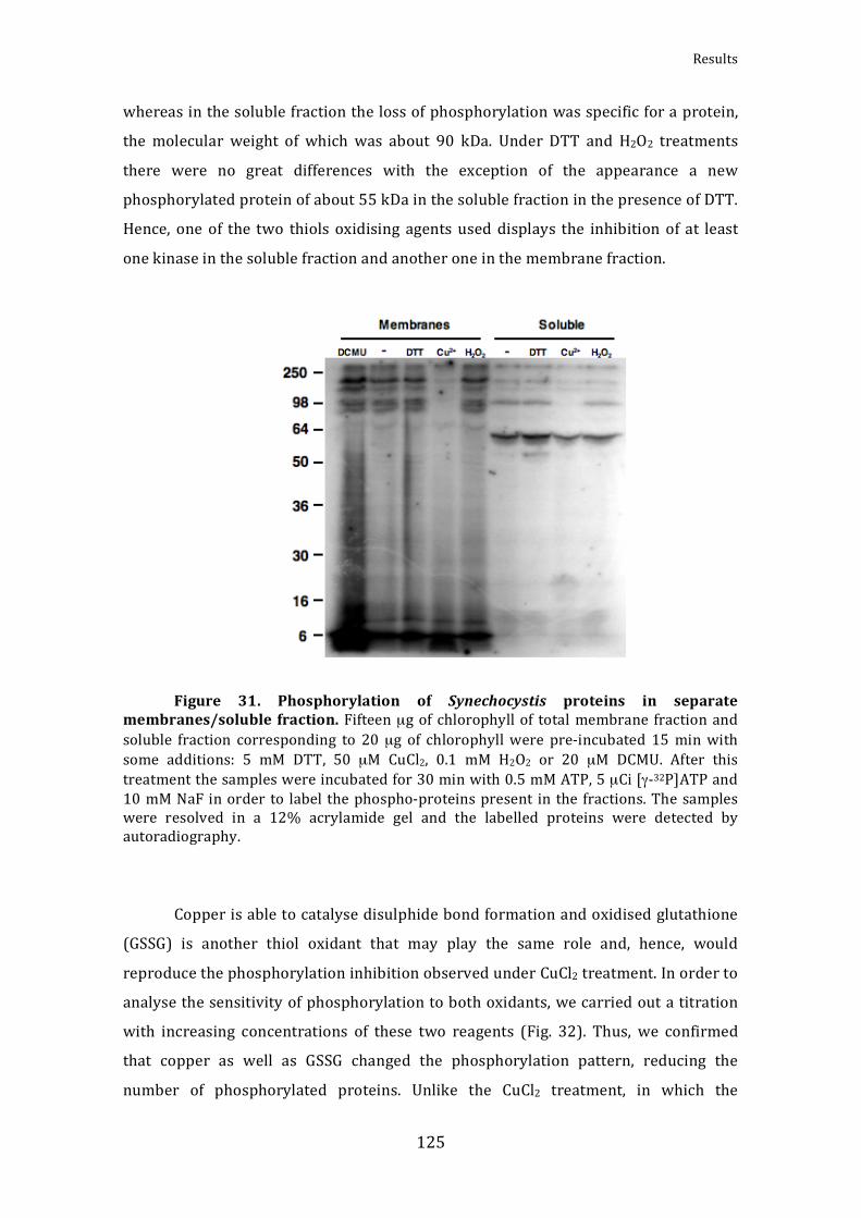
Results
125
whereasinthesolublefractionthelossofphosphorylationwasspecificforaprotein,
the molecular weight of which was about 90 kDa. Under DTT and H2O2 treatments
there were no great differences with the exception of the appearance a new
phosphorylatedproteinofabout55kDainthesolublefractioninthepresenceofDTT.
Hence,oneof the two thiolsoxidising agentsused displays the inhibitionof at least
onekinaseinthesolublefractionandanotheroneinthemembranefraction.
Figure 31. Phosphorylation of Synechocystis proteins in separate
membranes/soluble fraction.Fifteenµgof chlorophyllof totalmembrane fractionandsoluble fraction corresponding to 20µg of chlorophyllwere pre‐incubated 15minwithsome additions: 5 mM DTT, 50 µM CuCl2, 0.1 mM H2O2 or 20 µM DCMU. After thistreatmentthesampleswereincubatedfor30minwith0.5mMATP,5µCi[γ‐32P]ATPand10mMNaFinorderto labelthephospho‐proteinspresent in thefractions.Thesampleswere resolved in a 12% acrylamide gel and the labelled proteins were detected byautoradiography.
Copperisabletocatalysedisulphidebondformationandoxidisedglutathione
(GSSG) is another thiol oxidant that may play the same role and, hence, would
reproducethephosphorylationinhibitionobservedunderCuCl2treatment.Inorderto
analysethesensitivityofphosphorylationtobothoxidants,wecarriedoutatitration
with increasing concentrations of these two reagents (Fig. 32). Thus, we confirmed
that copper as well as GSSG changed the phosphorylation pattern, reducing the
number of phosphorylated proteins. Unlike the CuCl2 treatment, in which the

AlejandroMataCabana
126
phosphorylation of a protein of about 90 kDa disappeared specifically, the effect of
GSSGwasmoregeneral,showingadrastic global inhibitionofphosphorylationat10
mMtreatment.However,forlowerconcentrationsofGSSG(2.5and5mM)thespecific
inhibition of the phosphorylation of a protein with high molecular weight was
observed(Fig.32).Inbothcases,relativelyhighconcentrations(from25µMofCuCl2
and 10 mM of GSSG) were necessary to yield visible effects. Moreover, the CuCl2
produced an increase in labelling of a protein around 22 kDa. No increase in
phosphorylationwasobserveduponGSSGtreatment.
Figure 32. Phosphorylation sensibility to oxidant agents of Synechocystis
cytosolicproteins.Before radioactive labelling, theSynechocystis cytosolic fractionwastreated for 15minwith increasingconcentrations of the thioloxidants CuCl2 (from0 to100µM)andGSSG(from0to10mM).Afterthetreatmentthephosphorylationassaywasperformedincubatingthesampleswith0.5mMATP,5µCi[γ‐32P]ATPand10mMNaFinorder to radioactively label the phospho‐proteins present in the fractions. The sampleswereresolvedina10%acrylamidegel,whichwasstainedwithCoomassie(lowerpanel),andthelabelledproteinsweredetectedbyautoradiography(upperpanel).

Results
127
Aiming at elucidating the mechanism for redox regulation of the oxidant‐
sensitive kinase activity present in the Synechocystis cytosol, we wanted to test
whetherareductantcouldrestorethekinaseactivity(Fig.33).Thus,wepre‐treateda
SynechocystiscytosolsamplewithCuCl2 inorderto inhibit thekinaseandthen,after
eliminationofCu2+,treateditwithreducingagents,suchasGSH(reducedglutathione),
DTT and purified TrxA. As a result, DTT and TrxA, but not the GSH, were able to
recoverthephosphorylationofthe90kDaprotein.Hence,thephosphorylationofthis
proteindependedonaredox‐regulatedkinase.Thenextstepwastotrytoidentifythe
kinase,whichdisplayedsuchactivity.
Figure33.Reactivationof the90kDaproteinphosphorylationbyreductant
agents.CytosolicextractsofSynechocystiscorrespondingto80µgofchlorophyllwaspre‐oxidised for 15 min by 100 µM CuCl2 and then the sample was diluted to 20 µg ofchlorophyllandtreatedfor15minwithsomereductantagents:5mMGSH,5mMDTTor4µMTrxAwith0.2mMDTT.Afterthetreatmentthephosphorylationassaywasperformedincubating the sampleswith 0.5mMATP, 5µCi [γ‐32P]ATP and 10mMNaF in order tolabel thephospho‐proteinspresent.Thesampleswereresolvedina10%acrylamidegeland the labelled proteins were detected by autoradiography (upper panel). A controlwithoutoxidisingpre‐treatmentwasloadedinthefirst laneaswellasacontrolwiththeCuCl2treatmentbutwithoutreductantinthesecondlane.Theblackarrowheadindicatesthe 90 kDa protein the phosphorylation of which that is specifically inhibited underoxidising conditions. In the right panel the arithmetic mean of the 90 kDa proteinphosphorylation between three independent experiments is shown. 100% ofphosphorylationistheoneobservedinthecontrollane.

AlejandroMataCabana
128
The Two‐Component system is the principal signal transduction system in
bacteria (Mizuno et al., 1996), and the histidine kinases, which form part of this
system,arethemostcommonkinasesintheseorganisms.Despitethemainroleofthe
histidine kinases we decided to start searching for Ser/Thr kinases mainly for two
reasons.ThefirstreasonisthatSynechocystisincludes44putativegenesforhistidine
kinasesinitschromosomeandthreemoreinitsplasmids(MurataandSuzuki,2006),
whereasthenumberofgenesforSer/Thrkinasesismuchsmaller(7Pkn2typeand5
ABC1 type). The second reason is that the bond of the phosphoryl group to the
histidine residue ismore labile than to the serine and threonine (Matthews, 1995),
makingthehistidinekinasephosphorylationmoredifficulttoobserve.Therefore,the
phosphorylationobservedinourexperimentsmorelikelycorrespondstotheSer/Thr
kinase activity in Synechocystis. Moreover, the histidine kinases are bifunctional
havingbothkinaseandphosphataseactivities,andthephosphorylationsignalin this
transduction system is more transient than in the eukaryotic type (Klumpp and
Krieglstein, 2002). The Synechocystis mutant strain collection of our laboratory
containsthestrains lackingeachof theSer/Thrkinases. Itallowedustoanalysethe
solubleproteinphosphorylationpatternofallmutantsandtocomparethemwiththe
pattern corresponding to the WT strain under normal, oxidising and reducing
conditions (Fig. 34A). Allmutants, including theΔSpkEmutant (not shown), strains
exhibited a phosphorylation pattern very similar to that of theWT strain, with the
exception of the ΔSpkB mutant. In all cases the 90 kDa protein appeared
phosphorylated under normal conditions and unphosphorylated under CuCl2
treatment,but intheΔSpkBthisproteinwasunphosphorylatedunderallconditions,
suggesting that the SpkB kinase could be responsible for this phosphorylation. It
should be taken into account that SpkE was reported not to be a functional kinase
(Kamei et al., 2002). With the exception of SpkD all Synechocystis Ser/Thr kinases
possess intheiraminoacid sequenceoneormorecysteines residues, someofwhich
arelocatedinthekinasedomain(Fig.34B).SpkBandSpkFcontainaCys‐motifintheir
N‐terminal domain as described inKamei et al., 2002. Thus, the Cys‐motif could be
relatedwith the redox regulation of SpkB. However, the same Cys‐motif is found in
SpkF but no cytosolic phosphoproteins were missing in ΔSpkF. This might be
explained by the presence of four transmembrane helix domains in its C‐terminus,
whichshouldanchorittothemembraneand,henceitwouldbeabsentinthesoluble
proteinpreparations.

Results
129
Figure 34. Pkn2 kinases of Synechocystis. (A)The phosphorylation pattern of
cytosolic proteins fromWT andmutant strains lacking the different Pkn2 type Ser/Thrkinases was observed under normal conditions and under reducing and oxidisingtreatment.CytosolicextractsfromeachstrainofSynechocystiscorresponding to20µgofchlorophyllwere treatedfor15minwith5mMDTT,100µMCuCl2orwithoutadditionsandthereafter incubatedwith0.5mMATP,5µCi[γ‐32P]ATPand10mMNaFinorder tolabel the phospho‐proteins present in the fractions. Labelled proteinswere detected byautoradiography.(B)SchematicrepresentationofthedifferentPkn2typeSer/Thrkinasesin Synechocystis. The location of the kinase domain, transmembrane domain and thecysteinespresentintheaminoacidsequenceofeachkinaseareindicated.

AlejandroMataCabana
130
2.SpkBredoxregulation
SpkBhasbeenrelatedtocellmotility,sincetheΔSpkBmutantdidnotdisplay
phototactic movement (Kamei et al., 2003). This kinase is composed by the kinase
domainfollowedbyapentapeptiderepeatdomain. InZhangetal.,2007,theauthors
performed aphylogenetic analysisof theSer/Thrkinases in cyanobacteriabased on
the catalytic domain. Despite the use of only the catalytic domain in this study, all
cyanobacterial Ser/Thr kinases containing a pentapeptide repeat domain clustered
togetherformingasubgroupofSer/Thrkinases(Zhangetal.,2007).Here,wepresent
amultiple alignment of some representative kinases of this subgroup including the
Synechocystis SpkB (Fig. 33). In the alignment we can observe the existence of five
completely conserved cysteines and one more highly conserved cysteine (in all
sequences except one cysteine). Four of the totally conserved cysteine residues are
grouped in pairs forming the so‐called Cys‐motif in theN‐terminal domain. Someof
theaminoacidsadjacenttothesecysteinesarehighlyconservedtoo.Thehighdegree
of conservation of the Cys‐motif suggests a possibly important role in the protein
activity, maybe involving redox regulation (Kamei et al., 2002). The other two
remainingconservedcysteinesarelocatedwithinthekinasedomain,wherethefirstis
presentintheATPbindingdomain.Thepentapeptidedomainiscomposedofseveral
repeats of the following amino acid sequence: A(D/N)LXX. This domain is more
diverseamongstthekinasesfromdifferentorganismsandacommonpatterndoesnot
exist.Thefunctionofthisdomainremainsunknown(Zhangetal.,2007).Basedonthe
domain structure and phylogenetic data we propose that this kind of kinase
constitutesa newsub‐typeofPkn2 Ser/Thrkinases. So far, nothing isknownabout
their functions, protein substrates or regulation. Other Ser/Thr kinases have been
identified in bacteria that conserve the organization of a Cys‐motif upstream the
kinase domain, though the pentapeptide domain is replaced with other kinds of
domains. This is the case of Pkn22 fromAnabaena, where the pentapeptide repeat
domain has been substituted with a PbHI, which is structurally similar, and the
function is also unknown (Jenkins et al., 1998). In the case of PknG from
Mycobacterium tuberculosis, this domain has been replaced with a tetratricopeptide
repeat region, which mediates protein‐protein interactions and the assembly of
multiprotein complexes (D’Andrea and Regan, 2003) although a similar N‐terminal

Results
131
Cys‐motif is conserved. This could suggest that the Cys‐motif is awidely distributed
regulatordomainamongstSer/Thrkinases.
InordertoanalysetheSpkBkinaseactivityand itsputativeredoxregulation,
weclonedtheslr1697geneintothepET28avector,wheretheexpressionofthecloned
gene is subjected to IPTG control and a His‐Tag is added to the protein. SpkB was
expressed in the E. coli BL21DE3 strain and the kinase activity was detected in
cytosolic extracts fromE. coli cells as described inKameietal., 2001 and2002 (Fig.
36).Forthisphosphorylationassaycaseinwasusedasartificialproteinsubstrate.The
caseinaloneandincubatedwithextractsofE.colithatwasnotexpressingthekinase
did not show appreciable phosphorylation of the substrate (Fig. 36). However,
incubation with the E. coli extracts expressing SpkB resulted in a clear
phosphorylation of casein, which decreased after CuCl2 treatment, confirming the
previousresultsobtainedfromSynechocystisextracts.Moreover,phosphorylationofa
proteinintheupperpartofthegel(about80kDa)isobserved.Thismaycorrespond
totheautophosphorylationofSpkB,assuggestedinKameietal.,2003.
Figure 35. Alignment of SpkB homologues.The Clustal X enginewas used toperform the alignment of cyanobacterial SpkB homologous proteins. The sequences arefrom: Synechocystis sp. PCC 6803 (slr1697), Cyanothece sp. ATCC 51142 (cce_2943),Microcystis aeruginosa NIES‐843 (MAE_53550), Synechococcus sp. PCC 7002(SYNPCC7002_A0446), Nostoc punctiforme PCC 73102 (Npun_R2916), Anabaena sp. PCC7120 (alr3268), Trichodesmium erythraeum IMS101 (Tery_2064) and Lyngbya sp. PCC8106 (L8106_12570). The aminoacids that constitute the kinase domain are coloured inpurple. The totally conserved cysteines are coloured in green and the pentapepteiderepeatsareinred.TheATPbindingdomainisindicatedwitharedboxandthecysteinesofSpkBfromSynechocystisnottotallyconservedareindicatedwithagreenbox.

AlejandroMataCabana
132
slr1697 MSFCVNPNCPHPKNPNNVQVCQACGNSLRLNGRYQTLGLLGKGGFGATFAAADVALPGTPICVVKQLRPQTDDPN cce_2943 MSYCVNPSCAQPKNPNNVAVCQSCGSQLRLNNRYQPLGILGKGGFGATFGAADISLPGNPICVVKQLRPATDDPQ MAE_53550 MTYCLNPNCTQPKNAPTALICESCGSKLLLHDRYQTLRILGKGGFGATFVSSDLSLPDKPLCVVKQLQPPPKDAS SYNPCC7002_A0446 MSYCINPKCSNPKNPLKARVCGTCGSSLILRQRYKPLKSLGHGGFGATFLAVDLNLPGKPSCVIKQLRLASNVPH Npun_R2916 MSYCLNPICPNPENLVNSQSCQSCGSQLLLRDRYQAIKPLGQGGFGATFLANDRGLPGEPSCVIKQLRPSGSAPH alr3268 MSYCINPTCPNPENVAFSQKCEACGSPLLLRDRYRVLKPLGQGGFGATFLAHDQILPGEPSCVIKQLRPSGTAPH Tery_2064 MSYCINPACPQPQNLNSVNKCTACGSQLILRDRYKIIKPIGKGGFGVTFLAKDISLPGAPICVIKELRPSSTKAS L8106_12570 MSYCINPACSHPENPEDASQCQTCRSKLVLRNRYRVIKPLGKGGFGATFLSEDLSLPGSPKCVVKQLRPVAKSAR *::*:** *.:*:* * :* . * *. **: : :*:****.** : * **. * **:*:*: . slr1697 VFRMAKELFEREAQTLGRVGNHPQVPRLLDYFEDDHQFYLVQEYVKGHNLHQEVKKNGTFTEGSVKQFLTEILPI cce_2943 VYKMAKELFEREAETLGKVGNHPQVPRLLDYFEQNKEFYLVQEYVKGYNLHQEVKKKGPFSEAGVKQFLTELLPI MAE_53550 VFRMAKDLFDREAETLAKLGVHPQIPRLLDYFEDNQEFYLVQEYVKGHNLHQEVKKRGPFSEAGVKQFLSELLPI SYNPCC7002_A0446 LFQMARELFEREAETLGRIGNHPQVPRLLDYFEDDNHFYLVQEYVKGNNLQQEVKKNGPFSEAGARQFLSEVLPV Npun_R2916 VLQMARELFEREAKTLGKIGNHPQVPRLLDYFEDHEQFYLVQEYISGDTLQEEVKLNGILTETGVKQFLSEILPL alr3268 ILQMARELFEREAKTLGTIGNHPQVPRLLDYFEEQEQFYLVQEYISGSTLQQEVKLNGTLSEPGVKQFLSEILPL Tery_2064 ELEMARKLFQREAETLGKIGHHPQVPRLLDYFEWKKRFHLVQEYVSGLTLKQEVKKQGIFTEEQGKKFLIEVLSV L8106_12570 VLEMARQLFQREATTLGKIGDHPQIPRLLDYFSGKQQFYLVQEYVDGRNLKEEVQQSGVYTEDDIKAFLREILPI .**:.**:*** **. :* ***:*******. ...*:*****:.* .*::**: * :* : ** *:*.: slr1697 LDYIHSQKVIHRDIKPANLIRRQTDQKLVLIDFGAVKNQIDSVLSSNTSAQTALTAFAVGTAGFAPPEQMAMRPV cce_2943 LEYIHSQKVIHRDIKPANLIRSQKDSKLVLIDFGAVKNQVNSMVANNT--QTAFTAFAVGTAGFAPPEQMAMRPV MAE_53550 LKYVHEQRVIHRDIKPANLIRRQTDRKLVLIDFGAVKNPVNSVMANADS-QTALTNFAVGTQGFAPPEQMAMRPV SYNPCC7002_A0446 IQYIHSQQVIHRDIKPANLIRRDQDKKLVLIDFGAVKNQINAEAIASTSEQTALTSFAVGTPGYAPPEQMAMRPV Npun_R2916 LQYIHEQKVIHRDIKPANLIRRTQDARMVLIDFGAVKNQVTQGAISQSG-QTALTAYAIGTPGFAPPEQMAMRPV alr3268 LQYIHEHKVIHRDIKPANLIRRSQDARMVLIDFGAVKNQVSQAITNQSA-NTALTAYAIGTPGFAPPEQMAMRPV Tery_2064 IDYIHTQGVIHRDIKPTNIIRRSEDGHLVLIDFGAVKDHVNQTYISGGTGETAFTNISIGTSGFSPPEQVLLRPV L8106_12570 LSYIHSHEVIHRDIKPANILRRAQDQRLVLIDFGAVKDEVKQVTEFG-TGQTAFTNFAIGTSGFAPPEQMALRPV :.*:* : ********:*::* * ::*********: : :**:* ::** *::****: :*** slr1697 YASDIYATGVTCLYLLTGKTPKEIDCNSQTGEMDWEKHVTVSSKFAEVIRKMLELSVRHRYKSAQQVLDALEMPT cce_2943 YASDIYAVGVTCVYLLTAKTPKDIGCDPETGEIAWEPYVNISDSLANVLKKMLEVSVKHRYKSAEQVLDAMAMAP MAE_53550 YASDIYAVGATCLYLLTGKSPKAIEVDINTGELLWEKLVKVSPQFAKVLKTMLEVSVRHRYKSAQEVMDALDMTA SYNPCC7002_A0446 YASDIYAVGITCLYLLTGKPPKDLDYDSKTGELLWESHVNISNSLASVLHKMLESSVKHRYQSAQDVLDALEMEP Npun_R2916 YASDIYALGVTCIYLLTSKTPKDLDYNPNTGEMIWEQLVQVSDHLSNVLRKMLDVSVRNRYQSAAEVLRALEIEP alr3268 YASDIYALGITCVYLLTSKTPKDLDYNPTTGEVMWEHLVQASDHLIGVLRKMLEVSVRSRYQSATDVLKALEIEP Tery_2064 YASDIYALGVTTIFLLTGKTPNNLGYNQVNGEILWHSQVNVSDMFREILKKMLEVSVEDRFQSAEELLAALKTK- L8106_12570 YASDIYAVGMTCVYLMTGKSPSSLDHNPITAEVLWRALVSVSDSFAAILQKMLEMSVYQRYQSAEEVLGALDHET ******* * * ::*:*.*.*. : : ..*: *. * * : :::.**: ** *::** ::: *: slr1697 --------YEDGMMQGMVSTPFTTLTGAGDEPATGIRMGNSSSPDYGDPSTRFNTNVQPRDPSSTSLNTGIKT-R cce_2943 --------YEQGMQDSMTTM------------ITGFKTPSSSSS----PNSSLSTGLVSSSPSTRTMGR-VNT-H MAE_53550 --------YADSLAQSLIAAP----------------MVTSQPP-------RQNSGSMATSSVSVPLKSKFRP-K SYNPCC7002_A0446 --------YMESLAQGMADLSG-------AESFTGTTGGQVSPTASSDDDGELTGGPSTLANSQLAMAIRARRDR Npun_R2916 --------YLESLAKGLLIKSDT--------------GSKERTH------NHLENSAVLCNNSSVAVTSAG-VAQ alr3268 --------YLESLAQGLLVKSE-----------------KEQTS------QRPENSAVLSSSSPVAATSVGGVAQ Tery_2064 -------QPEKKQVQPRLDDA----------------VKTIFRDEITVFNDNKDNFSSMSANIKIAQKIRQRKYG L8106_12570 EFYTDFSDGLSVQPEPRVSEAPT-------------QVETHQQDNQTTIYCD-DESTALRPFASQAKQIRERNAR . . . slr1697 TAKPRQSPRDRATSNIESPTTRVRPASNMADGGSVGAGGIDYNMVNPKPFSRREEEKQAIAN------QPETKRW cce_2943 ISK--GSP-----MSSVSPEDKHSVATKIQ-GRVAGCGNANYNIAHGQTSSRRRGKGQGISNSDTIATKKNQKKW MAE_53550 NQNPEASP------------------DNML-GGQMAFSLFNTNVLN-KSKARSSTELTLLKK---------KPRL SYNPCC7002_A0446 QKKPYGAP---------SQTDTYHPSASAQRINRLTAGRNKPRSTMGKTMRVEPNKANRTNP--------METKL Npun_R2916 VAAAIRAR---------------RAKAAEAAGSHHASGMGKSTTLANSNSNGSQVQNSKVER-----------KL alr3268 VAAAIRAR---------------RAKDAAAKLAILPN--------SNSNGNGLPTQQSKAGR-----------RL Tery_2064 LEKNSTNQ---------------VLPN--SVKTKPKHIEGYLTVVPPKSKATNTTQLGNETP----------IKW L8106_12570 RQKKEYTQ---------------LGENFASVDGGISATQG----APTATMFTSANTPARNKL----------HRW : slr1697 NGKTFLAEYAQGKRDFADQNLVGIVLAKAFVPGINCYQANLTNANFEQAELTRADFGKARLKNVIFKGANLSDAY cce_2943 QEKTLLTAYENGRRDFTNQELNELNLSKAFLPGINCYQAKLSRINLQGAELTRADLGRADLTQAVMKNANLSEAY MAE_53550 DEKALLDAYNNGRRNFAQEELPNLNLAKAKLVGVNFCQSKLTRANLQGADLSNADLGRANLSQAILKSTNLNNAY SYNPCC7002_A0446 TAQDMLSAYGQGRRNFAQLQLLGLSLPRMNLSGCIFHQSQMIQANFQGANLSRADFGKANLHQANLRDANLEQAY Npun_R2916 DTQGLLTAYQKGRRDFALHNLSLLNLQGADLSGTNFHSTQLQKTNLQGANLHNSDFGRASLSKANLKDANLTKAY alr3268 DSQTLMKAYLKGRRDFALHNLNFLNLQGVDLSETNFHSAQLQSANLQGANLHNSDFGRASLTRANLKDANLNKAY Tery_2064 TAATLRSAYMKGRRDFAECNLIRLNLCNAKLSGANFYGANLGGANLQGADLSDSNFGHANLTNTILKNATLTKAY L8106_12570 DESSLHDAYIRGDRYFTDCDLQGLNLRSEKFSGAYFTKSRLEQTNFQNADLSKVDFARASLSDTIFKDANLTHAR : * .* * *: :* : * . :.: *:: *:* ::.:* * . ::.:.* .*: slr1697 FGYADLRGADLRGANLNGVNFKYANLQGANFSGADLGSAKVSPEQLKLAKTNWRTVMPGSGRRR--- cce_2943 LGYANLNGADLRGANLCGANLTYANLQGANLCGVDLSSARITEAQLSVAKTNWRTVMP-SGKRGFW- MAE_53550 LGYADLEKADLRGANLTGAHLRYANLKDANLCGANLSDAQVTREQIALAKTNWLTVMP-NGKRGSW- SYNPCC7002_A0446 LSYTNLSQADLRGANLKQAYLNYALLDGANLCGANLRNAKITDQQLKTAKTNWATIMP-NGRRGLF- Npun_R2916 FNHADLEGADLRGADLSNAYLSNANLRGANLCGANLTSAKISDEQLALAKTNWMTIRP-NGKRGLL- alr3268 FNHADLEGADLRGADLSHAYLSNANLRGTNLCGANLTGAKITDEQLALAKTNWMTIRP-NGKRGLL- Tery_2064 LSTASLANSNLQNANMKNTYLSGANLGGANLYGANLTNAIVSDRQLETAKTNWWTIFP-NGKRGSIF L8106_12570 LSYANLENADLRGANLTYANLSYANLRGTNLCGANLTHALVSEQQLALAKTNWRTILP-NRKRKFGL . :.* ::*:.*:: . : * * .:*: *.:* * :: *: ***** *: * . :*

Results
133
Figure36.ActivityofSpkBkinaseexpressedinE.coli.KinaseactivityofSpkB
wasanalysedperformingaphosphorylationassayusing20µgofE.colicytosolicproteinextracts from cells expressing (SE) or non‐expressing (NE) the SpkB kinase ofSynechocystis and 2.5 µg of casein as artificial kinase substrate. A sample of theE. coliexpressing the kinase was pre‐trated 15 min with 100 µM CuCl2. The phosphorylationassaywasperformed incubating the sampleswith 0.5mMATP,5µCi [γ‐32P]ATPand10mMNaFinordertoradioactivelabelthephospho‐proteins.Thesampleswereresolvedina15%acrylamidegel,whichwasstainedwith Coomassie (right panel), and the labelledproteins were detected by autoradiography (left panel). A control with only casein inabsenceofE.coli extractsuntreatedand treatedwithCuCl2,wereloadedin the twofirstlanesofgel.
In order to purify recombinant SpkB from E. coli extracts we applied nickel
affinitychromatography.However,SpkBcouldnotbepurifiedtohomogeneity,though
themajorityof contaminantswere removed.The identityof SpkB in the Coomassie‐
stained band was verified by MALDI‐TOF analysis and peptide mass fingerprinting
(PMF). The data for the identification of SpkB by PMF were MOWSE score: 189;
numberofpeptidesmatched:16;andsequencecoverage:29%.Thispartiallypurified
preparationwasused for phosphorylationassays,with caseinas substrate (Fig. 37).
Hence, the sensitivity of the semi‐purified SpkB to oxidising treatment and the
restorationofthekinaseactivityunderreducingtreatmentswastested.Sampleswere
pre‐trated with either Cu2+ or GSSG in order to inactivate the kinase activity and
attemptsweremadetoreactivatewith5mMDTT,5mMGSHor4µMTrxA(Fig.37A).
When the samplewas treatedwith TrxA a low concentration of DTT (0.2mM)was
AlejandroMataCabana
134
usedaselectrondonor.The phosphorylationassay showed thatboth Cu2+ andGSSG
inhibitedthekinaseactivityofSpkBandthat thisactivitywasefficiently restoredby
DTT and TrxA, but not by GSH (Fig 37A). This result is in agreement with the
inactivationandreactivationofthekinaseactivityobservedinSynechocystiscell‐free
extracts (Fig.33).The inefficiencyof theGSH treatment, as comparedwithDTTand
TrxAmaybeexplainedbytheredoxpotentialofGSH,whichismorepositivethanthat
of DTT and TrxA (Sevier and Kaiser, 2002). In order to test the concentration
dependence on TrxA for restoration of the SpkB kinase activity we performed a
titrationusingincreasingconcentrationsofTrxAasreductant(Fig.37B).Inthecontrol
withoutTrxA,butwith0.2mMDTT,partofthekinaseactivitywasrestored,although
1µMTrxAwas needed to reach themaximum level of casein phosphorylation. The
maximumphosphorylation level reachedupon restorationafterCuCl2 treatmentwas
slightlyhigherthanthatoftheuntreatedsample.
InordertotestwhetherrecombinantSpkBisabletophosphorylatethe90kDa
substrateprotein,which is non‐phosphorylated in theΔSpkBmutant,we performed
complementation assays. Hence, cytosolic extracts from the Synechocystis ΔSpkB
mutantwere incubatedwith increasingconcentrationsofpurifiedrecombinantSpkB
(Fig.38).Thephosphorylationof the90kDaproteinwasspecifically restored inthe
presenceoflowconcentrationsofexogenouslyaddedSpkB.Withincreasingamounts
ofSpkB, the90kDaproteinphosphorylationwasenhancedandnewphosphorylated
proteins appeared. One of them was most strongly labelled when the maximum
concentration of SpkB was used (Fig. 38). This probably corresponds to the
autophosphorylatedSpkB,sincethemolecularweightmatcheswiththeobservedone
for SpkB of about 80 kDa. In conclusion, the exogenous addition of SpkB is able to
replaceinvitrotheSpkBactivitylackinginthemutantstrain.
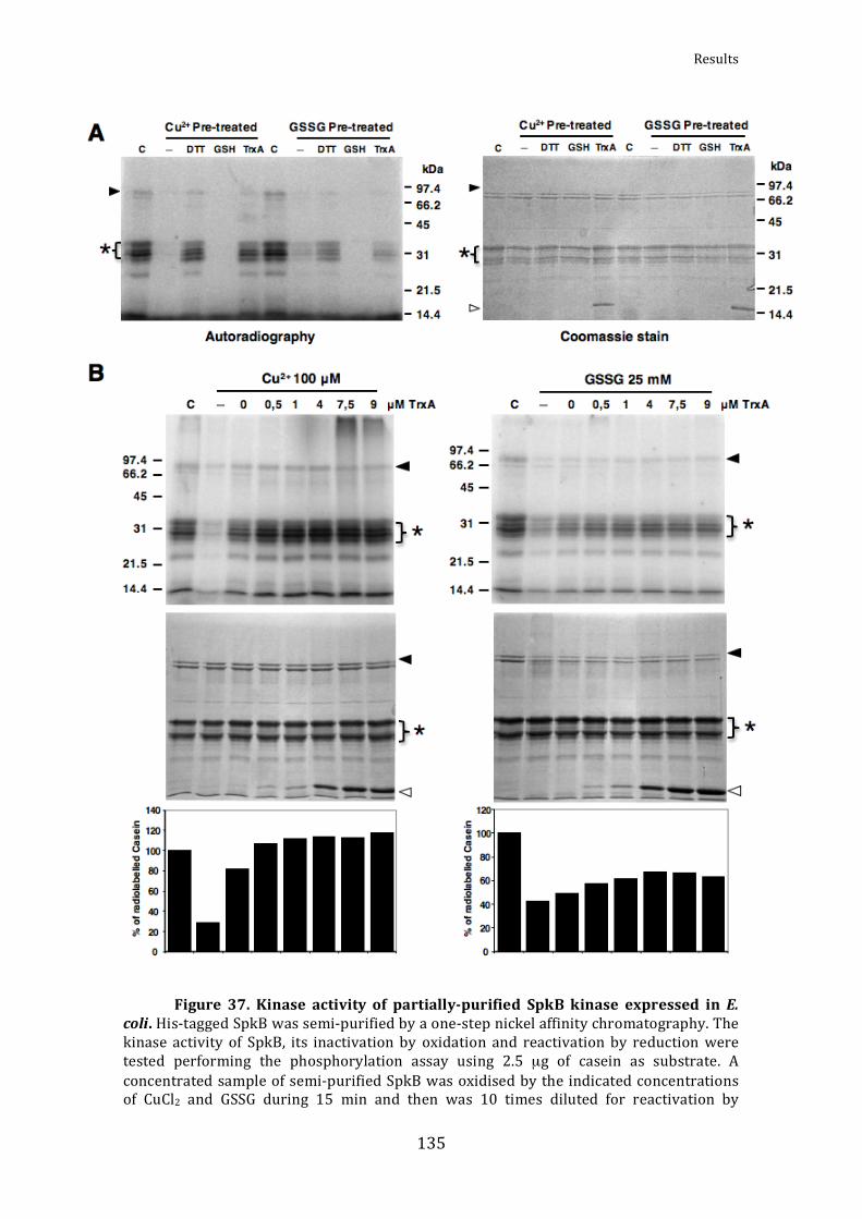
Results
135
Figure 37. Kinase activity of partiallypurified SpkB kinase expressed in E.
coli.His‐taggedSpkBwassemi‐purifiedbyaone‐stepnickelaffinitychromatography.Thekinase activity of SpkB, its inactivation by oxidation and reactivation by reductionweretested performing the phosphorylation assay using 2.5 µg of casein as substrate. Aconcentratedsampleofsemi‐purifiedSpkBwasoxidisedbytheindicatedconcentrationsof CuCl2 and GSSG during 15 min and then was 10 times diluted for reactivation by
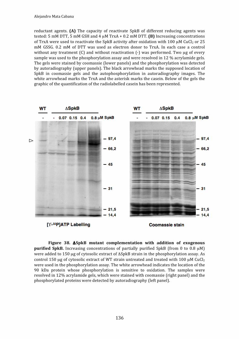
AlejandroMataCabana
136
reductant agents. (A) The capacity of reactivate SpkB of different reducing agents wastested:5mMDTT,5mMGSHand4µMTrxA+0.2mMDTT.(B)IncreasingconcentrationsofTrxAwereusedtoreactivatetheSpkBactivityafteroxidationwith100µMCuCl2or25mM GSSG. 0.2 mM of DTT was used as electron donor to TrxA. In each case a controlwithoutany treatment (C)andwithout reactivation (‐)was performed.Twoµgofeverysamplewasusedtothephosphorylationassayandwereresolvedin12%acrylamidegels.Thegelswerestainedbycoomassie(lowerpanels)andthephosphorylationwasdetectedbyautoradiography(upperpanels).TheblackarrowheadmarksthesupposedlocationofSpkB in coomassie gels and the autophosphorylation in autoradiography images. ThewhitearrowheadmarkstheTrxAandtheasteriskmarksthecasein.Belowofthegelsthegraphicofthequantificationoftheradiolabelledcaseinhasbeenrepresented.
Figure 38. ΔSpkB mutant complementation with addition of exogenous
purified SpkB. Increasing concentrations of partially purified SpkB (from 0 to 0.8µM)wereaddedto150µgofcytosolicextractofΔSpkBstraininthephosphorylationassay.Ascontrol150µgofcytosolicextractofWTstrainuntreatedandtreatedwith100µMCuCl2wereusedinthephosphorylationassay.Thewhitearrowheadindicatesthelocationofthe90 kDa protein whose phosphorylation is sensitive to oxidation. The samples wereresolvedin12%acrylamidegels,whichwerestainedwithcoomassie(rightpanel)andthephosphorylatedproteinsweredetectedbyautoradiography(leftpanel).

Results
137
3.IdentificationofatargetforSpkBphosphorylation
Wehadyet todeterminethe identityof theSpkBphysiological target.Tothis
end,thefirststepwastoresolvetheradioactivelylabelledcytosolicextractsfromWT
andΔSpkBstrainsinanacrylamidegelbymeansof2‐DE,inwhichthefirstdimension
consisted of a separation by the isoelectric point of proteins (pH 4.7 – 5.9). In the
seconddimensionSDS‐PAGEtheproteinsmigratedaccordingtheirmolecularweight
(Fig.39).TheresultinggelswereCBB‐stainedandthephosphorylatedproteinswere
detected by autoradiography. The comparative analyses of gels from both samples
showedthelackofphosphorylationofaproteinwiththeexpectedmolecularweightin
the mutant strain, being the apparent molecular weight about 85 kDa and the
isoelectric point next to 4.7. Importantly, his loss did not correspond to the
disappearance of the protein, but only its phosphorylation, since the Coomassie‐
stained spot remainedsimilar inWTandmutant.ThePMFidentificationof thisspot
revealed that the SpkB target is the glycyl t‐RNA synthetase β‐chain (GlyS). The
theoreticalmolecularweight is80.3kDaand the isoelectric point is 4.7.Thedata of
PMF for the spot identified as GlyS were MOWSE score: 248; number of peptides
matched:23;andsequencecoverage:33%.
The glycyl‐tRNA synthetase catalyses the esterification of glycine with the
terminal3’‐hydroxylgroupofglycinetransferRNAresultinginthesynthesisofglycyl‐
tRNA,whichisrequiredtointroduceglycine intoproteins.Besides,inasidereaction
the enzyme also synthesises dinucleoside phosphate, which may participate in
regulation of cell functions (Freistetal., 1996).Glycyl‐tRNAsynthetase is oneof the
few aminoacyl‐tRNA synthetases that exhibits different oligomeric structures in
differentorganisms.Atetramericstructureofthetypeα2β2hasbeensuggestedtobe
distributed only in bacteria and a dimeric structure of the type α2 can be mainly
observed in eukaryotes (Mazauric et al., 1998). The α‐subunit and parts of the β‐
subunit are required for aminoacylation of tRNA and the α‐chain contributes
specificallytoaminoacidandATPbinding(Freistetal.,1996).Treatmentwiththiol‐
reductant and thiol‐alkylating agents led to inhibition of the enzyme from E. coli
(Ostrem and Berg, 1974; Profy and Schimmel, 1986), S. aureus (Niyomporn et al.,
1968) and yeast (Kern et al., 1981). In general, this effect has been structurally
interpreted as an inactivation by dissociation of the oligomer (Freist et al., 1996).

AlejandroMataCabana
138
However,GlyShasbeenidentifiedtopossessareactivethiolinS.aureus(Pötheretal.,
2009) as well as in E. coli (Hu et al., 2010). This suggests a putative direct redox
regulation of glycyl‐tRNA synthetase. Phosphorylation also may be a means of
regulation of this enzyme. Phosphorylated aminoacyl‐tRNA synthetases have been
isolated from uterus and liver cells of mice and some of these were activated by
phosphorylation,whileotherswereinactivatedbythispost‐translationalmodification
(Berg, 1977, 1978). Later studies showed that 15 different aminoacyl‐tRNA
synthetases, including glycyl‐tRNA synthetase, from mouse liver were present in
phosphorylatedandnon‐phosphorylatedforms(Berg,1990).
Figure39. SpkBsubstrate identification.Fourhundredµg of cytocolicprotein
extracts of WT and ΔSpkB strains were resolved in 2‐D SDS‐PAGE gels after thephosphorylationassaywasperformedincubatingthesampleswith0.5mMATP,15µCi[γ‐32P]ATP and10mMNaF in order to label the phospho‐proteins present in the samples.ThegelswerestainedwithCoomassie(leftpanels)andthephosphorylatedproteinsweredetected by autoradiography (right panels).White arrowheadmarks the location of theendogenous SpkB substrate and the red ellipse localizes the Phenylalanyl‐tRNAsynthetase.Diamondsindicatethephycobilisomelinkerproteins,whichareconstitutivelyphosphorylatedinSynechocystis(Pivenetal.,2005).

Results
139
Synechocystis possesses in its genome two genes that codify for theα (GlyQ)
andβ(GlyS)subunitsofglycyl‐tRNAsynthetase,indicatingthattheenzymebelongsto
theα2β2 type, as in themajority of prokaryotes. Surprisingly, looking at the genetic
environment of SpkB we realised that an adjacent gene, which probably shares
promotorwithSpkB,wasthephenylalanyl‐tRNAsynthetaseβ‐chain(PheT).InE.coli
thephenylalanyl‐tRNAsynthetaseisalsoaα2β2typeenzymeandexhibitssimilarities
withGlyQS(Freistetal.,1996).Infact,despitelittlesequencehomologiesbetweenthe
two enzymes, antibodies raised specifically against GlyQS displayed cross‐reaction
withphenylalanyl‐tRNAsynthetase(Nageletal.,1988).Toclarifythepossibledoubts
aboutourSpkBtargetidentificationwelocalisedPheTinthe2‐Dgel(Fig.39).Hence,
weensuredthe lossofGlySphosphorylationwas realand itwasnotderivedfroma
geneticeffectonPheTintheΔSpkBmutantstrain.TheCBB‐stained2‐DEgelshowed
thatPheTandGlySwereclose,butfarenoughsoasnottobeconfused.Moreover,the
PheT protein was present in both strains in a similar quantity indicating that its
expressionwasnotalteredinthemutant(Fig.39).ThedataforthePheTidentification
by PMW were MOWSE score: 299; number of peptides matched: 26; and sequence
coverage:35%.
In order to verify in vitro the GlyS phosphorylation by SpkB we intended to
purify recombinant GlyS. TheORF (slr0220) that codes for GlySwas cloned into the
pET28avectorandexpressedinE.colibuttheproteinproducedwasnotsolubleand
impossible to purify in its native conformation. Since the glycyl‐tRNA synthetase
enzyme is composed by two subunits and maybe the solubility of the complex is
higherthanthatoftheseparatesubunits,weaimedatimprovingthesolubilityofGlyS
co‐expressing both subunits GlyS and GlyQ together. The glyQ gene (slr0638) was
cloned into pET22b. pET28a vector confers resistance to kanamycin antibiotic and
pET22btoampicillin,hencetheE.colicloneexpressingbothproteinswasselectedby
growingwithbothantibiotics. InthismannerweachievedpartiallysolubleGlySand
GlyQ in cytosolic extracts used to obtain an enriched preparation of GlyQS through
nickel affinity chromatography. The GlyQS preparation was used as substrate in a
SpkB phosphorylation assay (Fig. 40). As a result, SpkB was proven able to
phosphorylate GlyS in vitro. GlyQ was radioactively labelled in the presence and
absence of SpkB. This would be expected since this is the ATP‐binding subunit of
GlyQS(Freistetal.,1996).

AlejandroMataCabana
140
Figure 40. SpkB phosphorylates GlyS in vitro. His‐tagged GlyS and GlyQwere
cloned and co‐expressed in E. coli. They were partially co‐purified by nickel affinitychromatography.TenµgoftherecombinantGlyQSwereusedinaphosphorylationassayin the presence and absence of 0.5µg of purified SpkB. The sampleswere by 1‐D SDS‐PAGE resolved in a 12% acrylamide gel and stained with Coomassie (right panel). Thephosphorylatedproteinsweredetectedbyautoradiography(leftpanel).
4.SpkBCysmotifinvolvementinredoxregulation
AtruncatedversionofSpkBlackingtheN‐terminalCys‐motifwasconstructed
in order to analyse the implications of this domain in the redox regulation of SpkB
(Fig.41A).The truncatedSpkBwas calledβ‐SpkB.Becauseof thedifficulty topurify
these kinases we used extracts as in (Kamei et al., 2001, 2002 and 2003) ofE. coli
expressingSpkBandβ‐SpkBtoperformphosphorylationassays.Hence,wetestedthe
kinaseactivityofβ‐SpkBanditssensitivitytoredoxtreatmentsincomparisonofWT
SpkB(Fig.41B).TheamountsofSpkBandβ‐SpkBkinaseswerenormalisedbasedon
westernblotanalysesoftherespectiveE.coliextractsusingα–Hisantibodies.Without
anytreatment,β‐SpkBdisplayedaveryweakcaseinkinaseactivityascomparedwith
the WT kinase and no autophosphorylation was observed. However, the β‐SpkB
activitywasinhibitedbyCu2+justliketheWTSpkB.ThisresultsuggestedthattheCys‐
motif was required for optimal catalytic activity, but not for redox regulation. The
protein kinase G (PknG) from Mycobacterium tuberculosis (Scherr et al., 2007) is
structurallysimilartoSpkB.Theso‐calledCys‐motifinSpkBiscomposedinPknGby
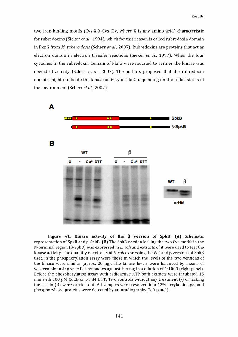
Results
141
two iron‐bindingmotifs (Cys‐X‐X‐Cys‐Gly, where X is any amino acid) characteristic
forrubredoxins(Siekeretal.,1994),whichforthisreasoniscalledrubredoxindomain
inPknGfromM.tuberculosis(Scherretal.,2007).Rubredoxinsareproteinsthatactas
electron donors in electron transfer reactions (Sieker et al., 1997). When the four
cysteines intherubredoxindomainofPknGweremutatedto serinesthekinasewas
devoid of activity (Scherr et al., 2007). The authors proposed that the rubredoxin
domainmightmodulatethekinaseactivityofPknGdependingontheredoxstatusof
theenvironment(Scherretal.,2007).
Figure 41. Kinase activity of the β version of SpkB. (A) Schematic
representationofSpkBandβ‐SpkB.(B)TheSpkBversionlackingthetwoCysmotifsintheN‐terminalregion(β‐SpkB)wasexpressedinE.coliandextractsofitwereusedtotestthekinaseactivity.ThequantityofextractsofE.coliexpressingtheWTandβversionsofSpkBused in thephosphorylationassaywere those inwhich the levels of the twoversionsofthe kinase were similar (aprox. 20 µg). The kinase levels were balanced by means ofwesternblotusingspecificanyibodiesagainstHis‐taginadilutionof1:1000(rightpanel).Before thephosphorylation assaywithradioactiveATPbothextractswere incubated15minwith100µMCuCl2or5mMDTT.Twocontrolswithoutanytreatment(‐)or lackingthecasein (Ø)werecarriedout.All sampleswere resolved ina12%acrylamide gel andphosphorylatedproteinsweredetectedbyautoradiography(leftpanel).

AlejandroMataCabana
142
Although the kinase activity of β‐SpkB was almost abolished, the slight
inhibition by CuCl2 raised the possibility that the cysteine responsible for redox
regulation is stillpresent, though the lackof theCys‐motif leavestheenzymealmost
completelyoxidised.Therefore,we tested furtherwhether thiskinasewas activated
by reducing agentswithout oxidising pre‐treatment. Thus,β‐SpkBwas incubated in
presenceof increasingconcentrationsofDTTandTrxApriortothephosphorylation
assay(Fig.42).Intheabsenceofreductantthekinaseactivitywashardlydetectable,
buttheadditionofDTTorTrxArestoredthekinaseactivity.ThetreatmentwithTrxA
asreducingagentwasmoreefficientthanthatwiththeDTTalone,recoveringahigher
phosphorylation level.The reactivationwas similar tothoseperformedwiththeWT
SpkB inhibited by CuCl2 (Fig. 37), though higher Trx concentrations (2 µM) were
neededtoreachmaximumactivity.ThisresultdemonstratedthattheCys‐motifisnot
requiredforcatalyticactivity,butforredoxregulation.

Results
143
Figure 42. Kinase activity reactivation of βSpkB. (A) Twenty µg of E. coli
extracts expressing β‐SpkB was incubated 15 min with growing concentrations of DTT(from0 to5mM)andTrxA(from0 to9µM),where0.2mMDTTwasusedas electrondonor to TrxA. A controlwith 0.2mMofDTT andwhitoutTrxAwas performed (DTT).Casein 2.5 µg was used as phosphorylation substrate. The phosphorylation assay wasperformedincubatingthesampleswith0.5mMATP,5µCi[γ‐32P]ATPand10mMNaF.Thesamples were resolved in 12% acrylamide gels and stained with Coomassie (lowerpanels). Phosphorylated proteins were detected by autoradiography (upper panels).Asterisks indicate the casein and the white arrowhead marks the TrxA. (B) Graphicrepresentationofthequantificationoftheradioactivesignalin(A).


145
DISCUSSION


Discussion
147
DISCUSSION
IMembraneproteinsfromthecyanobacteriumSynchocystissp.
PCC6803interactingwiththethioredoxin
Gram‐negative bacteria possess outer membranes and plasma membranes.
Cyanobacteriaareparticularamongst theGram‐negativebacteria,sincetheypossess
anadditionalmembranesystemformingthethylakoids.Oneofthecentralprocesses
in nature, the photosynthesis, takes place in the thylakoids. Furthermore, the
cyanobacterialrespirationoccursinthylakoidsandsharessomecomponentswiththe
photosynthetic electron transport. Hence, membranes in cyanobacteria have an
important role.However,because of technicaldifficulties the characterisation of the
membrane proteome in cyanobacteria hasbeen very limited. Aboutone‐thirdof the
more than 3000 ORFs present in the Synechocystis genome have been predicted to
encodeproteinsthatpossesstransmembranedomains(WallinandvonHeijne,1998).
In addition, there are peripheral membrane proteins, which are associated to
membranes or to integralmembrane proteins. Nevertheless, the number of integral
membraneproteins identified exclusively by standard proteomicapproaches is very
lowinthiscyanobacterium,withonly66outofthe706predictedintegralmembrane
proteins(Huangetal.,2002;Huangetal.,2006;Srivastavaetal.,2005;Pisarevaetal.,
2007;Wangetal.,2009).Thetechnicallimitationsforidentificationoflargenumbers
of membrane proteins are related to the hydrophobic nature of transmembrane
domains.Hydrophobicproteinsmayprecipitateat their isoelectricpointsduringthe
IEFseparationprocess,resultinginsamplelossanddifficultiestoperformthesecond
dimensionseparation(Santonietal.,2000).Moreover, in‐geltrypticdigestioncovers
principally exposed hydrophilic domains of integral membrane proteins, leaving
behind the hydrophobic transmembrane domains in the gel (Blackler et al., 2008).
However, progress has been made for membrane proteome analyses. One of the
solutionsproposed involvesa2‐DSDS‐PAGE,where inthe firstdimensiona cationic
detergent is used and in the second an anionic detergent, thus avoiding the IEF
separation (Zahedi et al., 2005). Another method developed is the Blue Native
PAGE/SDS‐PAGE inwhich the firstdimensionconsists of a nativeelectrophoresis in
the presence of a negatively charged CBB, which binds to exposed hydrophobic
surfacesofproteins(SchäggerandvonJagow,1991).Inthepast fewyears,agel‐free

AlejandroMataCabana
148
technical platform has been developed for membrane proteome analyses (Wu and
Yates,2003;WeinerandLi,2008). In thismethod theentireproteinmixture is first
digestedandpeptidesareanalysedbyLC‐MS/MS.Akeystep for thedevelopmentof
thismethod is finding a solvent for solubilisation ofmembrane proteins compatible
with enzymatic or chemical protein digestion (Weiner and Li, 2008). An efficient
meansofdegradingproteinsintopeptidesofsuitablelengthforLCseparationandMS
analysisisalsovitalforthesuccessofthegel‐freemethod(WeinerandLi,2008).The
methodsused to study integralmembraneproteins inSynechocystis include1‐DEof
membranepreparations(Srivastavaetal.,2005;Pisarevaetal.,2007)ortheanalyses
of thesolubilisedmembraneproteinsusingclassicalproteomicprodedure(Huanget
al., 2002 and 2006; Srivastava et al., 2005). Recently, a systematic analyses of
Synechocystis membrane proteome has been reported, in which the isolated total
membraneproteinsunderwent acidhydrolysis followedby trypsinor chymotrypsin
digestion and LC‐MS/MS analyses (Kwon et al., 2010). In this study 155 proteins
containing at least 1 transmembrane domain and 36 peripheralmembrane proteins
wereidentified(Kwonetal.,2010),whichmakesthisnewlydevelopedmethodtwice
asefficientastheclassicalones.
The association of thioredoxin with membranes (Srivastava et al., 2005;
DeRocher et al., 2008; Meng et al., 2010) as well as a thioredoxin‐like membrane
protein (Motohashi andHisabori,2006)havebeen reported.This facthasprompted
the search formembraneproteinsable to interactwith thioredoxins.Twoscreening
studies forTrx‐targetsinplantthylakoidmembraneshavebeenreported.Inthe first
study, Triton X‐100‐solubilised proteins reduced by Trx were labelled with
monobromobimane(mBBr)andseparatedusing2‐DE,whichledtotheidentification
of 14 proteins (Balmer et al., 2006). Since the authors applied a classical 2‐DE
procedurewithafirstIEFstepandasecondSDS‐PAGEseparation,theyprobablyonly
identified the most abundant proteins. In the second study, the authors used Trx‐
affinity chromatography to identify Trx‐targets amongst octylglucoside solubilised
membrane proteins (Motohashi and Hisabori, 2006). The proteins eluted from the
chromatography were resolved using 1‐DE. Hence, they identified 9 putative Trx‐
targets,5ofwhichwerepredictedtohavetransmembranedomains.However,since
both methods involve solubilisation prior to interaction with Trx, the exposure of
normallyburiedcysteinesmighthaveresultedinidentificationoffalsepositives.
Discussion
149
Contrary to previous studies, in the approach that we developed for
identification of membrane proteins interacting with Trx, the membranes were not
solubilisedpriortotheprocedure.Therefore,themonocysteinicTrxwasonlyableto
interact with the exposed cysteines in situ. After subsequent solubilisation, the Trx‐
target complexes were isolated and the separation of membrane proteins was
performedby2‐DSDS‐PAGE,andhence,theproblemsarisingfromtheuseofIEFwere
prevented. However, only 4 of the 50 Trx‐targets detected were transmembrane
proteins.ThiscouldmeanthatthereisnotahighnumberofTrx‐targetsamongstthe
integralmembraneproteinsorthatwewerenotabletoidentifythem,whichwouldbe
consistent with the fact that we performed in‐gel tryptic digestion and it has been
demonstratednottobethebestforhydrophobicproteins(Blackleretal.,2008).FtsH
is the only integral membrane protein in common between our study of
cyanobacterialmembranesandthatofplantthylakoidsmadebyMotashiandHisabori,
2006.Inthefuture,thenewadvancesintheanalysisofthemembraneproteomecould
be applied to improve the screening for Trx‐targets amongst integral membrane
proteins.
In our study we identified 50 Trx‐targets amongst the membrane bound
proteins.Thirty‐eightwere identified for the first time, though10 of thesehadbeen
previouslydescribedtointeractwithTrxinotherorganisms(Table3).Regardingthe
distributionofthetotalTrx‐targetsidentifiedinSynechocystis(LindahlandFlorencio,
2003;Pérez‐Pérezetal.,2006;andthepresentstudy)(Fig.13),itissurprisingthatthe
numberoftargetsassociatedtomembranesisalmosttwicethenumberofthesoluble
targets. This could be representative for the real distribution of proteins in
Synechocystis and emphasises the importance of the membranes in this kind of
organisms.Theoverlapofidentifiedtargetsbetweenouranalysesandthosemadeby
MotohashiandHisabori,2006,andBalmeretal.,2006 isminimal.Theonlytarget in
common with the study of Balmer et al., 2006 is the ATP synthase β‐subunit. This
proteinisalsofoundinMotoashiandHisabori,2006,whereothercommontargetsare
theATPsynthaseα‐subunitandFtsH.Thelargedifferencesintotalnumberoftargets
identifiedbetweenourstudyandtheothertwomaypartiallyexplainthisdivergence.
OnereasonfortherelativelylownumberoftargetsinMotohashiandHisabori,2006is
that theyusedasbait forTrx‐chromatographytheTrx‐likeproteinHCF164,whereas
weusedacanonicalTrx.AnimportantdifferencewiththeprocedureofBalmeretal.,

AlejandroMataCabana
150
2006, is that theyusedamBBr2‐DEapproach, lackinganenrichmentstep,whereas
we performed a Trx‐chromatography, in which the targets were enriched prior to
resolution.
It is striking that a large number of targets identified here are involved in
amino acid‐ and protein metabolism. This may suggest a redox‐dependent
readjustment of amino acid and protein metabolism in response to e.g. oxidative
stress,butfurtherinvestigationsarenecessarytoconfirmit.Moreover,herewehave
identified as Trx‐target the serine‐O‐acetyltransferase, which is involved in the
productionofcysteine.Inaddition,twoenzymesinvolvedinsulphurmetabolismhave
been identified, ferredoxin‐sulfite reductase (Lindahl and Florencio, 2003; and the
present study) and sulfate adenylyltransferase (Lindahl and Florencio, 2003). This
couldindicateaputativeredoxregulationofthecysteinemetabolism,whichwouldbe
ausefulmechanismofdefenceagainstoxidativestress.Anotherremarkablefeatureis
theabundanceofTrx‐targetscontainingaconservedcysteinewithinanATP‐binding
domain. This could indicate a putative regulation of the ATP‐binding by redox
modificationofthecysteine.Forexample,thedisulphidebondformationwithanother
molecule,suchasglutathione,oraprotein,wouldblockthebindingofATP.Another
possibilitywouldbethedirect interactionofthecysteinewiththeATP.Recently,the
autophosphorylationoftheresolvingcysteineofthe2‐CysPrxfromrapeseedhasbeen
reported(Aranetal.,2008).Thisstudydescribeshowtheoxidisedformsofcysteines
areabletobindATPandhowthisbinding impairs thecatalyticactivityof2‐CysPrx.
Thus, it opens the possibility that cysteinesare able to directlybindATP andwould
representtheconvergencebetweentheredoxandphosphorylationsignalling.
Twoofthetargetsanalysedinthepresentworkwerethe1‐Cysand2‐CysPrx.
1‐Cys Prx together with type II Prx have been reported as Trx‐targets in previous
proteomic studies in Synechocystis (Lindahl and Florencio, 2003; Pérez‐Pérez et al.,
2006;Florencioetal.,2006).However,2‐CysPrxhadneverbeendetectedandithad
evenbeenproposedtobeanchoredtomembrane(Pérez‐Pérezetal.,2006).Here,we
havedemonstrated that2‐CysPrx is located inthecytosolandthat itsabundance is
verylowcomparedto1‐CysPrxandtypeIIPrx.Thismaybethereasonwhy1‐Cysand
typeIIPrxshadbeendetected,butnot2‐CysPrx.Thedifferenceofabout50timesin
abundance could indicate a possible divergence in function between the different

Discussion
151
peroxiredoxins, each one playing a different role in redox signalling and peroxide
detoxification.
Aiming at verifying whether these two Prxs were truly Trx‐targets in
Synechocystis,wetestedtheirinteractionwithTrxA35invitrousingpurifiedproteins
and the capacity of TrxA to sustain their peroxidase activities. Eventually, we could
concludethat2‐CysPrxand1‐CysPrxareabletointeractwithTrxA inSynechocystis
andthattheseinteractionsarefunctional.Previously,averylowperoxidaseactivityof
1‐CysPrxusingx‐typeTrx(TrxB) inSynechocystis (Hosoya‐Matsuda,2005)hasbeen
shown, and the 2‐Cys Prx and 1‐Cys Prx activities in Synechococcus elongatus PCC
7942 using the complete reducing system of E. coli TrxA (Stork et al., 2009). The
activitiescalculatedbyStorketal.,2009weremuchhigherthanthosemeasuredhere,
butthedifferencebetweentheactivitiesofthetwoPrxwassimilar.However,noneof
themusedtheendogenousTrxA(m‐typeTrx)aselectrondonorandneitherusedthe
FOX method to monitor the peroxidase activity. In Arabidopsis the 2‐Cys Prx was
reducedbythem, fandx‐typesofTrx.However, thebestreductant for thisPrxwas
not them‐type Trx, but the x‐type Trx (TrxB in Synechocystis) (Collin et al., 2004).
Thesedataare consistentwith thoseobtained inour laboratory,which indicate that
TrxBisthethioredoxinthatbindswithmoreaffinitytothe2‐CysPrxinSynechocystis.
In Pérez‐Pérez et al., 2009, we performed an extensive study of the
peroxiredoxin system inSynechocystis.Wehaveexamined the interactionsof all five
Synechocystis Prx with the three different Trx from this organism with regard to
affinity and catalytic efficiency. In addition, we have analysed the expression ofprx
and trx genesunderdifferentconditions inorderto searchforcorrelationsbetween
theirexpressionpatterns.InthisstudyweconcludedthatallthesePrxareexpressed
andaregenuineTrx‐dependentperoxidases,allofwhichareabletointeractwithand
receiveelectronsfromthedifferentSynechocystisTrx(Pérez‐Pérezetal.,2009).With
respectto2‐CysPrx,TrxBisthethioredoxinwiththehighestaffinity,butTrxQisthe
thioredoxin that reduces most efficiently this Prx. However, the 50‐fold higher
abundance of TrxA as compared to TrxQ could mean that in vivo TrxA would have
morerelativeimportanceaselectrondonorforthisPrx.Inthecaseof1‐CysPrx,TrxB
is the Trx, which binds in with the highest affinity, but TrxA is the most efficient
electron donor. 1‐Cys Prx and TrxA coincided in the unusual expression pattern of
AlejandroMataCabana
152
reduced transcriptionunder conditionsof elevatedROSconcentrations (Pérez‐Pérez
etal.,2009).Thus,itseemsverylikelythatTrxAistheelectrondonorfor1‐CysPrxin
vivo.Sincethecatalase/peroxidaseKatGhashighercapacityforscavengingofH2O2in
Synechocystisascomparedtothethiolperoxidases,thisenzymeisresponsibleforthe
main peroxidase activity in the cell and for the high peroxide tolerance of this
cyanobacterium (Tichy and Vermaas, 1999). Thus, although the role of the Prxs
remains still unknown, it is possible that they do not participate in goal defence
againstROS,buttheycouldbeinvolvedincellularsignallingorinthelocalelimination
ofperoxidesatlowconcentrations.
Other putative targets found in our screening, in which we were interested,
were theUsp proteins, Usp1andUsp2.Wedemonstrated that the cysteinespresent
within the amino acid sequence of Usp1 played an important role in the
oligomerisationoftheUsp1underoxidisingconditions(Fig.20and21).Therefore,the
treatmentswithreductants,includingTrxA,wereabletoreducethehomodimericand
oligomeric formsofUsp1andtoreleasethemonomers.Interestingly,treatmentwith
H2O2 specifically inducesoligomerisation (Fig.21),which suggests thepresenceof a
redox‐activecysteinethat formssulfenicacid reaction intermediates(Salsburyetal.,
2008). Previously, the formation of homodimers of Usp proteins from diverse
organisms has been reported (Sousa and McKay, 2001; Zarembinski et al., 1998;
Weber and Jung, 2006) and also larger complexes in native E. coli protein extracts
(Gustavsson et al., 2002). However, neither of them reported the involvement of
cysteines in the oligomer formation. We have also demonstrated that the
autophosphorylation activity of Usp1 was impaired by reduction (Fig. 22). The
structure of onemember of the UspAprotein family fromMethanococcus jannaschii
wasresolvedbyX‐raycrystallographyandthisanalysisdemonstratedthattheprotein
formsATP‐bindinghomodimers (Zarembinskietal., 1998).Weobserved thatunder
oxidising conditions, when the oligomers are formed, is also when the Usp1 is
phosphorylated. Hence, when the monomers appear after reducing treatment the
autophosphorylation in inhibited. Therefore, the autophosphorylation could be
regulatedbytheoligomericstate,whichinturncouldberegulatedbytheredoxstate
ofthecysteines.TheUsp1fromSynechocystis is formedbytwoUspA‐likedomains in
tandem and each UspA contain an ATP‐binding motif, inside which is located one

Discussion
153
cysteine. Thus again, we found an ATP‐binding protein that is redox regulated,
supportingtheideathatthiscouldbeageneralregulationmechanism.
Althoughdiversephenotypesofhypersensitivitytostressconditionshavebeen
assigned to the ΔUsp mutant strains (Nyström and Neidhart, 1994 and 1996;
Gustavsson et al., 2002; Bochkareva et al., 2002;Nachin et al., 2005), themolecular
mechanism for the function of these proteins remains certainly unknown. With
respect toapossibleroleofUsp inresponse tooxidative stress, theE. coliUspAhas
been shown to be important for H2O2 resistance during growth (Nyström and
Neidhart,1996).Inaddition,theclassIofUsp(UspAandUspD)fromE.colihavealso
been suggested toplayan important role in the resistance to superoxideof growing
cells (Nachin et al., 2005). UspA has a single cysteine, which is highly conserved,
whereasUspDhastwocysteines,butonlythe secondone,whichcorrespondstothe
conservedcysteineinUspA,isextensivelyconservedamongstdiverseorganisms.
FtsH was an interesting putative target of Trx, since it would imply a redox
regulationofthephotoinhibitionprocess.Thus,underhighlighttheTrxcouldreduce
the only cysteine of FtsH located within the ATP‐binding domain activating the
proteaseactivityofFtsH,whichwould participate in theD1 degradation. Under low
light the cysteine could remain oxidasedor forminga disulphidebondwithanother
molecule, such as glutathione, which would block the ATP binding and the enzyme
wouldbeinactive.Ourdatastronglysuggestaninsitu interactionbetweenTrxAand
FtsH in Synechocystis. However, the data concerning the FtsH2 site directed mutant
werenotconclusive,sincethiswouldresemblethereducedformofFtsH2,andithad
the samephenotypeasWTstrain. Sincewewerenotable topurify FtsH2,wecould
notclarifytheredoxregulationofthisenzymeanditsputativeinhibitionbyoxidation.
Another possibility to explain the role of the only cysteine is that suggested in the
Figures22and23inwhichtheoxidationandreductionofthecysteinecouldmodulate
the involvementofFtsH insupercomplexes formationwithotherFtsHorwithother
kindsofprotein, suchasthose reported inE.coli (Saikawaetal.,2004)and inyeast
(Steglich etal., 1999).However, in this case the FtsHwouldbeactivewhen forming
thecomplexesinitsoxidisedstate,butthisisnotinaccordancewithourdatainwhich
theC266S‐versionofFtsH2isasactiveastheWT,atleastintheD1repairmechanism.

AlejandroMataCabana
154
Further investigations are necessary to completely elucidate whether the cysteine
playsaroleintheFtsHregulation.
II–Thioredoxintargetsinthethylakoidlumen
Since it is not possible to isolate intact thylakoids from cyanobacteria, the
thylakoid lumen proteome of cyanobacteria is not verywell characterised, although
some lumenal proteins have been identified (Mulo et al., 2008). Evidence for redox
regulationinthethylakoidlumenofthechloroplasthasbeenreported(Gopalanetal.,
2004;Marchandetal.,2004;Marchandetal.,2006;BuchananandBalmer,2005),but
no solubleTrxhasbeendetected insidethiscompartment.However,HCF164, aTrx‐
likeproteinanchoredtothethylakoidmembranewasidentified(Lennartzetal.,2001;
MotohashiandHisabori,2006).Thisprotein,whichishomologoustoE.coliDsbD,may
be responsible for transduction of reducing equivalents across the thylakoid
membranethroughdithiol/disulphideexchange(Poratetal.,2004).Thehomologueof
HCF164incyanobacteriawascalledTxlA(CollierandGrossman,1995).Thissuggests
a putative redox regulation in the thylakoid lumen of cyanobacteria. The disulphide
proteome studies performed hitherto in cyanobacteria have used total cell extracts
including soluble and membrane fractions (Florencio et al., 2006; and the present
work). Thus,a priori lumenal Trx‐targets could have been identified, but the fact is
that nonewas detected. Therefore, itwould be necessary to isolate the thylakoid to
analysetheproteinscontained,butsincethethylakoidandplasmamembraneare in
close connection, in cyanobacteria is not possible to separate intact thylakoids from
the rest of the cell. However, the chloroplast thylakoids are more independent
compartments and, then, therefore they can be isolated. Thus, we decided to use
chloroplast thylakoid preparations fromArabidopsis and the Trx‐targets foundwere
searched against the Synechocystis genome for comparison. Hence, we identified 12
putativeTrx‐targetsthathavehomologuesinSynechocystis(Table6).
This screening forms part of a systematic study, in which complementary
approacheshavebeenusedtodiscover lumenalTrx targets(Halletal.,2010).Apart
fromtheTrxaffinitychromatography,heredescribed,themethodsusedinthisstudy
are fluorescence labelling using mBBr and differential alkylation. The Trx affinity

Discussion
155
chromatographywastheprocedurethathasledtodetectionofthelargestnumberof
Trx‐targetproteins,atotalof14lumenalproteins,whereas,thetwootherapproaches
combined detected 11 targets. Four stromal Trx‐targets, which were contaminating
the lumenal preparation,were identified namely, theRuBisCo large subunit, the two
ferredoxin‐NADPoxidoreductaseFNR1andFNR2,andtheunknownproteinencoded
by the gene At3g15840.3. All these four contaminant proteinswere detected by the
Trx‐affinitychromatographymethod,butonlytheRuBisColargesubunitandtheFNR
1weredetectedbydifferentialalkylation.The locationof theplastid lipid‐associated
protein is unknown in plants and was only identified in the Trx affinity approach.
Thesedatasuggestthatthemethodhereexplainedwasthemostsensitiveofthethree
used.Moreover,anadvantageofthisprotocolisthatitcandiscriminatebetweentrue
targetsandpossiblefalsepositivesthatwouldbeequallyretainedinbothdimensions.
Therefore,proteinsthatmigratedexclusivelyalongthediagonalarenotconsideredas
Trxtargets.
The only two target proteins, which conserved the cysteines between
ArabidopsisandSynechocystiswerePsbOandPrxQ2(Table4).ThetwoPsbOisoforms
foundasputativetargetsintheArabidopsispreparationsharethesamehomologuein
Synechocystis. PsbO contains two conserved cysteines, whichmay form a disulphide
bondthatcould function inthe regulationof theoxygen‐evolvingactivity.Moreover,
PsbOisahighlyflexiblemoleculethatundergoesconformationalchangesandbound‐
unbound transitions of PsbO toPSIIhave been suggested (DeLasRivasandBarber,
2004).UndernormalconditionsthePsbO‐proteinsareverystableinisolatedluminal
preparations.However, the presenceofpossibledegradation productsofPsbO1and
PsbO2 in our eluates from Trx‐affinity chromatography (Fig. 26B) raised the
possibilitythatdisulphidereductionpromotestheirdegradation.Thus,weperformed
experiments in vitro and showed that the PsbO1 and PsbO2 subunits of the OEC in
ArabidopsisundergoapronouncedTrx‐dependentdegradation(Halletal.,2010).The
reductionofthedisulphidewithinthePsbOproteinscouldchangetheirconformation
and render them accessible to proteolysis. Taken into account the presence of two
lumenal proteases, Deg1 and Deg5, among the Trx‐targets found in the lumenal
fractionfromArabidopsis, onecouldenvisagea redox‐regulateddegradationofPsbO
subunits involving the Deg proteases. Deg proteases have been proposed to be
involved in heat and high light protection in cyanobacteria (Barker et al., 2006).

AlejandroMataCabana
156
However, their physiological substrates have not been identified (Huesgen et al.,
2009).
Another target that possesses conserved cysteines is the Trx‐dependent
peroxidase PrxQ. In plants the PrxQ is located in the chloroplast thylakoid lumen
(Petersson et al., 2006), but in bacteria its localisation is controversial. It has been
described as a periplasmic protein (Cha et al., 1995; Link et al., 1997) as well as
cytosolic(Tao,2008).SynechocystishastwoPrxQ,namedPrxQ1andPrxQ2.Wehave
reported here that PrxQ2 contains in its amino acid sequence a N‐terminal signal
peptide,whichcoulddirecttheenzymetothethylakoidlumenortheperiplasm.Thus,
itsuggeststhateachPrxQinSynechocystis islocatedinadifferentcompartment;one
inthecytosolandanotherprobablyinthethylakoidlumen.Thephylogeneticanalyses
revealed that this dual localisation isvery common amongst cyanobacteria (Fig. 26)
andthatthePrxQscontainingsignalpeptidesgrouptogether.
Here we have demonstrated that PrxQ2 is a functional Trx‐dependent
peroxidase. It is able to interact physically in vitro with TrxA (Fig. 27) and this
thioredoxin is also an effective electron donor to PrxQ2 (Fig. 28). The peroxidase
activityofPrxQ2 is intermediatebetweenactivitiesdetermined for 2‐CysPrxand1‐
CysPrxinthisstudy.Theseactivitiesaresignificantlylowerthanthosecalculatedfor
Anabaenasp.PCC7120(Chaetal.,2007),Synechococcuselongatus(Storketal.,2009)
and Arabidopsis (Lamkemeyer et al., 2006). However, these values are not entirely
comparablesincetheTrxsystem,Trxconcentrationsandtheassayusedineachcase
were different. For the PrxQ2 of Anabaena and Synechocuccus the E. coli TrxA
reduction system (TrxA, Thioredoxin reductase, NADPH) was used, where the
concentrations of TrxA were 5 µM and 3 µM, respectively. In the study of the
Arabidopsis PrxQ, Trx x was used with 0.4 mM DTT as reductant. The thioredoxin
concentrationinthiscasewas10µM,i.e.morethantwicetheconcentrationusedhere.
The peroxidase activity was measured by the FOX assay also in Anabaena as in
Arabidopsis,butinSynechococcustheabsorptionofNADPHat340nmwasmonitored.
TheTrxxofArabidopsiswastheonlyendogenousTrxusedasanelectrondonor for
the PrxQ2. However, it corresponds to the TrxB of Synechocystis, not to the TrxA,
which is anm‐type Trx. In Pérez‐Pérez et al., 2009, we reported that TrxQ is the

Discussion
157
Synechocystis Trxwith highest affinity for PrxQ2, but that TrxB is themost efficient
electrondonorforPrxQ2.
SincethePrxQ2seemstobelocatedinthethylakoidlumenorintheperiplasm,
noneoftheTrxusedasreductantinourassayswouldactuallyinteractphysiologically
with thiskind of peroxiredoxin.No solubleTrx has been shown tobe located in the
thylakoid lumen, and therefore this function may be attributed to the thylakoid
membrane anchored thioredoxin‐like proteinHCF164.Synechocystis possesses in its
genometwoORFs,sll1980andsll0685,whichencodetwoproteinshomologoustothe
Arabidopsis HCF164, and both contain a possibleN‐terminal signal peptides in their
amino‐acid sequences.Neitherof theseputativeHCF164havebeencharacterised in
Synechocystis or other cyanobacteria, with the exception of TxlA in Synechococcus,
whichwasreportedtobeinvolvedinredoxregulationofthestructureandfunctionof
photosynthetic apparatus, although the authors propose that TxlA acts as a protein
disulphide isomerase more than a thioredoxin (Collier and Grossman, 1995).
Definitely, this is only the beginning of the knowledge on redox regulation in the
thylakoid lumen and further investigations are necessary to understand completely
the thiol‐dependent signalling in this compartment and across the thylakoid
membrane.
III–Thioredoxinmediatedredoxregulationofaeukaryotetype
Ser/ThrkinaseinthecyanobacteriumSynechocystissp.PCC6803
Thepresenceofamechanismincyanobacteriasimilartothestatetransitionin
plants (Piven et al., 2005) could suggest the existence of a redox regulated Ser/Thr
kinase similar to the one in algae (Depègeet al., 2003) and plants (Bellafiore et al.,
2005).Thefirsteukaryotic typeSer/Thrkinase inprokaryoteswasreportedin1991
(Muñoz‐Dorado et al., 1991). Since then, the number of genes predicted to encode
bacterial Ser/Thr kinases has increased steadily and they are widespread amongst
prokaryotic genomes (Krupa and Srinivasan, 2005). However, the function of these
kinases isnotwell studied inbacteriaand no regulationof theseenzymes hasbeen
described. Two Ser/Thr kinases have been suggested to be involved in redox
signalling in bacteria.ThePkn22kinase (Alr2502) fromAnabaena sp.PCC 7120 has

AlejandroMataCabana
158
been reported to be induced by iron starvation and oxidative stress and shown to
regulate theexpressionof isiA,which is required for cell growthunder iron‐limiting
conditions (Xu et al., 2003). Pkn22 forms a cluster with the peroxiredoxin PrxQA,
which isthoughttoplayaroleinoxidativestressprotection(Latifietal.,2007).This
kinasepossessesin itsaminoacidsequenceaN‐terminal“Cys‐motif”containing four
cysteines and a C‐terminal PbHI domain, which is composed by parallel beta‐helix
repeats. Proteins containing these repeats are usually enzymes with polysaccharide
substrates and some of them belong to the amino acid metabolism of arginine and
proline (Jenkins et al., 1998). PknG is a Ser/Thr kinase from Mycobacterium
tuberculosis and it is an essential virulence factor (Walburger et al., 2004). The N‐
terminal part of this protein contains four cysteines grouped into two iron‐binding
motifs (Cys‐X‐X‐Cys‐Gly) characteristic for rubredoxins (Scherr et al., 2007).
Rubredoxinsactaselectrondonorsinelectrontransferreactions,sothisdomainhas
beensuggestedtoplayaregulatoryrole(Siekeretal.,1994;Scherretal.,2007).The
mutantversion ofPknG, inwhich the four cysteinesof the rubredoxindomainwere
replacedwith serine residues,wasdevoidofkinaseactivity.Thissuggests thatPknG
activitymightberedoxregulatedviatherubredoxindomain(Scherretal.,2007).
Withthisbackground,wedecidedtoundertakethesearchforaSer/Thrkinase
in Synechosystis that undergoes redox regulation.A priori, the search for a Ser/Thr
kinase should be easier than of a His kinase due to the lower number of predicted
genes inSynechocystis (Kanekoetal.,1996;Leonardetal.,1998;MurataandSuzuki,
2006)andsincethephosphorylationofHisresiduesismorelabile(Matthews,1995).
Hence,we firstly identifieda protein phosphorylation sensitive to copper treatment,
whichwasrestoredbyreductants(Fig.31and33).TheanalysesoftheSer/Thrkinase
mutant strains revealed that SpkB is responsible for this redox‐dependent
phosphorylation(Fig.34).AllPkn2typeSer/Thrkinases,withtheexceptionofSpkD,
contain Cys residues within the kinase domain, but SpkB and SpkF possess an
aditional Cysmotif in the N‐terminus in a manner similar to that ofM. tuberculosis
PknG (Fig. 34). This suggested that both could be redox regulated, but only SpkB
showedanalteredpatternofphosphorylationinthecytosol.SinceSpkFispredictedto
be an integral membrane protein (Fig. 34) it may be responsible for the
phosphorylation inhibitedby copper treatment inmembrane preparations (Fig.31).
Thetreatmentwithoxidisedglutathionealsocausedinhibitionofphosphorylationin

Discussion
159
cytoplasmicextracts(Fig.32).However,thisinhibitionwasmoredrasticandglobalat
high concentrations than the one displayed by copper. Since most of the signal
visualisedintheautoradiographyshouldcorrespondtoSer/Thr/Tyrphosphorylation,
theglobaleffectofGSSGmight involveageneral inhibitionof theSer/Thrkinases in
Synechocystis. It is consistentwith thepresence of cysteines in thekinasedomainof
almost all Ser/Thr in Synechocystis and with the idea here suggested of a redox
regulationoftheATP‐bindingandhydrolysisasaglobalmechanism.Curiously,allSpk
proteins, with the exceptions of SpkE (which is not a functional kinase), SpkA and
SpkD, containa cysteine intheATP‐binding region.Thus, theglutathione couldbind
the thiol group and block the ATP binding and, then, inhibit the kinase activity.
However,thishypothesisneedstobetested.
In order to characterise the redox regulation of SpkB in vitrowe carried out
phosphorylation assays using extracts of E. coli expressing SpkB as well as a
preparation of partially purified SpkB. Hence, we demonstrated that this kinase is
inhibitedbyoxidantsandthatitsactivityisrestoredbyreductants.Thetitrationwith
TrxA displayed a restored activity higher than the basal one before the inactivation
with copper (Fig. 37). Thismaybe explained by a progressive oxidation during the
storageofsemi‐purifiedSpkBprevioustotheassay.Inthecaseoftheinactivationby
GSSGcompleterestorationofactivitycouldnotbeachieved.Sofar,wehavenotbeen
abletoobtainabsolutelypurepreparationsofSpkBduetoextremelylowexpression
levels in E. coli. However, the appropriate controls using E. coli extracts from non‐
expressingcellsandmockpurificationshaveenabledusmakeconclusiveexperiments
regardingSpkBactivity.
In thecytosolicextractsoftheSynechocystisWTstraintreatedwithcopperor
inthoseoftheΔSpkBmutantstrainthephosphorylationofaproteinofabout90kDa
ismissing.Thephosphorylationofthisproteinwasreconstitutedincytosolicextracts
from the ΔSpkB mutant by addition of partially purified SpkB. Applying proteomic
approachescombinedwithradioactivelabellingwecouldidentifythisSpkBtargetas
GlyS, theβ–subunit of theglycyl‐tRNA synthetase.Thisenzymewas suggested tobe
subjectedtoredoxregulationinE.coli (OstremandBerg,1974;ProfyandSchimmel,
1986;Hu et al., 2010), aswell as inS. aureus (Niyomporn et al., 1968; Pöther etal.,
2009) and in yeast (Kern et al., 1981). In addition, the activity of some aminoacyl‐

AlejandroMataCabana
160
tRNA synthetases, including glycyl‐tRNA synthetase, has been reported to be
modulated by phosphorylation in mice (Berg, 1977, 1978, 1980). Therefore, this
enzymecouldundergoadoublepost‐translationalregulationbydirect redox‐control
andredox‐dependentphosphorylation.Takingintoaccountthepossibledoubleredox‐
regulationoftheglycyl‐tRNAsynthetasethissuggestsanimportantroleinthecellular
redoxhomeostasis.
AimingatelucidatingtheimplicationoftheCysmotifintheredoxregulationof
SpkB,weconstructeda truncatedversionof thekinase lackingthismotif, termedβ–
SpkB. The truncated version displayed a very reduced kinase activity that was still
inactivatedbythecoppertreatment(Fig.41).ThisresultsuggestedthattheCys‐motif
wasnotinvolvedintheredoxmodulationofSpkB,butinthekinaseactivity.However,
the β–SpkB kinase activity was efficiently restored by reductants, such as DTT and
TrxA(Fig.42),indicatingthatindeedtheCys‐motifisimplicatedintheredoxcontrol.
Thereby, this domain could have a trigger effect on other cysteines as an electron
donor, sowhen it is absent, thekinase is inactive,but inthepresenceofanexternal
reductant the activity is restored. The presence of a thiol oxidant, such as CuCl2 or
GSSG, has the same effect as the absence of the Cys‐motif; the target cysteine stays
oxidised and then the kinase inactivated. As in PknG fromM. tuberculosis, the Cys‐
motif could be a metal ion‐binding domain. If so, the Cys‐motif is not exactly a
rubredoxindomainsinceonlytwoofthefourcysteinesformacanonicaliron‐binding
domain.ThepossibilitythatSpkBbindsironoranothermetalcannotbediscarded.If
itweretrue,thiskinasecouldbeametalsensororusethisdomaintoreceivearedox
signalthatistranslatedintoaphosphorylationsignal.Maybethereasonwhyweneed
a high concentration of copper is due to this metal is competing with the metal
coordinatedbythefourcysteinesandcopperactuallyisinhibitingbydisplacementof
the other metal. Supporting the hypothesis of the iron sensor, a mutant strain of
Anabaenasp.PCC7120lackingtheSer/ThrkinasePkn22wasgrowth‐deficientunder
ironstarvation(Xuetal.,2003).Irondeficiencygeneratesoxidativestress,sothecell
needstoadapttosurviveunderthisconditions.Hypothetically,SpkBcouldsensethe
environmentalironlevelsandtriggeracellularresponsebymeansofmodificationof
the amino acidmetabolism in order to increase the cysteinepool to protect the cell
against oxidative damage. In that case under normal conditions SpkB would
phosphorylate GlyS and this enzyme would be active transferring the Gly to the

Discussion
161
corresponding tRNA and under oxidative stress generated by iron starvation SpkB
would not phosphorylate GlyS leaving this inactive, and the Gly would not be
incorporated to the tRNA increasing the pool of free Gly, which could be used to
synthesise cysteines. Free cysteines or incorporated into some molecules such as
glutathione play a protective role against oxidative damage. It is worth noting that
SpkBinvivo ispresent inthecompletely reduced,active formundernormalgrowth
conditions.Notably,undernormalgrowthconditionsSynechocystisSpkBisfullyactive
asshownbythefactthattheDTTtreatmenthasnoeffectonSpkBactivityincytosolic
extracts(Fig.33andFig.34).
In mammals some Ser/Thr kinases undergoing redox regulation have been
reported. Protein kinase C (PKC), MEKK1 and c‐Abl have been described to be
inhibited by glutathionylation (Ward et al., 1998; Cross and Templeton, 2004;
Leonberg and Chai, 2007). Both regulatory and catalytic domains of PKC contain
cysteinerichregionsthataretargetsforredoxregulation(Gopalakrishnaetal.,1995).
The cysteines within the regulatory domain coordinate zinc atoms and a redox
modificationofthesecysteinesreleaseszincpromotingstructuralchanges,whichlead
toconstitutiveactivationofPKC(GopalakrishnaandJaken,2000).Ontheotherhand
the cysteines within the catalytic domain are susceptible to react with alkylating
agentsandglutathione,which irreversibly inactivatetheenzyme (Wardetal.,1998;
GopalakrishnaandJaken,2000).PKCactivityisalsosensitivetootheroxidizingagents
suchasH2O2orotherperoxides (Gopalakrishna and Jaken,2000).MEKK1andc‐Abl
are both inactivated by alkylation and glutathionylation but in theses cases the
inhibitionbyglutathionylation is reversiblebytreatmentwithsomereducingagents
suchasDTTorglutaredoxin(CrossandTempleton,2004;LeonbergandChai,2007).
The cysteine residue that undergoes glutathionylation in MEKK has been located
withintheATP‐bindingpocketof thekinase, this isanunconservedresidue inother
kinases (Cross and Templeton, 2004). In the case of c‐Abl, it was reported that
glutathionylation affects a cysteine present in the catalytic domain (Leonberg and
Chai, 2007). Glutathionylation of a cysteine anywhere within the interior of the
catalyticdomainhasbeenproposedasageneralregulatorymechanismforkinasesin
mammals (Anselmo and Cobb, 2004). The Synechocystis SpkB possesses three
cysteinesinthekinasedomain,andoneormoreofthemarelikelytoberesponsible
fortheredoxregulationherereported.GlutathionecouldbeblockingtheATPbinding

AlejandroMataCabana
162
oranothertypeofinteractionorfunctioninsidethecatalyticdomainandthustherole
ofCys‐motifcouldbethereleaseofglutathionetomaintainactivethekinaseactivity.
It might explain the incapacity of GSH to reactive SpkB, since its presence would
favour the process of glutathionylation (Fig. 37). However, a data that support the
glutathionylationofMEKKwasthatGSHwascapableof reactivatethiskinase(Cross
and Templeton, 2004). Nonetheless, the glutathionylation may occur by direct
interactionbetweenanoxidisedsulphydrylradicalandGSH(Dalle‐Donneetal.,2007).
Hence,thehypothesisofaglutathionemoleculelinkedtoacysteinewithinthekinase
domain that inhibits SpkB could not be discarded. However, the inactivation of the
partially purifiedSpkBby Cu2+ alone speaks against anexclusive roleof glutathione
andglutathionylation.
Further investigation is needed to completely explain the redox regulation
mechanismofSpkBincyanobacteria.

163
MATERIALSANDMETHODS


MaterialsandMethods
165
MATERIALSANDMETHODS
1. Organismsandcultureconditions
1.1. Cyanobacteria
1.1.1. Cyanobacterialstrains
ThepresentworkhasbeencarriedoutusingthecyanobacteriumSynechocystis
strain sp. PCC 6803 (hereafter called Synechocystis). Synechocystis is a unicellular
cyanobacterium unable to fix nitrogen and it belongs to the taxonomic Group I
(formerlyChroococcales)(Rippkaetal.,1979).Synechocystiscangrowunderdiverse
physiologicalconditions,suchasphotoauto‐,mixo‐,andheterotrophycally.Duringthis
research theWTstrainwasusedaswell asdifferentmutant strains, someofwhich
were constructedhereandotherswere gifts. In theTable7 themutant strainsused
arelistedwiththeirprincipalfeatures.
Table7.Synechocystismutantstrainsusedinthethesis
Strain Features Source
GST‐FtsH2 ΔftsH2::ftsH2‐gst ProvidedbyPeterNixon’sLab.
ΔFtsH2 ΔftsH2::npt,Kmr Hereconstructed
FtsH266 ΔftsH2::ftsH2C266S::aadA+,SprStr Hereconstructed
ΔSpkA ΔspkA::npt,Kmr Lab.collection
ΔSpkB ΔspkB::npt,Kmr Lab.collection
ΔSpkC ΔspkC::npt,Kmr Lab.collection
ΔSpkD ΔspkD::npt,Kmr Lab.collection
ΔSpkE ΔspkE::npt,Kmr Lab.collection
ΔSpkF ΔspkF::npt,Kmr Lab.collection
ΔSpkG ΔspkG::npt,Kmr Lab.collection

AlejandroMataCabana
166
1.1.2 Cyanobacterialculturemediumandconditions
The different Synechocystis strains were grown at axenic conditions in a
mediumbasedontheBG11describedinRippkaetal.,1979.TheBG11compositionis:
NaNO3 17.6mM Ammoniumiron(III)citrate 6mg/l
MgSO4 0.30mM MnCl2 9.1μM
CaCl2 0.24mM Na2‐EDTA 2.4μM
Na2CO3 0.20mM Na2MoO4 1.6μM
K2HPO4 0.20mM ZnSO4 0.8μM
H3BO3 46μM CuSO4 0.3μM
Citricacid 28.5μM CoCl2 0.2μM
ThismediumsupplementedwithNaHCO312mMisdenotedBG11C,whichwas
themediumusedhere.ThemediumBG11Cwaspreparedfromasolution100 times
concentrated, containing all components except K2HPO4, NaHCO3 and NaNO3, which
were added to the medium just before being sterilized. Sterilisation of media and
materials was carried out during 20 min in an autoclave under one atmosphere of
over‐pressureandat120oC.Teng/l(1%w/v)ofagar(Bacto‐Agar,Difco)wereadded
topreparesolidmediumandwassterilisedseparately.
TheliquidSyechocystisculturesweregrownin250mlE‐flaskscontaining50to
100mlofmedium.E‐flakswereincubatedat30ºCundercontinuousillumination(25‐
50 µEm‐2s‐1) and with 100 rpm of constant stirring in orbital shakers (Gallenkamp
INR.401.Wmodel). Alternatively, cultures were bubbled with a stream of 1% (v/v)
CO2 in air under continuous illumination at light intensity of 50 to 70 µEm‐2s‐1
provided by fluorescents lamps (Sylvania daylight F20w/D). The bubbled cultures
weregrowninvolumesof750mlmediuminRoux flasksorat150ml incylindrical
flasks.PetriplateswithSynechocystisplatedonsolidmediumwereincubatedat30ºC
undercontinuousillumination(25‐30µEm‐2s‐1).
Antibioticswere added to culturemedia after their sterilisation by filtration.
Thefinalconcentrationsusedofeachofthemwere:

MaterialsandMethods
167
Kanamycin 50µg/ml
Streptomycin 2.5‐5µg/ml
Spectinomycin 2.5‐5µg/ml
1.2. Escherichiacoli
1.2.1. StrainsofE.coli
TheE.colistrainsusedandtheirgenotypesarelistedintheTable8.
Table8.StainsofE.coliusedinthiswork
Strains Genotype Reference
DH5α F,endA1,hsdR17(mK+,rK)supE44,thi1recA1
gyrA96relA1ΔlacU169(Ø80lacZΔM15)
(Hanahan,1983)
BL21(DE3) hsdSgal(λcIts857ind1Sam7nin5lacUV5T7
gene1)
(StudieryMoffatt,
1986)
Generally,theDH5αstrainwasusedforcloningandtheBL21(DE3)strainwas
usedfortheexpressionofproteinsclonedintothepETvectors.
1.2.2. E.coliculturemediumandconditions
ThenormalculturemediumforthegrowthoftheE.colistrainswastheLuria‐
Bertani(LB)(Sambrook,etal.,1989).TheLBcompositionwas:
NaCl 10g/l
Tryptone 10g/l
Yeastextract 5g/l
Inordertopreparesolidmedium,afinalconcentrationof15g/l(1.5%w/v)of
agar (Bacto‐Agar, Difco)was added to the LB prior to sterilisation. The sterilisation

AlejandroMataCabana
168
wasperformedusingthesameconditionsdescribedabovefortheBG11C.
In addition, the LB medium was supplemented with different antibiotics
dependingonthepurpose.Thefinalconcentrationsofantibioticswere:
Ampicillin 100µg/ml
Kanamycin 50µg/ml
Spectinomycin 100µg/ml
Differentvolumesofculturewereusedindifferentrecipients,tubesandflasks,
depending on the objective. Small volumes were used for DNA cloning and
manipulation and bigger volumes for protein expression. As a rule, the volume of
mediumtoinoculatecomprised1/5ofthecapacityoftherecipient.Theliquidcultures
as well as solid cultures were incubated at 37 ºC. Liquid cultures were stirred
continuouslyat200rpminNewBrunswickScientificshakers,modelG25.
1.3. Harvestingofcells
Cyanobacterial cells as well asE. coli cells were harvested by centrifugation.
Dependingonthevolumeofcultureamicro‐centrifugeEppendorf(13000xg,3min)
wasusedoraBeckmanrefrigeratedcentrifuge(9000xg,10min),modelAvantiJ‐25.
Centrifugationwasperformedeitheratroomtemperatureorat4ºCdependingonthe
requirements.
2. DNAanalysisandmanipulation
2.1. Plasmidsandprimers
Commercial and gifts plasmids as well as the plasmids constructed here are
listedintheTables9and10,wheresomeimportantcharacteristicsaredescribed.

MaterialsandMethods
169
Table9.Commercialplasmidsandgifts
Plasmid Features Reference Resistance
pGEMT Thisvectorcarriesterminalthymidinesinboth
ends thatallow the ligationofPCRproducts. It
also contains the β‐lactamase coding region,
whichallowsselectionofpositiveclones.
Promega Ap
pET28a Expression vector. The expression is under
controlofthepromoterofT7phage.Thisvector
encodes an N‐terminal and an optional C‐
terminalHis‐Tag.
Novagen Km
pET24a Expression vector. The expression is under
controlofthepromoterofT7phage.Thisvector
addsaC‐terminalHis‐Tagtothegeneproducts.
Novagen Km
pET22b Expression vector. The expression is under
controlofthepromoterofT7phage.Thisvector
encodesaC‐terminalHis‐Tag.
Novagen Ap
pFNT4 This plasmid derived from a pBS vector
containing the whole trxA gene from
Synechocystis.
(Navarroand
Florencio,1996)
Ap
pTrxA35 This plasmidderived frompET22b, intowhich
the trxA35 gene has been cloned. The vector
addsaC‐terminalHis‐tagtotheprotein.
(Lindahland
Florencio,2004)
Ap
Table10.Plasmidsconstructedinthiswork
Plasmid Features Resistance
pTrxA pET22b vector containing the whole trxA gene cloned from
pFNT4 in the restriction sites NdeI‐XhoI. This plasmid was
constructedtoexpressandpurifytheHis‐taggedTrxA.
Ap
p1CP pET24a vector containing the slr1198 gene cloned by PCR
from Synechocystis gDNA. The gene was inserted into the
NdeI‐XhoI restriction sites. This plasmidwas constructed to
expressandpurifytheHis‐tagged1‐CysPrx.
Km
AlejandroMataCabana
170
p2CP pET28a vector containing the sll0755 gene cloned by PCR
from Synechocystis gDNA. The gene was inserted into the
NdeI‐XhoI restriction sites. This plasmidwas constructed to
expressandpurifytheN‐terminalHis‐tagged2‐CysPrx.
Km
pPQ2 pET28a vector containing the slr0221 gene cloned by PCR
from Synechocystis gDNA. The gene was inserted into the
NdeI‐XhoI restriction sites. This plasmidwas constructed to
expressandpurifytheHis‐taggedPrxQ2.
Km
pUSP1 pET28a vector containing the slr0244 gene cloned by PCR
from Synechocystis gDNA. The gene was inserted into the
NdeI‐XhoI restriction sites. This plasmidwas constructed to
expressandpurifytheHis‐taggedUsp1.
Km
pFtsH2 pGEM‐TcarryingthePCRthatcontainstheslr0228geneplus
500pbpriorandafterthegene.Thisvectorwasconstructed
tomaketheFtsH2knockoutmutantstrain.
Ap
pΔFtsH pGEM‐T carrying the partially deleted and interrupted
slr0228 gene plus 500 bp before and after the gene. The
partialdeletionofslr0228wascarriedoutbyfusingthePCR
productscorrespondingtothebeginningofthegeneplusthe
500 bp upstream and the end of the gene plus the 500 bp
downstream. A StuI restriction site was introduced in the
fusion in order to replace the deleted sequence by a Km
resistance gene. This plasmid was used to transform
SynechocystistogeneratetheΔFtsH2mutantstrain.
Ap,Km
pFtsH266 pGEM‐T carrying the directed sitemutant slr0228 geneplus
500bpbeforeandafterthegene,inwhichthecysteine266is
replacedbyaserine.Attheendofthegene,aSp/Stresistance
gene was inserted in a BamHI restriction site generated by
PCR. This plasmid was used to transform and generate the
sitedirectedmutantFtsH266Synechocystisstrain.
Ap,Sp,St
pSpkB pET28a vector containing the slr1697 gene cloned by PCR
from Synechocystis gDNA. The gene was inserted into the
NdeI‐XhoI restriction sites. This plasmidwas constructed to
Km

MaterialsandMethods
171
expressandpurifytheHis‐taggedSpkB.
pβ‐SpkB pET28a vector containing the slr1697 gene lacking the N‐
terminal Cysmotif. Itwas clonedbyPCR fromSynechocystis
gDNA and inserted into the plasmid in the NdeI‐XhoI
restrictionsites.Thisplasmidwasconstructedtoexpressand
purifytheHis‐taggedβ‐SpkB.
Km
pGlyS pET28a vector containing the slr0220 gene cloned by PCR
from Synechocystis gDNA. The gene was inserted into the
NdeI‐XhoI restriction sites. This plasmidwas constructed to
expressandpurifytheHis‐taggedGlyS.
Km
pGlyQ pET22b vector containing the slr0638 gene cloned by PCR
from Synechocystis gDNA. The gene was inserted in NdeI‐
HindIII restriction sites. This plasmid was constructed to
expressandpurifytheHis‐taggedGlyQ.
Ap
TheprimersusedinthisworkweresynthesisedbySIGMA‐ALDRICH.Theyare
detailedintheTable11,includingtheirsequencesandtheiruse.
Table11.Syntheticprimersusedinthiswork
Primer Sequence Use
1CPF
1CPR
5’‐GCTACATATGGCCTTACAACTCGGTGATG‐3’
5’‐GCTACTCGAGCTTATTGGGTTGGGGGGTC‐3’
Used to clone the slr1198geneintothepET24avector
2CPF
2CPR
5’‐GCTACATATGACAGAGGTATTAAGGGTAG‐3’
5’‐GCTACTCGAGCTAAGGTTCCGCCACTGTCTC‐3’
Used to clone the sll0755geneintothepET28avector
PQ2F
PQ2R
5’‐CCTGGGGCATATGCAACCAGAGTTG‐3’
5’‐CGGGATTATTTACGTCGACGGGGAG‐3’
Used to clone the slr0221geneintothepET28avector
USP1F
USP1R
5’‐CAAGGAACTCAATTCATATGCTTAGCAAA‐3’
5’‐CCGGGAAACAAAGCTCGAGCTAAGCCTTG‐3’
Used to clone the slr0244geneintothepET28avector
FtsHUF
FtsHLR
5’‐GGTCAGAAATGTAGGTAACATC‐3’
5’‐CCGACGTGGGTTGCTGTGCAGAAG‐3’
Used to clone the slr0228plus the500pbadjacent toboth sides of the gene intothepGEN‐Tvector
FtsHUR
FtsHLF
5’‐GATAGGCCTTGGGGATTTAGTGTAATCGGTTAAC‐3’
5’‐CCAAGGCCTATCCCAACCAATTTAGTATGGTCAG‐3’
Used, along with FtsHUFand FtsHLR, to make theFtsH2 knock‐out mutant

AlejandroMataCabana
172
construction
FtsHCSF
FtsHCSR
FtsHCSLF
FtsHCSLR
5’‐GAATGCCCCCAGTTTGATCTTC‐3’
5’‐GAAGATCAAACTGGGGGCATTC‐3’
5’‐GATTCTTCGGATCCGGGCTTTTTTC‐3’
5’‐GAAAAAAGCCCGGATCCGAAGAATC‐3’
Used, along with FtsHUFand FtsHLR, to make theFtsH2 site directed mutantconstruction, in which theCys 266 was replaced withaserine
SpkBF
SpkBR
5’‐CTGGTGAACCCATATGAGTTTTTGCG‐3’
5’‐GCTTGGTTTGGTCAGAAACACTCGAGATT‐3’
Used to clone the slr1697geneintothepET28avector
β‐SpkBF 5’‐GTGGGCATATGCTGCGCCTC‐3’ Used, along with SpkBR, toclone the slr1697 genelacking the Cys motif intothepET28avector
GlySF
GlySR
5’‐GCTCCCTTGCCATATGCCCCTGC‐3’
5’‐GGAGGCCTCGAGGACATTTAAAAC‐3’
Used to clone the slr0220geneintothepET28avector
GlyQF
GlyQR
5’‐CTTGTTGGCTTCATATGACCATTACTTTCC‐3’
5’‐GTTTTTAAATCAAGCTTTACGAGGGAAA‐3’
Used to clone the slr0638gene into the pET22bvector
2.2. DNAisolation
2.2.1. PlasmidDNAisolationfromE.coli
Inorder to isolateplasmidDNA fromE. coli at small scaleweusedamethod
developedbyBirnboimandDoly,1979,basedonalkalinelysisofcellsinthepresence
of sodium dodecyl sulphate (SDS) and NaOH. Genomic DNA as well as the proteins
wereremovedbyprecipitationwithpotassiumacetate.Theprocedurewascarriedout
as described in Sambrook et al., 1989. As an option, the plasmid DNA preparations
were treatedwith a solution of phenol:chloroform (1:1, v/v) in order to reduce the
protein content. The plasmid DNA was precipitated at ‐20 ºC by 2.5 volumes of
absolute ethanol. Thereafter, theDNAwas resuspended in deionisedwater or in 10
mMTris‐HClpH8.ToeliminatetheRNApresentinthesamplethepreparationswere
treated during 1 h at 37 ºC with Ribonuclease A from bovine pancreas (SIGMA‐
ALDRICH). When a high quality preparation of plasmid DNA was required the
commercialkit“GFXMicroPlasmidPrepKit”(GEHealthcare,UK)wasusedfollowing
themanufacturer’sinstructions.
MaterialsandMethods
173
2.2.2. CyanobacterialgenomeDNAisolation
ThemethodusedtoisolatethegDNAfromcyanobacteriawasdescribedinCai
andWolk,1990,andisbasedoncelllysisusingglassbeads.Cellswereharvestedfrom
a 35 ml liquid BG11C culture by centrifugation at 4 ºC. The pellet of cells was
resuspendedin400µlTE(10mMTris‐HClpH8,0.1mMEDTApH8).Avolumeof150
µlofglassbeads(diameter0.25‐0.3mm,SIGMA‐ALDRICH),20µl10%(w/v)SDSand
400µlphenol:chloroform(1:1,v/v)wereaddedtothecellsuspension.Thismixture
wassubjectedto10cyclesofvortexing1minfollowedby1minofincubationonice.
The lysatewascentrifuged for15minat4 ºCat12000xg andthesupernatantwas
subjected to sucessive treatments with 400 µl phenol:chloroform (1:1, v/v). The
released gDNA was precipitated with 2 volumes of cold ethanol and 0.3 M sodium
acetate.Finally,theprecipitatedgDNAwasresuspendedin50µlTEandtheremaining
RNA was removed by Ribonuclease A from bovine pancreas (SIGMA‐ALDRICH)
treatment.
2.3. DNAanalysisandquantification
2.3.1. DNAelectrophoresisinagarosegels
ElectrophoreticresolutionofDNAinagarosegelswascarriedoutasdescribed
inSambrooketal.,1989.ThegelsweremadeinTBE0.5X(45mMTris‐borate,1mM
EDTA,pH8)buffercontaining0.7‐1.5%(w/v)agaroseand5µg/mlethidiumbromide.
The electrophoresis was run in Mini Sub‐cell GT systems (Bio‐Rad). DNA loading
bufferwasaddedtoeverysampleinaproportionof1/10.Thecompositionofloading
bufferwas 0.25% (w/v) bromophenol blue, 0.25% (w/v) xilene cyanol FF and 50%
(v/v)glycerol.Themolecularweightmarkerusedtoinferthemolecularweightofthe
DNA molecules migrated was the 1kb DNA Ladder (Invitrogen). After the
electrophoresis the DNA was visualised by illumination with UV light in a Bio‐Rad
transilluminatormodelGelDocXR.

AlejandroMataCabana
174
2.3.2. DNAquantification
Thefluorescenceemittedbytheethidiumbromideintercalatedintothedouble
DNAhelix isproportional to theamountofDNA.Thus, inordertoestimatetheDNA
concentration, the fluorescence emitted by the samples under UV illumination was
comparedtothatemittedbythemolecularmarkerofknownconcentration.
DNA was also quantified spectrophotometrically. DNA absorbs at 260 nm
wavelength and 1 unit of absorbance at 260 nm in a cuvette with 1 cm pathlength
correspondsto50µg/mlofDNA.
2.3.3. DNAextractionfromagarosegels
To purify DNA bands from the agarose gels we used the commercial system
“GFXPCRandGelBandpurification”(GEHealthcare,UK)followingthemanufacturers’
instructions.
2.3.4. EnzymaticmanipulationofDNA
The DNA enzymatic digestions were carried out by restriction nucleases
provided by Takara, Roche and New England BioLabs. The dephosphorylation of
vector ends cut open with restriction enzymes was performed by treatment with
alkaline phosphatase provided by Roche. For sticky end DNA fragments, cut with
restriction enzymes leaving the 5'‐strand protruding, filler treatments were
performedwith the Klenow fragment ofDNA polymerase I fromE. coli supplied by
Promega.TheligationofDNAfragmentswascarriedoutbytreatmentwithDNAligase
ofphageT4providedbyPromega.Inallcasesthetreatmentsweremadeaccordingto
themanufacturers'instructions.
2.4. PolymeraseChainReaction(PCR)
PCRreactionswereperformedinaMastercycler5330Eppendorfdevice.Each
PCR reaction was performed in a volume of 100 µl containing 2.5 U of Biotaq

MaterialsandMethods
175
polymerase(Bioline),Biotaqbuffer(suppliedwithenzyme)1.5mMMgCl2,dNTPsat
final concentrations of 0.2 mM each, 1.5 ng of template DNA and 50 pmol of each
primer. Oligonucleotides used are listed in Table 11. Generally, the amplification
programconsistedofan initialdenaturationcycleat94°C for5min, followedby30
cyclesof reactionanda finalpolymerizationcycleat72°C for10min.Each reaction
cycleconsistedofadenaturingstageat94°C(1min),arenaturationstepat45‐65°C
(1min)andapolymerizationstepat72°C(1minuteperkbofDNAtobeamplified).
The amplified DNA fragments were subjected to agarose gel electrophoresis and
purifiedessentiallyasdescribedinsection2.3.3.
2.5. DNAsequencing
The sequencings were carried out by the commercial service MWG Biotech
(Germany). The samples were prepared to sequencing according to the service
instructions.
2.6. IntroductionofDNAintodifferentorganismsbytransformation
2.6.1. E.colitransformation
Competent cells of the different strains of E. coli were supplied by the
responsible serviceof the Instituto deBioquímicaVegetal yFotosíntesis. Inorder to
transform the DH5α competent cells, the exogenous DNA (in a volume maximum
volumeof10µl)wasgentlymixedwithanaliquotofcompetentcells(100µl)thawed
onice.After30minincubationonice,themixturewassubjectedto2minheatshock
at42°Cfollowedby2minonice.Subsequently,0.9mlofLBmediumwasaddedand,
then,themixwasincubatedforonehourat37°C.Thenthecellswerespreadonsolid
LBmediumwith the antibiotic suitable for selecting transformants orwere used to
inoculate a liquid culture. When we used plasmids and E. coli strains, which allow
identifyingtheclonescarryingrecombinantmoleculesduetoinactivationbyinsertion
of lacZgene, themediumwassupplementedwith0.2mMIPTGand40mg/mlX‐gal.
Colonies with recombinant plasmids had white colour, whereas negative colonies
AlejandroMataCabana
176
wereblue.
TheBL21(DE3)competentcellsweretransformedbyelectroporation.Forthis
transformation,1‐2µgofplasmidDNA,inamaximumvolumeof5µl,wasmixedwith
100 µl of competent cells and the mixture was incubated on ice for 5 min.
Subsequently,itwastransferredtoaGenePulsercuvette(BioRad),whichhad2mmof
distancebetweentheelectrodes,andwassubjectedtoanelectricpulseof2500Vina
Easyject Optima (Equibio) device. Immediately, 1ml of cold LB was added and the
mixturewasincubated1hat37°C.Finally,thecellswerespreadonLBplateswiththe
appropiate antibiotic to select the transformant clones or they were inoculated
directlyinliquidLBcultures.
2.6.2. Synechocystistransformation
Synechocystis sp. PCC 6803 is naturally capable of incorporating into the cell
DNAmolecules from the culturemedium.Once inside, if these DNAmolecules have
homologytoanysequenceofthegenome,theyarestablyintegratedintothegenome
byrecombination.Theprincipleformutagenesisand introductionofexogenousDNA
fragments in Synechocystis sp. PCC 6803 is based on this property. Therefore, if a
fragment of DNA (exogenous DNA or an antibiotic resistance gene) is flanked by
sequences homologous to the genome sequences, the exogenous DNA fragment is
incorporatedinatleastonechromosomecopy.
The Synechocystis transformation method is based on that described by
Chauvatetal., 1986.Culturesweregrownuntil they reachedaOD580nmequal to1.A
volumeof50mlofculturewasharvestedbycentrifugationandthecellswerewashed
twicewithfreshmediumtoeliminatepossibleextracellularnucleases.Afterwashing,
cellswereresuspendedin1mlfreshmediumthatwasdistributedinaliquotsof200µl
in polystyrene tubes of 10 ml (Soria Greiner S.A.) and the DNA was added. Each
transformationwascarriedouttypicallywith2µgofpurifiedDNA.ThecellandDNA
mixturewasincubatedfor1.5hoursundernormallightconditions.Afterthistime,the
cells were spread on solid BG11C plates, without antibiotics, over 85mm diameter
Nucleopore filters (Whatman). After a period of 20 h to allow the expression of
resistance genes, introduced by the foreign DNA, the filters were moved to plates

MaterialsandMethods
177
containing the appropriate antibiotic, e.g. 50 µg/ml kanamycin or 2.5 µg/ml of
spectinomycin and streptomycin. Antibiotic resistant colonies appeared after 8‐10
days of incubation. To force segregation ofmutant chromosome copies, filterswere
transferredtonewplateswithincreasingconcentrationsofantibiotics.
The degree of segregation of mutant chromosomes was analysed by PCR,
performed as explained in section2.4., using gDNA isolated from themutant strains
usingtheprimerslistedintheTable11.
2.7. Sitedirectedmutagenesis
The site directed mutagenesis was performed using an overlapping PCR
approach inwhich two complementary synthetic primerswere designed containing
the site directed mutation. These oligonucleotides, along with two other external
primers,wereusedtoamplifythegeneofinterestintotwoseparatefragments.Based
on these fragments we carried out the overlapping PCR that could be divided into
threesteps:
1. Overlapping of the complementary sequence. At this stage both PCR
fragments were denatured and hybridised together through the
complementary sequence, corresponding to the primers that carry the
mutation. To facilitate the hybridisation, the temperature dropped
gradually from95 °C to72 °Cat a rateof0.01 °C/s.The reactionmixture
onlycontainedinthissteptheenzymebufferandthetwoPCRfragmentsin
aproportionof1:1.
2. Complementary chain polymerisation step. This phase completed the
polymerisation of each DNA chain from its 3' end, generating a double‐
stranded fragment resulting from the merge of the two initial PCR
fragments.AtthisstagethepolymeraseenzymeandthedNTPswereadded
tothereactionmix.Thecycleranfor10minat72°C.
3. Reaction cycles. The double‐stranded fragment obtained in the previous

AlejandroMataCabana
178
step was used as a template to start the typical PCR reaction cycles
describedabove(2.4.section),whichconsistsofdenaturation,renaturation
andpolymerization.Inthisphase,theadditionofspecificexternalprimers
is necessary. After 30‐35 cycles the PCR was completed with a
polymerisationcycleof5minat72°C.
3. Proteinsynthesis,purificationandanalysis
3.1. ProteinexpressioninE.coli
For the expression of various proteins used in this study we used a
heterologous expression system inE. coli, cloning the gene under the control of the
phageT7promoter(pETseriesplasmidssuppliedbyNovagen).Toinduceexpression
of the genes cloned, E. coli BL21 (DE3) cells were used, since they carry the gene
encodingtheT7RNApolymeraseunderthecontrolofthelacUV5promoter.Thus,the
geneisinduciblebyisopropyl‐β‐thiogalactopyranoside(IPTG).
TransformedE.coliBL21(DE3)cellswereculturedat37°CinLBmediumwith
the appropriate antibiotic until the culture reached an OD580nm equal to 0.5.
Subsequently, gene expressionwas induced by addition 1mM IPTG and the culture
incubationwascontinuedfor2‐4hours,beforecollectingthecells.
3.2. Preparationofcellextracts
3.2.1. Celllysisusingglassbeads
Generally, the cell extracts from Synechocystis and small quantities of E. coli
werepreparedbybreakagewithglassbeads.Tothisend,cultureswereharvestedby
centrifugationandthepelletwasresuspendedinasmallvolumeofbuffer25mMTris‐
HClpH8,50mMNaCl.ThissuspensionwasdividedintoEppendorftubesinaliquots
ofamaximumvolumeof500µlandanamountequivalentto50µlofglassbeads(0.25
to0.30mmindiameter,SIGMA‐ALDRICH)per100µlofsuspensionwasadded.Phenyl
MaterialsandMethods
179
methyl sulfonyl fluoride (PMSF), a serine prtease inhibitor, was added to a final
concentrationof1mM.Thismixturewassubjectedtotenpulsesof1minofvigorous
stirringalternatingwithperiodsof1minonice.Cellulardebrisandinsolubleproteins
wereseparatedfromthesolublefractionbycentrifugationfor30minutesat13000xg
at4ºC.Thesupernatantconstitutedthecell‐freecrudesolubleextract.
3.2.2. Celllysisbysonication
This method was used for large volumes of E. coli cultures (1‐1.5 l). The
harvested cells were resuspended in 25mM Tris‐HCl pH 8, 50mMNaCl buffer and
PMSFwasaddedtoafinalconcentrationof1mM.Thecellsuspensionwaskeptonice
andsonicatedin6pulsesof30sseparatedby30scoolingintervals.Thefrequencyof
ultrasoundproducedbyaBransonsonicatormodelB12,was20KHzandthepower
was40W.Afterlysis,thecelllysatewassubjectedtocentrifugationfor30minutesat
18000xgand4ºC.Thesupernatantconstitutedthecell‐freecrudesolubleextract.
3.2.3. PreparationofSynechocystistotalmembranes
Synechocystisculturesweregrownphotoautotrophicallywithbubblingat30ºC
undercontinuousilluminationwithwhitelightatanintensityof50µEm‐2s‐1.Cellsof
exponentially growing cultures were harvested and broken with glass beads in a
buffercontaining25mMHEPES‐NaOHpH7.0,15mMCaCl2,5mMMgCl2,15%(v/v)
glyceroland1mMPMSF(BufferA).Unbrokencellswereremovedbycentrifugationat
2300xgfor5minandtotalmembraneswerethereafterpelletedbycentrifugationat
16000xg for 20min.The pelletwaswashedonce by resuspension inBuffer A and
centrifugationat16000xg for20min.Finally, themembraneswere resuspended in
BufferAtoachlorophyllconcentrationof1mg/mL,whichcorrespondedtoaprotein
concentrationof23mg/ml.Thechlorophyllconcentrationwasmeasuredasdescribed
insection6.1.,andtheproteinconcentrationwasdeterminedaccordingtosection3.3.
3.3. Proteinquantification
The protein concentration of intact cells, cell extracts and isolated proteins

AlejandroMataCabana
180
were determined by applying the modification of the Lowry method (Lowry et al.,
1951)describedinMarkwelletal.,1978.ForthismethodtheFolin‐Ciocalteu’sreagent
(Merk,Germany)wasused.ThemethoddescribedbyBradford(Bradford,1976)was
alsousedtoquantifyproteins.InthiscaseweusedaBioRadreagentaccordingtothe
manufacturer’s instructions. In both cases we used known concentrations of
commercialBSA(Sigma)tomakethecalibrationcurve.
3.4. Proteinelectrophoresis
3.4.1. 1‐DSDS‐PAGE
The analytical separations of proteins using denaturing electrophoresis in
polyacrylamide gelswereperformedbasedon theLaemmli system(Laemmli,1970)
as described in (Sambrook et al., 1989), using Miniprotean BioRad II or III
apparatuses. Stackingandseparatinggelsaswellaselectrodebuffercontained0.1%
(w/v) SDS (sodium dodecyl sulphate). The concentration of polyacrylamide
(acrylamide:bisacrylamide29:1)inseparatinggelsvariedbetween10and15%(w/v)
dependingonthemolecularmassoftheproteinsanalysed.Samplesweremixedwith
an equal volume of solubilising buffer, containing: 0.125 M Tris‐HCl pH 6.8, 20%
glycerol (v/v), SDS 4% (w/v), 2‐mercaptoethanol 10% (v/v) and bromophenol blue
0.0025%(w/v).When theSDS‐PAGEwasperformedundernon‐reducing conditions
the 2‐mercaptoethanol was omitted. In some cases indicated in the text the 2‐
mercaptoethanol was replaced with 50 mM DTT to reach reducing conditions.
Denaturation of sampleswas carried out by heating at 65 °C for 5minutes and the
electrophoresiswasrunatroomtemperaturebyapplyingaconstantvoltageof200V
for45‐60minutes.
WeusedthemolecularmassstandardsLowMolecularWeightRangeprovided
by BioRad, which contains the following proteins: rabbit muscle phosphorylase B
(97.4 kDa), bovine serum albumin (BSA) (66.2 kDa), chicken ovalbumin (45.0 kDa),
bovinecarbonicanhydrase(31.0kDa),soybeanseedtrypsinogeninhibitor(21.5kDa)
andhenegglysozyme(14.4kDa).TheSeeBluePre‐StainedStandard(Invitrogen)was
also used, containing the following proteins: myosin (250 kDa), BSA (98 kDa),
MaterialsandMethods
181
glutamic dehydrogenase (64 kDa), alcohol dehydrogenase (50 kDa), carbonic
anhydrase (36 kDa),myoglobin (30 kDa), lysozyme (16 kDa) aprotinin (6 kDa) and
insulin,Bchain(4kDa).
3.4.2. Twodimensionalproteinelectrophoresis
3.4.2.1. Non‐reducing/reducing2‐DSDS‐PAGE
Thismethodwasused to isolate the putative Trx‐targets afterpurification of
protein complexes by Ni‐affinity chromatography. Thus, the imidazole eluates
containing target‐Trx mixed disulphides from Ni‐affinity chromatography were
subjected to 2‐D SDS‐PAGE under nonreducing/reducing conditions as described in
LindahlandFlorencio,2004.Forthefirstdimension,150µlofeluatewasmixedwith
10 µl of SeeBlue Prestained protein standard (Invitrogen) and 50µl of solubilising
bufferwithoutreductantconsistingof0.25MTris‐HClpH6.8,12.5%v/vglyceroland
10%w/vSDS.Themixturewasloadedona10%acrylamidegel,16x18cmand1.5
mmthick(AmershamBiosciences).Theelectrophoresiswascarriedoutbyapplyinga
maximum voltage of 220 V and a constant current of 35 mA per gel at room
temperature for about 4 h. The second dimension was carried out under reducing
conditions,whichallowedthereleaseofthetargetproteins.Thegellanescontaining
the proteins were excised, incubated for 1 h at room temperature in solubilisation
buffer diluted2.5 timesandsupplementedwithDTT toa final concentration of100
mM and were mounted at the top of the second dimension 10% acrylamide gels.
Electrophoresiswasperformedinthesamewayasforthefirstgel.Thegelswererun
usingaSE600Hoefersystem.
3.4.2.2. 2‐DIEF/SDS‐PAGE
The Synechocystis cytosolic extracts were prepared as described in 3.2.1.
section. The protein samples were salt precipitated and washed by means of the
commercial system 2‐D Clean Up Kit (GE Healthcare) and resuspended in DeStreak
rehydratation Solution (GE Healthcare) supplemented with ampholytes with a pH
range of 3‐10 (BioRad) at 0.5%, following the manufacturer's instructions. The
samples were separated in the first dimension according to their isoelectric point,

AlejandroMataCabana
182
using acrylamide strips with immobilized pH "DryStrips Immobiline gel (GE
Healtcare)withpHrangeof4‐7.Thesamplewasloadedbyanactiverehydrationat20
°C for 12 h. The IEF was performed applying the program according to the
manufacturer’s instruction depending on the strip used. For the indicated strips the
focusingtookplaceinfoursteps:S1:250V,15min,S2:250to8000V,2h30min;S3:
8000 V, 35000 Vh, S4: 500 V. Both rehydration and focusing were performed in a
Protean IEF Cell (BioRad)device.After IEF, and before the seconddimension, strips
wereimmersedinequilibrationsolution(6MUrea,2%SDS(w/v),375mMTris‐HCl
pH8.8,20%glycerol(p/v))initiallysupplementedwith130mMDTTfor15minand
subsequentlywith135mMIAAfor15moremin.TheincubationwiththeSDSsolution
wasnecessarytoconferanegativechargetotheproteinsandDTTtreatmentbrokeall
disulfidebonds, leavingeach protein in its reduced state.The IAA prevented the re‐
oxidation of proteins during electrophoresis. After that, the strips were washed in
electrophoresisbufferandplacedhorizontallyovertheseconddimensionacrylamide
gelsandfixedwithagarosesolution(0.5%agarose(w/v)and0.002%bromophenol
blue(w/v) inelectrophoresisbuffer).Acrylamideconcentrationsrangedbetween10
and15%.Thus,theseconddimensionwasperformedasanormalSDS‐PAGE,inwhich
proteinsareseparatedaccordingtotheirmolecularmass,atafixedcurrentof70mA.
ThegelswereruninaSE600Hoefersystem.
3.5. Proteinstaining
AftertheelectrophoresistheproteinswerevisualisedbyCBB‐staining.Hence,
the gelswere immersed in a solution of 0.1%(w/v) Coomassie blueR‐250 (SIGMA‐
ALDRICH),10%(v/v)aceticacidand40%(v/v)methanol.After15minutesstaining
atroomtemperature,gelswerewashedseveraltimeswitha40%methanol(v/v)and
10% (v/v) acetic acid solution. After this treatment, the proteins appeared as blue
bandsorspots.
3.6. Proteinidentificationbypeptidemassfingerprinting(PMF)
CBB‐stained proteins were excised manually from the gels, destained, dried

MaterialsandMethods
183
and digested with trypsin by rehydration in 100 µl of 50 mM NH4HCO3 with the
additionof15µlof0.1mg/mltrypsinin1mMHClovernightat37ºC.Peptideswere
extractedwith20µl of0.5%TFA,0.5µl of each samplewasapplied on the sample
plate and MALDI‐TOF MS spectra were acquired on an Autoflex apparatus (Bruker
Daltonics). External calibration was performed using Peptide Calibration Standard
(BrukerDaltonics)andthetrypsinautodigestionproductsofm/zvalues842.5094and
2211.1046wereusedfor internalcalibration.Proteinswere identifiedas thehighest
ranked result by searching the databases NCBInr 20061215 or MSDB 20050929,
including all species, using the MASCOT search engine (Matrix Science). The search
parameters included carbamidomethylation of cysteines, oxidation of methionines,
onemiscleavagebytrypsinandbetween30and80ppmmassaccuracy.
3.7. Proteininmunodetection(Westernblot)
The proteins subjected to electrophoresis on polyacrylamide gel were
transferredtonitrocellulosemembranes(Bio‐Rad)of0.45mmporesize,inasemidry
transfersystem(Biometrics),followingtheinstructionsofmanufacturer.Bothgeland
nitrocellulosemembranewerewet in transfer buffer (49.4mMTris pH 8.3, 39mM
glycine,1.3mMSDSand20%methanol(v/v))and,afterthat,thetransferwascarried
out at room temperature for 60‐90 min, depending on size of the proteins to be
transferred,at2.5mApercm2ofgelandamaximumof20V.Theefficiencyoftransfer
was checked by the disappearance of pre‐stained standard from the gel. After the
transfer, themembranewasincubatedforatleast1h inblockingsolutionconsisting
of5%skimmedmilkpowderLaAsturianaand0.1%Tween20inPBSbuffer(26mM
NaCl,0.54mMKCl,0.8mMNa2HPO4,0.352mMKH2PO4).Subsequently,themembrane
wasincubatedwiththeantiserumappropriatelydilutedinblockingsolutionfor15h
at 4 °C and constant gentle shaking. Afterwards, the membrane was subjected to 4
washesof15mininwashingsolution,whichconsistsofPBSbuffersupplementedwith
0.1%Tween20(v/v).Afterwashingtheincubationwiththesecondaryantibodywas
performed. Hence, the goat anti‐rabbit immunoglobulin G, conjugated with horse‐
radishperoxidase(BoehringerMannheim)diluted(1:10000)intheblockingsolution
was incubated for 1 hour at room temperature and constant shaking. Then, the
washing process was repeated as described above. The detection of peroxidase

AlejandroMataCabana
184
activity was carried out using the commercial system ECL Plus (GE Healthcare)
followingthemanufacturer'sinstructions.
3.8. Proteingeldrying
The stained gels containing radioactively labelled proteinswere dried before
autoradiography. The gels were incubated in a 10 % glycerol solution for 30 min.
Thereafter,thegelswereplacedbetweentwosheetsofCellophaneSupport(BioRad)
andtheyweredriedinaGelDryerModel583(BioRad)for2hat80ºC.
3.9. Detectionofradioactivity
Todetecttheradioactivesignaloftheradioactivelylabelledproteinsseparated
in acrylamide gels we used the Cyclone Storage Phosphor autoradiography system
(Packard). Alternatively, the dried gels were subjected to autoradiography by
exposure of the Kodak film X‐OMAT model S or X‐OMAT model LS in an exposure
cassette(SIGMA‐ALDRICH)usingLightningPlus intensifyingscreens(Dupont)at‐80
ºC. After the exposure time, the films were developed using developing and fixing
reagentssuppliedbyKodakfollowingthemanufacturer'sinstructions.
3.10. Proteinpurification
3.10.1. Metalaffinitychromatography
TheHis‐tagged proteinswere purifiedby nickel‐affinity chromatography in a
manual system using the matrix (His‐Bind Resin, Novagen) packaged into a plastic
column, Poly‐Prep Chromatography Columns (BioRad), or by using 1 ml Hitrap
columns connected to the Äkta FPLC systemof Amersham Biosciences according to
manufacturer's instructions. In both cases the columnswerewashedwith 5 column
volumesofdistilledwater, loadedwith50mMNiSO4and theexcesswaswashedoff
with 5 volumes of distilledwater. For both purificationmethods the binding buffer

MaterialsandMethods
185
was composed of 20 mM Tris‐HCl pH 8, 500 mM NaCl, and the elution buffer was
composedof20mMTris‐HClpH8,500mMNaCland500mMimidazole.
3.10.2. Gelfiltration
We used a preparative column HiLoad 16/60 Superdex 75 (Amersham
Biosciences)withaninnerdiameterof16mm,whichwas filledwithporousagarose
beadsboundcovalentlytodextrane,thatcouldseparateproteins,themolecularmass
ofwhicharebetween3000and70000Da.ThecolumnwasconnectedtoaÄktaFPLC
system(AmershamBiosciences)anda coolingbath formaintaining the temperature
at4ºC.Thecolumnwasequilibratedwithtwovolumesofabuffercontaining50mM
Tris‐HClpH8and150mMNaCl,previously filteredandvacuumdegassedtoremove
impurities and gases. The sample was passed through a filter of 0.2 μm pore size
before injection into the FPLC. The same bufferwas used to elute the sample. Each
proteinelutedinfractionscorrespondingtoitsmolecularmass.
3.10.3. Proteinspurifiedinthiswork
In the Table 12 are listed the proteins purified during the course of this
researchalongwiththemethodusedforpurification.
Table12.Proteinspurifiedduringthecourseofthiswork
Protein Plasmid Purificationmethod
TrxA pTrxA Ni‐affinitychromatography
TrxA35 pTrxA35 Ni‐affinitychromatography
Gelfiltrationchromatography
1‐CysPrx p1CP Ni‐affinitychromatography
2‐CysPrx p2CP Ni‐affinitychromatography
PrxQ2 pPQ2 Ni‐affinitychromatography
Usp1 pUSP1 Ni‐affinitychromatography

AlejandroMataCabana
186
SpkB pSpkB Ni‐affinitychromatography
β‐SpkB pβ‐SpkB Ni‐affinitychromatography
GlyS pGlyS Ni‐affinitychromatography
GlyQ pGlyQ Ni‐affinitychromatography
3.10.4. Trx‐targetisolation
3.10.4.1. Isolationofmembrane‐boundTrx‐targetproteincomplexes
AsuspensionofSynechocystistotalmembranes(See3.2.3.section)inBufferA
(25mMHEPES‐NaOHpH7.0,15mMCaCl2,5mMMgCl2,15%(v/v)glyceroland1mM
PMSF)ataproteinconcentrationof9.4mg/mlwasincubatedinafinalvolumeof2.5
ml for16hat4ºCundergentleagitationwith2.5mgof theHis‐taggedrecombinant
TrxA35, in which the second cysteine of the active site has been substituted for a
serine.Thesuspensionwascentrifugedat16000xgfor30minyieldingasupernatant,
whichwasdenotedfractionI.Thepelletwasresuspendedto1.75ml in20mMTris‐
HCl pH 8.0, 0.75M NaCl and 0.02% (v/v) Triton X‐100, kept on ice for 30min and
centrifuged at 16000 x g for 30 min. The supernatant was collected and named
fractionII.Thepelletedmembraneswereresuspendedtoavolumeof800µlin20mM
Tris‐HCl pH 8.0, an equal volume of 2.6% (w/v) n‐dodecyl‐β‐D‐maltoside (Roche
Diagnostics) in20mMTris‐HClpH8.0wasaddedandthemixturewassubsequently
incubated for 30 min at 4 ºC under gentle agitation. The resulting solubilised
membraneswerenamedfraction III.Eachof the fractionsI, IIandIIIwereaddedto
500µl ofHis‐bindNi‐affinitymatrix (Novagen) and incubated for 2 h at 4 ºC under
gentleagitation.Followingincubation,theHis‐bindresinwasallowedtosettleandthe
supernatant containing unbound proteins was removed. The His‐bind matrix was
washed four timeswith 1ml of 20mMTris‐HCl pH 8.0, 0.5MNaCl (Buffer B) and
thereafterthreetimeswith0.5mlofBufferBcontaining60mMimidazoletoremove
weakly bound contaminants, and three more times in 1 ml Buffer B without
supplements.Finally,theTrxA35‐targetproteincomplexeswerereleasedbyaddition
of120µlBufferBcontaining1Mimidazoleandincubatedat4ºCwithgentleagitation
for1h.ForfractionIII,BufferBwassupplementedwith0.1%n‐dodecylmaltoside.The

MaterialsandMethods
187
supernatant containing unbound proteins from fraction III was subjected to
centrifugation at 16000 xg for 15min. The pellet, which contained non‐solubilised
and/orprecipitatedmembraneproteinswasresuspendedin500µlof20mMTris‐HCl
pH 8.0 containing 8Murea and centrifuged at 16000 xg for 15min. The resulting
clear supernatant, fraction IV, was added to 250 µl of His‐bind matrix, which was
thereaftertreatedasdescribedabove,exceptthatBufferBincluded4Murea.
3.10.4.2. IsolationoflumenalTrx‐targetproteinscomplexes
Ineachexperiment1mgofrecombinanthistidine‐taggedTrxA35wasboundto
the Ni‐affinity matrix and 10 ml of lumenal fraction containing about 2 mg of total
proteinwasapplied.Afterwashes, the target‐TrxA35mixeddisulphideswereeluted
in250‐µl fractionswithbindingbuffer containing1M imidazole.Thebindingbuffer
compositionwas 20mMTris‐HCl pH 8.0, 0.5MNaCl. Thewash bufferwas binding
buffercontaining60mMimidazole.
3.11. Proteinconcentration
Theconcentrationofproteinswascarriedout,eitherinAmiconconcentrators
(Millipore),usingultrafiltrationmembraneswithacut‐off from3to30kDa,orusing
Vivaspin500(SartoriusStedimBiotech)withamolecularmasscut‐ofof30to50kDa,
in a refrigerated Eppendorf centrifuge. The method used, the time of concentration
andthemolecularmasscut‐offdependedontheprotein,thestartingvolumeandthe
desired final volume. These methods were also used for removal off salts, urea or
imidazole from theproteinpreparations.TheVivaspinconcentratorswerealsoused
toremovecopperandGSSGafteroxidativetretatments.
3.12. Productionofpolyclonalantibodies
SpecificpolyclonalantibodiesagainstSynechocystis2‐CysPrxwereobtainedat
theCentrodeProductionyExperimentaciónAnimalof theUniversidaddeSevilla. In
order to generate the antibodies, 0.75 mg of purified protein was injected

AlejandroMataCabana
188
subcutaneouslyforeachrabbit.
4. Enzymaticassays
4.1. Peroxidaseassay
PeroxidedecompositioncatalyzedbyPrxwasmeasuredusingthe ferrous ion
oxidation(FOX)assay inthepresenceofxylenolorangeasdescribed inWolff,1994.
The FOX1 reagentwas composed of 100µMxylenol orange (SIGMA‐ALDRICH), 250
µMammoniumferroussulphate,100mMsorbitoland25mMH2SO4.Hydroperoxides
oxidiseselectivelyferroustoferricionsindiluteacidandtheresultantferricionscan
be determined using ferric‐sensitive dyes as an indirect measure of hydroperoxide
concentration. Xylenol‐orange binds ferric ion with high selectivity to produce a
coloured (blue‐purple) complexwith an extinction coefficient of 1.5 x10⁴M‐1cm‐1 at
560 nm, the absorbance maximum. Hence, the H2O2 decomposition could be
spectrophotometricallymonitoredat560 nm.The assaymixture typically contained
50mMHEPES‐NaOHpH7,5µMPrx,4µMTrxA,0.2mMDTTand100µMH2O2.The
reactionwasstartedwiththeadditionoftheH2O2anditsconcentrationwasmeasured
atdifferenttimepointsbywithdrawing50µlof theassaymixtureto950µlofFOX1
reagent.Trxwas includedasanelectrondonorandwaskept reducedbyadditionof
0.2mMDTT.ControlswereperformedomittingTrxAand intheabsenceofbothPrx
andTrxA.
4.2. ProteinphosphorylationincellextractsofSynechocystis
One hundred and eighty ml of growing cultures of Synechocystis in the
exponentialphasewere harvested by centrifugationas described in1.3. sectionand
the cell extractswere prepared as described in 3.2. sectionwith the difference that
herewe resuspend the cells in 25mMHepes‐NaOHpH7.6, 15% (v/v) glycerol, 10
mM MgCl2 (phosphorylation buffer) prior to lysis. After the corresponding pre‐
treatmentsduring15minatroomtemperature,indicatedineachexperiment,thecell
extracts were incubated for 30 min at 25 ºC with the phosphorylation mixture

MaterialsandMethods
189
composedof0.5mMATP,10mMNaFand5µCi[γ‐32P]ATP.Theradioactivelylabelled
proteins were resolved in acrylamide gel electrophoresis according to section 3.4.
usingaHoeferSE600system.
ForidentificationoftheSpkBsubstrate400µgofcytosolicproteincellextract
wereused inthephosphorylationassay. Inthis casethe [γ‐32P]ATPwas increasedto
15µCiandtheproteinswereresolvedin2‐DIEF/SDS‐PAGEasdescribedin3.4.2.2.
Thegelsweredriedasexplained in3.8.andthe radioactivitywasdetectedas
describedin3.9.ProteinswereidentifiedbyPMFasdescribedin3.6.
4.3. Assaysforproteinkinaseactivity
AutophosphorylationofUSPandkinaseactivityofSpkBwereassayedinvitro
with[γ‐32P]ATP.ThreeµgofUSP,2µgofSpkBor20µgofE.coliextractsexpressing
SpkBwereusedintheassaysforkinaseactivity.InthecaseofSpkB2.5µgofcaseinor
10µgofGlySQwere usedas substrates.Themixtureswere incubated for15minat
roomtemperature inphosphorylationbufferwiththeir respectiveredox treatments,
indicated ineachexperiment. Subsequently, thephosphorylationmixturewasadded
to the sample, including2µCi [γ‐32P]ATP to USP and 5µCi [γ‐32P]ATP to SpkB, and
wereincubatedat25ºCfor30min.Theradioactivelylabelledproteinswereresolved
inacrylamideproteingels asdescribed in3.4.Afterdryingthegels the radioactivity
wasvisualisedasexplainedin3.9.
5. Bioinformaticmethods
5.1. DNAandProteinsequencesanalysis
The sequences of the genes and proteins ofSynechocystis andwere obtained
from the database Cyanobase (http://www.kazusa.or.jp/cyano.html) or from the
database of NCBI (National Center for Biotechnology Information)
(http://www.ncbi.nlm.nih.gov). To search for open reading frames, the location of

AlejandroMataCabana
190
restriction sites and translation of nucleotide sequences to protein, we used DNA‐
Strider,version1.3,designedbyChristianMark(ServiceofBiochimie,Centred'Etudes
Nucléaire de Saclay, France). The search for nucleotide or amino acid sequence
similarity with the sequences available in databases was made using the BLAST
application from NCBI (http://blast.ncbi.nlm.nih.gov/Blast.cgi) or Cyanobase
(http://blast.kazusa.or.jp/blast_search/cyanobase/genes).
In order to predict transmembrane regions, the DAS server
(http://www.sbc.su.se/~miklos/DAS/) as well as the TMHMM server
(http://www.cbs.dtu.dk/services/TMHMM‐2.0/)wasused.
The presence of signal peptide was analysed by the SignalP
(http://www.cbs.dtu.dk/services/SignalP/) and TargetP
(http://www.cbs.dtu.dk/services/TargetP/)servers.
The patterns and motifs present within the amino acid sequences were
analysedbydiverseservers:ScanProsite(http://www.expasy.ch/tools/scanprosite/),
Motif Scan (http://myhits.isb‐sib.ch/cgi‐bin/motif_scan) and Prosite Scan
(http://npsa‐pbil.ibcp.fr/cgi‐bin/npsa_automat.pl?page=npsa_prosite.html).
ThephylogeneticalignmentsandtreeswerecarriedoutbyusingtheClustalX
engine(EuropeanBioinformaticsInstitute).
6. Othermethods
6.1. Determinationofchlorophyllconcentration
The cellular content of chlorophyll was determined spectrophotometrically
following themethod described byMacKinney, 1941. The cells contained in 1ml of
culturewereharvesyedbycentrifugationat12000xg.Thepelletwasresuspendedin
1ml of 90%methanol, and after mixing the solution by vigorous vortexing for 30
seconds, the sample was centrifuged for 2 minutes at 12000 x g. Finally, we
determined the chlorophyll concentration in the supernatant measuring the

MaterialsandMethods
191
absorbanceat665nmusingaextinctioncoefficientof74.46mM‐1cm‐1.
6.2. Spectrophotometricmeasurements
The absorbance measurements of visible light or ultraviolet light were
performedinaBiomat5spectrophotometer(ThermoElectronCorporation).
6.3. pHmeasurements
ThepHofthesolutionswasdeterminedwithapHmeter,equippedwithdigital
scale,modelpHMeterBasic20(Crison).


193
CONCLUSIONS


Conclussions
195
CONCLUSIONS
1. Aboutone‐thirdoftheTrxAproteinpresentinSynechocystisisassociatedwith
membranes. TrxA appears distributed in equal proportions between plasma
membranesandthylakoids.
2. At least50Trx‐targetsaremembranes‐bound and 38of thesehave notbeen
previouslyreportedasTrx‐targetsinSynechocistis.Only10oftheseTrx‐targets
have earlier been reported as such in other organisms. Four of the newly
identifiedtargetswerepredictedtobeintegralmembraneproteins.
3. The distribution of all reported Trx‐targets in Synechocystis shows that the
majorityareinassociationwithmembranes.
4. A large number of Trx‐membrane targets identified here are involved in the
aminoacid‐andproteinmetabolism.ProteinscontainingATP‐bindingdomain
arehighlyrepresentedamongstthedetectedtargets.
5. 1‐CysPrx and 2‐CysPrxare two thiolperoxidases able to interactphysically
withTrxA.InbothcasesTrxAisabletosustaintheirperoxidaseactivities.
6. Oligomerisation as well as autophosphorylation activity of Usp1 is redox
regulated.TrxAcanreduceefficientlyUsp1.
7. FtsH proteins are able to interact in situ with TrxA35 using total membrane
preparations,buttheirredoxregulationcouldnotbedemonstrated.
8. Usingplantthylakoidlumenpreparationswehaveinferred7potentialluminal
Trx‐targetsinSynechocystis.
9. Synechocystis possesses two PrxQ, PrxQ1 and PrxQ2. PrxQ2 contains in its
aminoacidsequenceasignalpeptidecommontoasubclassofcyanobacterial
PrxQ.PrxQ2isabletointeractphysicallywithTrxA,whichsustainsthePrxQ2
peroxidaseactivity.

AlejandroMataCabana
196
10. The protein phosphorylation pattern ofSynechocystis is altered by treatment
with the oxidising agents copper and oxidised glutathione. Reducing
treatments with DTT or TrxA restore this phosphorylation indicating redox
regulationofproteinphosphorylationinthisprokaryote.
11. SpkB is a redox‐regulated Ser/Thr kinase. The kinase activity of SpkB is
inhibited by oxidising treatmentswith copper or oxidised glutathione and is
restoreduponreductioncatalysedbyTrxAinvitro.ThiskinasecontainsaCys‐
motifinitsN‐terminus,whichisinvolvedintheredoxregulation.
12. The physiological substrate of SpkB in Synechocystis is the glycyl‐tRNA
synthetase (GlyS), which has potential implications for indirect redox‐
regulationofitsactivity.

197
BIBLIOGRAPHY

Bibliography
199
BIBLIOGRAPHY
Akiyama Y, Yoshihisa T, Ito K (1995) FtsH, a membrane‐bound ATPase, forms a
complexinthecytoplasmicmembraneofEscherichiacoli.JBiolChem270:23485‐23490
AlkhalfiouiF,RenardM,FrendoP,KeichingerC,MeyerY,GelhayeE,HirasawaM,
KnaffDB,RitzenthalerC,MontrichardF (2008)Anovel typeof thioredoxindedicated to
symbiosisinlegumes.PlantPhysiol148:424‐435
Allen JF (1992) How does protein phosphorylation regulate photosynthesis?
TrendsBiochemSci17:12‐17
AluruMR, Yu F, Fu A, Rodermel S (2006)Arabidopsis variegationmutants: new
insightsintochloroplastbiogenesis.JExpBot57:1871‐1881
AnselmoAN andCobbMH (2004)Protein kinase function and glutathionylation.
BiochemJ381(Pt3):e1‐2
AranM,CaporalettiD,SennAM,TellezdeIñonMT,GirottiMR,LleraAS,Wolosiuk
RA (2008) ATP‐dependent modulation and autophosphorylation of rapeseed 2‐Cys
peroxiredoxin.FEBSJ275:1450‐1463
Aro EM, Virgin I, Andersson B (1993) Photoinhibition of photosystem II.
Inactivation,proteindamageandturnover.BiochimBiophysActa1143:113‐134
ÅslundF,ZhengM,BeckwithJ,StorzG(1999)RegulationoftheOxyRtranscription
factorbyhydrogenperoxideandthecellularthiol‐disulfidestatus.ProcNatlAcadSciUSA
96:6161–6165
Baier M and Dietz KJ (1996) Primary structure and expression of plant
homologues of animal and fungal thioredoxin‐dependent peroxide reductases and
bacterialalkylhydroperoxidereductases.PlantMolBiol31:553‐564
Bailey S,ThompsonE,NixonPJ,HortonP,MullineauxCW,RobinsonC,MannNH
(2002) A critical role for the Var2 FtsH homologue of Arabidopsis thaliana in the
photosystemIIrepaircycleinvivo.JBiolChem277:2006‐2011

AlejandroMataCabana
200
BaileyS,MelisA,MackeyKR,CardolP,FinazziG,vanDijkenG,BergGM,ArrigoK,
Shrager J, Grossman A (2008) Alternative photosynthetic electron flow to oxygen in
marineSynechococcus.BiochimBiophysActa1777:269‐276
BalmerYandBuchananBB(2002)Yetanotherplantthioredoxin.TrendsPlantSci
7:191–193
Balmer Y, Koller A, del Val G, Manieri W, Schürmann P, Buchanan BB (2003)
Proteomicsgivesinsightintotheregulatoryfunctionofchloroplastthioredoxins.ProcNatl
AcadSciUSA100:370‐375
Balmer Y, Koller A, Val GD, Schürmann P, Buchanan BB (2004) Proteomics
uncovers proteins interacting electrostatically with thioredoxin in chloroplasts.
PhotosynthRes79:275–280
BalmerY,VenselWH,TanakaCK,HurkmanWJ,GelhayeE,RouhierN, JacquotJP,
ManieriW,SchürmannP,DrouxM,BuchananBB(2004b)Thioredoxinlinksredoxtothe
regulation of fundamental processes of plant mitochondria. Proc Natl Acad Sci USA
101:2642‐2647
Balmer Y, Vensel WH, Hurkman WJ, Buchanan BB (2006) Thioredoxin target
proteinsinchloroplastthylakoidmembranes.AntioxidRedoxSignal8:1829–1834
BarberJandAnderssonB(1992).Toomuchofagoodthing–Lightcanbebadfor
photosynthesis.TrendsBiochemSci17:62‐66
Barker M, de Vries R, Nield J, Komenda J, Nixon PJ (2006) The Deg proteases
protectSynechocystissp.PCC6803duringheatandlightstressesbutarenotessentialfor
removal of damaged D1 protein during the photosystem two repair cycle. J Biol Chem
281:30347‐30355
BaumannUand Juttner J (2002)Plant thioredoxins: themultiplicity conundrum.
CellMolLifeSci59:1042‐1057
Bellafiore S, Barneche F, Peltier G, Rochaix JD (2005) State transitions and light

Bibliography
201
adaptationrequirechloroplastthylakoidproteinkinaseSTN7.Nature433:892‐895
Bendall DS and Manasse RS (1995) Cyclic photophosphorylation and electron
transport.BiochimBiophysActa1229:23‐38
BergBH (1977)The early influence of 17‐beta‐oestradiol on 17 aminoacyl‐tRNA
synthetases of mouse uterus and liver. Phosphorylation as a regulation mechanism.
BiochimBiophysActa479:152‐171
BergBH (1978)Therole of cyclic3',5'‐AMP in theregulationof aminoacyl‐tRNA
synthetase activities in mouse uterus and liver following 17beta‐oestradiol treatment.
Activationofaphosphoaminoacyl‐tRNAsynthetasephosphatasebyphosphorylationwith
cyclic3',5'‐AMPdependentproteinkinase.BiochimBiophysActa521:274‐287
BergBH(1990)Chromatofocusingofaminoacyl‐tRNAsynthetases,extractedfrom
NMRImouseliver.BiochimBiophysActa1038:391‐394
Bernroitner M, Zamocky M, Furtmüller PG, Peschek GA, Obinger C (2009)
Occurrence, phylogeny, structure, and function of catalases and peroxidases in
cyanobacteria.JExpBot60:423‐440
BirnboimHCandDolyJ(1979)Arapidalkalineextractionprocedureforscreening
recombinantplasmidDNA.NucleicAcidsRes7:1513‐1523
BlacklerAR,SpeersAE,LadinskyMS,WuCC(2008)Ashotgunproteomicmethod
for the identification of membrane‐embedded proteins and peptides. J Proteome Res
7:3028‐3034
BochkarevaES,GirshovichAS,BibiE(2002)Identificationandcharacterizationof
the Escherichia coli stress protein UP12, a putative in vivo substrate of GroEL. Eur J
Biochem269:3032‐3040
BockholtR,MasepohlB,Pistorius EK(1991) Insertional inactivation of the psbO
geneencodingthemanganesestabilizingproteinofphotosystemIIinthecyanobacterium
Synechococcus PCC7942. Effect on photosynthetic water oxidation and L‐amino acid
oxidaseactivity.FEBSLett294:59‐63

AlejandroMataCabana
202
Bradford MM (1976) A rapid and sensitive method for the quantitation of
microgram quantities of protein utilizing the principle of protein‐dye binding. Anal
Biochem72:248‐254
Brandes N, Rinck A, Leichert LI, Jakob U (2007) Nitrosative stress treatment of
E.colitargetsdistinctsetofthiolcontainingproteins.MolMicrobiol66:901‐914
BrickerTM,MorvantJ,MasriN,SuttonHM,FrankelLK(1998).Isolationofahighly
active photosystem II preparation from Synechocystis 6803 using a histidine‐tagged
mutantofCP47.BiochimBiophysActa1409:50–57
BroinMandReyP(2003)Potatoplants lackingtheCDSP32plastidicthioredoxin
exhibit overoxidation of the BAS1 2‐cysteine peroxiredoxin and increased lipid
Peroxidationinthylakoidsunderphotooxidativestress.PlantPhysiol132:1335‐1343
BrownAHandWebsterGC(1953)Theinfluenceoflightontherateofrespiration
oftheblue‐greenalgaAnabaena.AmJBot40:753‐758
Bonaventura C and Myers J (1969) Fluorescence and oxygen evolution from
Chlorellapyrenoidosa.BiochimBiophysActa189:366‐383.
BuchananBB(1980)Roleof light in theregulationofchloroplastenzymes.Annu
RevPlantPhysiol31:341–374
Buchanan BB, Schurmann P, Wolosiuk RA, Jacquot JP (2002) The
ferredoxin/thioredoxin system: from discovery to molecular structures and beyond.
PhotosynthRes73:215–222
BuchananBBandBalmerY(2005)Redoxregulation:abroadeninghorizon.Annu
RevPlantBiol56:187‐220
Burnap RL and Sherman LA (1991) Deletion mutagenesis in Synechocystis sp.
PCC6803indicatesthattheMn‐stabilizingproteinofphotosystemIIisnotessentialforO2
evolution.Biochemistry30:440‐446

Bibliography
203
CaiYPandWolkCP(1990)UseofaconditionallylethalgeneinAnabaenasp.strain
PCC7120toselectfordoublerecombinantsandtoentrapinsertionsequences.JBacteriol
172:3138‐3145.
Cha MK, Kim HK, Kim IH (1995) Thioredoxin‐linked "thiol peroxidase" from
periplasmicspaceofEscherichiacoli.JBiolChem270:28635‐28641
ChaMK,HongSK,KimIH(2007)FourthiolperoxidasescontainaconservedGCT
catalyticmotifandactasaversatilearrayof lipidperoxidasesinAnabaenasp.PCC7120.
FreeRadicBiolMed42:1736‐1748
ChauvatF,Labarre J,VanderEndeA,VanArkelGA (1986).Develomentofgene
transfersystemforthecyanobacteriumSynechocystissp.PCC6803.PlantPhysiolBiochem
26,629‐637
Choi YA, Kim SG, KwonYM (1999)Theplastidic glutamine synthetase activity is
directlymodulatedbymeansof redoxchange at twouniquecysteineresidues.Plant Sci
149:175–182
Christman MF, Morgan RW, Jacobson FS, Ames BN (1985) Positive control of a
regulonfordefensesagainstoxidativestressandsomeheat‐shockproteinsinSalmonella
typhimurium.Cell41:753‐762
Collier JL and Grossman AR (1995) Disruption of a gene encoding a novel
thioredoxin‐like protein alters the cyanobacterial photosynthetic apparatus. J Bacteriol
177:3269‐3276
Collin V, Issakidis‐Bourguet E, Marchand C, Hirasawa M, Lancelin JM, Knaff DB,
Miginiac‐Maslow M (2003) The Arabidopsis plastidial thioredoxins: new functions and
newinsightsintospecificity.JBiolChem278:23747‐23752
CotgreaveIA,GerdesR,Schuppe‐KoistinenI,LindC(2002)S‐glutathionylationof
glyceraldehyde‐3‐phosphate dehydrogenase: role of thiol oxidation and catalysis by
glutaredoxin.MethodsEnzymol348:175‐182
Cooper CE, Patel RP, Brookes PS, Darley‐Usmar VM (2002) Nanotransducers in

AlejandroMataCabana
204
cellular redox signaling: modification of thiols by reactive oxygen and nitrogen species.
TrendsBiochemSci27:489‐492
CouturierJ,JacquotJP,RouhierN(2009)Evolutionanddiversityofglutaredoxins
inphotosyntheticorganisms.CellMolLifeSci66:2539‐2557
CrossJVandTempletonDJ(2004)OxidativestressinhibitsMEKK1bysite‐specific
glutathionylationintheATP‐bindingdomain.BiochemJ381:675‐683
Dai S, SaarinenM, Ramaswamy S,Meyer Y, Jacquot JP, EklundH (1996) Crystal
structure of Arabidopsis thaliana NADPH dependent thioredoxin reductase at 2.5A
resolution.JMolBiol264:1044–1110
Dalle‐Donne I, Rossi R, Giustarini D, Colombo R, Milzani A (2007) S‐
glutathionylationinproteinredoxregulation.FreeRadicBiolMed43:883‐898
D'AndreaLDandReganL(2003)TPRproteins:theversatilehelix.TrendsBiochem
Sci28:655‐662
D'AutréauxBandToledanoMB(2007)ROSassignallingmolecules:mechanisms
thatgeneratespecificityinROShomeostasis.NatRevMolCellBiol8:813‐824
De la RosaMA, Navarro JA, Díaz‐Quintana A, De la Cerda B, Molina‐Heredia FP,
Balme A, Murdoch Pdel S, Díaz‐Moreno I, Durán RV, Hervás M (2002) An evolutionary
analysisof thereactionmechanismsofphotosystemIreductionbycytochromec(6)and
plastocyanin.Bioelectrochemistry55:41‐45
DeLasRivas JandBarber J (2004)Analysisof theStructureof thePsbOProtein
anditsImplications.PhotosynthRes81:329‐343
Debus RJ (2000) The polypeptides of Photosystem II and their influence on
manganotyrosyl/basedoxygenevolution.MetIonsBiolSyst37:657–711
DepègeN,BellafioreS,RochaixJD(2003)RoleofchloroplastproteinkinaseStt7in
LHCIIphosphorylationandstatetransitioninChlamydomonas.Science299:1572‐1575

Bibliography
205
DemmigB,WinterK,KrügerA,CzyganFC(1987)Photoinhibitionandzeaxanthin
formation in intact leaves. A possible role of the xanthophyll cycle in the dissipation of
excesslight.PlantPhysiol84:218–224
DeRocherAE,CoppensI,KarnatakiA,GilbertLA,RomeME,FeaginJE,BradleyPJ,
Parsons M (2008) A thioredoxin family protein of the apicoplast periphery identifies
abundantcandidatetransportvesiclesinToxoplasmagondii.EukaryotCell7:1518‐1529
DeRuyter YS and Fromme P (2008) Molecular structure of the photosynthetic
apparatus.InHerreroAandFloresE(ed.),TheCyanobacteria:MolcularBiology,Genomics
andEvolution.CaisterAcademicPress,Norfolk,UK.p217‐270
DufresneA,GarczarekL,PartenskyF(2005)Acceleratedevolutionassociatedwith
genomereductioninafree‐livingprokaryote.GenomeBiol6:R14
Eaton‐Rye JJ (2005) Requirements for different combinations of the extrinsic
proteinsinspecificcyanobacterialPhotosystemIImutants.PhotosynthRes84:275–281
Edge R, McGarvey DJ, Truscott TG (1997) The carotenoids as anti‐oxidants‐‐a
review.JPhotochemPhotobiolB41:189‐200
Ellis HR and Poole LB (1997) Roles for the two cysteine residues of AhpC in
catalysis of peroxide reduction by alkyl hydroperoxide reductase from Salmonella
typhimurium.Biochemistry36:13349–13356
EnamiI,IwaiM,AkiyamaA,SuzikiT,OkumuraA,KatohT,TadaO,OhtaH,ShenJR
(2003)Comparisonofbindingandfunctionalpropertiesoftwoextrinsiccomponents,Cyt
c550anda12kDaprotein,incyanobacterialPSIIwiththoseinreadalgalPSII.PlantCell
Physiol44:820–827
FayP(1992)Oxygenrelationsofnitrogenfixationincyanobacteria.MicrobiolRev
56:340‐373
Florencio FJ, Yee BC, Johnson TC, Buchanan BB (1988) An NADP/ thioredoxin
system in leaves: purification and characterization of NADP‐thioredoxin reductase and
thioredoxinhfromspinach.ArchBiochemBiophys266:496–507

AlejandroMataCabana
206
Florencio FJ, Gadal P, Buchanan BB (1993) Thioredoxin‐linked activation of the
chloroplast and cytosolic forms of Chlamydomonas reinhardtii glutamine synthetase.
PlantPhysiolBiochem31:649–655
Florencio JF, Pérez‐PérezME, López‐Maury L, Mata‐Cabana A, Lindahl M (2006)
Thediversityandcomplexityof thecyanobacterial thioredoxinsystems.PhotosynthRes
89:157‐171
Flores E and Herrero A (1994) Assimilatory nitrogen metabolism and its
regulation. In Bryant DA (ed.), Advances in Photosynthesis. The molecular biology of
cyanobacteria.KluwerAcademicPublishers,Dordretch,Holland.p487‐517
Flores E, and Wolk CP (1985) Identification of facultatively heterotrophic, N2‐
fixingcyanobacteriaabletoreceiveplasmidvectorsfromEscherichiacolibyconjugation.J
Bacteriol162:1339‐1341.
FormanHJ,Fukuto JM,TorresM(2004)Redoxsignaling: thiol chemistrydefines
whichreactiveoxygenandnitrogen speciescan actas secondmessengers. Am J Physiol
CellPhysiol287:C246‐56
Freestone P, Nyström T, TrineiM, Norris V (1997) The universal stress protein,
UspA,ofEscherichiacoliisphosphorylatedinresponsetostasis.JMolBiol274:318‐324
FreistW, LoganDT, GaussDH (1996) Glycyl‐tRNA synthetase. Biol ChemHoppe
Seyler377:343‐356
FrommeP and Grotjohann I (2008a) Structure of Photosystems I and II. Results
ProblCellDiffer45:33‐72
Fromme P and Grotjohann I (2008b) Overview of photosynthesis. In Fromme P
(ed.),PhotosyntheticProteinComplex.WILEY‐VCHVerlagGmbHandCo.KGaA,Weinheim,
Germany.p1‐22
FunkCandVermaasW(1999)Acyanobacterialgenefamilycodingforsingle‐helix
proteinsresemblingpartofthelight‐harvestingproteinsfromhigherplants.Biochemistry

Bibliography
207
38:9397‐9404
GaberA,YoshimuraK,TamoiM,TakedaT,NakanoY,ShigeokaS(2004)Induction
and functional analysis of two reduced nicotinamide adenine dinucleotide phosphate‐
dependent glutathione peroxidase‐like proteins in Synechocystis PCC 6803 during the
progressionofoxidativestress.PlantPhysiol136:2855‐2861
Gantt E (1994) Supramolecular membrane organization. In Bryant DA (ed.),
Advances in Photosynthesis. The molecular biology of cyanobacteria. Kluwer Academic
Publishers,Dordretch,Holland.p409‐435
GleasonFKandHolmgrenA (1981) Isolationandcharacterizationof thioredoxin
fromthecyanobacterium,Anabaenasp.JBiolChem256:8306‐8309
GleasonFK(1996)Glucose‐6‐phosphatedehydrogenasefromthecyanobacterium,
Anabaena sp. PCC 7120: purification and kinetics of redox modulation. Arch Biochem
Biophys334:277‐283.
Gonzalez Porqué P, Baldesten A, Reichard P (1970) The involvement of the
thioredoxin system in the reduction of methionine sulfoxide and sulfate. J Biol Chem
245:2371‐2374
Gopalakrishna R, Chen ZH, Gundimeda U (1995) Modifications of cysteine‐rich
regions in protein kinase C induced by oxidant tumor promoters and enzyme‐specific
inhibitors.MethodsEnzymol252:132‐146
Gopalakrishna R and Jaken S (2000) Protein kinase C signaling and oxidative
stress.FreeRadicBiolMed28:1349‐1361
Gopalan G, He Z, Balmer Y, Romano P, Gupta R, Héroux A, Buchanan BB,
SwaminathanK,LuanS(2004)Structuralanalysisuncoversaroleforredoxinregulating
FKBP13, an immunophilin of the chloroplast thylakoid lumen. Proc Natl Acad Sci USA
101:13945‐13950
Granlund I, StormP, SchubertM,García‐Cerdán JG, FunkC, SchröderWP (2009)
TheTL29proteinislumenlocated,associatedwithPSIIandnotanascorbateperoxidase.
PlantCellPhysiol50:1898‐1910

AlejandroMataCabana
208
GuerreroMG andLaraC (1987)Assimilation of inorganic nitrogen. In FayPand
vanBaalenC(ed.),Thecyanobacteria.ElsevierSciencePublishers,NewYork,USA.p163‐
186
GustavssonN,DiezAA,NyströmT(2002)Theuniversalstressproteinparalogues
ofEscherichiacoliareco‐ordinatelyregulatedandco‐operateinthedefenceagainstDNA
damage.MolMicrobiol43:107‐117
Hall M, Mata‐Cabana A, Akerlund HE, Florencio FJ, Schröder WP, Lindahl M,
Kieselbach T (2010) Thioredoxin targets of the plant chloroplast lumen and their
implicationsforplastidfunction.Proteomics10:987‐1001
Hägglund P, Bunkenborg J, Maeda K, Svensson B (2008) dentification of
thioredoxindisulfidetargetsusingaquantitativeproteomicsapproachbasedonisotope‐
codedaffinitytags.JProteomeRes7:5270‐5276
Han G, ZhangCC (2001)On the origin of Ser/Thr kinases in a prokaryote. FEMS
MicrobiolLett200:79‐84
HankamerB,Morris E,Nield J,CarneA,Barber J (2001) Subunitpositioning and
transmembrane helix organisation in the core dimer of Photosystem II. FEBS Lett
504:142–151
HeideH,KaliszHM,FollmannH(2004)Theoxygenevolvingenhancerprotein 1
(OEE) of photosystem II in green algae exhibits thioredoxin activity. J Plant Physiol
161:139‐149
Herranen M, Battchikova N, Zhang P, Graf A, Sirpiö S, Paakkarinen V, Aro EM
(2004)Towards functional proteomics ofmembraneproteincomplexes inSynechocystis
sp.PCC6803.PlantPhysiol134:470‐481
Hishiya S,HatakeyamaW,Mizota Y,Hosoya‐MatsudaN,MotohashiK, IkeuchiM,
Hisabori T (2008) Binary reducing equivalent pathways using NADPH‐thioredoxin
reductaseandferredoxin‐thioredoxinreductasein thecyanobacteriumSynechocystissp.
strainPCC6803.PlantCellPhysiol49:11‐18

Bibliography
209
Hochgräfe F, Mostertz J, Albrecht D, Hecker M (2005) Fluorescence thiol
modification assay: oxidatively modified proteins in Bacillus subtilis. Mol Microbiol
58:409‐425
Hochgräfe F, Mostertz J, Pöther DC, Becher D, Helmann JD, Hecker M (2007) S‐
cysteinylationisageneralmechanismforthiolprotectionofBacillussubtilisproteinsafter
oxidativestress.JBiolChem282:25981‐25985
Hoiczyk E andHansel A (2000) Cyanobacterial cell walls: news from an inusual
prokaryoticenvelope.JBacteriol182:1191–1199
Hondorp ER and Matthews RG (2004) Oxidative stress inactivates cobalamin‐
independentmethioninesynthase(MetE)inEscherichiacoli.PLoSBiol2:e336
Hosoya‐Matsuda N, Motohashi K, Yoshimura H, Nozaki A, Inoue K, Ohmori M,
Hisabori T (2005) Anti‐oxidative stress system in cyanobacteria. Significance of type II
peroxiredoxinand theroleof1‐Cysperoxiredoxin inSynechocystissp. strainPCC6803. J
BiolChem280:840‐846
Hosoya‐Matsuda N, Inoue K, Hisabori T (2009) Roles of thioredoxins in the
obligate anaerobic green sulfur photosynthetic bacterium Chlorobaculum tepidum. Mol
Plant2:336‐343
Huang F, Parmryd I, Nilsson F, Persson AL, Pakrasi HB, Andersson B, Norling B
(2002) Proteomics of Synechocystis sp. strain PCC 6803: identification of plasma
membraneproteins.MolCellProteomics1:956‐966
HuangF,HedmanE,FunkC,KieselbachT,SchröderWP,NorlingB(2004)Isolation
ofoutermembraneofSynechocystissp.PCC6803anditsproteomiccharacterization.Mol
CellProteomics3:586‐595
Huang F, Fulda S, Hagemann M, Norling B (2006) Proteomic screening of salt‐
stress‐induced changes in plasma membranes of Synechocystis sp. strain PCC 6803.
Proteomics6:2733‐2745
AlejandroMataCabana
210
Huber HE, Russel M, Model P, Richardson CC (1986) Interaction of mutant
thioredoxins of Escherichia coli with the gene 5 protein of phage T7. J Biol Chem
261:15006‐15012
Huber HE, Tabor S, RichardsonCC (1987) Escherichia coli thioredoxin stabilizes
complexes of bacteriophage T7 DNA polymerase and primed templates. J Biol Chem
262:16224‐16232
HuesgenPF,SchuhmannH,AdamskaI(2009)Deg/HtrAproteasesascomponents
of a network for photosystem II quality control in chloroplasts and cyanobacteria. Res
Microbiol160:726‐732
Ikegami A, Yoshimura N, Motohashi K, Takahashi S, Romano PG, Hisabori T,
Takamiya K, Masuda T (2007) The CHLI1 subunit of Arabidopsis thaliana magnesium
chelataseisatargetproteinofthechloroplastthioredoxin.JBiolChem282:19282–19291
ImlayJA(2003)Pathwaysofoxidativedamage.AnnuRevMicrobiol57:395–418
Ishikawa Y, Schröder WP, Funk C (2005) Functional analysis of the PsbP‐like
protein(sll1418)inSynechocystissp.PCC6803.PhotosynthRes84:257‐262
JacquotJP,EklundH,RouhierN,SchürmannP(2009)Structuralandevolutionary
aspectsof thioredoxinreductases inphotosyntheticorganisms.TrendsPlant Sci14:336‐
343
Jahns P, Latowski D, Strzalka K (2009) Mechanism and regulation of the
violaxanthin cycle: the role of antenna proteins andmembrane lipids. Biochim Biophys
Acta1787:3‐14
Jakob U, Muse W, Eser M, Bardwell JC (1999) Chaperone activity with a redox
switch.Cell96:341‐352
JangHH,LeeKO,ChiYH,JungBG,ParkSK,ParkJH,LeeJR,LeeSS,MoonJC,YunJW,
Choi YO, Kim WY, Kang JS, Cheong GW, Yun DJ, Rhee SG, Cho MJ, Lee SY (2004) Two
enzymes in one; two yeast peroxiredoxins display oxidative stress‐dependent switching
fromaperoxidasetoamolecularchaperonefunction.Cell117:625‐635

Bibliography
211
Jeanjean R, Latifi A, Matthijs HC, Havaux M (2008) The PsaE subunit of
photosystem I prevents light‐induced formation of reduced oxygen species in the
cyanobacteriumSynechocystissp.PCC6803.BiochimBiophysActa1777:308‐316
JenkinsJ,MayansO,PickersgillR(1998)Structureandevolutionofparallelbeta‐
helixproteins.JStructBiol122:236‐246
KameiA,YuasaT,OrikawaK,GengXX,IkeuchiM(2001)Aeukaryotic‐typeprotein
kinase, SpkA, is required for normal motility of the unicellular cyanobacterium
Synechocystissp.strainPCC6803.JBacteriol183:1505‐1510
Kamei A, Yuasa T, Geng X, Ikeuchi M (2002) Biochemical examination of the
potentialeukaryotic‐typeproteinkinasegenesinthecompletegenomeoftheunicellular
cyanobacteriumSynechocystissp.PCC6803.DNARes9:71‐78
Kamei A, Yoshihara S, Yuasa T, Geng X, Ikeuchi M (2003) Biochemical and
functional characterization of a eukaryotic‐type protein kinase, SpkB, in the
cyanobacterium,Synechocystissp.PCC6803.CurrMicrobiol46:296‐301
Kaneko T, Sato S, Kotani H, Tanaka A, Asamizu E, Nakamura Y, Miyajima N,
HirosawaM,SugiuraM,SasamotoS,KimuraT,HosouchiT,MatsunoA,MurakiA,Nakazaki
N,NaruoK,OkumuraS,ShimpoS,TakeuchiC,WadaT,WatanabeA,YamadaM,YasudaM,
Tabata S (1996) Sequence analysis of the genome of the unicellular cyanobacterium
Synechocystis sp. strain PCC6803. II. Sequence determination of the entire genome and
assignmentofpotentialprotein‐codingregions.DNARes3:109–136
Kaneko T, Nakamura Y, Wolk CP, Kuritz T, Sasamoto S, Watanabe A, IriguchiM,
Ishikawa A, Kawashima K, Kimura T, Kishida Y, Kohara M, Matsumoto M, Matsuno A,
MurakiA,NakazakiN,ShimpoS,SugimotoM,TakazawaM,YamadaM,YasudaM,TabataS
(2001) Complete genomic sequence of the filamentous nitrogen‐fixing cyanobacterium
Anabaenasp.strainPCC7120.DNARes8:205–213,227–253
KanesakiY,YamamotoH,PaithoonrangsaridM,ShoumskayaM,SuzukiI,Hayashi
H,Murata N (2007)Histidine kinases play important roles in the perception and signal
transduction of hydrogen peroxide in the cyanobacterium, Synechocystis sp. PCC 6803.

AlejandroMataCabana
212
PlantJ49:313–324
Kashino Y, Lauber WM, Carroll JA, Wang Q, Whitmarsh J, Satoh K, Pakrasi HB
(2002) Proteomic analysis of a highly active Photosystem II preparation from the
cyanobacterium Synechocystis sp. PCC 6803 reveals the presence of novel polypeptides.
Biochemistry41:8004–8012
Kern D, Giegé R, Ebel JP (1981) Glycyl‐tRNA synthetase from baker's yeast.
Interconversionbetweenactiveand inactive formsof the enzyme.Biochemistry20:122‐
131
KieselbachTand SchröderWP (2003)Theproteomeof thechloroplast lumenof
higherplants.PhotosynthRes78:249‐264
KimuraY,KakemizuA,MatsubaraY,TakegawaK(2009)Enzymaticcharacteristics
ofaSer/Thrproteinkinase,SpkA,fromMyxococcusxanthus.JBiosciBioeng107:10‐15
Kirilovsky D (2007) Photoprotection in cyanobacteria: the orange carotenoid
protein(OCP)‐relatednon‐photochemical‐quenchingmechanism.PhotosynthRes93:7‐16
Klughammer B, Baier M, Dietz KJ (1998) Inactivation by gene disruption of 2‐
cysteine‐peroxiredoxininSynechocystissp.PCC6803leadstoincreasedstresssensitivity.
PhysiolPlant104:699–706
Klumpp S, Krieglstein J (2002) Phosphorylation and dephosphorylation of
histidineresiduesinproteins.EurJBiochem269:1067‐1071
KnollAH(2008)CyanobacteriaandEarthHistory.InHerreroAandFloresE(ed.),
The Cyanobacteria: Molcular Biology, Genomics and Evolution. Caister Academic Press,
Norfolk,UK.p1‐19
Kobayashi M, Ishizuka T, Katayama M, Kanehisa M, Bhattacharyya‐Pakrasi M,
PakrasiHB,IkeuchiM(2004)Responsetooxidativestressinvolvesanovelperoxiredoxin
geneintheunicellularcyanobacteriumSynechocystissp.PCC6803.PlantCellPhysiol45:
290–299

Bibliography
213
Komenda J, BarkerM, Kuviková S, DeVries R,Mullineaux CW, TichyM, Nixon PJ
(2006).TheFtsHproteaseslr0228isimportantforqualitycontrolofphotosystemIIinthe
thylakoidmembraneofSynechocystisPCC6803.JBiolChem281:1145‐1151
König J, BaierM, Horling F, Kahmann U, Harris G, SchürmannP, Dietz KJ (2002)
Theplant‐specificfunctionof2‐Cysperoxiredoxin‐mediateddetoxificationofperoxidesin
theredox‐hierarchyofphotosyntheticelectronflux.ProcNatlAcadSciUSA99:5738‐5743
KooninEV(1993)Acommonsetofconservedmotifs inavastvarietyofputative
nucleic acid‐dependent ATPases including MCM proteins involved in the initiation of
eukaryoticDNAreplication.NucleicAcidsRes21:2541‐2547
Krone FA, Westphal G, Meyer HE, Schwenn JD (1990) PAPS‐reductase of
Escherichiacoli.CorrelatingtheN‐terminalaminoacidsequencewiththeDNAofgenecys
H.FEBSLett260:6–9
Krupa A and Srinivasan N (2005) Diversity in domain architectures of Ser/Thr
kinasesandtheirhomologuesinprokaryotes.BMCGenomics6:129
Kumar JK, Tabor S, Richardson CC (2004) Proteomic análisis of thioredoxin‐
targetedproteinsinEscherichiacoli.ProcNatlAcadSciUSA101:3759‐3764
KvintK,NachinL,DiezA,NyströmT(2003)Thebacterialuniversalstressprotein:
functionandregulation.CurrOpinMicrobiol6:140‐145
KwonJ,OhJ,ParkC,ChoK,KimSI,KimS,LeeS,BhakJ,NorlingB,ChoiJS(2010)
Systematiccyanobacterialmembraneproteomeanalysisbycombiningacidhydrolysisand
digestive enzymes with nano‐liquid chromatography‐Fourier transform mass
spectrometry.JChromatogrA1217:285‐293
Labarre J, Chauvat F, Thuriaux P (1989) Insertional mutagenesis by random
cloningofantibioticresistancegenesintothegenomeofthecyanobacteriumSynechocystis
strainPCC6803.JBacteriol171:3449‐3457
Laemmli UK (1970) Cleavage of structural proteins during the assembly of the
headofbacteriophageT4.Nature227:680‐685

AlejandroMataCabana
214
Laloi C,RayapuramN,ChartierY, Grienenberger JM,BonnardG,MeyerY (2001)
Identificationandcharacterizationofamitochondrial thioredoxinsystem inplants.Proc
NatlAcadSciUSA98:14144–14149
Lamkemeyer P, Laxa M, Collin V, LiW, Finkemeier I, SchottlerMA, Holtkamp V,
Tognetti VB, Issakidis‐Bourguet E, Kandlbinder A,Weis E, Miginiac‐MaslowM, Dietz KJ
(2006)PeroxiredoxinQofArabidopsisthalianaisattachedtothethylakoidsandfunctions
incontextofphotosynthesis.PlantJ45:968‐981
Lamparter T (2004) Evolution of cyanobacterial and plant phytochromes. FEBS
Lett573:1‐5
Latifi A, Ruiz M, Jeanjean R, Zhang CC (2007) PrxQ‐A, a member of the
peroxiredoxin Q family, plays a major role in defense against oxidative stress in the
cyanobacteriumAnabaenasp.strainPCC7120.FreeRadicBiolMed42:424‐431
LatifiA,RuizM,ZhangCC(2009)Oxidativestressincyanobacteia.FEMSMicrobiol
Rev33:258‐278
Laurent TC, Moore EC, Reichard P (1964) Enzymatic synthesis of
deoxyribonucleotides. IV. Isolation and characterization of thioredoxin, the hydrogen
donorfromEscherichiacoliB.JBiolChem239:3436–3444
LeeSP,HwangYS,KimYJ,KwonKS,KimHJ,KimK,ChaeHZ(2001)Cyclophilina
binds to peroxiredoxins and activates its peroxidase activity. J Biol Chem 276:29826‐
29832
LeichertLI,ScharfC,HeckerM(2003)Globalcharacterizationofdisulfidestressin
Bacillussubtilis.JBacteriol185:1967‐1975
LeichertLIandJakobU(2004)Proteinthiolmodificationsvisualizedinvivo.PLOS
Biol2:1723‐1737
LeichertLI,GehrkeF,GudisevaHV,BlackwellT, IlbertM,WalkerAK,StrahlerJR,
Andrews PC, Jakob U (2008) Quantifying changes in the thiol redox proteome upon

Bibliography
215
oxidativestressinvivo.ProcNatlAcadSciUSA105:8197‐8202
LemaireSD,GuillonB,LeMaréchalP,KeryerE,Miginiac‐MaslowM,DecottigniesP
(2004) New thioredoxin targets in the unicellular photosynthetic eukaryote
Chlamydomonasreinhardtii.ProcNatlAcadSciUSA101:7475–7480
Lemaire SD, Michelet L, Zaffagnini M, Massot V, Issakidis‐Bourguet E (2007)
Thioredoxinsinchloroplasts.CurrGenet51:343‐365
Lennartz K, Plücken H, Seidler A, Westhoff P, Bechtold N, Meierhoff K (2001)
HCF164encodesa thioredoxin‐likeproteininvolvedin thebiogenesisofthecytochrome
b(6)fcomplexinArabidopsis.PlantCell13:2539‐2551
Lennon BW, Williams CH Jr, Ludwig ML (2000) Twists in catalysis: alternating
conformationsofEscherichiacolithioredoxinreductase.Science289:1190–1194
LeonardCJ,AravindL,KooninEV(1998)Novelfamiliesofputativeproteinkinases
inbacteriaandarchaea:evolutionofthe"eukaryotic"proteinkinasesuperfamily.Genome
Res8:1038‐1047
LeonbergAKandChaiYC(2007)Thefunctionalroleofcysteineresiduesforc‐Abl
kinaseactivity.MolCellBiochem.304:207‐212
LiH, SinghAK,McIntyre LM, ShermanLA (2004)Differential gene expression in
response tohydrogenperoxideand theputativePerRregulonofSynechocystis sp. strain
PCC6803.JBacteriol186:3331–3345
LibertonM,HowardBergR,HeuserJ,RothR,PakrasiHB(2006)Ultrastructureof
the membrane systems in the unicellular cyanobacterium Synechocystis sp. strain PCC
6803.Protoplasma227:129‐138
LichterAandHäberleinI(1998)Alight‐dependentredoxsignalparticipatesinthe
regulation of ammonia fixation in chloroplasts of higher plants – ferredoxin: glutamate
synthaseisathioredoxin‐dependentenzyme.JPlantPhysiol153:83–90
Liebeke M, Pöther DC, van Duy N, Albrecht D, Becher D, Hochgräfe F, Lalk M,

AlejandroMataCabana
216
Hecker M, Antelmann H (2008) Depletion of thiol‐containing proteins in response to
quinonesinBacillussubtilis.MolMicrobiol69:1513‐1529
Lillig CH, Prior A, Schwenn JD, Åslund F, Ritz D, Vlamis‐Gardikas A, Holmgren A
(1999) New thioredoxins and glutaredoxins as electron donors of 3’‐
phosphoadenylylsulfatereductase.JBiolChem274:7695‐7698
Lindahl M, Tabak S, Cseke L, Pichersky E, Andersson B, Adam Z (1996)
Identification,characterizationandmolecularcloningofahomologueofthebacterialFtsH
proteaseinchloroplastsofhigherplants.JBiolChem271:29329‐29334
LindahlM,SpeteaC,HundalT,OppenheimAB,AdamZ,AnderssonB (2000)The
thylakoidFtsHproteaseplaysaroleinthelight‐inducedturnoverofthephotosystemIID1
protein.PlantCell12:419‐431
LindahlMandFlorencioFJ(2003)Thioredoxin‐linkedprocessesincyanobacteria
are as numerous as in chloroplasts, but targets are different. Proc Natl Acad Sci USA
100:16107–16112
Lindahl M and Florencio FJ (2004) Systematic screening of reactive cysteine
proteomes.Proteomics4:448–450
Lindahl M and Kieselbach T (2009)Disulphide proteomes and interactionswith
thioredoxin on the track towards understanding redox regulation in chloroplasts and
cyanobacteria.JProteomics72:416‐438
Link AJ, Robison K, Church GM (1997) Comparing the predicted and observed
properties of proteins encoded in the genome of Escherichia coli K‐12. Electrophoresis
18:1259‐1313
LiuXG,ZhaoJJ,WuQY(2005)Oxidativestressandmetalionseffectsonthecores
ofphycobilisomesinSynechocystissp.PCC6803.FEBSlett579:4571–4576
López‐Maury L, Sánchez‐Riego AM, Reyes JC, Florencio FJ (2009) The
glutathione/glutaredoxin system is essential for arsenate reduction in Synechocystis sp.
strainPCC6803.JBacteriol191:3534‐3543

Bibliography
217
LowryOH,RosebroughNJ,FarrAL,RandallRJ (1951)Proteinmeasurementwith
theFolinphenolreagent.JBiolChem193:265‐275
Lu JZ, Fujiwara T, Komatsuzawa H, Sugai M, Sakon J (2006) Cell wall‐targeting
domainofglycylglycineendopeptidasedistinguishesamongpeptidoglycancross‐bridges.J
BiolChem281:549‐558
LundinB,ThuswaldnerS,ShutovaT,EshaghiS,SamuelssonG,BarberJ,Andersson
B,SpeteaC(2007)SubsequenteventstoGTPbindingbytheplantPsbOprotein:structural
changes, GTP hydrolysis and dissociation from the photosystem II complex. Biochim
BiophysActa1767:500‐508
Mackinney G (1941) Absorption of light by chlorophyll solutions. J Biol Chem
140:109‐112.
Mann NH, Novac N, Mullineaux CW, Newman J, Bailey S, Robinson C (2000)
Involvement of an FtsH homologue in the assembly of functional photosystem I in the
cyanobacteriumSynechocystissp.PCC6803.FEBSLett479:72‐77
Marchand C, Le Maréchal P, Meyer Y, Miginiac‐MaslowM, Issakidis‐Bourguet E,
Decottignies P (2004) New targets of Arabidopsis thioredoxins revealed by proteomic
analysis.Proteomics4:2696‐2706
Marchand C, Le Maréchal P, Meyer Y, Decottignies P (2006) Comparative
proteomic approaches for the isolation of proteins interacting with thioredoxin.
Proteomics6:6528‐6537
MarkwellMA,HaasSM,BieberLL,TolbertNE(1978)AmodificationoftheLowry
procedure tosimplifyproteindeterminationinmembraneandlipoproteinsamples.Anal
Biochem87:206‐210
Marx C, Wong JH, Buchanan BB (2003) Thioredoxin and germinating barley:
targetsandproteinredoxchanges.Planta216:454‐460
MasipL,VeeravalliK,Georgiou,G(2006)Themanyfacesofglutathioneinbacteria.

AlejandroMataCabana
218
AntioxRedoxSignal8:753‐762
Matthews HR (1995) Protein kinases and phosphatases that act on histidine,
lysine, or arginine residues in eukaryotic proteins: a possible regulator of themitogen‐
activatedproteinkinasecascade.PharmacTher67:323‐350
MazauricMH, KeithG, LoganD,KreutzerR, Giegé R,KernD (1998) Glycyl‐tRNA
synthetase from Thermus thermophilus‐‐wide structural divergence with other
prokaryotic glycyl‐tRNA synthetases and functional inter‐relation with prokaryotic and
eukaryoticglycylationsystems.EurJBiochem251:744‐57
Meng L, Wong JH, Feldman LJ, Lemaux PG, Buchanan BB (2010) A membrane‐
associated thioredoxinrequired forplant growthmoves fromcell tocell, suggestiveofa
roleinintercellularcommunication.ProcNatlAcadSciUSA107:3900‐3905
Meyer Y, Reichheld JP, Vignols F (2005) Thioredoxins in Arabidopsis and other
plants.PhotosynthRes86:419–433
Meyer Y, Buchanan BB, Vignols F, Reichheld JP (2009) Thioredoxins and
glutaredoxins:unifyingelementsinredoxbiology.AnnuRevGenet43:335‐367
Michelet L, Zaffagnini M, Massot V, Keryer E, Vanacker H, Miginiac‐Maslow M,
Issakidis‐Bourguet E, Lemaire SD (2006) Thioredoxins, glutaredoxins and
glutathionylation:newcrosstalkstoexplore.PhotosynthRes89:225‐245
MikkelsenR,MutendaKE,MantA,SchürmannP,BlennowA(2005)Alpha‐glucan,
waterdikinase(GWD):aplastidicenzymewithredox‐regulatedandcoordinatedcatalytic
activityandbindingaffinity.ProcNatlAcadSciUSA102:1785‐1790
MizunoT,KanekoT,TabataS (1996)Compilationofallgenesencodingbacterial
two‐componentsignaltransducersinthegenomeofthecyanobacterium,Synechocystissp.
strainPCC6803.DNARes3:407‐414
MotohashiK,KondohA,StumppMT,HisaboriT(2001)Comprehensivesurveyof
proteinstargetedbychloroplastthioredoxin.ProcNatlAcadSciUSA98:11224‐11229

Bibliography
219
Motohashi K, Koyama F, Nakanishi Y, Ueoka‐Nakanishi H, Hisabori T (2003)
Chloroplastcyclophilinisatargetproteinofthioredoxin.Thiolmodulationofthepeptidyl‐
prolylcis‐transisomeraseactivity.JBiolChem278:31848‐31852
Motohashi K andHisabori T (2006) HCF164 receives reducing equivalents from
stromal thioredoxin across the thylakoid membrane and mediates reduction of target
proteinsinthethylakoidlumen.JBiolChem281:35039–35047
Montrichard F, Alkhalfioui F, Yano H, Vensel WH, Hurkman WJ, Buchanan BB
(2009)Thioredoxintargetsinplants:thefirst30years.JProteomics72:452‐474
MullineuxCW(2008)BiogenesisandDynamicsofThylakoidMembranesand the
PhotosyntheticApparatus. InHerreroA andFloresE (ed.),TheCyanobacteria:Molcular
Biology,GenomicsandEvolution.CaisterAcademicPress,Norfolk,UK.p289‐303
Mulo P, Sirpiö S, Suorsa M, Aro EM (2008) Auxiliary proteins involved in the
assemblyandsustenanceofphotosystemII.PhotosynthRes98:489‐501
Muñoz‐Dorado J, Inouye S, Inouye M (1991) A gene encoding a protein
serine/threonine kinase is required for normal development of M. xanthus, a gram‐
negativebacterium.Cell67:995‐1006
MurataN(1969)Controlofexcitationtransferinphotosynthesis.I.Light‐induced
change of chlorophyll a fluorescence in Porphyridium cruentum. Biochim Biophys Acta
172:242‐251
MurataNand Suzuki I (2006)Exploitationof genomic sequences ina systematic
analysistoaccesshowcyanobacteriasenseenvironmentalstress.JExpBot57:235‐247
MutsudaM, IshikawaT, TakedaT, Shigeoka S (1996)The catalase‐peroxidase of
Synechococcus PCC 7942: purification, nucleotide sequence analysis and expression in
Escherichiacoli.BiochemJ316:251‐257
NachinL,NannmarkU,NyströmT(2005)Differentialrolesoftheuniversalstress
proteinsofEscherichiacoliinoxidativestressresistance,adhesionandmotility.JBacteriol
187:6265‐6272

AlejandroMataCabana
220
Nagel GM, Johnson MS, Rynd J, Petrella E, Weber BH (1988) Glycyl‐tRNA
synthetase of Escherichia coli: immunological homology with phenylalanyl‐tRNA
synthetase.ArchBiochemBiophys262:409‐415
NeuwaldAF,AravindL,SpougeJL,KooninEV(1999)AAA+:Aclassofchaperone‐
like ATPases associated with the assembly, operation and disassembly of protein
complexes.GenomeRes9:27‐43
NieldJ,MorrisEP,BibbyTS,BarberJ(2003)Structuralanalysisofthephotosystem
Isupercomplexofcyanobacteriainducedbyirondeficiency.Biochemistry42:3180‐3188
NishiyamaY,AllakhverdievSI,MurataN(2006)Anewparadigmfortheactionof
oxygenspecies in thephotoinhibitionofphotosystem II.BiochmBiophyActa 1757:742–
749
NiyogiKK,GrossmanAR,BjörkmanO(1998)Arabidopsismutantsdefineacentral
roleforthexanthophyllcycleintheregulationofphotosyntheticenergyconversion.Plant
Cell10:1121–1134
NixonPJ,BarkerM,BoehmM,deVriesR,KomendaJ(2005)FtsH‐mediatedrepair
of the photosystem two complex in response to light stress in the cyanobacterium
Synechocystissp.PCC6803.JExpBot56:347‐356
Nixon PJ, Michoux F, Yu J, Boehm M, Komenda J (2010) Recent advances in
understandingtheassemblyandrepairofphotosystemII.AnnBot106:1‐16
NiyompornB,Dahl JL, Strominger JL (1968)Biosynthesisof thepeptidoglycanof
bacterial cell walls. IX. Purification and properties of glycyl transfer ribonucleic acid
synthetasefromStaphylococcusaureus.JBiolChem243:773‐778
NorlingB,ZakE,AnderssonB,PakrasiH(1998)2D‐isolationofpureplasmaand
thylakoid membranes from the cyanobacterium Synechocystis sp. PCC 6803. FEBS Lett
436:189‐192
NyströmT andNeidhardt FC (1994)Expression and role of the universal stress

Bibliography
221
protein,UspA,ofEscherichiacoliduringgrowtharrest.MolMicrobiol11:537–544
NystromTandNeidhardtFC(1996)Effectsofoverproducingtheuniversalstress
proteinUspA,inEscherichiacoliK‐12.JBacteriol178:927–930
Obinger C, Regelsberger G, Strasser G, BurnerU, Peschek GA (1997)Purification
and characterization of a homodimeric catalase‐peroxidase from the cyanobacterium
Anacystisnidulans.BiochemBiophysResCommun235:545‐552
OguraTandWilkinsonAJ(2001)AAA+superfamilyATPases:commonstructure–
diversefunction.GenesCells6:575‐597
Ostrem DL and Berg P (1974) Glycyl transfer ribonucleic acid synthetase from
Escherichia coli: purification, properties, and substrate binding. Biochemistry 13:1338‐
1348
PanichkinVB,Arakawa‐Kobayashi S, Kanaseki T, Suzuki I, LosDA, Shestakov SV,
Murata N (2006) Serine/threonine protein kinase SpkA in Synechocystis sp. strain PCC
6803 is a regulator of expression of three putative pilA operons, formation of thick pili,
andcellmotility.JBacteriol188:7696‐7699
PaumannM,RegelsbergerG,ObingerC,PeschekGA(2005)Thebioenergeticrole
of dioxygen and the terminal oxidase(s) in cyanobacteria. Biochim Biophys Acta
1707:231‐253
Pe’er I, Felder CE,Man O, Silman I, Sussman JL, Beckmann JS (2004) Proteomic
signatures:Aminoacidandoligopeptidecompositionsdifferentiateamongphyla.Proteins
54:20‐40
PeltierJB,EmanuelssonO,KalumeDE,YtterbergJ,FrisoG,RudellaA,LiberlesDA,
Söderberg L, Roepstorff P, von Heijne G, van Wijk KJ (2002) Central functions of the
lumenal and peripheral thylakoid proteome of Arabidopsis determined by
experimentationandgenome‐wideprediction.PlantCell14:211‐236
Perelman A, Uzan A, Hacohen D, Schwarz R (2003) Oxidative stress in
Synechococcus sp. strain PCC 7942: various mechanisms for H2O2 detoxification with

AlejandroMataCabana
222
differentphysiologicalroles.JBacteriol185:3654‐3660
Pérez‐Pérez ME, Florencio FJ, Lindahl M (2006) Selecting thioredoxins for
disulphideproteomics: targetproteomesof three thioredoxins fromthecyanobacterium
Synechocystissp.PCC6803.Proteomics6(Suppl1):S186–195
Pérez‐PérezME,Mata‐CabanaA,Sánchez‐RiegoAM,LindahlM,FlorencioFJ(2009)
Acomprehensiveanalysisof the peroxiredoxinreduction system in the Cyanobacterium
Synechocystis sp. strain PCC 6803 reveals that all five peroxiredoxins are thioredoxin
dependent.JBacteriol191:7477‐7489
Pérez‐RuizJM,SpínolaMC,KirchsteigerK,MorenoJ,SahrawyM,CejudoFJ(2006)
RiceNTRC is a high‐efficiency redox system for chloroplast protection against oxidative
damage.PlantCell18:2356‐2368
Petersson UA, Kieselbach T, García‐Cerdán JG, Schröder WP (2006) The Prx Q
protein of Arabidopsis thaliana is a member of the luminal chloroplast proteome. FEBS
Lett580:6055‐6061
Pisareva T, Shumskaya M, Maddalo G, Ilag L, Norling B (2007) Proteomics of
Synechocystis sp. PCC 6803. Identification of novel integral plasmamembrane proteins.
FEBSJ274:791‐804
Piven I, Ajlani G, Sokolenko A (2005) Phycobilisome linker proteins are
phosphorylatedinSynechocystissp.PCC6803.JBiolChem280:21667‐21672
PooleLB,KarplusPA,ClaiborneA(2004)Proteinsulfenicacidsinredoxsignaling.
AnnuRevPharmacolToxicol44:325‐47
Poole LB (2005) Bacterial defenses against oxidants: mechanistic features of
cysteine‐based peroxidases and their flavoprotein reductases. Arch Biochem Biophys
433:240‐254
Poole LB (2007) The catalytic mechanism of peroxiredoxins. Subcell Biochem
44:61–81
Bibliography
223
Poole LB and Nelson KJ (2008) Discovering mechanisms of signaling‐mediated
cysteineoxidation.CurrOpinChemBiol12:18‐24
Porat A, Cho SH, Beckwith J (2004) The unusual transmembrane electron
transporter DsbD and its homologues: a bacterial family of disulfide reductases. Res
Microbiol155:617‐622
PötherDC,LiebekeM,HochgräfeF,AntelmannH,BecherD,LalkM,LindequistU,
Borovok I, Cohen G, Aharonowitz Y, Hecker M (2009) Diamide triggers mainly S
ThiolationsinthecytoplasmicproteomesofBacillussubtilisandStaphylococcusaureus.J
Bacteriol191:7520‐7530
PriceGD,BadgerMR,WoodgerFJ,LongBM(2008)Advancesinunderstandingthe
cyanobacterial CO2‐concentrating‐mechanism (CCM): functional components, Ci
transporters,diversity,geneticregulationandprospectsforengineeringintoplants.JExp
Bot59:1441‐1461
ProfyATandSchimmelP(1986)Asulfhydrylpresumedessential isnotrequired
forcatalysisbyanaminoacyl‐tRNAsynthetase.JBiolChem261:15474‐15479
ReyP,CuinéS,EymeryF,GarinJ,CourtM,JacquotJP,RouhierN,BroinM(2005)
Analysis of the proteins targeted by CDSP32, a plastinic thioredoxin participating in
oxidativestressresponses.PlantJ41:31–42
RheeKY,Erdjument‐BromageH,TempstP,NathanCF(2004)S‐nitrosoproteome
of Mycobacterium tuberculosis: Enzymes of intermediary metabolism and antioxidant
defense.ProcNatlAcadSciUSA102:467‐472
RintamäkiE,MartinsuoP,PursiheimoS,AroEM(2000)Cooperativeregulationof
light‐harvesting complex II phosphorylation via the plastoquinol and ferredoxin‐
thioredoxinsysteminchloroplasts.ProcNatlAcadSciUSA97:11644‐11649
Rippka R, Deruelles J, Waterbury JB, Herman M, Stanier RY (1979) Generic
assignments, strain histories and properties of pure cultures of cyanobacteria. J Gen
Microbiol111:1–61

AlejandroMataCabana
224
RouhierN,GelhayeE, SautierePE,BrunA, LaurentP,TaguD, Gerard J,deFaÿE,
Meyer Y, Jacquot JP (2001) Isolation and characterization of a new peroxiredoxin from
poplar sieve tubes thatuseseither glutaredoxinor thioredoxinas a protondonor.Plant
Physiol127:1299‐1309
RouhierN,GelhayeE,GualbertoJM,JordyMN,DeFayE,HirasawaM,DuplessisS,
LemaireSD,FreyP,MartinF,ManieriW,KnaffDB,JacquotJP(2004)Poplarperoxiredoxin
Q. A thioredoxin‐linked chloroplast antioxidant functional in pathogen defense. Plant
Physiol134:1027‐1038
RusselM,ModelP(1985)Thioredoxinisrequiredforfilamentousphageassembly.
ProcNatlAcadSciUSA82:29‐33
SaikawaN,AkiyamaY,ItoK(2004)FtsHexistsasanexceptionallylargecomplex
containingHflKCintheplasmamembraneofEscherichiacoli.JStructBiol146:123‐129
SakamotoW,ZaltsmanA,AdamZ,TakahashiY(2003)Coordinatedregulationand
complex formation of yellow variegated1 and yellow variegated2, chloroplastic FtsH
metalloproteases involved in therepair cycleofphotosystem II inArabidopsis thylakoid
membranes.PlantCell15:2843‐2855
SakamotoW(2006)Proteindegradationmachineries inplastids.AnnuRevPlant
Biol57:599‐621
Salsbury FR Jr, Knutson ST, Poole LB, Fetrow JS (2008) Functional site profiling
and electrostatic analysis of cysteines modifiable to cysteine sulfenic acid. Protein Sci
17:299‐312
Sambrook J, Fritsch EF, Maniatis T (1989) Molecular Cloning: A Laboratory
Manual.ColdSpringHarbor,N.Y.:ColdSpringHarborLaboratory
SantoniV,MolloyM,RabilloudT (2000)Membraneproteinsandproteomics:Un
amourimpossible?.Electrophoresis21:1054‐1070
Schägger H and von Jagow G (1991) Blue native electrophoresis for isolation of
membraneproteincomplexesinenzymaticallyactiveform.AnalBiochem199:223‐231

Bibliography
225
Scherr N, Honnappa S, Kunz G, Mueller P, Jayachandran R, Winkler F, Pieters J,
Steinmetz MO (2007) Structural basis for the specific inhibition of protein kinase G, a
virulencefactorofMycobacteriumtuberculosis.ProcNatlAcadSciUSA104:12151‐12156
SchillingB,YooCB, Collins CJ, GibsonBW (2004)Determiningcysteineoxidation
statususingdifferentialalkylation.IntJMassSpectrom236:117‐127
Schneider GJ, Tumer NE, Richaud C, Borbely G, Haselkorn R (1987) Purification
andcharacterizationofRNApolymerasefromthecyanobacteriumAnabaena7120. JBiol
Chem262:14633‐14639
SchriekS,Aguirre‐von‐WobeserE,NodopA,BeckerA, IbelingsBW,BokJ,Staiger
D, Matthijs HC, Pistorius EK, Michel KP (2008) Transcript profiling indicates that the
absenceofPsbOaffectsthecoordinationofCandNmetabolisminSynechocystissp.PCC
6803.PhysiolPlant133:525‐543
SchubertH,MatthijsHCP,MurLR(1995)InvivoassayofP700redoxchangesin
the cyanobacterium Fremyella diplosiphon and the role of cytochrome‐C‐oxidase in
regulationofphotosyntheticelectron‐transfer.Photosynthetica31:517‐527
Schubert M, Petersson UA, Haas BJ, Funk C, Schröder WP, Kieselbach T (2002)
Proteomemapof the chloroplast lumen of Arabidopsis thaliana. J Biol Chem277:8354‐
8365
SchürmannP,BuchananBB(2008)Theferredoxin/thioredoxinsystemofoxygenic
photosynthesis.AntioxidRedoxSignal10:1235‐1274
Schürmann P, Jacquot JP (2000) Plant thioredoxin systems revisited. Annu Rev
PlantPhysiolPlantMolBiol51:371–400
Serrato AJ, Pérez‐Ruiz JM, Spinola MC, Cejudo FJ (2004) A novel NADPH
thioredoxin reductase, localized in the chloroplast, which deficiency causes
hypersensitivitytoabioticstressinArabidopsisthaliana.JBiolChem279:43821‐43827
SétifP,FischerN,LagoutteB,BottinH,RochaixJD(2002)Theferredoxindocking
siteofphotosystemI.BiochimBiophysActa1555:204‐209
AlejandroMataCabana
226
Sevier CS and Kaiser CA (2002) Formation and transfer of disulphide bonds in
livingcells.NatRevMolCellBiol3:836‐847
Shi J, Vlamis‐Gardikas A, Aslund F, Holmgren A, Rosen BP (1999) Reactivity of
glutaredoxins1,2, and3 fromEscherichia coli shows thatglutaredoxin2 is theprimary
hydrogendonortoArsC‐catalyzedarsenatereduction.JBiolChem274:36039–36042
SiekerLC,StenkampRE,LeGall J(1994)Rubredoxinincrystallinestate.Methods
Enzymol243:203‐216
Silva P, Thompson E, Bailey S, Kruse O, Mullineaux CW, Robinson C, Mann NH,
Nixon PJ (2003) FtsH is involved in the early stages of repair of photosystem II in
Synechocystissp.PCC6803.PlantCell15:2152‐2164
SimonRD(1976)Thebiosynthesisofmulti‐L‐arginyl‐poly(L‐asparticacid) in the
filamentouscyanobacteriumAnabaenacylindrica.BiochimBiophysActa422:407‐418
Simon RD (1987) Inclusion bodies in the cyanobacteria: cyanophycin,
polyphosphate, polyhedral bodies. In Fay P and Van Baalen C (ed.) The Cyanobacteria.
Elsevier,Amsterdam,Netherlands.p199–225
Singh AK and Sherman LA (2000) Identification of iron‐responsive, differential
gene expression in the cyanobacterium Synechocystis sp. strain PCC 6803 with a
customizedamplificationlibrary.JBacteriol182:3536‐3543
SinghAKandShermanLA(2002) Characterizationofa stress‐responsiveoperon
inthecyanobacteriumSynechocystissp.strainPCC6803.Gene297:11‐19
SippolaKandAroEM(1999)ThiolredoxstateregulatesexpressionofpsbAgenes
inSynechococcussp.PCC7942.PlantMolBiol41:425‐433
SmulevichG,JakopitschC,DroghettiE,ObingerC(2006)Probingthestructureand
bifunctionalityofcatalase‐peroxidase(KatG).JInorgBiochem100:568‐85
Sousa MC and McKay DB (2001) Structure of the universal stress protein from
Haemophilusinfluenzae.Structure9:1135–1141

Bibliography
227
Srivastava R, Pisareva T, Norling, B (2005) Proteomic studies of the thylakoid
membraneofSynechocystissp.PCC6803.Proteomics5:4905‐4916
Stacy RA, Munthe E, Steinum T, Sharma B, Aalen RB (1996) A peroxiredoxin
antioxidant is encoded by a dormancy‐related gene, Per1, expressed during late
developmentinthealeuroneandembryoofbarleygrains.PlantMolBiol31:1205‐1216
StacyRA,NordengTW,Culiáñez‐MaciàFA,AalenRB(1999)Thedormancy‐related
peroxiredoxin anti‐oxidant, PER1, is localized to the nucleus of barley embryo and
aleuronecells.PlantJ19:1‐8
Stanier RY, Cohen‐Bazire G (1977) Phototrophic prokaryotes: the cyanobacteria.
AnnuRevMicrobiol31:225‐274
Steglich G, Neupert W, Langer T (1999) Prohibitins regulate membrane protein
degradationbythem‐AAAproteaseinmitochondria.MolCellBiol19:3435‐3442
Stewart WD (1980) Some aspects of structure and function in N2‐fixing
cyanobacteria.AnnuRevMicrobiol34:497‐536
Stock JB, Stock AM,Mottonen JM (1990) Signal transduction in bacteria. Nature
344:395‐400
Stork T, Michel KP, Pistorius EK, Dietz KJ (2005) Bioinformatic analysis of the
genomes of the cyanobacteria Synechocystis sp. PCC 6803 and Synechococcus elongatus
PCC7942forthepresenceofperoxiredoxinsandtheirtranscriptregulationunderstress.J
ExpBot56:3193‐3206
Stork T, Laxa M, Dietz MS, Dietz KJ (2009) Functional characterisation of the
peroxiredoxingenefamilymembersofSynechococcuselongatusPCC7942.ArchMicrobiol
191:141‐151
StorzGandImlayJA(1999)Oxidativestress.CurrOpiMicrobiol2:188–194
StröherEandDietzKJ(2008)Thedynamicthiol‐disulphideredoxproteomeofthe
Arabidopsis thaliana chloroplast as revealed by differential electrophoretic mobility.

AlejandroMataCabana
228
PhysiolPlant133:566‐583
SugishimaM,MigitaCT,ZhangX,YoshidaT,FukuyamaK(2004)Crystalstructure
of heme oxygenase‐1 from cyanobacteriumSynechocystis sp. PCC6803 in complexwith
heme.EurJBiochem271:4517‐4525
Sun QA, Kirnarsky L, Sherman S, Gladyshev VN (2001) Selenoprotein
oxidoreductasewith specificity for thioredoxin and glutathione systems. Proc Natl Acad
SciU.S.A.98:3673–3678
Sveshnikov D, Funk C, Schröder WP (2007) The PsbP‐like protein (sll1418) of
Synechocystis sp. PCC 6803 stabilises the donor side of Photosystem II. Photosynth Res
93:101‐109
Tao K (2008) Subcellular localization and in vivo oxidation‐reduction kinetics of
thiolperoxidaseinEscherichiacoli.FEMSMicrobiolLett289:41‐45
Thiel T and Poo H (1989) Transformation of a filamentous cyanobacterium by
electroporation.JBacteriol171:5743‐5746
Thornton LE, Ohkawa H, Roose JL, Kashino Y, Keren N, Pakrasi HB (2004)
HomologsofplantPsbPandPsbQproteinsarenecessaryforregulationofphotosystemii
activityinthecyanobacteriumSynechocystis6803.PlantCell16:2164‐75
TichyMandVermaasW(1999)Invivoroleofcatalase‐peroxidaseinSynechocystis
sp.strainPCC6803.JBacteriol181:1875‐1882
TingCS,RocapG,KingJ,ChisholmSW(2002)Cyanobacterialphotosynthesisinthe
oceans: the origins and significance of divergent lightharvesting strategies. Trends
Microbiol10:134–142
Tomitani A, Knoll AH, Cavanaugh CM, Ohno T (2006) The evolutionary
diversificationofcyanobacteria:molecular‐phylogeneticandpaleontologicalperspectives.
ProcNatlAcadUSA103:5442‐5447
Tsiotis G, Haase W, Engel A, Michel H (1995) Isolation and structural

Bibliography
229
characterization of trimeric cyanobacterial photosystem I complex with the help of
recombinantantibodyfragments.EurJBiochem231:823‐830
vandeMeeneAM,Hohmann‐MarriottMF,VermaasWF,RobersonRW(2006)The
three‐dimensional structure of the cyanobacterium Synechocystis sp. PCC 6803. Arch
Microbiol184:259‐270
Vassiliev SandBruceD (2008)Towardunderstandingmolecularmechanismsof
lightharvestingandchargeseparationinphotosystemII.PhotosynthRes97:75‐89
Verdoucq L, Vignols F, Jacquot JP, Chartier Y, Meyer Y (1999) In vivo
characterization of a thioredoxin h target protein defines a new peroxiredoxin family. J
BiolChem274:19714‐19722.
Walburger A, Koul A, Ferrari G, Nguyen L, Prescianotto‐Baschong C, Huygen K,
Klebl B, Thompson C, Bacher G, Pieters J (2004) Protein kinase G from pathogenic
mycobacteriapromotessurvivalwithinmacrophages.Science304:1800‐1804
Walker JE,SarasteM,RunswickMJ, GayNJ (1982)Distantly relatedsequences in
theα‐andβ‐subunitsofATPsynthase,myosin,kinasesandotherATP‐requiringenzymes
andacommonnucleotidebindingfold.EMBOJ1:945‐951
Wallin E, and von Heijne G (1998) Genome‐wide analysis of integral membrane
proteinsfromeubacterial,archaeanandeukaryoticorganisms.ProtSci7:1029‐1038
Walters EM, Garcia‐Serres R, Jameson GN, Glauser DA, Bourquin F, Manieri W,
SchürmannP,JohnsonMK,HuynhBH(2005)Spectroscopiccharacterizationofsitespecific
[Fe(4)S(4)] cluster chemistry in ferredoxin:thioredoxin reductase: implications for the
catalyticmechanism.JAmChemSoc127:9612–9624
WangY, XuW, ChitnisPR (2009) Identification andbioinformaticanalysisof the
membraneproteinsofSynechocystissp.PCC6803.ProteomeSci7:11
Ward NE, Pierce DS, Chung SE, Gravitt KR, O'Brian CA (1998) Irreversible
inactivationofproteinkinaseCbyglutathione.JBiolChem273:12558‐12566

AlejandroMataCabana
230
Weber A and Jung K (2006) Biochemical properties of UspG, a universal stress
proteinofEscherichiacoli.Biochemistry45:1620‐1628
Weber M, Suter M, Brunold C, Kopriva S (2000) Sulfate assimilation in higher
plants characterization of a stable intermediate in the adenosine 5'‐phosphosulfate
reductasereaction.EurJBiochem267:3647‐3653
WeberH,EngelmannS,BecherD,HeckerM(2004)Oxidativestresstriggersthiol
oxidation in the glyceraldehydes‐3‐phosphate dehydrogenase of Staphylococcus aureus.
MolMicrobiol52:133‐140
Weiner JH and Li L (2008) Proteome of the Escherichia coli envelope and
technological challenges in membrane proteome analysis. Biochim Biophys Acta
1778:1698‐16713
Wieles B, van Soolingen D, Holmgren A, Offringa R, Ottenhoff T, Thole J (1995)
Unique gene organization of thioredoxin and thioredoxin reductase in Mycobacterium
leprae.MolMicrobiol16:921–929
Wolf C, Hochgräfe F, Kusch H, Albrecht D, Hecker M, Engelmann S (2008)
ProteomicanalysisofantioxidantstrategiesofStaphylococcusaureus:diverseresponses
todifferentoxidants.Proteomics8:3139‐3153
Wolff SP (1994) Ferrous ionoxidation in presenceof ferric ion indicatorxylenol
orangeformeasurementofhydroperoxides.MethodsEnzymol233:182–189
Wolk CP (1982) Heterocysts. In Carr NG and Whitton BA (ed.), The biology of
cyanobacteria.BlackwellScientificPublications,Oxford,UK.p359‐386
WolkCP,VonshakA,KehoeP,ElhaiJ(1984)Constructionofshuttlevectors
capable of conjugative transfer from Escherichia coli to nitrogen‐fixing filamentous
cyanobacteria.ProcNatlAcadSciUSA81:1561‐1565.
Wong JH, Balmer Y, Cai N, Tanaka CK, Vensel WH, Hurkman WJ, Buchanan BB
(2003) Unraveling thioredoxin‐linked metabolic processes of cereal starchy endosperm
usingproteomics.FEBSLett547:151‐156

Bibliography
231
WoodZA, SchröderE,RobinHarris J,Poole LB (2003)Structure,mechanismand
regulationofperoxiredoxins.TrendsBiochemSci28:32‐40
WuCCandYatesIIIJR(2003)Theapplicationofmassspectrometrytomembrane
proteomics.NatBiotechnol21:262‐267
WuG,Ortiz‐FloresG,Ortiz‐LopezA,OrtDR(2007)ApointmutationinatpC1raises
the redox potential of the Arabidopsis chloroplast ATP synthase ‐subunit regulatory
disulphideabovetherangeofthioredoxinmodulation.JBiolChem282:36782–36789
Xu H, Vavilin D, Funk C, VermaasW (2004)Multiple deletions of small Cab‐like
proteins in the cyanobacterium Synechocystis sp. PCC 6803: consequences for pigment
biosynthesisandaccumulation.JBiolChem279:27971‐9
XuWL,JeanjeanR,LiuYD,ZhangCC(2003)pkn22 (alr2502)encodingaputative
Ser/Thr kinase in the cyanobacterium Anabaena sp. PCC 7120 is induced by both iron
starvation and oxidative stress and regulates the expression of isiA. FEBSLett 553:179‐
182
Yamamoto H, Miyake C, Dietz KJ, Tomizawa K, Murata N, Yokota A (1999)
ThioredoxinperoxidaseinthecyanobacteriumSynechocystissp.PCC6803.FEBSLett447:
269‐273
Yano H, Wong JH, Lee YM, Cho MJ, Buchanan BB (2001) A strategy for the
identificationofproteinstargetedbythioredoxin.ProcNatlAcadSciUSA98:4794‐4799
Yano H, Kuroda S, Buchanan BB (2002) Disulfide proteome in the analysis of
proteinfunctionandstructure.Proteomics2:1090‐1096
YeeBC,delaTorreA,CrawfordNA,LaraC,CarlsonDE,BuchananBB(1981)The
ferredoxin/thioredoxinenzymeregulationinacyanobacterium.ArchMicrobiol130:14‐18
Zahedi RP,Meisinger C, Sickmann A (2005) Two‐dimensional benzyldimethyl‐n‐
hexadecylammoniumchloride/SDS‐PAGE formembraneproteomics.Proteomics5:3581‐
3588

AlejandroMataCabana
232
ZarembinskiTI,HungLW,Müller‐DieckmannHJ,KimKK,YokotaH,KimR,KimSH
(1998)Structure‐basedassignmentofthebiochemicalfunctionofahypotheticalprotein:
atestofstructuralgenomics.ProcNatlAcadSciUSA95:15189–15193
Zeller T, Klug G (2006) Thioredoxins in bacteria: functions in oxidative stress
responseandregulationofthioredoxingenes.Naturwissenschaften93:259‐266
ZhangCC(1993)Ageneencodingaproteinrelatedtoeukaryoticproteinkinases
fromthefilamentousheterocystouscyanobacteriumAnabaenaPCC7120.ProcNatlAcad
SciUSA90:11840‐11844
Zhang X, Zhao F, Guan X, Yang Y, Liang C, Qin S (2007) Genome‐wide survey of
putativeserine/threonineproteinkinasesincyanobacteria.BMCGenomics8:395
ZhengM,ÅslundF, StorzG (1998)Activationof theOxyR transcription factorby
reversibledisulfidebondformation.Science279:1718‐1721