New evaluation of the Castel di Guido ‘hyoid’ · Castel di Guido is located west of Rome and...
6
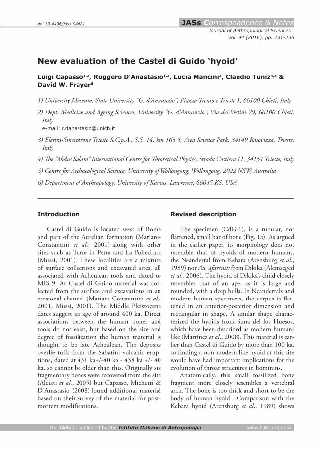
JASs Correspondence & Notes Journal of Anthropological Sciences the JASs is published by the Istituto Italiano di Antropologia www.isita-org.com Vol. 94 (2016), pp. 231-235 New evaluation of the Castel di Guido ‘hyoid’ Luigi Capasso 1,2 , Ruggero D’Anastasio 1,2 , Lucia Mancini 3 , Claudio Tuniz 4,5 & David W. Frayer 6 1) University Museum, State University “G. d’Annunzio”, Piazza Trento e Trieste 1, 66100 Chieti, Italy 2) Dept. Medicine and Ageing Sciences, University “G. d’Annunzio”, Via dei Vestini 29, 66100 Chieti, Italy e-mail: [email protected] 3) Elettra-Sincrotrone Trieste S.C.p.A., S.S. 14, km 163.5, Area Science Park, 34149 Basovizza, Trieste, Italy 4) e “Abdus Salam” International Centre for eoretical Physics, Strada Costiera 11, 34151 Trieste, Italy 5) Centre for Archaeological Science, University of Wollongong, Wollongong, 2022 NSW, Australia 6) Department of Anthropology, University of Kansas, Lawrence, 66045 KS, USA Introduction Castel di Guido is located west of Rome and part of the Aurelian formation (Mariani- Constantini et al., 2001) along with other sites such as Torre in Petra and La Polledrara (Mussi, 2001). These localities are a mixture of surface collections and excavated sites, all associated with Acheulean tools and dated to MIS 9. At Castel di Guido material was col- lected from the surface and excavations in an erosional channel (Mariani-Constantini et al., 2001; Mussi, 2001). The Middle Pleistocene dates suggest an age of around 400 ka. Direct associations between the human bones and tools do not exist, but based on the size and degree of fossilization the human material is thought to be late Acheulean. The deposits overlie tuffs from the Sabatini volcanic erup- tions, dated at 431 ka+/-40 ka - 438 ka +/- 40 ka. so cannot be older than this. Originally six fragmentary bones were recovered from the site (Alciati et al., 2005) but Capasso, Michetti & D’Anastasio (2008) found additional material based on their survey of the material for post- mortem modifications. Revised description The specimen (CdG-1), is a tubular, not flattened, small bar of bone (Fig. 1a). As argued in the earlier paper, its morphology does not resemble that of hyoids of modern humans, the Neandertal from Kebara (Arensburg et al., 1989) nor Au. afarensis from Dikika (Alemseged et al., 2006). The hyoid of Dikika’s child closely resembles that of an ape, as it is large and rounded, with a deep bulla. In Neandertals and modern human specimens, the corpus is flat- tened in an anterior-posterior dimension and rectangular in shape. A similar shape charac- terized the hyoids from Sima del los Huesos, which have been described as modern human- like (Martínez et al., 2008). This material is ear- lier than Castel di Guido by more than 100 ka, so finding a non-modern-like hyoid at this site would have had important implications for the evolution of throat structures in hominins. Anatomically, this small fossilized bone fragment more closely resembles a vertebral arch. The bone is too thick and short to be the body of human hyoid. Comparison with the Kebara hyoid (Arensburg et al., 1989) shows doi 10.4436/jass.94021
Transcript of New evaluation of the Castel di Guido ‘hyoid’ · Castel di Guido is located west of Rome and...
JASs Correspondence & NotesJournal of Anthropological Sciences
the JASs is published by the Istituto Italiano di Antropologia www.isita-org.com
Vol. 94 (2016), pp. 231-235
New evaluation of the Castel di Guido ‘hyoid’
Luigi Capasso1,2, Ruggero D’Anastasio1,2, Lucia Mancini3, Claudio Tuniz4,5 & David W. Frayer6
1) University Museum, State University “G. d’Annunzio”, Piazza Trento e Trieste 1, 66100 Chieti, Italy
2) Dept. Medicine and Ageing Sciences, University “G. d’Annunzio”, Via dei Vestini 29, 66100 Chieti, Italy e-mail: [email protected]
3) Elettra-Sincrotrone Trieste S.C.p.A., S.S. 14, km 163.5, Area Science Park, 34149 Basovizza, Trieste, Italy
4) The “Abdus Salam” International Centre for Theoretical Physics, Strada Costiera 11, 34151 Trieste, Italy
5) Centre for Archaeological Science, University of Wollongong, Wollongong, 2022 NSW, Australia
6) Department of Anthropology, University of Kansas, Lawrence, 66045 KS, USA
Introduction
Castel di Guido is located west of Rome and part of the Aurelian formation (Mariani-Constantini et al., 2001) along with other sites such as Torre in Petra and La Polledrara (Mussi, 2001). These localities are a mixture of surface collections and excavated sites, all associated with Acheulean tools and dated to MIS 9. At Castel di Guido material was col-lected from the surface and excavations in an erosional channel (Mariani-Constantini et al., 2001; Mussi, 2001). The Middle Pleistocene dates suggest an age of around 400 ka. Direct associations between the human bones and tools do not exist, but based on the size and degree of fossilization the human material is thought to be late Acheulean. The deposits overlie tuffs from the Sabatini volcanic erup-tions, dated at 431 ka+/-40 ka - 438 ka +/- 40 ka. so cannot be older than this. Originally six fragmentary bones were recovered from the site (Alciati et al., 2005) but Capasso, Michetti & D’Anastasio (2008) found additional material based on their survey of the material for post-mortem modifications.
Revised description
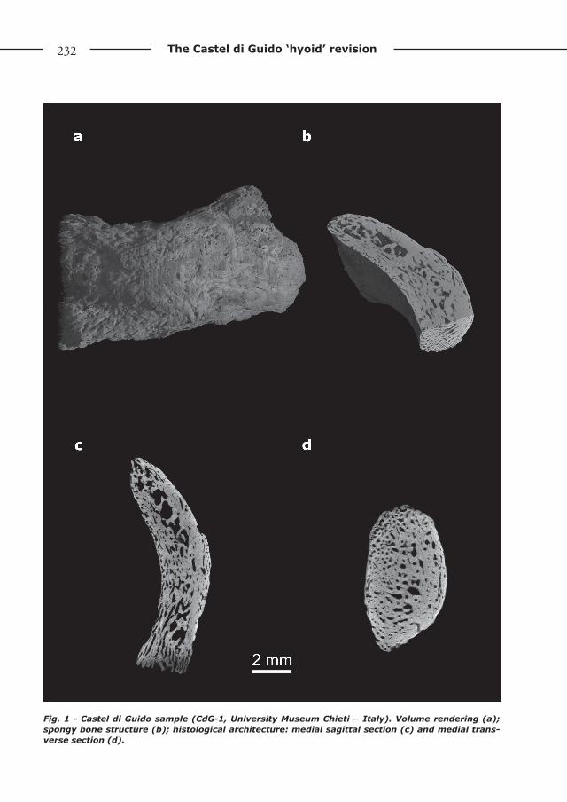
The specimen (CdG-1), is a tubular, not flattened, small bar of bone (Fig. 1a). As argued in the earlier paper, its morphology does not resemble that of hyoids of modern humans, the Neandertal from Kebara (Arensburg et al., 1989) nor Au. afarensis from Dikika (Alemseged et al., 2006). The hyoid of Dikika’s child closely resembles that of an ape, as it is large and rounded, with a deep bulla. In Neandertals and modern human specimens, the corpus is flat-tened in an anterior-posterior dimension and rectangular in shape. A similar shape charac-terized the hyoids from Sima del los Huesos, which have been described as modern human-like (Martínez et al., 2008). This material is ear-lier than Castel di Guido by more than 100 ka, so finding a non-modern-like hyoid at this site would have had important implications for the evolution of throat structures in hominins.
Anatomically, this small fossilized bone fragment more closely resembles a vertebral arch. The bone is too thick and short to be the body of human hyoid. Comparison with the Kebara hyoid (Arensburg et al., 1989) shows
doi 10.4436/jass.94021
232 The Castel di Guido ‘hyoid’ revision
Fig. 1 - Castel di Guido sample (CdG-1, University Museum Chieti – Italy). Volume rendering (a); spongy bone structure (b); histological architecture: medial sagittal section (c) and medial trans-verse section (d).

www.isita-org.com
233L. Capasso et al.
Fig. 2 - Homo sapiens hyoid (Canne della Battaglia T58, University Museum Chieti - Italy). Volume rendering (a); spongy bone structure (b); histological architecture: medial sagittal section (c) and medial transverse section (d).

234 The Castel di Guido ‘hyoid’ revision
that it is approximately three times thicker at the base and shorter in height by more than one-third. Comparisons to a modern human hyoid (Fig. 2) reveal similar results, with the modern hyoid thinner in the anterior-poste-rior view. CdG-1 is a stocky bone, unlike the Kebara and modern human hyoids, which are long and thin. Part of the reason it was identi-fied as a hyoid corpus is the presence of a prom-inent crest on the anterior face (Capasso et al., 2008). In fact the whole face is swollen into a tubercle, which is positioned lower than in the Kebara or modern human hyoids. The anterior face lacks the depressions lateral to the tubercle, typical of modern human and Kebara hyoids. On the anterior face of Kebara’s hyoid the fos-sae for the geniohyoid muscles are deep and pro-nounced. Nothing similar appears in Castel di Guido bone. Unlike Kebara, the superior margin does not form a shelf, but has a rounded margin. Even young individuals have a superior border, which forms a shelf that extends back a few mil-limeters and definitely makes a break with the anterior face. In Castel di Guido the bone surface does not angle posteriorly in the superior-most aspect, but forms a margin of a few millimeters, then it drops vertically and inferiorly. Viewed superiorly one can look down the anterior and posterior face and follow bone surfaces. In the Kebara and modern human hyoids (including young individuals) the interior face is obscured by the superior shelf.
The overall morphology best matches the posterior arch of the atlas and the tubercle is the attachment for ligamentum nuchae. The bone fragment does not appear to be the anterior rim, since its internal surface lacks the dens articu-lar facet. Corroboration of this new assessment comes from microtomographic sections, which show CdG1 to be very different in basic details from a modern hyoid bone.
A modern human hyoid of an adult male from Canne della Battaglia (T58) and the CdG-1 bone (both stored in the University Museum of Chieti, Italy) were analysed by X-ray computed microtomography (Tab. 1) at the TomoLab sta-tion of the Elettra Synchrotron Light Laboratory in Trieste (Italy). The TomoLab station is based on a microfocus source which guarantees a mini-mum focal spot size of 5 microns, in an energy range from 40-130 kV and a maximum cur-rent of 300 µA. A set of 2D slices was recon-structed by using the commercial software COBRA (Exxim) from tomographic projections acquired by the detector (a water-cooled, 12bit, 4008x2672 pixels CCD camera with a pixel size of 12.5×12.5 mm2) during a full sample rotation (360 degrees). Volume renderings of the sam-ples were obtained by the commercial software VGStudio MAX 2.0©.
Scanned images of the CdG-1 show that it is mostly composed of cancellous bone with a moderate cortical cover. This is very different from the modern human hyoid body, that is primarily a thin plate of cortical bone with very thin cancellous bone and well developed inter-trabecular spaces (Figs. 2b-d). Besides, the body of the modern human hyoid presents many thin trabeculae that form a complicated net, more evident in the lateral regions of the hyoid’s body. The Castel di Guido bone has a thick cancellous bone and covered with cortical bone, narrow inter-trabecular spaces and an irregular orienta-tion of the bony trabeculae (Figs. 1b-d). So, it is a thick bone, metrically different from hyoid bones of modern humans, Kebara Neanderthal or Sima de los Huesos hominins (Arensburg et al., 1989; Capasso et al., 2008; Martínez et al., 2008).
SAMPLE T58 CdG-1
VOLTAGE 80 kV 100 kV
CURRENT 100 µA 80 µA
NUMBER OF PROJECTIONS 2400 2400
EXPOSURE TIME / PROJECTION
5.5 sec 2.8 sec
FILTER 1 mm Al 1 mm Al
CUBIC VOXEL WITH SIDE 10.7 micron
10.0 micron
Tab. 1 - X-ray computed microtomography set-up.

www.isita-org.com
235L. Capasso et al.
Conclusions
The Castel di Guido specimen has an exter-nal and micro-structural anatomy that is very different from extant and fossil hyoid bones. It is better identified as the posterior rim of the first cervical vertebra.
References
Alciati G., Pesce Delfino V. & Vacca E. 2005. Catalogue of Italian Fossil Human Remains from the Palaeolithic to the Mesolithic. J. Anthropol. Sci. (Suppl), 83: 1-184.
Alemseged Z., Spoor F., Kimbel W.H., Bobe R., Geraads D., Reed D. & Wynne J.W. 2006. A juvenile early hominin skeleton from Dikika, Ethiopia. Nature, 443: 296-301.
Arensburg B., Tillier A., Vandermeersch B., Duday H., Schepartz L. & Rak Y. 1989. A Middle
Palaeolothic human hyoid bone. Nature, 338: 758-710.
Capasso L., Michetti E. & D’Anatasio R. 2008. A Homo Erectus Hyoid Bone: Possible Implications for the Origin of the Human Capability for Speech. Coll. Antropol., 4: 1007-1011.
Mariani-Constantini R., Ottini L., Carmiello S., Palmirotta R., Mallegni F., Rossi A., Frati L. & Capasso L. 2001. Taphonomy of the fossil hominid bones from the Acheulean site of Castel di Guido near Rome, Italy. J. Hum. Evol., 41: 211-225.
Martínez I., Arsuaga J.L., Quinn R., Carretero J.K., Gracia A. & Rodríguez L. 2008. Human hyoid bones from the middle Pleistocene site of the Sima de los Huesos (Sierra de Atapuerca, Spain). J. Hum. Evol., 54: 118-124.
Mussi M. 2001. Earliest Italy. Kluwer Academic/Plenum, New York.
Editor, Giovanni Destro Bisol
This work is distributed under the terms of a Creative Commons Attribution-NonCommercial 4.0
Unported License http://creativecommons.org/licenses/by-nc/4.0/