Neurotransmitter release through the V0 sector of V … Neurotransmitter release through the V0...
4
q 2001 International Society for Neurochemistry, Journal of Neurochemistry, 79, 485–488 485 Journal of Neurochemistry, 2001, 79, 485–488 MINI-REVIEW Neurotransmitter release through the V0 sector of V-ATPase N. Morel,* Y. Dunant² and M. Israe ¨l* *Laboratoire de Neurobiologie Cellulaire et Mole ´culaire, CNRS, Gif sur Yvette, France ²De ´partement de Pharmacologie, CMU, Geneva, Switzerland Neurotransmitter release occurs at specialized areas of the nerve terminal membrane, the active zones, where clusters of synaptic vesicles, the neurotransmitter-storing organelles, are observed (Couteaux and Pe ´cot-Dechavassine 1974; Harlow et al. 2001). In resting conditions, a population of synaptic vesicles is docked to the active zone membrane, close to voltage-gated calcium channels (Robitaille et al. 1990), within microdomains where, upon stimulation, cytosolic calcium reaches transiently a very high concentra- tion (Llinas et al. 1992). In spite of the high specialization of the active zone structure and high speed of synaptic transmission, proteins involved in docking and fusion of synaptic vesicles are similar to those operating for much slower membrane fusions, from yeast to neurones (Wickner and Haas 2000). In this respect, the role of SNARE complexes for docking synaptic vesicles at the active zones has been well documented (Rothman 1994; Jahn and Su ¨dhof 1999). A detailed genetic and pharmacological dissection of yeast homotypic vacuole fusion revealed the existence, after vacuole docking by trans-SNARE complex formation, of a Ca 21 /calmodulin reaction preceeding the final microcystin- inhibited step of membrane fusion (Wickner and Haas 2000). Recently, Peters et al. (2001) showed that it was the proteolipids of the membrane sector (V0) of V-ATPase which bind to calmodulin and initiate the final step of membrane fusion. The vacuolar-type H 1 -ATPase is indeed composed of a proteolipid membrane sector (V0) and a catalytic sector (V1). The association between V0 and V1 is reversible and participates in the regulation of proton pumping (Nelson and Harvey 1999). Reconstituted V0 pro- teolipids form a pore that opens in the presence of calcium and calmodulin. During the fusion of two yeast vacuoles, a V0 trans-complex is formed by the apposition of two proteolipid rings, brought into close contact by the SNARE proteins. The V0 trans-complex may therefore form a pro- teolipid channel spanning the two interacting membranes at the fusion site (Peters et al. 2001). We would like to discuss the relevance of this model for neurotransmitter release. Synaptosomal membranes were shown to contain a proteolipid oligomer that supported a calcium-dependent release of acetylcholine (ACh) when reconstituted in artificial membranes (Israe ¨l et al. 1986; see Fig. 1). This oligomer (mediatophore) turned out to be made of the proteolipid c subunit of V-ATPase (Birman et al. 1990). When cells were transfected for this proteolipid, they acquired a Ca 1 -dependent ACh release mechanism that displayed quantal properties (Falk-Vairant et al. 1996; see Fig. 2). Such reconstitution experiments, using liposomes, transfected cells or Xenopus oocytes (Cavalli et al. 1993), showed that a single proteolipid ring not only opens upon calcium action but is sufficient to let ACh out down its concentration gradient. This was confirmed by Peters et al. (2001) who measured the release of choline through reconstituted yeast V-ATPase proteolipids, release that required Ca 21 and, in this case, calmodulin. In synapses, the neurotransmitter is pre-concentrated in synaptic vesicles. This process depends on the proton gradient generated by the V-ATPase, and is blocked by N-N 0 -dicyclohexylcarbodiimide (DCCD). In contrast, the efflux of ACh from already loaded synaptic vesicles is not affected by DCCD (Dolezal et al. 1993). This illustrates that ACh and protons follow different routes. Protons bind to a glutamic residue facing the exterior of the proteolipid ring (Harrison et al. 2000) and are translocated during the ATP- driven rotation of this ring (see Nelson and Harvey 1999 for a review on V-ATPases). ACh is most probably released through a pore found in the middle of the proteolipid oligomer by Jones et al. (1995). Received June 13, 2001; revised manuscript received August 8, 2001; accepted August 10, 2001. Address correspondence and reprint requests to N. Morel, Labora- toire de Neurobiologie Cellulaire et Mole ´culaire, CNRS, 91198 Gif sur Yvette, France. E-mail: [email protected] Abbreviations: ACh, acetylcholine; DCCD, N-N 0 -dicyclohexyl- carbodiimide.
Transcript of Neurotransmitter release through the V0 sector of V … Neurotransmitter release through the V0...

q 2001 International Society for Neurochemistry, Journal of Neurochemistry, 79, 485±488 485
Journal of Neurochemistry, 2001, 79, 485±488
MINI-REVIEW Neurotransmitter release through the V0 sector of
V-ATPase
N. Morel,* Y. Dunant² and M. IsraeÈl*
*Laboratoire de Neurobiologie Cellulaire et MoleÂculaire, CNRS, Gif sur Yvette, France
²DeÂpartement de Pharmacologie, CMU, Geneva, Switzerland
Neurotransmitter release occurs at specialized areas of the
nerve terminal membrane, the active zones, where clusters
of synaptic vesicles, the neurotransmitter-storing organelles,
are observed (Couteaux and PeÂcot-Dechavassine 1974;
Harlow et al. 2001). In resting conditions, a population of
synaptic vesicles is docked to the active zone membrane,
close to voltage-gated calcium channels (Robitaille et al.
1990), within microdomains where, upon stimulation,
cytosolic calcium reaches transiently a very high concentra-
tion (Llinas et al. 1992). In spite of the high specialization of
the active zone structure and high speed of synaptic
transmission, proteins involved in docking and fusion of
synaptic vesicles are similar to those operating for much
slower membrane fusions, from yeast to neurones (Wickner
and Haas 2000). In this respect, the role of SNARE
complexes for docking synaptic vesicles at the active
zones has been well documented (Rothman 1994; Jahn
and SuÈdhof 1999).
A detailed genetic and pharmacological dissection of
yeast homotypic vacuole fusion revealed the existence, after
vacuole docking by trans-SNARE complex formation, of a
Ca21/calmodulin reaction preceeding the ®nal microcystin-
inhibited step of membrane fusion (Wickner and Haas
2000). Recently, Peters et al. (2001) showed that it was the
proteolipids of the membrane sector (V0) of V-ATPase
which bind to calmodulin and initiate the ®nal step of
membrane fusion. The vacuolar-type H1-ATPase is indeed
composed of a proteolipid membrane sector (V0) and a
catalytic sector (V1). The association between V0 and V1 is
reversible and participates in the regulation of proton
pumping (Nelson and Harvey 1999). Reconstituted V0 pro-
teolipids form a pore that opens in the presence of calcium
and calmodulin. During the fusion of two yeast vacuoles, a
V0 trans-complex is formed by the apposition of two
proteolipid rings, brought into close contact by the SNARE
proteins. The V0 trans-complex may therefore form a pro-
teolipid channel spanning the two interacting membranes at
the fusion site (Peters et al. 2001). We would like to discuss
the relevance of this model for neurotransmitter release.
Synaptosomal membranes were shown to contain a
proteolipid oligomer that supported a calcium-dependent
release of acetylcholine (ACh) when reconstituted in
arti®cial membranes (IsraeÈl et al. 1986; see Fig. 1). This
oligomer (mediatophore) turned out to be made of the
proteolipid c subunit of V-ATPase (Birman et al. 1990).
When cells were transfected for this proteolipid, they
acquired a Ca1-dependent ACh release mechanism that
displayed quantal properties (Falk-Vairant et al. 1996; see
Fig. 2). Such reconstitution experiments, using liposomes,
transfected cells or Xenopus oocytes (Cavalli et al. 1993),
showed that a single proteolipid ring not only opens upon
calcium action but is suf®cient to let ACh out down its
concentration gradient. This was con®rmed by Peters et al.
(2001) who measured the release of choline through
reconstituted yeast V-ATPase proteolipids, release that
required Ca21 and, in this case, calmodulin.
In synapses, the neurotransmitter is pre-concentrated in
synaptic vesicles. This process depends on the proton
gradient generated by the V-ATPase, and is blocked by
N-N 0-dicyclohexylcarbodiimide (DCCD). In contrast, the
ef¯ux of ACh from already loaded synaptic vesicles is not
affected by DCCD (Dolezal et al. 1993). This illustrates that
ACh and protons follow different routes. Protons bind to a
glutamic residue facing the exterior of the proteolipid ring
(Harrison et al. 2000) and are translocated during the ATP-
driven rotation of this ring (see Nelson and Harvey 1999 for
a review on V-ATPases). ACh is most probably released
through a pore found in the middle of the proteolipid
oligomer by Jones et al. (1995).
Received June 13, 2001; revised manuscript received August 8, 2001;
accepted August 10, 2001.
Address correspondence and reprint requests to N. Morel, Labora-
toire de Neurobiologie Cellulaire et MoleÂculaire, CNRS, 91198 Gif sur
Yvette, France. E-mail: [email protected]
Abbreviations: ACh, acetylcholine; DCCD, N-N 0-dicyclohexyl-
carbodiimide.

At the active zone, in addition to synaptic vesicles
(Yamagata and Parsons 1989), the proteolipid rings are
present in the pre-synaptic membrane. Using the fracture-
label technique (Taubenblatt et al. 1999), we observed the
presence of proteolipid subunits of V-ATPase in the plasma-
lemma, particularly at the site of contact between docked
synaptic vesicles and the synaptosomal membrane. Strick-
ingly, the V0 proteolipid (mediatophore) in the pre-synaptic
membrane is associated with large intramembrane particles
(Fig. 3). The number of such large intramembrane particles
abruptly increases at the peak of ACh release (Muller et al.
1987), when the fusion pore is supposed to open. Similar
particles also increase in number during release in cells
transfected with the V0 proteolipid (Bugnard et al. 1999).
In addition, large intramembrane particles are stably present
in the active zone membrane, organized in relation with
docked synaptic vesicles (Heuser et al. 1979; Harlow et al.
2001). Besides pre-synaptic Ca21 channels, the large V0
trans-complex identi®ed by Peters et al. (2001) could well
be constituents of a population of active zone intramem-
brane particles.
The two proteolipid rings, i.e. the vesicular V0 and the
membrane V0 (mediatophore), would still have to meet to
form the fusion pore. The V0 domain of V-ATPase interacts
in synaptic vesicles with the v-SNARE VAMP (Galli et al.
1996), and with the t-SNARE syntaxin at the plasma mem-
brane (Shiff et al. 1996). It is therefore possible, as suggested
by Peters et al. (2001) in yeast experiments, that, at the active
zone also, the trans-SNARE complex acts by facilitating the
pairing of the two opposite proteolipid rings; rather than
forming the fusion pore itself (Weber et al. 1998). This
would explain the inhibitory effect of SNARE knock-outs
on the synchronized evoked transmitter release, while spon-
taneous release is only reduced (Broadie et al. 1995).
Similarly, clostridial neurotoxins by cleaving the SNARE
proteins block synaptic transmission while desynchronized
quantal release persists (Dunant et al. 1987; Molgo et al.
1990).
In contrast to the collapse of synaptic vesicles in the
plasma membrane, the kiss-and-run concept states that
synaptic vesicles brie¯y interact with the plasma membrane
and are retrieved at the same site (Fesce et al. 1994). In this
process, transmitter release would occur without full fusion
(Neher 1993) through a transient fusion pore (Almers and
Tse 1990). Possibly, according to cytosolic Ca21 concentra-
tion, the V0 trans-complex fusion pore may either ¯icker
and close, or expand to full fusion (AleÁs et al. 1999). In
Fig. 1 SDS-gels (a) comparing total synaptosomal proteins (lane 1)
and puri®ed proteolipid V0 (mediatophore) subunit (lane 2). (b) Puri-
®ed mediatophore (arrows) in its lipidic membrane environment. (c)
Calcium-dependent acetylcholine (ACh) release through puri®ed
mediatophore reconstituted in proteoliposomes.
Fig. 2 Transfection of N18-TG2 neuroblastoma cells with a plasmid
encoding for Torpedo mediatophore. The western blot shows that
the transfected cells (T) expressed in their membranes the Torpedo
proteolipid (detected with monoclonal antibody 15K1; C: control
cells). Acetylcholine release from stimulated N18-TG2 cells, loaded
with acetylcholine, was detected using a patch-clamped Xenopus
myocyte (upper panel). Traces compare a control cell which does
not release transmitter and a transfected cell that displays release
with clear quantal jumps.
486 N. Morel et al.
q 2001 International Society for Neurochemistry, Journal of Neurochemistry, 79, 485±488
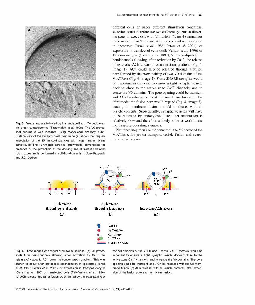
different cells or under different stimulation conditions,
secretion could therefore use two different systems, a ¯icker-
ing pore, or exocytosis with full fusion. Figure 4 summarizes
three modes of ACh release. After proteolipid reconstitution
in liposomes (IsraeÈl et al. 1986; Peters et al. 2001), or
expression in transfected cells (Falk-Vairant et al. 1996) or
Xenopus oocytes (Cavalli et al. 1993), V0 proteolipids form
hemichannels allowing, after activation by Ca21, the release
of cytosolic ACh down its concentration gradient (Fig. 4,
image 1). ACh could also be released through a fusion
pore formed by the trans-pairing of two V0 domains of the
V-ATPase (Fig. 4, image 2). Trans-SNARE complex would
be important in this case to ensure a tight synaptic vesicle
docking close to the active zone Ca21 channels, and to
center the V0 domains. The pore opening could be transient
and ACh be released without full membrane fusion. In the
third mode, the fusion pore would expand (Fig. 4, image 3),
leading to membrane fusion and ACh release, with all
vesicle contents. Subsequently, synaptic vesicles will have
to be reformed by endocytosis. The latter mechanism is
relatively slow and therefore unlikely to be at work in the
most rapidly operating synapses.
Neurones may then use the same tool, the V0 sector of the
V-ATPase, for proton transport, vesicle fusion and neuro-
transmitter release.
Fig. 3 Freeze fracture followed by immunolabelling of Torpedo elec-
tric organ synaptosomes (Taubenblatt et al. 1999). The V0 proteo-
lipid subunit c was localized using monoclonal antibody 15K1.
Surface view of the synaptosomal membrane (a) shows the frequent
association of the 15 nm gold particles with large intramembrane
particles. (b) The 15 nm gold particles (arrowheads) demonstrate the
presence of the proteolipid at the docking site of synaptic vesicles
(SV). Experiments performed in collaboration with T. Gulik-Krzywicki
and J.C. Dedieu.
Fig. 4 Three modes of acetylcholine (ACh) release. (a) V0 proteo-
lipids form hemichannels allowing, after activation by Ca21, the
release of cytosolic ACh down its concentration gradient. This was
shown to occur after proteolipid reconstitution in liposomes (IsraeÈ l
et al. 1986; Peters et al. 2001), or expression in Xenopus oocytes
(Cavalli et al. 1993) or transfected cells (Falk-Vairant et al. 1996).
(b) ACh release through a fusion pore formed by the trans-pairing of
two V0 domains of the V-ATPase. Trans-SNARE complex would be
important to ensure a tight synaptic vesicle docking close to the
active zone Ca21 channels, and to centre the V0 domains. The pore
opening could be transient and ACh be released without full mem-
brane fusion. (c) ACh release, with all vesicle contents, after expan-
sion of the fusion pore and membrane fusion.
Neurotransmitter release through the V0 sector of V-ATPase 487
q 2001 International Society for Neurochemistry, Journal of Neurochemistry, 79, 485±488

References
AleÁs E., Tabares L., Poyato J. M., Valero V., Lindau M. and Alvarez de
Toledo G. (1999) High calcium concentrations shift the mode of
exocytosis to the `kiss and run' mechanism. Nature Cell Biol. 1,
40±44.
Almers W. and Tse F. W. (1990) Transmitter release from synapses:
does a pre-assembled fusion pore initiate exocytosis? Neuron 4,
813±818.
Birman S., Meunier F.-M., Lesbats B., Le Caer J. P., Rossier J. and
IsraeÈl M. (1990) A 15-kDa proteolipid found in mediatophore
preparations from Torpedo electric organ presents high sequence
homology with the bovine chromaf®n granule protonophore.
FEBS Lett. 261, 303±306.
Broadie K., Prokop A., Bellen H. J., O'Kane C. J., Schulze K. I. and
Sweeney S. T. (1995) Syntaxin and synaptobrevin function
downstream of vesicle docking in Drosophila. Neuron 15,
663±673.
Bugnard E., Sors P., Roulet E., Bloc A., Loctin F. and Dunant Y. (1999)
Morphological changes related to reconstituted acetylcholine
release in a release-de®cient cell line. Neuroscience 94, 329±338.
Cavalli A., Dunant Y., Leroy C., Meunier F. M., Morel N. and IsraeÈl M.
(1993) Antisense probes against mediatophore block transmitter
release in oocytes primed with neuronal mRNAs. Eur. J. Neurosci.
5, 1539±1544.
Couteaux R. E. and PeÂcot-Dechavassine M. (1974) Les zones speÂcial-
iseÂes des membranes preÂsynaptiques. C. R. Acad. Sci. Paris 278,
291±293.
Dolezal V., Sbia M., Diebler M.-F., Varoqui H. and Morel N. (1993)
Effect of N,N 0-dicyclohexylcarbodiimide on compartmentation
and release of newly synthesized and preformed acetylcholine in
Torpedo synaptosomes. J. Neurochem. 61, 1454±1460.
Dunant Y., Esquerda J. E., Loctin F., Marsal J. and Muller D. (1987)
Botulinum toxin inhibits quantal acetylcholine release and energy
metabolism in the Torpedo electric organ. J. Physiol. 385,
677±692.
Falk-Vairant J., CorreÁges P., Eder-Colli L., Salem N., Roulet E., Bloc
A., Meunier F., Lesbats B., Loctin F., Synguelakis M., IsraeÈl
M. and Dunant Y. (1996) Quantal acetylcholine release induced
by mediatophore transfection. Proc. Natl. Acad. Sci. USA 93,
5203±5207.
Fesce R., Grohovaz F., Valtorta F. and Meldolesi J. (1994) Neuro-
transmitter release: fusion or `kiss and run'. Trends Cell Biol. 4,
1±4.
Galli T., McPherson P. S. and De Camilli P. (1996) The V0 sector of the
V-ATPase, synaptobrevin and synaptophysin are associated on
synaptic vesicles in a Triton X-100 resistant, freeze±thaw sensi-
tive complex. J. Biol. Chem. 271, 2193±2198.
Harlow M. L., Ress D., Stoschek A., Marshall R. M. and McMahan U. J.
(2001) The architecture of the active zone material at the frog's
neuromuscular junction. Nature 409, 479±484.
Harrison M., Powell B., Finbow M. E. and Findlay J. B. C. (2000)
Identi®cation of lipid-accessible sites on the Nephrops 16 kDa
proteolipid incorporated into a hybrid vacuolar H1-ATPase.
Biochemistry 39, 7531±7537.
Heuser J. E., Reese T. S., Dennis M. J., Jan Y., Jan L. and Evans L.
(1979) Synaptic vesicle exocytosis captured by quick freezing
and correlated with quantal transmitter release. J. Cell Biol. 81,
275±300.
IsraeÈl M., Morel N., Lesbats B., Birman S. and Manaranche R. (1986)
Puri®cation of a pre-synaptic membrane protein that mediates a
calcium-dependent translocation of acetylcholine. Proc. Natl.
Acad. Sci. USA 83, 9226±9230.
Jahn R. and SuÈdhof T. C. (1999) Membrane fusion and exocytosis.
Annu. Rev. Biochem. 68, 863±911.
Jones P. C., Harrison M. A., Kim Y. I., Finbow M. E. and Findlay J. B.
C. (1995) The ®rst putative transmembrane helix of the 16-kDa
proteolipid lines a pore in the V0 sector of the vacuolar H1-
ATPase. Biochem. J. 312, 739±747.
Llinas R., Sugimori M. and Silver R. B. (1992) Microdomains of high
calcium concentration in a pre-synaptic terminal. Science 256,
677±679.
Molgo J., Comella J. X., Angaut-Petit D., Pecot-Dechavassine M., Tabti
N., Faille L., Mallart A. and Thesleff S. (1990) Presynaptic actions
of botulinal neurotoxins at vertebrate neuromuscular junctions.
J. Physiol. 84, 152±166.
Muller D., Garcia-Segura L. M., Parducz A. and Dunant Y. (1987) Brief
occurrence of a population of pre-synaptic intramembrane
particles coincides with transmission of a nerve impulse. Proc.
Natl. Acad. Sci. USA 84, 590±594.
Neher E. (1993) Secretion without full fusion. Nature 363, 497±498.
Nelson N. and Harvey W. R. (1999) Vacuolar and plasma membrane
proton-adenosinetriphosphatases. Physiol. Rev. 79, 361±385.
Peters C., Bayer M. J., BuÈhler S., Andersen J. S., Mann M. and Mayer
A. (2001) Trans-complex formation by proteolipid channels in the
terminal phase of membrane fusion. Nature 409, 581±588.
Robitaille R., Adler E. M. and Charlton M. P. (1990) Strategic location
of calcium channels at transmitter release sites of frog neuro-
muscular synapse. Neuron 5, 773±779.
Rothman J. E. (1994) Mechanisms of intracellular protein transport.
Nature 372, 55±63.
Shiff G., Synguelakis M. and Morel N. (1996) Association of syntaxin
with SNAP 25 and VAMP (synaptobrevin) in Torpedo synapto-
somes. Neurochem. Int. 29, 659±667.
Taubenblatt P., Dedieu J. C., Gulik-Krzywicki T. and Morel N. (1999)
VAMP (synaptobrevin) is present in the plasma membrane of
nerve terminals. J. Cell Sci. 112, 3559±3567.
Weber T., Zemelman B. V., McNew J. A., Westermann B., Gmachl M.,
Parlati F., SoÈllner T. H. and Rothman J. E. (1998) Minimal
machinery for membrane fusion. Cell 92, 759±772.
Wickner W. and Haas A. (2000) Yeast homotypic vacuole fusion: a
window on organelle traf®cking mechanisms. Annu. Rev. Biochem.
69, 247±275.
Yamagata S. K. and Parsons S. M. (1989) Cholinergic synaptic vesicles
contain a V-type and a P-type ATPase. J. Neurochem. 53,
1354±1362.
488 N. Morel et al.
q 2001 International Society for Neurochemistry, Journal of Neurochemistry, 79, 485±488