Neurotransmission. Receptors and signal transduction ... · 2019. 09. 11. 2 6. Receptors, signal...
25
2019. 09. 11. 1 Neurotransmission. Receptors and signal transduction mechanisms. Learning objectives 6. and 7. Dr. Gabriella Kékesi 2019 7. Neurotransmission. • Characterize electric synapses including the description of the molecular structure of gap junction operating in these synapses. Compare transmission between electric and chemical synapses (direction of information, speed and way of transmission). • Describe the consecutive events of chemical neurotransmission (starting with the depolarization of presynaptic membrane ending with the development of the graded electric response of the postsynaptic membrane (postsynaptic potential, PSP). Describe the ion currents involved in the development of the following local potentials: excitatory postsynaptic potential (EPSP), inhibitory postsynaptic potential (IPSP), end plate potential (EPP). • Describe the common features of classical neurotransmitters. • Group the neurotransmitters based on their chemical structure: 1. acetyl- choline, 2. amino acids (glutamate, glycine, GABA), 3. biogenic amines (dopamine, noradrenaline, adrenaline, histamine, serotonin), 4. gases (NO, CO), 5. lipids (endocannabinoids, 6. peptides (endophins, encephalins, dynorphins, substance P, CGRP, VIP), 7. purines. Describe the synthesis, mechanism of action and significance of NO. • Describe the fate of released neurotransmitters: receptor binding, enzymatic degradation, diffusion, reuptake. Normal values: synaptic delay: 1-1.5 ms
Transcript of Neurotransmission. Receptors and signal transduction ... · 2019. 09. 11. 2 6. Receptors, signal...

2019. 09. 11.
1
Neurotransmission.Receptors and signal transduction
mechanisms.
Learning objectives 6. and 7.
Dr. Gabriella Kékesi
2019
7. Neurotransmission.
• Characterize electric synapses including the description of the molecular structure of gap junction
operating in these synapses. Compare transmission between electric and chemical synapses
(direction of information, speed and way of transmission).
• Describe the consecutive events of chemical neurotransmission (starting with the depolarization
of presynaptic membrane ending with the development of the graded electric response of the
postsynaptic membrane (postsynaptic potential, PSP). Describe the ion currents involved in the
development of the following local potentials: excitatory postsynaptic potential (EPSP),
inhibitory postsynaptic potential (IPSP), end plate potential (EPP).
• Describe the common features of classical neurotransmitters.
• Group the neurotransmitters based on their chemical structure: 1. acetyl- choline, 2. amino acids
(glutamate, glycine, GABA), 3. biogenic amines (dopamine, noradrenaline, adrenaline, histamine,
serotonin), 4. gases (NO, CO), 5. lipids (endocannabinoids, 6. peptides (endophins, encephalins,
dynorphins, substance P, CGRP, VIP), 7. purines. Describe the synthesis, mechanism of action and
significance of NO.
• Describe the fate of released neurotransmitters: receptor binding, enzymatic degradation,
diffusion, reuptake.
Normal values: synaptic delay: 1-1.5 ms

2019. 09. 11.
2
6. Receptors, signal transduction mechanisms.
• Describe the main types of signaling molecules (mediators): autocrine and paracrine signaling
molecules, hormones, neurotransmitters, neurohormones and cytokines.
• Define the terms: receptor, ligand, agonist, antagonist (competitive, non-competitive).
• Classification of receptors: 1. based on localization (cell membrane receptors, cytosolic receptors,
nuclear receptors, intracellular membrane receptors (IP3, ryanodin), 2. based on function
(ionotropic receptors, metabotropic receptors, receptor enzymes, and enzyme-linked receptors).
• Ionotropic receptors: selective and non-selective receptors, cation and anion channels. Give 1-1
examples.
• G-protein coupled metabotropic receptors. Heterotrimer G-proteins: types (Gs/Gi/Gq) and
functions.
• Define the term second messengers, describe the most important members (cAMP, cGMP, calcium,
IP3/DAG).
• Explain the function of receptor enzymes and enzyme-linked receptors through 1-1 example
(tyrosine kinase receptors).
• Describe the following terms related to membrane receptors: activation, inactivation,
internalization, up-regulation, down-regulation, sensitization, desensitization.
• Signal transduction via intracellular receptors: the function of cytosolic and nuclear receptors
explained through 1-1 example (steroid and thyroid hormone receptors).
Communication of cells
• Gap-junction
• Extracellular signaling molecules
• Local or distant/remote origin
• Signal transduction
• Cellular response (e.g. electrical change, modified enzymatic activity,
proliferation, moified transcription…)

2019. 09. 11.
3
Extracellular signalmolecules
• Enchored molecules (A)
– Cell surface molecules of thesurrounding cells; EC matrixmacromolecules…
• Mobile molecules – MEDIATORS -Chemical communication
– Paracrin signaling (B)
– Autocrin signaling
– Endocrine signaling – hormones (D)
– Neurotransmitters & neurohormones (C)

2019. 09. 11.
4
Synapse
• is a structure that permits a neuron to pass an electrical or chemical signal to another cell (neural or otherwise – muscles, glands).
• Neurotransmission
• Presynaptic - refers to components of a synapse specialized for transmitter release
• Postsynaptic - refers to those components designed for transmitter reception
• Classified by the type of cellular structure serving as the postsynaptic target:
– Axodendritic / axosomatic / axoaxonicaxons
Interneuronal synapses
• ~ 2 000 synapses / neuron
• CNS: 1011 neuron
• 2 x 10¹⁴ synapses
• By the type of cellular structure serving as the postsynaptic target
– Axo-dendritic synapses
– Axo-somatic synapses
– Axo-axonal synapses

2019. 09. 11.
5
Electrical synapses
• Direct flow of electrical current at gap junctions
• Voltage sensor
• Ions/glucose, soluble secondmessengers, amino acids passively flow through
• Bidirectional flow of material is possible
• No delay – immediate response
• Regulating rhythmic or synchronized electrical activity (e.g. breathing, heart)
Chemical synapses
• Otto Loewi (Provided experimentalevidence of chemical transmission)
• Pre- and postsynaptic cell membranes
• Synaptic cleft (20-40 nm)
• Neurotransmitter release
• Synaptic delay (1-1.5 ms)
• Localised changes in membranepotential (PSPs)
– EPSP / IPSP

2019. 09. 11.
6
ELECTRICAL SYNAPSE CHEMICAL SYNAPSE
a scanning electron microscope shows an axon terminal that was broken open to reveal synaptic vesicles
Steps of transmitter release

2019. 09. 11.
7
Docking (SNARE) proteins to mediate vesicle fusion: syntaxin, SNAP-25 and synaptobrevin.
Transmitter release
• Dale’s principle: the same neurotransmitter is released from all axons of the neuron
• Co-localisation: cotransmitters
– Ach + VIP; NE + NPY; SP + CGRP …
• Co-release
– frequency-dependent:
• Low-frequency stimulation: classical ntrm. release
• High-frequency stimulation: peptide ntrms. are also released
• Quantal release (Bernard Katz)

2019. 09. 11.
8
Figure 14-6 Neurotransmitter is released in fixed increments, or quanta. Each quantum of transmitter produces a unit postsynaptic potential of fixed amplitude. The amplitude of the postsynaptic potential evoked by nerve stimulation is equal to the unit amplitude multiplied by the number of quanta of transmitter released. (Adapted from Boyd and Martin 1956.)
Quantal release
Fate of released transmitters
1. Binding to specific receptors
2. Diffusion from the synaptic cleft
• Into capillaries
• Into Lymphatic vessels
• To extrasynapticreceptors
3. Enzymatic degradation inthe synaptic cleft
4. Reuptake into thepresynaptic axon terminalor into the gilal cells1.
2.
3.
4.

2019. 09. 11.
9
Presynaptic modifications of neurotransmission
Postsynaptic modifications of neurotransmission

2019. 09. 11.
10
Postsynaptic electric events
• Local, electrotonic potential changes (PSPs)
• EPSP (Excitatory postsynaptic potential); IPSP (inhibitory postsynaptic potential)
The time course of Ca2+ influx in the presynaptic cell determines the onset of synaptic transmission. An action potential in the presynaptic cell (1) causes voltage-gated Ca2+ channels in the terminal to open and a Ca2+ current (2) to flow into the terminal. The Ca2+ influx triggers release of neurotransmitter. The postsynaptic response to the transmitter begins soon afterward (3) and, if sufficiently large (reaches the threshold), will trigger an action potential in the postsynaptic cell (4). (EPSP = excitatory postsynaptic potential.) (Adapted from Llinás 1982.)
Synaptic integrationsummations of PSPs
Temporal summation: PSPs occur one after another at the same site, thereby combining potential in the postsynaptic terminal
Spatial summation: inputs from several presynaptic neurons acting on different areas combine to form an action potential

2019. 09. 11.
11
Presynaptic regulation of transmitter release
an axoaxonic synapse causes an action potential arriving at the axon terminal to release more neurotransmitter and thereby cause a larger EPSP
the neurotransmitter from the presynaptic neuron atthe axonic synapse causes the postsynaptic axonterminal to release less neurotransmitter
Retrograde signalisatione.g. endocannabinoids
• Refers to the process by which a retrograde messenger, such as anandamide or nitric oxide, is released by a postsynaptic dendrite or cell body, and travels "backwards" across a chemical synapse to bind to the axon terminal of a presynaptic neuron

2019. 09. 11.
12
Chemical classification of neurotransmitters
• Classical, small molecule neurotransmitters– Acetylcholine
– Biogenic amines
• Catecholamines: dopamine, epinephrine, norepinephrine
• Serotonine
• Histamine
– Amino acid derivatives
• excitatory: glutamate, aspartate
• inhibitory: GABA (γ-amino-butyric acid) glycin
• Purine nucleotides– ATP, adenosine
• Gases (gasotransmitters)– NO, CO, H2S
• Peptides – neuropeptides (TRH, vazopressine, oxytocin, VIP, opioids, CGRP, SP), hormones, cytokines, growth factors
• Other neurotransmitters– Lipids: prostaglandines, leucotrienes, anandamide
– Steroids (hormones): cortisol, aldosterone, sexual steroids…
Synthesis and metabolism of acetylcholine(Ach)

2019. 09. 11.
13
Cholinergic synapse
Neuromuscular junction (motor endplate
• The axon of a motorneuron (presynaptic) establishes synaptic contact with a striated muscle (postsynaptic) fiber
• Synaptic cleft: 500 nm
• Neurotransmitter: Acetylcholine (Ach)

2019. 09. 11.
14
Synthesis and metabolism of catecholamines
Neurotransmission in adrenergic neurons

2019. 09. 11.
15
Neurotransmission in serotonergic neurons
Synthesis and release of steroids
• Steroids are synthesized and released as needed by enzymes in smooth ER and mitochondria from cholesterol.
• All readily diffuse through cell membranes because of their lipophilic character.
• Steroids cannot be stored in membrane bound vesicles and this is why they are synthesized as needed and released immediately.

2019. 09. 11.
16
Gaseous mediators
Hem
Synthesis and metabolism of peptides
• 1. A prepropeptide is synthesized and released into the rough ER.
• 2. Proteolytic enzymes in the RER cleave off some amino acids to yield propeptides.
• 3. In the smooth ER propeptides are packaged into transport vesicles.
• 4. The vesicles are transported to Golgi complexes.
• 5. Golgi complexes package the propeptide into secretory vesicles. Either in the GA or secretory vesicles more amino acids are cleaved to yield the final peptide.
• 6. The peptides are released by exocytosis.

2019. 09. 11.
17
Lipids - eicosanoids
• Lipophilic
• Released on demand
• Release of arachidonic acid from phospholipids in the cell membrane by phospholipase A2
Transport of chemical messengers
• Simple diffusion (autocrines, paracrines, neurotransmitters) or by blood transport (hormones, neurohormones)
• Hydrophilic messengers travel in blood in dissolved form or bound to carrier proteins (catecholamines)
• Hydrophobic messengers (steroides and thyroid hormones) bound to carrier proteins
• Blood-born messengers are:
– Degraded by the liver or
– Secreted by the kidney
• Dissolved messengers have a shorter half-life than messengers bound to carrier proteins.

2019. 09. 11.
18
Receptors
• Receptor: special protein molecule that binds to specific chemical signals (ligand) from outside the cell resulting some form of cellular/tissue responses
• High affinity, reversible binding
• Structural changes (conformation, dimerizsation)
• Ligand: any kinds of substances, that bind specifically to receptors with biologicalactivity
• Agonist: are able to activate the receptor and result in biological responseinducing signaltransduction
• Antagonist: bind to receptors but do not activate them resulting in receptor blockade, inhibiting the binding of agonists
– Competitive vs non-cmpetitive
• Multiple receptors for most transmitters
• Pre- and/or postsynaptic localisation
• Receptor families
• Desensitisation (Internalisation, phosphorilation)
Classification of receptors
• Based on localisation:
– Cell membrane receptors
– Intracellular receptors
• Cytosolic and nuclear r. (steroids, thyroid hormones)
• Intracellular membrane r. (ryanodin, IP3)
• Based on function:
– Metabotropic (G-protein coupled ; A)
– Enzyme-linked or receptor enzymes(B; insuline receptor))
– Ionotropic (C)

2019. 09. 11.
19
Ionotropic receptors I.
• Tetrameric or pentameric structure
• Receptor is an ion channel
• Ligand binding (1.) – conformational change –ion channel opens (2.) –change in electrical properties (3.)
• Excitatory / Inhibitory
• Fast; brief response
• E.g.:
– GABA A, GABA C– Glycin receptor
– nACh receptor
– NMDA, non-NMDA (AMPA, Kainate) receptor
– Px (ATP) receptor
– 5HT3 receptor

2019. 09. 11.
20
Metabotropic receptors I.
• 7 TM structure
• G-protein coupling
– Gi/Gs/Gq
– Trimer: α,β and γ subunits
– GDP/GTP binding
– GTP-ase activity
• Direct influence of ion channels (activation or inhibition) OR second messenger release

2019. 09. 11.
21
Action mechanisms of G protein
1. regulate a slow ligand-gated ion channel by either opening or closing the channel. This is in contrast to the more direct ligand-gated channels that only open in response to messenger binding. G protein-linked ion channels also take longer to open and stay open longer.
2. by activating or inhibiting an amplifier protein through the GTP- subunit. The amplifier protein catalyzes the synthesis of a second messenger. The second messenger activates a protein kinase that catalyzes the phosphorylation of a protein. This leads to the response in the cell.
Second messenger systems induce signalamplification
1. Enables one molecule to activate an enormous number of proteins.
2. Enables the cell to be very sensitive in response to the presence of messenger even at very low concentrations.
3. a cascade in which there are sequential steps that progressively increase in magnitude.

2019. 09. 11.
22
Metabotropic receptors II.second messengers
• cAMP (cyclic adenosine-monophosphate)
• cGMP (cyclic guanosine-monophosphate)
• IP3 (inositol-triphosphate)
• DAG (diacylglycerol)
• Calcium
• (Arachidonic acid)

2019. 09. 11.
23
Metabotropic receptors III/1.signaling pathways
• Gs/Gi
– Adenylyl-cyclase enzyme (AC) – cAMP – protein kinase A (PKA)• Pl.: α2 (↓) and β1-3 adrenergic receptors (↑); M2,4 acetylcholine receptor (↓);
– Guanylyl-cyclase enzyme (GC) – cGMP – protein kinase G enzyme(PKG)• Pl.: atrial natriuretic peptide (ANP) receptor; NO
Metabotropic receptors III/2.signaling pathways
• Gq
– Phospholipase C enzyme (PLC) – Phosphatidylinositol (4,5)-bisphosphate (PIP2) – Phosphatidylinositol (3,4,5)-trisphosphate (IP3) + diacylglycerol (DAG)
• IP3 – IC calcium release
• DAG – protein kinase C enzyme (PKC)
• Pl.: α1 adrenergic receptor; M1, 3, 5 acetylcholine receptor; mGLUT, H1 receptor, 5HT2 receptor

2019. 09. 11.
24
GsGiGq
Metabotropic receptors IV.examples
• Muscarinic acetylcholine receptors (M1-5)
• Adrenergic receptors (α1-2, β1-3)
• Serotonine receptors (5HT1-2, 4-7)
• Histamine receptors
• GABA-B receptor
• Dopamine receptors
• Metabotropic glutamate receptors
• Adenosine receptors
• Peptide receptors
• Py receptors (ATP)
2019. 09. 11.
25
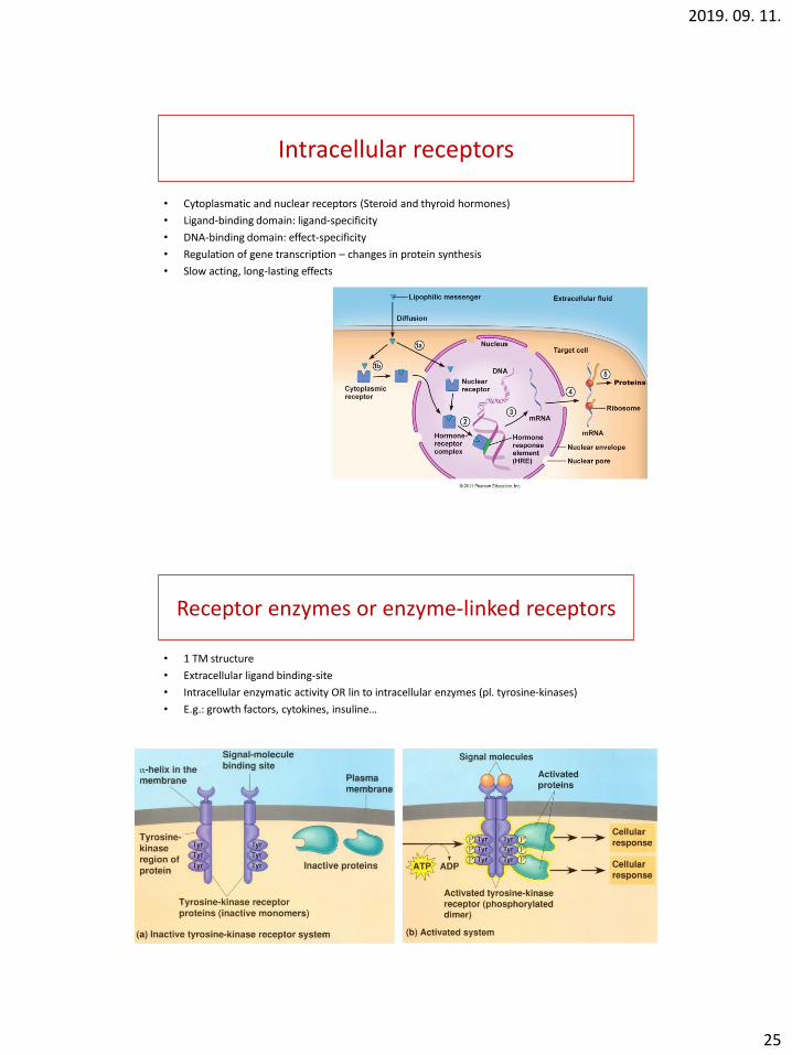
Intracellular receptors
• Cytoplasmatic and nuclear receptors (Steroid and thyroid hormones)
• Ligand-binding domain: ligand-specificity
• DNA-binding domain: effect-specificity
• Regulation of gene transcription – changes in protein synthesis
• Slow acting, long-lasting effects
Receptor enzymes or enzyme-linked receptors
• 1 TM structure
• Extracellular ligand binding-site
• Intracellular enzymatic activity OR lin to intracellular enzymes (pl. tyrosine-kinases)
• E.g.: growth factors, cytokines, insuline…