Neurotoxic Effects of Gibberellic Acid (GA3) and its ...3)15/1.pdf · In conclusion: results of the...
12
Global Journal of Pharmacology 9 (3): 222-233, 2015 ISSN 1992-0075 © IDOSI Publications, 2015 DOI: 10.5829/idosi.gjp.2015.9.3.95134 Corresponding Author: Nadia Ragheb Ali Abou-Zeid, Electron Microscope Lab, Ain Shams Specialized Hospital, Ain Shams University, Cairo, Egypt. Tel/Mob: +2 01003679324, E-mail: [email protected]. 222 Neurotoxic Effects of Gibberellic Acid (GA3) and its Withdrawal in Adult Male Albino Rats: A Light and Electron Microscopic Study Nadia R.A. Abou-zeid and Hala F. Abd-Ellah 1 2 Electron Microscope Lab., Ain Shams Specialized Hospital, Ain Shams University, Cairo, Egypt 1 Department of Zoology, Women College for Arts, 2 Science and Education, Ain Shams University, Cairo, Egypt Abstract: The present investigation aims to illustrate the effects of gibberellic acid (GA3) on the histology and ultrastructure of the cerebellum of rats as well as the possibility of recovery after GA3 withdrawal. Rats were equally divided into five groups. Group I served as control. Groups II&III received GA3 daily in drinking water in two gradually increasing doses of 100 and 200 ppm, respectively, for 8 weeks. Groups IV&V received the same treatment of GA3 as the second and third groups then were left without any treatment for another 8 weeks. Light microscopic examination of the cerebellar cortex of GA3-treated animals revealed its prominent neurotoxic effect on Purkinje cells and granular nerve cells in association with vacuolation in the molecular layer. Ultrastructure of the cerebellum of GA3-treated rats confirmed the light microscopic observations. The previous changes were more severe in rats treated with 200 ppm GA3. However, stoppage of GA3 administration for 8 weeks has resulted in some sort of regression of the previously mentioned effects. In conclusion: results of the current study suggested that the toxic effects of GA3 were dose-dependent. While, after GA3 stoppage, 8 weeks period of follow up showed incomplete recovery of these toxic effects. So, gibberellic acid should be used cautionary. Key words: Plant Growth regulators Gibberellic acid Neurotoxicity Rats INTRODUCTION months. The Environmental Protection Agency has Many chemicals are currently used in agriculture People may be exposed to residues of GA3 in diet derived nowadays. One of these chemicals is the plant growth from consumption of different types of fruits and regulators (PGRs) where its use began in 1930s [1]. vegetables treated with GA3. Exposure to residues may PGRs are also known as plant hormones or also be through drinking water [6] Occupational exposure phytohormones. They are chemicals that regulate plant of the agricultural workers to GA3 may occur through growth [2]. Gibberellins are one of the six major classes of inhalation of powder and dermal contact with this plant growth regulators according to the American compound at work places where GA3 is produced or used Society of Agricultural Science [1]. Gibberellic acid giving the picture of acute toxicity [7]. The liver is the (GA3) is one of the most active hormones of gibberellins. main target for the toxicity of several compounds. Recent It affects many mechanisms of plant growth including reports indicate that GA3 may induce oxidative stress, stem elongation by stimulating cell division leading to the generation of free radicals and causing cells flowering, fruit development and breaking dormancy damage in many organs, including the heart, kidney, [3]. Sakr et al., [4] mentioned that gibberellic acid (GA3) stomach and spleen [8] and the liver [9] of adult rats. is used extensively in Egypt and other countries, to However, little information is available regarding the increase the growth of many fruits (such as neurotoxicity effect of this compound. The increasing use strawberries and grapes) and vegetables (such as of this substance in agriculture making it as an interesting tomatoes, cabbages and cauliflower). Gibberellic acid subject to investigate its possible adverse effects on the (GA3) is highly persistent and bioactive in soil for cerebellum. So, the aim of this study was to evaluate determined its use to be only allowed in low doses [5]. .
Transcript of Neurotoxic Effects of Gibberellic Acid (GA3) and its ...3)15/1.pdf · In conclusion: results of the...
Global Journal of Pharmacology 9 (3): 222-233, 2015ISSN 1992-0075© IDOSI Publications, 2015DOI: 10.5829/idosi.gjp.2015.9.3.95134
Corresponding Author: Nadia Ragheb Ali Abou-Zeid, Electron Microscope Lab, Ain Shams Specialized Hospital,Ain Shams University, Cairo, Egypt. Tel/Mob: +2 01003679324, E-mail: [email protected].
222
Neurotoxic Effects of Gibberellic Acid (GA3) and its Withdrawal inAdult Male Albino Rats: A Light and Electron Microscopic Study
Nadia R.A. Abou-zeid and Hala F. Abd-Ellah1 2
Electron Microscope Lab., Ain Shams Specialized Hospital, Ain Shams University, Cairo, Egypt1
Department of Zoology, Women College for Arts, 2
Science and Education, Ain Shams University, Cairo, Egypt
Abstract: The present investigation aims to illustrate the effects of gibberellic acid (GA3) on the histology andultrastructure of the cerebellum of rats as well as the possibility of recovery after GA3 withdrawal. Rats wereequally divided into five groups. Group I served as control. Groups II&III received GA3 daily in drinking waterin two gradually increasing doses of 100 and 200 ppm, respectively, for 8 weeks. Groups IV&V received thesame treatment of GA3 as the second and third groups then were left without any treatment for another 8 weeks.Light microscopic examination of the cerebellar cortex of GA3-treated animals revealed its prominent neurotoxiceffect on Purkinje cells and granular nerve cells in association with vacuolation in the molecular layer.Ultrastructure of the cerebellum of GA3-treated rats confirmed the light microscopic observations. The previouschanges were more severe in rats treated with 200 ppm GA3. However, stoppage of GA3 administration for8 weeks has resulted in some sort of regression of the previously mentioned effects. In conclusion: results ofthe current study suggested that the toxic effects of GA3 were dose-dependent. While, after GA3 stoppage,8 weeks period of follow up showed incomplete recovery of these toxic effects. So, gibberellic acid should beused cautionary.
Key words: Plant Growth regulators Gibberellic acid Neurotoxicity Rats
INTRODUCTION months. The Environmental Protection Agency has
Many chemicals are currently used in agriculture People may be exposed to residues of GA3 in diet derivednowadays. One of these chemicals is the plant growth from consumption of different types of fruits andregulators (PGRs) where its use began in 1930s [1]. vegetables treated with GA3. Exposure to residues mayPGRs are also known as plant hormones or also be through drinking water [6] Occupational exposurephytohormones. They are chemicals that regulate plant of the agricultural workers to GA3 may occur throughgrowth [2]. Gibberellins are one of the six major classes of inhalation of powder and dermal contact with thisplant growth regulators according to the American compound at work places where GA3 is produced or usedSociety of Agricultural Science [1]. Gibberellic acid giving the picture of acute toxicity [7]. The liver is the(GA3) is one of the most active hormones of gibberellins. main target for the toxicity of several compounds. RecentIt affects many mechanisms of plant growth including reports indicate that GA3 may induce oxidative stress,stem elongation by stimulating cell division leading to the generation of free radicals and causing cellsflowering, fruit development and breaking dormancy damage in many organs, including the heart, kidney,[3]. Sakr et al., [4] mentioned that gibberellic acid (GA3) stomach and spleen [8] and the liver [9] of adult rats.is used extensively in Egypt and other countries, to However, little information is available regarding theincrease the growth of many fruits (such as neurotoxicity effect of this compound. The increasing usestrawberries and grapes) and vegetables (such as of this substance in agriculture making it as an interestingtomatoes, cabbages and cauliflower). Gibberellic acid subject to investigate its possible adverse effects on the(GA3) is highly persistent and bioactive in soil for cerebellum. So, the aim of this study was to evaluate
determined its use to be only allowed in low doses [5].
.
Global J. Pharmacol., 9 (3): 222-233, 2015
223
neurotoxic effects of two different doses of GA3 in adult Group IV: Animals of the fourth group (recovery ofmale albino rats for 8 weeks and also to determine the 100 ppm GA3-treated group) received the same treatmenteffects of GA3 withdrawal on the affected organ following as the second group), then were left without any8 weeks of follow up. treatment for another 8 weeks.
MATERIALS AND METHODS Group V: Animals of the fifth group (recovery of 200 ppm
Chemicals: Gibaifar (5% Gibberellic acid) supplied by third group), then were left without any treatment forAIFAR AGROCHIMICA SRL ViaBazzano, 12 16019 another 8 weeks.RoncoScrivia (Genoa) Italy.www.aifar.it.
Preparation of GA3: 2ml and 4ml of 5 % GA3 (equivalent from each group were sacrificed by cervicalto 100 mg and 200 mg of GA3, respectively) were diluted decapitation and cerebellum were excised and immediatelywith tap water until 1000 ml to obtain 100 and 200 ppm of processed for histopathological and ultrastructuralGA3, respectively. studies.
Experimental Animals: Adult male Wistar albino rats Determination of GA3 Content in Cerebellum: A residueweighing 180-200 g were obtained from animal house in of GA3 was estimated in cerebellum tissues by thin layerMedical Research Center (MRC), Faculty of Medicine, chromatography method described by Official MethodsAin Shams University. Animals were kept in stainless of Analysis [13].cages under standard laboratory conditions withtemperature maintained at 25 ± 3°C, relative humidity of Light Microscopic Study: For light microscopic50% ± 5% and 12 h alternating light and dark cycles. examination, the cerebellum tissues were collected from allThe animals were provided with an adequate rat chow diet the animal groups, fixed in aqueous Bouin’s fixative,and drinking tap water ad libitum throughout the study. dehydrated in ascending grades of ethyl alcohol, clearedThe experiment was conducted at the animal house in in xylene, impregnated in paraffin wax and sections ofMedical Research Center (MRC), Faculty of Medicine, 5-7 µm thickness were taken and stained with Harri’sAin Shams University. The experiment was performed in hematoxylin & eosin (H&E) [14] and examined for anyaccordance with the "Guide for the Care and Use of structural changes under light microscope.Laboratory Animals"[10] and was approved by the localEthics Committee. Transmission Electron Microscopic Study: For
Experimental Protocol: After an acclimatization period of pieces of about 1-mm3 in size and immediately fixed in1 week, animals were divided into five groups, 10 rats each 2.5% glutaraldehyde for 24-48 hr. The specimens wereas follows: then washed in phosphate buffer (pH 7.2–7.4) 3-4 times
Group I: Animals of the first group served as control and solution of 1% osmium tetraoxide for 2 hr., after thatwere received tap water. washed in the same buffer 4 times for 20 min. each.
Group II: Animals of the second group received 100 ppm ethyl alcohol (30%, 50%, 70%, 90% and 100%), cleared inof GA3 [11] daily in drinking tap water for eight weeks. two changes of propylene oxide and embedded in
Group III: Animals of the third group were received stained with toluidine blue and examined under light200 ppm of GA3 [9, 12] daily in drinking tap water for eight microscope. The resin blocks were retrimmed to get rid ofweeks. the undesired tissue. Ultrathin sections (60–90 nm thick)
Since all rats have the same physiologic characters, with uranyl acetate and lead citrate [16]. The grids weredaily water consumption of all rats was approximately examined and photographed using a transmission electron30 ± 3ml during the tests. Consequently, the GA3 intake microscope (JEOL JEM-1010, Japan) operated at 60-70 kV,amount of each rat was about 3± 0.3mg and 6± 0.6 mg per Regional Center for Mycology and Biotechnologyday for groups II and III, respectively. (RCMB), Al-Azhar University.
GA3-treated group) received the same treatment as the
On completion of experimental period, animals
ultrastructural examination, cerebellum was cut into small
for 20 min. every times and post-fixed in a buffered
Fixed specimens were dehydrated in ascending grades of
Epon resin [15]. Semi-thin sections (1µm thick) were
were cut, mounted on copper grids and double stained
Global J. Pharmacol., 9 (3): 222-233, 2015
224
RESULTS deposition of Purkinje cells with marked loss in their
Concentrations of GA3 in Cerebellum: The present with the morphological alterations seen in the Purkinjeexperiment showed that, in rats treated with 100 and layer, the molecular layer displayed increased vascularity200 ppm of GA3 (Groups II& III, respectively) the higher with dilatation of the capillaries and prominent spongiosiscontent of GA3 was found in cerebellum of rats treated in the form of multiple vacuolated areas (Fig. 3a, b &d).with the high dose of GA3 (200ppm) than those in rats Congested, dilated blood vessels in the vicinity oftreated with the low dose of GA3 (100ppm). In the granular layer were also seen (Fig. 3 b). Also, the granularrecovery groups (IV&V), following 8 weeks of GA3 layer showed variable thickening and revealed patchy cellwithdrawal, a reduction in GA3 content was observed in loss or depletion in some areas (Fig. 3a & b). In betweencerebellum of both groups, mainly Group IV (Table 1). the affected cells, the cerebellar islands (Glomeruli)
Light Microscopic Findings of the Cerebellum: Animals treated with 100ppm GA3 for 8 weeks thenLight microscopic examination of the cerebellar cortex of left without treatment for another 8 weeks (Group IV)the control group revealed its well- known architecture. showed marked regression in the neurotoxic effect ofThe cortex of the cerebellum which constituted its grey GA3. Purkinje cells were more or less normal in shape.matter is formed of three layers; outer molecular layer, However, edema was observed in the molecular andmiddle Purkinje layer and inner granular layer (Fig. 1). Purkinje layers (Fig. 4).The outer molecular layer has sparse population of Animals treated with 200ppm GA3 for 8 weeks thenneurons. The Purkinje layer shows large flask-shaped left without treatment for another 8 weeks (Group V)Purkinje cells arranged typically in a single row at the showed partial recovery in the cerebellar cortex layers.junction of the molecular layer with the granular layer. However, few Purkinje cells were affected in between theThese cells display a characteristic centrally located normal ones. Also, depletion of granular cells androunded open face nucleus with prominent nucleolus. downward deposition of some deformed Purkinje cellsPurkinje cells are surrounded by Bergmann astrocytes into the granular layer were still noticed (Fig. 5).that have pale nuclei and pale cytoplasm. The nextgranular layer contains numerous compactly disposed Ultrastructural Findings of the Cerebellum:granular nerve cells with darkly stained nuclei surrounded Ultrastructural examination of the cerebellar cortex ofby very little cytoplasm. Small clear lightly stained areas control rat revealed that the molecular layer is mostlycalled cerebellar islands (glomeruli) are also seen in formed of an extension of axons and dendrites of cells ofbetween the granular nerve cells (Fig. 1). the same molecular layer including basket and stellate
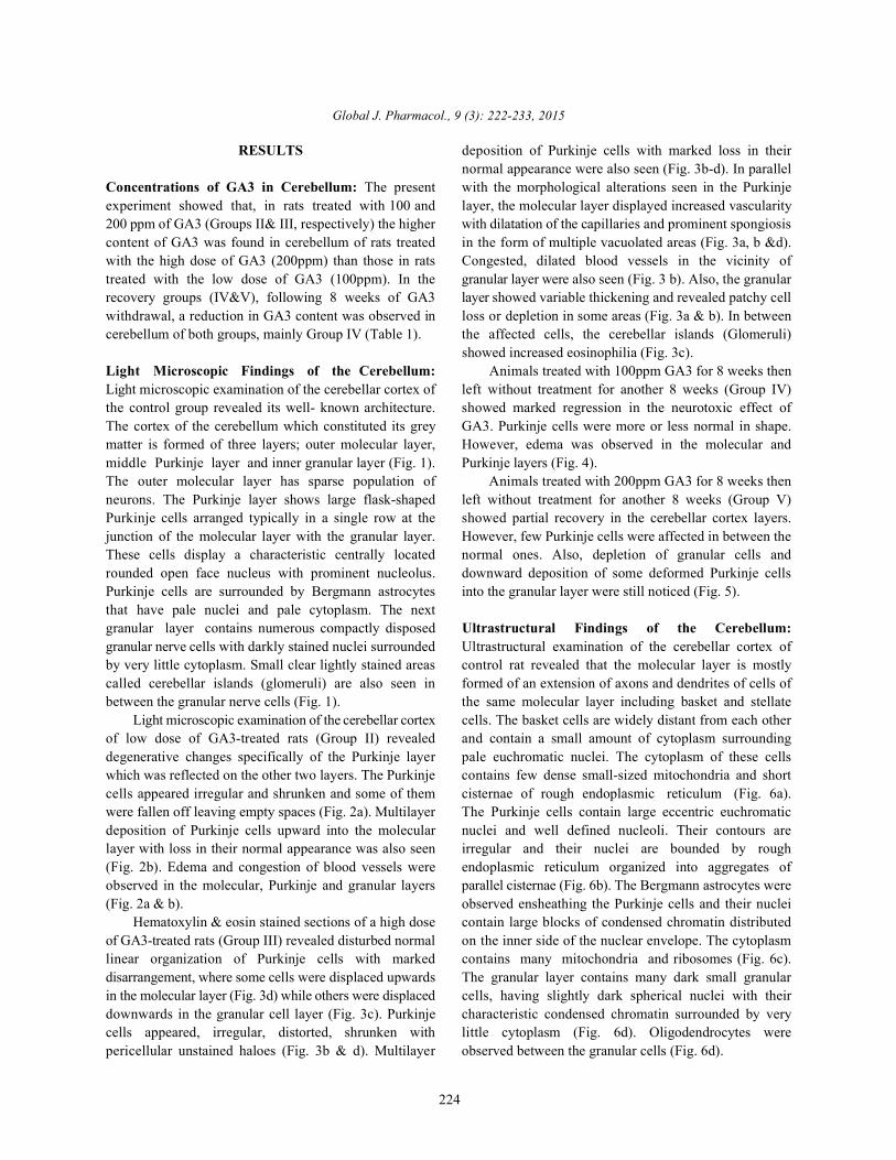
Light microscopic examination of the cerebellar cortex cells. The basket cells are widely distant from each otherof low dose of GA3-treated rats (Group II) revealed and contain a small amount of cytoplasm surroundingdegenerative changes specifically of the Purkinje layer pale euchromatic nuclei. The cytoplasm of these cellswhich was reflected on the other two layers. The Purkinje contains few dense small-sized mitochondria and shortcells appeared irregular and shrunken and some of them cisternae of rough endoplasmic reticulum (Fig. 6a).were fallen off leaving empty spaces (Fig. 2a). Multilayer The Purkinje cells contain large eccentric euchromaticdeposition of Purkinje cells upward into the molecular nuclei and well defined nucleoli. Their contours arelayer with loss in their normal appearance was also seen irregular and their nuclei are bounded by rough(Fig. 2b). Edema and congestion of blood vessels were endoplasmic reticulum organized into aggregates ofobserved in the molecular, Purkinje and granular layers parallel cisternae (Fig. 6b). The Bergmann astrocytes were(Fig. 2a & b). observed ensheathing the Purkinje cells and their nuclei
Hematoxylin & eosin stained sections of a high dose contain large blocks of condensed chromatin distributedof GA3-treated rats (Group III) revealed disturbed normal on the inner side of the nuclear envelope. The cytoplasmlinear organization of Purkinje cells with marked contains many mitochondria and ribosomes (Fig. 6c).disarrangement, where some cells were displaced upwards The granular layer contains many dark small granularin the molecular layer (Fig. 3d) while others were displaced cells, having slightly dark spherical nuclei with theirdownwards in the granular cell layer (Fig. 3c). Purkinje characteristic condensed chromatin surrounded by verycells appeared, irregular, distorted, shrunken with little cytoplasm (Fig. 6d). Oligodendrocytes werepericellular unstained haloes (Fig. 3b & d). Multilayer observed between the granular cells (Fig. 6d).
normal appearance were also seen (Fig. 3b-d). In parallel
showed increased eosinophilia (Fig. 3c).
Global J. Pharmacol., 9 (3): 222-233, 2015
225
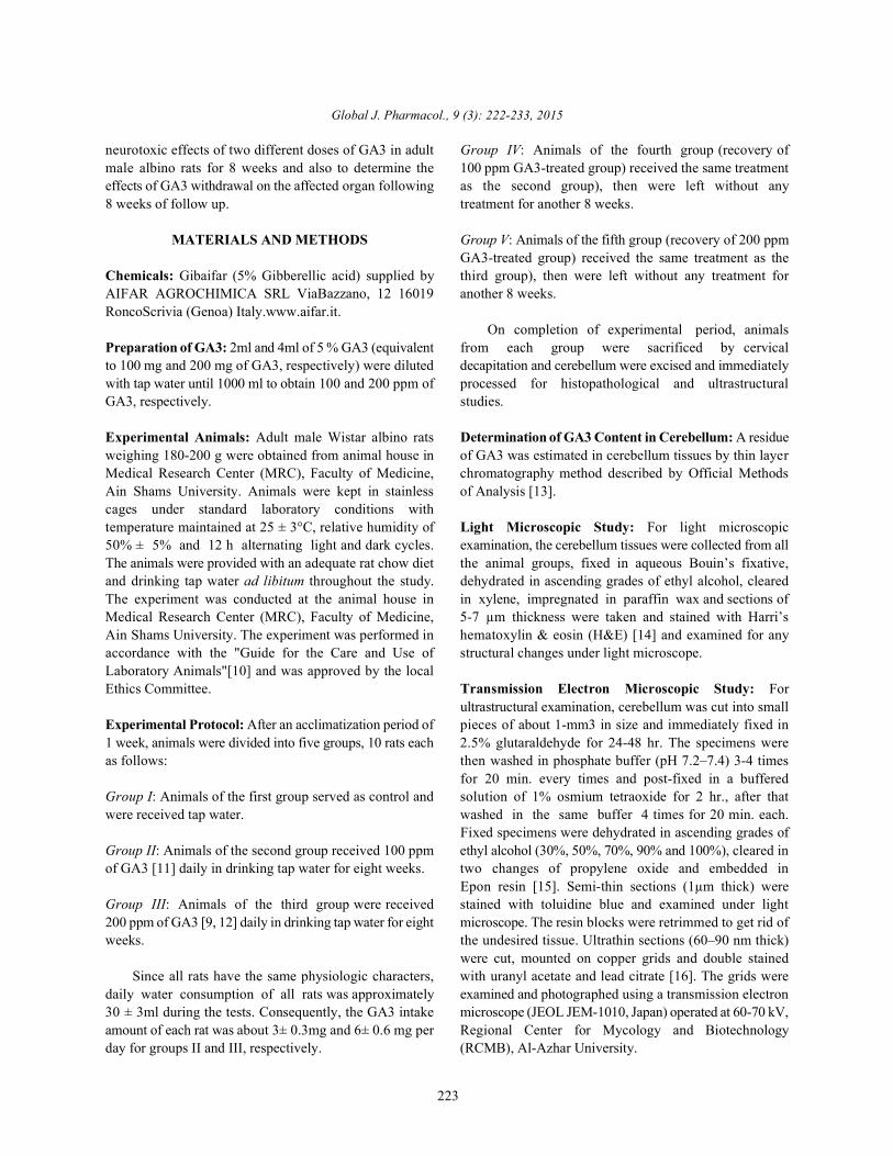
Table 1: Concentrations of GA3 in cerebellum in all the studied groups [Data are expressed as means ± S.E. (n = 10 in each group]Groups Concentration of GA3 (µg/g) in cerebellum (Mean ± S.E.)Group I (control) nilGroup II (100 ppm) 18.25± 1.90Group III (200 ppm) 55.25± 2.66Group IV (Recovery of 100 ppm) 8.75 ± 0.75Group V (Recovery of 200 ppm) 17.00± 1.22
Fig. 1: Photomicrograph of a section of cerebellar cortex of a control rat showing the outer molecular layer (M), themiddle Purkinje layer (P) having large flask-shaped Purkinje cells (PC) with centrally located large vesicular nucleiand are surrounded by Bergmann astrocytes (B). Note that the inner granular layer (G) is composed of closelypacked deeply stained numerous cells (arrow) and cerebellar islands (C) (H & E, X400)
Fig. 2: (a & b) Photomicrographs of sections of cerebellar cortex of rats treated with 100 ppm GA3 for 8 weeks. (a)Showing edema (arrows) in the molecular, Purkinje and granular layers and shrunken Purkinje cells with pyknotic(PK) nuclei. Some Purkinje cells are fallen off leaving empty space (S). Congestion of blood vessels andperivascular edema (arrow heads) appear in the molecular layer. (b) Showing multilayer deposition of Purkinje cellsupward into the molecular layer (arrows), pyknotic (PK) nuclei of Purkinje cells and congestion of blood vessels(arrow heads) in the molecular and granular layers (H&E, a &b X400)
Electron microscopic examination of the cerebellar indistinct nucleoli. Their cytoplasm had deformed roughcortex of rats treated with 100 ppm GA3 (Group II) endoplasmic reticulum and distorted mitochondriarevealed marked alteration in the cerebellar cortex layers. (Fig. 7b). Bergmann astrocytes appeared swollen withThe molecular layer revealed basket cell with marked clear cytoplasm (Fig. 7b). Furthermore, Granular cells withinfolding of the nuclear envelope and ill-defined irregular nuclear envelopes or pyknotic nuclei werecytoplasmic organelles (Fig. 7a). Nerve axons appeared detected (Fig. 7b &c). An increase in electron density ofwith distorted myelin sheath and the neuropil was the nuclei of oligodendrocytes and distortion in thevacuolated (Fig. 7a&b). In Purkinje layer, some Purkinje contour of the myelinated axons surrounding the granularcells had nuclei with ill-defined nuclear envelopes and cells were also noticed (Fig. 7c).
Global J. Pharmacol., 9 (3): 222-233, 2015
226
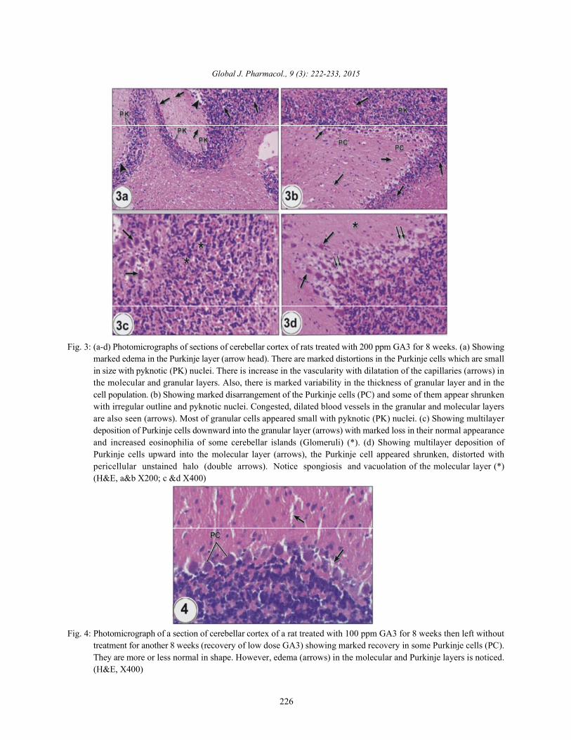
Fig. 3: (a-d) Photomicrographs of sections of cerebellar cortex of rats treated with 200 ppm GA3 for 8 weeks. (a) Showingmarked edema in the Purkinje layer (arrow head). There are marked distortions in the Purkinje cells which are smallin size with pyknotic (PK) nuclei. There is increase in the vascularity with dilatation of the capillaries (arrows) inthe molecular and granular layers. Also, there is marked variability in the thickness of granular layer and in thecell population. (b) Showing marked disarrangement of the Purkinje cells (PC) and some of them appear shrunkenwith irregular outline and pyknotic nuclei. Congested, dilated blood vessels in the granular and molecular layersare also seen (arrows). Most of granular cells appeared small with pyknotic (PK) nuclei. (c) Showing multilayerdeposition of Purkinje cells downward into the granular layer (arrows) with marked loss in their normal appearanceand increased eosinophilia of some cerebellar islands (Glomeruli) (*). (d) Showing multilayer deposition ofPurkinje cells upward into the molecular layer (arrows), the Purkinje cell appeared shrunken, distorted withpericellular unstained halo (double arrows). Notice spongiosis and vacuolation of the molecular layer (*)(H&E, a&b X200; c &d X400)
Fig. 4: Photomicrograph of a section of cerebellar cortex of a rat treated with 100 ppm GA3 for 8 weeks then left withouttreatment for another 8 weeks (recovery of low dose GA3) showing marked recovery in some Purkinje cells (PC).They are more or less normal in shape. However, edema (arrows) in the molecular and Purkinje layers is noticed.(H&E, X400)
Global J. Pharmacol., 9 (3): 222-233, 2015
227
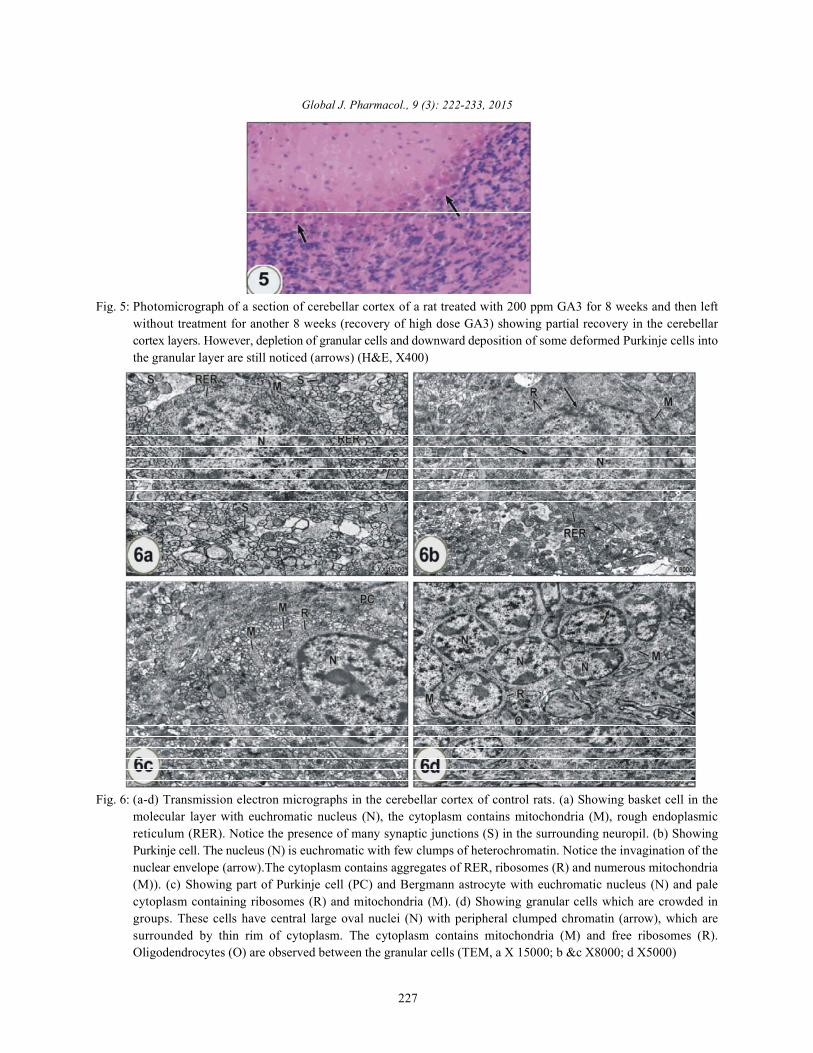
Fig. 5: Photomicrograph of a section of cerebellar cortex of a rat treated with 200 ppm GA3 for 8 weeks and then leftwithout treatment for another 8 weeks (recovery of high dose GA3) showing partial recovery in the cerebellarcortex layers. However, depletion of granular cells and downward deposition of some deformed Purkinje cells intothe granular layer are still noticed (arrows) (H&E, X400)
Fig. 6: (a-d) Transmission electron micrographs in the cerebellar cortex of control rats. (a) Showing basket cell in themolecular layer with euchromatic nucleus (N), the cytoplasm contains mitochondria (M), rough endoplasmicreticulum (RER). Notice the presence of many synaptic junctions (S) in the surrounding neuropil. (b) ShowingPurkinje cell. The nucleus (N) is euchromatic with few clumps of heterochromatin. Notice the invagination of thenuclear envelope (arrow).The cytoplasm contains aggregates of RER, ribosomes (R) and numerous mitochondria(M)). (c) Showing part of Purkinje cell (PC) and Bergmann astrocyte with euchromatic nucleus (N) and palecytoplasm containing ribosomes (R) and mitochondria (M). (d) Showing granular cells which are crowded ingroups. These cells have central large oval nuclei (N) with peripheral clumped chromatin (arrow), which aresurrounded by thin rim of cytoplasm. The cytoplasm contains mitochondria (M) and free ribosomes (R).Oligodendrocytes (O) are observed between the granular cells (TEM, a X 15000; b &c X8000; d X5000)
Global J. Pharmacol., 9 (3): 222-233, 2015
228
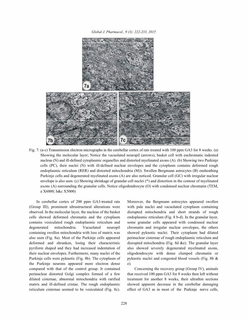
Fig. 7: (a-c) Transmission electron micrographs in the cerebellar cortex of rats treated with 100 ppm GA3 for 8 weeks. (a)Showing the molecular layer; Notice the vacuolated neuropil (arrows), basket cell with euchromatic indentednucleus (N) and ill-defined cytoplasmic organelles and distorted myelinated axons (A). (b) Showing two Purkinjecells (PC), their nuclei (N) with ill-defined nuclear envelopes and the cytoplasm contains deformed roughendoplasmic reticulum (RER) and distorted mitochondria (M)). Swollen Bergmann astrocytes (B) ensheathingPurkinje cells and degenerated myelinated axons (A) are also noticed. Granular cell (GC) with irregular nuclearenvelope is also seen. (c) Showing shrinkage of granular cell nuclei (*) and distortion in the contour of myelinatedaxons (A) surrounding the granular cells. Notice oligodendrocyte (O) with condensed nuclear chromatin (TEM,a X6000; b&c X5000)
In cerebellar cortex of 200 ppm GA3-treated rats Moreover, the Bergmann astrocytes appeared swollen(Group III), prominent ultrastructural alterations were with pale nuclei and vacuolated cytoplasm containingobserved. In the molecular layer, the nucleus of the basket disrupted mitochondria and short strands of roughcells showed deformed chromatin and the cytoplasm endoplasmic reticulum (Fig. 8 b-d). In the granular layer,contains vesiculated rough endoplasmic reticulum and some granular cells appeared with condensed nucleardegenerated mitochondria. Vacuolated neuropil chromatin and irregular nuclear envelopes, the otherscontaining swollen mitochondria with loss of matrix was showed pyknotic nuclei. Their cytoplasm had dilatedalso seen (Fig. 8a). Most of the Purkinje cells appeared perinuclear cisternae of rough endoplasmic reticulum anddeformed and shrunken, losing their characteristic disrupted mitochondria (Fig. 8d &e). The granular layerpyriform shaped and they had increased indentation of also showed severely degenerated myelinated axons,their nuclear envelopes. Furthermore, many nuclei of the oligodendrocyte with dense clumped chromatin orPurkinje cells were pyknotic (Fig. 8b). The cytoplasm of pyknotic nuclei and congested blood vessels (Fig. 8b &the Purkinje neurons appeared more electron dense e).compared with that of the control group. It contained Concerning the recovery group (Group IV), animalsperinuclear distorted Golgi complex formed of a few that received 100 ppm GA3 for 8 weeks then left withoutdilated cisternae, abnormal mitochondria with rarified treatment for another 8 weeks, their ultrathin sectionsmatrix and ill-defined cristae. The rough endoplasmic showed apparent decrease in the cerebellar damagingreticulum cisternae seemed to be vesiculated (Fig. 8c). effect of GA3 as in most of the Purkinje nerve cells,
Global J. Pharmacol., 9 (3): 222-233, 2015
229
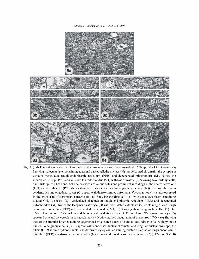
Fig. 8: (a-d) Transmission electron micrographs in the cerebellar cortex of rats treated with 200 ppm GA3 for 8 weeks. (a)Showing molecular layer containing abnormal basket cell, the nucleus (N) has deformed chromatin, the cytoplasmcontains vesiculated rough endoplasmic reticulum (RER) and degenerated mitochondria (M). Notice thevacuolated neuropil (VN) contains swollen mitochondria (M1) with loss of matrix. (b) Showing two Purkinje cells,one Purkinje cell has abnormal nucleus with active nucleolus and prominent infoldings in the nuclear envelope(PC1) and the other cell (PC2) shows shrunken pyknotic nucleus. Some granular nerve cells (GC) show chromatincondensation and oligodendrocytes (O) appear with dense clumped chromatin. Vacuolization (V) is also observedin the cytoplasm of Bergmann astrocyte (B). (c) Showing Purkinje cell (PC) with dense cytoplasm containingdilated Golgi vesicles (Gg), vesiculated cisternea of rough endoplasmic reticulum (RER) and degeneratedmitochondria (M). Notice the Bergmann astrocyte (B) with vacuolated cytoplasm (V) containing dilated roughendoplasmic reticulum (RER) and degenerated mitochondria (M1). (d) Showing abnormal granular cells (GC). Oneof them has pyknotic (PK) nucleus and the others show deformed nuclei. The nucleus of Bergmann astrocyte (B)appeared pale and the cytoplasm is vacuolated (V). Notice marked vacuolation of the neuropil (VN). (e) Showingarea of the granular layer containing degenerated myelinated axons (A) and oligodendrocyte (O) with pyknoticnuclei. Some granular cells (GC1) appear with condensed nuclear chromatin and irregular nuclear envelope, theothers (GC2) showed pyknotic nuclei and deformed cytoplasm containing dilated cisternae of rough endoplasmicreticulum (RER) and disrupted mitochondria (M). Congested blood vessel is also noticed (*) (TEM, a-e X5000)
Global J. Pharmacol., 9 (3): 222-233, 2015
230
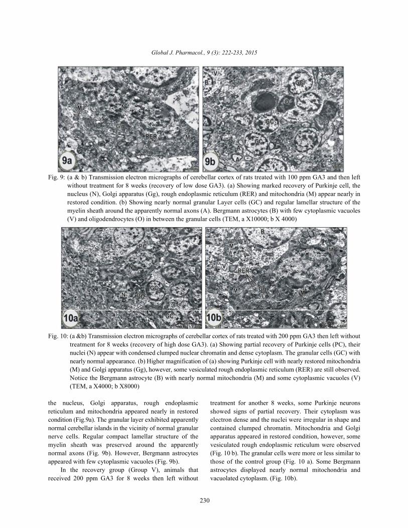
Fig. 9: (a & b) Transmission electron micrographs of cerebellar cortex of rats treated with 100 ppm GA3 and then leftwithout treatment for 8 weeks (recovery of low dose GA3). (a) Showing marked recovery of Purkinje cell, thenucleus (N), Golgi apparatus (Gg), rough endoplasmic reticulum (RER) and mitochondria (M) appear nearly inrestored condition. (b) Showing nearly normal granular Layer cells (GC) and regular lamellar structure of themyelin sheath around the apparently normal axons (A). Bergmann astrocytes (B) with few cytoplasmic vacuoles(V) and oligodendrocytes (O) in between the granular cells (TEM, a X10000; b X 4000)
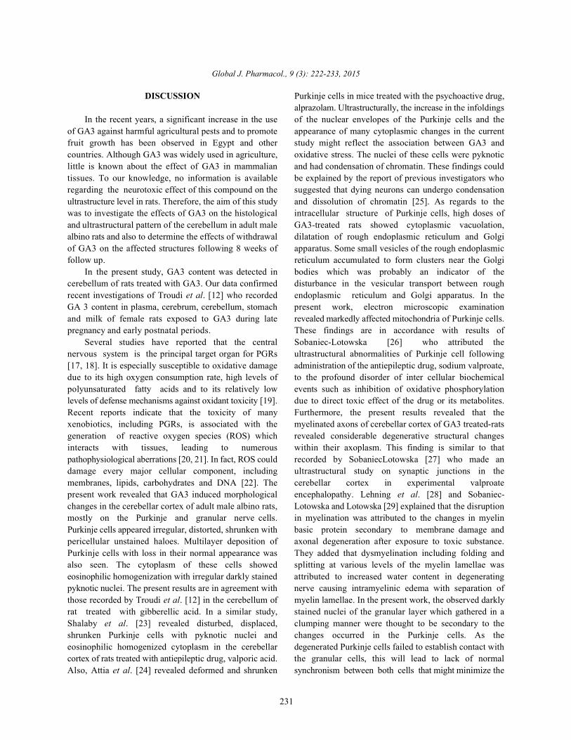
Fig. 10: (a &b) Transmission electron micrographs of cerebellar cortex of rats treated with 200 ppm GA3 then left withouttreatment for 8 weeks (recovery of high dose GA3). (a) Showing partial recovery of Purkinje cells (PC), theirnuclei (N) appear with condensed clumped nuclear chromatin and dense cytoplasm. The granular cells (GC) withnearly normal appearance. (b) Higher magnification of (a) showing Purkinje cell with nearly restored mitochondria(M) and Golgi apparatus (Gg), however, some vesiculated rough endoplasmic reticulum (RER) are still observed.Notice the Bergmann astrocyte (B) with nearly normal mitochondria (M) and some cytoplasmic vacuoles (V)(TEM, a X4000; b X8000)
the nucleus, Golgi apparatus, rough endoplasmic treatment for another 8 weeks, some Purkinje neuronsreticulum and mitochondria appeared nearly in restored showed signs of partial recovery. Their cytoplasm wascondition (Fig.9a). The granular layer exhibited apparently electron dense and the nuclei were irregular in shape andnormal cerebellar islands in the vicinity of normal granular contained clumped chromatin. Mitochondria and Golginerve cells. Regular compact lamellar structure of the apparatus appeared in restored condition, however, somemyelin sheath was preserved around the apparently vesiculated rough endoplasmic reticulum were observednormal axons (Fig. 9b). However, Bergmann astrocytes (Fig. 10 b). The granular cells were more or less similar toappeared with few cytoplasmic vacuoles (Fig. 9b). those of the control group (Fig. 10 a). Some Bergmann
In the recovery group (Group V), animals that astrocytes displayed nearly normal mitochondria andreceived 200 ppm GA3 for 8 weeks then left without vacuolated cytoplasm. (Fig. 10b).
Global J. Pharmacol., 9 (3): 222-233, 2015
231
DISCUSSION Purkinje cells in mice treated with the psychoactive drug,
In the recent years, a significant increase in the use of the nuclear envelopes of the Purkinje cells and theof GA3 against harmful agricultural pests and to promote appearance of many cytoplasmic changes in the currentfruit growth has been observed in Egypt and other study might reflect the association between GA3 andcountries. Although GA3 was widely used in agriculture, oxidative stress. The nuclei of these cells were pyknoticlittle is known about the effect of GA3 in mammalian and had condensation of chromatin. These findings couldtissues. To our knowledge, no information is available be explained by the report of previous investigators whoregarding the neurotoxic effect of this compound on the suggested that dying neurons can undergo condensationultrastructure level in rats. Therefore, the aim of this study and dissolution of chromatin [25]. As regards to thewas to investigate the effects of GA3 on the histological intracellular structure of Purkinje cells, high doses ofand ultrastructural pattern of the cerebellum in adult male GA3-treated rats showed cytoplasmic vacuolation,albino rats and also to determine the effects of withdrawal dilatation of rough endoplasmic reticulum and Golgiof GA3 on the affected structures following 8 weeks of apparatus. Some small vesicles of the rough endoplasmicfollow up. reticulum accumulated to form clusters near the Golgi
In the present study, GA3 content was detected in bodies which was probably an indicator of thecerebellum of rats treated with GA3. Our data confirmed disturbance in the vesicular transport between roughrecent investigations of Troudi et al. [12] who recorded endoplasmic reticulum and Golgi apparatus. In theGA 3 content in plasma, cerebrum, cerebellum, stomach present work, electron microscopic examinationand milk of female rats exposed to GA3 during late revealed markedly affected mitochondria of Purkinje cells.pregnancy and early postnatal periods. These findings are in accordance with results of
Several studies have reported that the central Sobaniec-Lotowska [26] who attributed thenervous system is the principal target organ for PGRs ultrastructural abnormalities of Purkinje cell following[17, 18]. It is especially susceptible to oxidative damage administration of the antiepileptic drug, sodium valproate,due to its high oxygen consumption rate, high levels of to the profound disorder of inter cellular biochemicalpolyunsaturated fatty acids and to its relatively low events such as inhibition of oxidative phosphorylationlevels of defense mechanisms against oxidant toxicity [19]. due to direct toxic effect of the drug or its metabolites.Recent reports indicate that the toxicity of many Furthermore, the present results revealed that thexenobiotics, including PGRs, is associated with the myelinated axons of cerebellar cortex of GA3 treated-ratsgeneration of reactive oxygen species (ROS) which revealed considerable degenerative structural changesinteracts with tissues, leading to numerous within their axoplasm. This finding is similar to thatpathophysiological aberrations [20, 21]. In fact, ROS could recorded by SobaniecLotowska [27] who made andamage every major cellular component, including ultrastructural study on synaptic junctions in themembranes, lipids, carbohydrates and DNA [22]. The cerebellar cortex in experimental valproatepresent work revealed that GA3 induced morphological encephalopathy. Lehning et al. [28] and Sobaniec-changes in the cerebellar cortex of adult male albino rats, Lotowska and Lotowska [29] explained that the disruptionmostly on the Purkinje and granular nerve cells. in myelination was attributed to the changes in myelinPurkinje cells appeared irregular, distorted, shrunken with basic protein secondary to membrane damage andpericellular unstained haloes. Multilayer deposition of axonal degeneration after exposure to toxic substance.Purkinje cells with loss in their normal appearance was They added that dysmyelination including folding andalso seen. The cytoplasm of these cells showed splitting at various levels of the myelin lamellae waseosinophilic homogenization with irregular darkly stained attributed to increased water content in degeneratingpyknotic nuclei. The present results are in agreement with nerve causing intramyelinic edema with separation ofthose recorded by Troudi et al. [12] in the cerebellum of myelin lamellae. In the present work, the observed darklyrat treated with gibberellic acid. In a similar study, stained nuclei of the granular layer which gathered in aShalaby et al. [23] revealed disturbed, displaced, clumping manner were thought to be secondary to theshrunken Purkinje cells with pyknotic nuclei and changes occurred in the Purkinje cells. As theeosinophilic homogenized cytoplasm in the cerebellar degenerated Purkinje cells failed to establish contact withcortex of rats treated with antiepileptic drug, valporic acid. the granular cells, this will lead to lack of normalAlso, Attia et al. [24] revealed deformed and shrunken synchronism between both cells that might minimize the
alprazolam. Ultrastructurally, the increase in the infoldings
Global J. Pharmacol., 9 (3): 222-233, 2015
232
regulatory role on them [30] In the present work, the ACKNOWLEDGEMENT.
structural changes seen in the cerebellum of rats treatedwith GA3 were also characterized by edema in the internal The authors thank the members in Medical Researchgranular layer. This might be due to the increased amount Center (MRC), Faculty of Medicine, Ain Shams Universityof ROS product which might attack membranes and for their kind help and support.enhance their permeability leading to vacuolization, theprimary response to cell injury [31] or to the marked REFERENCES.
disturbances in lipid inclusions as shown by Sherlock andDooley [32]. 1. Fishel, F.M., 2006. Gibberellins.The Agronomy
In the present study, withdrawal effect of GA3 Department, Florida Cooperative Extention Service,treatment on the cerebellar cortex was also investigated. Institute of Food and Agricultural SciencesRecovery of some Purkinje neuron was observed where (University of Florida, USA).ultrastructure alterations were less evident than those in 2. Osborne, D.J. and M.T. McManus, 2005. Hormonesthe treated animals. Their cytoplasm showed that most of signals and target cells in plant development. 1 ed.,RER appeared in nearly restored condition. Abundant Vol.1 (Cambridge University Press), pp: 266.SER in the recovery period could be also explained by 3. Neil, A.C. and J.B. Reece, 2002. PhytohormonesCsala [33] who stated that it takes part in the synthesis of (plant hormones) and other growth regulators:.
phospholipids for building of cell membrane and Gibberellins. In: Biology. 6 ed., (San Fransisco,membranes of cell organelles. On the other hand, the Benjamin Cummings), pp: 145.increased RER was explained by Kimball [34] who 4. Sakr, S.A., Y.A. Okdah and S.F. EL-Abd, 2003.mentioned that RER had a very important role in the Gibberellin A3 induced histological andsynthesis and packaging of proteins. Some of these histochemical alterations in the liver of albino rats.proteins might be used by the cell to synthesize Science Asia, 29: 327-331.membrane, other cell organelles and the others were sent 5. Schwechheimer, C. and B.C. Willige, 2009. Sheddingout. This was considered as a structural-functional light on gibberellic acid signaling. Curr. Opin. Plantresponse to enable cells to release the oxidative stress Biol., 12: 57-62.secondary to GA3 toxicity. Also, mitochondria almost 6. Tomlin, C.D.S., 2004. Gibberellic acid. In: The e-retained their normal appearance suggesting increased Pesticide Manual. 13 ed., edited by Tomlin C.D.S.active detoxifying mechanisms in those Purkinje neurons (Hampshire, UK, British Crop. Protection Council),which might need longer period of withdrawal to escape Chapter 3, 5. from the toxic effect of GA3. In contradiction with our 7. Arteca, R., 1996. Plant growth substances: Principlesresults, Abdelhamid et al. [35] reported that feeding and Applications. (New York, Chapman & Hall),2-weeks old broiler chicks on gibberellic acid (GA3)- pp: 1-95.containing diets (0, 1, 5, 25 and 125 ppm) for 3-weeks led 8. Celik, I., Y. Tuluce and I. Isik, 2007 Evalution ofto numerous histological lesions in different organs. toxicity of abcisic acid and gibberellic acid in rats:Two-week withdrawal period did not ameliorate the 50 days drinking water study. J. Enzyme Inhib. Med.negative effects of GA3. This controversy could be Chem., 2: 219-226.attributed to variations in animals species, intensity of the 9. Troudi, A., A.M. Samet and N. Zeghal, 2010.doses and differences in administered and withdrawal Hepatotoxicity induced by gibberellic acid inperiods. adult rats and their progeny. Exp. Toxicol. Pathol.,
CONCLUSION 10. Institute of Laboratory Animal Resources,
From the previously mentioned results we can Council, Guide for the Care and Use of Laboratoryconclude that GA3 has a dose-dependent toxic effect on Animals. (National Academy Press, Washington,the cerebellar cortex of adult male albino rats. On the other D.C), 1996, 21.http://www.nap.edu/openbook.php.hand, 8 weeks period of follow up was insufficient for 11. Celik, I., Y. Tuluce and I. Isik, 2006. Influence ofcomplete recovery of these toxic effects. subacute treatment of some plant growth regulators
Conflict of Interest: The authors declared no conflicts of antioxidant defense and lipid peroxidation in rats.interest. J. Biochem. Mol. Toxicol., 20: 174-182.
st
th
th
62: 637-642.
Commission on Life Sciences, National Research
on serum marker enzymes and erythrocyte and tissue
Global J. Pharmacol., 9 (3): 222-233, 2015
233
12. Troudi, A., H. Bouaziz, N. Soudani, I. Ben Amara, 24. Attia, A.A., N.A. Kheirallah and S.A. Khalifa, 2014.T. Boudawara, H. Touzani, B. Lyoussi and N. Zeghal, Biochemical and ultrastructural studies of the effect2012. Neurotoxicity and oxidative stress induced by of alprazolam as an anxiolytic drug on the cerebellumgibberellic acid in rats during late pregnancy and of adult male mice. J. Appl. Pharm. Sci., 4: 74-83.early postnatal periods: Biochemical and histological 25. Olivi, A., M. Gilbert, K.L. Duncan, B. Corden,changes. Exp. Toxicol. Pathol., 6: 583-590. D. Lenartz and H. Brem, 1993. Direct delivery of
13. A.O.A.C, 1990. Official Methods of Analysis. The platinum-based antineoplastics to the centralAssociation of Official Analytical Chemists. 15 ed., nervous system: A toxicity and ultrastructural study.th
(Arlington, West Virginia, USA Washington D.C.) Cancer Chemother. Pharmacol., 31: 449-454.14. Bancroft, J.D. and M. Gamble, 2008. Theory and 26. Sobaniec-Lotowska, M.E., 2001. Ultrastructure of
Practice of Histological Techniques. 6 ed., (Churchill Purkinje cell perikarya and their dendritic processesth
Livingstone, Elsevier, Philadelphia). in the rat cerebellar cortex in experimental15. Hayat, M.A., 1986. Principles of tissue preparation encephalopathy induced by chronic application of
for electron microscopy. In: “Basic Techniques for valproate. Int. J. Exp. Pathol., 82: 337-348.Transmission Electron Microscopy”. 1 ed., 27. Sobaniec-Lotowska, M.E., 2002. Ultrastructure ofst
(Macmillan Press, New York) Chapter 2, pp: 1-22. synaptic junctions in the cerebellar cortex in16. Weakley, B.S., 1981. A Beginner's Handbook in experimental valproate encephalopathy and after
Biological Transmission Electron Microscopy. 2 terminating chronic application of the antiepileptic.nd
ed., (Churchill Livingstone, London) Chapter 2, Folia Neuropathol., 40: 87-96.pp: 24. 28. Lehning, E.J., C.D. Balabanm, J.F. Ross, M.A. Reid
17. Furukawa, S., M. Abe, K. Usuda and I. Ogawa, 2004. and R.M. LoPachin, 2002. Acrylamide neuropathy. I.Indole-3-acetic acid induces microencephaly in rat Spatiotemporal characteristics of nerve cell damagefetuses. J. Toxicol. Pathol., 32: 659-667. in rat cerebellum. Neurotoxicology, 23: 397-414.
18. Yilmaz, Z. and I. Celik, 2009. Neurotoxic and 29. Sobaniec-Lotowska, M.E. and J.M. Lotowska, 2005.immunotoxic effects of indole-3-butyric acid on rats Ultrastructural study of cerebellar dentate nucleusat subacute and subchronic exposure. astrocytes in chronic experimental model withNeurotoxicology, 30: 382-385. valproate. Folia Neuropathol., 43: 166-171.
19. Samson, F.E. and S.R. Nelson, 2000. The aging brain, 30. Saad El-Dien, H.M., D.A. EL Gamal, H.A. Mubarakmetals and oxygen free radicals. Cell Mol. Biol., and S.M. Saleh, 2010. Effect of fluoride on rat46: 699-707. cerebellar cortex: Light and electron microscopic
20. Celik, I., M. Turker and Y. Tuluce, 2007. Abscisic acid studies. Egypt. J. Histol., 33: 245-256.and gibberellic acid cause increased lipid 31. Robbins, S.L. and M. Angell, 1976. Basic Pathology.peroxidation and fluctuated antioxidant defense 2 ed., (W. B. Saunders Company, Philadelphia).systems of various tissues in rats. J. Haz. Mat., 32. Sherlock, S. and J. Dooley, 2002. Diseases of the148: 623-629. Liver and Biliary System. 9 ed., (Oxford, Boston:
21. Yu, F., Z. Wang, B. Ju, Y. Wang, J. Wang and D. Bai, Blackwell Scientific Publications), pp: 114. 2 0 0 8 . A p o p t o t i c e f f e c t o f 33. Csala, M., G. Bánhegyi and A. Benedetti, 2006.organophosphorusinsecticide chlorpyrifos on mouse Endoplasmic reticulum: a metabolic compartment.retina in vivo oxidative stress and protection of FEBS Letters, 580: 2160-2165.combination of vitamin C and E. Exp. Toxicol. Pathol., 34. Kimball, J.W., 2008. Growth regulating hormones.59: 415-423. http://users.rcn.com/jkimball.ultranet/biologypages.
22. Bhalla, P. and D.K. Dhawan, 2009. Protective role of 35. Abdelhamid, A.M., T.M. Dorra, M.A. Ali andlithium in ameliorating the aluminium-induced E.H. Abou-Egla, 1994. Effect of gibberellic acid onoxidative stress and histological changes in rat brain. broiler chickens performance and some metabolicCell Mol. Neuro. Biol., 29: 513-521. parameters. Arch. Tierernahr., 46: 269-276.
23. Shalaby, N.M. and N.I. Sarhan, 2008. Light andelectron microscopic study on the effect of valproicacid on cerebellar cortex of adult male albino rats andthe possible protective effect of L-carnitine. EgyptJ. Histol., 31: 256-265.
nd
th