Neuroprotection or Neurotoxicity of Illicit Drugs on ...
36
life Review Neuroprotection or Neurotoxicity of Illicit Drugs on Parkinson’s Disease Carla Ferreira 1,2,3 , Catarina Almeida 1 , Sandra Tenreiro 4 and Alexandre Quintas 1,2, * 1 Molecular Pathology and Forensic Biochemistry Laboratory, Centro de Investigação Interdisciplinar Egas Moniz, P-2825-084 Caparica, Portugal; [email protected] (C.F.); [email protected] (C.A.) 2 Laboratório de Ciências Forenses e Psicológicas Egas Moniz, Campus Universitário–Quinta da Granja, Monte de Caparica, P-2825-084 Caparica, Portugal 3 Faculty of Medicine of Porto University, Al. Prof. Hernâni Monteiro, P-4200–319 Porto, Portugal 4 CEDOC–Chronic Diseases Research Center, Faculdade de Ciências Médicas, Universidade Nova de Lisboa, P-1150-082 Lisboa, Portugal; [email protected] * Correspondence: [email protected] Received: 23 May 2020; Accepted: 9 June 2020; Published: 11 June 2020 Abstract: Parkinson’s Disease (PD) is currently the most rapid growing neurodegenerative disease and over the past generation, its global burden has more than doubled. The onset of PD can arise due to environmental, sporadic or genetic factors. Nevertheless, most PD cases have an unknown etiology. Chemicals, such as the anthropogenic pollutant 1-methyl-4-phenyl-1,2,3,6-tetrahydropyridine (MPTP) and amphetamine-type stimulants, have been associated with the onset of PD. Conversely, cannabinoids have been associated with the treatment of the symptoms’. PD and medical cannabis is currently under the spotlight, and research to find its benefits on PD is on-going worldwide. However, the described clinical applications and safety of pharmacotherapy with cannabis products are yet to be fully supported by scientific evidence. Furthermore, the novel psychoactive substances are currently a popular alternative to classical drugs of abuse, representing an unknown health hazard for young adults who may develop PD later in their lifetime. This review addresses the neurotoxic and neuroprotective impact of illicit substance consumption in PD, presenting clinical evidence and molecular and cellular mechanisms of this association. This research area is utterly important for contemporary society since illicit drugs’ legalization is under discussion which may have consequences both for the onset of PD and for the treatment of its symptoms. Keywords: Parkinson’s Disease; phytocannabinoids; amphetamine-type stimulants; novel psychoactive substances; cocaine; opioids 1. Introduction Neurodegenerative diseases are progressive incapacitating conditions involving the function loss of nerve cells in the brain or peripheral nervous system. These types of diseases affect millions of people worldwide, who may suffer an early death. The burden of neurodegenerative diseases has significantly increased worldwide over the past 25 years, mostly due to incremental increases in the population numbers, population ageing and to the environmental stress associated to contemporary societies [1–3]. These diseases have become the leading cause of disability and death in developed countries and, in spite of decades of research, there is no way to cure them or slow down their progression [4]. Neurodegenerative diseases include Alzheimer’s, amyotrophic lateral sclerosis, Huntington’s, Lewy body disease and Parkinson’s, among others. The two most prevalent neurodegenerative diseases are Alzheimer’s (AD) and Parkinson’s disease (PD). About 5% of people over the age of 85 suffer from PD, Life 2020, 10, 86; doi:10.3390/life10060086 www.mdpi.com/journal/life
Transcript of Neuroprotection or Neurotoxicity of Illicit Drugs on ...

life
Review
Neuroprotection or Neurotoxicity of Illicit Drugs onParkinson’s Disease
Carla Ferreira 1,2,3, Catarina Almeida 1, Sandra Tenreiro 4 and Alexandre Quintas 1,2,*1 Molecular Pathology and Forensic Biochemistry Laboratory, Centro de Investigação Interdisciplinar Egas
Moniz, P-2825-084 Caparica, Portugal; [email protected] (C.F.);[email protected] (C.A.)
2 Laboratório de Ciências Forenses e Psicológicas Egas Moniz, Campus Universitário–Quinta da Granja,Monte de Caparica, P-2825-084 Caparica, Portugal
3 Faculty of Medicine of Porto University, Al. Prof. Hernâni Monteiro, P-4200–319 Porto, Portugal4 CEDOC–Chronic Diseases Research Center, Faculdade de Ciências Médicas, Universidade Nova de Lisboa,
P-1150-082 Lisboa, Portugal; [email protected]* Correspondence: [email protected]
Received: 23 May 2020; Accepted: 9 June 2020; Published: 11 June 2020�����������������
Abstract: Parkinson’s Disease (PD) is currently the most rapid growing neurodegenerative diseaseand over the past generation, its global burden has more than doubled. The onset of PD can arise dueto environmental, sporadic or genetic factors. Nevertheless, most PD cases have an unknown etiology.Chemicals, such as the anthropogenic pollutant 1-methyl-4-phenyl-1,2,3,6-tetrahydropyridine(MPTP) and amphetamine-type stimulants, have been associated with the onset of PD. Conversely,cannabinoids have been associated with the treatment of the symptoms’. PD and medical cannabisis currently under the spotlight, and research to find its benefits on PD is on-going worldwide.However, the described clinical applications and safety of pharmacotherapy with cannabis productsare yet to be fully supported by scientific evidence. Furthermore, the novel psychoactive substancesare currently a popular alternative to classical drugs of abuse, representing an unknown healthhazard for young adults who may develop PD later in their lifetime. This review addresses theneurotoxic and neuroprotective impact of illicit substance consumption in PD, presenting clinicalevidence and molecular and cellular mechanisms of this association. This research area is utterlyimportant for contemporary society since illicit drugs’ legalization is under discussion which mayhave consequences both for the onset of PD and for the treatment of its symptoms.
Keywords: Parkinson’s Disease; phytocannabinoids; amphetamine-type stimulants; novelpsychoactive substances; cocaine; opioids
1. Introduction
Neurodegenerative diseases are progressive incapacitating conditions involving the function lossof nerve cells in the brain or peripheral nervous system. These types of diseases affect millions of peopleworldwide, who may suffer an early death. The burden of neurodegenerative diseases has significantlyincreased worldwide over the past 25 years, mostly due to incremental increases in the populationnumbers, population ageing and to the environmental stress associated to contemporary societies [1–3].These diseases have become the leading cause of disability and death in developed countries and,in spite of decades of research, there is no way to cure them or slow down their progression [4].Neurodegenerative diseases include Alzheimer’s, amyotrophic lateral sclerosis, Huntington’s, Lewybody disease and Parkinson’s, among others. The two most prevalent neurodegenerative diseases areAlzheimer’s (AD) and Parkinson’s disease (PD). About 5% of people over the age of 85 suffer from PD,
Life 2020, 10, 86; doi:10.3390/life10060086 www.mdpi.com/journal/life

Life 2020, 10, 86 2 of 36
overwhelming health support systems and families [5]. The mechanisms underlying these particularconditions are related to the accumulation of misfolded and aggregated proteins, which may promotethe disease either by a gain of the toxic activity or by loss of biological function [6–9]. The misfoldedprotein may aggregate into amyloid fibrils which deposit in the form of extracellular amyloid plaques,neuro-fibrillary tangles and other intracytoplasmic or intranuclear inclusions [8–10].
PD is currently the most rapid growing neurodegenerative disease [3,11] and, over the pastgeneration, its global burden has more than doubled, as a result of human life span increment,better health conditions and environmental factors, such as exposition to the anthropogenic pollutant1-methyl-4-phenyl-1,2,3,6-tetrahydropyridine (MPTP) and pesticides [11–15]. The onset of this diseaseusually occurs at near to average 60 years of age [16]. However, in some cases, PD takes place muchearlier, between 21 and 50 years of age (early-onset PD (EOPD)) [17]. Symptomatically, initial phases ofthe disease are characterized by movement disorders, such as shaking, rigidity, slowness and aberrantgait [18]. Other symptoms, such as cognitive and behavioral problems, appear as dementia in the laterphases of the disease [18]. The onset of PD and EOPD can arise as a consequence of genetic and/orenvironmental factors but most of the cases are sporadic [15,17,19–23]. It is generally accepted thatknown genetic causes may account for over 5% of the total PD population. A recent study was able toexplain 16–36% of PD heritability, prevalence estimates (0.5–2.0%) [24]. The heritability in EOPD ishigher than in later-onset PD [19]. These low values are in agreement with clinical reports describingthe vast majority of PD cases as having an unknown etiology [3,11,17,25,26]. Several studies highlightthat the combination of genetic and environmental factors, such as the consumption of substancesand the variability of brain vulnerability, may also increase the risk to PD onset. These studies showthat variability in genes associated with the cellular response and metabolism of xenobiotics or toxinsincreases the predisposition to develop PD [27]. Actually, the neurotoxic effect of MPTP changes theexpression of genes associated with PD [28], which may alter the resilience of neurons to toxics [29,30].Interestingly, scientific reports suggest that the incidence of PD and EOPD are increasing [11,17,31,32].
The onset of PD is tightly associated with the neuronal protein α-synuclein (α-syn). α-synphysiological function is to promote N-ethylmaleimide-sensitive factor (NSF) attachment to thesoluble NSF attachment receptor (SNARE) protein complex assembly during synaptic exocytosis [33].Thus, α-syn seems to regulate the synaptic vesicle release from presynaptic cells [33]. However, inpathological conditions, the overexpression and/or modification of α-syn forms neurotoxic aggregateswhich promote the selective loss of dopaminergic neurons in the substantia nigra pars compacta [34–36].Most of the studies associate PD to several defected cellular and physiological mechanisms, such asneuroinflammation, excitotoxicity, mitochondrial dysfunction, reduced trophic support, abnormalkinase activity, disruption of calcium homeostasis and proteostasis dysfunction [37–43]. Nevertheless,there is still much to clarify, such as the precise molecular mechanism of amyloid fibrils formation,and its relationship with glial activation and peripheral immune cell infiltration in the inflammatoryresponses [7,44].
Recently, it has been hypothesized that the increase in the risk of PD might be associated withthe consumption of drugs of abuse, such as stimulants [45]. The supporting pieces of evidencesuggest that the neurotoxic effects of amphetamine-like stimulants on the nigrostriatal pathway areintriguingly similar to those in neurodegeneration observed in PD [46]. Furthermore, Parkinsonismwas also described as a consequence of heroin consumption, which is a depressor of the central nervoussystem [47,48]. Conversely, scientific data supports that other substances of abuse, such as somephytocannabinoids, which interact with the endocannabinoid system, may play a neuroprotective rolein PD [49–55]. Interestingly, both neurotoxic and neuroprotective effects are described as the outcomesfrom different drugs of abuse consumption. Finally, it is important to mention that during the lastdecade a new trend of synthetic molecules with similar effects to traditional illicit drugs, the novelpsychoactive substances (NPS), has emerged and little is known about their toxicological impact.Consumption of NPS among young people may be a promotor of the initial stages of neurodegeneration

Life 2020, 10, 86 3 of 36
and may well increase the future incidence of PD. Therefore, further studies with these substancesare essential.
In this article, the current knowledge connecting illicit drugs of abuse and PD is reviewedand discussed.
2. Methodology
Searches were conducted in the online database PubMed and an advanced search were performedusing the following boolean equations: (i) “misfolding diseases” AND “amphetamine”; (ii) “Parkinson”AND “amphetamine” with a cut-off filter to select just the papers from the last 10 years; (iii) “Parkinson”AND “phytocannabinoids” with a cut-off filter to select just the papers from the last 10 years; (iv)“Parkinson” AND “cathinone”; (v) “amyloid formation” AND “amphetamine” AND “Parkinson”;(vi) “heroin” AND “Parkinson”; (vii) “opioid” AND ”Parkinson”; (viii) “cocaine” AND “Parkinson”.The search was limited to English-language peer-reviewed journal publications. In the eliminationprocess, papers that did not focus on illicit drugs of abuse or had no relationship between PD andillicit drugs of abuse were excluded. Further sources were identified by following up internal citationsand references within the documents retrieved in the initial search.
3. Phytocannabinoids and Parkinson’s Disease
Phytocannabinoids are chemical substances present in Cannabis sativa and indica, commonlyknown as marijuana. Marijuana has been consumed for recreational, religious and medicinal purposesfor at least five millennia [56]. Cannabis continues to be the most widely used drug worldwide [57].United Nations Office on Drugs and Crime (UNODC) estimates that roughly 3.8% of the globalpopulation aged 15–64 years old used cannabis at least once during 2017 [57]. The cannabis is marketas herb, resin and as hash oil [57]. The cannabis herb consists of the dried and crumbled leaves andflowering tops of the cannabis plant, which is generally smoked. In contrast, cannabis resin, theconcentrated extract of cannabis flower and plant, is usually mixed with tobacco to be smoked [57].Hash oil is a cannabis product which is extracted from any part of the plant using organic solvents [57].Cannabis is controlled under the Single Convention on Narcotic Drugs of 1961 amended by the 1972protocol [57]. Nowadays, the use of cannabis has been legalized in several countries. However,it remains illegal in the vast majority of the countries that have signed the UN convention on Narcoticdrugs. Independently of its licit or illicit consumption, several studies point out medicinal benefitsof products derived from marijuana for cases of glaucoma, neurodegeneration, multiple sclerosis,schizophrenia, cancer, epilepsy and eating disorders [58–61].
The two main cannabinoids present in marijuana, responsible for its psychoactive and medicinaleffects, are ∆9-tetrahydrocannabinol (∆9-THC) and cannabidiol (CBD), respectively. The effects of∆9-THC and CBD are mediated by receptors [62,63]. There are two known cannabinoid receptorssubtypes, cannabinoid receptor 1 (CB1) and cannabinoid receptor 2 (CB2), which are also receptors forendogenous cannabinoids. The view of the endocannabinoid system as a therapeutic target has beenbringing some researchers to explore potential defects in endocannabinoids metabolism, cannabinoidsreceptors and other components of the endocannabinoid system in PD onset [64–67].
3.1. Endocannabinoid System and Parkinson’s Disease
CB1 and CB2, encoded by cannabinoid receptor 1 and 2 genes, CNR1 and CNR2 genes, respectively.Both receptors exert biological effects by activating heterotrimeric Gi/o type G proteins, which lead to theinhibition of adenylyl cyclase and consequently to the reduction of cyclic AMP levels. These receptorsalso activate different members of the family of mitogen-activated protein kinases (MAPKs) [68].The two receptors are differently distributed in the human body. Overall, CB1 distribution is moreprominent in the central nervous system, whereas CB2 is mainly present in the immune system and, in alesser extent, in the central nervous system [69,70]. CB1 has three isoforms from different expressionCNR1 patterns. The full-length product of CNR1 is mostly found in the brain and skeletal muscles,

Life 2020, 10, 86 4 of 36
while the two isoforms are present in the liver and in pancreatic islet β-cells [69]. CB2 has also threeisoforms, CB2A and CB2B resulting from alternative splicing of CNR2. The full-length CB2 is presentin the central nervous system, mainly in glial cells, and in the immune system. The two isoforms CB2Aand CB2B are predominantly expressed in testis and in the spleen [70].
Although several studies have shown the presence of CB2 in the brain, the role of CB2 inendocannabinoid-mediated synaptic transmission is still largely elusive [71,72]. However, it wasreported that in medial prefrontal cortical pyramidal neurons, intracellular CB2 reduces neuronal firingthrough the opening of Ca2+-activated chloride channels, suggesting its involvement in the regulationof neuronal activity [73]. Moreover, CB2 receptors are involved in neuroinflammation by modulatingmicroglia activation and migration [74] and are consequently associated with neurodegenerativedisorders [75]. Accordingly, CB2 receptor is seen as a potential therapeutic target for PD since itmodulates the inflammatory process and does not trigger undesirable psychoactive effects [76,77].Furthermore, several shreds of evidence suggest that cannabinoid compounds may interact with otherreceptors, besides CB1 and CB2, namely the transient receptor potential cation channel subfamily Vmember 1 (TRPV1), the peroxisome proliferator-activated receptors (PPARs) and the G-protein-coupledreceptor (GPCR) [78,79].
CB1 receptor structure has a classical 7TM fold (Figure 1a) similar to other rhodopsin family classA GPCRs [80]. Endocannabinoids bind to the CB1 hydrophobic binding pocket, activating the receptor.However, the position of the ligand-binding pocket of CB1 is different from the previously describedbinding sites of other class A GPCRs. Ligands lie low in the binding pocket of CB1, immediately abovethe conserved W356 [81]. Conformational changes in the surroundings of the residue W356 have beenproposed as triggering CB1 activation [81].
Life 2020, 10, x FOR PEER REVIEW 4 of 38
system. The two isoforms CB2A and CB2B are predominantly expressed in testis and in the spleen
[70].
Although several studies have shown the presence of CB2 in the brain, the role of CB2 in
endocannabinoid-mediated synaptic transmission is still largely elusive [71,72]. However, it was
reported that in medial prefrontal cortical pyramidal neurons, intracellular CB2 reduces neuronal
firing through the opening of Ca2+-activated chloride channels, suggesting its involvement in the
regulation of neuronal activity [73]. Moreover, CB2 receptors are involved in neuroinflammation by
modulating microglia activation and migration [74] and are consequently associated with
neurodegenerative disorders [75]. Accordingly, CB2 receptor is seen as a potential therapeutic target
for PD since it modulates the inflammatory process and does not trigger undesirable psychoactive
effects [76,77]. Furthermore, several shreds of evidence suggest that cannabinoid compounds may
interact with other receptors, besides CB1 and CB2, namely the transient receptor potential cation
channel subfamily V member 1 (TRPV1), the peroxisome proliferator-activated receptors (PPARs)
and the G-protein-coupled receptor (GPCR) [78,79].
CB1 receptor structure has a classical 7TM fold (Figure 1a) similar to other rhodopsin family
class A GPCRs [80]. Endocannabinoids bind to the CB1 hydrophobic binding pocket, activating the
receptor. However, the position of the ligand-binding pocket of CB1 is different from the previously
described binding sites of other class A GPCRs. Ligands lie low in the binding pocket of CB1,
immediately above the conserved W356 [81]. Conformational changes in the surroundings of the
residue W356 have been proposed as triggering CB1 activation [81].
CB2 has a high degree of homology with CB1, sharing 44% sequence identity [82]. The
architecture of CB2 is also comprised of a 7TM fold (Figure 1b). The structural data indicates a critical
role for the residue W258 as the toggle switch for CB2 activation [83]. Overall, structural data points
out differences in the binding pockets between CB1 and CB2.
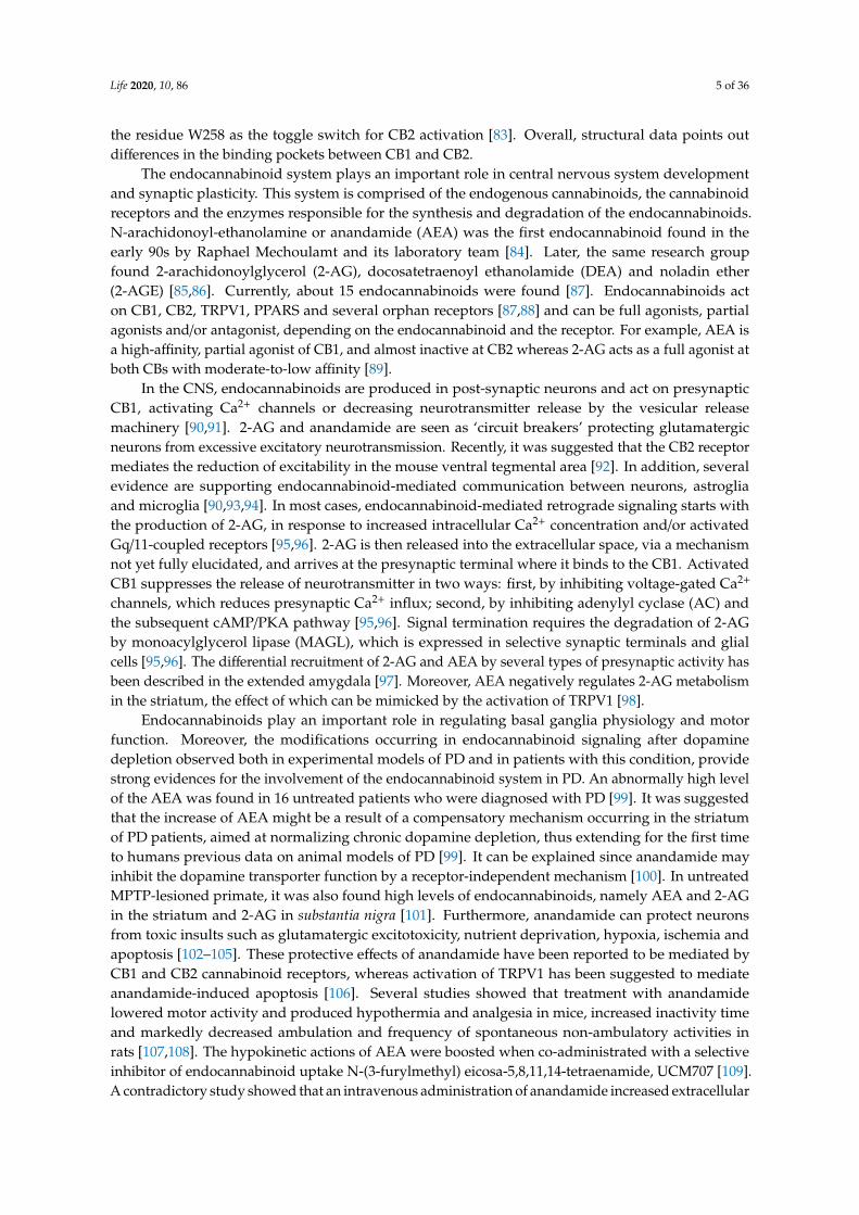
Figure 1. (a) Cannabinoids receptors 1 (PDB: 5u09) and (b) 2 (PDB: 6kpc). Helix 1 to 7 (red to purple)
is mostly located inside the cell plasma membrane. (a) The cannabinoids receptor 1 (CB1) and (b) CB2
N-terminal loop (red) occupy the polar zone of the binding pockets. The CB1 receptor represented is
bound to Tranabant, and CB2 receptor is bound to AM10257.
The endocannabinoid system plays an important role in central nervous system development
and synaptic plasticity. This system is comprised of the endogenous cannabinoids, the cannabinoid
Figure 1. (a) Cannabinoids receptors 1 (PDB: 5u09) and (b) 2 (PDB: 6kpc). Helix 1 to 7 (red to purple) ismostly located inside the cell plasma membrane. (a) The cannabinoids receptor 1 (CB1) and (b) CB2N-terminal loop (red) occupy the polar zone of the binding pockets. The CB1 receptor represented isbound to Tranabant, and CB2 receptor is bound to AM10257.
CB2 has a high degree of homology with CB1, sharing 44% sequence identity [82]. The architectureof CB2 is also comprised of a 7TM fold (Figure 1b). The structural data indicates a critical role for
Life 2020, 10, 86 5 of 36
the residue W258 as the toggle switch for CB2 activation [83]. Overall, structural data points outdifferences in the binding pockets between CB1 and CB2.
The endocannabinoid system plays an important role in central nervous system developmentand synaptic plasticity. This system is comprised of the endogenous cannabinoids, the cannabinoidreceptors and the enzymes responsible for the synthesis and degradation of the endocannabinoids.N-arachidonoyl-ethanolamine or anandamide (AEA) was the first endocannabinoid found in theearly 90s by Raphael Mechoulamt and its laboratory team [84]. Later, the same research groupfound 2-arachidonoylglycerol (2-AG), docosatetraenoyl ethanolamide (DEA) and noladin ether(2-AGE) [85,86]. Currently, about 15 endocannabinoids were found [87]. Endocannabinoids acton CB1, CB2, TRPV1, PPARS and several orphan receptors [87,88] and can be full agonists, partialagonists and/or antagonist, depending on the endocannabinoid and the receptor. For example, AEA isa high-affinity, partial agonist of CB1, and almost inactive at CB2 whereas 2-AG acts as a full agonist atboth CBs with moderate-to-low affinity [89].
In the CNS, endocannabinoids are produced in post-synaptic neurons and act on presynapticCB1, activating Ca2+ channels or decreasing neurotransmitter release by the vesicular releasemachinery [90,91]. 2-AG and anandamide are seen as ‘circuit breakers’ protecting glutamatergicneurons from excessive excitatory neurotransmission. Recently, it was suggested that the CB2 receptormediates the reduction of excitability in the mouse ventral tegmental area [92]. In addition, severalevidence are supporting endocannabinoid-mediated communication between neurons, astrogliaand microglia [90,93,94]. In most cases, endocannabinoid-mediated retrograde signaling starts withthe production of 2-AG, in response to increased intracellular Ca2+ concentration and/or activatedGq/11-coupled receptors [95,96]. 2-AG is then released into the extracellular space, via a mechanismnot yet fully elucidated, and arrives at the presynaptic terminal where it binds to the CB1. ActivatedCB1 suppresses the release of neurotransmitter in two ways: first, by inhibiting voltage-gated Ca2+
channels, which reduces presynaptic Ca2+ influx; second, by inhibiting adenylyl cyclase (AC) andthe subsequent cAMP/PKA pathway [95,96]. Signal termination requires the degradation of 2-AGby monoacylglycerol lipase (MAGL), which is expressed in selective synaptic terminals and glialcells [95,96]. The differential recruitment of 2-AG and AEA by several types of presynaptic activity hasbeen described in the extended amygdala [97]. Moreover, AEA negatively regulates 2-AG metabolismin the striatum, the effect of which can be mimicked by the activation of TRPV1 [98].
Endocannabinoids play an important role in regulating basal ganglia physiology and motorfunction. Moreover, the modifications occurring in endocannabinoid signaling after dopaminedepletion observed both in experimental models of PD and in patients with this condition, providestrong evidences for the involvement of the endocannabinoid system in PD. An abnormally high levelof the AEA was found in 16 untreated patients who were diagnosed with PD [99]. It was suggestedthat the increase of AEA might be a result of a compensatory mechanism occurring in the striatumof PD patients, aimed at normalizing chronic dopamine depletion, thus extending for the first timeto humans previous data on animal models of PD [99]. It can be explained since anandamide mayinhibit the dopamine transporter function by a receptor-independent mechanism [100]. In untreatedMPTP-lesioned primate, it was also found high levels of endocannabinoids, namely AEA and 2-AGin the striatum and 2-AG in substantia nigra [101]. Furthermore, anandamide can protect neuronsfrom toxic insults such as glutamatergic excitotoxicity, nutrient deprivation, hypoxia, ischemia andapoptosis [102–105]. These protective effects of anandamide have been reported to be mediated byCB1 and CB2 cannabinoid receptors, whereas activation of TRPV1 has been suggested to mediateanandamide-induced apoptosis [106]. Several studies showed that treatment with anandamidelowered motor activity and produced hypothermia and analgesia in mice, increased inactivity timeand markedly decreased ambulation and frequency of spontaneous non-ambulatory activities inrats [107,108]. The hypokinetic actions of AEA were boosted when co-administrated with a selectiveinhibitor of endocannabinoid uptake N-(3-furylmethyl) eicosa-5,8,11,14-tetraenamide, UCM707 [109].A contradictory study showed that an intravenous administration of anandamide increased extracellular
Life 2020, 10, 86 6 of 36
dopamine levels in the nucleus accumbens shell of awake, freely moving rats, a characteristic effect ofmost drugs of abuse in humans [110].
Levels and activities of AEA and 2-AG can be manipulated by inhibition of fatty acid amidehydrolase (FAAH) enzyme, the action of which is reduced in experimental models of PD [111,112].However, it was evidenced that FAAH inhibition remarkably increases AEA tissue levels but reduces2-AG levels [113]. The systemic administration of N-(4-hydroxyphenyl)-arachidonamide (AM404)enhances anandamide (AEA) availability in the biophase and exerts antiparkinsonian effects in6-hydroxydopamine-lesioned rats. This is due to a reduction of D2 dopamine receptor functiontogether with a positive modulation of 5-HT1B serotonin receptor function [114].
TRPV1 receptors also seem to play an important role in development and expression of dyskinesiasin PD. The systemic administration of oleoylethanolamide, an agonist of PPARα and antagonist ofTRPV1 receptors, reduces the development of dyskinesias dependent of a TRPV1-pathway in mousemodel of PD, not involving PPARα receptors [115]. The intake of this compound induced thereduction of FosB striatal protein overexpression and the phosphoacetylation of histone 3, which aremolecular markers of L-DOPA-induced dyskinesias. Actually, FOSB overexpression was previouslyassociated with L-DOPA-induced dyskinesia in nitric oxide synthase-positive striatal interneurons inhemiparkinsonian mice [116]. This observation was correlated with the activation of ERK1/2 due toincreased phosphorylation of its regulatory kinases [116]. It was suggested that GPR55 modulatesanti-neuroinflammatory responses and movement control. These observations point to this receptor asa therapeutic target for the non-dopaminergic symptomatic treatment of PD [78,117].
The anti-inflammatory, antioxidant and proneurogenic properties of the endocannabinoidsystem make it a potential target to reduce the symptomatics of a number of neurodegenerativeconditions [65,118–120]. Nonetheless, there are challenges in the development of drugs lackingpsychoactive side effects and, to that end, targeting the anti-inflammatory non-psychotropic CB2receptor is particularly promising [119–121].
There are shreds of evidence supporting the involvement of the endocannabinoid system in PD.Magnetic resonance imaging studies have shown regional differences in CB1 receptor availability in PDpatients’ brains. According to one of these studies, CB1 availability was increased in mesolimbic andmesocortical regions of the brain, which are usually dopamine depleted in PD, and decreased in thesubstantia nigra [122]. Two other studies shown active involvement of CB1 in the regulation of L-DOPAaction during PD therapy, preventing motor fluctuation through modulation of the striatonigral andstriatopallidal pathway [123,124]. Regarding CB2, this receptor was found at significantly lower levels intyrosine hydroxylase-containing neurons from substantia nigra of PD patients [125]. In contrast, studiesin glial elements from post-mortem tissues of PD patients showed an increase in CB2 availability, eitherquantified by immunochemistry or by gene expression. These observations were then corroborated bystudies in animal models [125–128]. The authors suggest that up-regulation of CB2 in glial cells is anindicator of the involvement of this receptor in neuroprotection.
3.2. Clinical Observations on Phytocannabinoids Use in Parkinson’s Disease
In countries where cannabis is legal, marijuana is used recreationally to self-medicate symptomsof disorders such as PD, multiple sclerosis, amyotrophic lateral sclerosis and schizophrenia [129].About 44% of the population with PD is currently using marijuana [130]. In addition to cannabis, threecannabis derived products are also in use: dronabinol, nabiximols and nabilone [129]. Despite the lackof solid scientific evidence, PD patients using cannabis mention a positive impact on mood, memory,fatigue, obesity, sleep, pain, tremor, rigidity and bradykinesia after its consumption.
A study with 85 PD patients combined half a teaspoon of cannabis leaves, along with theirprescribed pharmacotherapy for PD. About 46% of these individuals reported relief of PD symptomson average 1.7 months after the first use of marijuana, suggesting chronic use of marijuana maybe required for improvement in symptoms [131,132]. Overall, patients using cannabis have beenreporting a lower level of disability after the intake of phytocannabinoids [130,133–135]. On the other

Life 2020, 10, 86 7 of 36
hand, Carroll et al. have conducted a clinical trial with an orally administered cannabis extract whichresulted in no objective or subjective improvement in dyskinesias or parkinsonism showing that resultsfor clinical cannabis in PD still seem to be inconsistent [136]. Therefore, in determining if medicalmarijuana is beneficial as a PD therapeutic, some factors, such as chemical constituents, dose, deliverysystem and clinical outcomes, must be carefully controlled [137]. Cannabis is a complex plant with twomain subspecies, namely Cannabis sativa and Cannabis indica, which can be differentiated by C. indicahaving higher cannabidiol content and C. sativa having a higher ∆9-THC content [138]. In addition,there are differences in ∆9-THC and CBD amount from strain to strain. Moreover, with the rising of themarijuana business, each sample might have different levels of ∆9-THC and CBD [139]. Furthermore,from the 538 natural compounds identified in C. sativa, more than 100 are phytocannabinoids. Therefore,the use of therapeutic cannabis is, surely a complex issue from the composition point of view [140,141].
Regarding the two most abundant phytocannabinoids found in C. Sativa, CBD showed to be themost promising, relieving some PD symptoms [142]. The first clinical studies with CBD pointed to adecrease in the psychotic symptoms [93] and significant improvements in measures of functioningand well-being of PD patients with no psychiatric comorbidities [94]. Overall, CBD shows significanttherapeutic effects in reducing tremor, dyskinesia, rigidity and some non-motor symptoms, such aspsychosis, rapid eye movement sleep behavior disorder, daily activities and stigma linked to relationaland communication problems in PD [68,142–144]. However, larger-scale studies and randomizeddouble-blind controlled studies are still needed to confirm the observations since several reports arementioning negative effects [143].
A major concern with phytocannabinoids use is the inherent risk of PD patients to developpsychosis and cognitive impairment. This aspect makes them more susceptible to psychomimeticsubstances agonists of CB1, such as ∆9-THC or Nabilone [145,146]. In fact, it is well known thatNabilone may induce psychosis, even in patients without a psychiatric history [146]. Thus, patients withdementia should not be treated with agonists of CB1 to avoid further aggravation of neuropsychiatricsymptoms [68]. Moreover, the PD patient’s personality must be taken into account to avoid thedevelopment of addictive behavior [147].
Overall, the evidence on the therapeutic use of medical marijuana and cannabinoid derivativesin patients with PD are heterogeneous and of poor quality [146]. Consequently, there is an urgentneed for further scientific studies and to educate the caretakers on the pharmacology, known risks andknown benefits of cannabis [148].
3.3. Studies on the Molecular and Cellular Mechanisms Underlying Clinical Observations
The positive clinical evidence observed in PD patients using medical marijuana and cannabinoidderivatives are leading researchers to address the cellular and molecular mechanisms underlyingsuch outcomes. Therefore, a couple of studies suggest that molecules which bind to CB1 and/or CB2receptors might be beneficial, since pharmacological modulation of the endocannabinoid system hasbeen shown to reduce chronic activation of the neuroinflammatory response, reduce mitochondrialdysfunction and keep calcium homeostasis, resulting in the decrease of oxidative stress, which preventsthe proapoptotic cascade, promoting neurotrophic support [59,122].
Studies in human differentiated neuroblastomas, which have dopamine beta-hydroxylase activity,show that ∆9-THC seems to be neuroprotective by up-regulating the expression of gene encoding CB1,suggesting a direct neuronal protective effect of ∆9-THC mediated via PPARγ not involving CB2 [55].The activity of dopamine beta-hydroxylase modulates the levels of dopamine [149]. Busquets-Garciaet al. (2016) observed that normal circulating adrenaline and noradrenaline levels are sustainedafter stress by AM6545 pre-treatment, a full agonist of CB1 [150]. Thus, the clinical observationthat the increment of CB1 availability in mesolimbic and mesocortical regions of brain seems to beneuroprotective [122]. Moreover, the involvement of PPARγ activation in the neuroprotective effect of∆9-THC is also suggested, as it induces the transcription of proteins involved in oxidative stress defenseand mitochondrial biogenesis, promoting mitochondrial normal function in PD [151]. In addition,

Life 2020, 10, 86 8 of 36
the reduction of oxidative stress was linked to the restored the Peroxisome proliferator-activatedreceptor-gamma coactivator (PGC-1α) levels which regulate energetic metabolism [151]. In fact, lowbasal levels of PGC-1α are expected to be associated with enhanced glycolytic metabolism, low oxygenconsumption and elevated reactive oxygen species (ROS) levels [152]. In addition, the observed∆9-THC mitochondrial biogenesis may be linked to its ability to induce the mitochondria transcriptionfactors (TFAM) expression and to restore mitochondrial DNA levels leading to increased cytochrome coxidase subunit 4 (COX4) [151], the terminal enzyme complex of the respiratory chain which is linkedto PD [153] (Figure 2).
Life 2020, 10, x FOR PEER REVIEW 8 of 38
A high concentration of glutamate induces deregulation of intracellular Ca2+ levels which results
in mitochondrial Ca2+ overload and membrane depolarization, triggering the mechanism of cell death
[94]. Δ9-THC also seems to play a neuroprotective effect against glutamate-induced neurotoxicity, in
neural primary cells, by restoring mitochondrial membrane potential which produces an anti-
apoptotic effect. In the same study, a decrease in the levels of glutamate was observed, which in turn
decreases capase-3 levels, one of the critical enzymes of apoptosis. Overall, CB1 activation by Δ9-THC
seems to slow down the degenerative processes in PD associated with the overflow of glutamate [154].
Cannabidiol also presents a neuroprotective activity against MPP+, a neurotoxin which triggers
PD, by the activation of nerve growth factor receptor (NGF) also known as Tropomyosin receptor
kinase A (TRKA), and the increment in the expression of axonal and synaptogenic proteins [155].
Other compounds found in Cannabis sativa, such as β-caryophyllene and Δ9-tetrahydrocannabivarin
(Δ9-THCV), showed the potential to prevent the onset of PD. β-caryophyllene activates CB2, leading
to a decrease of oxidative/nitrosative stress, to a decrease of pro-inflammatory cytokines release and
to an inhibition of gliosis, which reduces neuroinflammation and nigrostriatal degeneration [156,157].
Δ9-THCV is a potent CB2 receptor partial agonist in vitro and it antagonizes cannabinoid receptor
agonists in CB1-expressing tissues. However, in vivo Δ9-THCV behaves both as an antagonist or, at higher
doses, an agonist of CB1 [58]. It has been shown that acute administration of this phytocannabinoid
attenuated the motor inhibition caused by changes in glutamatergic transmission, and the chronic
administration of Δ9-THCV has reduced the loss of tyrosine hydroxylase–positive neurons caused by 6-
hydroxydopamine in the substantia nigra [158] (Figure 2).
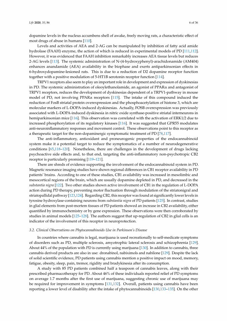
Figure 2. Neuroprotective mechanisms in Parkinson’s disease activated by phytocannabinoids. (a)
The dysregulation of intracellular Ca2+ levels result in excitotoxicity which results in mitochondrial
depolarization. CB1 activation restores membrane potential; (b) Activation CB1 promotes gene
expression of cannabinoid receptor 1 (CNR1) reducing neurotransmitters release; (c) Increasing levels
of reactive oxygen species (ROS) promote the formation of protein toxic oligomers. CB1 activation
decrease ROS levels by expressing mitochondrial transcription factors (TFAM) and restoring
mitochondrial DNA levels; (d) CB2 activation decrease pro-inflammatory cytokines release; and (e)
CB2 activation inhibit apoptosis by nerve growth factor receptor (NGF) also known as Tropomyosin
receptor kinase A (TRKA).
In general, the effects of some phytocannabinoids on PD appear to be protective either by
binding to the CB1 receptor, inhibiting dopamine beta hydroxylase activity and decreasing glutamate
levels or by binding to CB2, reducing neuroinflammation.
Figure 2. Neuroprotective mechanisms in Parkinson’s disease activated by phytocannabinoids. (a) Thedysregulation of intracellular Ca2+ levels result in excitotoxicity which results in mitochondrialdepolarization. CB1 activation restores membrane potential; (b) Activation CB1 promotes geneexpression of cannabinoid receptor 1 (CNR1) reducing neurotransmitters release; (c) Increasing levels ofreactive oxygen species (ROS) promote the formation of protein toxic oligomers. CB1 activation decreaseROS levels by expressing mitochondrial transcription factors (TFAM) and restoring mitochondrialDNA levels; (d) CB2 activation decrease pro-inflammatory cytokines release; and (e) CB2 activationinhibit apoptosis by nerve growth factor receptor (NGF) also known as Tropomyosin receptor kinase A(TRKA).
A high concentration of glutamate induces deregulation of intracellular Ca2+ levels which results inmitochondrial Ca2+ overload and membrane depolarization, triggering the mechanism of cell death [94].∆9-THC also seems to play a neuroprotective effect against glutamate-induced neurotoxicity, in neuralprimary cells, by restoring mitochondrial membrane potential which produces an anti-apoptotic effect.In the same study, a decrease in the levels of glutamate was observed, which in turn decreases capase-3levels, one of the critical enzymes of apoptosis. Overall, CB1 activation by ∆9-THC seems to slowdown the degenerative processes in PD associated with the overflow of glutamate [154].
Cannabidiol also presents a neuroprotective activity against MPP+, a neurotoxin which triggersPD, by the activation of nerve growth factor receptor (NGF) also known as Tropomyosin receptorkinase A (TRKA), and the increment in the expression of axonal and synaptogenic proteins [155].Other compounds found in Cannabis sativa, such as β-caryophyllene and ∆9-tetrahydrocannabivarin(∆9-THCV), showed the potential to prevent the onset of PD. β-caryophyllene activates CB2, leadingto a decrease of oxidative/nitrosative stress, to a decrease of pro-inflammatory cytokines release and toan inhibition of gliosis, which reduces neuroinflammation and nigrostriatal degeneration [156,157].∆9-THCV is a potent CB2 receptor partial agonist in vitro and it antagonizes cannabinoid receptor

Life 2020, 10, 86 9 of 36
agonists in CB1-expressing tissues. However, in vivo ∆9-THCV behaves both as an antagonistor, at higher doses, an agonist of CB1 [58]. It has been shown that acute administration of thisphytocannabinoid attenuated the motor inhibition caused by changes in glutamatergic transmission,and the chronic administration of ∆9-THCV has reduced the loss of tyrosine hydroxylase–positiveneurons caused by 6-hydroxydopamine in the substantia nigra [158] (Figure 2).
In general, the effects of some phytocannabinoids on PD appear to be protective either by bindingto the CB1 receptor, inhibiting dopamine beta hydroxylase activity and decreasing glutamate levels orby binding to CB2, reducing neuroinflammation.
3.4. Is There Enough Data Supporting Protective or Therapeutic Role of Cannabinoids on PD?
Overall, clinical observations and research outcomes support the endocannabinoid system asa target to alleviate the symptoms of PD. Actually, patients using cannabis have been reportinga lower level of disability after the intake of phytocannabinoids [130,133–135]. However, theevidence on the therapeutic use of cannabinoids in patients with PD are heterogeneous and ofpoor quality [146]. At molecular and cellular levels, the evidence is promising for the use ofphytocannabinoids in PD. Phytocannabinoids reduce neuroinflammatory response, mitochondrialdysfunction and oxidative stress [59,122]. Additionally, ∆9-THC plays a neuroprotective effect againstglutamate-induced neurotoxicity, in neural primary cells slowing down neuron degeneration due tooverflow of glutamate [154]. Epidemiological studies are also encouraging. A retrospective surveyfound an improvement of PD symptoms with medical cannabis in the initial stages of treatment, withno evidence of major adverse effects [49]. Another epidemiological study pointed to the possibleeffect of cannabidiol in improving the quality of life of PD patients without psychiatric comorbidities.However, the authors found no statistically significant differences concerning the motor symptoms ofPD [159].
Despite the shreds of evidence suggesting that the consumption of cannabinoids can reducePD symptoms, some authors argue that there are not enough studies for such a conclusion [50–54].Stampanoni Bassi et al. (2017) concluded that results from available clinical studies are controversialand inconclusive due to several limitations, including small sample size, lack of standardized outcomemeasures and expectancy bias [54,160]. They propose studies involving a larger sample of patients,appropriate molecular targets, objective biological measures (i.e., cannabinoids blood level) andspecific clinical outcome measures to clarify the effectiveness of cannabinoids-based therapies [54].Moreover, most of the studies investigating the therapeutic potential of cannabinoids in PD havebeen conducted in animal models, and an insufficient number of clinical trials have been carriedout. Furthermore, the therapeutic benefits demonstrated in animal models will require further studyin humans avoiding extrapolation between them, since animal models may not properly induce orrecapitulate PD pathology [51,52,119,148,161–164]. Thereby, in the present, the studies investigating therole of phytocannabinoids are few and limited to understand its beneficial effects. The improvementsneeded for further successful research in this area are (i) larger sample size; (ii) well-designedstudies testing cannabis in PD patients population to establish evidence-based data on the scopeof pharmacological benefits and adverse effects; (iii) long term evaluation of disease progression;(iv) identification of the precise formulation for each type of pathology and each subset of patients forachieving a neuroprotective effect [51,52,119,148,162–164]. Overall, there is a clear need for furtherstudies in humans.
4. Amphetamine-Type Stimulants and Parkinson’s Disease
According to the World Health Organization, amphetamine-type stimulants is a group of drugsof abuse whose principal members include amphetamine and methamphetamine [165]. Amphetaminewas firstly synthesized in 1887 in Germany as phenylisopropylamine by Romanian chemist LazărEdeleanu [166]. Methamphetamine was synthetized in 1893 by Nagayoshi from ephedrine [167], analkaloid present in the plant Ephedra, isolated for the first time in 1885 by G. Yamanashi and named
Life 2020, 10, 86 10 of 36
by Nagai in 1887 [168]. Amphetamine-type stimulants have been used for recreational purposes toimprove physical and mental performance in fatigued subjects. During World War II, amphetamineand methamphetamine were used extensively by Allied and Axis forces for their stimulant andperformance-enhancing effects [169].
As the addictive properties of the drugs became known, governments began to place strict controlson the sale of the drugs. As a result of the United Nations 1971 Convention on Psychotropic Substances,amphetamine became a schedule II-controlled substance, as defined in the treaty, ratified by all 183state members at the time. Despite strict government controls, amphetamine and methamphetamineare used for recreation purposes, and according to the European Monitoring Centre for Drugs andDrug Addiction (EMCDDA), amphetamines are associated with a large number of health emergenciesin the north and east of Europe [170]. The monitoring center estimates that 1.2 million of Europeanyoung people between 15–34 age have consumed amphetamine-type stimulants in the last year, and12.4 million of European people have consumed them somewhere during their lifetime [170].
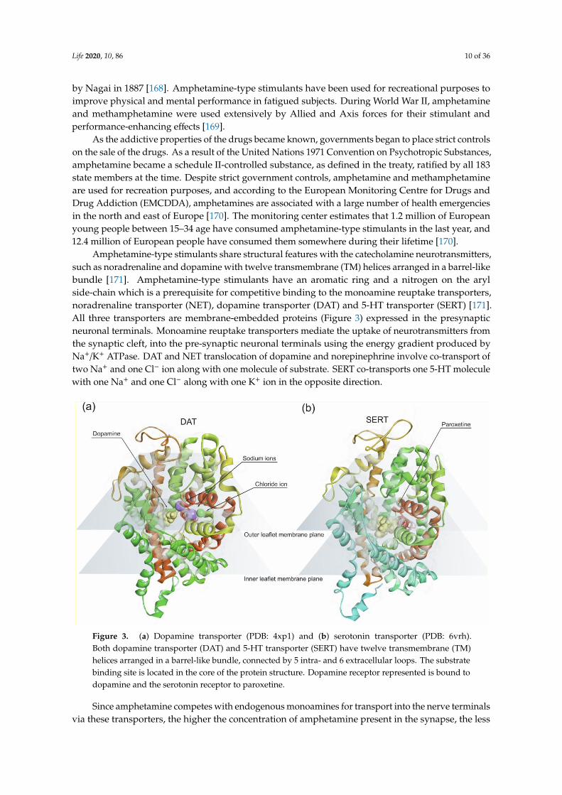
Amphetamine-type stimulants share structural features with the catecholamine neurotransmitters,such as noradrenaline and dopamine with twelve transmembrane (TM) helices arranged in a barrel-likebundle [171]. Amphetamine-type stimulants have an aromatic ring and a nitrogen on the arylside-chain which is a prerequisite for competitive binding to the monoamine reuptake transporters,noradrenaline transporter (NET), dopamine transporter (DAT) and 5-HT transporter (SERT) [171].All three transporters are membrane-embedded proteins (Figure 3) expressed in the presynapticneuronal terminals. Monoamine reuptake transporters mediate the uptake of neurotransmitters fromthe synaptic cleft, into the pre-synaptic neuronal terminals using the energy gradient produced byNa+/K+ ATPase. DAT and NET translocation of dopamine and norepinephrine involve co-transport oftwo Na+ and one Cl− ion along with one molecule of substrate. SERT co-transports one 5-HT moleculewith one Na+ and one Cl− along with one K+ ion in the opposite direction.
Life 2020, 10, x FOR PEER REVIEW 10 of 38
million of European young people between 15–34 age have consumed amphetamine-type stimulants
in the last year, and 12.4 million of European people have consumed them somewhere during their
lifetime [170].
Amphetamine-type stimulants share structural features with the catecholamine
neurotransmitters, such as noradrenaline and dopamine with twelve transmembrane (TM) helices
arranged in a barrel-like bundle [171]. Amphetamine-type stimulants have an aromatic ring and a
nitrogen on the aryl side-chain which is a prerequisite for competitive binding to the monoamine
reuptake transporters, noradrenaline transporter (NET), dopamine transporter (DAT) and 5-HT
transporter (SERT) [171]. All three transporters are membrane-embedded proteins (Figure 3)
expressed in the presynaptic neuronal terminals. Monoamine reuptake transporters mediate the uptake
of neurotransmitters from the synaptic cleft, into the pre-synaptic neuronal terminals using the energy
gradient produced by Na+/K+ ATPase. DAT and NET translocation of dopamine and norepinephrine
involve co-transport of two Na+ and one Cl- ion along with one molecule of substrate. SERT co-transports
one 5-HT molecule with one Na+ and one Cl− along with one K+ ion in the opposite direction.
Figure 3. (a) Dopamine transporter (PDB: 4xp1) and (b) serotonin transporter (PDB: 6vrh). Both
dopamine transporter (DAT) and 5-HT transporter (SERT) have twelve transmembrane (TM) helices
arranged in a barrel-like bundle, connected by 5 intra- and 6 extracellular loops. The substrate binding
site is located in the core of the protein structure. Dopamine receptor represented is bound to
dopamine and the serotonin receptor to paroxetine.
Since amphetamine competes with endogenous monoamines for transport into the nerve
terminals via these transporters, the higher the concentration of amphetamine present in the synapse,
the less molecules of endogenous catecholamines are uptake due to competitive inhibition of DAT
by amphetamine. Consequently, there is a greater stimulation effect on postsynaptic receptors by
dopamine[171]. Amphetamine also has an affinity for vesicular monoamine transporter 2, preventing
the translocation of monoamines into the intraneuronal storage vesicles and reversing the direction
of the reuptake transporter. Therefore, it pumps neurotransmitters out of neurons into the synapse
[94,172]. In addition, amphetamine also increases synaptic monoamine concentrations inhibiting
monoamine oxidase, which catalyzes the breakdown of monoamine neurotransmitters in the CNS.
The abuse of amphetamines-type stimulants has been largely described as affecting
dopaminergic transmission and function, inducing dopamine depletion, rising extracellular
dopamine levels and prolonging dopamine receptor signaling in the striatum. The consequences of
amphetamines-type stimulants intake have been suggesting a relationship between its consumption
and the onset of PD [173,174]. The studies performed with amphetamine and methamphetamine
Figure 3. (a) Dopamine transporter (PDB: 4xp1) and (b) serotonin transporter (PDB: 6vrh).Both dopamine transporter (DAT) and 5-HT transporter (SERT) have twelve transmembrane (TM)helices arranged in a barrel-like bundle, connected by 5 intra- and 6 extracellular loops. The substratebinding site is located in the core of the protein structure. Dopamine receptor represented is bound todopamine and the serotonin receptor to paroxetine.
Since amphetamine competes with endogenous monoamines for transport into the nerve terminalsvia these transporters, the higher the concentration of amphetamine present in the synapse, the less
Life 2020, 10, 86 11 of 36
molecules of endogenous catecholamines are uptake due to competitive inhibition of DAT byamphetamine. Consequently, there is a greater stimulation effect on postsynaptic receptors bydopamine [171]. Amphetamine also has an affinity for vesicular monoamine transporter 2, preventingthe translocation of monoamines into the intraneuronal storage vesicles and reversing the direction ofthe reuptake transporter. Therefore, it pumps neurotransmitters out of neurons into the synapse [94,172].In addition, amphetamine also increases synaptic monoamine concentrations inhibiting monoamineoxidase, which catalyzes the breakdown of monoamine neurotransmitters in the CNS.
The abuse of amphetamines-type stimulants has been largely described as affecting dopaminergictransmission and function, inducing dopamine depletion, rising extracellular dopamine levels andprolonging dopamine receptor signaling in the striatum. The consequences of amphetamines-typestimulants intake have been suggesting a relationship between its consumption and the onset ofPD [173,174]. The studies performed with amphetamine and methamphetamine show that these twosubstances have similar pharmacokinetic profiles and their dopamine responses in the striatum areequivalent [175]. Some studies tried to verify the amphetamine-like stimulants effects in PD symptomstreatment, however no significant improvement has been found [176].
4.1. Clinical Observations of Amphetamine-Type Stimulants Use in Parkinson’s Disease
Amphetamine-type stimulants effects are euphoria, mood elevation, sense of wellbeing, energy,wakefulness, fatigue decrease, focus and alertness increase [177–179]. Repeated administration ofamphetamine-type stimulants leads to neuroadaptation and impaired basal functioning, which canresult in a depressed mood, cognitive impairment, leakage of the blood-brain barrier by hypoperfusionin the striatum, causing hypoxia and dopamine reduction [180–182]. Chronic methamphetamineuse causes neurotoxicity, damaging the dopamine neurons in the nigrostriatal pathway, due toa rise in α-syn levels in substantia nigra, which may increase the risk of developing PD in laterlife [180,183–185]. In addition, it has been observed that the intake of these substances during miceadolescence may later increase their vulnerability for neuroinflammation and cell death by toxins,such as 1-methyl-4-phenyl-1,2,3,6-tetrahydropyridine (MPTP) [186]. Over the years, these damagedcells may die precociously, depleting the reserve of neural cells necessary for normal neurologicalfunction and, when a critical number of cells are lost, parkinsonism starts developing [183]. In addition,neurotoxic doses of methamphetamine cause depletion in the dopamine content of striated tissue.This depletion should also be considered as a clinical consequence to the brain, regardless of theabsence of neuronal loss or physiological nerve changes [187]. Thereby, amphetamine-type stimulantsmake dopamine pathways, involved in motor function and limbic-motor integration, vulnerable toprogressive degeneration increasing the predisposition to PD [188].
4.2. Studies on the Molecular and Cellular Mechanisms Underlying Clinical Observations
Protein misfolding and aggregation processes are involved in several neurodegenerative diseasesand are a consequence of conformational changes in the amyloid protein precursors. In PD, α-synaggregates form amyloid deposits in the brain called Lewy bodies, which are associated with the lossof dopaminergic neurons in the substantia nigra. Thus, some researchers are studying the relationshipbetween α-syn and amphetamine-type stimulants, such as conformational changes, post-translationalmodification and increased protein expression [189–191]. Amphetamine and methamphetaminebind tightly to N-terminus of intrinsically unstructured α-syn inducing a folded conformation.A putative fold conformation increases the likelihood of misfolding and aggregation. Consequently,the authors suggest that this mechanism may increase the incidence of PD amongst amphetamineand methamphetamine users [189,190]. Additionally, Wang et al. (2014) suggested an incrementof α-syn levels due to methamphetamine-induced excessive heat. Actually, the temperature in themid-brain region can exceed 41 ◦C upon ingestion of this stimulant [191]. Repeated bouts of excessiveheat increase α-syn expression to prevent cells from heat damage by inhibition of stress signaling.Consequently, this causes an accumulation of α-syn promoting its aggregation, which in turn damages

Life 2020, 10, 86 12 of 36
neurons [191]. Moreover, post-translational modifications of α-syn, such as phosphorylation, nitration,acetylation and ubiquitination, have also been pointed as a risk or beneficial factor for PD [192–195].However, only protein nitration has been linked to the use of methamphetamine, which is pointedout as a risk due to the increased post-translational modifications of α-syn which seems to mediateneurotoxicity, as judged by studies in human neuronal lines and mice brain cells [192].
Methamphetamine influences gene expression of normal dopaminergic innervation in striatumvia stimulation of dopamine and glutamate receptors [196–198]. Low doses of methamphetaminewere also found to induce the expression of a different set of genes in lesioned denervated striatum,completely lacking dopamine. These observations implicate an alternative gene expression activationindependent from dopamine in the presence of methamphetamine [196]. In addition, the authorssuggest that the absence of dopamine might cause plastic changes that render the striatum differentiallyresponsive to the effects of methamphetamine [196]. Another study observed the neurotoxic effects ofmethamphetamine in rodent models using epigenetics assays, showing that the consumption of thissubstance decreased cytosine methylation in SNCA promoter region, and consequently upregulatesα-syn in substantia nigra, contributing to the Parkinson’s-like behavior [199].
Regarding the cellular mechanisms underlying the neurotoxic effects of amphetamine-typestimulants, these substances activate nicotinic alpha-7 receptors, which increase intra-synaptosomalcalcium, nitric oxide synthase and protein kinase C, leading to the production of high levels of nitricoxide and to dopamine oxidation, which promotes neurodegeneration [200]. The increase of nitric oxidesynthase may modulate fundamental functions since nitric oxide is involved in almost all vital functions,from platelet aggregation to neurotransmission [201]. Cells treated for 24 h with methamphetaminessignificantly increased its nitric oxide synthase, causing a rise in nitric oxide and α-syn levels, thatconsequently promoted the aggregation of α-syn [202]. Another study has also suggested that tyrosinehydroxylase, dopamine transporter, vesicular monoamine transporter 2, nitric oxide synthase andreactive oxygen species may be involved in α-syn mediated methamphetamine-induced neuronaltoxicity [203] (Figure 4).
Life 2020, 10, x FOR PEER REVIEW 12 of 38
consumption of this substance decreased cytosine methylation in SNCA promoter region, and
consequently upregulates α-syn in substantia nigra, contributing to the Parkinson’s-like behavior [199].
Regarding the cellular mechanisms underlying the neurotoxic effects of amphetamine-type
stimulants, these substances activate nicotinic alpha-7 receptors, which increase intra-synaptosomal
calcium, nitric oxide synthase and protein kinase C, leading to the production of high levels of nitric
oxide and to dopamine oxidation, which promotes neurodegeneration [200]. The increase of nitric
oxide synthase may modulate fundamental functions since nitric oxide is involved in almost all vital
functions, from platelet aggregation to neurotransmission [201]. Cells treated for 24 h with
methamphetamines significantly increased its nitric oxide synthase, causing a rise in nitric oxide and
α-syn levels, that consequently promoted the aggregation of α-syn [202]. Another study has also
suggested that tyrosine hydroxylase, dopamine transporter, vesicular monoamine transporter 2,
nitric oxide synthase and reactive oxygen species may be involved in α-syn mediated
methamphetamine-induced neuronal toxicity [203] (Figure 4).
Figure 4. Parkinson’s disease neurotoxic pathways triggered by amphetamine-type stimulants. (a)
Amphetamine-like stimulants can promote the formation of protein aggregates by (i) increasing the
α-syn level; (ii) bind tightly to N-terminus of intrinsically unstructured α-syn adopting a folded
conformation; (iii) post-translational modification of α-syn by nitration; (b) decrease the dopamine
levels; (c) generation of ROS by (i) dysregulated cellular Ca2+ which activate nitric oxide synthetase or
(ii) dopamine oxidation.
Oxidative stress-induced by amphetamine-type stimulants is also linked to PD since it increases
dopamine neurons vulnerability. A study in pregnant primates exposed to methamphetamine
showed that high levels of oxidative stress in pregnancy can compromise the population of
nigrostriatal dopamine neurons and potentially elevate the risk of PD in the born child’s later life
[204]. It was proposed that the higher levels of oxidative stress, induced by amphetamine-like
stimulants, are a consequence of dopamine autoxidation which increases excitotoxicity [205].
However, there are also evidences that the exposure to low levels of methamphetamine induces a
certain degree of cellular stress that can reduce the vulnerability of dopamine neurons to insults. The
activation of a small stress response can be used to protect neuron against neurodegeneration and
might be used pharmacologically [196]. The cellular mechanisms underlying stress-induced
protection are associated to (i) decrease of basal ERK 1/2 and kinase b levels, involved in multiple
cellular processes such as apoptosis; (ii) reduced activity of protein phosphatase 2, a protein
Figure 4. Parkinson’s disease neurotoxic pathways triggered by amphetamine-type stimulants.(a) Amphetamine-like stimulants can promote the formation of protein aggregates by (i) increasingthe α-syn level; (ii) bind tightly to N-terminus of intrinsically unstructured α-syn adopting a foldedconformation; (iii) post-translational modification of α-syn by nitration; (b) decrease the dopaminelevels; (c) generation of ROS by (i) dysregulated cellular Ca2+ which activate nitric oxide synthetase or(ii) dopamine oxidation.

Life 2020, 10, 86 13 of 36
Oxidative stress-induced by amphetamine-type stimulants is also linked to PD since it increasesdopamine neurons vulnerability. A study in pregnant primates exposed to methamphetamine showedthat high levels of oxidative stress in pregnancy can compromise the population of nigrostriataldopamine neurons and potentially elevate the risk of PD in the born child’s later life [204]. It wasproposed that the higher levels of oxidative stress, induced by amphetamine-like stimulants, area consequence of dopamine autoxidation which increases excitotoxicity [205]. However, there arealso evidences that the exposure to low levels of methamphetamine induces a certain degree ofcellular stress that can reduce the vulnerability of dopamine neurons to insults. The activation of asmall stress response can be used to protect neuron against neurodegeneration and might be usedpharmacologically [196]. The cellular mechanisms underlying stress-induced protection are associatedto (i) decrease of basal ERK 1/2 and kinase b levels, involved in multiple cellular processes such asapoptosis; (ii) reduced activity of protein phosphatase 2, a protein phosphatase implicated in ERK1/2dephosphorylation, inhibiting it; and (iii) upregulation of the pro-survival protein BCL-2, which playsan anti-apoptotic role [196].
4.3. Is There Enough Data Supporting a Neurotoxic Role of Amphetamine-Type Stimulants on PD?
Overall, clinical observations point out amphetamine-type stimulants as neurotoxic.These substances damage dopaminergic neurons, involved in motor function and limbic-motorintegration, increasing the predisposition to PD [188]. The molecular studies show that amphetamineupregulates α-syn in substantia nigra which accumulates leading to aggregation, which in turn damagesneurons [191] contributing to the Parkinson’s-like behavior [199]. Conversely, there is evidencethat exposure to low levels of methamphetamine may reduce dopamine neurons vulnerability toinsults. Epidemiological studies suggest an increased risk of PD for amphetamine-type stimulantsusers independently of the lifestyle [45,206–208]. In fact, a nearly 3-fold increased risk of PD inamphetamine-type stimulants users vs. non-consumers was described [209]. Moreover, a retrospectivecase-control study revealed that prolonged use of amphetamines is associated with 8-fold increasedrisk of PD, with an average of 27 years between amphetamine exposure and the onset of diseasesigns [183].
Despite this epidemiological evidence, some studies suggest that there is not enough datato indicate that amphetamine-type stimulants exposure causes loss of dopamine neurons inhumans, and consequently the appearance of PD [187,210]. In some consumers, the exposureto methamphetamine resulted in dopamine loss, more marked in caudate than in putamen, whereas inPD the putamen is distinctly more affected [187,210]. However, striatal dopamine deficiency is evidentin methamphetamine consumers which are explained by a loss of dopamine in intact neurons and/orloss of dopaminergic neurons. According to the authors, this can be partially resolved by dopaminesubstitution medication in some individuals [210].
Other studies agree that these drugs may not directly evoke PD, but might predispose the centralnervous system for Parkinson-like syndromes in long-term exposure [174,211]. Perfeito et al. (2013)showed the evidence of neurotoxic events linked to dopamine-induced oxidative stress and decreasedprotein quality control and Volkow et al. (2015) showed an acceleration of the age-related loss ofdopamine neuronal function [174,211]. Therefore, the use of amphetamine-type stimulants may be aninitiating event in the development of PD and parkinsonism, in conjugation to other risk factors that agiven individual may hold [212]. Corroborating that the interplay of genetic and environmental riskfactors increases the susceptibility to sporadic PD, a recent study found a significantly higher alleleand genotype frequency of the CYP2D6*4 variant in 174 sporadic PD patients when compared to 200controls [27] providing evidence on the hypothesis that a poor metabolizer status may increase the riskto develop PD especially in populations that are exposed to environmental toxins [27].

Life 2020, 10, 86 14 of 36
5. Cocaine
Cocaine is extracted from leaves of two distinct species of the genus Erythroxylum (familyErythroxylaceae): Erythroxylum coca Lam. and Erythroxylum novogranatense (Morris) Hieron [213]. Cocaleaves chewing is part of the Andean lifestyle for thousands of years. At the end of the 19th Century,pharmaceutical and food products with coca leave extracts were introduced in the market achievinghigh popularity. Later, the active principle present in coca leaves was purified and used in medicineboth as a stimulant for psychanalysis and as an anesthetic. Simultaneously, the use of pure cocainefor recreative purposes also started. Nowadays, the cocaine market is the second-largest illicit drugmarket in the EU, after cannabis [214]. According to the EMCDDA 2019 drug report, about 4 millionpeople in the EU have used cocaine in 2018 [214].
Cocaine acts on presynaptic monoamine reuptake transporters inhibiting monoamineneurotransmitters reuptake which increases its levels in the synaptic cleft [215,216]. The atomicstructure of dopamine transporter of Drosophila melanogaster bound to cocaine was obtained in2015 [217]. This structure was used as a template in combination with computational tools to study thebinding and modulation of human dopamine transporter function by dopamine and cocaine. Thisstudy showed that cocaine competitively binds dopamine transporter. However, the binding affinity isdependent on the conformational state of dopamine transporter [216]. Notwithstanding, cocaine bindscompetitively to dopamine transporter inhibiting dopamine reuptake [218].
5.1. Clinical Observations of Cocaine Use in Parkinson’s Disease
Cocaine exposure may have neurotoxic effects on dopaminergic neurons since the total numberof melanized dopamine cells in the anterior midbrain is reduced in cocaine users [219]. In addition,chronic cocaine use leads to down-regulation of post-synaptic dopamine receptors which results inputamen hypertrophy as a compensatory process to produce more dopamine to maintain dopaminergictransmission [220,221]. However, in PD brains, both caudate and putamen volumes were smallerwhen compared to controls [222,223]. On the other hand, there is a published case report of a youngadult that developed early parkinsonism after chronic cocaine use [224]. To date, the effect of cocaineuse in PD is still controversial.
5.2. Studies on the Molecular and Cellular Mechanisms
In a similar way to amphetamine, cocaine binds tightly to N-terminus of intrinsically unstructuredα-syn, inducing a folded conformation, which increases the likelihood of misfolding, and possiblyleading to an increased incidence of PD amongst drug users [189]. Moreover, cocaine has been shownto increase the levels of α-synuclein [225–227]. A recent genetic study links the cocaine abuse tosecondary Parkinsonism as a consequence of a potential gene-environmental interaction, namely adetected leucine-rich repeat kinase 2 (LRRK2) risk variant [224].
5.3. Is There Enough Data Supporting a Neurotoxic role of Cocaine on PD?
Despite the scarcity of the clinical and bench research data on cocaine and PD, several studieshave shown that cocaine is not a risk factor for PD onset [45,228–230]. There is a consensus thathigh levels of cytosol dopamine are neurotoxic. It was observed that after cocaine administration thecytosol levels of dopamine remained unchanged suggesting that cocaine administration may not beconsidered a risk factor in terms of dopamine-induced neurodegeneration [228]. This is reinforced bythe observations that cocaine enhances dopamine levels in the dorsal, but not in ventral, striatum [229].Finally, a three-day administration via implanted minipumps of cocaine hydrochloride did not produceaxonal degeneration in the frontal agranular cortex or neostriatum [230]. Interestingly, cocaine hasbeen shown to alleviate the symptoms of PD in monkeys [231].

Life 2020, 10, 86 15 of 36
6. Opiates and Parkinson’s Disease
Opiates comprise the naturally occurring alkaloids found in the opium poppy from the plantPapaver somniferum, such as morphine, codeine and also their semi-synthetic derivatives, heroin,hydrocodone, oxycodone and buprenorphine among others [232]. Most pharmaceutical opioidsare controlled under the Single Convention on Narcotic Drugs of 1961 with some exceptions, suchas buprenorphine, which are controlled under the Convention on Psychotropic Substances of 1971.The prevalence of opiates consumption in Europe in 2017 was estimated at 0.7% of the adult population,representing nearly 3.8 million opioid users. In Western and Central Europe, where there are anestimated 2 million opioid users (0.6% of the adult population), the use of opioids is dominated byheroin [233]. In addition to heroin, the most common opioids are opium, morphine, methadone,buprenorphine, tramadol and various fentanyl analogues.
Opioid binds to G protein-coupled (Gi and/or Go) receptors [234,235]. The opioid receptors arepresent in CNS and are classified into four types: µ, κ, δ and nociceptin [236]. µ-receptors mediatenatural rewards initiating addictive behaviors [237], whereas δ and κ-receptor activity appears toplay a role in improving mood states [238,239]. These receptors bind to endogenous and exogenousopioids structurally related to the natural plant alkaloids found in opium, but also to small opioidpeptides. Nociceptin receptor binds to medium size endogenous opioid peptides such as nociceptionand orphanin. Theses receptors exhibit seven transmembrane helices, typical of GPCR structures.The atomic structures of opioid receptors revealed common features for opioid recognition as predictedpreviously [240–242]. Opioid receptors binding sites contain an anionic aspartic acid residue that formsan ionic bond with the amino group of opioid ligands (Figure 5). The binding hydrophobic pocketaccommodates the aliphatic substituents on the amino group and the phenolic group of morphineengages an extended hydrogen-bonding network between two water molecules and a conservedhistidine residue in transmembrane helix 6 (TM6) [243]. The activation of the receptor is due to aconformational change displacing TM6 10 A and, to a lesser extent, TM5 and TM7. These movementsopen a large pocket in the intracellular side of the receptor allowing to couple the heterotrimeric Gproteins [243].
1
Figure 5. Opioid Receptors (a) µ (PDB: 4dkl), (b) κ (PDB: 4djh) and (c) δ (PDB: 4ej4). Theses receptorsexhibit seven transmembrane helices, typical of G-protein-coupled receptor (GPCR) structures. Thebinding hydrophobic pocket accommodates the aliphatic substituents on the amino group and thephenolic group of morphine engages an extended hydrogen-bonding network between two watermolecules and a conserved histidine residue in transmembrane helix 6 (TM6).
After binding to these receptors, opioids inhibit voltage-dependent Ca2+ channels or activateinwardly rectifying potassium channels, thereby diminishing neuronal excitability [234]. Opioids alsoinhibit the cyclic adenosine monophosphate pathway and activate mitogen-activated protein kinasecascades, both of which affect cytoplasmic events and transcriptional activity of the cell [234]. Overall,opioids inhibit neurons by decreasing either neuronal firing on the postsynaptic localization orneurotransmitter release on presynaptic localization of the receptors. Finally, since opioid receptors

Life 2020, 10, 86 16 of 36
are expressed on both excitatory and inhibitory neurons, they can exert activation or inhibition ofthe neural circuits [234]. In addition to opioids, opioid peptides are sharing a common N-terminalTyr-Gly-Gly-Phe signature sequence that also interact with opioid receptors, namely β-endorphin,enkephalins and dynorphins which bind to µ, δ and κ, respectively [234].
In the rat model of PD, studies suggest the involvement of opioid pathways in the mechanismsmodulating nociceptive thresholds [244]. Another study observed an increase in the survival rate ofdopaminergic neurons treated with δ opioid peptide, when exposed to the neurotoxin 6-OHDA, bothin vitro and in vivo [245]. These results suggest that δ-opioid receptors may be protective in PD [245].Moreover, more studies performed in rat and primate models of PD indicates that δ-opioid receptorsreduce dyskinesia induced by levodopa [246,247].
Regarding endogenous opioids, it is now accepted that endogenous morphine, structurally similarto vegetal morphine-alkaloid, is synthesized by mammalian cells from dopamine [248]. It binds to µ
opioid receptor and induces antinociceptive effects. In PD patients the levels of endogenous morphineand its metabolites were increased [249]. This increment may be associated with fatigue, depressionand pain symptoms experienced by PD patients [249]. Opioids affect locomotion and reward behaviormediated by the basal ganglia [250,251]. Since the striatum is rich in both µ-and δ-opioid receptors,these substances can act as modulators of dopamine, gamma-aminobutyric acid (GABA) and glutamateneurotransmission [250,252].
An increase in opioid transmission in the two main striatal outputs has been observed in monkeysor humans with dyskinesis induced by levodopa, which may indicate that the endogenous opioidsystem must be involved in mitigating the effect of abnormal dopaminergic stimuli. This knowledgecan help to find therapeutic strategies for the treatment and prevention of motor complications inPD [253]. On the other hand, prolonged treatment with oxycodone-naloxone seems to affect onlyspecific subgroups of PD patients with pain, which suggests that successful clinical improvementsrequire a careful identification and characterization of PD patients [254].
Cellular studies showed that δ-receptor activation attenuates α-synuclein expression andaggregation reducing cytotoxicity in vitro PD model exposed to MPP(+) stress [255]. δ-receptoractivation can largely attenuate α-synuclein expression via DJ-1 upregulation in both genetic (α-synwild-type or A53T-mutant α-syn) and environmental (hypoxic) conditions. Moreover, the δ-receptoraction involves transducer of regulated CREB1 (TORC1) / salt-inducible kinase 1 (SIK1) downregulationin the former condition and cAMP response element-binding protein (CREB) phosphorylation in thelatter condition [256]. The activation of δ-receptor seems to be cytoprotective against both hypoxiaand MPP+ through the regulation of PTEN-induced kinase 1 (PINK1) and caspase 3 pathways [257]Although, activation of δ-receptors has anti-parkinsonian effect, adverse effects of opioids were alsoobserved. A long-term exposure to tramadol is known to induce tremor, muscular rigidity and tardivedyskinesia [258]. These symptoms are possibly related to: (i) serotonin’s inhibitory effect on dopamineneurotransmission within the basal ganglion system, which may result in the altered function in thestriatum [259]; and (ii) the inhibition of serotonin reuptake inhibitors [260].
6.1. Morphine and Parkinson’s Disease
Morphine is a partial agonist for µ-opioid receptors and acts as a weak agonist for δ-opioidreceptors. However, morphine does not seem to act through κ-opioid receptors [236]. It was suggestedthat morphine raises dopamine levels in the brain by stimulating µ opioid receptors, which inhibitGABA release and consequently enhances dopamine release [261,262]. Therefore, µ opioid receptorsare a potential therapeutic target in PD symptom relief. This is reinforced by clinical observationsshowing that morphine alleviates tremor significantly [263]. However, the levels of α-syn protein inmice withdrawn from morphine for 48 h were significantly increased in the ventral striatum, namelynucleus accumbens and two weeks after treatment cessation the protein levels were still high [264].
According to Fan et al. (2019), morphine increases the cell viability in PC12 cells after MPP+
exposition. MPP+ reduces cell viability and tyrosine hydroxylase expression, but this effect was

Life 2020, 10, 86 17 of 36
reversed in the presence of morphine which acts on the P13K/Akt pathway [265]. Moreover, it wasshown that morphine have neuroprotective effects against 6-OHDA-induced SH-SY5Y dopaminergiccell damage and neurodegeneration [266,267]. Moreover, it was shown that morphine contributes forCa2+ homeostasis and ROS production decreasing in 6-OHDA-treated SH-SY5Y cells [266].
Morphine potentially changes the expression of PD-associated genes. Mantione (2014) reportedthat PARK2 was up-regulated and PINK1 was down-regulated [268]. These two genes are associatedwith juvenile PD. Mutations in PARK2 are the cause of near 50% of autosomal recessive juvenileParkinsonism [269]. PINK 1 overexpression activates Parkin’s E3 ubiquitin ligase and recruits Parkintriggering selective autophagy [270,271].
6.2. Heroin
Heroin is a semisynthetic product obtained by acetylation of morphine, which occurs as a naturalproduct in opium, the dried latex of certain poppy species (e.g., Papaver somniferum L.). Heroin is anarcotic analgesic used in the treatment of severe pain. This substance crosses the blood-brain barrierwithin twenty seconds and almost 70% of the dose reaches the brain after injection. Heroin is 2–3fold more potent than morphine. It is difficult to detect in the blood since it is rapidly hydrolyzed to6-monoacetylmorphine and slowly converted to morphine, the main active metabolite.
Heroin is the most common opioid on the European Union drug market and, in 2017, itwas second in the rank of drugs responsible for emergency attendance in hospitals [170]. Likeother opioids, this drug of abuse interacts with opioid receptors localized in the peripheral andcentral nervous system. Heroin is usually injected or smoked. The inhalation of the vapor,resulting from heroin heated on aluminum foil (chasing the dragon) has been associated withspongiform encephalopathy [272–280] and Parkinsonism [47,48]. However, despite of a casereport describing temporary Parkinsonism in a patient who inhaled heroin vapor, it was founda reversible deficiency of tetrahydrobiopterin underlying the altered dopamine metabolism [48].Another case report refers that, twenty four hours after snorting heroin, a patient exhibiteda generalized dyskinetic syndrome and impaired vision, and severe parkinsonian symptomswhich have worsened in the following two weeks [281]. More cases were reported, however,in all these cases the heroin was analyzed and contaminants were found, namely MPTP,1-methyl-4-phenyl-4-propionoxypiperidine (MPPP) (1-methyl-4-phenyl-4-propionoxypiperidine) and1-methyl-4-phenylpyridinium ion (MPP+) [14,282,283]. The relationship between these contaminantsand PD has already been described. MPPP, a heroin analogue, is quickly converted in its metaboliteMPP+, which promotes a syndrome indistinguishable from Parkinsonism, after cell uptake by thedopamine transporter of dopaminergic neurons inhibiting the activity of the mitochondrial nicotinamideadenine dinucleotide hydride (NADH)-Q dehydrogenase complex (EC 1.6.5.3) [284–287]. Moreover,MPTP also destroys dopamine-making cells in substantia nigra [283]. In addition to the contaminantspresent in heroin preparation, this drug is normally combined with other substances of abuse, namelymephedrone [282]. In this context, there are no molecular evidence that heroin is associated with PDonset and the case reports are unclear, as heroin is mostly consumed with contaminants.
7. Future Issues: Novel Psychoactive Substances-Protective or Neurotoxic?
The emergence of NPS over the last decade has been challenging drug policies [288]. An NPS isdefined as “a new narcotic or psychotropic drug, in pure form or in preparation, that is not controlledby the United Nations drug conventions, but which may pose a public health threat comparable to thesubstances of abuse listed in these conventions” [289]. NPS have been emerging in the street marketand on the Internet on a regular basis. Their properties change regularly, due to structural modificationto circumvent legislation. This practice makes it almost impossible to characterize its toxicologicalprofiles on an acceptable time scale, mostly due to the time-consuming experiments that must be heldin animal models or human cells by standard methods [290]. NPS are associated with deaths and acute

Life 2020, 10, 86 18 of 36
intoxications in Europe, as well as changes to current drug policy models [170]. NPS are classified inseveral groups, of which the most consumed are synthetic cannabinoids and synthetic cathinones [170].
7.1. Synthetic Cannabinoids
Cannabinoid receptor agonists have been developed with therapeutic purposes. However, mostof these substances were not approved by medicine regulatory agencies. These substances werehijacked and misused for recreational purposes [291]. Synthetic cannabinoids are a group of NPSwith similar properties to ∆9-THC that appeared in the drug market in 2004 as a herbal blend [288].Since then, the composition of these cannabinoid containing herbal products has substantially changedto include more potent new psychoactive compounds and circumvent the law. Synthetic cannabinoidsare divided into several classes which JWH series are among the oldest. These synthetic cannabinoidsare representative of scientific research which the illegal market turned them into potent, dangerousrecreational drugs that strongly activate the brain reward pathway and expose the user to a higher riskof developing addiction and other severe illnesses, including psychosis [292].
In vivo studies showed that synthetic cannabinoids 2-(2-methoxyphenyl)-1-(1-pentyl-1H-indol-3-yl)ethenone (JWH-250) and 1-butyl-3-(1-naphthoyl)indole (JWH-073), major agonists of CB1, inducedseveral effects when administered separately, such as impaired sensorimotor responses (visual, acousticand tactile), caused seizures, myoclonia, hyperreflexia and promote aggressiveness in mice [293].However, a co-administration of ineffective doses of JWH-250 and JWH-073 impaired visual stimulatedmesolimbic dopamine transmission in mice. Such experiments have shown the potential synergisticaction of synthetic cannabinoids suggesting that co-administration of these NPS may potentiate theharmful effects of individual compounds increasing their dangerousness [293].
Recently, the impact of 1-pentyl-3-(1-naphthoyl)indole (JWH-018), a more potent agonist of CB1than ∆9-THC, was described at the molecular and cellular level, using yeast as a eukaryotic model [294].In the presence of JWH-018, the cells’ growth rate is higher due to an enhanced glycolytic flux at expensesof a decrease in pentose phosphate pathway (PPP) [294]. PPP generates NADPH, which is criticallyimportant since its production provides reducing power to deal with the oxidative stress [295]. In thebrain, reactive oxygen species come mainly from dopamine metabolism, mitochondrial dysfunctionand neuroinflammation, and its oxidative stress contributes to PD [296]. Consequently, the reductionof NADPH production during a long period might allow the proliferation of reactive oxygen species,thus increasing the probability of developing PD in JWH-018 consumers. However, further studiesmust be performed to understand such implications in PD models.
Despite the possible neurotoxic effects of synthetic cannabinoids, (6aR,10aR)-6,6,9-trimethyl-3-(2-methylpentan-2-yl)-6a,7,10,10a-tetrahydrobenzo[c]chromene (JWH-133), a potentagonist of CB2, showed to be neuroprotective in a MPTP model of PD. This response was associated withthe suppression of blood-brain barrier damage, astroglial myeloperoxidase expression, infiltration ofperipheral immune cells and production of inducible nitric oxide synthase, proinflammatory cytokinesand chemokines by activated microglia [127].
In synthetic cannabinoids, the affinity to CB1 or CB2 seems to be essential in understanding if acompound might be neurotoxic or protective to PD, respectively.
7.2. Synthetic Cathinones
Synthetic cathinones appeared in drug markets in the mid-2000s [288]. They are derivedfrom cathinone, which is the principal active ingredient in the leaves of the khat plant (Cathaedulis). Since cathinones are structurally similar to amphetamine, it has been hypothesized thatthese substances also share the same mechanisms of action. Synthetic cathinones are divides intotwo major groups according to its effects. On group includes cathinones with amphetamine-relatedpharmacological activities, lacking the methylenedioxy ring, and another group comprises thosewith effects similar to “ecstasy”, bearing the methylenedioxy ring [297]. Mephedrone is a syntheticcathinone causing Parkinson type symptomatology (in the form of spasms and ‘wobbling’) [282].

Life 2020, 10, 86 19 of 36
This powerful stimulant seems to work as a monoamine reuptake inhibitor, increasing serotonin,norepinephrine and dopamine levels at neuronal synapses, which leads to dangerous neurologicalcomplications, such as reversible encephalopathy [298–300]. Mephedrone is normally combined withother drugs of abuse, and the literature indicates that it significantly enhances the neurotoxicityto dopamine nerve endings of the striatum caused by methamphetamine, amphetamine and3,4-methylenedioxymethamphetamine (MDMA), showing that the consumption of these mixtures maystill represent a greater risk [301]. Moreover, a recent study conducted in human differentiated neuronalcells revealed that amphetamine does not generate significant amounts of reactive oxygen speciescompared to the negative control. However, the cells showed significant levels of reactive oxygen speciesin the presence of 3,4-dimethylmethcathinone (3,4-DMMC), methcathinone and pentedrone [302],meaning that these new compounds could be more dangerous than the natural cathinone. In addition,the reactive oxygen species production by cells in the presence of these compounds might be a riskfactor for developing PD. Furthermore, neurodegenerative effects have also been observed. A recentstudy with methylenedioxypyrovaleron (MDPV) proved that long-term use increases the risk ofimpaired cognitive function and neurodegeneration in the prefrontal cortex or hippocampus [303].In sum, synthetic cathinones appear to induce neurocognitive dysfunction and cytotoxicity, which aredependent on drug type, dose, frequency and time following exposure [304].
8. Synthesis of the Available Data on Illicit Drugs and Parkinson’s Disease
The study of illicit substances’ effects on PD combines data from basic, clinic and epidemiologicalresearch. Table 1 abridge all the scientific data previously exposed, aiming to understand whetherdrugs of abuse are neuroprotective or neurotoxic regarding PD.
Phytocannabinoids are used legally or illegally by PD patients worldwide despite their use havenot been approved by the EMA and FDA for PD. In general, the effects of phytocannabinoids on PDappear to be protective either by binding to the CB1 receptor or by CB2. As an example, the bind of∆9-THC to CB1 restores membrane potential; decrease ROS; increase CB1 protein level which willincrease the amount of synaptic vesicles inhibition. Moreover, after a decrease of the mitochondrialpotential membrane by MPP+ exposition, the activation of CB2 might be neuroprotective by inhibitingthe apoptosis by Trka/NGF.
The effects of amphetamine-type stimulants consumption in PD is fairly scientifically documented.The effects of amphetamines seem to be neurotoxic and the several molecular and cellular interactionof amphetamines-type substances seems to result in the aggregation or in the increment of ROS bydysregulated cellular Ca2+ which activate nitric oxide synthetase or dopamine oxidation. Cocaine isalso a stimulant, but studies do not seem to point a protective or neurotoxic effect of cocaine. However,there are few studies suggesting that cocaine might be neurotoxic promoting PD by increasing α-synlevels and consequently its aggregation.
Morphine is vastly used in a medical context, mostly as an analgesic. It is suggested that morphineis neuroprotective in PD by increasing the brain dopamine levels, stabilizing Ca2+ homeostasis,decreasing ROS production and altering the expression of PD-associates genes, which counteract theneurodegeneration triggers.

Life 2020, 10, 86 20 of 36
Table 1. The effect and mechanism of action of illicit drugs of abuse on PD as described in the literature.
Class of Substance Substance Mechanism of Action Related to PD Neurotoxic orNeuroprotector Ref
Phytocannabinoids
∆9-THC
upregulates the expression of gene encoding CB1
Neuroprotector
[55]
induce the transcription of proteins involved in oxidative stress defense andmitochondrial biogenesis, promoting mitochondrial normal function [151]
expresses mitochondria transcription factors (TFAM) and restore mitochondrialDNA levels leading to increased cytochrome c oxidase subunit 4 (COX4) [151]
effective against glutamate-induced neurotoxicity restoring mitochondrialmembrane potential which produces an anti-apoptotic effect. [154]
cannabidiol effective against MPP+ neurotoxin by the activation of NGF/TRKA receptors and theincrement in expression of axonal and synaptogenic proteins Neuroprotector [155]
β-caryophyllene decreases oxidative/nitrosative stress, decrease pro-inflammatory cytokines releaseand to an inhibition of gliosis Neuroprotector [156,157]
∆9-THCVacute administration changes glutamatergic transmission, and the chronic
administration was shown to reduce the loss of tyrosine hydroxylase–positiveneurons caused by 6-hydroxydopamine in the substantia nigra
Neuroprotector [158]
Stimulants
Amphetamine andmethamphetamine
bind tightly to N-terminus of intrinsically unstructured α-syn adopting a foldedconformation, increasing the likelihood of misfolding Neurotoxic [189,190]
Amphetamine andmethamphetamine
involvement of tyrosine hydroxylase, dopamine transporter and vesicularmonoamine transporter 2 in the decrease of dopamine levels Neurotoxic [203]
methamphetamine
increments α-syn levels induced by excessive heat
Neurotoxic
[191]
causes post-translational modification of α-syn by nitration increase expression ofnT39 α-syn. [192]
decreases cytosine methylation in SNCA promoter region, and consequentlyupregulates α-syn in the in substantia nigra [199]
activates nicotinic alpha-7 receptors, which increase intra-synaptosomal calcium,nitric oxide synthase and protein kinase C, leading to the production of unjustified
nitric oxide and dopamine oxidation[200]
induces higher levels of oxidative stress as a consequence of dopamine autoxidationand increasing excitotoxicity as a result of perturbations in energy metabolism. [205]
low doses induce the expression of a different set of genes in lesioned denervatedstriatum, completely lacking dopamine
(i) decreases basal ERK 1/2 and kinase b levels, involved in multiple cellularprocesses such as apoptosis; (ii) reduces the activity of protein phosphatase 2, a
protein phosphatase implicated in ERK1/2 dephosphorylation, inhibiting it; and (iii)upregulates the pro-survival protein BCL-2, which plays an anti-apoptotic role
Neuroprotector [196]
Cocainebinds tightly to N-terminus of intrinsically unstructured α-syn adopting a folded
conformation, increasing the likelihood of misfolding Neurotoxic[189]
increments α-syn levels [225–227]
Opioids
Morphine
elevates brain dopamine levels by stimulating µ opioid receptor, which inhibitsGABA release and consequently enhances dopamine release
Neuroprotector
[261,262]
reverses MPP+ toxicity through activating P13K/Akt pathway [265]
stabilizes Ca2+ homeostasis and decreases ROS production and cytochrome c in6-OHDA-treated cells.
[266,267]
alters PD-associated genes expression, whereas PARK2 is up-regulated and PINK1 isdown-regulated. [268]
Syntheticcannabinoid
JWH-018 enhances glycolytic flux at expenses of a decrease in pentose phosphate pathway Neurotoxic [294]
JWH-133suppresses blood–brain barrier damage, astroglial myeloperoxidase expression,infiltration of peripheral immune cells and production of inducible nitric oxidesynthase, proinflammatory cytokines and chemokines by activated microglia
Neuroprotector [127]
Syntheticcathinone
mephedrone monoamine reuptake inhibitor, increasing serotonin, norepinephrine and dopaminelevels at neuronal synapses Neurotoxic [298–300]
3,4-DMMC,methcathinone and
pentedroneIncreases the levels of reactive oxygen species Neurotoxic [302]
9. Conclusions
If drugs of abuse are neuroprotective or neurotoxic regarding PD onset is highly dependentson the type of substance and is still a matter of debate. However, the evidence obtained bydifferent research groups have been pointing to similar effects for the same class of illicit substances.Concerning cannabinoids, evidence from basic and clinical research along with epidemiological studiessuggest that phytocannabinoids have the potential to prevent and alleviate PD. Notwithstanding, someresults are inconsistent. Discrepancies are partially explained by differences in research methodologiesand by the translation of data gathered in animal models to humans. Actually, animal models do notrecapitulate the timeline of PD pathology in humans. The effects of amphetamine-type stimulantsconsumption in PD seem to be better scientifically documented from basic to clinical research andepidemiological evidence. In general, these studies associate amphetamine stimulants consumption

Life 2020, 10, 86 21 of 36
to PD onset. Regarding opioids, the basic, clinical and epidemiological studies suggest that they areneuroprotective in PD. Opioids regulate levels of dopamine, calcium and ROS, which counteractthe neurodegeneration triggers. Although heroin was associated with PD clinical observations, thescientific studies do not support this association, and the evidence point out heroin cutting agents asthe cause of PD. The impact of NPS consumption in PD is yet to be revealed since it is a recent trend.However, the few scientific results in the literature point to neurotoxicity and the hypothesis of thesesubstances being implicated in PD onset cannot be discarded.
To sum up, phytocannabinoids and morphine seem to be neuroprotective while amphetamine-typestimulants seem to be neurotoxic. In addition, there is not enough data to support the involvement ofcocaine and heroin in PD. Overall, this review gathers current knowledge on the relationship betweenillicit drugs and PD, which is utterly important for contemporary society, as illicit drug legalization isunder discussion in many countries worldwide, and the health consequences must be balanced.
Author Contributions: Conceptualization, C.F. and A.Q.; methodology, C.F.; writing—original draft preparation,C.F. and A.Q.; writing—review and editing, C.A., S.T. and A.Q.; figures designing, C.F. and A.Q; supervision, A.Q.All authors have read and agreed to the published version of the manuscript.
Funding: S.T. was supported by Fundação para a Ciência e Tecnologia project 02/SAICT/2017/029656and iNOVA4Health-UID/Multi/04462/2013, a program financially supported by Fundação para a Ciência eTecnologia/Ministério da Educação e Ciência, Portugal, through national funds and co-funded by FEDERunder the PT2020 Partnership Agreement. C.F., C.A. and A.Q. were supported by Egas Moniz Cooperativa deEnsino Superior.
Conflicts of Interest: The authors declare no conflict of interest.
References
1. Bondy, S.C. Anthropogenic pollutants may increase the incidence of neurodegenerative disease in an agingpopulation. Toxicology 2016, 341–343, 41–46. [CrossRef] [PubMed]
2. Hou, Y.; Dan, X.; Babbar, M.; Wei, Y.; Hasselbalch, S.G.; Croteau, D.L.; Bohr, V.A. Ageing as a risk factor forneurodegenerative disease. Nat. Rev. Neurol. 2019, 15, 565–581. [CrossRef] [PubMed]
3. Feigin, V.L.; Krishnamurthi, R.V.; Theadom, A.M.; Abajobir, A.A.; Mishra, S.R.; Ahmed, M.B.; Abate, K.H.;Mengistie, M.A.; Wakayo, T.; Abd-Allah, F.; et al. Global, regional, and national burden of neurologicaldisorders during 1990–2015: A systematic analysis for the Global Burden of Disease Study 2015. Lancet Neurol.2017, 16, 877–897. [CrossRef]
4. Castillo, X.; Castro-Obregón, S.; Gutiérrez-Becker, B.; Gutiérrez-Ospina, G.; Karalis, N.; Khalil, A.A.;Lopez-Noguerola, J.S.; Rodríguez, L.L.; Martínez-Martínez, E.; Perez-Cruz, C.; et al. Re-thinking theEtiological Framework of Neurodegeneration. Front. Neurosci. 2019, 13, 1–25. [CrossRef]
5. Reeve, A.; Simcox, E.; Turnbull, D. Ageing and Parkinson’s disease: Why is advancing age the biggest riskfactor? Ageing Res. Rev. 2014, 14, 19–30. [CrossRef]
6. Soto, C. Protein misfolding and disease; protein refolding and therapy. FEBS Lett. 2001, 498, 204–207.[CrossRef]
7. Lee, C.-J.; Yu, M.-H. Protein Folding and Diseases. BMB Rep. 2005, 38, 275–280. [CrossRef]8. Quintas, A.; Vaz, D.C.; Cardoso, I.; Saraiva, M.J.M.; Brito, R.M.M. Tetramer Dissociation and Monomer
Partial Unfolding Precedes Protofibril Formation in Amyloidogenic Transthyretin Variants. J. Biol. Chem.2001, 276, 27207–27213. [CrossRef]
9. Thomas, P.J.; Qu, B.-H.; Pedersen, P.L. Defective protein folding as a basis of human disease. Trends Biochem.Sci. 1995, 20, 456–459. [CrossRef]
10. Eva, Ž. Amyloid-fibril formation. Eur. J. Biochem. 2002, 269, 3362–3371. [CrossRef]11. Dorsey, E.R.; Elbaz, A.; Nichols, E.; Abd-Allah, F.; Abdelalim, A.; Adsuar, J.C.; Ansha, M.G.; Brayne, C.;
Choi, J.-Y.J.; Collado-Mateo, D.; et al. Global, regional, and national burden of Parkinson’s disease, 1990–2016:A systematic analysis for the Global Burden of Disease Study 2016. Lancet Neurol. 2018, 17, 939–953.[CrossRef]
12. Miyazaki, I.; Isooka, N.; Imafuku, F.; Sun, J.; Kikuoka, R.; Furukawa, C.; Asanuma, M. Chronic SystemicExposure to Low-Dose Rotenone Induced Central and Peripheral Neuropathology and Motor Deficits in

Life 2020, 10, 86 22 of 36
Mice: Reproducible Animal Model of Parkinson’s Disease. Int. J. Mol. Sci. 2020, 21, 3254. [CrossRef][PubMed]
13. Kumar, V.; Gill, K.D. Oxidative stress and mitochondrial dysfunction in aluminium neurotoxicity and itsamelioration: A review. Neurotoxicology 2014, 41, 154–166. [CrossRef] [PubMed]
14. Langston, J.; Ballard, P.; Tetrud, J.; Irwin, I. Chronic Parkinsonism in humans due to a product ofmeperidine-analog synthesis. Science 1983, 219, 979–980. [CrossRef] [PubMed]
15. Pan-Montojo, F.; Reichmann, H. Considerations on the role of environmental toxins in idiopathic Parkinson’sdisease pathophysiology. Transl. Neurodegener. 2014, 3, 10. [CrossRef] [PubMed]
16. Pagano, G.; Ferrara, N.; Brooks, D.J.; Pavese, N. Age at onset and Parkinson disease phenotype. Neurology2016, 86, 1400–1407. [CrossRef]
17. Tysnes, O.-B.; Storstein, A. Epidemiology of Parkinson’s disease. J. Neural Transm. 2017, 124, 901–905.[CrossRef]
18. Khan, A.U.; Akram, M.; Daniyal, M.; Zainab, R. Awareness and current knowledge of Parkinson’s disease:A neurodegenerative disorder. Int. J. Neurosci. 2019, 129, 55–93. [CrossRef]
19. Thacker, E.L.; Ascherio, A. Familial aggregation of Parkinson’s disease: A meta-analysis. Mov. Disord. 2008,23, 1174–1183. [CrossRef]
20. Billingsley, K.J.; Bandres-Ciga, S.; Saez-Atienzar, S.; Singleton, A.B. Genetic risk factors in Parkinson’s disease.Cell Tissue Res. 2018, 373, 9–20. [CrossRef]
21. Barrett, M.J.; Hac, N.E.; Yan, G.; Harrison, M.B.; Wooten, G.F. Relationship of age of onset and family historyin Parkinson disease. Mov. Disord. 2015, 30, 733–735. [CrossRef] [PubMed]
22. Sellbach, A.N.; Boyle, R.S.; Silburn, P.A.; Mellick, G.D. Parkinson’s disease and family history. ParkinsonismRelat. Disord. 2006, 12, 399–409. [CrossRef] [PubMed]
23. Bertram, L. The genetic epidemiology of neurodegenerative disease. J. Clin. Investig. 2005, 115, 1449–1457.[CrossRef] [PubMed]
24. Nalls, M.A.; Blauwendraat, C.; Vallerga, C.L.; Heilbron, K.; Bandres-Ciga, S.; Chang, D.; Tan, M.; Kia, D.A.;Noyce, A.J.; Xue, A.; et al. Identification of novel risk loci, causal insights, and heritable risk for Parkinson’sdisease: A meta-analysis of genome-wide association studies. Lancet Neurol. 2019, 18, 1091–1102. [CrossRef]
25. Fahn, S. Description of Parkinson’s disease as a clinical syndrome. Ann. N. Y. Acad. Sci. 2003, 991, 1–14.[CrossRef]
26. Ferri, C.P.; Prince, M.; Brayne, C.; Brodaty, H.; Fratiglioni, L.; Ganguli, M.; Hall, K.; Hasegawa, K.; Hendrie, H.;Huang, Y.; et al. Global prevalence of dementia: A Delphi consensus study. Lancet 2005, 366, 2112–2117.[CrossRef]
27. Anwarullah; Aslam, M.; Badshah, M.; Abbasi, R.; Sultan, A.; Khan, K.; Ahmad, N.; von Engelhardt, J. Furtherevidence for the association of CYP2D6*4 gene polymorphism with Parkinson’s disease: A case controlstudy. Genes Environ. 2017, 39, 18. [CrossRef]
28. Xu, Z.; Cawthon, D.; McCastlain, K.A.; Slikker, W.; Ali, S.F. Selective alterations of gene expression in miceinduced by MPTP. Synapse 2005, 55, 45–51. [CrossRef] [PubMed]
29. Takahashi, N.; Miner, L.L.; Sora, I.; Ujike, H.; Revay, R.S.; Kostic, V.; Jackson-Lewis, V.; Przedborski, S.;Uhl, G.R. VMAT2 knockout mice: Heterozygotes display reduced amphetamine-conditioned reward,enhanced amphetamine locomotion, and enhanced MPTP toxicity. Proc. Natl. Acad. Sci. USA 1997, 94,9938–9943. [CrossRef] [PubMed]
30. Kwakye, G.F.; McMinimy, R.A.; Aschner, M. Disease-Toxicant Interactions in Parkinson’s DiseaseNeuropathology. Neurochem. Res. 2017, 42, 1772–1786. [CrossRef]
31. Ylikotila, P.; Tiirikka, T.; Moilanen, J.S.; Kääriäinen, H.; Marttila, R.; Majamaa, K. Epidemiology of early-onsetParkinson’s disease in Finland. Parkinsonism Relat. Disord. 2015, 21, 938–942. [CrossRef] [PubMed]
32. Savica, R.; Grossardt, B.R.; Bower, J.H.; Ahlskog, J.E.; Rocca, W.A. Time Trends in the Incidence of ParkinsonDisease. JAMA Neurol. 2016, 73, 981. [CrossRef]
33. Burré, J.; Sharma, M.; Südhof, T.C. α-Synuclein assembles into higher-order multimers upon membranebinding to promote SNARE complex formation. Proc. Natl. Acad. Sci. USA 2014, 111, E4274–E4283.[CrossRef] [PubMed]
34. Wegrzynowicz, M.; Bar-On, D.; Calo’, L.; Anichtchik, O.; Iovino, M.; Xia, J.; Ryazanov, S.; Leonov, A.; Giese, A.;Dalley, J.W.; et al. Depopulation of dense α-synuclein aggregates is associated with rescue of dopamine
Life 2020, 10, 86 23 of 36
neuron dysfunction and death in a new Parkinson’s disease model. Acta Neuropathol. 2019, 138, 575–595.[CrossRef] [PubMed]
35. Cookson, M.R. α-Synuclein and neuronal cell death. Mol. Neurodegener. 2009, 4, 9. [CrossRef] [PubMed]36. Yasuda, T.; Nakata, Y.; Mochizuki, H. α-Synuclein and Neuronal Cell Death. Mol. Neurobiol. 2013, 47,
466–483. [CrossRef] [PubMed]37. Ambrosi, G.; Cerri, S.; Blandini, F. A further update on the role of excitotoxicity in the pathogenesis of
Parkinson’s disease. J. Neural Transm. 2014, 121, 849–859. [CrossRef]38. Hirsch, E.C.; Hunot, S. Neuroinflammation in Parkinson’s disease: A target for neuroprotection? Lancet
Neurol. 2009, 8, 382–397. [CrossRef]39. Park, J.-S.; Davis, R.L.; Sue, C.M. Mitochondrial Dysfunction in Parkinson’s Disease: New Mechanistic
Insights and Therapeutic Perspectives. Curr. Neurol. Neurosci. Rep. 2018, 18, 21. [CrossRef] [PubMed]40. Peterson, A.L.; Nutt, J.G. Treatment of Parkinson’s disease with trophic factors. Neurotherapeutics 2008, 5,
270–280. [CrossRef] [PubMed]41. Dzamko, N.; Zhou, J.; Huang, Y.; Halliday, G.M. Parkinson’s disease-implicated kinases in the brain; insights
into disease pathogenesis. Front. Mol. Neurosci. 2014, 7, 57. [CrossRef] [PubMed]42. Zaichick, S.V.; McGrath, K.M.; Caraveo, G. The role of Ca2+ signaling in Parkinson’s disease. Dis. Model.
Mech. 2017, 10, 519–535. [CrossRef] [PubMed]43. Cook, C.; Stetler, C.; Petrucelli, L. Disruption of Protein Quality Control in Parkinson’s Disease. Cold Spring
Harb. Perspect. Med. 2012, 2, a009423. [CrossRef] [PubMed]44. Wang, Q.; Liu, Y.; Zhou, J. Neuroinflammation in Parkinson’s disease and its potential as therapeutic target.
Transl. Neurodegener. 2015, 4, 19. [CrossRef] [PubMed]45. Callaghan, R.C.; Cunningham, J.K.; Sykes, J.; Kish, S.J. Increased risk of Parkinson’s disease in individuals
hospitalized with conditions related to the use of methamphetamine or other amphetamine-type drugs.Drug Alcohol Depend. 2012, 120, 35–40. [CrossRef] [PubMed]
46. Gramage, E.; Herradón, G. Connecting Parkinson’s disease and drug addiction: Common players revealunexpected disease connections and novel therapeutic approaches. Curr. Pharm. Des. 2011, 17, 449–461.[CrossRef] [PubMed]
47. Ribaric, S. The pharmacological properties and therapeutic use of apomorphine. Molecules 2012, 17, 5289–5309.[CrossRef]
48. Heales, S.; Crawley, F.; Rudge, P. Reversible parkinsonism following heroin pyrolysate inhalation is associatedwith tetrahydrobiopterin deficiency. Mov. Disord. 2004, 19, 1248–1251. [CrossRef]
49. Balash, Y.; Bar-Lev Schleider, L.; Korczyn, A.D.; Shabtai, H.; Knaani, J.; Rosenberg, A.; Baruch, Y.; Djaldetti, R.;Giladi, N.; Gurevich, T. Medical Cannabis in Parkinson Disease. Clin. Neuropharmacol. 2017, 40, 268–272.[CrossRef]
50. Koppel, B.S.; Brust, J.C.M.; Fife, T.; Bronstein, J.; Youssof, S.; Gronseth, G.; Gloss, D. Systematic review: Efficacyand safety of medical marijuana in selected neurologic disorders: Report of the Guideline DevelopmentSubcommittee of the American Academy of Neurology. Neurology 2014, 82, 1556–1563. [CrossRef]
51. More, S.V.; Choi, D. Promising cannabinoid-based therapies for Parkinson’s disease: Motor symptoms toneuroprotection. Mol. Neurodegener. 2015, 10, 17. [CrossRef] [PubMed]
52. Andrzejewski, K.; Barbano, R.; Mink, J. Cannabinoids in the treatment of movement disorders: A systematicreview of case series and clinical trials. Basal Ganglia 2016, 6, 173–181. [CrossRef]
53. Kluger, B.; Triolo, P.; Jones, W.; Jankovic, J. The therapeutic potential of cannabinoids for movement disorders.Mov. Disord. 2015, 30, 313–327. [CrossRef] [PubMed]
54. Stampanoni Bassi, M.; Sancesario, A.; Morace, R.; Centonze, D.; Iezzi, E. Cannabinoids in Parkinson’s Disease.Cannabis Cannabinoid Res. 2017, 2, 21–29. [CrossRef] [PubMed]
55. Carroll, C.B.; Zeissler, M.-L.; Hanemann, C.O.; Zajicek, J.P. ∆9 -tetrahydrocannabinol (∆9 -THC) exerts a directneuroprotective effect in a human cell culture model of Parkinson’s disease. Neuropathol. Appl. Neurobiol.2012, 38, 535–547. [CrossRef] [PubMed]
56. Mandelbaum, D.E.; de la Monte, S.M. Adverse Structural and Functional Effects of Marijuana on the Brain:Evidence Reviewed. Pediatr. Neurol. 2017, 66, 12–20. [CrossRef]
57. United Nations. World Drug Report 2019—Cannabis and Hallucinogens; United Nations: New York, NY, USA,2019.

Life 2020, 10, 86 24 of 36
58. Pertwee, R.G. The diverse CB1 and CB2 receptor pharmacology of three plant cannabinoids:Delta9-tetrahydrocannabinol, cannabidiol and delta9-tetrahydrocannabivarin. Br. J. Pharmacol. 2008,153, 199–215. [CrossRef] [PubMed]
59. Fagan, S.G.; Campbell, V.A. The influence of cannabinoids on generic traits of neurodegeneration. Br. J.Pharmacol. 2014, 171, 1347–1360. [CrossRef] [PubMed]
60. Alexander, S.P.H. Therapeutic potential of cannabis-related drugs. Prog. Neuro-Psychopharmacol. Biol.Psychiatry 2016, 64, 157–166. [CrossRef]
61. Leweke, F.M.; Mueller, J.K.; Lange, B.; Rohleder, C. Therapeutic Potential of Cannabinoids in Psychosis.Biol. Psychiatry 2016, 79, 604–612. [CrossRef]
62. Gérard, C.M.; Mollereau, C.; Vassart, G.; Parmentier, M. Molecular cloning of a human cannabinoid receptorwhich is also expressed in testis. Biochem. J. 1991, 279, 129–134. [CrossRef]
63. Matsuda, L.A.; Lolait, S.J.; Brownstein, M.J.; Young, A.C.; Bonner, T.I. Structure of a cannabinoid receptorand functional expression of the cloned cDNA. Nature 1990, 346, 561–564. [CrossRef] [PubMed]
64. Di Filippo, M.; Picconi, B.; Tozzi, A.; Ghiglieri, V.; Rossi, A.; Calabresi, P. The Endocannabinoid System inParkinsons Disease. Curr. Pharm. Des. 2008, 14, 2337–2346. [CrossRef] [PubMed]
65. Basavarajappa, B.S.; Shivakumar, M.; Joshi, V.; Subbanna, S. Endocannabinoid system in neurodegenerativedisorders. J. Neurochem. 2017, 142, 624–648. [CrossRef] [PubMed]
66. Navarrete, F.; García-Gutiérrez, M.S.; Aracil-Fernández, A.; Lanciego, J.L.; Manzanares, J. CannabinoidCB1 and CB2 Receptors, and Monoacylglycerol Lipase Gene Expression Alterations in the Basal Ganglia ofPatients with Parkinson’s Disease. Neurotherapeutics 2018, 15, 459–469. [CrossRef] [PubMed]
67. Rojo-Bustamante, E.; Abellanas, M.A.; Clavero, P.; Thiolat, M.-L.; Li, Q.; Luquin, M.R.; Bezard, E.;Aymerich, M.S. The expression of cannabinoid type 1 receptor and 2-arachidonoyl glycerolsynthesizing/degrading enzymes is altered in basal ganglia during the active phase of levodopa-induceddyskinesia. Neurobiol. Dis. 2018, 118, 64–75. [CrossRef] [PubMed]
68. Katz, I.; Katz, D.; Shoenfeld, Y.; Porat-Katz, B.S. Clinical Evidence for Utilizing Cannabinoids in the Elderly.Isr. Med. Assoc. J. 2017, 19, 71–75. [PubMed]
69. González-Mariscal, I.; Krzysik-Walker, S.M.; Doyle, M.E.; Liu, Q.-R.; Cimbro, R.; Santa-Cruz Calvo, S.;Ghosh, S.; Ciesla, Ł.; Moaddel, R.; Carlson, O.D.; et al. Human CB1 Receptor Isoforms, present in Hepatocytesand β-cells, are Involved in Regulating Metabolism. Sci. Rep. 2016, 6, 33302. [CrossRef] [PubMed]
70. Liu, Q.-R.; Pan, C.-H.; Hishimoto, A.; Li, C.-Y.; Xi, Z.-X.; Llorente-Berzal, A.; Viveros, M.-P.; Ishiguro, H.;Arinami, T.; Onaivi, E.S.; et al. Species differences in cannabinoid receptor 2 ( CNR2 gene): Identification ofnovel human and rodent CB2 isoforms, differential tissue expression and regulation by cannabinoid receptorligands. Genes Brain Behav. 2009, 8, 519–530. [CrossRef] [PubMed]
71. Dhopeshwarkar, A.; Mackie, K. CB 2 Cannabinoid Receptors as a Therapeutic Target—What Does the FutureHold? Mol. Pharmacol. 2014, 86, 430–437. [CrossRef]
72. Gong, J.-P.; Onaivi, E.S.; Ishiguro, H.; Liu, Q.-R.; Tagliaferro, P.A.; Brusco, A.; Uhl, G.R. Cannabinoid CB2receptors: Immunohistochemical localization in rat brain. Brain Res. 2006, 1071, 10–23. [CrossRef] [PubMed]
73. den Boon, F.S.; Chameau, P.; Schaafsma-Zhao, Q.; van Aken, W.; Bari, M.; Oddi, S.; Kruse, C.G.; Maccarrone, M.;Wadman, W.J.; Werkman, T.R. Excitability of prefrontal cortical pyramidal neurons is modulated by activationof intracellular type-2 cannabinoid receptors. Proc. Natl. Acad. Sci. USA 2012, 109, 3534–3539. [CrossRef][PubMed]
74. Walter, L.; Franklin, A.; Witting, A.; Wade, C.; Xie, Y.; Kunos, G.; Mackie, K.; Stella, N. NonpsychotropicCannabinoid Receptors Regulate Microglial Cell Migration. J. Neurosci. 2003, 23, 1398–1405. [CrossRef][PubMed]
75. Carta, A.R.; Pisanu, A.; Carboni, E. Do PPAR-Gamma Agonists Have a Future in Parkinson’s DiseaseTherapy? Parkinsons. Dis. 2011, 2011, 1–14. [CrossRef] [PubMed]
76. Tanveer, R.; McGuinness, N.; Daniel, S.; Gowran, A.; Campbell, V.A. Cannabinoid receptors andneurodegenerative diseases. Wiley Interdiscip. Rev. Membr. Transp. Signal. 2012, 1, 633–639. [CrossRef]
77. Prandi, C.; Blangetti, M.; Namdar, D.; Koltai, H. Structure-Activity Relationship of Cannabis DerivedCompounds for the Treatment of Neuronal Activity-Related Diseases. Molecules 2018, 23, 1526. [CrossRef]

Life 2020, 10, 86 25 of 36
78. Celorrio, M.; Rojo-Bustamante, E.; Fernández-Suárez, D.; Sáez, E.; Estella-Hermoso de Mendoza, A.;Müller, C.E.; Ramírez, M.J.; Oyarzábal, J.; Franco, R.; Aymerich, M.S. GPR55: A therapeutic target forParkinson’s disease? Neuropharmacology 2017, 125, 319–332. [CrossRef]
79. Katz, D.; Katz, I.; Porat-Katz, B.S.; Shoenfeld, Y. Medical cannabis: Another piece in the mosaic ofautoimmunity? Clin. Pharmacol. Ther. 2017, 101, 230–238. [CrossRef]
80. Shao, Z.; Yin, J.; Chapman, K.; Grzemska, M.; Clark, L.; Wang, J.; Rosenbaum, D.M. High-resolution crystalstructure of the human CB1 cannabinoid receptor. Nature 2016, 540, 602–606. [CrossRef]
81. Hua, T.; Vemuri, K.; Pu, M.; Qu, L.; Han, G.W.; Wu, Y.; Zhao, S.; Shui, W.; Li, S.; Korde, A.; et al. CrystalStructure of the Human Cannabinoid Receptor CB1. Cell 2016, 167, 750–762.e14. [CrossRef]
82. Munro, S.; Thomas, K.L.; Abu-Shaar, M. Molecular characterization of a peripheral receptor for cannabinoids.Nature 1993, 365, 61–65. [CrossRef] [PubMed]
83. Li, X.; Hua, T.; Vemuri, K.; Ho, J.; Wu, Y.; Wu, L.; Popov, P.; Benchama, O.; Zvonok, N.; Locke, K.; et al. CrystalStructure of the Human Cannabinoid Receptor CB2. Cell 2019, 176, 459–467.e13. [CrossRef] [PubMed]
84. Devane, W.; Hanus, L.; Breuer, A.; Pertwee, R.; Stevenson, L.; Griffin, G.; Gibson, D.; Mandelbaum, A.;Etinger, A.; Mechoulam, R. Isolation and structure of a brain constituent that binds to the cannabinoidreceptor. Science 1992, 258, 1946–1949. [CrossRef]
85. Mechoulam, R.; Ben-Shabat, S.; Hanus, L.; Ligumsky, M.; Kaminski, N.E.; Schatz, A.R.; Gopher, A.; Almog, S.;Martin, B.R.; Compton, D.R.; et al. Identification of an endogenous 2-monoglyceride, present in canine gut,that binds to cannabinoid receptors. Biochem. Pharmacol. 1995, 50, 83–90. [CrossRef]
86. Hanus, L.; Gopher, A.; Almog, S.; Mechoulam, R. Two new unsaturated fatty acid ethanolamides in brainthat bind to the cannabinoid receptor. J. Med. Chem. 1993, 36, 3032–3034. [CrossRef]
87. Fernández-Ruiz, J.; Romero, J.; Ramos, J.A. Endocannabinoids; Pertwee, R.G., Ed.; Handbook ofExperimental Pharmacology; Springer International Publishing: Cham, Switzerland, 2015; Volume 231, ISBN978-3-319-20824-4.
88. Di Marzo, V.; De Petrocellis, L. Why do cannabinoid receptors have more than one endogenous ligand?Philos. Trans. R. Soc. B Biol. Sci. 2012, 367, 3216–3228. [CrossRef]
89. Pertwee, R.G.; Howlett, A.C.; Abood, M.E.; Alexander, S.P.H.; Di Marzo, V.; Elphick, M.R.; Greasley, P.J.;Hansen, H.S.; Kunos, G. Cannabinoid Receptors and Their Ligands: Beyond CB 1 and CB 2. Pharmacol. Rev.2010, 62, 588–631. [CrossRef]
90. Katona, I.; Freund, T.F. Endocannabinoid signaling as a synaptic circuit breaker in neurological disease.Nat. Med. 2008, 14, 923–930. [CrossRef]
91. Busquets-Garcia, A.; Bains, J.; Marsicano, G. CB1 Receptor Signaling in the Brain: Extracting Specificity fromUbiquity. Neuropsychopharmacology 2018, 43, 4–20. [CrossRef]
92. Ma, Z.; Gao, F.; Larsen, B.; Gao, M.; Luo, Z.; Chen, D.; Ma, X.; Qiu, S.; Zhou, Y.; Xie, J.; et al. Mechanisms ofcannabinoid CB2 receptor-mediated reduction of dopamine neuronal excitability in mouse ventral tegmentalarea. EBioMedicine 2019, 42, 225–237. [CrossRef]
93. Han, J.; Kesner, P.; Metna-Laurent, M.; Duan, T.; Xu, L.; Georges, F.; Koehl, M.; Abrous, D.N.;Mendizabal-Zubiaga, J.; Grandes, P.; et al. Acute Cannabinoids Impair Working Memory through AstroglialCB1 Receptor Modulation of Hippocampal LTD. Cell 2012, 148, 1039–1050. [CrossRef] [PubMed]
94. Sierra, H.; Cordova, M.; Chen, C.-S.J.; Rajadhyaksha, M. Confocal Imaging–Guided Laser Ablation of BasalCell Carcinomas: An Ex Vivo Study. J. Investig. Dermatol. 2015, 135, 612–615. [CrossRef] [PubMed]
95. Kano, M.; Ohno-Shosaku, T.; Hashimotodani, Y.; Uchigashima, M.; Watanabe, M. Endocannabinoid-MediatedControl of Synaptic Transmission. Physiol. Rev. 2009, 89, 309–380. [CrossRef] [PubMed]
96. Castillo, P.E.; Younts, T.J.; Chávez, A.E.; Hashimotodani, Y. Endocannabinoid Signaling and SynapticFunction. Neuron 2012, 76, 70–81. [CrossRef] [PubMed]
97. Puente, N.; Cui, Y.; Lassalle, O.; Lafourcade, M.; Georges, F.; Venance, L.; Grandes, P.; Manzoni, O.J.Polymodal activation of the endocannabinoid system in the extended amygdala. Nat. Neurosci. 2011, 14,1542–1547. [CrossRef] [PubMed]
98. Maccarrone, M.; Rossi, S.; Bari, M.; De Chiara, V.; Fezza, F.; Musella, A.; Gasperi, V.; Prosperetti, C.;Bernardi, G.; Finazzi-Agrò, A.; et al. Anandamide inhibits metabolism and physiological actions of2-arachidonoylglycerol in the striatum. Nat. Neurosci. 2008, 11, 152–159. [CrossRef]

Life 2020, 10, 86 26 of 36
99. Pisani, A.; Fezza, F.; Galati, S.; Battista, N.; Napolitano, S.; Finazzi-Agrò, A.; Bernardi, G.; Brusa, L.;Pierantozzi, M.; Stanzione, P.; et al. High endogenous cannabinoid levels in the cerebrospinal fluid ofuntreated Parkinson’s disease patients. Ann. Neurol. 2005, 57, 777–779. [CrossRef]
100. Oz, M.; Jaligam, V.; Galadari, S.; Petroianu, G.; Shuba, Y.M.; Shippenberg, T.S. The endogenous cannabinoid,anandamide, inhibits dopamine transporter function by a receptor-independent mechanism. J. Neurochem.2010, 112, 1454–1464. [CrossRef] [PubMed]
101. Stelt, M.; Fox, S.H.; Hill, M.; Crossman, A.R.; Petrosino, S.; Di Marzo, V.; Brotchie, J.M. A role forendocannabinoids in the generation of parkinsonism and levodopa-induced dyskinesia in MPTP-lesionednon-human primate models of Parkinson’s disease. FASEB J. 2005, 19, 1140–1142. [CrossRef]
102. Mnich, K.; Finn, D.P.; Dowd, E.; Gorman, A.M. Inhibition by Anandamide of 6-Hydroxydopamine-InducedCell Death in PC12 Cells. Int. J. Cell Biol. 2010, 2010, 1–10. [CrossRef]
103. Matas, D.; Juknat, A.; Pietr, M.; Klin, Y.; Vogel, Z. Anandamide Protects from Low Serum-induced Apoptosisvia Its Degradation to Ethanolamine. J. Biol. Chem. 2007, 282, 7885–7892. [CrossRef] [PubMed]
104. Shouman, B.; Fontaine, R.H.; Baud, O.; Schwendimann, L.; Keller, M.; Spedding, M.; Lelièvre, V.; Gressens, P.Endocannabinoids potently protect the newborn brain against AMPA-kainate receptor-mediated excitotoxicdamage. Br. J. Pharmacol. 2009, 148, 442–451. [CrossRef] [PubMed]
105. Sinor, A.D.; Irvin, S.M.; Greenberg, D.A. Endocannabinoids protect cerebral cortical neurons from in vitroischemia in rats. Neurosci. Lett. 2000, 278, 157–160. [CrossRef]
106. Contassot, E.; Wilmotte, R.; Tenan, M.; Belkouch, M.-C.; Schnüriger, V.; de Tribolet, N.; Bourkhardt, K.;Dietrich, P.-Y. Arachidonylethanolamide Induces Apoptosis of Human Glioma Cells through VanilloidReceptor-1. J. Neuropathol. Exp. Neurol. 2004, 63, 956–963. [CrossRef]
107. Babayeva, M.; Assefa, H.; Basu, P.; Chumki, S.; Loewy, Z. Marijuana Compounds: A NonconventionalApproach to Parkinson’s Disease Therapy. Parkinsons. Dis. 2016, 2016, 1–19. [CrossRef]
108. Romero, J.; Garcia, L.; Cebeira, M.; Zadrozny, D.; Fernández-Ruiz, J.J.; Ramos, J.A. The endogenouscannabinoid receptor ligand, anandamide, inhibits the motor behavior: Role of nigrostriatal dopaminergicneurons. Life Sci. 1995, 56, 2033–2040. [CrossRef]
109. De Lago, E.; Fernández-Ruiz, J.; Ortega-Gutiérrez, S.; Viso, A.; López-Rodríguez, M.L.; Ramos, J.A. UCM707,a potent and selective inhibitor of endocannabinoid uptake, potentiates hypokinetic and antinociceptiveeffects of anandamide. Eur. J. Pharmacol. 2002, 449, 99–103. [CrossRef]
110. Solinas, M.; Justinova, Z.; Goldberg, S.R.; Tanda, G. Anandamide administration alone and after inhibitionof fatty acid amide hydrolase (FAAH) increases dopamine levels in the nucleus accumbens shell in rats.J. Neurochem. 2006, 98, 408–419. [CrossRef]
111. Gubellini, P.; Picconi, B.; Bari, M.; Battista, N.; Calabresi, P.; Centonze, D.; Bernardi, G.; Finazzi-Agrò, A.;Maccarrone, M. Experimental Parkinsonism Alters Endocannabinoid Degradation: Implications for StriatalGlutamatergic Transmission. J. Neurosci. 2002, 22, 6900–6907. [CrossRef]
112. Deutsch, D.G.; Ueda, N.; Yamamoto, S. The fatty acid amide hydrolase (FAAH). Prostaglandins Leukot. Essent.Fat. Acids 2002, 66, 201–210. [CrossRef]
113. Di Marzo, V.; Maccarrone, M. FAAH and anandamide: Is 2-AG really the odd one out? Trends Pharmacol. Sci.2008, 29, 229–233. [CrossRef] [PubMed]
114. Fernandez-Espejo, E.; Caraballo, I.; Rodriguez de Fonseca, F.; Ferrer, B.; El Banoua, F.; Flores, J.A.;Galan-Rodriguez, B. Experimental Parkinsonism Alters Anandamide Precursor Synthesis, and FunctionalDeficits are Improved by AM404: A Modulator of Endocannabinoid Function. Neuropsychopharmacology2004, 29, 1134–1142. [CrossRef] [PubMed]
115. González-Aparicio, R.; Moratalla, R. Oleoylethanolamide reduces L-DOPA-induced dyskinesia via TRPV1receptor in a mouse model of Parkinson’s disease. Neurobiol. Dis. 2014, 62, 416–425. [CrossRef] [PubMed]
116. Pavón, N.; Martín, A.B.; Mendialdua, A.; Moratalla, R. ERK Phosphorylation and FosB Expression AreAssociated with L-DOPA-Induced Dyskinesia in Hemiparkinsonian Mice. Biol. Psychiatry 2006, 59, 64–74.[CrossRef] [PubMed]
117. Saliba, S.W.; Jauch, H.; Gargouri, B.; Keil, A.; Hurrle, T.; Volz, N.; Mohr, F.; van der Stelt, M.; Bräse, S.;Fiebich, B.L. Anti-neuroinflammatory effects of GPR55 antagonists in LPS-activated primary microglial cells.J. Neuroinflamm. 2018, 15, 322. [CrossRef] [PubMed]

Life 2020, 10, 86 27 of 36
118. Fernández-Ruiz, J.; Romero, J.; Ramos, J.A. Endocannabinoids and Neurodegenerative Disorders: Parkinson’sDisease, Huntington’s Chorea, Alzheimer’s Disease, and Others. Handb. Exp. Pharmacol. 2015, 231, 233–259.[PubMed]
119. Little, J.P.; Villanueva, E.B.; Klegeris, A. Therapeutic potential of cannabinoids in the treatment ofneuroinflammation associated with Parkinson’s disease. Mini Rev. Med. Chem. 2011, 11, 582–590.[CrossRef]
120. Gowran, A.; Noonan, J.; Campbell, V.A. The Multiplicity of Action of Cannabinoids: Implications for TreatingNeurodegeneration. CNS Neurosci. Ther. 2011, 17, 637–644. [CrossRef]
121. Concannon, R.M.; Okine, B.N.; Finn, D.P.; Dowd, E. Differential upregulation of the cannabinoid CB2 receptorin neurotoxic and inflammation-driven rat models of Parkinson’s disease. Exp. Neurol. 2015, 269, 133–141.[CrossRef] [PubMed]
122. Van Laere, K.; Casteels, C.; Lunskens, S.; Goffin, K.; Grachev, I.D.; Bormans, G.; Vandenberghe, W. Regionalchanges in type 1 cannabinoid receptor availability in Parkinson’s disease in vivo. Neurobiol. Aging 2012, 33,620.e1–620.e8. [CrossRef]
123. Farkas, S.; Nagy, K.; Jia, Z.; Harkany, T.; Palkovits, M.; Donohou, S.R.; Pike, V.W.; Halldin, C.; Máthé, D.;Csiba, L.; et al. The decrease of dopamine D2/D3 receptor densities in the putamen and nucleus caudatusgoes parallel with maintained levels of CB1 cannabinoid receptors in Parkinson’s disease: A preliminaryautoradiographic study with the selective dopamine D2/D3 antagoni. Brain Res. Bull. 2012, 87, 504–510.[CrossRef] [PubMed]
124. Liu, Z.; Song, L.; Yang, X.; Ma, Y.; Wu, N. The CB1 cannabinoid receptor agonist reduces L-DOPA-inducedmotor fluctuation and ERK1/2 phosphorylation in 6-OHDA-lesioned rats. Drug Des. Devel. Ther. 2014, 8,2173–2179. [CrossRef]
125. García, M.C.; Cinquina, V.; Palomo-Garo, C.; Rábano, A.; Fernández-Ruiz, J. Identification of CB2 receptorsin human nigral neurons that degenerate in Parkinson’s disease. Neurosci. Lett. 2015, 587, 1–4. [CrossRef][PubMed]
126. Gómez-Gálvez, Y.; Palomo-Garo, C.; Fernández-Ruiz, J.; García, C. Potential of the cannabinoid CB2 receptoras a pharmacological target against inflammation in Parkinson’s disease. Prog. Neuro-Psychopharmacol. Biol.Psychiatry 2016, 64, 200–208.
127. Chung, Y.C.; Shin, W.; Baek, J.Y.; Cho, E.J.; Baik, H.H.; Kim, S.R.; Won, S.; Jin, B.K. CB2 receptor activationprevents glial-derived neurotoxic mediator production, BBB leakage and peripheral immune cell infiltrationand rescues dopamine neurons in the MPTP model of Parkinson’s disease. Exp. Mol. Med. 2016, 48, e205.[CrossRef] [PubMed]
128. Cerri, S.; Levandis, G.; Ambrosi, G.; Montepeloso, E.; Antoninetti, G.F.; Franco, R.; Lanciego, J.L.; Baqi, Y.;Müller, C.E.; Pinna, A.; et al. Neuroprotective Potential of Adenosine A 2A and Cannabinoid CB 1 ReceptorAntagonists in an Animal Model of Parkinson Disease. J. Neuropathol. Exp. Neurol. 2014, 73, 414–424.[CrossRef]
129. Suryadevara, U.; Bruijnzeel, D.M.; Nuthi, M.; Jagnarine, D.A.; Tandon, R.; Bruijnzeel, A.W. Pros and Cons ofMedical Cannabis use by People with Chronic Brain Disorders. Curr. Neuropharmacol. 2017, 15, 800–814.[CrossRef]
130. Kindred, J.H.; Li, K.; Ketelhut, N.B.; Proessl, F.; Fling, B.W.; Honce, J.M.; Shaffer, W.R.; Rudroff, T. Cannabisuse in people with Parkinson’s disease and Multiple Sclerosis: A web-based investigation. Complement. Ther.Med. 2017, 33, 99–104. [CrossRef]
131. Patel, R.S.; Kamil, S.; Shah, M.R.; Bhimanadham, N.N.; Imran, S. Pros and Cons of Marijuana in Treatment ofParkinson’s Disease. Cureus 2019, 11, 3–7. [CrossRef]
132. Venderová, K.; Ružicka, E.; Voríšek, V.; Višnovský, P. Survey on cannabis use in Parkinson’s disease:Subjective improvement of motor symptoms. Mov. Disord. 2004, 19, 1102–1106.
133. Arjmand, S.; Vaziri, Z.; Behzadi, M.; Abbassian, H.; Stephens, G.J.; Shabani, M. Cannabinoids and TremorInduced by Motor-related Disorders: Friend or Foe? Neurotherapeutics 2015, 12, 778–787. [CrossRef] [PubMed]
134. Lotan, I.; Treves, T.A.; Roditi, Y.; Djaldetti, R. Cannabis (Medical Marijuana) Treatment for Motor andNon–Motor Symptoms of Parkinson Disease. Clin. Neuropharmacol. 2014, 37, 41–44. [CrossRef] [PubMed]
135. De Tommaso, M.; Kunz, M.; Valeriani, M. Therapeutic approach to pain in neurodegenerative diseases:Current evidence and perspectives. Expert Rev. Neurother. 2017, 17, 143–153. [CrossRef] [PubMed]

Life 2020, 10, 86 28 of 36
136. Carroll, C.B.; Bain, P.G.; Teare, L.; Liu, X.; Joint, C.; Wroath, C.; Parkin, S.G.; Fox, P.; Wright, D.; Hobart, J.;et al. Cannabis for dyskinesia in Parkinson disease: A randomized double-blind crossover study. Neurology2004, 63, 1245–1250. [CrossRef] [PubMed]
137. Noel, C. Evidence for the use of “medical marijuana” in psychiatric and neurologic disorders. Ment. Heal.Clin. 2017, 7, 29–38. [CrossRef]
138. Atakan, Z. Cannabis, a complex plant: Different compounds and different effects on individuals. Ther. Adv.Psychopharmacol. 2012, 2, 241–254. [CrossRef]
139. ElSohly, M.A.; Mehmedic, Z.; Foster, S.; Gon, C.; Chandra, S.; Church, J.C. Changes in Cannabis PotencyOver the Last 2 Decades (1995–2014): Analysis of Current Data in the United States. Biol. Psychiatry 2016, 79,613–619. [CrossRef]
140. Bonini, S.A.; Premoli, M.; Tambaro, S.; Kumar, A.; Maccarinelli, G.; Memo, M.; Mastinu, A. Cannabis sativa:A comprehensive ethnopharmacological review of a medicinal plant with a long history. J. Ethnopharmacol.2018, 227, 300–315. [CrossRef]
141. Hanuš, L.O.; Meyer, S.M.; Muñoz, E.; Taglialatela-Scafati, O.; Appendino, G. Phytocannabinoids: A unifiedcritical inventory. Nat. Prod. Rep. 2016, 33, 1357–1392. [CrossRef]
142. Crippa, J.A.S.; Hallak, J.E.C.; Zuardi, A.W.; Guimarães, F.S.; Tumas, V.; dos Santos, R.G. Is cannabidiol theideal drug to treat non-motor Parkinson’s disease symptoms? Eur. Arch. Psychiatry Clin. Neurosci. 2019, 269,121–133. [CrossRef]
143. Zuardi, A.; Crippa, J.; Hallak, J.; Pinto, J.; Chagas, M.; Rodrigues, G.; Dursun, S.; Tumas, V. Cannabidiol forthe treatment of psychosis in Parkinson’s disease. J. Psychopharmacol. 2009, 23, 979–983. [CrossRef] [PubMed]
144. Chagas, M.H.N.; Eckeli, A.L.; Zuardi, A.W.; Pena-Pereira, M.A.; Sobreira-Neto, M.A.; Sobreira, E.T.;Camilo, M.R.; Bergamaschi, M.M.; Schenck, C.H.; Hallak, J.E.C.; et al. Cannabidiol can improve complexsleep-related behaviours associated with rapid eye movement sleep behaviour disorder in Parkinson’sdisease patients: A case series. J. Clin. Pharm. Ther. 2014, 39, 564–566. [CrossRef] [PubMed]
145. Hamilton, I.; Monaghan, M. Cannabis and Psychosis: Are We any Closer to Understanding the Relationship?Curr. Psychiatry Rep. 2019, 21, 48. [CrossRef]
146. Udow, S.J.; Freitas, M.E.; Fox, S.H.; Lang, A.E. Exacerbation of psychosis triggered by a synthetic cannabinoidin a 70-year-old woman with Parkinson disease. Can. Med. Assoc. J. 2018, 190, E50–E52. [CrossRef] [PubMed]
147. Mursaleen, L.R.; Stamford, J.A. Drugs of abuse and Parkinson’s disease. Prog. Neuro-Psychopharmacol. Biol.Psychiatry 2016, 64, 209–217. [CrossRef] [PubMed]
148. Bega, D.; Simuni, T.; Okun, M.S.; Chen, X.; Schmidt, P. Medicinal Cannabis for Parkinson’s Disease: Practices,Beliefs, and Attitudes Among Providers at National Parkinson Foundation Centers of Excellence. Mov.Disord. Clin. Pract. 2017, 4, 90–95. [CrossRef] [PubMed]
149. Shao, P.; Yu, Y.; Bao, J. Association of Dopamine Beta-Hydroxylase (DBH) Polymorphisms with Susceptibilityto Parkinson’s Disease. Med. Sci. Monit. 2016, 22, 1617–1622. [CrossRef]
150. Busquets-Garcia, A.; Gomis-González, M.; Srivastava, R.K.; Cutando, L.; Ortega-Alvaro, A.; Ruehle, S.;Remmers, F.; Bindila, L.; Bellocchio, L.; Marsicano, G.; et al. Peripheral and central CB1 cannabinoid receptorscontrol stress-induced impairment of memory consolidation. Proc. Natl. Acad. Sci. USA 2016, 113, 9904–9909.[CrossRef] [PubMed]
151. Zeissler, M.; Eastwood, J.; McCorry, K.; Hanemann, C.O.; Zajicek, J.P.; Carroll, C.B.Delta-9-tetrahydrocannabinol protects against MPP+ toxicity in SH-SY5Y cells by restoring proteins involvedin mitochondrial biogenesis. Oncotarget 2016, 7, 46603–46614. [CrossRef]
152. Fabregat-Andres, O.; Paredes, F.; Monsalve, M.; Milara, J.; Ridocci-Soriano, F.; Gonzalez-Hervas, S.; Mena, A.;Facila, L.; Hornero, F.; Morell, S.; et al. mRNA PGC-1α levels in blood samples reliably correlates with itsmyocardial expression: Study in patients undergoing cardiac surgery. Anatol. J. Cardiol. 2015, 16, 622–629.[CrossRef]
153. Kadenbach, B. Mitochondrial Oxidative Phosphorylation; Kadenbach, B., Ed.; Advances in ExperimentalMedicine and Biology; Springer: New York, NY, USA, 2012; Volume 748, ISBN 978-1-4614-3572-3.
154. Nguyen, C.H.; Krewenka, C.; Radad, K.; Kranner, B.; Huber, A.; Duvigneau, J.C.; Miller, I.; Moldzio, R.THC (∆9-Tetrahydrocannabinol) Exerts Neuroprotective Effect in Glutamate-affected Murine PrimaryMesencephalic Cultures Through Restoring Mitochondrial Membrane Potential and Anti-apoptosis InvolvingCB 1 Receptor-dependent Mechanism. Phyther. Res. 2016, 30, 2044–2052. [CrossRef] [PubMed]
Life 2020, 10, 86 29 of 36
155. Santos, N.A.G.; Martins, N.M.; Sisti, F.M.; Fernandes, L.S.; Ferreira, R.S.; Queiroz, R.H.C.; Santos, A.C.The neuroprotection of cannabidiol against MPP + -induced toxicity in PC12 cells involves trkA receptors,upregulation of axonal and synaptic proteins, neuritogenesis, and might be relevant to Parkinson’s disease.Toxicol. Vitr. 2015, 30, 231–240. [CrossRef] [PubMed]
156. Ojha, S.; Javed, H.; Azimullah, S.; Haque, M.E. β-Caryophyllene, a phytocannabinoid attenuates oxidativestress, neuroinflammation, glial activation, and salvages dopaminergic neurons in a rat model of Parkinsondisease. Mol. Cell. Biochem. 2016, 418, 59–70. [CrossRef] [PubMed]
157. Javed, H.; Azimullah, S.; Haque, M.E.; Ojha, S.K. Cannabinoid Type 2 (CB2) Receptors Activation Protectsagainst Oxidative Stress and Neuroinflammation Associated Dopaminergic Neurodegeneration in RotenoneModel of Parkinson’s Disease. Front. Neurosci. 2016, 10, 1–14. [CrossRef] [PubMed]
158. García, C.; Palomo-Garo, C.; García-Arencibia, M.; Ramos, J.; Pertwee, R.; Fernández-Ruiz, J.Symptom-relieving and neuroprotective effects of the phytocannabinoid ∆ 9 -THCV in animal models ofParkinson’s disease. Br. J. Pharmacol. 2011, 163, 1495–1506. [CrossRef]
159. Chagas, M.H.N.; Zuardi, A.W.; Tumas, V.; Pena-Pereira, M.A.; Sobreira, E.T.; Bergamaschi, M.M.; dosSantos, A.C.; Teixeira, A.L.; Hallak, J.E.; Crippa, J.A.S. Effects of cannabidiol in the treatment of patients withParkinson’s disease: An exploratory double-blind trial. J. Psychopharmacol. 2014, 28, 1088–1098. [CrossRef][PubMed]
160. Mohanty, D.; Lippmann, S. Marijuana for Parkinson’s Disease? Innov. Clin. Neurosci. 2019, 16, 33–34.161. Turner, H.; Chueh, D.; Ortiz, T.; Stokes, A.J.; Small-Howard, A.L. Cannabinoid Therapeutics in Parkinson’s
Disease: Promise and Paradox. J. Herbs. Spices Med. Plants 2017, 23, 231–248. [CrossRef]162. Brucki, S.M.D.; Frota, N.A.; Schestatsky, P.; Souza, A.H.; Carvalho, V.N.; Manreza, M.L.G.; Mendes, M.F.;
Comini-Frota, E.; Vasconcelos, C.; Tumas, V.; et al. Cannabinoids in neurology—Brazilian Academy ofNeurology. Arq. Neuropsiquiatr. 2015, 73, 371–374. [CrossRef] [PubMed]
163. Aymerich, M.S.; Aso, E.; Abellanas, M.A.; Tolon, R.M.; Ramos, J.A.; Ferrer, I.; Romero, J.; Fernández-Ruiz, J.Cannabinoid pharmacology/therapeutics in chronic degenerative disorders affecting the central nervoussystem. Biochem. Pharmacol. 2018, 157, 67–84. [CrossRef] [PubMed]
164. Peres, F.F.; Lima, A.C.; Hallak, J.E.C.; Crippa, J.A.; Silva, R.H.; Abílio, V.C. Cannabidiol as a PromisingStrategy to Treat and Prevent Movement Disorders? Front. Pharmacol. 2018, 9, 1–12. [CrossRef] [PubMed]
165. United Nations Office on Drugs and Crime. World Drug Report—2009; United Nations Office on Drugs andCrime: Vienna, Austria, 2009.
166. Edeleano, L. Ueber einige Derivate der Phenylmethacrylsäure und der Phenylisobuttersäure. Berichte derDtsch. Chem. Gesellschaft 1887, 20, 616–622. [CrossRef]
167. Nagai, N. Studies on the components of Ephedraceaein herb medicine. Yakugaku Zasshi 1893, 139, 901–933.168. Chen, K.K.; Kao, C.H. Ephedrine and Pseudoephedrine, their Isolation, Constitution, Isomerism, Properties,
Derivatives and Synthesis. (with a Bibliography) **The expense of this work has been defrayed by a part of agrant from the Committee on Therapeutic Research, Council on. J. Am. Pharm. Assoc. 1926, 15, 625–639.
169. Rasmussen, N. Medical Science and the Military: The Allies’ Use of Amphetamine during World War II. J.Interdiscip. Hist. 2011, 42, 205–233. [CrossRef] [PubMed]
170. European Monitoring Centre for Drugs and Drug Addiction. European Drug Report—Trends and Developments;European Monitoring Centre for Drugs and Drug Addiction: Lisbon, Portugal, 2019.
171. Heal, D.J.; Smith, S.L.; Gosden, J.; Nutt, D.J. Amphetamine, past and present—A pharmacological and clinicalperspective. J. Psychopharmacol. 2013, 27, 479–496. [CrossRef]
172. Teng, L.; Crooks, P.A.; Dwoskin, L.P. Lobeline Displaces [3H]Dihydrotetrabenazine Binding and Releases[3H]Dopamine from Rat Striatal Synaptic Vesicles: Comparison with d-Amphetamine. J. Neurochem. 2002,71, 258–265. [CrossRef]
173. Calipari, E.S.; Ferris, M.J. Amphetamine Mechanisms and Actions at the Dopamine Terminal Revisited.J. Neurosci. 2013, 33, 8923–8925. [CrossRef]
174. Perfeito, R.; Cunha-Oliveira, T.; Rego, A.C. Reprint of: Revisiting oxidative stress and mitochondrialdysfunction in the pathogenesis of Parkinson disease—resemblance to the effect of amphetamine drugs ofabuse. Free Radic. Biol. Med. 2013, 62, 186–201. [CrossRef]
175. Melega, W.P.; Williams, A.E.; Schmitz, D.A.; DiStefano, E.W.; Cho, A.K. Pharmacokinetic andpharmacodynamic analysis of the actions of D-amphetamine and D-methamphetamine on the dopamineterminal. J. Pharmacol. Exp. Ther. 1995, 274, 90–96.

Life 2020, 10, 86 30 of 36
176. Franssen, M.; Winward, C.; Collett, J.; Wade, D.; Dawes, H. Interventions for fatigue in Parkinson’s disease:A systematic review and meta-analysis. Mov. Disord. 2014, 29, 1675–1678. [CrossRef] [PubMed]
177. De Wit, H. Acute Administration of d-Amphetamine Decreases Impulsivity in Healthy Volunteers.Neuropsychopharmacology 2002, 27, 813–825. [CrossRef]
178. Johanson, C.E.; Uhlenhuth, E.H. Drug preference and mood in humans: Repeated assessment ofd-amphetamine. Pharmacol. Biochem. Behav. 1981, 14, 159–163. [CrossRef]
179. Chapotot, F.; Pigeau, R.; Canini, F.; Bourdon, L.; Buguet, A. Distinctive effects of modafinil and d-amphetamineon the homeostatic and circadian modulation of the human waking EEG. Psychopharmacology (Berl). 2003,166, 127–138. [CrossRef] [PubMed]
180. Cruickshank, C.C.; Dyer, K.R. A review of the clinical pharmacology of methamphetamine. Addiction 2009,104, 1085–1099. [CrossRef]
181. Kousik, S.M.; Graves, S.M.; Napier, T.C.; Zhao, C.; Carvey, P.M. Methamphetamine-induced vascular changeslead to striatal hypoxia and dopamine reduction. Neuroreport 2011, 22, 923–928. [CrossRef]
182. Rusyniak, D.E. Neurologic Manifestations of Chronic Methamphetamine Abuse. Psychiatr. Clin. North Am.2013, 36, 261–275. [CrossRef] [PubMed]
183. Garwood, E.R.; Bekele, W.; McCulloch, C.E.; Christine, C.W. Amphetamine exposure is elevated in Parkinson’sdisease. Neurotoxicology 2006, 27, 1003–1006. [CrossRef] [PubMed]
184. Rumpf, J.; Albers, J.; Fricke, C.; Mueller, W.; Classen, J. Structural abnormality of substantia nigra induced bymethamphetamine abuse. Mov. Disord. 2017, 32, 1784–1788. [CrossRef] [PubMed]
185. Jung, B.D.; Shin, E.; Nguyen, X.T.; Jin, C.; Bach, J.; Park, S.J.; Nah, S.; Wie, M.; Bing, G.; Kim, H. Potentiationof methamphetamine neurotoxicity by intrastriatal lipopolysaccharide administration. Neurochem. Int. 2010,56, 229–244. [CrossRef] [PubMed]
186. Costa, G.; Frau, L.; Wardas, J.; Pinna, A.; Plumitallo, A.; Morelli, M. MPTP-induced dopamine neurondegeneration and glia activation is potentiated in MDMA-pretreated mice. Mov. Disord. 2013, 28, 1957–1965.[CrossRef] [PubMed]
187. Moszczynska, A.; Fitzmaurice, P.; Ang, L.; Kalasinsky, K.S.; Schmunk, G.A.; Peretti, F.J.; Aiken, S.S.;Wickham, D.J.; Kish, S.J. Why is parkinsonism not a feature of human methamphetamine users? Brain 2004,127, 363–370. [CrossRef] [PubMed]
188. Kousik, S.M.; Carvey, P.M.; Napier, T.C. Methamphetamine self-administration results in persistentdopaminergic pathology: Implications for Parkinson’s disease risk and reward-seeking. Eur. J. Neurosci.2014, 40, 2707–2714. [CrossRef] [PubMed]
189. Kakish, J.; Lee, D.; Lee, J.S. Drugs That Bind to α-Synuclein: Neuroprotective or Neurotoxic? ACS Chem.Neurosci. 2015, 6, 1930–1940. [CrossRef] [PubMed]
190. Tavassoly, O.; Lee, J.S. Methamphetamine binds to α-synuclein and causes a conformational change whichcan be detected by nanopore analysis. FEBS Lett. 2012, 586, 3222–3228. [CrossRef] [PubMed]
191. Wang, S.; Witt, S.N. The Parkinson’s Disease-Associated Protein α-Synuclein Disrupts Stress Signaling—APossible Implication for Methamphetamine Use? Microb. Cell (Graz, Austria) 2014, 1, 131–132. [CrossRef]
192. Qiao, H.; Zhu, L.; Wang, Y.; Hui, J.; Xie, W.; Liu, C.; Chen, L.; Qiu, P. Implications of alpha-synuclein nitrationat tyrosine 39 in methamphetamine-induced neurotoxicity in vitro and in vivo. Neural Regen. Res. 2019, 14,319.
193. Tenreiro, S.; Reimão-Pinto, M.M.; Antas, P.; Rino, J.; Wawrzycka, D.; Macedo, D.; Rosado-Ramos, R.; Amen, T.;Waiss, M.; Magalhães, F.; et al. Phosphorylation Modulates Clearance of Alpha-Synuclein Inclusions in aYeast Model of Parkinson’s Disease. PLoS Genet. 2014, 10, e1004302. [CrossRef]
194. Watson, M.D.; Lee, J.C. N-Terminal Acetylation Affects α-Synuclein Fibril Polymorphism. Biochemistry 2019,58, 3630–3633. [CrossRef]
195. Rott, R.; Szargel, R.; Haskin, J.; Shani, V.; Shainskaya, A.; Manov, I.; Liani, E.; Avraham, E.; Engelender, S.Monoubiquitylation of α-Synuclein by Seven in Absentia Homolog (SIAH) Promotes Its Aggregation inDopaminergic Cells. J. Biol. Chem. 2008, 283, 3316–3328. [CrossRef]
196. Cadet, J.L.; Brannock, C.; Krasnova, I.N.; Ladenheim, B.; McCoy, M.T.; Chou, J.; Lehrmann, E.; Wood, W.H.;Becker, K.G.; Wang, Y. Methamphetamine-Induced Dopamine-Independent Alterations in Striatal GeneExpression in the 6-Hydroxydopamine Hemiparkinsonian Rats. PLoS ONE 2010, 5, e15643. [CrossRef][PubMed]

Life 2020, 10, 86 31 of 36
197. Berke, J.D.; Paletzki, R.F.; Aronson, G.J.; Hyman, S.E.; Gerfen, C.R. A Complex Program of Striatal GeneExpression Induced by Dopaminergic Stimulation. J. Neurosci. 1998, 18, 5301–5310. [CrossRef] [PubMed]
198. Bustamante, D.; You, Z.-B.; Castel, M.-N.; Johansson, S.; Goiny, M.; Terenius, L.; Hökfelt, T.;Herrera-Marschitz, M. Effect of single and repeated methamphetamine treatment on neurotransmitterrelease in substantia nigra and neostriatum of the rat. J. Neurochem. 2002, 83, 645–654. [CrossRef] [PubMed]
199. Jiang, W.; Li, J.; Zhang, Z.; Wang, H.; Wang, Z. Epigenetic upregulation of alpha-synuclein in the rats exposedto methamphetamine. Eur. J. Pharmacol. 2014, 745, 243–248. [CrossRef] [PubMed]
200. Ali, S.F.; Itzhak, Y. Effects of 7-Nitroindazole, an NOS Inhibitor on Methamphetamine-Induced Dopaminergicand Serotonergic Neurotoxicity in Micea. Ann. N. Y. Acad. Sci. 1998, 844, 122–130. [CrossRef] [PubMed]
201. Gupta, S.P.; Yadav, S.; Singhal, N.K.; Tiwari, M.N.; Mishra, S.K.; Singh, M.P. Does Restraining Nitric OxideBiosynthesis Rescue from Toxins-Induced Parkinsonism and Sporadic Parkinson’s Disease? Mol. Neurobiol.2014, 49, 262–275. [CrossRef] [PubMed]
202. Wu, X.; Wang, A.-F.; Chen, L.; Huang, E.-P.; Xie, W.; Liu, C.; Huang, W.; Chen, C.; Qiu, P.; Wang, H.S-nitrosylating protein disulphide isomerase mediates α-synuclein aggregation caused by methamphetamineexposure in PC12 cells. Toxicol. Lett. 2014, 230, 19–27. [CrossRef]
203. Chen, L.; Huang, E.; Wang, H.; Qiu, P.; Liu, C. RNA interference targeting α-synuclein attenuatesmethamphetamine-induced neurotoxicity in SH-SY5Y cells. Brain Res. 2013, 1521, 59–67. [CrossRef][PubMed]
204. Morrow, B.A.; Roth, R.H.; Redmond, D.E.; Elsworth, J.D. Impact of methamphetamine on dopamine neuronsin primates is dependent on age: Implications for development of Parkinson’s disease. Neuroscience 2011,189, 277–285. [CrossRef] [PubMed]
205. Betarbet, R.; Sherer, T.B.; Greenamyre, J.T. Animal models of Parkinson’s disease. BioEssays 2002, 24, 308–318.[CrossRef]
206. Kuehn, B.M. Meth use linked to risk of Parkinson disease. JAMA 2011, 306, 814. [CrossRef] [PubMed]207. Ceccatelli, S. Mechanisms of neurotoxicity and implications for neurological disorders. J. Intern. Med. 2013,
273, 426–428. [CrossRef] [PubMed]208. Callaghan, R.C.; Cunningham, J.K.; Sajeev, G.; Kish, S.J. Incidence of Parkinson’s disease among hospital
patients with methamphetamine-use disorders. Mov. Disord. 2010, 25, 2333–2339. [CrossRef] [PubMed]209. Curtin, K.; Fleckenstein, A.E.; Robison, R.J.; Crookston, M.J.; Smith, K.R.; Hanson, G.R.
Methamphetamine/amphetamine abuse and risk of Parkinson’s disease in Utah: A population-basedassessment. Drug Alcohol Depend. 2015, 146, 30–38. [CrossRef] [PubMed]
210. Kish, S.J.; Boileau, I.; Callaghan, R.C.; Tong, J. Brain dopamine neurone ‘damage’: Methamphetamine usersvs. Parkinson’s disease—A critical assessment of the evidence. Eur. J. Neurosci. 2017, 45, 58–66. [CrossRef][PubMed]
211. Volkow, N.D.; Wang, G.; Smith, L.; Fowler, J.S.; Telang, F.; Logan, J.; Tomasi, D. Recovery of dopaminetransporters with methamphetamine detoxification is not linked to changes in dopamine release. Neuroimage2015, 121, 20–28. [CrossRef] [PubMed]
212. Lappin, J.M.; Darke, S.; Farrell, M. Methamphetamine use and future risk for Parkinson’s disease: Evidenceand clinical implications. Drug Alcohol Depend. 2018, 187, 134–140. [CrossRef] [PubMed]
213. Plowman, T. The identification of coca (Erythroxylum species): 1860-1910. Bot. J. Linn. Soc. 1982, 84, 329–353.[CrossRef]
214. EMCDDA. EU Drug Markets Report 2019; EMCDDA: Lisbon, Portugal, 2019; ISBN 9789294974211.215. Ciccarone, D. Stimulant Abuse: Pharmacology, Cocaine, Methamphetamine, Treatment, Attempts at
Pharmacotherapy. Prim. Care Clin. Off. Pract. 2011, 38, 41–58. [CrossRef] [PubMed]216. Cheng, M.H.; Block, E.; Hu, F.; Cobanoglu, M.C.; Sorkin, A.; Bahar, I. Insights into the Modulation of
Dopamine Transporter Function by Amphetamine, Orphenadrine, and Cocaine Binding. Front. Neurol. 2015,6, 8966–8975. [CrossRef]
217. Penmatsa, A.; Wang, K.H.; Gouaux, E. X-ray structure of dopamine transporter elucidates antidepressantmechanism. Nature 2013, 503, 85–90. [CrossRef] [PubMed]
218. Kuhar, M.J.; Ritz, M.C.; Boja, J.W. The dopamine hypothesis of the reinforcing properties of cocaine.Trends Neurosci. 1991, 14, 299–302. [CrossRef]
219. Little, K.Y.; Ramssen, E.; Welchko, R.; Volberg, V.; Roland, C.J.; Cassin, B. Decreased brain dopamine cellnumbers in human cocaine users. Psychiatry Res. 2009, 168, 173–180. [CrossRef]

Life 2020, 10, 86 32 of 36
220. Bunney, P.E.; Zink, A.N.; Holm, A.A.; Billington, C.J.; Kotz, C.M. Orexin activation counteracts decreasesin nonexercise activity thermogenesis (NEAT) caused by high-fat diet. Physiol. Behav. 2017, 176, 139–148.[CrossRef]
221. Volkow, N.D.; Tomasi, D.; Wang, G.-J.; Logan, J.; Alexoff, D.L.; Jayne, M.; Fowler, J.S.; Wong, C.; Yin, P.; Du, C.Stimulant-induced dopamine increases are markedly blunted in active cocaine abusers. Mol. Psychiatry 2014,19, 1037–1043. [CrossRef] [PubMed]
222. Pitcher, T.L.; Melzer, T.R.; MacAskill, M.R.; Graham, C.F.; Livingston, L.; Keenan, R.J.; Watts, R.;Dalrymple-Alford, J.C.; Anderson, T.J. Reduced striatal volumes in Parkinson’s disease: A magneticresonance imaging study. Transl. Neurodegener. 2012, 1, 17. [CrossRef] [PubMed]
223. Wang, J.; Zhang, J.-R.; Zang, Y.-F.; Wu, T. Consistent decreased activity in the putamen in Parkinson’s disease:A meta-analysis and an independent validation of resting-state fMRI. Gigascience 2018, 7, 1–13. [CrossRef]
224. Illés, A.; Balicza, P.; Molnár, V.; Bencsik, R.; Szilvási, I.; Molnar, M.J. Dynamic interaction of genetic riskfactors and cocaine abuse in the background of Parkinsonism—A case report. BMC Neurol. 2019, 19, 260.[CrossRef]
225. Brenz Verca, M.S.; Bahi, A.; Boyer, F.; Wagner, G.C.; Dreyer, J.-L. Distribution of alpha- and gamma-synucleinsin the adult rat brain and their modification by high-dose cocaine treatment. Eur. J. Neurosci. 2003, 18,1923–1938. [CrossRef] [PubMed]
226. Mash, D.C.; Ouyang, Q.; Pablo, J.; Basile, M.; Izenwasser, S.; Lieberman, A.; Perrin, R.J. Cocaine AbusersHave an Overexpression of α-Synuclein in Dopamine Neurons. J. Neurosci. 2003, 23, 2564–2571. [CrossRef][PubMed]
227. Qin, Y.; Ouyang, Q.; Pablo, J.; Mash, D.C. Cocaine abuse elevates alpha-synuclein and dopamine transporterlevels in the human striatum. Neuroreport 2005, 16, 1489–1493. [CrossRef]
228. Pregeljc, D.; Teodorescu-Perijoc, D.; Vianello, R.; Umek, N.; Mavri, J. How Important Is the Use of Cocaineand Amphetamines in the Development of Parkinson Disease? A Computational Study. Neurotox. Res. 2020,37, 724–731. [CrossRef] [PubMed]
229. Salinas, A.G.; Davis, M.I.; Lovinger, D.M.; Mateo, Y. Dopamine dynamics and cocaine sensitivity differbetween striosome and matrix compartments of the striatum. Neuropharmacology 2016, 108, 275–283.[CrossRef] [PubMed]
230. Ryan, L.J.; Martone, M.E.; Linder, J.C.; Groves, P.M. Cocaine, in contrast to D-amphetamine, does not causeaxonal terminal degeneration in neostriatum and agranular frontal cortex of long-evans rats. Life Sci. 1988,43, 1403–1409. [CrossRef]
231. Madras, B.K.; Fahey, M.A.; Goulet, M.; Lin, Z.; Bendor, J.; Goodrich, C.; Meltzer, P.C.; Elmaleh, D.R.; Livni, E.;Bonab, A.A.; et al. Dopamine Transporter (DAT) Inhibitors Alleviate Specific Parkinsonian Deficits inMonkeys: Association with DAT Occupancy in Vivo. J. Pharmacol. Exp. Ther. 2006, 319, 570–585. [CrossRef][PubMed]
232. LiverTox: Clinical and Research Information on Drug-Induced Liver Injury [Internet]. Available online:https://pubmed.ncbi.nlm.nih.gov/31643176/ (accessed on 11 June 2020).
233. UNODC. World Drug Report 2019—Depressants; UNODC: Vienna, Austria, 2019.234. Kieffer, B.L. Opioid Peptides and Receptors. In Encyclopedia of Neuroscience; Elsevier: Amsterdam, The
Netherlands, 2009; pp. 235–240.235. Wei, L. Regulation of opioid receptor expression. Curr. Opin. Pharmacol. 2002, 2, 69–75. [CrossRef]236. Minami, M.; Satoh, M. Molecular biology of the opioid receptors: Structures, functions and distributions.
Neurosci. Res. 1995, 23, 121–145. [CrossRef]237. Moles, A. Deficit in Attachment Behavior in Mice Lacking the -Opioid Receptor Gene. Science 2004, 304,
1983–1986. [CrossRef]238. Filliol, D.; Ghozland, S.; Chluba, J.; Martin, M.; Matthes, H.W.D.; Simonin, F.; Befort, K.; Gavériaux-Ruff, C.;
Dierich, A.; LeMeur, M.; et al. Mice deficient for δ- and µ-opioid receptors exhibit opposing alterations ofemotional responses. Nat. Genet. 2000, 25, 195–200. [CrossRef]
239. Lutz, P.-E.; Kieffer, B.L. Opioid receptors: Distinct roles in mood disorders. Trends Neurosci. 2013, 36, 195–206.[CrossRef]
240. Beckett, A.H.; Casy, A.F. Synthetic analgesics: Stereochemical considerations. J. Pharm. Pharmacol. 1954, 6,986–1001. [CrossRef]

Life 2020, 10, 86 33 of 36
241. Beckett, A.H.; Casy, A.F.; Harper, N.J. Analgesics and their antagonists: Some steric and chemicalconsiderations: Part III. J. Pharm. Pharmacol. 1956, 8, 874–884. [CrossRef] [PubMed]
242. Beckett, A.H.; Casy, A.F. Analgesics and their Antagonists: Biochemical Aspects and Structure-ActivityRelationships. In Progress in Medicinal Chemistry; Elsevier: Amsterdam, The Netherlands, 1965; Volume 4,pp. 171–218.
243. Manglik, A. Molecular Basis of Opioid Action: From Structures to New Leads. Biol. Psychiatry 2020, 87, 6–14.[CrossRef] [PubMed]
244. Madeo, G.; Schirinzi, T.; Natoli, S.; Pierantozzi, M.; Stefani, A.; Dauri, M.; Pisani, A. Efficacy and safety profileof prolonged release oxycodone in combination with naloxone (OXN PR) in Parkinson’s disease patientswith chronic pain. J. Neurol. 2015, 262, 2164–2170. [CrossRef] [PubMed]
245. Borlongan, C.V.; Su, T.-P.; Wang, Y. Treatment with delta opioid peptide enhances in vitro and in vivo survivalof rat dopaminergic neurons. Neuroreport 2000, 11, 923–926. [CrossRef] [PubMed]
246. Billet, F.; Costentin, J.; Dourmap, N. Influence of corticostriatal δ-opioid receptors on abnormal involuntarymovements induced by L-DOPA in hemiparkinsonian rats. Exp. Neurol. 2012, 236, 339–350. [CrossRef]
247. Henry, B.; Fox, S.H.; Crossman, A.R.; Brotchie, J.M. µ- and δ-Opioid Receptor Antagonists ReduceLevodopa-Induced Dyskinesia in the MPTP-Lesioned Primate Model of Parkinson’s Disease. Exp. Neurol.2001, 171, 139–146. [CrossRef]
248. Stefano, G.B.; Goumon, Y.; Casares, F.; Cadet, P.; Fricchione, G.L.; Rialas, C.; Peter, D.; Sonetti, D.; Guarna, M.;Welters, I.D.; et al. Endogenous morphine. Trends Neurosci. 2000, 23, 436–442. [CrossRef]
249. Charron, G.; Doudnikoff, E.; Laux, A.; Berthet, A.; Porras, G.; Canron, M.H.; Barroso-Chinea, P.; Li, Q.;Qin, C.; Nosten-Bertrand, M.; et al. Endogenous morphine-like compound immunoreactivity increases inparkinsonism. Brain 2011, 134, 2321–2338. [CrossRef]
250. Yue, X.; Falk, T.; Zuniga, L.A.; Szabò, L.; Porreca, F.; Polt, R.; Sherman, S.J. Effects of the novel glycopeptideopioid agonist MMP-2200 in preclinical models of Parkinson’s disease. Brain Res. 2011, 1413, 72–83.[CrossRef]
251. Begum M, E.T.; Sen, D. DOR agonist (SNC-80) exhibits anti-parkinsonian effect via downregulatingUPR/oxidative stress signals and inflammatory response in vivo. Neurosci. Lett. 2018, 678, 29–36. [CrossRef][PubMed]
252. Fox, S.H.; Lang, A.E.; Brotchie, J.M. Translation of nondopaminergic treatments for levodopa-induceddyskinesia from MPTP-lesioned nonhuman primates to phase IIa clinical studies: Keys to success and roadsto failure. Mov. Disord. 2006, 21, 1578–1594. [CrossRef] [PubMed]
253. Samadi, P. Opioid antagonists increase the dyskinetic response to dopaminergic agents in parkinsonianmonkeys: Interaction between dopamine and opioid systems. Neuropharmacology 2003, 45, 954–963.[CrossRef]
254. Antonini, A.; Tinazzi, M. Targeting pain in Parkinson’s disease. Lancet Neurol. 2015, 14, 1144–1145. [CrossRef]255. Chen, T.; Li, J.; Chao, D.; Sandhu, H.K.; Liao, X.; Zhao, J.; Wen, G.; Xia, Y. δ-Opioid receptor activation reduces
α-synuclein overexpression and oligomer formation induced by MPP+ and/or hypoxia. Exp. Neurol. 2014,255, 127–136. [CrossRef]
256. Chen, T.; Wang, Q.; Chao, D.; Xia, T.C.; Sheng, S.; Li, Z.R.; Zhao, J.N.; Wen, G.Q.; Ding, G.; Xia, Y.δ-Opioid Receptor Activation Attenuates the Oligomer Formation Induced by Hypoxia and/or α-SynucleinOverexpression/Mutation Through Dual Signaling Pathways. Mol. Neurobiol. 2019, 56, 3463–3475. [CrossRef][PubMed]
257. Xu, Y.; Zhi, F.; Shao, N.; Wang, R.; Yang, Y.; Xia, Y. Cytoprotection against hypoxic and/or MPP+ injury: Effectof δ-opioid receptor activation on caspase 3. Int. J. Mol. Sci. 2016, 17, 1179. [CrossRef] [PubMed]
258. Singh, R. Tramadol-induced parkinsonism: A case report of a 75-year-old woman. J. Basic Clin. Physiol.Pharmacol. 2019, 30, 275–278. [CrossRef] [PubMed]
259. Di Rocco, A.; Brannan, T.; Prikhojan, A.; Yahr, M.D. Sertraline induced parkinsonim. A case report andan in-vivo study of the effect of sertraline on dopamine metabolism. J. Neural Transm. 1998, 105, 247–251.[CrossRef]
260. Gonul, A.S.; Aksu, M. SSRI-Induced Parkinsonism May Be an Early Sign of Future Parkinson’s Disease.J. Clin. Psychiatry 1999, 60, 410. [CrossRef] [PubMed]
261. Johnson, S.; North, R. Opioids excite dopamine neurons by hyperpolarization of local interneurons. J. Neurosci.1992, 12, 483–488. [CrossRef] [PubMed]

Life 2020, 10, 86 34 of 36
262. Pierce, R.C.; Kumaresan, V. The mesolimbic dopamine system: The final common pathway for the reinforcingeffect of drugs of abuse? Neurosci. Biobehav. Rev. 2006, 30, 215–238. [CrossRef] [PubMed]
263. Yan, T.; Rizak, J.D.; Yang, S.C.; Li, H.; Huang, B.H.; Ma, Y.Y.; Hu, X.T. Acute morphine treatments alleviatetremor in 1-methyl-4-phenyl-1,2,3,6- tetrahydropyridine-treated monkeys. PLoS ONE 2014, 9, e88404.[CrossRef] [PubMed]
264. Ziolkowska, B. Regulation of -Synuclein Expression in Limbic and Motor Brain Regions of Morphine-TreatedMice. J. Neurosci. 2005, 25, 4996–5003. [CrossRef] [PubMed]
265. Fan, Y.; Chen, Y.; Zhang, S.; Huang, M.; Wang, S.; Li, Y.; Bai, J. Morphine reverses the effects of1-methyl-4-phenylpyridinium in PC12 cells through activating PI3K/Akt. Int. J. Neurosci. 2019, 129,30–35. [CrossRef]
266. Elyasi, L.; Eftekhar-Vaghefi, S.H.; Esmaeili-Mahani, S. Morphine protects SH-SY5Y human neuroblastomacells against 6-hydroxydopamine-induced cell damage: Involvement of anti-oxidant, calcium blocking, andanti-apoptotic properties. Rejuvenation Res. 2014, 17, 255–263. [CrossRef]
267. Elyasi, L.; Eftekhar-Vaghefi, S.H.; Asadi-Shekaari, M.; Esmaeili-Mahani, S. Induction of cross-tolerancebetween protective effect of morphine and nicotine in 6-hydroxydopamine-induce neurotoxicity in SH-SY5Yhuman dopaminergic neuroblastoma cells. Int. J. Neurosci. 2019, 129, 131–140. [CrossRef]
268. Mantione, K. The effects of morphine on Parkinson’s-related genes PINK1 and PARK2. Med. Sci. Monit.Basic Res. 2014, 20, 63–69. [CrossRef]
269. Spratt, D.E.; Julio Martinez-Torres, R.; Noh, Y.J.; Mercier, P.; Manczyk, N.; Barber, K.R.; Aguirre, J.D.;Burchell, L.; Purkiss, A.; Walden, H.; et al. A molecular explanation for the recessive nature of parkin-linkedParkinson’s disease. Nat. Commun. 2013, 4, 1983. [CrossRef]
270. Shimura, H.; Hattori, N.; Kubo, S.; Mizuno, Y.; Asakawa, S.; Minoshima, S.; Shimizu, N.; Iwai, K.; Chiba, T.;Tanaka, K.; et al. Familial Parkinson disease gene product, parkin, is a ubiquitin-protein ligase. Nat. Genet.2000, 25, 302–305. [CrossRef]
271. Healy, D.G.; Abou-Sleiman, P.M.; Gibson, J.M.; Ross, O.A.; Jain, S.; Gandhi, S.; Gosal, D.; Muqit, M.M.K.;Wood, N.W.; Lynch, T. PINK1 (PARK6) associated Parkinson disease in Ireland. Neurology 2004, 63, 1486–1488.[CrossRef] [PubMed]
272. Rizzuto, N.; Morbin, M.; Ferrari, S.; Cavallaro, T.; Sparaco, M.; Boso, G.; Gaetti, L. Delayed spongiformleukoencephalopathy after heroin abuse. Acta Neuropathol. 1997, 94, 87–90. [CrossRef] [PubMed]
273. Kass-Hout, T.; Kass-Hout, O.; Darkhabani, M.Z.; Mokin, M.; Mehta, B.; Radovic, V. “Chasing theDragon”—Heroin-Associated Spongiform Leukoencephalopathy. J. Med. Toxicol. 2011, 7, 240–242. [CrossRef][PubMed]
274. Villella, C.; Iorio, R.; Conte, G.; Batocchi, A.P.; Bria, P. Toxic leukoencephalopathy after intravenous heroininjection: A case with clinical and radiological reversibility. J. Neurol. 2010, 257, 1924–1926. [CrossRef]
275. Cilia, R. Molecular Imaging of the Cannabinoid System in Idiopathic Parkinson’s Disease. In InternationalReview of Neurobiology; Elsevier: Amsterdam, The Netherlands, 2018; Volume 141, pp. 305–345.ISBN 9780128154182.
276. Weber, W.; Henkes, H.; Möller, P.; Bade, K.; Kühne, D. Toxic spongiform leucoencephalopathy after inhalingheroin vapour. Eur. Radiol. 1998, 8, 749–755. [CrossRef] [PubMed]
277. Halloran, O.; Ifthikharuddin, S.; Samkoff, L. Leukoencephalopathy from “chasing the dragon”. Neurology2005, 64, 1755. [CrossRef] [PubMed]
278. Chang, W.-C.; Lo, C.-P.; Kao, H.-W.; Chen, C.-Y. MRI features of spongiform leukoencephalopathy followingheroin inhalation. Neurology 2006, 67, 504. [CrossRef] [PubMed]
279. Buxton, J.A.; Sebastian, R.; Clearsky, L.; Angus, N.; Shah, L.; Lem, M.; Spacey, S.D. Chasing thedragon—characterizing cases of leukoencephalopathy associated with heroin inhalation in British Columbia.Harm Reduct. J. 2011, 8, 3. [CrossRef]
280. Hedley-Whyte, E.T.; Kriegstein, A.R.; Shungu, D.C.; Millar, W.S.; Armitage, B.A.; Brust, J.C.; Chillrud, S.;Goldman, J.; Lynch, T. Leukoencephalopathy and raised brain lactate from heroin vapor inhalation. Neurology2000, 54, 2027–2028. [CrossRef]
281. Matzler, W.; Nagele, T.; Gasser, T.; Kruger, R. Acute parkinsonism with corresponding lesions in the basalganglia after heroin abuse. Neurology 2007, 68, 414. [CrossRef]
282. Van Hout, M.C.; Bingham, T. “A Costly Turn On”: Patterns of use and perceived consequences of mephedronebased head shop products amongst Irish injectors. Int. J. Drug Policy 2012, 23, 188–197. [CrossRef] [PubMed]

Life 2020, 10, 86 35 of 36
283. Youdim, M.B.H.; Riederer, P. Understanding Parkinson’s Disease. Sci. Am. 1997, 276, 52–59. [CrossRef][PubMed]
284. Kashiwaya, Y.; Takeshima, T.; Mori, N.; Nakashima, K.; Clarke, K.; Veech, R.L. D-β-hydroxybutyrate protectsneurons in models of Alzheimer’s and Parkinson’s disease. Proc. Natl. Acad. Sci. USA 2000, 97, 5440–5444.[CrossRef]
285. Miyoshi, H.; Iwata, J.; Sakamoto, K.; Furukawa, H.; Takada, M.; Iwamura, H.; Watanabe, T.; Kodama, Y.Specificity of Pyridinium Inhibitors of the Ubiquinone Reduction Sites in Mitochondrial Complex I. J. Biol.Chem. 1998, 273, 17368–17374. [CrossRef] [PubMed]
286. Dunnett, S.B.; Björklund, A. Prospects for new restorative and neuroprotective treatments in Parkinson’sdisease. Nature 1999, 399, A32–A39. [CrossRef] [PubMed]
287. Sablin, S.O.; Krueger, M.J.; Yankovskaya, V.L.; Tkachenko, S.E.; Razdolsky, A.N.; Bachurin, S.O.; Ramsay, R.R.;Singer, T.P. Inhibition of NADH oxidation by 1-methyl-4-phenylpyridinium analogs as the basis for theprediction of the inhibitory potency of novel compounds. J. Biochem. Toxicol. 1996, 11, 33–43. [CrossRef]
288. United Nations Office on Drugs and Crime. The Challenge of New Psychoactive Substances; United NationsOffice on Drugs and Crime: Vienna, Austria, 2013.
289. European Union. Council Decision 2005/387/JHA; European Union: Brussels, Belgium, 2005; Volume 2005,pp. 32–37.
290. Ferreira, C.; Vaz, A.R.; Florindo, P.R.; Lopes, Á.; Brites, D.; Quintas, A. Development of a high throughputmethodology to screen cathinones’ toxicological impact. Forensic Sci. Int. 2019, 298, 1–9. [CrossRef]
291. Wiley, J.; Marusich, J.; Huffman, J.W.; Balster, R.L.; Thomas, B. Hijacking of Basic Research: The Case of SyntheticCannabinoids; RTI Press publication No. OP-0007-1111; Research Triangle Park: Raleigh, NC, USA, 2011.
292. De Luca, M.A.; Fattore, L. Therapeutic Use of Synthetic Cannabinoids: Still an Open Issue? Clin. Ther. 2018,40, 1457–1466. [CrossRef]
293. Ossato, A.; Canazza, I.; Trapella, C.; Vincenzi, F.; De Luca, M.A.; Rimondo, C.; Varani, K.; Borea, P.A.;Serpelloni, G.; Marti, M. Effect of JWH-250, JWH-073 and their interaction on “tetrad”, sensorimotor,neurological and neurochemical responses in mice. Prog. Neuro-Psychopharmacol. Biol. Psychiatry 2016, 67,31–50. [CrossRef]
294. Ferreira, C.; Couceiro, J.; Família, C.; Jardim, C.; Antas, P.; Santos, C.N.; Outeiro, T.F.; Tenreiro, S.; Quintas, A.The synthetic cannabinoid JWH-018 modulates Saccharomyces cerevisiae energetic metabolism. FEMS YeastRes. 2019, 19, 1–11. [CrossRef]
295. Mullarky, E.; Cantley, L.C. Diverting Glycolysis to Combat Oxidative Stress; Springer: Tokyo, Japan, 2015;ISBN 9784431556503.
296. Dias, V.; Junn, E.; Mouradian, M.M. The role of oxidative stress in Parkinson’s disease. J. Parkinsons. Dis.2013, 3, 461–491. [CrossRef]
297. Valente, M.J.; Guedes de Pinho, P.; de Lourdes Bastos, M.; Carvalho, F.; Carvalho, M. Khat and syntheticcathinones: A review. Arch. Toxicol. 2014, 88, 15–45. [CrossRef]
298. Jerry, J.; Collins, G.; Streem, D. Synthetic legal intoxicating drugs: The emerging “incense” and “bath salt”phenomenon. Cleve. Clin. J. Med. 2012, 79, 258–264. [CrossRef]
299. Omer, T.A.; Doherty, C. Posterior reversible encephalopathy syndrome (PRES) complicating the “legal high”mephedrone. Case Rep. 2011, 2011, bcr0220113904. [CrossRef] [PubMed]
300. Baumann, M.H.; Ayestas, M.A.; Partilla, J.S.; Sink, J.R.; Shulgin, A.T.; Daley, P.F.; Brandt, S.D.; Rothman, R.B.;Ruoho, A.E.; Cozzi, N.V. The Designer Methcathinone Analogs, Mephedrone and Methylone, are Substratesfor Monoamine Transporters in Brain Tissue. Neuropsychopharmacology 2012, 37, 1192–1203. [CrossRef][PubMed]
301. Angoa-Pérez, M.; Kane, M.J.; Briggs, D.I.; Francescutti, D.M.; Sykes, C.E.; Shah, M.M.; Thomas, D.M.;Kuhn, D.M. Mephedrone does not damage dopamine nerve endings of the striatum, but enhances theneurotoxicity of methamphetamine, amphetamine, and MDMA. J. Neurochem. 2013, 125, 102–110. [CrossRef][PubMed]
302. Soares, J.; Costa, V.M.; Gaspar, H.; Santos, S.; de Lourdes Bastos, M.; Carvalho, F.; Capela, J.P.Structure-cytotoxicity relationship profile of 13 synthetic cathinones in differentiated human SH-SY5Yneuronal cells. Neurotoxicology 2019, 75, 158–173. [CrossRef] [PubMed]

Life 2020, 10, 86 36 of 36
303. Sewalia, K.; Watterson, L.R.; Hryciw, A.; Belloc, A.; Ortiz, J.B.; Olive, M.F. Neurocognitive dysfunctionfollowing repeated binge-like self-administration of the synthetic cathinone 3,4-methylenedioxypyrovalerone(MDPV). Neuropharmacology 2018, 134, 36–45. [CrossRef]
304. Leyrer-Jackson, J.M.; Nagy, E.K.; Olive, M.F. Cognitive deficits and neurotoxicity induced by syntheticcathinones: Is there a role for neuroinflammation? Psychopharmacology (Berl). 2019, 236, 1079–1095. [CrossRef]
© 2020 by the authors. Licensee MDPI, Basel, Switzerland. This article is an open accessarticle distributed under the terms and conditions of the Creative Commons Attribution(CC BY) license (http://creativecommons.org/licenses/by/4.0/).