Neuropilin-1 is a T cell memory checkpoint limiting long ...

Cerebral Cortex July 2009;19:i11--i21doi:10.1093/cercor/bhp027Advance Access publication April 8, 2009
Neuropilin 1-Sema Signaling RegulatesCrossing of Cingulate Pioneering Axonsduring Development of the CorpusCallosum
Michael Piper1, Celine Plachez3, Oressia Zalucki1,2,Thomas Fothergill1, Guy Goudreau4, Reha Erzurumlu3,Chenghua Gu5 and Linda J. Richards1--3
1Queensland Brain Institute, 2The School of BiomedicalSciences, The University of Queensland, Brisbane, Queensland4072, Australia, 3Deptartment of Anatomy and Neurobiologyand the Program in Neuroscience, University of Maryland,Baltimore, School of Medicine, Baltimore, MD 21201, USA, 4MaxPlanck Institute for Biophysical Chemistry, Am Fassberg 11,37077 Gottingen, Germany and 5Department of Neurobiology,Harvard Medical School, Boston, MA 02115, USA
Michael Piper and Celine Plachez contributed equally to thiswork.
Pioneer axons from the cingulate cortex initiate corpus callosum(CC) development, yet nothing is known about the molecules thatregulate their guidance. We demonstrate that neuropilin 1 (Npn1)plays an integral role in the development of the CC. Npn1 islocalized to axons of cingulate neurons as they cross the midline,and multiple class 3 semaphorins (Semas) are expressed aroundthe developing CC, implicating these guidance molecules in theregulation of Npn1-expressing axons emanating from the cingulatecortex. Furthermore, axons from the cingulate cortex displayguidance errors in Npn1Sema- mice, a knockin mouse line in whichNpn1 is unable to bind Semas. Analysis of mice deficient in thetranscription factor Emx2 demonstrated that the cingulate cortex ofthese mice was significantly reduced in comparison to wild-typecontrols at E17 and that the CC was absent in rostral sections.Expression of Npn1 was absent in rostral sections of Emx2 mutants,suggesting that Npn1-expressing cingulate pioneers are required forCC formation. These data highlight a central role for Npn1 in thedevelopment of projections from the cingulate cortex and furtherillustrate the importance of these pioneer axons in the formation ofthe CC.
Keywords: axon guidance, cingulate cortex, Emx2, neuropilin 1, Sema
Introduction
The corpus callosum (CC) comprises the largest fiber tractwithin the cortex and consists of axons that join the 2 cerebralhemispheres, enabling interhemispheric communication andsubsequent coordination of activity (Yorke and Caviness 1975).In rodents, axons crossing the CC originate predominantlyfrom neurons located in layers II, III, and V of the cerebralcortex. Development of the CC begins embryonically andcontinues postnatally; for instance, in mice, CC developmentbegins at approximately embryonic day 15 (E15) and continuesto postnatal day 14 (P14) (Plachez and Richards 2005). Manymolecular determinants regulating the guidance of neocorticalaxons across the CC have recently been identified (Lindwallet al. 2007). These include axon guidance molecules such asSlit2 (Shu and Richards 2001; Bagri et al. 2002) and Netrin1(Serafini et al. 1996) and axon guidance receptors includingRobo (Andrews et al. 2006), Deleted in Colorectal Cancer(DCC; Fazeli et al. 1997) and Ryk (Keeble et al. 2006).Furthermore, midline glial populations, including the glialwedge and indusium griseum glia, are also critical for CCdevelopment. Defects in midline glial development, such as
that observed in mice lacking Nfia or Fgfr1, correlate withdeficits in CC formation (Shu et al. 2003; Smith et al. 2006).
Pioneer neurons are the first to project axons alongpathways in the nervous system that later form axon tracts.The first pioneer axons to cross the midline, and so initiate CCdevelopment, derive from the most medial aspect of the cortex,the cingulate cortex. These pioneering axons cross the midlineat around E15 in mice and E17 in rats (Koester and O’Leary1994; Rash and Richards 2001). Such pioneers are critical foraxon tract development. For instance, the other main efferentaxonal projection from the mammalian cortex, the cortico-thalamic pathway, is pioneered by subplate neurons thatproject into the internal capsule (McConnell et al. 1989;De Carlos and O’Leary 1992). Ablation of subplate pioneerneurons results in aberrant corticothalamic (McConnell et al.1994) and thalamocortical pathway formation (Ghosh et al.1990; Ghosh and Shatz 1993). Two distinct groups of pioneerneurons, Cajal--Retzius cells and GABAergic neurons, have alsobeen demonstrated to regulate the development of entorhinaland commissural connections to the hippocampus (Super et al.1998), further emphasizing the importance of pioneeringneurons in cortical development. However, although Pax6 andR-cadherin have been implicated in regulating the guidance oflongitudinal axons pioneering the tract of the postopticcommissure (Andrews and Mastick 2003) and Slit--Robosignaling has recently been shown to regulate the guidance ofaxons that pioneer longitudinal tracts between the brain andspinal cord (Farmer et al. 2008), little is known about themolecules that regulate the guidance of neocortical pioneers,including those of the CC.
Here, we demonstrate that neuropilin 1 (Npn1) is expressedon the axons of cingulate pioneers, as they traverse the CC, andplays an important role in the development of this axon tract.Npn1 is a high-affinity receptor for the class 3 semaphorins(Semas) (He and Tessier-Lavigne 1997; Takahashi et al. 1999)and regulates axon guidance in a variety of contexts, includinghippocampal formation (Pozas et al. 2001; Gu et al. 2003),dorsal root ganglion development (He and Tessier-Lavigne1997), and the innervation of the inner ear and spinal cord bysensory afferents (Gu et al. 2003). Using immunohistochemistyand in situ hybridization, we demonstrate that cingulatepioneer neurons express Npn1, L1, and members of the plexinA subfamily. Multiple class 3 Semas are also expressed at thecortical midline from E15 to E17, and using an in vitrococulture paradigm, we show that Sema3C acts as an attractantfor cingulate cortex neurites. Furthermore, we use mice
! The Author 2009. Published by Oxford University Press. All rights reserved.For permissions, please e-mail: [email protected]
at Francis A Countw
ay Library of Medicine on July 12, 2011
cercor.oxfordjournals.orgD
ownloaded from

lacking the transcription factor Emx2, which have a reducedcingulate cortex (Pellegrini et al. 1996), to demonstrate a strongcorrelation between the presence of Npn1-expressing pioneersand the presence of the CC. These data provide a description ofthe molecular mechanisms driving guidance of corticalpioneers during development and demonstrate that Npn1 playsa crucial role in the formation of the CC.
Materials and Methods
Mouse StrainsAll the animals were bred on-site at The University of Queensland withapproval from The University of Queensland Animal Ethics and AnimalWelfare Unit. Animals used in this study were wild-type C57Bl/6J,Npn1Sema- (Gu et al. 2003), Emx2-deficient (Pellegrini et al. 1996), andSema3A-deficient (Behar et al. 1996) mice. Timed-pregnant femaleswere obtained by placing male and female mice together overnight. Thefollowing day was designated as embryonic day 0 (E0) if the female hada vaginal plug. Heterozygous mice were bred to obtain wild-type,heterozygous, and homozygous progeny. Embryos were genotyped bypolymerase chain reaction.
ImmunohistochemistryOn the required gestational day, embryos were transcardially perfusedwith 0.9% saline, followed by 4% paraformaldehyde (PFA), and thenpostfixed in 4% PFA for 24 h before being stored in phosphate bufferedsaline at 4 "C until sectioning. Brains were removed, blocked in 3%noble agar (DIFCO, Sparks, MS), and then sectioned coronally at 45 lmon a vibroslicer (Leica, Nussloch, Germany). Sections were processedfree floating for immunohistochemistry using the chromogen 3,3#-diaminobenzidine as described previously (Campbell et al. 2008;Plachez et al. 2008). Primary antibodies used for immunohistochemistrywere anti-Npn1 (a gift from Prof. David Ginty, Johns HopkinsUniversity; 1/75 000), anti-L1 (Chemicon International, Bedford, MA;1/5000), and anti-DCC (a gift from Assoc. Prof. Helen Cooper,Queensland Brain Institute, The University of Queensland; 1/30 000).Secondary antibodies used were biotinylated goat anti-rabbit IgG(Vector Laboratories, Burlingame, CA; 1/1000) and biotinylated donkeyanti-rat IgG (Jackson ImmunoResearch, West Grove, PA; 1/500).
In Situ HybridizationFor in situ hybridization, embryos were transcardially perfused withPFA, blocked, and sectioned at 45 lm on a vibroslicer as describedabove. Sections were then mounted onto Superfrost slides (Menzel-Glaser, Brunswick, Germany) and allowed to dry. In situ hybridizationwas performed as described previously (Piper et al. 2000) with minormodifications. All hybridizations were carried out at 68 "C using thecolor substrate BM Purple (Roche, Mannheim, Germany). Expressionpatterns were assessed using digoxygenin-labeled antisense riboprobesspecific for Npn1, Sema3A, Sema3B, Sema3C, Sema3F, plexin A1,plexin A2, or plexin A3.
Image Acquisition and AnalysisOnce mounted and coverslipped, sections were imaged using anupright microscope (Zeiss Z1, Zeiss, Goettingen, Germany) attached toa digital camera (Zeiss AxioCam HRc), and images were captured usingAxioVision software (Zeiss). When comparing wild-type to knockouttissue, sections from matching positions along the rostrocaudal axiswere selected. For all immunohistochemistry and in situ hybridizationexperiments, sections from n = 3 different brains of each genotypewere analyzed.
Sema3A and Sema3C Transfections in HEK293T CellsHEK293T cells were transfected either with a full-length mammalianexpression vector containing Sema3A or a Sema3C (in which the firstfurin cleavage site was mutated; both constructs were a gift fromDr Andreas Puschel, Genetik, Westfalische Wilhelms-Universitat
Munster, Munster, Germany) (Adams et al. 1997) or mock transfectedas a control with Lipofectamine 2000 (Invitrogen, Carlsbad, CA) usingstandard protocols. For both Sema3C- and mock-transfected controls,cells were cotransfected with a plasmid encoding red fluorescentprotein (RFP). Following transfection, cells were cultured for 24 h andthen checked for transfection efficiency via expression of the RFPmarker. Cells were then trypsinized, centrifuged, and resuspended in100 ll of 13 Opti-MEM (Invitrogen), 0.09% v/v NaHCO3 (Biowhittaker,Charles City, IA), 1% v/v Penicillin--Streptomycin--Fungizone (Invitro-gen), and 58% v/v type 1 rat-tail collagen (BD Biosciences, FranklinLakes, NJ). To make cell blocks, 20 ll of cells were added to 120 ll ofwarm 2% low melting point agarose (Lonza, Basel, Switzerland). Onceset, the agar was cut into cubes of approximately 500 lm.
Collagen Gel Assay for Neurite Guidance and OutgrowthThe neocortex or cingulate cortex were dissected from E15.5 or E16embryos and cut into 350 lm explants using a McIlwain Tissue Chopper(The Mickle Laboratory Engineering Co., Guildford, United Kingdom).For hemisected slices, E16 embryonic brains were cut into 300 lm slicesusing a vibroslicer. Brain slices were then hemisected for the cultureassay. Four-well plates were prepared by layering 250 ll of 0.2% collagenmix (in 13 Opti-MEM, Invitrogen; 0.09% v/v NaHCO3, Biowhittaker; 1%v/v Penicillin--Streptomycin--Fungizone, Invitrogen; and 58% v/v type 1rat-tail collagen, BD Biosciences) in each well, which was then allowedto set. On top of this bottom layer of collagen, 3 explants surroundinga mock-transfected control, Sema3A, or Sema3C-transfected cell block,were then quickly embedded in a further 50 ll of 0.2% collagen mix.Explants were placed approximately 500 lm from the cell block. Afterpositioning the explants, the top collagen layer was allowed to set beforeincubating for 48 h in a 5% CO2 humidified incubator at 37 "C. Explantswere then fixed overnight in 10% formaldehyde, before being rinsed andprocessed for immunohistochemistry. For the Sema3A culture assay,explants were injected with DiI (Invitrogen; in a 10% solution ofdimethylformamide) to measure neurite outgrowth. Cultures were thenkept at 37 "C in the dark to allow dye transport and then counterstainedwith Sytox green (Invitrogen) to visualize the cell blocks and explants.Cultures were imaged using a confocal microscope (Fluoview FV5000,Olympus, NY). For the Sema3C culture assay, the primary antibody usedwas antineuronal-specific bIII tubulin (TuJ-1 clone, R&D Systems,Minneapolis, MN; 1/1000). The secondary antibody was goat anti-mouseAlexa Fluor 488 (Invitrogen; 1/1000). Following staining for neurites,explants were imaged at 53 magnification. Approximately 10 to 15optical sections were imaged through each explant with an uprightAxio-Imager Z1 (Zeiss) microscope fitted with ApoTome (Zeiss) and anAxioCam HRm camera (Zeiss) at 20 lm across the explant to obtain allneurites in focus. Optical sections were then flattened into a multipleimage projection. To quantify neurite outgrowth, each explant imagewas processed using an algorithm and implementation modified fromWeaver et al. (2003). This consisted of pre- and postprocessing inMATLAB (The Mathworks Inc., Natick, MA) and C code, whichautomatically identified explants and used a ridge-tracing algorithm tolocate neurite pixels. The total number of neurite pixels per explant wasquantified, as was the total number of neurite pixels on both the side ofthe explant proximal to, and that distal to, the cell block. These resultswere then used to calculate the guidance ratio (GR) and the outgrowthfor each explant (Rosoff et al. 2004). The GR was defined as: GR =(proximal neurite pixels – distal neurite pixels)/(total neurite pixels).Outgrowth was defined as: outgrowth = (proximal neurite pixels + distalneurite pixels)/total explant pixels. Explants displaying low growth wereeliminated by removing all explants with an outgrowth value less thanone standard deviation below the mean for that experiment. Statisticalanalyses were performed using a 2-tailed unpaired t-test. Error barsindicate standard error of the mean (SEM). Data represent pooled resultsfrom 3 independent experiments.
Hematoxylin Staining and Statistical AnalysisIn all, 45 lm coronal sections of E17 wild-type C57Bl/6J or Emx2 –/–brains were mounted and stained with Mayer’s hematoxylin asdescribed previously (Barry et al. 2008). Sections were imaged asabove, and the dorsoventral width of the brain and of the cingulate
i12 Npn1-Sema Signaling Regulates Crossing of Cingulate Pioneering Axons d Piper et al.
at Francis A Countw
ay Library of Medicine on July 12, 2011
cercor.oxfordjournals.orgD
ownloaded from

cortex was measured. Data from sections encompassing rostral, middle,and caudal CC were pooled from wild-type and mutant embryos.Statistical analyses were performed using a 2-tailed unpaired t-test.Error bars represent the SEM. Data represent pooled results from 4wild-type and 4 Emx2 –/– brains.
DiI and DiA LabelingFor carbocyanine tract tracing, brains were fixed in 4% PFA and smallinjections of DiI and DiA (Invitrogen; each in a 10% solution ofdimethylformamide) were made into the cingulate cortex (DiI) orhippocampus (DiA) using pulled glass pipettes attached to a Picospritzer.Brains were stored in the dark at 37 "C in 4% PFA for at least 4 weeks toallow for dye transport. They were then sectioned coronally at 45 lmusing a vibroslicer and imaged using a confocal microscope (FluoviewFV5000, Olympus). Nuclei were counterstained with 4#,6-diamidino-2-phenylindole (blue). (n = 3 brains analyzed for each genotype.)
Results
Cingulate Pioneering Axons Express Npn1
Mouse models have proven extremely useful in studying themechanisms underlying the development of the CC (Lindwallet al. 2007; Piper et al. 2007). However, surprisingly little isknown about the molecules involved in the guidance ofpioneer axons from the cingulate cortex. We recently reportedexpression of Npn1 on the axons of cingulate pioneers duringhuman embryonic development (Ren et al. 2006). To de-termine if such expression was evolutionarily conserved, weanalyzed the expression of Npn1 during the period in whichcingulate axons first cross the CC in mouse (E15--E17). In situhybridization showed the localization of Npn1 mRNA to thecingulate cortex at E15--E17 (Fig. 1A--C), and immunohisto-chemical analysis further demonstrated that Npn1 protein islocalized to axons of cingulate cortex neurons as they pioneerthe CC (Fig. 1D--I). L1, a component of the Npn1 receptorcomplex (Castellani et al. 2000; Castellani 2002), is alsoexpressed on cingulate axons as well as on other callosal andcorticofugal axons (Fig. 1J--L). Axons of the cingulate cortexoccupy the dorsal region of the tract, pioneering a path for thelater arriving neocortical axons that make up the bulk of theCC (compare Fig. 1I with Fig. 1L). Thin optical sections haverevealed that ventrally crossing neocortical axons likelyfasciculate with the cingulate pioneers as they cross themidline (Rash and Richards 2001). These data suggest thatNpn1 may regulate the guidance of cingulate pioneer axons.
Expression of Plexins in the Cingulate Cortex
The plexin family of transmembrane receptors are involved inSema signaling via either direct interactions with Semas or inconjunction with neuropilins in multimeric receptor complexes(Tamagnone et al. 1999). Multiple plexins are expressed in thedeveloping cortex (Perala et al. 2005), and expression of plexinsA1, A2, and A3 has been reported in the cortical plate of thedeveloping neocortex (Murakami et al. 2001). As these plexinsinteract with Npn1 to transduce secreted class 3 Sema signaling(Tamagnone et al. 1999), we investigated their expression in the
Figure 1. Npn1 and plexin expression in the cingulate cortex. Coronal sections ofwild-type brains. Npn1 mRNA is expressed by cells in the cingulate cortex at E15,E16, and E17 (arrows, A--C). Npn1 protein is expressed on the axons of cingulatepioneer neurons (arrowheads) at E15 (D, G), E16 (E, H), and E17 (F, I). Panels G, H,and I are higher magnification views of the boxed regions in D, E, and F, respectively.L1, a coreceptor for Sema signaling, is expressed by all axons crossing the CC at E15(J), E16 (K), and E17 (L). Plexin A1 mRNA is expressed in the cingulate cortex(arrows) and septum at E15, E16, and E17 (M--O), whereas plexin A2 mRNA
expression is restricted to the cingulate cortex (double arrowheads, P--R). Plexin A3mRNA is expressed in the cingulate cortex (open arrowheads) and the septumbetween E15 and E17 (S--U). Sp, septum. Scale bar: 600 lm (D), 550 lm (E),500 lm (F), 250 lm (A, G, J, M, P, and S), 235 lm (B, H, K, N, Q, and T), and200 lm (C, I, L, O, R, and U).
Cerebral Cortex July 2009, V 19 N Supplement1 i13
at Francis A Countw
ay Library of Medicine on July 12, 2011
cercor.oxfordjournals.orgD
ownloaded from

cingulate cortex by in situ hybridization. Plexin A1 and plexinA3 were both expressed in the cingulate cortex and in theseptum from E15 to E17 (Fig. 1M--O, S--U), whereas theexpression of plexin A2 was restricted to the cingulate cortex(Fig. 1P--R). These expression patterns implicate members of theplexin A subfamily in cingulate axon guidance.
Class 3 Sema Expression at the Cortical Midline
The cortical midline is a source of axon guidance cues such asSlit2 (Shu and Richards 2001). As Npn1--plexin receptorcomplexes transduce class 3 Sema signaling (Takahashi et al.1999; Tamagnone et al. 1999), we next used in situhybridization to determine if ligands from this family wereexpressed at the cortical midline. Sema3A is expressed in theneocortex and is a known repellent for neocortical axons invitro (Bagnard et al. 1998; Castellani et al. 2000). At E15,Sema3A mRNA was expressed in the intermediate zone (IZ) ofthe cingulate cortex, where it could act to regulate theguidance of cingulate axons (Fig. 2A). A similar expressionpattern was observed at E16 (Fig. 2B), but by E17, expressionhad become restricted to a band below the CC (Fig. 2C),corresponding to the subcallosal sling (Shu et al. 2003).Expression of Sema3B and Sema3F was also observed in theIZ at E15 and E16 and in the subcallosal sling at E17, albeit at
lower levels than Sema3A (data not shown). Interestingly,Sema3C, a known attractant for neocortical axons in vitro(Bagnard et al. 1998), was also highly expressed at the corticalmidline between E15 and E17. Expression was observed in theIZ at E15 (Fig. 2D), and this had intensified by E16 (Fig. 2E). AtE17, Sema3C mRNA expression was clearly evident in thesubcallosal sling as well as the indusium griseum (Fig. 2F),a population of glia located just dorsal to the CC that isrequired for callosal formation (Smith et al. 2006).
Sema3A and Sema3C Guide Cingulate Cortex Axons InVitro
The expression of Sema3A in the IZ and the subcallosal slingfrom E15 to E17 indicated that this ligand was ideally placed toguide cingulate axons. In vitro coculture experiments showedthat Sema3A secreted by cell blocks repelled axons fromexplants taken from both the neocortex (Bagnard et al. 1998)and the cingulate cortex at E16 (Supplementary Fig. 1).Furthermore, the ability of Sema3A to repel callosal axonswas supported by experiments in which coronal sections of therostral cortex were hemisected at the midline and culturednext to Sema3A-expressing cell blocks. The axons emanatingfrom the hemisected slices, which correspond to callosalaxons, were also repelled by Sema3A (Supplementary Fig. 1).
Figure 2. Expression of class 3 Semas in the cingulate cortex. Coronal sections of wild-type brains demonstrating expression of Sema3A (A--C) and Sema3C (D--F) mRNA at thecortical midline. Sema3A is expressed in the IZ (arrowheads) at E15 (A) and E16 (B) and is expressed in the subcallosal sling (double arrowhead) at E17 (C). Sema3C is highlyexpressed in the IZ (arrowheads) at E15 (D) and E16 (E) and is expressed in both the subcallosal sling (double arrowhead) and the indusium griseum glia (arrow) at E17 (F). Scalebar: 250 lm (A and D), 235 lm (B and E), and 200 lm (C and F).
i14 Npn1-Sema Signaling Regulates Crossing of Cingulate Pioneering Axons d Piper et al.
at Francis A Countw
ay Library of Medicine on July 12, 2011
cercor.oxfordjournals.orgD
ownloaded from

We further investigated a potential role for Sema3A in guidingcingulate pioneering axons by analyzing Sema3A-deficient mice.However, these mice did not possess any gross defects in CCformation, with no apparent deficits in cingulate pioneer axonguidance (Npn1 expression, Fig. 3A--D) or callosal axon guidance(L1 expression, Fig. 3E--H) at E17 or in the adult (data not shown).This is perhaps not surprising as other major central nervoussystem axon tracts are relatively normal in Sema3A knockoutmice (Catalano et al. 1998). Major axonal defects seen in theaxonal projections of primary sensory neurons (Ulupinar et al.
1999) are also corrected later in development (White and Behar2000), suggesting that a level of redundancy and compensationmay ameliorate any deficiencies in this guidance cue.
The expression of Sema3C dorsally and ventrally to thedeveloping CC makes it another ideal candidate to attract andguide Npn1-expressing cingulate pioneer axons as they firsttraverse the CC. A Sema3C knockout has been generatedrecently, and these mice display aortic arch interruption andpersistent truncus arteriosus (Feiner et al. 2001). However, thedevelopment of axon tracts in these mice, including the CC,
Figure 3. The CC forms normally in Sema3A-deficient mice. Coronal sections of wild-type and Sema3A-deficient brains at E17. Npn1 is expressed on the axons of cingulatepioneers in both wild-type (A; arrow in C) and Sema3A mutants (B; arrow in D), although a possible reduction in Npn1-expressing axons in the mutant is evident. The CC inSema3A-deficient mice appears grossly normal in comparison to wild-type controls as assessed by L1 expression (E--H). Panels C, D, G, and H are higher magnification views ofthe boxed regions A, B, E, and F, respectively. Scale bar: 500 lm (A, B, E, and F) and 200 lm (C, D, G, and H).
Cerebral Cortex July 2009, V 19 N Supplement1 i15
at Francis A Countw
ay Library of Medicine on July 12, 2011
cercor.oxfordjournals.orgD
ownloaded from

has not been investigated. To determine if Sema3C canguide axons from the cingulate cortex, we performed in vitrococulture experiments. Explants from E15.5 cingulate cortexwere cultured for 48 h beside cell blocks expressing eitherSema3C or a mock-transfected control (Fig. 4A,B). Neuriteoutgrowth from explants cultured next to Sema3C-expressingcell blocks was not significantly different from controls (Fig.4C). However, explants grown next to Sema3C-expressing cellblocks displayed a significantly higher GR in comparison tothose grown next to mock-transfected control cell blocks (P <0.0005, t-test; Fig. 4D), indicating that Sema3C attractscingulate cortex neurites in vitro. Together, these resultssuggest that cingulate pioneering axons are guided by bothrepellent and attractant Sema activities at the midline.
Emx2-Deficient Mice Have a Reduced Cingulate Cortex
The homeobox gene Emx2 is expressed in the presumptivecerebral cortex in a high caudomedial to low rostrolateralgradient, where it functions in cortical arealization (Bishopet al. 2000). As well as exhibiting deficiencies in corticalpatterning, Emx2-deficient mice display morphological abnor-malities in medial allocortical structures such as the dentategyrus (Pellegrini et al. 1996). Pellegrini et al. (1996) alsoreported that the medial limbic cortex, which includes thecingulate cortex, was shortened in these mutants, buta quantification of this defect and, crucially, how it relates tocingulate pioneers and subsequent CC development was notaddressed. We investigated this by first quantifying the size of
the cingulate cortex in Emx2 mutants. Coronal sections of E17wild-type or Emx2-deficient brains were mounted and stainedwith hematoxylin (Fig. 5A,B). Sections were then imaged andthe dorsoventral width of the cingulate cortex at rostral,middle, and caudal levels measured in wild-type and mutantsamples. The dorsoventral width of the cingulate cortex wassignificantly reduced in comparison to the wild-type control atrostral (P < 0.0001, t-test), middle (P < 0.0001, t-test), andcaudal levels (P < 0.005, t-test; Fig. 5C). Importantly, becausethe brains of these mutants are also comparatively smaller(Pellegrini et al. 1996), we also analyzed the width of thecingulate cortex as a ratio of the total dorsoventral width of thebrain to determine if there was a disproportionate decrease incingulate size in Emx2-deficient mice. These analyses revealedthat the ratio of cingulate cortex width to dorsoventral widthwas significantly reduced in Emx2 mutants compared withcontrols at both the rostral and middle levels (P < 0.005, t-test)but not at the caudal level (Fig. 5D). This implies that there isindeed a significant decrease in the size of the cingulate cortexin mice lacking Emx2. As this region is the source of Npn1-expressing neurons that pioneer the CC, we next investigatedexpression of this receptor in Emx2 mutants.
CC Formation Correlates with the Presence of Npn1-Expressing Pioneering Axons
Analysis of sections stained with hematoxylin demonstratedthat in Emx2-deficient mice, no callosal axons were observedcrossing the midline at rostral levels (Fig. 5A,B), but at more
Figure 4. Sema3C attracts cingulate cortex neurites in vitro. Cingulate cortex explants grown next to mock-transfected cell blocks (A) or Sema3C-expressing cell blocks (B) for48 h. Explants have been stained with antineuronal-specific bIII tubulin to visualize neurites. There is no significant difference in neurite outgrowth from explants from either group(C). However, the GR (defined as [(proximal neurite pixels ! distal neurite pixels)/(total neurite pixels)) was significantly greater in explants grown next to Sema3C-expressing cellblocks in comparison to controls (*P\ 0.0005), indicating that Sema3C acts as an attractant for neurites from cingulate cortex explants. The arrows in A and B indicate thedirection of the cell block relative to the explant. Scale bar: 200 lm.
i16 Npn1-Sema Signaling Regulates Crossing of Cingulate Pioneering Axons d Piper et al.
at Francis A Countw
ay Library of Medicine on July 12, 2011
cercor.oxfordjournals.orgD
ownloaded from

Figure 5. Expression of Npn1 correlates with the presence of the CC in Emx2 mutants. Coronal sections of E17 wild-type (A) and Emx2 knockout (B) brains stained withhematoxylin. (C) The dorsoventral size of the cingulate cortex in Emx2 mutants at rostral, middle, and caudal levels is significantly reduced in comparison to wild-type controls. (D)At rostral and middle levels, the ratio of the cingulate cortex dorsoventral size to the total dorsoventral size is significantly reduced in comparison to the control (**P\ 0.0001,*P\ 0.005). (E--P) Coronal sections of E17 wild-type (E--J) or Emx2 mutant (K--P) brains. Npn1 expression is observed on cingulate pioneer axons at rostral, middle, and caudallevels of the wild type (arrows in E--G), and L1-expressing axons are seen crossing the CC (H--J). In the mutant, no Npn1 expression is observed in rostral sections (K), and noaxons can be seen crossing the CC using L1 staining (N). At more caudal levels in the mutant, Npn1 expression becomes detectable (arrows in L, M) and L1-expressing axonscan be seen crossing the midline through the CC (O, P). Scale bar: 500 lm (A, B) and 200 lm (E--P).
Cerebral Cortex July 2009, V 19 N Supplement1 i17
at Francis A Countw
ay Library of Medicine on July 12, 2011
cercor.oxfordjournals.orgD
ownloaded from
caudal levels, the CC was evident, though much reduced.Immunohistochemical analysis of wild-type brains at E17demonstrated that Npn1 was expressed by cingulate pioneersat rostral, middle, and caudal levels of the CC (Fig. 5E--G) andthat L1-expressing callosal axons form the entire CC at theselevels (Fig. 5H--J). In contrast, at rostral levels in Emx2-deficientmice, Npn1 expression was absent (Fig. 5K) and L1-expressingcallosal axons did not cross the midline (Fig. 5N). Importantly,at middle and caudal levels, Npn1 expression became apparent,though at reduced levels (Fig. 5L,M), and the CC was present, asdemonstrated by L1 expression (Fig. 5O,P). These data indicatethat the presence of Npn1-expressing cingulate pioneers andthe presence of the CC are correlated, suggesting that Npn1expression on cingulate pioneers may be critical for theformation of this axon tract.
Cingulate Pioneering Axons Are Misguided inNpn1Sema- Mutants
Mice lacking Npn1 die at ~E13.5 (Kawasaki et al. 1999), and assuch, the role of Npn1 in the formation of the CC cannot beascertained by the study of these animals. To examine the role ofNpn1-Sema signaling in callosal axon pathfinding, we used
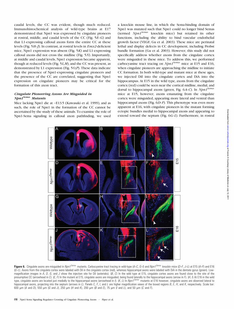
a knockin mouse line, in which the Sema-binding domain ofNpn1 was mutated such that Npn1 could no longer bind Semas(termed Npn1Sema- knockin mice) but retained its otherfunctions, including the ability to bind vascular endothelialgrowth factor (VEGF; Gu et al. 2003). These mice are perinatallethal and display defects in CC development, including Probstbundle formation (Gu et al. 2003). However, this study did notspecifically address whether axons from the cingulate cortexwere misguided in these mice. To address this, we performedcarbocyanine tract tracing on Npn1Sema- mice at E15 and E16,when cingulate pioneers are approaching the midline to initiateCC formation. In both wild-type and mutant mice at these ages,we injected DiI into the cingulate cortex and DiA into thehippocampus. At E15 in the wild type, axons from the cingulatecortex (red) could be seen near the cortical midline, medial, anddorsal to hippocampal axons (green, Fig. 6A--C). In Npn1Sema-
mice at E15, however, axons emanating from the cingulatecortex were misguided, appearing more lateral and ventral thanhippocampal axons (Fig. 6D--F). This phenotype was even moreapparent at E16, with cingulate pioneers in the mutant formingectopic bundles medial to hippocampal axons and appearing toextend toward the septum (Fig. 6G--L). Furthermore, in rostral
Figure 6. Cingulate axons are misguided in Npn1Sema- mutants. Carbocyanine tract tracing in wild-type (A--C, G--I) and Npn1Sema- knockin mice (D--F, J--L) at E15 (A--F) and E16(G--L). Axons from the cingulate cortex were labeled with DiI in the cingulate cortex (red), whereas hippocampal axons were labeled with DiA in the dentate gyrus (green). Low-magnification images in A, D, G, and J show the injection site for DiI (asterisks). (B, C) In the wild type at E15, cingulate cortex axons are found close to the site of thepresumptive CC (arrowhead in C). (E, F) In the mutant at E15, cingulate axons are misguided, being found laterally to the hippocampal axons (arrow in F). (H, I) At E16 in the wildtype, cingulate axons are located just medially to the hippocampal axons (arrowhead in I). (K, L) In Npn1Sema- mutants at E16 however, cingulate axons are observed lateral tohippocampal axons, projecting into the septum (arrows in L). Panels C, F, I, and L are higher magnification views of the boxed regions B, E, H, and K, respectively. Scale bar:600 lm (A and D), 550 lm (G and J), 250 lm (H and K), 200 lm (B and E), 75 lm (I and L), and 50 lm (C and F).
i18 Npn1-Sema Signaling Regulates Crossing of Cingulate Pioneering Axons d Piper et al.
at Francis A Countw
ay Library of Medicine on July 12, 2011
cercor.oxfordjournals.orgD
ownloaded from
sections of E17 Npn1Sema- mice, we observed aberrant extensionof axons into the septum (Supplementary Fig. 2) using anothermarker for cingulate pioneer axons, DCC (Shu et al. 2000).These data demonstrate that pioneering axons from thecingulate cortex are misguided in Npn1Sema- mutants, suggest-ing that the callosal defects reported in these mice (Gu et al.2003) are likely to stem from this defect. This further implicatesNpn1 as a key player in CC development.
Discussion
The formation of mature axon tracts is preceded by theextension of axons from pioneer neurons, whose processesinitiate tract formation by projecting along specific trajectories.For instance, the 2 major efferent projections from the cortex,the corticofugal pathway, and the CC are pioneered by axonsoriginating in the subplate (McConnell et al. 1989) and thecingulate cortex (Koester and O’Leary 1994), respectively. Wehave demonstrated here that Npn1 plays a pivotal role inguiding pioneer axons from the cingulate cortex, providing aninsight into the molecular mechanisms regulating how corticalpioneering axons are guided as they initiate tract formation.Three pieces of evidence, in particular, emphasize the im-portance of Npn1 for the guidance of cingulate pioneer axons.First, the spatiotemporal expression of Npn1 and its corecep-tors in cingulate neurons and axons coupled with theexpression of class 3 Semas at the cortical midline is indicativeof a role in axon guidance during the period in which the firstaxons cross the CC (Rash and Richards 2001). Second, thepresence of guidance defects in cingulate pioneer axons fromNpn1Sema- knockin mice demonstrates a requirement for Npn1function in the pathfinding of these processes. Third, theabsence (rostral) or presence (caudal) of Npn1-expressingpioneers correlates with the presence of the CC in Emx2-deficient mice, a finding that is indicative of a causal linkbetween expression of this receptor on cingulate pioneers andsubsequent formation of the callosal tract.
The expression of Npn1, its coreceptors, and its ligandswithin the cingulate cortex and the cortical midline, during theperiod in which the CC is first formed, provide us witha framework in which to comprehensively investigate the mole-cular determinants underlying callosal formation. One immedi-ate challenge is to determine which molecules are the primaryligands for Npn1 expressed on cingulate pioneers. Our dataindicate that axons from cingulate cortex explants or fromhemisected cortical slices are repelled from cell blocks transi-ently transfected with Sema3A. These findings are consistentwith the published reports that in in vitro culture experiments,axons from cortical explants (collectively including bothcallosally and subcortically projecting axons) are repelled bySema3A (Bagnard et al. 1998) and imply that the repulsiveactivity of Sema3A in the IZ and subcallosal sling may channelcingulate pioneers to the midline during development. Indeed,Sema3A has been demonstrated to be a repellent for axons ina variety of experimental paradigms in vitro. However, althoughSema3A-deficient mice demonstrate defects in sensory axonprojections in the periphery and the brainstem (Ulupinar et al.1999), these defects are later corrected (White and Behar 2000),and moreover, no significant defects have been noted in theforebrain axonal projections of these mice (Catalano et al. 1998),including the CC (this study). We did observe that Npn1expression was slightly reduced in the Sema3A mutants,
indicating that some Npn1 positive axons may have beeneliminated during development. Thus, although relevant in an invitro context, the contribution of Sema3A in vivo to theguidance of axonal populations, including cingulate pioneers,is unclear. Our in situ hybridization analysis also indicates thatSema3C is an excellent candidate for guiding cingulate pioneeraxons. Sema3C, expressed in the IZ (E15 and E16) and theindusium griseum glia and subcallosal sling (E17), is an attractantin vitro for neurites from neocortical explants (Bagnard et al.1998) and cingulate cortex explants (this study, Fig. 4). Althougha Sema3C knockout mouse has been generated (Feiner et al.2001), a thorough analysis to determine if CC formation isaberrant in these mice has yet to be performed.
Our study demonstrates that axons from cingulate pioneersare misguided in Npn1Sema- knockin mice. This confirms therequirement for Npn1-Sema signaling in the guidance of thecallosal pioneer axons. Our data suggest that both attractive(Sema3C) and repulsive (Sema3A) cues may be required forthe guidance of the callosal pioneers. For example, Sema3C couldbe required to attract the pioneer axons to the midline, whereafter crossing, they could then be repelled by Sema3A. Suchdifferential guidance mechanisms would be possible throughspecific coreceptor binding or different intracellular signalingpathways being activated at the appropriate developmental stage.
When interpreting the phenotype of Npn1Sema- knockinmice, it is important to note that Npn1, although being unableto bind Semas (Gu et al. 2003), is still expressed on cingulateaxons and may be capable of binding to VEGF (Gu et al. 2002)or promoting cell--cell adhesion (Shimizu et al. 2000). Assuch, we cannot determine the full extent of Npn1 functionin the Npn1Sema- knockin mice. A recent report using RNAinterference-mediated knockdown of Npn1 in vivo has dem-onstrated that this receptor is required for radial migrationof cortical neurons during development (Chen et al. 2008).A similar experimental approach may provide insights into thefunction of Npn1 during CC development, as would examiningcallosal development in a conditional knockout of Npn1.
Emx2 is a homeodomain transcription factor expressed ina graded pattern within the developing neocortex that is highcaudally and medially and low rostrally and laterally (Mallamaciet al. 1998). Emx2 is postulated to regulate arealization withinthe neocortex in a combinatorial fashion with Pax6 (Bishopet al. 2000). This hypothesis is supported by analyses of Emx2-deficient mice, in which the rostral and lateral neocortical areasare expanded at the expense of caudal and medial areas(Bishop et al. 2000). How can we frame our findings within thishypothesis? Although Emx2 is a transcription factor, it seemsunlikely that it directly regulates Npn1 expression, as Npn1expression is only lost in rostral regions of Emx2-deficientbrains. Although another transcription factor may be compen-sating for Emx2 function in more caudal areas of the mutant,a more likely explanation is that the expansion of rostral andlateral regions results in a more caudal localization of the areaof the cingulate cortex that would normally express Npn1.Driving expression of Npn1 in vivo using electroporationdirectly into the rostral cingulate cortex in Emx2 mutantswould provide a way to rigorously analyze the contribution ofthis receptor to the guidance of cingulate pioneers and,moreover, to address the importance of these pioneers to theguidance of later arriving neocortical axons. In summary, ourstudy indicates that Npn1 plays a central role in the guidance ofpioneering axons from the cingulate cortex.
Cerebral Cortex July 2009, V 19 N Supplement1 i19
at Francis A Countw
ay Library of Medicine on July 12, 2011
cercor.oxfordjournals.orgD
ownloaded from

Supplementary Material
Supplementary Figures 1 and 2 can be found at: http://www.cercor.oxfordjournals.org/.
Funding
National Health and Medical Research Council (NHMRC;401616 and 456027 to L.J.R.); National Institutes of Health(NS039050 and NS049048 to R.E.); L.J.R. is a NHMRC SeniorResearch Fellow; M.P. holds an NHMRC Biomedical CareerDevelopment Award.
Notes
We thank Sarah Croft, Daphne Kusters, Lynnette Knowles, ChantelleReid, Erica Little, Barri King, and John Baisden for technical assistance.We are very grateful to Dr David Ginty (John Hopkins University,Baltimore, MD) for providing the Npn1Sema- knockin mouse line, theNpn1 antibody, and Npn1 in situ probe; Dr Helen Cooper (QueenslandBrain Institute, Brisbane, Australia) for providing the DCC antibody; DrPeter Gruss (Max Planck Society, Munich, Germany) for providing theEmx2-deficient line; and Dr Andreas W. Puschel (Genetik, WestfalischeWilhelms-Universitat Munster, Munster, Germany) for providing theSema expression constructs and Sema in situ probes. We also thankJonathan Hunt (Queensland Brain Institute, Brisbane, Australia) for helpin modifying and implementing the algorithm for measuring neuriteoutgrowth and guidance. Conflict of Interest : None declared.
Address correspondence to email: [email protected].
References
Adams RH, Lohrum M, Klostermann A, Betz H, Puschel AW. 1997. Thechemorepulsive activity of secreted semaphorins is regulated byfurin-dependent proteolytic processing. EMBO J. 16:6077--6086.
Andrews GL, Mastick GS. 2003. R-cadherin is a Pax6-regulated, growth-promoting cue for pioneer axons. J Neurosci. 23:9873--9880.
Andrews W, Liapi A, Plachez C, Camurri L, Zhang J, Mori S, Murakami F,Parnavelas JG, Sundaresan V, Richards LJ. 2006. Robo1 regulates thedevelopment of major axon tracts and interneuron migration in theforebrain. Development. 133:2243--2252.
Bagnard D, Lohrum M, Uziel D, Puschel AW, Bolz J. 1998. Semaphorinsact as attractive and repulsive guidance signals during the de-velopment of cortical projections. Development. 125:5043--5053.
Bagri A, Marin O, Plump AS, Mak J, Pleasure SJ, Rubenstein JL, Tessier-Lavigne M. 2002. Slit proteins prevent midline crossing and determinethe dorsoventral position of major axonal pathways in the mammalianforebrain. Neuron. 33:233--248.
Barry G, Piper M, Lindwall C, Moldrich R, Mason S, Little E, Sarkar A,Tole S, Gronostajski RM, Richards LJ. 2008. Specific glial populationsregulate hippocampal morphogenesis. J Neurosci. 28:12328--12340.
Behar O, Golden JA, Mashimo H, Schoen FJ, Fishman MC. 1996.Semaphorin III is needed for normal patterning and growth ofnerves, bones and heart. Nature. 383:525--528.
Bishop KM, Goudreau G, O’Leary DD. 2000. Regulation of area identity inthe mammalian neocortex by Emx2 and Pax6. Science. 288:344--349.
Campbell CE, PiperM,PlachezC,YehYT, Baizer JS, Osinski JM, LitwackED,Richards LJ, Gronostajski RM. 2008. The transcription factor Nfixis essential for normal brain development. BMC Dev Biol. 8:52.
Castellani V. 2002. The function of neuropilin/L1 complex. Adv ExpMed Biol. 515:91--102.
Castellani V, Chedotal A, Schachner M, Faivre-Sarrailh C, Rougon G.2000. Analysis of the L1-deficient mouse phenotype reveals cross-talk between Sema3A and L1 signaling pathways in axonal guidance.Neuron. 27:237--249.
Catalano SM, Messersmith EK, Goodman CS, Shatz CJ, Chedotal A. 1998.Many major CNS axon projections develop normally in the absenceof semaphorin III. Mol Cell Neurosci. 11:173--182.
Chen G, Sima J, Jin M, Wang KY, Xue XJ, Zheng W, Ding YQ, Yuan XB.2008. Semaphorin-3A guides radial migration of cortical neuronsduring development. Nat Neurosci. 11:36--44.
De Carlos JA, O’Leary DD. 1992. Growth and targeting of subplate axonsand establishment of major cortical pathways. J Neurosci. 12:1194--1211.
Farmer WT, Altick AL, Nural HF, Dugan JP, Kidd T, Charron F,Mastick GS. 2008. Pioneer longitudinal axons navigate using floorplate and Slit/Robo signals. Development. 135:3643--3653.
Fazeli A, Dickinson SL, Hermiston ML, Tighe RV, Steen RG, Small CG,Stoeckli ET, Keino-Masu K, Masu M, Rayburn H, et al. 1997.Phenotype of mice lacking functional Deleted in colorectal cancer(Dcc) gene. Nature. 386:796--804.
Feiner L, Webber AL, Brown CB, Lu MM, Jia L, Feinstein P, Mombaerts P,Epstein JA, Raper JA. 2001. Targeted disruption of semaphorin 3Cleads to persistent truncus arteriosus and aortic arch interruption.Development. 128:3061--3070.
Ghosh A, Antonini A, McConnell SK, Shatz CJ. 1990. Requirement forsubplate neurons in the formation of thalamocortical connections.Nature. 347:179--181.
Ghosh A, Shatz CJ. 1993. A role for subplate neurons in the patterningof connections from thalamus to neocortex. Development. 117:1031--1047.
Gu C, Limberg BJ, Whitaker GB, Perman B, Leahy DJ, Rosenbaum JS,Ginty DD, Kolodkin AL. 2002. Characterization of neuropilin-1structural features that confer binding to semaphorin 3A andvascular endothelial growth factor 165. J Biol Chem. 277:18069--18076.
Gu C, Rodriguez ER, Reimert DV, Shu T, Fritzsch B, Richards LJ,Kolodkin AL, Ginty DD. 2003. Neuropilin-1 conveys semaphorin andVEGF signaling during neural and cardiovascular development. DevCell. 5:45--57.
He Z, Tessier-Lavigne M. 1997. Neuropilin is a receptor for the axonalchemorepellent Semaphorin III. Cell. 90:739--751.
Kawasaki T, Kitsukawa T, Bekku Y, Matsuda Y, Sanbo M, Yagi T,Fujisawa H. 1999. A requirement for neuropilin-1 in embryonicvessel formation. Development. 126:4895--4902.
Keeble TR, Halford MM, Seaman C, Kee N, Macheda M, Anderson RB,Stacker SA, Cooper HM. 2006. The Wnt receptor Ryk is required forWnt5a-mediated axon guidance on the contralateral side of thecorpus callosum. J Neurosci. 26:5840--5848.
Koester SE, O’Leary DD. 1994. Axons of early generated neurons incingulate cortex pioneer the corpus callosum. J Neurosci. 14:6608--6620.
Lindwall C, Fothergill T, Richards LJ. 2007. Commissure formation inthe mammalian forebrain. Curr Opin Neurobiol. 17:3--14.
Mallamaci A, Iannone R, Briata P, Pintonello L, Mercurio S, Boncinelli E,Corte G. 1998. EMX2 protein in the developing mouse brain andolfactory area. Mech Dev. 77:165--172.
McConnell SK, Ghosh A, Shatz CJ. 1989. Subplate neurons pioneer thefirst axon pathway from the cerebral cortex. Science. 245:978--982.
McConnell SK, Ghosh A, Shatz CJ. 1994. Subplate pioneers and theformation of descending connections from cerebral cortex. JNeurosci. 14:1892--1907.
Murakami Y, Suto F, Shimizu M, Shinoda T, Kameyama T, Fujisawa H.2001. Differential expression of plexin-A subfamily members in themouse nervous system. Dev Dyn. 220:246--258.
Pellegrini M, Mansouri A, Simeone A, Boncinelli E, Gruss P. 1996.Dentate gyrus formation requires Emx2. Development. 122:3893--3898.
Perala NM, Immonen T, Sariola H. 2005. The expression of plexinsduring mouse embryogenesis. Gene Expr Patterns. 5:355--362.
Piper M, Dawson AL, Lindwall C, Barry G, Plachez C, Richards LJ. 2007.Emx and Nfi genes regulate cortical development and axonguidance in the telencephalon. Novartis Found Symp.288:230--242;[discussion 242--235, 276--281].
Piper M, Georgas K, Yamada T, Little M. 2000. Expression of thevertebrate Slit gene family and their putative receptors, the Robogenes, in the developing murine kidney. Mech Dev. 94:213--217.
Plachez C, Lindwall C, Sunn N, Piper M, Moldrich RX, Campbell CE,Osinski JM, Gronostajski RM, Richards LJ. 2008. Nuclear factor I geneexpression in the developing forebrain. J CompNeurol. 508:385--401.
Plachez C, Richards LJ. 2005. Mechanisms of axon guidance in thedeveloping nervous system. Curr Top Dev Biol. 69:267--346.
i20 Npn1-Sema Signaling Regulates Crossing of Cingulate Pioneering Axons d Piper et al.
at Francis A Countw
ay Library of Medicine on July 12, 2011
cercor.oxfordjournals.orgD
ownloaded from

Pozas E, Pascual M, Nguyen Ba-Charvet KT, Guijarro P, Sotelo C,Chedotal A, Del Rio JA, Soriano E. 2001. Age-dependent effects ofsecreted Semaphorins 3A, 3F, and 3E on developing hippocampalaxons: in vitro effects and phenotype of Semaphorin 3A (–/–) mice.Mol Cell Neurosci. 18:26--43.
Rash BG, Richards LJ. 2001. A role for cingulate pioneering axons in thedevelopment of the corpus callosum. J Comp Neurol. 434:147--157.
Ren T, Anderson A, Shen WB, Huang H, Plachez C, Zhang J, Mori S,Kinsman SL, Richards LJ. 2006. Imaging, anatomical, and molecularanalysis of callosal formation in the developing human fetal brain.Anat Rec A Discov Mol Cell Evol Biol. 288:191--204.
Rosoff WJ, Urbach JS, Esrick MA, McAllister RG, Richards LJ,Goodhill GJ. 2004. A new chemotaxis assay shows the extremesensitivity of axons to molecular gradients. Nat Neurosci. 7:678--682.
Serafini T, Colamarino SA, Leonardo ED, Wang H, Beddington R,Skarnes WC, Tessier-Lavigne M. 1996. Netrin-1 is required forcommissural axon guidance in the developing vertebrate nervoussystem. Cell. 87:1001--1014.
Shimizu M, Murakami Y, Suto F, Fujisawa H. 2000. Determination of celladhesion sites of neuropilin-1. J Cell Biol. 148:1283--1293.
Shu T, Butz KG, Plachez C, Gronostajski RM, Richards LJ. 2003.Abnormal development of forebrain midline glia and commissuralprojections in Nfia knock-out mice. J Neurosci. 23:203--212.
Shu T, Richards LJ. 2001. Cortical axon guidance by the glial wedgeduring the development of the corpus callosum. J Neurosci.21:2749--2758.
Shu T, Valentino KM, Seaman C, Cooper HM, Richards LJ. 2000.Expression of the netrin-1 receptor, deleted in colorectal cancer
(DCC), is largely confined to projecting neurons in the developingforebrain. J Comp Neurol. 416:201--212.
Smith KM, Ohkubo Y, Maragnoli ME, Rasin MR, Schwartz ML, Sestan N,Vaccarino FM. 2006. Midline radial glia translocation and corpuscallosum formation require FGF signaling. Nat Neurosci. 9:787--797.
Super H, Martinez A, Del Rio JA, Soriano E. 1998. Involvement of distinctpioneer neurons in the formation of layer-specific connections inthe hippocampus. J Neurosci. 18:4616--4626.
Takahashi T, Fournier A, Nakamura F, Wang LH, Murakami Y, Kalb RG,Fujisawa H, Strittmatter SM. 1999. Plexin-neuropilin-1 complexesform functional semaphorin-3A receptors. Cell. 99:59--69.
Tamagnone L, Artigiani S, Chen H, He Z, Ming GI, Song H, Chedotal A,Winberg ML, Goodman CS, Poo M, et al. 1999. Plexins are a largefamily of receptors for transmembrane, secreted, and GPI-anchoredsemaphorins in vertebrates. Cell. 99:71--80.
Ulupinar E, Datwani A, Behar O, Fujisawa H, Erzurumlu R. 1999. Role ofsemaphorin III in the developing rodent trigeminal system. Mol CellNeurosci. 13:281--292.
Weaver CM, Pinezich JD, Lindquist WB, Vazquez ME. 2003. Analgorithm for neurite outgrowth reconstruction. J NeurosciMethods. 124:197--205.
White FA, Behar O. 2000. The development and subsequent eliminationof aberrant peripheral axon projections in Semaphorin3A nullmutant mice. Dev Biol. 225:79--86.
Yorke CH, Jr, Caviness VS, Jr. 1975. Interhemispheric neocorticalconnections of the corpus callosum in the normal mouse: a studybased on anterograde and retrograde methods. J Comp Neurol.164:233--245.
Cerebral Cortex July 2009, V 19 N Supplement1 i21
at Francis A Countw
ay Library of Medicine on July 12, 2011
cercor.oxfordjournals.orgD
ownloaded from