Neuropathology of Schizophrenia: Cortex, Thalamus, Basal Ganglia
19
Neuropathology of Schizophrenia: Cortex, Thalamus, Basal Ganglia, and Neurotransmitter-Specific Projection Systems by Stephan Heckers Abstract This article reviews neuropathological studies in the search for an anatomical correlate of schizophrenia. Replication of many results has proven to be difficult. A consistent finding is the lack of significant gliosis in the neocortex. Intriguing findings that need further corroboration include decreased volume and cell num- ber of the mediodorsal thalamic nucleus, cytoarchitec- tural alterations of the prefrontal cortex and upper layers of the anterior cingulate gyms, and superior temporal gyrus abnormalities. Most neuropathological studies investigate regional brain volume and cell den- sity. Highly variable shrinkage of brain tissue post- mortem makes these estimates prone to bias and often not comparable across studies. So far, no strong clini- copathological correlations and no pathological crite- ria to diagnose schizophrenia have been established. Schizophrenia Bulletin, 23(3):403--421,1997. Neuropathologists have made substantial progress in the search for causes of neuropsychiatric disorders. Initial findings such as ventricular enlargement or gross brain atrophy were often neither specific nor sensitive, but they allowed for the differentiation of abnormal from normal brains. Some pathological findings, such as plaques and tangles in Alzheimer's disease (Khachaturian 1985) or loss of striatal neurons in Huntington's disease (VonSattel et al. 1985) are now used to make and stage a diagnosis. Ventricular enlargement, reported for many but not all schizophrenia patients, has long been regarded as indica- tion of an underlying pathology of cortex, white matter, or subcortical structures in schizophrenia (Southard 1915). Is ventricular enlargement a clue that points to a neurode- generative or neurodevelopmental abnormality in schizo- phrenia (Benes 19936)? This review assesses progress in the neuropathologi- cal understanding of schizophrenia—with an emphasis on more recent studies of the prefrontal cortex, the anterior cingulate cortex, the auditory cortex, the thalamus, the basal ganglia, and the brainstem. Limbic structures of the medial temporal lobe are reviewed by Dwork (1997, this issue). Studies of the ventricular system, now performed more accurately by in vivo imaging techniques, are reviewed elsewhere (Shelton and Weinberger 1986; Kotrla and Weinberger 1995) and are not included here. Structural neuroimaging studies that are directly relevant to interpreting postmortem studies will be included. Requirements for Neuropathological Studies of Schizophrenia The scientific rigor of neuropathological studies of patients with schizophrenia has improved in recent years. I will briefly address requirements now considered essen- tial for a sound neuropathological study (table 1; for fur- ther review, see Benes 1988; Casanova and Kleinman 1990; Kleinman et al. 1995) and will focus on a less well- known source of error: postmortem artifacts. Reliable Diagnosis of Schizophrenia. Only recently have investigators begun to study patients who were diag- nosed using standardized criteria. This makes it difficult to compare older studies with more recent ones: patients with dementia praecox and schizophrenia diagnosed before 1980 might be quite different from the schizophrenia patients diagnosed according to DSM-III or DSM-III-R criteria (American Psychiatric Association 1980, 1987). Even if only those patients who fulfill the current diagnos- tic criteria are included, they still may represent different subgroups of schizophrenia (catatonic, disorganized, para- noid, undifferentiated, and residual type). Conflicting Reprint requests should be sent to Dr. S. Heckers, Dept. of Psychiatry, Massachusetts General Hospital, Warren 605, Fruit St., Boston, MA 02114. 403 Downloaded from https://academic.oup.com/schizophreniabulletin/article/23/3/403/1886122 by guest on 22 December 2021
Transcript of Neuropathology of Schizophrenia: Cortex, Thalamus, Basal Ganglia

Neuropathology of Schizophrenia:Cortex, Thalamus, Basal Ganglia, and
Neurotransmitter-Specific Projection Systemsby Stephan Heckers
Abstract
This article reviews neuropathological studies in thesearch for an anatomical correlate of schizophrenia.Replication of many results has proven to be difficult.A consistent finding is the lack of significant gliosis inthe neocortex. Intriguing findings that need furthercorroboration include decreased volume and cell num-ber of the mediodorsal thalamic nucleus, cytoarchitec-tural alterations of the prefrontal cortex and upperlayers of the anterior cingulate gyms, and superiortemporal gyrus abnormalities. Most neuropathologicalstudies investigate regional brain volume and cell den-sity. Highly variable shrinkage of brain tissue post-mortem makes these estimates prone to bias and oftennot comparable across studies. So far, no strong clini-copathological correlations and no pathological crite-ria to diagnose schizophrenia have been established.
Schizophrenia Bulletin, 23(3):403--421,1997.
Neuropathologists have made substantial progress in thesearch for causes of neuropsychiatric disorders. Initialfindings such as ventricular enlargement or gross brainatrophy were often neither specific nor sensitive, but theyallowed for the differentiation of abnormal from normalbrains. Some pathological findings, such as plaques andtangles in Alzheimer's disease (Khachaturian 1985) orloss of striatal neurons in Huntington's disease (VonSattelet al. 1985) are now used to make and stage a diagnosis.Ventricular enlargement, reported for many but not allschizophrenia patients, has long been regarded as indica-tion of an underlying pathology of cortex, white matter, orsubcortical structures in schizophrenia (Southard 1915). Isventricular enlargement a clue that points to a neurode-generative or neurodevelopmental abnormality in schizo-phrenia (Benes 19936)?
This review assesses progress in the neuropathologi-cal understanding of schizophrenia—with an emphasis on
more recent studies of the prefrontal cortex, the anteriorcingulate cortex, the auditory cortex, the thalamus, thebasal ganglia, and the brainstem. Limbic structures of themedial temporal lobe are reviewed by Dwork (1997, thisissue). Studies of the ventricular system, now performedmore accurately by in vivo imaging techniques, arereviewed elsewhere (Shelton and Weinberger 1986;Kotrla and Weinberger 1995) and are not included here.Structural neuroimaging studies that are directly relevantto interpreting postmortem studies will be included.
Requirements for NeuropathologicalStudies of Schizophrenia
The scientific rigor of neuropathological studies ofpatients with schizophrenia has improved in recent years.I will briefly address requirements now considered essen-tial for a sound neuropathological study (table 1; for fur-ther review, see Benes 1988; Casanova and Kleinman1990; Kleinman et al. 1995) and will focus on a less well-known source of error: postmortem artifacts.
Reliable Diagnosis of Schizophrenia. Only recentlyhave investigators begun to study patients who were diag-nosed using standardized criteria. This makes it difficult tocompare older studies with more recent ones: patients withdementia praecox and schizophrenia diagnosed before1980 might be quite different from the schizophreniapatients diagnosed according to DSM-III or DSM-III-Rcriteria (American Psychiatric Association 1980, 1987).Even if only those patients who fulfill the current diagnos-tic criteria are included, they still may represent differentsubgroups of schizophrenia (catatonic, disorganized, para-noid, undifferentiated, and residual type). Conflicting
Reprint requests should be sent to Dr. S. Heckers, Dept. ofPsychiatry, Massachusetts General Hospital, Warren 605, Fruit St.,Boston, MA 02114.
403
Dow
nloaded from https://academ
ic.oup.com/schizophreniabulletin/article/23/3/403/1886122 by guest on 22 D
ecember 2021

Schizophrenia Bulletin, Vol. 23, No. 3, 1997 S. Heckers
results of methodologically comparable studies mighttherefore be due to a true difference in the patient popula-tion (Andreasen et al. 1994b).
Comparison of Schizophrenia and Control Cases.The majority of earlier studies compared brains of schizo-phrenia patients with those the individual investigatorconsidered to be "normal." Many important contribu-tions—including those of Alzheimer (1897, 1913) andSouthard (1914, 1915)—still await confirmation in con-trolled studies.
Matching of Cases for Gender and Age. Samples ofnormal individuals as well as schizophrenia patients showsignificant gender differences and age-related changes ofparameters, such as regional brain volume (e.g., Schroderet al. 1975; DeLisi et al. 1989; de Lacoste et al. 1990;Heckers et al. 1990, 1991b). Differences between schizo-phrenia patients and normal samples are difficult toattribute to diagnosis if the samples have incompatiblegender or age distributions.
Controlling for Presence of Neurological Disorders.The search for pathological changes in schizophreniapatients should not preclude a thorough screening forother pathological entities that could explain the psychoticfeatures observed during a lifetime (Jellinger 1985). Forexample, patients with tumors and vascular lesions, evenin areas not under study, should be excluded. Age-relatedchanges (e.g., neurofibrillary tangles, neuritic plaques)should be documented.
Quantifying Abnormalities Without Knowledge of theDiagnosis. When schizophrenia cases are comparedwith control cases, they should be studied without knowl-edge of the diagnosis. Furthermore, the parameter understudy should be quantified to allow for the comparison ofdifferent studies.
Table 1. Requirements for neuropathologlcalstudies of schizophrenia
1. Reliable diagnosis of schizophrenia2. Comparison of schizophrenia and control cases3. Matching of cases for gender and age4. Controlling for presence of neurological disorders5. Quantification of abnormalities without knowledge of the
diagnosis
Postmortem ArtifactsWhen an investigator has taken all the precautions out-lined above, the major challenge will be to assess and
control for tissue changes after autopsy. The most signifi-cant effect to control for is the individual shrinkage ofbrain tissue. Previous studies have demonstrated thatbrain tissue shrinks in a highly variable fashion (between40% and 60%) during fixation and staining (Schroder etal. 1975; Mouritzen Dam 1979; Heckers et al. 1990).
Tissue shrinkage affects the two most-studied para-meters, volume and cell density. If the entire brain isavailable, brain volume can be determined after autopsyand fixation using the Archimedes principle (Weibel1979) and after staining using the Cavalieri estimator(Gundersen et al. 1988a, 1988b). An individual shrinkagefactor can then be calculated and the volume estimate canbe corrected. Most investigators, however, do not have theentire brain at hand, and they estimate volume in a seriesof brain sections by multiplying the area of the slice by itsthickness (Rosen and Harry 1990). If this volume estimateis not corrected by an individual shrinkage factor, it ishighly biased and may reflect more the individual post-mortem shrinkage of brain tissue than the proposed vol-ume in vivo (Schroder et al. 1975; Gundersen 1992).
Cell-density estimates (cell number per unit volume)are equally biased if they are based on uncorrected volumeestimates. Two additional sources of bias can affect theaccuracy of cell-density estimates. First, counting all cellswithin one defined volume leads to overcounting cells thatare only partially located within the scanned volume. Thelarger the cell and the thinner the section, the greater theoverestimate. There are now simple and unbiased methodsto avoid this counting error (Williams and Rakic 1988;Gundersen 1992). Second, estimates of cell density areoften established in only a few microscopic fields that arenot sampled systematically throughout the entire anatomi-cal area studied. While such studies may demonstrate highreliability among raters, they do not necessarily reflect thetrue cell number in the anatomical area studied.
Total cell number (cell density X volume) is a muchmore powerful parameter. It is independent of any tissueshrinkage and can therefore be compared across studies(Coggeshall 1992; Gundersen 1992; Pakkenberg 1992).Total cell-number estimates require two important steps:First, volume is determined in a systematic sample ofslices (typically about five sections in simple structuressuch as the amygdala or the thalamus). Second, cell den-sity is determined in systematically sampled microscopicfields, which ensures that the estimates are a true represen-tation of the sample. Each investigator can calculate thecoefficient of error for the total cell-number estimate,which predicts the accuracy and reproducibility of the esti-mate. For example, two studies of the total neuron numberin the subfields of the human hippocampus arrived at verysimilar estimates (both with coefficients of error below10%), although volumes and cell densities were quite dif-
404
Dow
nloaded from https://academ
ic.oup.com/schizophreniabulletin/article/23/3/403/1886122 by guest on 22 D
ecember 2021
Neuropathology of Schizophrenia Schizophrenia Bulletin, Vol. 23, No. 3, 1997
ferent due to different fixation and staining procedures(West and Gundersen 1990; Heckers et al. 1991a).
If total cell number cannot be calculated, it is stillpossible to compare different cell populations within thesame specimen (e.g., neuronal versus glial cells, pyrami-dal versus nonpyramidal neurons, or cytochemically dif-ferent cells visualized by immunohistochemistry or in situhybridization), since they are theoretically affectedequally by postmortem tissue shrinkage.
Investigators using immunohistochemical or in situhybridization techniques must consider the possibility thatthe amount of protein or the messenger ribonucleic acid(mRNA) levels change postmortem before they are visu-alized. Rigorous matching for autolysis time and incuba-tion parameters, as well as quantitative analysis, is neces-sary (Zoli et al. 1990). One additional way to reducepotential artifacts is to compare different cell populationslabeled with the same or even different probes within thesame specimen. A differential change—when one class ofcells is altered between samples but another isunchanged—makes it less likely that the observed differ-ence across samples is due to postmortem alterations.
The most likely contributors to the divergence of
results of the studies reviewed here include true biologicaldifferences in subgroups of schizophrenia subjects;methodological errors, such as insufficient matching forgender and age; and the use of biased estimates, such asvolume and cell density.
Neuropathological Findings
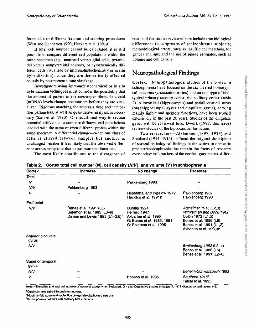
Cortex. Neuropathological studies of the cortex inschizophrenia have focused on the six-layered homotypi-cal isocortex (association cortex) and on one type of idio-typical primary sensory cortex, the auditory cortex (table2). Allocortical (hippocampus) and periallocortical areas(parahippocampal gyms and cingulate gyms), servingmainly limbic and memory functions, have been studiedextensively in the past 20 years. Studies of the cingulategyrus will be reviewed here; Dwork (1997, this issue)reviews studies of the hippocampal formation.
Two researchers—Alzheimer (1897, 1913) andSouthard (1914, 1915)—offered the original descriptionof several pathological findings in the cortex in dementiapraecox/schizophrenia that remain the focus of researcheven today: volume loss of the cortical gray matter, differ-
Table 2. Cortex total cell number (N), cell density (N/V), and volume (V) In schizophreniaCortex
TotalNN/VV
PrefrontalN/V
Anterior cingulategyrusN/V
Superior temporalgyrus
N/VV
Increase
-
Pakkenberg 1993-
Benesetal. 1991 (L5)Selemon et al. 1995 (L3-6)Daviss and Lewis 1993 (L1-3.5)1
-
-
No change
Pakkenberg 1993-
Rosenthal and Bigelow 1972Heckers et al. 1991b
Dunlap 1924Ferrero 1947Akbarian et al. 1995G: Benes et al. 1986, 1991G: Selemon et al. 1995
—
-
Hobsonetal. 1995
Decrease
--
Pakkenberg 1987Pakkenberg 1993
Alzheimer 1913 (L2,3)Winkelman and Book 1949Colon 1972 (L4,5)Benesetal. 1986 (L6)Benesetal. 1991 (L1.2)Akbarian et al. 1993a*
Braitenberg 1952 (L2-4)Benesetal. 1986 (L5)Benesetal. 1991 (L2-6)
Beheim-Schwarzbach 1952
Southard 19153
Falkaietal. 1995Note.—Densities and total cell number of neurons except when Indicated. Q = glia; Qualitative studies in Italics; P-1-6] indicates cortical layers 1-6.
'Caltnndin- and calretWn-posittve neurons.2Nlcotinam)de adenine dlnucfeotlde phosphate-diaphorase neurons.'Schizophrenia patients with auditory hallucinations.
405
Dow
nloaded from https://academ
ic.oup.com/schizophreniabulletin/article/23/3/403/1886122 by guest on 22 D
ecember 2021

Schizophrenia Bulletin, Vol. 23, No. 3, 1997 S. Hcckers
ential vulnerability of cortical neurons toward injury, lowglial/neuronal cell ratio, and abnormalities in the positionof neurons within the cortical layers. Alzheimer's firststudy (1897) described three cases with overall decreasedneuronal density and unchanged glial cells and two caseswith normal neuronal density with "Schiefstellung"(skewed position) of neurons and increased glial density.His second report (1913) of 18 cases stressed neuronalloss primarily in layers 2 and 3, skewed position of pyra-midal cells, and decreased neuropil.
Southard (1910), initially skeptical about morpholog-ical changes in schizophrenia, studied 25 cases of demen-tia praecox and found internal hydrocephalus, more left-sided than right-sided lesions, temporal lesions (especiallyleft superior temporal gyrus hypoplasia) in associationwith auditory hallucinations, parietal atrophy and sclero-sis in catatonia, and frontal-lobe aplasia or atrophy inpatients with delusions (Southard 1914, 1915). Theselesions primarily affected the upper cortical layers andwere not associated with pronounced gliosis (Southard1919).
Neither investigator classified his patients accordingto current criteria, used quantitative techniques, or wasblind to the diagnosis during the study. It is thereforeremarkable that recent quantitative studies described neu-ronal loss in layers 2 and 3 of the prefrontal cortex andcingulate gyrus (see below), lack of gliosis (see below andDwork 1997, this issue), abnormal orientation of pyrami-dal cells (see Dwork 1997, this issue), and superior tem-poral gyrus abnormalities (see below). However, many ofthese findings are still subject to debate.
Entire cortex. Pakkenberg (1987) reported a 12percent reduction of total cortical volume in schizophreniapatients, whereas others found no significant differencebetween schizophrenia subjects and controls (Rosenthaland Bigelow 1972; Heckers et al. 19916). Pakkenberg'sfinding (1987) of decreased cortex volume was derivedfrom a sample of 29 institutionalized, chronic schizophre-nia patients, with a mean age of 74 years, compared with asample of 30 controls with a mean age of 71 years. A sub-sequent study of a subset of eight schizophrenia casesshowed increased cell density and, therefore, a normaltotal neuron number in all four lobes of the cerebral cortex(Pakkenberg 1993). This stereological study demonstratedthat decreased volume or increased cell density alone can-not be used to predict the total cell number.
Prefrontal cortex. The dorsolateral-prefrontal cor-tex—here broadly defined as an associational isocorticalregion, including Brodmann areas 8 to 10 and 44 to 47(Broca's area) (Zilles 1990)—has been studied repeatedly,with conflicting results. Many early studies, with methodssimilar to those of Alzheimer's, described qualitativechanges, but often with different results (for reviews, see
Winkelman and Book 1949; Peters 1956; David 1957).There is even one report of neuronal density in frontal-cortex specimens removed during surgery from 22 schizo-phrenia patients and 1 manic-depressive patient (Rowlandand Mettler 1949). Here I will review the quantitativestudies of neuronal density, specific neuronal markers, andglial density. Quantitative studies of neuronal density areconflicting (i.e., reporting normal, increased, anddecreased cell density) and inconclusive.
Normal neuronal density: Dunlap (1924) studiedeight cases of dementia praecox (ages 20 to 44 years) andfive controls (ages 17 to 45 years), finding normal neu-ronal density in the superior frontal gyrus. Ferrero (1947)studied area 9 in five different groups of neuropsychiatricpatients, including 12 schizophrenia subjects (9 femalesand 3 males, with a mean age of 67). He found somewhatthicker layers 3b, 5b, and 6a in the schizophrenia sampleand highly variable cell densities, which was not differentfrom the normal variation. Akbarian et al. (1995, seebelow) found no difference in the cell density of all orsmall neurons of area 9.
Increased neuronal density: Benes et al. (1991)described increased pyramidal cell density selectively inlayer 5 of area 10 in a sample of 18 schizophrenia andschizoaffective patients, compared with 12 controls.Selemon et al. (1995) studied neuronal and glial density inprefrontal area 9 and occipital area 17 in 16 schizophreniasubjects, 19 control cases, 6 schizoaffective patients, and9 with Huntington's disease. Schizophrenia patients hadsignificantly increased neuronal density in areas 9 (17%)and 17 (10%). In area 9, pyramidal and nonpyramidalneuronal density was increased in layers 3 to 6, with thelargest increase (24%) in layers 5 to 6. Cortical thicknesswas reduced by 8 percent in the schizophrenia sample,reaching significance for layer 5 (7% decrease) only.Mean glial density was not significantly changed. Sinceglial density correlated negatively with the time of fixa-tion in formaldehyde, all cases (14 controls, 6 people withschizophrenia) fixated for less than 5 years were analyzedseparately. This revealed a 22 percent higher glial densityin those with schizophrenia, which did not reach signifi-cance. Cell density was estimated using an unbiasedmethod (Williams and Rakic 1988), but was not correctedfor the significant shrinkage of the tissue blocks duringfixation in formaldehyde and embedding in celloidin.
The fact that the schizophrenia cases were fixated sig-nificantly longer than the control cases—and the extremeindividual variation of tissue shrinkage during fixation andstaining (40% to 60%, see above)—make the finding ofincreased cell density in an unknown reference volumedifficult to interpret. Furthermore, increased neuronal den-sity in schizophrenia was not specific for the prefrontalcortex, since it was also found in the occipital cortex.
406
Dow
nloaded from https://academ
ic.oup.com/schizophreniabulletin/article/23/3/403/1886122 by guest on 22 D
ecember 2021

Neuropathology of Schizophrenia Schizophrenia Bulletin, Vol. 23, No. 3, 1997
Daviss and Lewis (1993) studied a subpopulation of corti-cal interneurons with antibodies against calbindin and cal-retinin. They found an increased density of these local cir-cuit neurons in layers 1 to 3 and 5 in area 46 of five schiz-ophrenia patients compared with five controls.
Decreased neuronal density: Winkelman and Book(1949) studied 10 schizophrenia subjects and founddecreased neuronal density primarily in the frontal cortex.They did not provide a quantitative analysis but describedgeneral as well as focal loss of neurons, increased numberof astroglia, and mild subcortical demyelinization. Colon(1972) reported marked neuronal loss (57%) primarily indeeper layers 4 and 5 of area 10 (and also for areas 4 and24). Benes et al. (1986) reported significantly decreasedneuronal density in layer 6 of area 10 in a sample of sevenschizophrenia cases compared with nine control cases. Ina subsequent study of a different sample of 18 schizophre-nia and schizoaffective patients and 12 controls, Benes etal. (1991) found decreased density of small neurons inlayers 1 and 2 of area 10.
Akbarian et al. (1993a) studied a small subset of cor-tical neurons that express the enzyme nicotinamide ade-nine dinucleotide phosphate-diaphorase (NADPH-d) infive schizophrenia patients matched for age, sex, andautolysis time with five controls. Some gray- as well aswhite-matter neurons of the adult human brain expressNADPH-d, which is coexpressed with, or even identicalto, nitric oxide synthase. These neurons have been shownto be particularly resistant to cytotoxic injury. In all schiz-ophrenia subjects, NADPH-d neurons were significantlyfewer in the cortex and directly adjacent white matter, andfour persons with schizophrenia showed a significantlyincreased number in the deep white matter. This bimodalpattern of altered NADPH-d cell density was interpretedas dislocation of neurons or impaired programmed celldeath rather than as cell loss; neurons that are destined toreach the subplate and then the cortex either arrest duringdevelopment in the white matter or are spared from pro-grammed cell death.
Studies of neuronal markers: Glantz and Lewis(1993) found a significant decrease of synaptophysinimmunoreactivity (a marker of synapse density) in a sam-ple of six schizophrenia patients compared with six sex-and age-matched controls in areas 9 and 46. Horton et al.(1993) used the immunoreactivity for ubiquitin (a heat-shock related protein) as a probe for neurodegeneration ina sample of 17 schizophrenia subjects (mean age 69) and26 controls (mean age 72). They found no difference inthe degree of ubiquination in the prefrontal cortex.Akbarian et al. (1995) studied the left superior frontalgyms (Brodmann area 9) in 10 chronic schizophreniapatients and 10 controls matched for age, gender, andautolysis time. As mentioned above, they found no differ-
ence in the density of all neurons or in the density ofsmall, round, or ovoid neurons in any of the six corticallayers. However, they found significantly fewer neuronsexpressing mRNA for glutamic acid decarboxylase67
(GAD67) in layers 1 to 5 in the schizophrenia sample.GAD67 is one of the two isoforms of GAD and is presentin both the cell bodies and the axon terminals of gamma-aminobutyric acid (GABA)ergic neurons throughout thecentral nervous system. The difference in GAD67 mRNAwas most pronounced in layers 2 (48% decrease) and 1(40% decrease) and was still significant in layers 3 to 5(30% decrease). In contrast, the density of neuronsexpressing calcium-calmodulin-dependent protein kinase(CamKH) mRNA was not different between samples.
This differential mRNA change in two neuronal pop-ulations makes methodological errors of the in situ proto-col less likely, although not impossible (see Lee and Tobin1995); it also makes postmortem shrinkage less of a prob-lem. Decreased GAD mRNA in the absence of significantcell loss could be a sign of functional down-regulation ofinhibitory interneurons in a cortex that has developed nor-mally and has not undergone significant degeneration dur-ing life.
Glial density: In contrast to the conflicting results ofneuronal density studies, glial density was consistentlyfound to be normal. The two studies by Benes et al.(1986, 1991) determined glial density in the prefrontalcortex and found no significant differences betweenschizophrenia patients and controls. Selemon et al.(1995) reported an increased neuronal density in theschizophrenia sample without a significant change inglial density.
Conclusion: The most robust finding of neuropatho-logical studies of the prefrontal cortex in schizophrenia isthe lack of significant gliosis. However, qualitative studieshave not consistently shown changes of neurons, as hasbeen suggested by the large body of qualitative studies.Even studies of the same anatomical areas (e.g., for area9) have revealed conflicting results (Dunlap 1924; Ferrero1947; Akbarian et al. 1995; Selemon et al. 1995).Different fixation techniques, staining methods, andcounting algorithms make it virtually impossible to com-pare results of the different studies. It is fair to say thatchanges in neuron number in the frontal cortex of schizo-phrenia patients are not as dramatic and widespread as inneurodegenerative diseases such as Huntington's diseaseor Alzheimer's disease. However, only unbiased estimatesof total cell number, which can be compared across stud-ies, will reliably assess glial and neuronal number inschizophrenia. The studies by Pakkenberg (1987, 1993),showing increased neuronal density in a significantlysmaller cortex in schizophrenia, exemplify that volume orcell density alone cannot predict total cell number.
407
Dow
nloaded from https://academ
ic.oup.com/schizophreniabulletin/article/23/3/403/1886122 by guest on 22 D
ecember 2021

Schizophrenia Bulletin, Vol. 23, No. 3, 1997 S. Heckers
If there is cell loss, are subsets of cortical neurons—such as GABAergic interneurons, glutamatergic pyrami-dal cells, and NADPH-d-positive neurons—selectivelyvulnerable to injury? If so, at which point in the life cycleof the cortical neuron does the injury occur? Studies usingimmunocytochemical, in situ hybridization, and receptor-binding techniques are needed to answer these questions.The increased neuron/glia ratio, as reported by someinvestigators, is often interpreted as evidence for develop-mental abnormalities in the prefrontal cortex in schizo-phrenia. The finding of displaced NADPH-d positive neu-rons in the prefrontal cortex by Akbarian et al. (1993a)adds new evidence to this line of thinking. It will beimportant to confirm this finding in a larger sample and tostudy whether other types of cortical cells are affected in asimilar fashion (i.e., displaced to the white matter).
Anterior cingulate cortex. The anterior part of thecingulate gyms (Brodmann area 24) is a periallocortical,agranular area that is generally considered to be part ofthe limbic system. Braitenberg (1952) studied this area in30 cases, including 7 with schizophrenia, from the Vogtcollection (table 2). In a purified sample of nine caseswithout any additional neuropathological alterations, hefound decreased neuronal density and decreased stainingof nerve cells with Nissl-stain, interpreted as a sign ofdegeneration, in layers 2 to 4 in the schizophreniapatients, but not in the controls. One of the very few elec-tronmicroscopical studies of schizophrenia patients(Aganova and Uranova 1992) reported swelling ofsynapses in layers 1 and 2 of area 24.
Benes and coworkers studied area 24 extensivelyusing Nissl-stain, immunohistochemistry, and autoradiog-raphy (Benes 1993a). Their initial finding (in a sample of10 schizophrenia subjects with a mean age of 60, and 10controls with a mean age of 66) was decreased neuronaldensity in layer 5 in the schizophrenia cases (Benes et al.1986). A second study of the same sample describedabnormal aggregates of layer 2 neurons, which weresmaller and separated by wider distances in the schizo-phrenia cases (Benes and Bird 1987). The hypothesis thatthese wider spaces were filled with vertical axons wasstudied using antibodies against the 200-kilodalton neuro-filament subunit (a component of the axonal cytoskeleton)and against glutamate (Benes et al. 1987, 1992a). Schizo-phrenia patients showed 25 percent more vertical axons,which might indicate an increased associative input intothe anterior cingulate gyms in schizophrenia.
A third cell density study in a sample of 18 schizo-phrenia and schizoaffective patients (mean ages 53 and 49,respectively) and 12 controls (with a mean age of 59)revealed a significant deficit in small interneurons in layers2 to 6 without changes in glial density (Benes et al. 1991).
To test whether the loss of small cortical neurons, pre-sumably GABAergic interneurons, is related to changes inGABA receptor density, autoradiography was used tolocalize the binding of bicuculline-sensitive titrated musci-mol in a sample of six schizophrenia patients and eightcontrols (Benes et al. 19926). This study showed a signifi-cant and preferential increase in GABAA receptor bindingin layers 2 and 3, but not in layers 5 and 6 in persons whohad schizophrenia, with and without exposure to neurolep-tics, which could suggest a compensatory up-regulation ofGABAA receptors secondary to the loss of GABAergicinterneurons of layers 2 and 3.
Conclusion: The quantitative findings of Benes et al.are in accordance with Braitenberg's (1952) previousqualitative description and have been integrated into acomprehensive neuroanatomical model of anterior cingu-late dysfunction in schizophrenia (Benes 1993a). Briefly,increased excitatory input via associate fibers, decreasedinhibitory input due to loss of inhibitory interneurons, andincreased dopaminergic tone cause excess firing of thepyramidal cell of the anterior cingulate gyrus. Themethodology of the Benes studies is sound (blind quanti-tation, matched samples, correction for confounding vari-ables). However, several features will make it difficult tocompare her results with those of others to come. First,cell-density estimates are highly sensitive to differentialtissue shrinkage postmortem. Second, the distinction ofsmall neurons from pyramidal neurons and from glialcells is not trivial. Further, her small neuron/pyramidalneuron ratio, as well as neuron/glia ratio, are very differ-ent from those of other investigators (for review, seeBraak and Braak 1986). Finally, the second-highest den-sity of small neurons in area 24 was found in layer 4,although area 24 is defined as agranular cortex, whichlacks layer 4 (Brodmann 1909; Zilles 1990).
Auditory cortex. The primary auditory and uni-modal auditory association areas (Brodmann areas 41, 42,22), located on the superior temporal gyrus, make up theonly sensory cortical region reported to be altered in schiz-ophrenia (table 2). Southard (1914, 1915) was the first todocument decreased volume in the left superior temporalgyrus in dementia praecox patients with auditory hallucina-tions. He studied 25 patients with dementia praecox, diag-nosed as either paranoid or catatonic. Of the 12 cases withauditory hallucinations, 9 showed "striking evidence oftemporal-lobe involvement" (especially a smaller left supe-rior temporal gyrus), whereas only 4 of the 13 cases with-out evidence of hallucinations showed some kind of tempo-ral-lobe pathology. This pathology was not significantlycorrelated with "internal hydrocephalus," which Southardfound in nine cases (five without hallucinations, two ofwhich showed temporal-lobe abnormalities, and four withhallucinations, three of which showed such abnormalities).
408
Dow
nloaded from https://academ
ic.oup.com/schizophreniabulletin/article/23/3/403/1886122 by guest on 22 D
ecember 2021

Neuropathology of Schizophrenia Schizophrenia Bulletin, Vol. 23, No. 3, 1997
Southard's study did not include controls or otherpsychiatric cases and was not quantitative. His studydesign was more directed toward detecting clinicopatho-logical correlations in individual cases, which he studiedexhaustively with the available techniques of his time. Itis therefore important to refer to two recent in vivo imag-ing studies (Barta et al. 1990; Shenton et al. 1992), whichreported a significant association between auditory hallu-cinations or thought-disorder index and superior temporalgyms volume in schizophrenia patients. However, prelim-inary data from a recent postmortem study of 19 suchpatients and 21 control subjects do not indicate a signifi-cant difference in the volume of the left or right superiortemporal gyms in schizophrenia subjects (Hobson et al.1995). Considering Southard's findings, it will be impor-tant to subdivide this sample into schizophrenia patientswith and without auditory hallucinations.
Beheim-Schwarzbach (1952) reported a qualitative,cytological study of the superior temporal gyms in a sam-ple of 21 schizophrenia cases from the Vogt collection.All cases showed normal cell densities in the internalHeschl gyrus but "gaps of cells" in the more lateralaspects of the superior temporal gyms, primarily in layers3 and 5. Abnormalities of NADPH-d-positive neurons inmedial and lateral parts of the temporal lobe (Akbarian etal. 1993b), similar to those described by the same authorsfor the frontal lobe (see above), are reviewed in detail byDwork (1997, this issue).
The planum temporale, of which the posterior supe-rior temporal gyms is an external landmark, is intimatelyinvolved in the production and comprehension of lan-guage. In right-handed individuals, the surface area of theleft planum temporale is usually much larger than that ofthe right. Falkai et al. (1995) reported disturbed planumtemporale asymmetry in a sample of 24 schizophreniapatients and 24 control subjects matched by sex and age.The asymmetry coefficients for the planum temporalecortex volume and anterior-posterior diameter were sig-nificantly different between schizophrenia patients andcontrols. Five magnetic resonance imaging (MRI) studiesof planum temporale asymmetry found either a signifi-cant reduction in the normal left > right asymmetry(DeLisi et al. 1994; Rossi et al. 1994; Petty et al. 1995)or no difference in the planum temporale asymmetrycoefficient (Kleinschmidt et al. 1994; Kulynych et al.1995).
One pathological study investigated sylvian fissureasymmetry, which is closely related to planum temporaleand superior temporal gyms asymmetry in the human cor-tex. Falkai et al. (1992) reported a 16 percent shorter leftsylvian fissure in schizophrenia subjects who lacked theusual left > right asymmetry. Unfortunately, they com-
pared photographs of lateral views from postmortemspecimens on which parts of the sylvian fissure cannot beassessed if hidden by overlapping cortex. One in vivostudy (Bartley et al. 1993) used the superior technique ofmeasuring sylvian fissure length in three-dimensional cor-tical renderings from MRI images of 10 normal monozy-gotic twins and 10 monozygotic twins discordant forschizophrenia. Both sets showed the normal sylvian fis-sure asymmetry, and there was no difference within the 10discordant twin pairs.
Conclusion: Left superior temporal gyms abnormali-ties have been described in schizophrenia patients (Falkaiet al. 1995), especially in those with auditory hallucina-tions (Southard 1914, 1915). The lack of such changes inschizophrenia cases without auditory hallucinations wouldestablish a stronger clinicopathological correlation.Furthermore, other brain areas that are potentiallyinvolved in abnormal auditory perception should beinvestigated. The intriguing finding by Southard, therecent pathological studies of Falkai et al., and the recentMRI studies (Barta et al. 1990; Shenton et al. 1992) raisefurther questions: How does decreased volume of thesuperior temporal gyrus correlate to functional abnormali-ties of the temporal lobe (McCarley et al. 1993;Waddington 1993)? What is the underlying cellular abnor-mality? Further analysis of this brain region in schizo-phrenia seems warranted.
Thalamus. Few investigators have studied the thalamus,and most have singled out the mediodorsal nucleus (thesecond largest and phylogenetically most progressed thal-amic nucleus) for study (table 3). This nucleus is charac-terized by two different neuronal types (magno- and par-vocellular) and topographical organization of afferent andefferent connections to the prefrontal cortex and limbicstructures.
The volume of individual thalamic nuclei, as well asthe entire group of thalamic nuclei, was found to beunchanged in at least two studies. Rosenthal and Bigelow(1972) found similar volumes of the thalamus in 10 schiz-ophrenia cases compared with 10 controls, matched forgender and age. Lesch and Bogerts (1984) studied 15schizophrenia and 12 control cases from the Vogt collec-tion, which were not matched for gender, and found thevolume of all large thalamic subnuclei and of the thala-mus as a whole unchanged in schizophrenia. However,they found a reduced thickness of the periventricular graymatter surrounding the third ventricle. Stein and Ziegler(1939) reported a morphometric analysis of the entirethalamus in 28 patients with dementia praecox and 6 withmanic-depressive illness. This well-performed quantita-tive study found no significant differences in total thala-
409
Dow
nloaded from https://academ
ic.oup.com/schizophreniabulletin/article/23/3/403/1886122 by guest on 22 D
ecember 2021

Schizophrenia Bulletin, Vol. 23, No. 3, 1997 S. Heckers
Table 3. Thalamus total cell number (N), cell density (N/V), and volume (V) In schizophreniaThalamus Increase No change Decrease
TotalN/V
Mediodorsal nucleus
N
N/V
Funfgeld 1954Baumer 1954
Hempel and Treff 1959 (G)
Stein and Ziegler 19391
Rosenthal and Bigelow 1972Lesch and Bogerts 1984
Dometal. 1981Pakkenberg 1990 (N+G)
Pakkenberg 1990 (N+G)Treff and Hempel 1958
Pakkenberg 1990Note.—Densities and total cell number of neurons except when indicated: G « glla, N+G = neurons and glia; Qualitatrve studies in italics.
'Schizophrenia patients compared with other psychiatric patients.
mic volume or cell density between these two samples ofneuropsychiatric patients.
Pakkenberg (1990) found a significantly decreasedvolume of the left mediodorsal nucleus in a sample of 12schizophrenia patients (mean age 63), compared with 12controls (mean age 62). Of interest, a recent MRI study of39 male schizophrenia patients with a mean age of 30 and47 male controls, matched for age, height, and educa-tional level, found thalamic abnormalities (Andreasen etal. 1994a). Effect-size mapping (i.e., subtracting the aver-aged MRIs of all patients from those of all controls andsearching for signal-intensity difference of each voxel)revealed significant differences primarily on the right sideand in lateral regions, as well as the adjacent white matter.This was interpreted as evidence of decreased thalamicsize, mainly due to changes in lateral parts of the thala-mus. However, Andreasen et al. did not perform area mea-surements to arrive at volume estimates, which makes itdifficult to compare this study with previous volumetricstudies.
Funfgeld (1954) and Baumer (1954) studied cellularchanges of the thalamus in brains of the Vogt collection.Funfgeld reported cell loss and "Schwundzellen" (dwarfor wasting cells, as defined by changes of Nissl-stainedcytoplasmic material) in seven out of eight cases of cata-tonic schizophrenia. Baumer studied 51 brains, including17 from schizophrenia cases, and also found cell loss andSchwundzellen—primarily in the mediodorsal, anterior,and ventral thalamic nuclei. In a series of quantitativestudies of the mediodorsal nucleus in primarily catatonicschizophrenia patients, Hempel and Treff reported areduction of neuronal density (maximally 44%), increasedglial density, increased volume of glial cells, andincreased volume of neuronal nuclei (Treff and Hempel1958, 1959; Hempel and Treff 1959). While they usedquantitative techniques, they were aware of the diagnosisat the time of the analysis, and the sample size in each of
the studies was smaller than 10. Dom et al. (1981) com-pared five catatonic schizophrenia cases with five normalones and found no differences in medial thalamus neu-ronal density.
The study by Pakkenberg (1990), which examinedonly left hemispheres, provides an unbiased estimate oftotal cell number in the mediodorsal nucleus of the thala-mus. In the sample of 12 schizophrenia subjects, cell den-sity was not significantly reduced, but the total number ofneurons (-40%), astrocytes (-44%), and oligodendrogliacells (—45%) was significantly decreased. All schizophre-nia subjects were chronically institutionalized, all hadbeen treated with neuroleptics, seven had developed dys-kinesias, and some had received electroconvulsive ther-apy and insulin coma treatment; however, these confound-ing variables were not correlated with cell number. Thiselegant stereological study provides absolute cell numbersthat can be compared across studies.
Conclusion: The most compelling finding is that byPakkenberg (1990) of decreased total neuronal and glialnumber in the mediodorsal thalamic nucleus of schizo-phrenia cases. The study design is optimal, and the findingis much more solid than the previous studies of cell den-sity in the brains of the Vogt collection. However,Pakkenberg's patient sample might not be representativefor schizophrenia, and the various treatments of the schiz-ophrenia subjects might have affected the finding.Furthermore, we do not know whether the neuronal lossaffects the different cell types or the medial and lateralparts of the mediodorsal nucleus differentially.Confirmation by an independent stereological study in adifferent sample would establish neuronal loss in themediodorsal nucleus as a very robust neuropathologicalfinding in schizophrenia research.
The finding of thalamic abnormalities, as reported byAndreasen et al. (1994a), is in contrast to at least two vol-umetric postmortem studies (Rosenthal and Bigelow
410
Dow
nloaded from https://academ
ic.oup.com/schizophreniabulletin/article/23/3/403/1886122 by guest on 22 D
ecember 2021

Neuropathology of Schizophrenia Schizophrenia Bulletin, Vol. 23, No. 3, 1997
1972; Lesch and Bogerts 1984). However, it is conceiv-able that the right thalamic abnormalities, attributed to lat-eral aspects of the thalamus in the Andreasen study, aredue to a primary shrinkage of the mediodorsal nucleus.This would be similar to the finding of a smaller leftmediodorsal thalamic nucleus in schizophrenia (Pakken-berg 1990). Several important questions deserve furtherstudy: Is the mediodorsal thalamic nucleus smaller inschizophrenia? If so, is there hemispheric asymmetry?Are sensory relay nuclei in more lateral parts of the thala-mus equally affected?
Basal Ganglia. The neostriatum (caudate nucleus, puta-men), the limbic striatum (nucleus accumbens), and theglobus pallidus have been studied neuropathologically inschizophrenia (table 4). Four studies found decreased ornormal volumes of basal ganglia structures. From the Vogtcollection, Bogerts et al. (1985) studied the left hemi-spheres of 13 schizophrenia cases (10 females, 3 males)with a mean age of 41, and 9 controls (3 females, 6 males)with a mean age of 53. They found normal volumes of thecaudate nucleus, putamen, nucleus accumbens, and exter-nal globus pallidus, but a 20 percent volume reduction ofthe internal globus pallidus. A second study (Bogerts et al.1990) of a new brain collection covered both hemispheresof 18 people with schizophrenia (9 females, 9 males, meanage 52) and 21 controls (7 females, 14 males, mean age54). This second study had essentially the same results.Brown et al. (1986) compared 41 schizophrenia patients(mean age 67) to those with affective psychosis,Huntington's disease, and Alzheimer's disease and found
no significant differences in the area of the striatum andpallidum sampled on only one slice per brain. Pakkenberg(1990) studied the nucleus accumbens and the ventral pal-lidum in the same sample that showed the thalamic abnor-malities (see above). The volume of the nucleus accum-bens was decreased in the schizophrenia subjects, and theventral pallidum was normal.
One stereological postmortem study (Heckers et al.19916) of 23 schizophrenia cases (13 females, 10 males,mean age 62) and 23 sex- and age-matched controls foundincreased volumes of the striatum and globus pallidus inthe schizophrenia sample, which was significant for thestriatum on the left and for the globus pallidus on theright. Of interest, this surprising postmortem finding wassubsequently corroborated by MRI studies (Jernigan et al.1991; Breier et al. 1992; Swayze et al. 1992; Buchanan etal. 1993; Elkashef et al. 1994). A recent MRI study(Chakos et al. 1994) of 29 first-episode schizophreniapatients and 10 healthy control subjects measured the vol-umes of cerebral cortex, lateral ventricles, and caudatenuclei at the beginning of the study and after 18 months.Patients were treated with neuroleptic medication duringthis time. On followup, the patients showed a 5.7 percentvolume increase of the caudate nuclei, whereas the con-trols showed a 1.6 percent volume decrease. This indi-cates that volume increase in the caudate nucleus can beseen in young first-episode schizophrenia cases and that itmay be related to exposure to neuroleptic medication. Asimilar change in caudate volume with neuroleptic treat-ment was reported by Keshavan et al. (1994). Of interest,Chakos et al. (1995) reported reversal of caudate volume
Table 4. Basal ganglia total cell number (N), cell density (N/V), and volume (V) In schizophreniaBasal ganglia
NeostriatumNA/V
Nucleus accumbensN
N/V
V
Globus pallidusNV
Increase
-
Heckers et al. 1991 b
-
Stevens 1982 (G)_
-
Heckers etal. 1991b
No change
-
Bogerts etal. 1985Brown etal. 19861
Bogerts etal. 1990
-
-
Bogerts etal. 1985Bogerts et al. 1990
Pakkenberg 19902 (N+G)
Pakkenberg 19902
Decrease
Hopf1954—
Pakkenberg 1990 (N+G)-
Pakkenberg 1990
-
Bogerts etal. 19853
Bogerts etal. 19903
Note.—Densities and total cell number of neurons except when Indicated: Q <= glia, N+G •> neurons and glia; Qualitative studies in Italics.
'Schizophrenia patients compared wtth other psychiatric patients; area measurement only.Central pallidum.'Internal globus pallidus.
411
Dow
nloaded from https://academ
ic.oup.com/schizophreniabulletin/article/23/3/403/1886122 by guest on 22 D
ecember 2021

Schizophrenia Bulletin, Vol. 23, No. 3, 1997 S. Heckers
increase seen with typical neuroleptics after switching toclozapine.
Neuronal density was investigated in four studies.Hopf (1954) found Schwundzellen (dwarf cells) in 10 outof 10 catatonic schizophrenia patients and no changes inparanoid and hebephrenic schizophrenia cases from theVogt collection. Stevens (1982) reported an extensive,qualitative neuropathological study of 25 schizophreniasubjects (mean age 44, diagnosed retrospectively accord-ing to the ninth revision of the International Classificationof Diseases [World Health Organization 1978] criteria),28 nonschizophrenia psychiatric patients, and 21 controls.For schizophrenia cases (with sarcoidosis, colloid cyst,astrocytoma, and Alzheimer's disease, respectively) wereexcluded, but two schizophrenia cases with bilateralglobus pallidus infarcts remained in the study. The impor-tant findings in the striatum of the schizophrenia samplewere patchy fibrillary gliosis in the nucleus accumbensand loss of primarily large neurons in the globus pallidus.Arendt et al. (1983) studied 14 controls and 58 cases ofneuropsychiatric disorders, including 3 female schizo-phrenia cases (mean age 48) who showed normal neuro-nal density in the external globus pallidus. The stereologi-cal study by Pakkenberg (1990) found normal neuronaland glial densities in the globus pallidus and nucleusaccumbens. The total number of neurons and glial cells inthe nucleus accumbens was decreased due to significantvolume loss (see above).
Two studies used immunohistochemical methods tostudy the striatum in schizophrenia. Heckers et al. (1993)found decreased expression of choline acetyltransferase instriatal cholinergic interneurons in a sample of six schizo-phrenia and schizoaffective patients compared with sixcontrols. Mai et al. (1993) reported a morphometric analy-sis of structures containing the neuropeptides vasopressinand oxytocin, using neurophysin-immunoreactivity as amarker, in 11 schizophrenia and 10 control cases of theVogt collection. Fiber density was decreased in the globuspallidus, most pronounced in the internal pallidum, andincreased in the substantia nigra.
Conclusion: Despite the long interest in an abnormaldopaminergic innervation of the striatum in schizophre-nia, there are only few and equivocal neuropathologicalstudies. The striatum shows a complex organization ofGABAergic projection neurons, cholinergic interneurons,and glutamatergic and dopaminergic afferents. There areno good quantitative, case-control studies of this complexcytochemical organization in schizophrenia. Do thenucleus accumbens and globus pallidus show degenerativechanges? The quantitative study by Pakkenberg (1990) didnot find decreased neuronal density and increased glialdensity, as suggested by the qualitative research of Hopf
(1954) and Stevens (1982). Recent MRI studies haveshown that the finding of basal ganglia volume increase(Heckers et al. 19916) seems to be closely related to theuse of typical neuroleptics. Which component of the basalganglia contributes to this plasticity? So far, we have con-flicting results and need more advanced neuropathologicalstudies of the striatum in schizophrenia.
Neurotransmitter-Specific Projection Systems. Fourgroups of neurons (dopaminergic neurons in the substan-tia nigra and ventral tegmental area, cholinergic neuronsin the basal forebrain and upper brainstem, noradrenergicneurons in the locus ceruleus, and serotonergic neurons inthe raphe nuclei) project diffusely to cortical and subcorti-cal areas and modulate signal transmission. In contrast tothe extensive search for neurochemical abnormalities inschizophrenia, few structural studies are reported for thesesystems (table 5).
Three qualitative studies described periventricularand periaqueductal gliosis potentially affecting diffuselyprojecting neurotransmitter systems. Fisman (1975) stud-ied 10 controls and 24 psychiatric patients using qualita-tive neuropathological methods. Controls and psychiatricpatients showed brainstem lesions, but six out of theseven schizophrenia cases without other cerebral diseaseshowed "glial nodules and perivascular infiltration," inter-preted as the result of a viral infection. The qualitativestudy by Stevens (1982), which reported striatal abnor-malities (see above), revealed patchy fibrillary gliosis"that was maximal in the periventricular and periaqueduc-tal regions." Bruton et al. (1990) reported a prospective,extensive pathological study of 56 schizophrenia patientsand 56 age- and sex-matched controls. They found a "sig-nificant increase in fibrous glia" in the cerebral cortex,white matter, and periventricular structures. Cortical andwhite matter gliosis were significantly correlated withfocal brain damage (e.g., degeneration of the substantianigra, calcification of the hippocampus, tumors, andstrokes) but periventricular gliosis was not. Thus, the eti-ology of periventricular gliosis might be different fromcortical and white matter gliosis.
Two studies showed no increased gliosis in the brain-stem of schizophrenia cases. Hankoff and Peress (1981)studied 27 patients (8 schizophrenia and 19 nonschizo-phrenia control cases) and described clusters of microglialcells or perivascular infiltrates in 7 of their cases, but inonly 1 schizophrenia patient. Stevens et al. (1988)counted astroglial cells that stained with an antibodyagainst glial fibrillary acidic protein (a marker for onesubtype of astrocytes). They found normal astrocyte den-sity in the periventricular gray matter of the third ventriclein five schizophrenia subjects, compared with seven con-
412
Dow
nloaded from https://academ
ic.oup.com/schizophreniabulletin/article/23/3/403/1886122 by guest on 22 D
ecember 2021

Neuropathology of Schizophrenia Schizophrenia Bulletin, Vol. 23, No. 3,1997
Table 5. Brainstem/basal forebrain total cell number (N), cell density (N/V), and volume (V) inschizophreniaBrainstem/basalforebrain Increase No change Decrease
BrainstemN/V
Substantia nigraN
V
Locus ceruleusN
Fisman 1975 (G)Stevens 1982 (G)Brutonetal. 1990 (G)
—
Stevens etal. 1988 (G)Hankoff and Peress 1981(G)
Bogertsetal. 1983 (N+G)
—
Karsonetal. 1991
N/V
Bogertsetal. 1983
Garcia-Rill et al. 1995Lohrand Jeste 1988
PPN + LTDN
NbMN/V
Karson etal. 1991Garcia-Rill et al. 1995
Manayeetal. 1995
ArendtetaJ. 1983EI-MaJlakh et al. 1991
Von Buttiar-Brentano 1952Averback 1981
Note.—Densities and total cell number of neurons except when Indicated: Q •= glla; N+Q « neurons + glla; Qualitative studies in Italics; PPN = peduncutopon-tine nucleus; LTD = lateral dorsal tegmental nucleus; NbM - nucleus basaBs of Meynert.
trol cases. Furthermore, the caudate nucleus of four schiz-ophrenia cases showed astrocyte density similar to thecontrol cases and significantly less compared with fourcases with Huntington's disease.
Four quantitative studies of dopaminergic and norad-renergic neurons revealed no significant changes in cellnumber. Bogerts et al. (1983) looked at the neuromelanin-containing neurons of the substantia nigra/ventral tegmen-tal area of a sample of six schizophrenia patients notexposed to neuroleptic treatment, comparing them withthose of six controls. The volume of the lateral substantianigra area was decreased, and the size of neuromelanin-containing perikarya in the mesolimbic part of thedopaminergic cell group decreased. There were nochanges in the total number of nerve cells and glial cells.Lohr and Jeste (1988) studied neuromelanin-containingcells of the locus ceruleus in 15 leukotomized schizophre-nia patients (mean age 46), 11 leukotomized patients withdiagnoses other than schizophrenia (mean age 52), and 13controls (mean age 52). There was a nonsignificant trendfor decreased locus ceruleus volume but normal cell den-sity and total neuron number. Karson et al. (1991) andGarcia-Rill et al. (1995) used an antibody against tyrosinehydroxylase to stain the catecholaminergic neurons of thelocus ceruleus. Both studies found no change of total neu-ron number, but Karson et al. (1991) reported a signifi-cantly reduced cell size of the locus ceruleus neurons inschizophrenia subjects.
Two qualitative studies described degeneration ofcholinergic basal forebrain neurons. Van Buttiar-Brentano(1952) studied nine schizophrenia cases from the Vogt col-lection and described a decrease in cell size, but did notgive a description of the normal cell. Averback (1981)studied 35 controls, 45 cases of Huntington's disease, 7 ofAlzheimer's disease, and 13 of schizophrenia. For all threedisease states (including 11 cases of schizophrenia), hereported vacuolar degeneration and glial cell clusters sur-rounding the large neurons of the ansa peduncularis, a sub-group of cholinergic neurons of the basal forebrain.
Two quantitative studies reported normal cell densityof cholinergic basal forebrain neurons: Arendt et al.(1983) for a sample of 3 schizophrenia patients and El-Mallakh et al. (1991) for a sample of 10 intellectuallyimpaired and 7 intellectually intact schizophrenia patients.The projections of the cholinergic basal forebrain, studiedwith acetyl cholinesterase histochemistry in different cor-tical areas, were found to be normal in a sample of sixschizophrenia and schizoaffective patients compared withsix controls (Heckers et al. 1993).
Karson et al. (1991) studied the pedunculopontinenucleus (PPN) and the lateral dorsal tegmental nucleus(LDT), using NADPH-d reactivity as a marker for cholin-ergic neurons. The total number of NADPH-d neuronswas significantly higher in the four schizophrenia cases(mean age 63), compared with the five control cases(mean age 60), which was interpreted as evidence for an
413
Dow
nloaded from https://academ
ic.oup.com/schizophreniabulletin/article/23/3/403/1886122 by guest on 22 D
ecember 2021

Schizophrenia Bulletin, Vol. 23, No. 3, 1997 S. Heckers
increased number of cholinergic brainstem neurons inschizophrenia. In a subsequent study by the same group(Garcia-Rill et al. 1995), this finding was corroborated inat least some of nine schizophrenia patients comparedwith four nonschizophrenia psychiatric cases and fivecontrol cases (eight of the nine previously analyzed caseswere included in the study). Total cell number, determinedby counting all PPN and LDT neurons in every eighthsection through the right brainstem, was increased in theschizophrenia sample by 64 and 56 percent for PPN andLDT, respectively, reaching significance only for the PPNdue to large variance mainly in the schizophrenia sample.As in the previous study, the number of locus ceruleusneurons was unchanged in the schizophrenia sample. TheNADPH-d neurons in all three groups showed a regionaldistribution and mean cell size similar to that known forneurons stained with choline acetyltransferase (ChAT)immunohistochemistry, which supported the assumptionthat NADPH-d can be used as a marker for cholinergicPPN and LDT neurons.
However, a recent study (Manaye et al. 1995) foundno differences in the number of ChAT-positive neurons inthe PPN and LDT of three schizophrenia cases and sixcontrols. Also, in 8 schizophrenia and 11 control cases,there was no difference in the number of Nissl-stainedPPN neurons.
A Western immunoblot study of the pontine tegmen-tum of younger individuals (25 schizophrenia cases with amean age of 34 and 28 nonschizophrenia cases with amean age of 35) revealed a significantly lower concentra-tion of ChAT in the schizophrenia sample (Karson et al.1993). A lower level of the acetylcholine-synthesizingenzyme ChAT seems at variance with an increased num-ber of cholinergic neurons in the same area. One possibleexplanation is a decreased cholinergic input into the pon-tine tegmentum, since most of the ChAT enzyme is local-ized in cholinergic axons and terminals (Garcia-Rill et al.1995).
Conclusion: Gliosis in the periventricular and peri-aqueductal white matter was demonstrated in three quali-tative studies (but not in the quantitative study by Stevenset al. 1988). In contrast to cortical and white matter glio-sis, found in some cases diagnosed with schizophreniathat revealed focal brain damage on pathological exami-nation, periventricular and periaqueductal gliosis were notrelated to any focal damage. The cause remains obscure(remnant of an infectious or inflammatory process?), andfurther studies are needed. Four methodologically soundstudies (providing total cell number) demonstrated normalneuronal and glial numbers in the sustantia nigra and thelocus ceruleus. Quantitative studies of the cholinergicbasal forebrain neurons have demonstrated no significant
changes. Studies of the cholinergic brainstem neurons areconflicting: NADPH-d—positive neurons in the PPN andLDT were found to be elevated in at least some schizo-phrenia subjects, whereas the number of ChAT-positiveneurons was unchanged and the level of ChAT was foundto be decreased.
Discussion
Neuropathological studies of schizophrenia have estab-lished neither a single lesion nor strong clinicopathologi-cal correlations. Moreover, there are no neuropathologicalcriteria to diagnose or stage schizophrenia. Although thestudies reviewed here have progressed from qualitativedescriptions to blind, quantitative, case-control studies,they still produce conflicting results. Neuronal and glialdensity, regional brain volume, and architecture of corticalneurons and gyri are the focus of most current studies.What are the promising leads in these studies? What isnecessary to resolve the dispute over divergent results?
Cell Density Studies. Altered neuronal density in post-mortem specimens can be due to abnormal developmentof neurons, changes of neuron numbers in the developedbrain, decreased or increased neuropil, or tissue changesduring fixation and staining. Decreased neuronal densityaccompanied by increased glial density is typically seenafter degenerative changes of the brain, whereasdecreased neuronal density accompanied by unchangedglial density is often interpreted as an indicator of braininjury during development (because this is a time whenthe glial response to injury is decreased or absent). So far,studies of neuronal density have reported decreased,increased, or unchanged density in the cortex; decreasedor unchanged density in the thalamus and the basal gan-glia; and increased or unchanged density in the brainstem(tables 2 to 5). As pointed out above, it will be importanteither to eliminate the bias introduced by postmortem tis-sue changes by using total cell-number estimates or toreduce this bias by comparing different subpopulations ofcells within the same specimen. This will allow us toanswer the question: Is the neuron number changed inschizophrenia? If there is neuronal loss, are subsets ofneurons differentially vulnerable to injury, and when doesthe injury occur?
Studies so far point to a vulnerability of supragranu-lar cortical neurons (layers 2 and 3) and NADPH-d andGABAergic cortical neurons (Alzheimer 1913; Jakob andBeckmann 1986; Benes 1993a; Akbarian et al. 1993a,1995). The attractive theory of cortical maldevelopmentin schizophrenia proposes an impaired migration of somecortical neurons from the ventricular wall to their final
414
Dow
nloaded from https://academ
ic.oup.com/schizophreniabulletin/article/23/3/403/1886122 by guest on 22 D
ecember 2021

Neuropathology of Schizophrenia Schizophrenia Bulletin, Vol. 23, No. 3, 1997
cortical destination (for review, see Weinberger 1995).This could explain the decreased neuronal density insupragranular layers unaccompanied by a significantincrease in glial density, the increased density of supra-granular neurons in deeper layers, the increased density ofcortical neurons in the underlying white matter, and(although less stringent) an abnormal gyration of the cor-tex. However, decreased neuronal density in supragranu-lar layers is still controversial, and evidence of misplacedcortical neurons is either qualitative or awaits corrobora-tion by further quantitative studies. The most robust find-ing so far is the lack of cortical gliosis. The neurodevelop-mental theory of schizophrenia will be greatly advanced ifa reduction or displacement of (supragranular) neuronscan be established by unbiased and independent studies.
Decreased neuronal density in the striatum, thalamus,and brainstem was reported in earlier studies, whereasmore recent studies have reported a reduced total cell num-ber in the nucleus accumbens and mediodorsal thalamicnucleus, an unchanged cell number in the globus pallidus,and even an increased cell number in the brainstem (tables3 to 5). Increased gliosis in the basal ganglia, thalamus,and brainstem, as reported in earlier studies, could not becorroborated in more recent studies (tables 3 to 5). Whilethere has been a renewed interest in cortical abnormalities,there are very few studies investigating the complex orga-nization of subcortical structures in schizophrenia. Thereare essentially no studies of the different neuronal subpop-ulations in the thalamus and basal ganglia and very few ofthe neurotransmitter-specific projection neurons of thebrainstem. Considering the importance of cortical-subcor-tical interaction and the variety of subcortical targets forneuroleptic medication, it will be a major challenge toexplore a subcortical pathology in schizophrenia.
Volumetric Studies. Three significant volumetricchanges in schizophrenia, originally observed postmortemand now increasingly by in vivo imaging, are a smallerthalamus, larger basal ganglia components, and a smallersuperior temporal gyrus. However, the results are not con-sistent. First, one study reported reduced volume of theleft mediodorsal thalamic nucleus in postmortem speci-mens (Pakkenberg 1990), but in vivo imaging of the thal-amus revealed lateral thalamic abnormalities on the right(Andreasen et al. 19946). Second, postmortem and in vivoimaging studies reported basal ganglia volume increase(Heckers et al. 19916; Jernigan et al. 1991; Breier et al.1992; Swayze et al. 1992; Buchanan et al. 1993), butrecent MRI studies reported a significant effect of neu-roleptic medication on the size of the caudate nucleus inschizophrenia patients (Chakos et al. 1994; Keshavan etal. 1994).
Postmortem volumetric studies, with their intrinsicproblem of tissue shrinkage, will become less important asin vivo imaging techniques become more accurate in delin-eating the boundaries of anatomical structures. Volumetricin vivo studies can follow patients over time, comparepatients on and off medication, and use the powerful twin-and family-study design. Such studies have already addedsignificantly to our understanding of schizophrenia (Kotrlaand Weinberger 1995). However, the major question of vol-umetric studies remains: What does volume change indi-cate? This has been the riddle of ventricular enlargementfound in many but not all schizophrenia patients (Sheltonand Weinberger 1986). Therefore, neuropathological stud-ies will remain crucial in elucidating the microscopicequivalent of volumetric abnormalities.
Architectural Studies. The architecture of cells withinthe cortical mantle and the configuration of gyri and fis-sures on the cortical surface have attracted the interest ofschizophrenia researchers since Alzheimer's study in1897. The current interest in cytoarchitectural abnormali-ties is mainly driven by the neurodevelopmental theory: Ifschizophrenia is not characterized by massive cell lossand significant gliosis in the adult brain (as suggested bymost of the studies), the "schizophrenia lesion" might beacquired early in development, could be more subtle, andcould involve qualitative alterations of cell shape, cellposition, connectivity, and the normally asymmetricdevelopment of gyri and fissures in both hemispheres.Most of the cytoarchitectural findings are reported for thelimbic and paralimbic structures of the temporal lobe (seeDwork 1997, this issue), but abnormalities of the superiortemporal gyrus (Southard 1915; Barta et al. 1990;Shenton et al. 1992; Falkai et al. 1995) and displacementof NADPH-d-positive cortical neurons to the underlyingwhite matter (Akbarian et al. 1993a, 19936) are twoimportant findings in the isocortex of schizophreniapatients that need to be replicated. The challenge of thisapproach is the question "What is normal?"—whichrequires quantitation that goes beyond the rather simpleschema of "more or less."
Cllnicopathological Correlations. It will be importantfor neuropathologists to establish strong clinicopathologi-cal correlations in schizophrenia. Studying patients withmore circumscribed symptoms (such as auditory halluci-nations), rather than studying syndromal constructs suchas schizophrenia, might yield stronger correlations of neu-ropathological changes and clinical symptoms. For exam-ple, do only schizophrenia subjects with auditory halluci-nations show abnormalities of the superior temporalgyrus? How do those with auditory hallucinations com-
415
Dow
nloaded from https://academ
ic.oup.com/schizophreniabulletin/article/23/3/403/1886122 by guest on 22 D
ecember 2021

Schizophrenia Bulletin, Vol. 23, No. 3, 1997 S. Heckers
pare with other patients with such hallucinations? It willalso be important to study the anatomical pattern of thelesions described in schizophrenia. For example, dopatients with abnormalities of the mediodorsal thalamicnucleus also show changes in closely connected areassuch as the prefrontal cortex, anterior cingulate gyms, orlimbic structures of the temporal lobe?
Neuropathological studies of schizophrenia havedemonstrated that changes are more subtle than those inestablished neurodegenerative diseases and more wide-spread than those in system diseases. The study of well-defined subgroups and the use of techniques that allowcomparison of results across studies will advance the neu-ropathology of schizophrenia. Then we can close the"graveyard of neuropathologists" (Plum 1972).
References
Aganova, E.A., and Uranova, N.A. Morphometric analy-sis of synaptic contacts in the anterior limbic cortex in theendogenous psychoses. Neuroscience and BehavioralPhysiology, 22:59-65,1992.
Akbarian, S.; Bunney, W.E.; Potkin, S.G.; Wigal, S.B.;Hagman, J.O.; Sandman, C.A.; and Jones, E.G. Altereddistribution of nicotinamide-adenine-dinucleotide phos-phate-diaphorase cells in frontal cortex of schizophrenicsimplies disturbances of cortical development. Archives ofGeneral Psychiatry, 50:169-177,1993a.
Akbarian, S.; Kim, J.J.; Potkin, S.G.; Hagman, J.O.;Tafazolli, A.; Bunney, W.E.; and Jones, E.G. Gene expres-sion for glutamic acid decarboxylase is reduced withoutloss of neurons in prefrontal cortex of schizophrenics.Archives of General Psychiatry, 52:258-266,1995.
Akbarian, S.; Vinuela, A.; Kim, J.J.; Potkin, S.G.; Bunney,W.E.; and Jones, E.G. Distorted distribution of nico-tinamide-adenine-dinucleotide phosphate-diaphorase neu-rons in temporal lobe of schizophrenics implies anom-alous cortical development. Archives of GeneralPsychiatry, 50:178-187,1993ft.
Alzheimer, A. Beitrage zur pathologischen Anatomie derHirnrinde und zur anatomischen Grundlage einigerPsychosen. Monatsschrift fUr Psychiatrie und Neurologie,2:82-120,1897.
Alzheimer, A. Beitrage zur pathologischen Anatomie derDementia praecox. Allgemeine Zeitschrift fiir Psychiatrieund gerichtliche Medizin, 70:810-812, 1913.
American Psychiatric Association. DSM-JH: Diagnosticand Statistical Manual of Mental Disorders. 3rd ed.Washington, DC: The Association, 1980.
American Psychiatric Association. DSM-II1-R: Diag-
nostic and Statistical Manual of Mental Disorders. 3rded., revised. Washington, DC: The Association, 1987.
Andreasen, N.C.; Arndt, S.; Swayze, V.W. II; Cizadlo, X;Flaum, M.; O'Leary, D.; Ehrhardt, J.C.; and Yuh, W.T.C.Thalamic abnormalities in schizophrenia visualizedthrough magnetic resonance image averaging. Science,266:294-298,1994a.
Andreasen, N.C.; Swayze, V.W. II; Flaum, M.; O'Leary,D.S.; and Alliger, R. The neural mechanisms of mentalphenomena. In: Andreasen, N.C., ed. Schizophrenia. FromMind to Molecule. Washington, DC: American PsychiatricPress, 1994ft. pp. 49-91.
Arendt, X; Bigl, V.; Arendt, A.; and Tennstedt, A. Loss ofneurons in the nucleus basalis of Meynert in Alzheimer'sdisease, paralysis agitans and Korsakoff's disease. AdaNeuropathologica, 61:101-108,1983.
Averback, P. Lesions of the nucleus ansae peduncularis inneuropsychiatric disease. Archives of Neurology, 38:230-235,1981.
Barta, P.E.; Pearlson, G.D.; Powers, R.E.; Richards, S.S.;and Tune, L.E. Auditory hallucinations and smaller supe-rior temporal gyral volume in schizophrenia. AmericanJournal of Psychiatry, 147:1457-1462,1990.
Bartley, A.J.; Jones, D.W.; Torrey, E.F.; Zigun, J.R.; andWeinberger, D.R. Sylvian fissure asymmetries in monozy-gotic twins: A test of laterality in schizophrenia. Bio-logical Psychiatry, 34:853-863,1993.
Baumer, H. Veranderungen des Thalamus bei Schizo-phrenic Journal fur Hirnfoschung, 1:156-172, 1954.
Beheim-Schwarzbach, D. Anatomische Veranderungen imSchlafenlappen bei "funktionellen" Psychosen. In: Pro-ceedings of the First International Congress of Neuro-pathology. Turin, Italy: Rosenberg & Sellier, 1952.pp. 609-620.
Benes, F.M. Postmortem structural analyses of schizo-phrenic brain: Study design and the interpretation of data.Psychiatric Developments, 6:213-226, 1988.
Benes, F.M. Neurobiological investigations in cingulatecortex of schizophrenic brain. Schizophrenia Bulletin,19(3):537-549,1993a.
Benes, F.M. The relationship between structural brainimaging and histopathologic findings in schizophreniaresearch. Harvard Review of Psychiatry, 1:100—109,1993ft.
Benes, F.M., and Bird, E.D. An analysis of the arrange-ment of neurons in the cingulate cortex of schizophrenicpatients. Archives of General Psychiatry, 44:608-616,1987.
Benes, F.M.; Davidson, J.; and Bird, E.D. Quantitative
416
Dow
nloaded from https://academ
ic.oup.com/schizophreniabulletin/article/23/3/403/1886122 by guest on 22 D
ecember 2021

Neuropathology of Schizophrenia Schizophrenia Bulletin, Vol. 23, No. 3, 1997
cytoarchitectural studies of the cerebral cortex of schizo-phrenics. Archives of General Psychiatry, 43:31-35,1986.
Benes, F.M.; Majocha, R.; Bird, E.D.; and Marotta, C.A.Increased vertical axon numbers in cingulate cortex ofschizophrenics. Archives of General Psychiatry, 44:1017-1021,1987.
Benes, F.M.; McSparren, J.; San Giovanni, J.P.; andVincent, S.L. Deficits in small interneurons in cingulatecortex of schizophrenic and schizoaffective patients.Archives of General Psychiatry, 48:996-1001,1991.
Benes, F.M.; Sorensen, I.; Vincent, S.L.; and Bird, E.D.Increased density of glutamate-immunoreactive verticalprocess in superficial laminae in cingulate cortex of schiz-ophrenic brain. Cerebral Cortex, 2:503-512,1992a.
Benes, F.M.; Vincent, S.L.; Alsterberg, G.; Bird, E.D.; andSan Giovanni, J.P. Increased GABAA receptor binding insuperficial layers of cingulate cortex in schizophrenics.Journal of Neuroscience, 12:924-929, 19926.
Bogerts, B.; Falkai, P.; Haupts, M.; Greve, B.; Ernst, S.;Tapernon-Franz, U.; and Heinzmann, U. Post-mortemvolume measurements of limbic system and basal gangliastructures in chronic schizophrenics: Initial results from anew brain collection. Schizophrenia Research, 3:295-301,1990.
Bogerts, B.; Hantsch, J.; and Herzer, M. A morphometricstudy of the dopamine-containing cell groups in the mes-encephalon of normals, Parkinson patients, and schizo-phrenics. Biological Psychiatry, 18:951-969,1983.
Bogerts, B.; Meertz, E.; and Schonfeldt-Bausch, R. Basalganglia and limbic system pathology in schizophrenia: Amorphometric study of brain volume and shrinkage.Archives of General Psychiatry, 42:784-791, 1985.
Braalc, H., and Braak, E. Ratio of pyramidal versus non-pyramidal cells in the human frontal isocortex andchanges in ratio with ageing and Alzheimer's disease. In:Swaab, D.F.; Fliers, E.; Mirmiran, M; Van Gool, W.A.;and Van Haaren, F. Progress in Brain Research. Vol. 70.New York, NY: Elsevier Science Publishers, 1986.pp. 185-212.
Braitenberg, V. Ricerche istopatologiche sulla cortecciafrontale di schizofrenici. In: Proceedings of the FirstInternational Congress of Neuropathology. Turin, Italy:Rosenberg & Sellier, 1952. pp. 621-626.
Breier, A.; Buchanan, R.W.; Elkashef, A.; Munson, R.C.;Kirkpatrick, B.; and Gellad, F. Brain morphology andschizophrenia. A magnetic resonance imaging study oflimbic, prefrontal cortex, and caudate structures. Archivesof General Psychiatry, 49:921-926,1992.
Brodmann, K. Vergleichende Loklisationslehre derGrofihirnrinde. Leipzig, Germany: Barth, 1909.
Brown, R.; Colter, N.; Corsellis, J.A.; Crow, T.J.; Frith,CD.; Jagoe, R.; Johnstone, E.C.; and Marsh, L. Post-mortem evidence of structural brain changes in schizo-phrenia: Differences in brain weight, temporal horn area,and parahippocampal gyrus compared with affective dis-order. Archives of General Psychiatry, 43:36—42, 1986.
Bruton, C.J.; Crow, T.J.; Frith, CD. ; Johnstone, E.C.;Owens, D.G.; and Roberts, G.W. Schizophrenia and thebrain: A prospective clinico-neuropathological study.Psychological Medicine, 20:285-304,1990.
Buchanan, R.W.; Breier, A.; Kirkpatrick, B.; Elkashef, A.;Munson, R.C; Gellad, F.; and Carpenter, W.T., Jr. Struc-tural abnormalities in deficit and nondeficit schizophrenia.American Journal of Psychiatry, 150:59-65,1993.
Casanova, M.F., and Kleinman, J.E. The neuropathologyof schizophrenia: A critical assessment of researchmethodologies. Biological Psychiatry, 27:353-362, 1990.
Chakos, M.H.; Lieberman, J.A.; Alvir, J.; Bilder, R.M.;and Ashtari, M. Caudate nuclei volumes in schizophrenicpatients treated with typical antipsychotics or clozapine.Lancet, 345:456-^57, 1995.
Chakos, M.H.; Lieberman, J.A.; Bilder, R.M.; Borenstein,M.; Lerner, G.; Bogerts, B.; Wu, H.; Kinon, B.; andAshtari, M. Increase in caudate nuclei volumes of first-episode schizophrenic patients taking antipsychotic drugs.American Journal of Psychiatry, 151:1430-1436, 1994.
Coggeshall, R.E. A consideration of neural countingmethods. Trends in Neurosciences, 15:9—13, 1992.
Colon, E.J. Quantitative cytoarchitectonics of the humancerebral cortex in schizophrenic dementia. Ada Neuro-pathologica, 20:1-10,1972.
David, G.B. The pathological anatomy of the schizophre-nias. In: Richter, D., ed. Schizophrenia: Somatic Aspects.New York, NY: Macmillan, 1957. pp. 93-130.
Daviss, S.R., and Lewis, D.A. Calbindin- and calretinin-immunoreactive local circuit neurons are increased indensity in the prefrontal cortex. [Abstract] Society forNeuroscience Abstracts, 19:199,1993.
de Lacoste, M.-C; Adesanya, T.; and Woodward, D.J.Measures of gender differences in the human brain andtheir relationship to brain weight. Biological Psychiatry,28:931-942, 1990.
DeLisi, L.E.; Dauphinais, D.; and Hauser, P. Gender dif-ferences in the brain: Are they relevant to the pathogene-sis of schizophrenia? Comprehensive Psychiatry, 30:197-208,1989.
417
Dow
nloaded from https://academ
ic.oup.com/schizophreniabulletin/article/23/3/403/1886122 by guest on 22 D
ecember 2021

Schizophrenia Bulletin, Vol. 23, No. 3, 1997 S. Heckers
DeLisi, L.E.; Hoff, A.L.; Neale, C ; and Kushner, M.Asymmetries in the superior temporal lobe in male andfemale first-episode schizophrenic patients: Measures ofthe planum temporale and superior temporal gyrus byMRI. Schizophrenia Research, 12:19-28,1994.
Dom, R.; De Saedeleer, J.; Bogerts, B.; and Hopf, A.Quantitative cytometric analysis of basal ganglia in cata-tonic schizophrenics. In: Perris, C ; Struwe, G.; andJansson, B., eds. Biological Psychiatry. Amsterdam, TheNetherlands: Elsevier/North-Holland Biomedical Press,1981. pp. 723-726.
Dunlap, C.B. Dementia praecox. Some preliminary obser-vations on brains from carefully selected cases, and a con-sideration of certain sources of error. American Journal ofPsychiatry, 3:403-^21,1924.
Dwork, A.J. Postmortem studies of the hippocampal for-mation in schizophrenia. Schizophrenia Bulletin,23(3):385-402,1997.
Elkashef, A.M.; Buchanan, R.W.; Gellad, R; Munson,R.C.; and Breier, A. Basal ganglia pathology in schizo-phrenia and tardive dyskinesia: An MRI quantitative study.American Journal of Psychiatry, 151:752-755, 1994.
El-Mallakh, RS.; Kirch, D.G.; Shelton, R.; Fan, K.-J.;Pezeshkpour, G.; Kanhouwa, S.; Wyatt, R.J.; andKleinman, J.E. The nucleus basalis of Meynert, senileplaques and intellectual impairment in schizophrenia.Journal of Neuropsychiatry and Clinical Neurosciences,3:383-386,1991.
Falkai, P.; Bogerts, B.; Greve, B.; Pfeiffer, U.; Machus,B.; Folsch-Reetz, B.; Majtenyi, C ; and Ovary, I. Loss ofsylvian fissure asymmetry in schizophrenia: A quantitativepost-mortem study. Schizophrenia Research, 7:23-32,1992.
Falkai, P.; Bogerts, B.; Schneider, T ; Greve, B.; Pfeiffer,U.; Pilz, K.; Gonsiorzcyk, C ; Majtenyi, C; and Ovary, I.Disturbed planum temporale asymmetry in schizophrenia.A quantitative post-mortem study. SchizophreniaResearch, 14:161-176,1995.
Ferrero, C. Le champ frontal granulaire magnocellulairede Pecorce cerebrale. Schweizer Archiv fur Neurologieund Psychiatrie, 59:41-70,1947.
Fisman, M. The brain stem in psychosis. British Journalof Psychiatry, 126:414-422, 1975.
Funfgeld, E.W. Der Nucleus anterior thalami bei Schizo-phrenic Journal fur Hirnforschung, 1:146-154,1954.
Garcia-Rill, E.; Biedermann, J.A.; Chambers, T.; Skinner,R.D.; Mrak, R.E.; Husain, M.; Karson, C.N. Mesopontineneurons in schizophrenia. Neuroscience, 66:321-335,1995.
Glantz, L.A., and Lewis, D.A. Synaptophysin immunore-activity is selectively decreased in the prefrontal cortex ofschizophrenic subjects. [Abstract] Society for Neuro-science Abstracts, 19:199,1993.
Gundersen, H.J.G. Stereology: The fast lane between neu-roanatomy and brain function—Or still a tightrope? ActaNeurologica Scandinavica, 137(Suppl.):8—13,1992.
Gundersen, H.J.G.; Bagger, P.; Bendtsen, T.F.; Evans,S.M.; Korbo, L.; Marcussen, N.; Moller, A.; Nielsen, K.;Nyengaard, J.R.; Pakkenberg, B.; Sorensen, F.B.;Vesterby, A.; and West, M.J. The new stereological tools:Disector, fractionator, nucleator and point sampled inter-cepts and their use in pathological research and diagnosis.Acta Pathologica, Microbiologica et ImmunologicaScandinavica, 96:857-881,1988a.
Gundersen, H.J.G.; Bendtsen, T.F.; Korbo, L.; Marcussen,N.; Moller, A.; Nielsen, K.; Nyengaard, J.R.; Pakkenberg,B.; Sorensen, F.B.; Vesterby, A.; and West, M.J. Somenew, simple and efficient stereological methods and theiruse in pathological research and diagnosis. Acta Patho-logica, Microbiologica et Immunologica Scandinavica,96:379-394,1988b.
Hankoff, L.D., and Peress, N.S. Neuropathology of thebrain stem in psychiatric disorders. Biological Psychiatry,16:945-952, 1981.
Heckers, S.; Heinsen, H.; Geiger, B.; and Beckmann, H.Hippocampal neuron number in schizophrenia. A stereo-logical study. Archives of General Psychiatry, 48:1002-1008,1991a.
Heckers, S.; Heinsen, H.; Heinsen, Y.C.; and Beckmann,H. Limbic structures and lateral ventricle in schizophre-nia. Archives of General Psychiatry, 47:1016-1022,1990.
Heckers, S.; Heinsen, H.; Heinsen, Y.C.; and Beckmann,H. Cortex, white matter, and basal ganglia in schizophre-nia: A volumetric postmortem study. BiologicalPsychiatry, 29:556-566,19916.
Heckers, S.; Mash, D.; Geula, G; and Mesulam, M.-M.Basal forebrain and striatal cholinergic neurons in schizo-phrenia. [Abstract] Society for Neuroscience Abstracts,19:838,1993.
Hempel, K.-J., and Treff, W.M. Die Gliazelldichte beiklinisch Gesunden und Schizophrenen. Journal furHirnforschung, 4:371-410,1959.
Hobson, T.; Falkai, P.; Bogerts, B.; Schneider, T.; andPfeiffer, U. The superior temporal gyrus in schizophrenia.A morphometric post-mortem study. [Abstract] Schizo-phrenia Research, 15:27,1995.
Hopf, A. Orientierende Untersuchungen zur Fragepathoanatomischer Veranderungen im Pallidum und
418
Dow
nloaded from https://academ
ic.oup.com/schizophreniabulletin/article/23/3/403/1886122 by guest on 22 D
ecember 2021

Neuropathology of Schizophrenia Schizophrenia Bulletin, Vol. 23, No. 3, 1997
Striatum bei Schizophrenic Journal fiir Hirnforschung,1:97-145,1954.
Horton, K.; Forsyth, C.S.; Sibtain, N.; Ball, S.; Bruton,C.J.; Royston, M.C.; and Roberts, G.W. Ubiquination as aprobe for neurodegeneration in the brain in schizophrenia:The prefrontal cortex. Psychiatry Research, 48:145-152,1993.
Jakob, H., and Beckmann, H. Prenatal developmental dis-turbances in the limbic allocortex of schizophrenics.Journal of Neural Transmission, 65:303-326,1986.
Jellinger, K. Neuromorphological background of patho-chemical studies in major psychoses. In: Beckmann, H.,and Riederer, P., eds. Pathochemical Markers in MajorPyschoses. New York, NY: Springer-Verlag, 1985.pp. 1-23.
Jernigan, T.L.; Zisook, S.; Heaton, R.K.; Moranville, J.T.;Hesselink, J.R.; and Braff, D.L. Magnetic resonanceimaging abnormalities in lenticular nuclei and cerebralcortex in schizophrenia. Archives of General Psychiatry,48:881-890,1991.
Karson, C.N.; Casanova, M.F.; Kleinman, J.E.; andGriffin, W.S.T. Choline acetyltransferase in schizophrenia.American Journal of Psychiatry, 150:454-459,1993.
Karson, C.N.; Garcia-Rill, E.; Biedermann, J.; Mrak,R.E.; Husain, M.M.; and Skinner, R.D. The brain stemreticular formation in schizophrenia. Psychiatry Research,40:31^8,1991.
Keshavan, M.S.; Bagwell, W.W.; Haas, G.L.; Sweeney,J.A.; Schooler, N.R.; and Pettegrew, J.W. Changes in cau-date volume with neuroleptic treatment. [Abstract]Lancet, 344:1434,1994.
Khachaturian, Z.S. Diagnosis of Alzheimer's disease.Archives of Neurology, 42:1097-1105,1985.
Kleinman, J.E.; Hyde, T.H.; and Herman, M.M.Methodological issues in the neuropathology of mentalillness. In: Bloom, F.E., and Kupfer, D.J., eds. Psycho-pharmacology: The Fourth Generation of Progress. NewYork, NY: Raven Press, 1995. pp. 859-864.
Kleinschmidt, A.; Falkai, P.; Huang, Y; Schneider, T.;Fust, G.; and Steinmetz, H. In vivo morphometry ofplanum temporale asymmetry in first-episode schizophre-nia. Schizophrenia Research, 12:9-18,1994.
Kotrla, K.J., and Weinberger, D.R. Brain imaging inschizophrenia. Annual Review of Medicine, 46:113-122,1995.
Kulynych, JJ.; Vladar, K.; Fantie, B.D.; Jones, D.W.; andWeinberger, D.R. Normal asymmetry of the planum tem-porale in patients with schizophrenia. British Journal ofPsychiatry, 166:742-749,1995.
Lee, D.E., and Tobin, A.J. Reduced inhibitory capacity inprefrontal cortex of schizophrenics. Archives of GeneralPsychiatry, 52:267-268,1995.
Lesch, A., and Bogerts, B. The diencephalon in schizo-phrenia: Evidence for reduced thickness of the periven-tricular grey matter. European Archives of Psychiatry andNeurological Sciences, 234:212-219,1984.
Lohr, J.B., and Jeste, D.V. Locus ceruleus morphometryin aging and schizophrenia. Acta Psychiatrica Scandi-navica, 77:689-697,1988.
Mai, J.K.; Berger, K.; and Sofroniew, M.V. Morphometricevaluation of neurophysin-immunoreactivity in the humanbrain: Pronounced inter-individual variability and evi-dence for altered staining patterns in schizophrenia.Journal fur Hirnforschung, 34:133-154,1993.Manaye, K.F.; Zweig, R.; Wu, D.; and German, D.C.Cholinergic pedunculopontine neurons in schizophrenia:Failure to find increased cell numbers. [Abstract] Societyfor Neuroscience Abstracts, 21(3):2127,1995.
McCarley, R.W.; Shenton, M.E.; O'Donnell, B.F.; Faux,S.F.; Kikinis, R.; Nestor, P.G.; and Jolesz, F.A. AuditoryP300 abnormalities and left posterior superior temporalgyrus volume reduction in schizophrenia. Archives ofGeneral Psychiatry, 50:190-197,1993.
Mouritzen Dam, A. Brain shrinkage during histologicalprocedures. Journal fiir Hirnforschung, 20:115-119,1979.
Pakkenberg, B. Post-mortem study of chronic schizo-phrenic brains. British Journal of Psychiatry, 151:744—752,1987.
Pakkenberg, B. Pronounced reduction of total neuronnumber in mediodorsal thalamic nucleus and nucleusaccumbens in schizophrenics. Archives of GeneralPsychiatry, 47:1023-1028,1990.
Pakkenberg, B. Stereological quantitation of humanbrains from normal and schizophrenic individuals. ActaNeurologica Scandinavica, 137(Suppl.):20-33,1992.
Pakkenberg, B. Total nerve cell number in neocortex inchronic schizophrenics and controls estimated using opti-cal disectors. Biological Psychiatry, 34:768-772,1993.
Peters, G. Dementia praecox. In: Lubarsch, O.; Henke, F.;and Rdssle, R., eds. Handbuch der speziellen Anatomieund Histologie. Vol. 13, Part 4. Berlin, Germany:Springer-Verlag, 1956. pp. 1-52.
Petty, R.G.; Barta, P.E.; Pearlson, G.D.; McGilchrist, I.K.;Lewis, R.W.; Tien, A.Y.; Pulver, A.; Vaughn, D.D.;Casanova, M.F.; and Powers, R.E. Reversal of asymmetryof the planum temporale in schizophrenia. AmericanJournal of Psychiatry, 152:715-721,1995.
419
Dow
nloaded from https://academ
ic.oup.com/schizophreniabulletin/article/23/3/403/1886122 by guest on 22 D
ecember 2021

Schizophrenia Bulletin, Vol. 23, No. 3, 1997 S. Heckers
Plum, F. Prospects for research on schizophrenia: 3.Neuropsychology—Neuropathological findings. Neuro-sciences Research Program Bulletin, 10:384-388,1972.
Rosen, G.D., and Hairy, J.D. Brain volume estimationfrom serial section measurements: A comparison ofmethodologies. Journal of Neuroscience Methods,35:115-124,1990.
Rosenthal, R., and Bigelow, L.B. Quantitative brain mea-surements in chronic schizophrenia. British Journal ofPsychiatry, 121:259-264, 1972.
Rossi, A.U.; Serio, A.; Stratta, P.; Petruzzi, C ; Schiazza,G.; Mancini, F.; and Casacchia, M. Planum temporaleasymmetry and thought disorder in schizophrenia. Schizo-phrenia Research, 12:1-7,1994.
Rowland, L.P., and Mettler, F.A. Cell concentration andlaminar thickness in the frontal cortex of psychoticpatients: Studies on cortex removed at operation. Journalof Comparative Neurology, 90:255-280,1949.
Schroder, K.F.; Hopf, A.; Lange, H.; and Thorner, G. Mor-phometrisch-statistische Strukturanalysen des Striatum,Pallidum, und Nucleus subthalamicus beim Menschen: I.Striatum. Journal fur Hirnforschung, 16:333-350,1975.
Selemon, L.D.; Rajkowska, G.; and Goldman-Rakic, P.S.Abnormally high neuronal density in the schizophreniccortex. A morphometric analysis of preftontal area 9 andoccipital area 17'. Archives of General Psychiatry, 52:805-818, 1995.
Shelton, R.C., and Weinberger, D.R. X-ray computerizedtomography studies in schizophrenia: A review and syn-thesis. In: Nasrallah, H.A., and Weinberger, D.R., eds.Handbook of Schizophrenia: The Neurology of Schizo-phrenia. Vol. 1. Amsterdam, The Netherlands: Elsevier,1986. pp. 207-250.
Shenton, M.E.; Kikinis, R.; Jolesz, F.A.; Pollak, S.D.;Lemay, M.; Wible, C.G.; Hokama, H.; Martin, J.; Metcalf,D.; Coleman, M.; and McCarley, R.W. Abnormalities ofthe left temporal lobe and thought disorder in schizophre-nia. A quantitative magnetic resonance imaging study.New England Journal of Medicine, 327:604-612, 1992.
Southard, E.E. A study of the dementia praecox group inthe light of certain cases showing anomalies or sclerosisin particular brain regions. American Journal of Insanity,67:119-176,1910.
Southard, E.E. On the topographical distribution of cortexlesions and anomalies in dementia praecox, with someaccount of their functional significance. Part 1. AmericanJournal of Insanity, 71:383-403,1914.
Southard, E.E. On the topographical distribution of cortexlesions and anomalies in dementia praecox, with some
account of their functional significance. Part 2. AmericanJournal of Insanity, 71:603-671,1915.
Southard, E.E. On the focality of microscopic brainlesions found in dementia praecox. Archives of Neurologyand Psychiatry, 1:172-192,1919.
Stein, RJ., and Ziegler, L.H. A comparison of the thala-mus in dementia praecox and manic-depressive brains: Abiometric analysis. Journal of Nervous and MentalDisease, 90:709-729,1939.
Stevens, CD.; Altshuler, L.L.; Bogerts, B.; and Falkai, P.Quantitative study of gliosis in schizophrenia and Hunt-ington's chorea. Biological Psychiatry, 24:697-700,1988.
Stevens, J.R. Neuropathology of schizophrenia. Archivesof General Psychiatry, 39:1131-1139, 1982.
Swayze, V.W.; Andreasen, N.C.; Alliger, R.J.; Yuh,W.T.C.; and Ehrhardt, J.C. Subcortical and temporalstructures in affective disorder and schizophrenia: A mag-netic resonance imaging study. Biological Psychiatry,31:221-240,1992.
Treff, W.M., and Hempel, K.-J. Die Zelldichte beiSchizophrenen und klinisch Gesunden. Journal fiirHirnforschung, 4:314-369,1958.
Treff, W.M., and Hempel, K.-J. Quantitative Unter-suchungen iiber relative Zell- und Zellkernvolumina beiklinisch Gesunden und Schizophrenen. Journal fiirHirnforschung, 4:412-454,1959.
Von Buttlar-Brentano, K. Pathohistologische Feststel-lungen am Basalkern Schizophrener. Journal of Nervousand Mental Disorders, 116:646-653, 1952.
VonSattel, J.P.; Myers, R.H.; Stevens, T.J.; Ferrante, R.J.;Bird, E.D.; and Richardson, E.P. Neuropathological clas-sification of Huntington's disease. Journal of Neuro-pathology and Experimental Neurology, 44:559-577,1985.
Waddington, J.L. Sight and insight: "Visualization" ofauditory hallucination in schizophrenia? Lancet, 342:692-693,1993.
Weibel, E.R. Stereological Methods: Practical Methodsfor Biological Morphometry. Orlando, FL: AcademicPress, 1979.
Weinberger, D.R. Neurodevelopmental perspectives onschizophrenia. In: Bloom, F.E., and Kupfer, D.J., eds.Psychopharmacology: The Fourth Generation of Progress.New York, NY: Raven Press, 1995. pp. 1171-1183.
West, M.J., and Gundersen, H.J.G. Unbiased stereologicalestimation of the number of neurons in the human hip-pocampus. Journal of Comparative Neurology, 296:1—22,1990.
420
Dow
nloaded from https://academ
ic.oup.com/schizophreniabulletin/article/23/3/403/1886122 by guest on 22 D
ecember 2021

Neuropathology of Schizophrenia Schizophrenia Bulletin, Val. 23, No. 3,1997
Williams, R.W., and Rakic, P. Three-dimensional count-ing: An accurate and direct method to estimate numbersof cells in sectioned material. Journal of ComparativeNeurology, 278:344-352,1988.
Winkelman, N.W., and Book, N.H. Observations on thehistopathology of schizophrenia. American Journal ofPsychiatry, 105:889-896,1949.
World Health Organization. Mental Disorders: Glossaryand Guide to Their Classification in Accordance With theNinth Revision of the International Classification ofDisease. Geneva, Switzerland: The Organization, 1978.
Zilles, K. Cortex. In: Paxinos, G., ed.: The HumanNervous System. San Diego, CA: Academic Press, 1990.pp. 757-802.
Zoli, M.; Zini, I.; Aganati, L.F.; Guidoiln, D.; Ferraguti,F.; and Fuxe, K. Aspects of neural plasticity in the centralnervous system: I. Computer assisted image analysismethods. Neurochemistry International, 16:383-418,1990.
The Author
Stephan Heckers, M.D., is Clinical Fellow, Department ofPsychiatry, Massachusetts General Hospital, HarvardMedical School, Boston, MA.
An Invitation to Readers
Providing a forum for a lively exchange of ideas rankshigh among the Schizophrenia Bulletin's objectives. In thesection At Issue, readers are asked to comment on spe-cific controversial subjects that merit wide discussion. Butremarks need not be confined to the issues we have identi-fied. At Issue is open to any schizophrenia-related topicthat needs airing. It is a place for readers to discuss arti-cles that appear in the Bulletin or elsewhere in the profes-sional literature, to report informally on experiences in theclinic, laboratory, or community, and to share ideas—
including those that might seem to be radical notions. Wewelcome all comments.—The Editors.
Send your remarks to:At IssueResearch Projects and
Publications BranchNational Institute of Mental
Health5600 Fishers Lane, Rm. 18C-06Rockville, MD 20857
421
Dow
nloaded from https://academ
ic.oup.com/schizophreniabulletin/article/23/3/403/1886122 by guest on 22 D
ecember 2021
![Radiology and Diagnostic Imaging - OAText · basal ganglia-thalamus pattern [8,9,10,11] (Figure 2 a-b). Periventricular leukomalacia (PVL) is a subset of HIE which occurs primarily](https://static.fdocuments.in/doc/165x107/5ed575e677a7be1f0d40fae7/radiology-and-diagnostic-imaging-oatext-basal-ganglia-thalamus-pattern-891011.jpg)