
Neuronal circuits of fear extinction
14
REVIEW ARTICLE Neuronal circuits of fear extinction Cyril Herry, 1 Francesco Ferraguti, 2 Nicolas Singewald, 3 Johannes J. Letzkus, 4 Ingrid Ehrlich 5 and Andreas Lu ¨thi 4 1 INSERM U862, Neurocentre Magendie, Bordeaux, France 2 Department of Pharmacology, Innsbruck Medical University, Innsbruck, Austria 3 Department of Pharmacology and Toxicology, Institute of Pharmacy and Center for Molecular Biosciences Innsbruck (CMBI), University of Innsbruck, Innsbruck, Austria 4 Friedrich Miescher Institute for Biomedical Research, Maulbeerstrasse 66, 4058 Basel, Switzerland 5 Hertie Institute for Clinical Brain Research, Centre for Integrative Neuroscience, Tu ¨ bingen, Germany Keywords: amygdala, learning, mPFC, plasticity, PTSD Abstract Fear extinction is a form of inhibitory learning that allows for the adaptive control of conditioned fear responses. Although fear extinction is an active learning process that eventually leads to the formation of a consolidated extinction memory, it is a fragile behavioural state. Fear responses can recover spontaneously or subsequent to environmental influences, such as context changes or stress. Understanding the neuronal substrates of fear extinction is of tremendous clinical relevance, as extinction is the cornerstone of psychological therapy of several anxiety disorders and because the relapse of maladaptative fear and anxiety is a major clinical problem. Recent research has begun to shed light on the molecular and cellular processes underlying fear extinction. In particular, the acquisition, consolidation and expression of extinction memories are thought to be mediated by highly specific neuronal circuits embedded in a large-scale brain network including the amygdala, prefrontal cortex, hippocampus and brain stem. Moreover, recent findings indicate that the neuronal circuitry of extinction is developmentally regulated. Here, we review emerging concepts of the neuronal circuitry of fear extinction, and highlight novel findings suggesting that the fragile phenomenon of extinction can be converted into a permanent erasure of fear memories. Finally, we discuss how research on genetic animal models of impaired extinction can further our understanding of the molecular and genetic bases of human anxiety disorders. Introduction In the past decade, increased interest has been directed at understanding the neuronal basis of fear extinction, in part because of its clinical relevance in the context of human anxiety disorders such as phobias and post-traumatic stress disorder (PTSD). In the laboratory, fear extinction is one of the most studied forms of behavioural inhibition. Operationally, fear extinction is defined as a reduction of previously acquired conditioned fear responses as a consequence of non-reinforced presen- tations of a conditioned stimulus (CS, usually a tone or a light) previously paired with a noxious unconditioned stimulus (US) such as a footshock. The resulting progressive decrease of conditioned fear responses is referred to as extinction learning and can lead to the formation of short- and long-lasting forms of extinction memory. From an historical point of view, research in the extinction field has been largely driven by recurrent questions that are still partially unresolved. The first set of questions relate to the nature of the extinction phenomenon in terms of the underlying associative or non-associative mechanisms. Essentially, they address whether extinction is a new learning of the association between the CS and the absence of US, an unlearning of the original association between the CS and the US, a habituation-like phenomenon or a combination of these mechanisms. Recently, the debate has been gravitating toward temporal and developmental aspects of fear extinction. The general idea is that depending on the developmental state and on the time interval between fear acquisition ⁄ reactivation and extinction learning, repeated non- reinforced CS presentations may recruit different extinction mecha- nisms. A second set of questions concern the nature of the neuronal substrates and the plasticity mechanisms underlying fear extinction. These include the identification of brain structures and neuronal circuits selectively implicated in different phases of extinction (acquisition vs. consolidation), the study of neuronal correlates of extinction memory and the analysis of the underlying molecular mechanisms. The last set of questions are clinically orientated and tackle the fundamental issue as to whether the development of animal models for impaired extinction may help to reveal specific neuronal circuits implicated in the failure to acquire or maintain an extinction memory, and to develop new therapeutic strategies for human anxiety disorders and related psychi- atric conditions. In this review, for each of the aforementioned set of questions, we will first briefly outline current knowledge and then discuss recent findings, some of which challenge previously held notions and the current model of fear extinction. Fear extinction: new learning or unlearning? Several behavioural studies have provided compelling evidence indicating that fear extinction reflects new learning rather than the erasure of the original fear memory trace. Learning theorists have long Correspondence: Dr A. Lu ¨thi, as above. E-mail: [email protected] Received 27 September 2009, revised 1 December 2009, accepted 14 December 2009 European Journal of Neuroscience, Vol. 31, pp. 599–612, 2010 doi:10.1111/j.1460-9568.2010.07101.x ª The Authors (2010). Journal Compilation ª Federation of European Neuroscience Societies and Blackwell Publishing Ltd European Journal of Neuroscience
Transcript of Neuronal circuits of fear extinction
REVIEW ARTICLENeuronal circuits of fear extinction
Cyril Herry,1 Francesco Ferraguti,2 Nicolas Singewald,3 Johannes J. Letzkus,4 Ingrid Ehrlich5 and Andreas Luthi41INSERM U862, Neurocentre Magendie, Bordeaux, France2Department of Pharmacology, Innsbruck Medical University, Innsbruck, Austria3Department of Pharmacology and Toxicology, Institute of Pharmacy and Center for Molecular Biosciences Innsbruck (CMBI),University of Innsbruck, Innsbruck, Austria4Friedrich Miescher Institute for Biomedical Research, Maulbeerstrasse 66, 4058 Basel, Switzerland5Hertie Institute for Clinical Brain Research, Centre for Integrative Neuroscience, Tubingen, Germany
Keywords: amygdala, learning, mPFC, plasticity, PTSD
Abstract
Fear extinction is a form of inhibitory learning that allows for the adaptive control of conditioned fear responses. Although fearextinction is an active learning process that eventually leads to the formation of a consolidated extinction memory, it is a fragilebehavioural state. Fear responses can recover spontaneously or subsequent to environmental influences, such as context changesor stress. Understanding the neuronal substrates of fear extinction is of tremendous clinical relevance, as extinction is thecornerstone of psychological therapy of several anxiety disorders and because the relapse of maladaptative fear and anxiety is amajor clinical problem. Recent research has begun to shed light on the molecular and cellular processes underlying fear extinction. Inparticular, the acquisition, consolidation and expression of extinction memories are thought to be mediated by highly specificneuronal circuits embedded in a large-scale brain network including the amygdala, prefrontal cortex, hippocampus and brain stem.Moreover, recent findings indicate that the neuronal circuitry of extinction is developmentally regulated. Here, we review emergingconcepts of the neuronal circuitry of fear extinction, and highlight novel findings suggesting that the fragile phenomenon of extinctioncan be converted into a permanent erasure of fear memories. Finally, we discuss how research on genetic animal models of impairedextinction can further our understanding of the molecular and genetic bases of human anxiety disorders.
Introduction
In the past decade, increased interest has been directed at understandingthe neuronal basis of fear extinction, in part because of its clinicalrelevance in the context of human anxiety disorders such as phobias andpost-traumatic stress disorder (PTSD). In the laboratory, fear extinctionis one of themost studied forms of behavioural inhibition.Operationally,fear extinction is defined as a reduction of previously acquiredconditioned fear responses as a consequence of non-reinforced presen-tations of a conditioned stimulus (CS, usually a tone or a light)previously paired with a noxious unconditioned stimulus (US) such as afootshock. The resulting progressive decrease of conditioned fearresponses is referred to as extinction learning and can lead to theformation of short- and long-lasting forms of extinction memory. Froman historical point of view, research in the extinction field has beenlargely driven by recurrent questions that are still partially unresolved.The first set of questions relate to the nature of the extinctionphenomenon in terms of the underlying associative or non-associativemechanisms. Essentially, they address whether extinction is a newlearning of the association between the CS and the absence of US, anunlearning of the original association between the CS and the US, ahabituation-like phenomenon or a combination of these mechanisms.Recently, the debate has been gravitating toward temporal and
developmental aspects of fear extinction. The general idea is thatdepending on the developmental state and on the time interval betweenfear acquisition ⁄ reactivation and extinction learning, repeated non-reinforced CS presentations may recruit different extinction mecha-nisms. A second set of questions concern the nature of the neuronalsubstrates and the plasticity mechanisms underlying fear extinction.These include the identification of brain structures and neuronal circuitsselectively implicated in different phases of extinction (acquisition vs.consolidation), the study of neuronal correlates of extinction memoryand the analysis of the underlyingmolecular mechanisms. The last set ofquestions are clinically orientated and tackle the fundamental issue as towhether the development of animal models for impaired extinction mayhelp to reveal specific neuronal circuits implicated in the failure toacquire or maintain an extinction memory, and to develop newtherapeutic strategies for human anxiety disorders and related psychi-atric conditions. In this review, for each of the aforementioned set ofquestions, we will first briefly outline current knowledge and thendiscuss recent findings, some of which challenge previously heldnotions and the current model of fear extinction.
Fear extinction: new learning or unlearning?
Several behavioural studies have provided compelling evidenceindicating that fear extinction reflects new learning rather than theerasure of the original fear memory trace. Learning theorists have long
Correspondence: Dr A. Luthi, as above.E-mail: [email protected]
Received 27 September 2009, revised 1 December 2009, accepted 14 December 2009
European Journal of Neuroscience, Vol. 31, pp. 599–612, 2010 doi:10.1111/j.1460-9568.2010.07101.x
ª The Authors (2010). Journal Compilation ª Federation of European Neuroscience Societies and Blackwell Publishing Ltd
European Journal of Neuroscience
proposed that extinction entails the development of a secondinhibitory association that competes with the original excitatory CS–US association without destroying it (Konorski, 1967). Behaviourally,after extinction has occurred, it has been shown that conditioned fearresponses can recover with the simple passage of time (spontaneousfear recovery) (Pavlov, 1927; Brooks & Bouton, 1993), a contextualshift (fear renewal) (Bouton & Bolles, 1979; Bouton & King, 1983) oran exposure to the original US (reinstatement) (Pavlov, 1927; Rescorla& Heth, 1975; Bouton & Peck, 1989; Delamater, 1997). These threeparadigms, along with savings of conditioned fear memories (seebelow), now constitute the gold standard for determining whether amemory has been erased or is being actively suppressed. However, theuse of a single read-out (like freezing) to assess memory erasure islikely to be inadequate (Lattal & Stafford, 2008), and more completeanalyses will be required to demonstrate memory erasure unequivo-cally. Recent experimental and theoretical approaches to extinctionhave emphasized the role of context in the recovery of conditionedfear responses and suggested that the extinction learning process, andtherefore extinction memory retrieval, is fundamentally context-dependent (Bouton, 2002, 2004). Therefore, any contextual changeafter extinction will prevent extinction memory retrieval and precip-itate recovery of conditioned fear responses. The strongest support forthe notion that extinction involves new learning probably comes fromreacquisition experiments demonstrating that after extinction addi-tional CS–US pairings result in the reacquisition of conditioned fearresponses at a faster rate than during the initial acquisition (Bouton &Swartzentruber, 1989; Napier et al., 1992). These so-called savingsstrongly support the idea that the original fear memory has not beenerased during extinction. In striking contrast to this hypothesis, aninfluential learning model postulates that extinction results in theunlearning of the original CS–US association (Rescorla & Wagner,1972). Although this assumption is not supported by most experi-mental results, the fact that conditioned fear recovers in most cases toa lesser extent after extinction compared with the original conditionedfear responses may suggest that part of the original memory trace hasbeen erased by extinction (Bouton & King, 1983; Rauhut et al., 2001).Theories of Pavlovian conditioning have postulated that duringconditioning, the CS can form associations with separate componentsof the US such as its sensory and motivational ⁄ affective properties(Konorski, 1967; Wagner & Brandon, 1989; Delamater, 1996;Rescorla, 1999). Although this has only been demonstrated in theappetitive domain (for a review see Delamater, 2004), one possibleway to reconcile the unlearning vs. new learning hypotheses could bethat during fear extinction specific associations between the CS andthe US are inhibited whereas others are erased. Finally, non-associative mechanisms have been proposed to explain extinctionand spontaneous recovery of conditioned fear responses (Robbins,1990; Kamprath & Wotjak, 2004). This hypothesis posits thatextinction triggers habituation-like processes associated with a lossof CS (Pavlov, 1927; Pearce & Hall, 1980) or US (Rescorla & Heth,1975) processing or with habituation of the conditioned responses(Hull, 1943). However, the fact that extinction is both CS- andcontext-dependent (Richards & Sargent, 1983; Kasprow et al., 1984;Bouton, 2002, 2004) is difficult to reconcile with habituation of USprocessing. Moreover, although it has been argued that the sponta-neous recovery of conditioned fear responses after extinction can beexplained by a recovery from habituation (McSweeney & Swindell,2002), the observation that spontaneous recovery often occurs only ina fraction of the animals tested (Herry & Garcia, 2002; Milad & Quirk,2002; Herry & Mons, 2004) argues against this hypothesis. Recentevidence indicates that habituation of CS processing contributes toextinction under certain conditions. When mice are conditioned with
few pairings, habituation-like mechanisms have been demonstrated toaccount for part of the suppression of fear behaviour during extinction(Kamprath & Wotjak, 2004). Additional evidence that extinction andhabituation share common mechanisms is provided by the finding thatthe CB1 endocannabinoid receptor is required for both processes(Marsicano et al., 2002; Kamprath et al., 2006; for a review seeLafenetre et al., 2007).In summary, it seems likely that extinction learning can engage
multiple associative and non-associative mechanisms, with the relativecontribution of these mechanisms determined by several factors. Thenext sections discuss two recently identified, dominant factorsgoverning the selection of extinction mechanisms.
Timing-dependence of fear extinction
Recently, the temporal relationship between fear learning, fear retrievaland extinction has attracted considerable attention as a potentialdeterminant of the mechanisms that are engaged during extinction.Findings from these studies may have important clinical implications asthey would allow the identification of time windows in whichpsychological interventions could promote long-lasting extinction ofstressful or traumatic events by triggering erasure-like mechanisms.Rescorla (2004) was one of the first to investigate the influence of
the time interval between acquisition and extinction in an appetitivePavlovian conditioning paradigm in animals. He reported that thestrength of spontaneous recovery was inversely correlated with thedelay between acquisition and extinction, short delays leading tomaximal spontaneous recovery of conditioned responses (Rescorla,2004). In striking contrast, Myers et al. (2006) using the fear-potentiated startle paradigm (FPS) reported that fear extinctioninitiated 10 min after acquisition leads to memory erasure, assuggested by the lack of spontaneous recovery, renewal andreinstatement of conditioned fear responses. These read-outs werestill partially impaired when extinction was initiated 1 h but not 72 hafter acquisition (Myers et al., 2006). However, several groups havefailed to reproduce these findings albeit under slightly differentexperimental conditions. For instance, Schiller et al. (2008) usedconditioned freezing as a read-out in a Pavlovian auditory fearconditioning paradigm with a 12- to 15-min delay between fearacquisition and extinction and found no evidence for weakerspontaneous fear recovery after immediate extinction. This indicatesthat immediate extinction training after classical fear conditioning maynot engage erasure mechanisms. The same conclusion was reached ina recent study using an aversive conditioning paradigm in whichsuppression of an operant behaviour served as an index of fear (Woods& Bouton, 2008). Maren & Chang (2006) reported that immediateextinction of an auditory CS in a classical fear conditioning paradigmyields poor long-term fear extinction, but rather induces a transientand context-independent suppression of conditioned freezing re-sponses. When the same animals were tested again 48 h later theconditioned freezing responses recovered (Maren & Chang, 2006;Chang & Maren, 2009). The authors concluded that immediateextinction might recruit a context-independent habituation-like mech-anism. Human studies using fear measures such as skin-conductanceresponses or startle amplitude (LaBar & Phelps, 2005; Milad et al.,2005; Alvarez et al., 2007; Schiller et al., 2008) are also mostlyinconsistent with the notion that early extinction engages unlearningmechanisms, the only exception being a recent study using the FPSparadigm which showed that immediate extinction is associated withless spontaneous recovery compared with delayed extinction in adiscriminative fear conditioning paradigm (Norrholm et al., 2008).Although reasons for discrepancies in these studies are not clear, they
600 C. Herry et al.
ª The Authors (2010). Journal Compilation ª Federation of European Neuroscience Societies and Blackwell Publishing LtdEuropean Journal of Neuroscience, 31, 599–612
may involve differences in subjects and behavioural paradigms.Overall, it appears that the time at which extinction is initiated afterfear conditioning is not a reliable factor determining the selection ofextinction mechanisms.
Another important factor for selective engagement of learning vs.unlearning mechanisms during extinction might be the time at whichextinction training is initiated after fear reactivation. Once reactivated,consolidated fear memory undergoes a labile phase during which it issensitive to disruption by protein synthesis inhibitors, a process knownas memory reconsolidation (Nader et al., 2000; Sara, 2000). Recon-solidation and extinction processes are similar in that they are initiatedupon non-reinforced presentations of the conditioned stimulus andthey are CS-specific (Richards & Sargent, 1983; Kasprow et al., 1984;Debiec et al., 2006). The finding that post-reactivation injection ofprotein synthesis inhibitors is associated with low fear level expressionon subsequent tests has been suggested to reflect an enhancement ofextinction learning rather than a blockade of reconsolidation process(Myers & Davis, 2002; Fischer et al., 2004). However, this view hasbeen challenged by the observation that post-reactivation injection ofprotein synthesis inhibitors also prevents spontaneous recovery,renewal or reinstatement of conditioned fear responses, threephenomena that should occur if fear extinction has been enhanced(Duvarci & Nader, 2004; Duvarci et al., 2006). Rather, these results
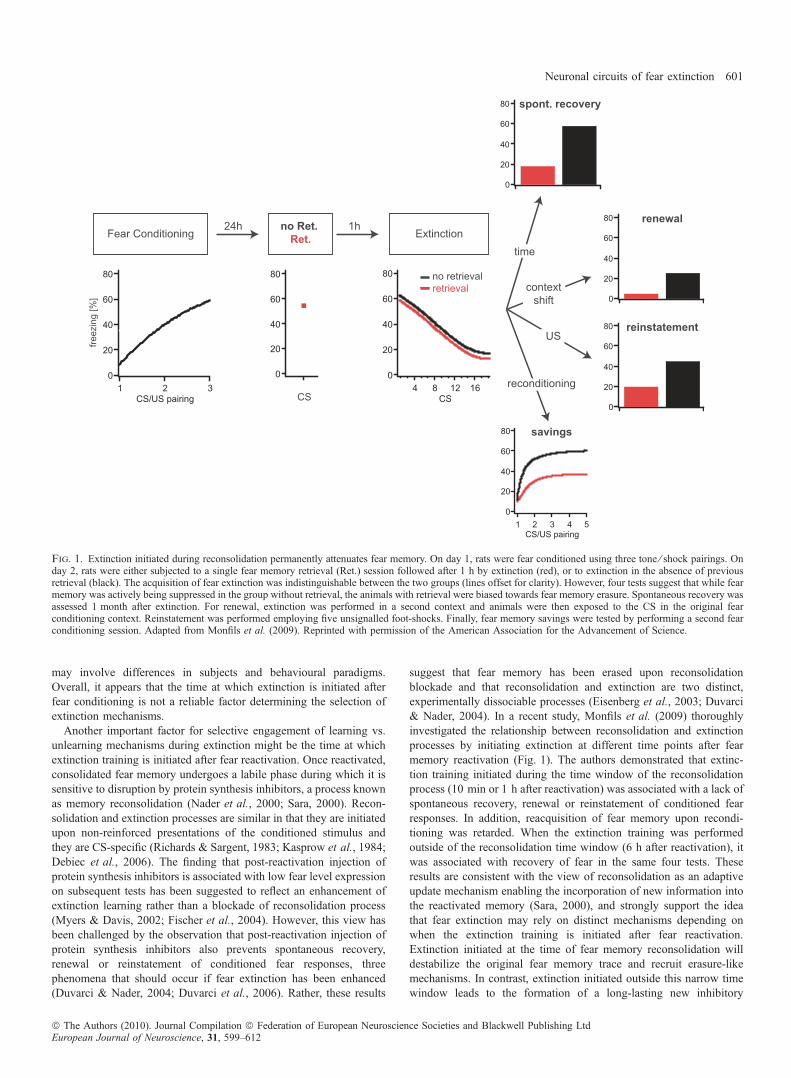
suggest that fear memory has been erased upon reconsolidationblockade and that reconsolidation and extinction are two distinct,experimentally dissociable processes (Eisenberg et al., 2003; Duvarci& Nader, 2004). In a recent study, Monfils et al. (2009) thoroughlyinvestigated the relationship between reconsolidation and extinctionprocesses by initiating extinction at different time points after fearmemory reactivation (Fig. 1). The authors demonstrated that extinc-tion training initiated during the time window of the reconsolidationprocess (10 min or 1 h after reactivation) was associated with a lack ofspontaneous recovery, renewal or reinstatement of conditioned fearresponses. In addition, reacquisition of fear memory upon recondi-tioning was retarded. When the extinction training was performedoutside of the reconsolidation time window (6 h after reactivation), itwas associated with recovery of fear in the same four tests. Theseresults are consistent with the view of reconsolidation as an adaptiveupdate mechanism enabling the incorporation of new information intothe reactivated memory (Sara, 2000), and strongly support the ideathat fear extinction may rely on distinct mechanisms depending onwhen the extinction training is initiated after fear reactivation.Extinction initiated at the time of fear memory reconsolidation willdestabilize the original fear memory trace and recruit erasure-likemechanisms. In contrast, extinction initiated outside this narrow timewindow leads to the formation of a long-lasting new inhibitory
Fig. 1. Extinction initiated during reconsolidation permanently attenuates fear memory. On day 1, rats were fear conditioned using three tone ⁄ shock pairings. Onday 2, rats were either subjected to a single fear memory retrieval (Ret.) session followed after 1 h by extinction (red), or to extinction in the absence of previousretrieval (black). The acquisition of fear extinction was indistinguishable between the two groups (lines offset for clarity). However, four tests suggest that while fearmemory was actively being suppressed in the group without retrieval, the animals with retrieval were biased towards fear memory erasure. Spontaneous recovery wasassessed 1 month after extinction. For renewal, extinction was performed in a second context and animals were then exposed to the CS in the original fearconditioning context. Reinstatement was performed employing five unsignalled foot-shocks. Finally, fear memory savings were tested by performing a second fearconditioning session. Adapted from Monfils et al. (2009). Reprinted with permission of the American Association for the Advancement of Science.
Neuronal circuits of fear extinction 601
ª The Authors (2010). Journal Compilation ª Federation of European Neuroscience Societies and Blackwell Publishing LtdEuropean Journal of Neuroscience, 31, 599–612
memory that competes with the original memory trace withoutdestroying it.
Developmental regulation of extinction mechanisms
Another factor that determines which mechanism becomes engagedduring extinction is the developmental stage of the animal at the timeof behavioural testing. Fear extinction is generally context-dependentin the adult (Bouton, 2004). In contrast, pre-weaning rats do notdisplay contextual fear conditioning (Rudy, 1993; Rudy & Morledge,1994; Pugh & Rudy, 1996). Therefore, it has been suggested thatextinction in juvenile rats may rely on mechanisms distinct from thoseengaged in adulthood. This hypothesis was first tested in the appetitivedomain by Carew & Rudy (1991) who reported a lack of renewal ofconditioned responses after extinction in 17-day-old compared with20-day-old rats. This finding was replicated in the aversive domain byRichardson and colleagues who used auditory and olfactory fearconditioning to show that juvenile 16- to 17-day-old rats do notexhibit spontaneous recovery, renewal or reinstatement of conditionedfear responses after extinction (Kim & Richardson, 2007a,b; Yap &Richardson, 2007). These findings strongly support the view of adevelopmental switch in extinction mechanisms. During early post-natal development, extinction appears to be permanent and has beensuggested to reflect an unlearning process leading to the erasure ofconditioned fear memories (Kim & Richardson, 2008). Currently, theneuronal mechanisms underlying the developmental regulation of fearextinction are still poorly understood. Further investigations (seebelow) could be important to devise novel and specific therapeuticinterventions for anxiety and related disorders in children.
Neuronal substrates and plasticity mechanismsunderlying fear extinction
This section discusses recent findings extending our knowledge on theneuronal substrates and molecular mechanisms underlying fearextinction in adulthood and in juveniles. For clarity, this part isdivided into separate sections covering the neuronal substrates andplasticity mechanisms of acquisition, consolidation and expression,and contextual modulation of fear extinction.
Acquisition of fear extinction
Synaptic signalling and plasticity in the BLA
Over the past decade evidence has accumulated pointing to a criticalrole of the basolateral amygdala (BLA) in the acquisition phase ofextinction. The BLA is composed of several anatomically andfunctionally distinct nuclei, including the lateral (LA) and basal(BA) nuclei. The LA is known to be a critical site of synaptic plasticityand N-methyl-d-aspartate (NMDA) receptor-dependent long-term-potentiation (LTP) during fear learning (Davis, 2000; LeDoux, 2000;Maren, 2001; Maren & Quirk, 2004). However, because of technicaldifficulties in restricting the local application of drugs to BLAsubnuclei, for most of the studies discussed below no claim can bemade on the specific contribution of LA and BA. The first studyindicating that cellular and synaptic plasticity in the BLA is alsoimplicated in acquisition of extinction was conducted by Falls et al.(1992), who demonstrated that intra-BLA injection of the NMDAreceptor (NMDAR) antagonist AP5 prevents fear extinction asassessed using the FPS paradigm. Conversely, intra-BLA infusion ofthe NMDAR partial agonist d-cycloserine facilitates fear extinction(Mao et al., 2006). Using the same paradigm, ERK ⁄ MAPK inhibitors
injected directly into the BLA also prevent fear extinction (Lu et al.,2001; Lin et al., 2003). However, a major caveat of these studies is themethodological limitation in the FPS paradigm that does not easilyallow for discriminating between the acquisition and consolidationphases of extinction. Recent studies, using intra-BLA infusions ofglutamate receptor antagonists (including NMDA and mGlu1 receptorantagonists) and ERK ⁄ MAPK inhibitors, have unambiguously dem-onstrated an impairment of extinction acquisition in classical auditoryfear conditioning (Herry et al., 2006; Kim et al., 2007; Sotres-Bayonet al., 2007). It is possible that NMDARs might not exclusively beengaged in synaptic plasticity, but could also mediate regular synaptictransmission, for example in GABAergic interneurons (Szinyei et al.,2003), contributing to spatio-temporal dynamics of amygdala networkactivity. Nevertheless, taken together, these studies strongly suggestthat glutamatergic synaptic plasticity in the amygdala is a keycomponent mediating extinction learning.To date, the pathways and cell types exhibiting NMDAR-dependent
synaptic plasticity during extinction acquisition have not beenidentified. One possibility is that NMDAR-dependent synapticplasticity occurs at glutamatergic inputs onto local BLA GABAergicinterneurons or onto neurons within the intercalated cell masses(ITCs), which are mostly GABAergic and surround the BLA (for areview see Ehrlich et al., 2009). Indeed, NMDAR-dependent LTP hasbeen observed at glutamatergic inputs onto LA (Mahanty & Sah,1998; Bauer & LeDoux, 2004) and BA (Mahanty & Sah, 1998)interneurons, and at LA and BA afferents onto medial paracapsularITCs (Royer & Pare, 2002, 2003). However, the relevance ofNMDAR-dependent LTP in amygdala inhibitory circuits remains tobe determined.Although additional studies are required for understanding the role
of specific amygdala GABAergic circuits in extinction acquisition,there is compelling evidence that enhanced inhibition contributes tothe expression of extinction (see below). In addition to plasticity ofinhibitory circuits, extinction acquisition might involve plasticityat glutamatergic synaptic inputs onto subpopulations of principalneurons (see below). Finally, consistent with the fact that extinctionacquisition is strongly regulated by various neuromodulatory, neurop-eptidergic and neuroendocrine systems (for a review see Myers &Davis, 2007), neuromodulation has been shown to regulate amygdalainhibitory circuits at different levels, and to gate activity-dependentplasticity of glutamatergic synaptic transmission (e.g. Shumyatskyet al., 2002; Bissiere et al., 2003; Shaban et al., 2006; Tully et al.,2007; Jungling et al., 2008).
Encoding of prediction errors in the vlPAG
During the acquisition of conditioned fear, the dependence ofNMDAR-mediated LTP on coincident synaptic input from sensoryafferents and postsynaptic depolarization is a good cellular modelaccounting for coding of CS–US contiguity (for a review see Sahet al., 2008). For extinction acquisition, the mechanisms of plasticityunderlying learning about the absence of the US are still unclear.Reinforcement learning theories posit that at the beginning ofextinction acquisition, when the animal predicts the occurrence ofthe US when presented with the CS, a so-called prediction error signalis generated when the US does not occur (Rescorla & Wagner, 1972).Associative learning then occurs as a product of learning rate and thediscrepancies (the prediction error) between the expected (theprediction) and obtained outcomes (Rescorla & Wagner, 1972).Prediction error signalling by dopaminergic neurons in the ventraltegmental area is well established in the appetitive domain (fora review see Schultz, 2006). In classical fear conditioning, thereis compelling evidence that a similar role might be performed by
602 C. Herry et al.
ª The Authors (2010). Journal Compilation ª Federation of European Neuroscience Societies and Blackwell Publishing LtdEuropean Journal of Neuroscience, 31, 599–612
mu-opioid receptors (MORs) in the ventro-lateral periaquaeductal gray(vlPAG; McNally & Westbrook, 2006). The vlPAG is rich in opioidreceptors and systemic or intra-vlPAG injection of the non-selectiveopioid receptor antagonist naloxone or the selective MOR antagonistCTAP dose-dependently impairs the acquisition of fear extinction(McNally & Westbrook, 2003; McNally et al., 2004, 2005). Con-versely, inhibiting enzymatic degradation of the endogenous opioidsaccelerates extinction acquisition (McNally, 2005). Based on addi-tional evidence obtained from related learning paradigms involvingprediction error signalling, such as Kamin blocking (Kamin, 1968;McNally et al., 2004; Cole & McNally, 2007) and overexpectation(McNally et al., 2004), it has been proposed that vlPAG MORs signalthe predicted US, and that the mismatch between this MOR signal andthe absence of the US at the onset of extinction training would benecessary to drive the acquisition of extinction (for a review seeMcNally & Westbrook, 2006). It is intriguing to postulate that whilevlPAG MORs encode the prediction error, NMDARs in the BLA arecrucial for determining learning rate in fear conditioning (Cole &McNally, 2007, 2008). In the future, it will be of particular interest touse electrophysiological approaches to investigate how MOR-depen-dent signalling in vlPAG generates a prediction error signal, and howsuch a signal interacts with synaptic plasticity in the BLA during theacquisition of extinction.
A critical role of the BA
An important step towards understanding the role of the BLA inextinction acquisition is the identification of the subnuclei, circuits andneurons involved. Recent studies using immediate early gene (IEG)analysis, classical lesion approaches or more sophisticated localreversible inactivation have refined the role of distinct amygdalasubnuclei during acquisition of extinction. Although NMDARsignalling in the LA may participate in acquisition of extinction(Sotres-Bayon et al., 2007), these studies strongly suggest a criticalrole for activity in the BA also. It has been shown for instance thatextinction training leads to the induction of the IEG c-fos in the BA(Herry & Mons, 2004). Conversely, in animal models showingimpaired extinction learning c-fos induction in the BA is stronglycompromised (Muigg et al., 2008; see below). However, pre-training(before fear conditioning) electrical lesions restricted to the BA haveno effect on subsequent fear extinction (Sotres-Bayon et al., 2004;Anglada-Figueroa & Quirk, 2005). There are two possible interpre-tations of these results: either the BA is not necessary for acquisitionof extinction, or compensatory mechanisms masked the role of the BAduring extinction. Nevertheless, the fact that BA-lesioned animals canstill acquire fear extinction suggests that additional structures areimplicated. Post-training lesions of the BA have also been conducted,but the fact that this manipulation alters fear expression prevents a firmconclusion about the role of the BA during extinction learning(Anglada-Figueroa & Quirk, 2005). Finally, studies using localreversible inactivation with the GABAA receptor agonist muscimolhave demonstrated that whereas inactivation of the entire amygdalahas no effect on extinction learning (Akirav et al., 2006; but see Hartet al., 2009), inactivation restricted to the BA completely blocksacquisition of extinction (Herry et al., 2008). Importantly, memoryretrieval and expression were not affected in the latter study, indicatingthat the BA is required for extinction learning, but not for the storageof extinction memories (Herry et al., 2008).
Which neuronal populations participate in extinction learning, andtheir properties and functions is still a matter of debate andinvestigation. First indications came from in vivo single unitrecordings that established that both the LA and the BA contain
distinct cell populations whose activity correlates with high fear levelsfollowing auditory fear conditioning (Quirk et al., 1995, 1997; Pare &Collins, 2000; Repa et al., 2001; Goosens et al., 2003; Herry et al.,2008). In particular, one subpopulation of neurons displayed persistentactivity throughout extinction learning whereas another showed adecline in conditioned responses upon non-reinforced presentation ofthe CS (Repa et al., 2001; Herry et al., 2008). It has been suggestedthat these subpopulations might underlie the encoding and themaintenance of the CS–US association, respectively (Repa et al.,2001). Alternatively, the rapid reversal of neuronal conditioned fearresponses during extinction may reflect an erasure of the original CS–US association. The cellular mechanism underlying this erasure couldreflect depotentiation of sensory afferent synapses onto LA principalneurons previously potentiated during fear conditioning (Mao et al.,2006; Kim et al., 2007). However, given that fear extinction does notlead to the erasure of the original fear memory under mostcircumstances, the role of synaptic depotentiation in extinctionacquisition remains debatable.We recently identified a novel cell population specific to the BA
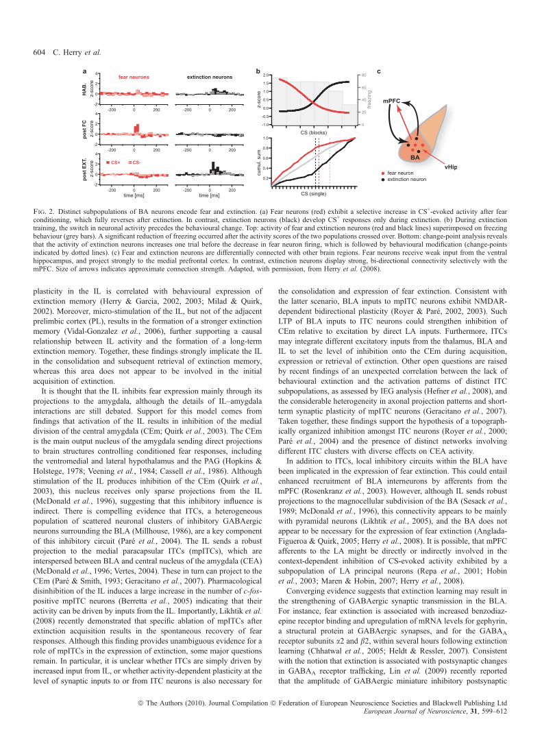
which becomes CS-responsive during extinction acquisition (‘extinc-tion neurons’) (Herry et al., 2008). In concert with another populationof BA neurons whose activity correlates with a high fear state (‘fearneurons’), activity-switching in these BA circuits is essential for rapidswitching between behavioural states (Fig. 2). Extinction neurons maytrigger a low fear state by exerting instructive effects on extinctionmemory consolidation in the medial prefrontal cortex (mPFC; seebelow). Notably, both ‘fear neurons’ and ‘extinction neurons’ projectto the mPFC (Herry et al., 2008), suggesting that there could bedistinct fear- and extinction-pathways connecting the BA to themPFC. These distinct neuronal circuits may modulate the transitionbetween high and low fear states by tipping the balance of activitybetween specific prefrontal circuits as recently suggested (Burgos-Robles et al., 2009). Consistent with the idea of discrete sub-networksof projection neurons interconnecting brain areas involved in extinc-tion, we found that inputs from the mPFC and the hippocampus weredifferentially targeting ‘extinction neurons’ and ‘fear neurons’,respectively (Fig. 2). In addition to specific long-range interactions,extinction neurons could also interact locally with other principal cellsor interneurons within the BLA, the central nuclei and ⁄ or the ITCs, toreduce fear responses during extinction acquisition and ⁄ or to triggerplasticity mediating extinction memory consolidation.
Consolidation and expression of fear extinction
Similar to other forms of learning, acquisition of extinction is followedby a consolidation phase, which lasts several hours and is required tostabilize plastic events into a long-term memory (McGaugh, 2000). Asoutlined above, extinction learning appears to be mediated by adistributed network of brain areas centred around activity in the BA,while consolidation recruits other circuits and brain areas. The firstarea found to be implicated in expression of extinction is the mPFC(Morgan et al., 1993). Subsequent studies demonstrated that lesions orinactivation of a subregion of the mPFC, the infralimbic cortex (IL)block retrieval of extinction, but not acquisition (Quirk et al., 2000;Laurent & Westbrook, 2009; for a review see Quirk & Mueller, 2008).In addition, immediate post-training infusions of an NMDARantagonist (Burgos-Robles et al., 2007) or a MAPK inhibitor (Hugueset al., 2004) impaired the retrieval of extinction. On the physiologicallevel, these findings concur with the fact that a subpopulation of ILneurons shows CS-evoked responses during extinction retrieval, butnot during acquisition (Milad & Quirk, 2002), and that neuronal
Neuronal circuits of fear extinction 603
ª The Authors (2010). Journal Compilation ª Federation of European Neuroscience Societies and Blackwell Publishing LtdEuropean Journal of Neuroscience, 31, 599–612
plasticity in the IL is correlated with behavioural expression ofextinction memory (Herry & Garcia, 2002, 2003; Milad & Quirk,2002). Moreover, micro-stimulation of the IL, but not of the adjacentprelimbic cortex (PL), results in the formation of a stronger extinctionmemory (Vidal-Gonzalez et al., 2006), further supporting a causalrelationship between IL activity and the formation of a long-termextinction memory. Together, these findings strongly implicate the ILin the consolidation and subsequent retrieval of extinction memory,whereas this area does not appear to be involved in the initialacquisition of extinction.It is thought that the IL inhibits fear expression mainly through its
projections to the amygdala, although the details of IL–amygdalainteractions are still debated. Support for this model comes fromfindings that activation of the IL results in inhibition of the medialdivision of the central amygdala (CEm; Quirk et al., 2003). The CEmis the main output nucleus of the amygdala sending direct projectionsto brain structures controlling conditioned fear responses, includingthe ventromedial and lateral hypothalamus and the PAG (Hopkins &Holstege, 1978; Veening et al., 1984; Cassell et al., 1986). Althoughstimulation of the IL produces inhibition of the CEm (Quirk et al.,2003), this nucleus receives only sparse projections from the IL(McDonald et al., 1996), suggesting that this inhibitory influence isindirect. There is compelling evidence that ITCs, a heterogeneouspopulation of scattered neuronal clusters of inhibitory GABAergicneurons surrounding the BLA (Millhouse, 1986), are a key componentof this inhibitory circuit (Pare et al., 2004). The IL sends a robustprojection to the medial paracapsular ITCs (mpITCs), which areinterspersed between BLA and central nucleus of the amygdala (CEA)(McDonald et al., 1996; Vertes, 2004). These in turn can project to theCEm (Pare & Smith, 1993; Geracitano et al., 2007). Pharmacologicaldisinhibition of the IL induces a large increase in the number of c-fos-positive mpITC neurons (Berretta et al., 2005) indicating that theiractivity can be driven by inputs from the IL. Importantly, Likhtik et al.(2008) recently demonstrated that specific ablation of mpITCs afterextinction acquisition results in the spontaneous recovery of fearresponses. Although this finding provides unambiguous evidence for arole of mpITCs in the expression of extinction, some major questionsremain. In particular, it is unclear whether ITCs are simply driven byincreased input from IL, or whether activity-dependent plasticity at thelevel of synaptic inputs to or from ITC neurons is also necessary for
the consolidation and expression of fear extinction. Consistent withthe latter scenario, BLA inputs to mpITC neurons exhibit NMDAR-dependent bidirectional plasticity (Royer & Pare, 2002, 2003). SuchLTP of BLA inputs to ITC neurons could strengthen inhibition ofCEm relative to excitation by direct LA inputs. Furthermore, ITCsmay integrate different excitatory inputs from the thalamus, BLA andIL to set the level of inhibition onto the CEm during acquisition,expression or retrieval of extinction. Other open questions are raisedby recent findings of an unexpected correlation between the lack ofbehavioural extinction and the activation patterns of distinct ITCsubpopulations, as assessed by IEG analysis (Hefner et al., 2008), andthe considerable heterogeneity in axonal projection patterns and short-term synaptic plasticity of mpITC neurons (Geracitano et al., 2007).Taken together, these findings support the hypothesis of a topograph-ically organized inhibition amongst ITC neurons (Royer et al., 2000;Pare et al., 2004) and the presence of distinct networks involvingdifferent ITC clusters with diverse effects on CEA activity.In addition to ITCs, local inhibitory circuits within the BLA have
been implicated in the expression of fear extinction. This could entailenhanced recruitment of BLA interneurons by afferents from themPFC (Rosenkranz et al., 2003). However, although IL sends robustprojections to the magnocellular subdivision of the BA (Sesack et al.,1989; McDonald et al., 1996), this connectivity appears to be mainlywith pyramidal neurons (Likhtik et al., 2005), and the BA does notappear to be necessary for the expression of fear extinction (Anglada-Figueroa & Quirk, 2005; Herry et al., 2008). It is possible, that mPFCafferents to the LA might be directly or indirectly involved in thecontext-dependent inhibition of CS-evoked activity exhibited by asubpopulation of LA principal neurons (Repa et al., 2001; Hobinet al., 2003; Maren & Hobin, 2007; Herry et al., 2008).Converging evidence suggests that extinction learning may result in
the strengthening of GABAergic synaptic transmission in the BLA.For instance, fear extinction is associated with increased benzodiaz-epine receptor binding and upregulation of mRNA levels for gephyrin,a structural protein at GABAergic synapses, and for the GABAA
receptor subunits a2 and b2, within several hours following extinctionlearning (Chhatwal et al., 2005; Heldt & Ressler, 2007). Consistentwith the notion that extinction is associated with postsynaptic changesin GABAA receptor trafficking, Lin et al. (2009) recently reportedthat the amplitude of GABAergic miniature inhibitory postsynaptic
Fig. 2. Distinct subpopulations of BA neurons encode fear and extinction. (a) Fear neurons (red) exhibit a selective increase in CS+-evoked activity after fearconditioning, which fully reverses after extinction. In contrast, extinction neurons (black) develop CS+ responses only during extinction. (b) During extinctiontraining, the switch in neuronal activity precedes the behavioural change. Top: activity of fear and extinction neurons (red and black lines) superimposed on freezingbehaviour (grey bars). A significant reduction of freezing occurred after the activity scores of the two populations crossed over. Bottom: change-point analysis revealsthat the activity of extinction neurons increases one trial before the decrease in fear neuron firing, which is followed by behavioural modification (change-pointsindicated by dotted lines). (c) Fear and extinction neurons are differentially connected with other brain regions. Fear neurons receive weak input from the ventralhippocampus, and project strongly to the medial prefrontal cortex. In contrast, extinction neurons display strong, bi-directional connectivity selectively with themPFC. Size of arrows indicates approximate connection strength. Adapted, with permission, from Herry et al. (2008).
604 C. Herry et al.
ª The Authors (2010). Journal Compilation ª Federation of European Neuroscience Societies and Blackwell Publishing LtdEuropean Journal of Neuroscience, 31, 599–612
currents (mIPSCs) recorded from LA principal neurons was increasedafter extinction of FPS. The increase in mIPSC amplitude andextinction of FPS were blocked by disruption of the interactionbetween GABAA receptors and the associated protein GABARAP,which has been implicated in GABAA receptor trafficking (for areview see Jacob et al., 2008). Moreover, extinction-associatedchanges in the expression of the GABA-synthesizing enzymeGAD67 and in the GABA uptake transporter GAT-1 have beenreported (Heldt & Ressler, 2007). Thus, extinction might lead toconcerted pre- and postsynaptic changes at GABAergic synapses inthe BLA. However, given that extinction is highly CS- and context-dependent, it seems unlikely that it should be associated with globalchanges in GABAergic drive. Indeed, the BLA contains severalfunctionally distinct subtypes of GABAergic interneurons (McDonald,1982; Rainnie et al., 1993; for a review see Ehrlich et al., 2009).Future studies will have to determine whether extinction affectsspecific inhibitory circuits that could inhibit BLA principal neurons, orsubsets thereof, at distinct levels.
Finally, although the mPFC may inhibit the expression of condi-tioned fear through its control over the ITCs and ⁄ or the BLA, thiscould also be accomplished through alternative routes involving, forexample, direct projections to the lateral capsular subdivision of theCEA (McDonald, 1998) or to the hypothalamus and the brain stem(Fisk & Wyss, 2000; Floyd et al., 2000). Future studies should bedesigned to test the validity of these different circuit models as well asto determine the integration of different components mediatingconsolidation of fear extinction.
Contextual modulation of fear extinction
Fear extinction is strongly modulated by contextual elements (Bouton,2002, 2004). Given the involvement of the hippocampus in theformation of contextual representations (Kim & Fanselow, 1992;Phillips & LeDoux, 1992), numerous studies have thoroughlyinvestigated its role in the contextual modulation of fear extinction(for reviews see Bouton et al., 2006; Ji & Maren, 2007). The mainquestions addressed so far relate to participation of the hippocampus inencoding the context-specificity of extinction, and its role in thecontext-dependent retrieval of fear after extinction.
Context specificity coding was evaluated by Corcoran et al. (2005)using acute pre-extinction inactivation of the dorsal hippocampus withmuscimol. Whereas extinction acquisition was delayed, rats wereunable to express fear extinction when tested the next day in the sameor in a different context. This effect has been suggested to reflect aninvolvement of the hippocampus in the contextual encoding andcontext-dependent retrieval of the extinction memory, but not in theencoding of extinction per se (Corcoran et al., 2005). These findingsare consistent with human functional magnetic resonance imaging datashowing that retrieval of extinction memory was associated with anactivation of the hippocampus in concert with the ventral mPFC(Kalisch et al., 2006; Milad et al., 2007).
Additionally, the hippocampus has a critical role in the context-dependent renewal of extinguished fear memories. Acute pharmaco-logical inactivation by local injection of muscimol into the dorsal orventral hippocampus prior to testing completely prevented context-dependent renewal of conditioned fear when tested in a novel context(Corcoran & Maren, 2001; Hobin et al., 2006), but not in the fearconditioning context (Corcoran & Maren, 2004). In contrast, pre-conditioning electrolytic lesions of the dorsal hippocampus alsointerfered with fear renewal in the conditioning context (Ji & Maren,2005). This indicates that the type of manipulation (lesion vs.inactivation) or the timing of the manipulation (pre-conditioning vs.
pre-test) play a critical role. Using single unit recordings in the LA,Hobin et al. (2003) demonstrated that renewal of conditioned fearresponses was associated with an increase in CS-evoked neuronalactivity. These renewal-associated neuronal responses were abolishedby inactivation of the dorsal hippocampus (Maren & Hobin, 2007).Furthermore, several human studies also support a role of the
hippocampus in context-dependent retrieval of fear after extinction.For example, recovery of conditioned fear responses after extinction iscontext- and CS-specific (LaBar & Phelps, 2005; Schiller et al., 2008),and patients with selective bilateral atrophy of the hippocampus do notexhibit context-dependent recovery of fear responses followingreinstatement (LaBar & Phelps, 2005). Collectively, these findingssuggest an important role for the hippocampus in controlling thecontext-dependency of extinguished fear responses in both humansand animals. Interestingly, acute pre-testing inactivation of thehippocampus does not abolish extinction, but rather renders extinctionbehaviour context-independent and impairs context-dependent renew-al of conditioned fear responses. Accordingly, it has been proposedthat the contextualization of previously acquired fear memories uponextinction training might reflect a more general principle governingthe interaction between different memories in the sense that memoriesbecome contextualized by a second learning episode (Harris et al.,2000; Bouton, 2004).What might be the neuronal circuitry by which the hippocampus
triggers the context-dependent renewal of conditioned fear responses?One possibility is that the hippocampus regulates neuronal activity inthe amygdala indirectly via its strong projections to the mPFC(Hoover & Vertes, 2007). Indeed, renewal of conditioned fearresponses is associated with opposite changes in c-fos expression inIL and PL (Knapska & Maren, 2009). Alternatively, or in addition, thehippocampus might exert contextual control of conditioned fearbehaviour through direct projections to the amygdala. Consistent withthis scenario, we recently found that BA neurons exhibiting context-dependent renewal of neuronal conditioned responses (i.e. ‘fearneurons’), but not BA ‘extinction neurons’, receive strong hippocam-pal inputs (Herry et al., 2008). Moreover, hippocampal inputs to theBA have been shown to exhibit NMDAR-dependent LTP (Maren &Fanselow, 1995). Thus, these data strongly suggest that hippocampalinputs onto specific subpopulations of BLA neurons contribute to thecontext-dependent renewal of conditioned fear responses.
Mechanisms of extinction in juveniles
As mentioned earlier, fear extinction is developmentally regulated andresults in the formation of a permanent extinction memory in juvenilerats and mice younger than about 3 weeks. In contrast to adultanimals, extinguished fear responses do not exhibit spontaneousrecovery, context-dependent renewal or reinstatement (Kim & Rich-ardson, 2007a,b; Yap & Richardson, 2007; Gogolla et al., 2009),strongly suggesting that extinction training leads to unlearning orerasure of the conditioned fear response. Mechanistically, extinction injuveniles seems to be based on mechanisms and circuits which overlapwith the adult, but which also display distinct features. As in adults,extinction in juveniles is an active process requiring an intact BLA(Kim & Richardson, 2008) and depends on NMDAR and MORactivation (Langton et al., 2007; Kim & Richardson, 2009). However,extinction in juveniles is mPFC-independent (Kim et al., 2009), and isinsensitive to pharmacological treatments that reduce the efficiency ofGABAergic transmission (Kim & Richardson, 2007b). This suggeststhat extinction in juveniles might involve distinct plasticity mecha-nisms in different BLA micro-circuits. In particular, the role ofinhibitory circuits appears to be developmentally regulated.
Neuronal circuits of fear extinction 605
ª The Authors (2010). Journal Compilation ª Federation of European Neuroscience Societies and Blackwell Publishing LtdEuropean Journal of Neuroscience, 31, 599–612
This is very reminiscent of the developmental regulation ofneuronal circuit plasticity in visual cortex. There, the developmentof inhibitory circuits containing parvalbumin-positive interneuronsmarks the opening and closure of critical periods, during whichenvironmental manipulations, such as eye closure, can have profoundeffects on circuit organization and function (for a review see Hensch,2005). Recently, we found in analogy to visual cortex that theformation of perineuronal nets (PNNs), an organized formof extracellular matrix around parvalbumin-positive interneurons(Berardi et al., 2003; Hensch, 2005), coincides with the switch ofextinction mechanisms to the adult phenotype (Gogolla et al., 2009).Interestingly, enzymatic degradation of PNNs in the BLA of adultanimals re-enabled the erasure of conditioned fear memories byextinction training, but only if PNNs were degraded before fearconditioning (Gogolla et al., 2009). Thus, in adults, PNNs appear toactively protect conditioned fear memories from extinction-inducederasure, allowing extinction memories to coexist with previouslyacquired fear memories, both of which can be retrieved in a context-dependent manner. In juveniles, contextualization cannot occurbecause extinction induces the erasure of fear memories. Consistentwith the proposed role of the mPFC and the hippocampus in thecontextualization of extinction memories (Hobin et al., 2003, 2006),connections between the BLA and these brain areas continue todevelop up to several months after birth (Bouwmeester et al., 2002;Cunningham et al., 2002). Thus, the developmental regulation ofamygdala circuit function underlying extinction-induced memoryerasure enables juvenile animals to adhere to the most recently learnedinformation, a strategy that might increase chances of survival.
Animal models of impaired extinction: evidence forextinction circuitry dysfunction
Impaired extinction of fear memories is thought to contribute to thedevelopment and persistence of anxiety disorders including phobias,PTSD and panic (e.g. Rosen & Schulkin, 1998; Lissek et al., 2005;Anderson & Insel, 2006; Mineka & Zinbarg, 2006; Rauch et al., 2006;Milad et al., 2008). Currently, a substantial proportion of anxietypatients do not respond effectively to standard treatments to inhibitpathological fear responses, including extinction-based cognitivebehavioural therapy and pharmacotherapy (Pull, 2007). For thedevelopment of novel therapeutic strategies to promote fear extinctionprocesses selectively, a better understanding of mechanisms underly-ing pathologically impaired fear extinction is necessary. Animalmodels mimicking impaired fear extinction could provide this insightand open avenues for therapeutic strategies applicable to humanpathology (Yehuda et al., 2006). There is considerable genetic as wellas environmental (e.g. psychological trauma, stress) contribution toindividual variability in the risk for anxiety disorders (True et al.,1993; Kendler, 2001; Yehuda & LeDoux, 2007), although the genesinvolved in resistance to extinction are not known. Interestingly, stresselicits structural changes in key areas implicated in extinction, such asthe mPFC (Holmes & Wellman, 2009), amygdala (Vyas et al., 2002;Roozendaal et al., 2009) and hippocampus (Watanabe et al., 1992).Both stress and genetic differences were exploited to generate rodentmodels displaying impaired extinction. Indeed, in these animalmodels, more persistent fear responses can be observed after exposureto different stressors (e.g. Izquierdo et al., 2006; Miracle et al., 2006;Mitra & Sapolsky, 2009; Yamamoto et al., 2008; Baran et al., 2009),in animals acutely selected according to their individual differences inextinction (Herry & Mons, 2004; Bush et al., 2007), after selectivebreeding (Ponder et al., 2007; Muigg et al., 2008; Lopez-Aumatellet al., 2009; see also Shumake et al., 2005; Wrubel et al., 2007) or in
naturally occurring extinction-deficient mouse strains (Falls et al.,1997; Stiedl et al., 1999; McCaughran et al., 2000; Waddell et al.,2004; Hefner et al., 2008; Camp et al., 2009), and following differentgenetic manipulations (e.g. Marsicano et al., 2002; Wellman et al.,2007). Extinction deficits were apparent in some models during theacquisition and ⁄ or during retrieval of extinction and were seen incontextual and ⁄ or cued fear extinction.Insight into the neuronal correlates of extinction failure in these
models comes mainly from IEG mapping studies. It has been shownpreviously that the molecular events which underlie synaptic plasticityaccompanying extinction learning and retrieval require activation ofspecific signalling cascades, including expression of IEGs (Morrowet al., 1999; Nader et al., 2000; Hall et al., 2001). Of these IEGs,mainly c-fos (Kovacs, 1998; Hoffman & Lyo, 2002; Singewald, 2007)and Zif268 (Egr1; Davis et al., 2003; Lee et al., 2004; Amin et al.,2006; Jenkins et al., 2006; Schulte et al., 2006) have been used tolabel activated neurons in widespread regions of the brain, includingcortical and amygdala regions.From these studies, it is emerging that resistance to extinction is
associated with distinct alterations in activation of IEGs in prefrontal-limbic circuits important in fear extinction. Specifically, hypoactivationof the IL is associated with extinction deficits. This was observed in129S1mice, a strain with pronounced deficits in fear extinction learningand retrieval (Hefner et al., 2008; Camp et al., 2009), and in acutelyselected mice showing impaired extinction retrieval and re-extinctionlearning (Herry & Mons, 2004) (Table 1). Furthermore, HAB rats, amodel of pathological anxiety (Landgraf et al., 2007) with impairedextinction acquisition and retrieval (Muigg et al., 2008), display asimilar hypoactivation of the IL with a prolonged extinction learningsession (Table 1), which may already involve extinction consolidation.In contrast, extinguishing control animals from all these studies showedrobust activation in the IL (Herry & Mons, 2004; Muigg et al., 2008;Hefner et al., 2008; see also Knapska & Maren, 2009). These findingssupport the notion that activity of IL neurons is correlated with andnecessary for extinction consolidation and expression, and suggest acommon failure to appropriately activate IL neurons in animalmodels ofimpaired fear extinction. Furthermore, they show intriguing parallelsbetween deficient activation of the IL in rodent models and ofhomologous brain regions in humans implicated in resistance toextinction in anxiety disorders including PTSD, panic and specificphobia (reviewed in Rauch et al., 2006; Hofmann, 2007; Quirk &Mueller, 2008; see also Michael et al., 2007; Milad et al., 2008).It has been suggested that PL activation is associated with a resistance
to induce fear extinction learning (Gilmartin &McEchron, 2005; Vidal-Gonzalez et al., 2006; Corcoran & Quirk, 2007). To date there is noevidence for enhanced PL activation in animal models displayingimpaired extinction (Table 1). However, thus far IEGmapping has onlybeen performed in response to multiple CS presentations duringextinction training (Muigg et al., 2008) or retrieval (Herry & Mons,2004; Hefner et al., 2008). This has the caveat that it may result in aceiling effect in PL activation, obscuring subtle differences in theactivation of neurons in this area. Indeed, preliminary results using onlyone CS presentation in extinction retrieval revealed enhanced PLactivation in non-extinguishing 129S1 compared with extinguishingC57BL ⁄ 6 mice (Whittle et al., 2009). In keeping with the idea that fearextinction may require down-regulation of PL activity, in a recent study,Burgos-Robles et al. (2009) demonstrated that in rats exhibiting goodextinction memory, CS-evoked activity in PL was low, whereas animalsexhibiting poor extinction memory showed persistent CS-evokedactivity in PL.Another common observation is that resistance to extinction is
associated with altered activation in specific amygdala regions. As
606 C. Herry et al.
ª The Authors (2010). Journal Compilation ª Federation of European Neuroscience Societies and Blackwell Publishing LtdEuropean Journal of Neuroscience, 31, 599–612
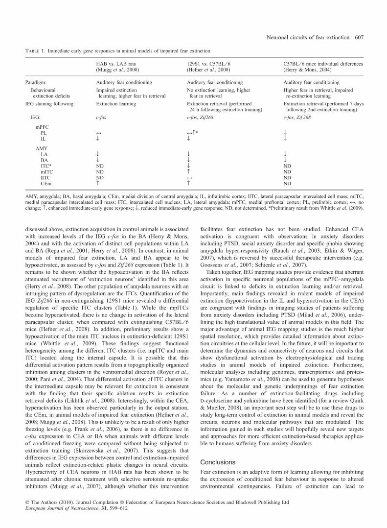
discussed above, extinction acquisition in control animals is associatedwith increased levels of the IEG c-fos in the BA (Herry & Mons,2004) and with the activation of distinct cell populations within LAand BA (Repa et al., 2001; Herry et al., 2008). In contrast, in animalmodels of impaired fear extinction, LA and BA appear to behypoactivated, as assessed by c-fos and Zif 268 expression (Table 1). Itremains to be shown whether the hypoactivation in the BA reflectsattenuated recruitment of ‘extinction neurons’ identified in this area(Herry et al., 2008). The other population of amydala neurons with anintruiging pattern of dysregulation are the ITCs. Quantification of theIEG Zif268 in non-extinguishing 129S1 mice revealed a differentialregulation of specific ITC clusters (Table 1). While the mpITCsbecome hyperactivated, there is no change in activation of the lateralparacapsular cluster, when compared with extinguishing C57BL ⁄ 6mice (Hefner et al., 2008). In addition, preliminary results show ahypoactivation of the main ITC nucleus in extinction-deficient 129S1mice (Whittle et al., 2009). These findings suggest functionalheterogeneity among the different ITC clusters (i.e. mpITC and mainITC) located along the internal capsule. It is possible that thisdifferential activation pattern results from a topographically organizedinhibition among clusters in the ventromedial direction (Royer et al.,2000; Pare et al., 2004). That differential activation of ITC clusters inthe intermediate capsule may be relevant for extinction is consistentwith the finding that their specific ablation results in extinctionretrieval deficits (Likhtik et al., 2008). Interestingly, within the CEA,hyperactivation has been observed particularly in the output station,the CEm, in animal models of impaired fear extinction (Hefner et al.,2008; Muigg et al., 2008). This is unlikely to be a result of only higherfreezing levels (e.g. Frank et al., 2006), as there is no difference inc-fos expression in CEA or BA when animals with different levelsof conditioned freezing were compared without being subjected toextinction training (Skorzewska et al., 2007). This suggests thatdifferences in IEG expression between control and extinction-impairedanimals reflect extinction-related plastic changes in neural circuits.Hyperactivity of CEA neurons in HAB rats has been shown to beattenuated after chronic treatment with selective serotonin re-uptakeinhibitors (Muigg et al., 2007), although whether this intervention
facilitates fear extinction has not been studied. Enhanced CEAactivation is congruent with observations in anxiety disordersincluding PTSD, social anxiety disorder and specific phobia showingamygdala hyper-responsivity (Rauch et al., 2003; Etkin & Wager,2007), which is reversed by successful therapeutic intervention (e.g.Goossens et al., 2007; Schienle et al., 2007).Taken together, IEG mapping studies provide evidence that aberrant
activation in specific neuronal populations of the mPFC–amygdalacircuit is linked to deficits in extinction learning and ⁄ or retrieval.Importantly, main findings revealed in rodent models of impairedextinction (hypoactivation in the IL and hyperactivation in the CEA)are congruent with findings in imaging studies of patients sufferingfrom anxiety disorders including PTSD (Milad et al., 2006), under-lining the high translational value of animal models in this field. Themajor advantage of animal IEG mapping studies is the much higherspatial resolution, which provides detailed information about extinc-tion circuitries at the cellular level. In the future, it will be important todetermine the dynamics and connectivity of neurons and circuits thatshow dysfunctional activation by electrophysiological and tracingstudies in animal models of impaired extinction. Furthermore,molecular analyses including genomics, transcriptomics and proteo-mics (e.g. Yamamoto et al., 2008) can be used to generate hypothesesabout the molecular and genetic underpinnings of fear extinctionfailure. As a number of extinction-facilitating drugs includingd-cycloserine and yohimbine have been identified (for a review Quirk& Mueller, 2008), an important next step will be to use these drugs tostudy long-term control of extinction in animal models and reveal thecircuits, neurons and molecular pathways that are modulated. Theinformation gained in such studies will hopefully reveal new targetsand approaches for more efficient extinction-based therapies applica-ble to humans suffering from anxiety disorders.
Conclusions
Fear extinction is an adaptive form of learning allowing for inhibitingthe expression of conditioned fear behaviour in response to alteredenvironmental contingencies. Failure of extinction can lead to
Table 1. Immediate early gene responses in animal models of impaired fear extinction
HAB vs. LAB rats(Muigg et al., 2008)
129S1 vs. C57BL ⁄ 6(Hefner et al., 2008)
C57BL ⁄ 6 mice individual differences(Herry & Mons, 2004)
Paradigm: Auditory fear conditioning Auditory fear conditioning Auditory fear conditioning
Behaviouralextinction deficits
Impaired extinctionlearning, higher fear in retrieval
No extinction learning, higherfear in retrieval
Higher fear in retrieval, impairedre-extinction learning
IEG staining following: Extinction learning Extinction retrieval (performed24 h following extinction training)
Extinction retrieval (performed 7 daysfollowing 2nd extinction training)
IEG: c-fos c-fos, Zif268 c-fos, Zif 268
mPFCPL M M›* flIL fl fl fl
AMYLA fl fl flBA fl fl flITC* ND fl NDmITC ND › NDlITC ND M NDCEm › › ND
AMY, amygdala; BA, basal amygdala; CEm, medial division of central amygdala; IL, infralimbic cortex; lITC, lateral paracapsular intercalated cell mass; mITC,medial paracapsular intercalated cell mass; ITC, intercalated cell nucleus; LA, lateral amygdala; mPFC, medial prefrontal cortex; PL, prelimbic cortex; M, nochange; ›, enhanced immediate-early gene response; fl, reduced immediate-early gene response; ND, not determined. *Preliminary result from Whittle et al. (2009).
Neuronal circuits of fear extinction 607
ª The Authors (2010). Journal Compilation ª Federation of European Neuroscience Societies and Blackwell Publishing LtdEuropean Journal of Neuroscience, 31, 599–612
excessive and inappropriate fear and anxiety behaviour as seen incertain forms of anxiety disorders such as PTSD. Accumulatingevidence indicates that extinction involves new learning resulting inthe formation of a long-term extinction memory. Phenomena such asspontaneous recovery, reinstatement and context-dependent fearrenewal strongly argue for the notion that extinction does not leadto the destruction of a previously acquired fear memory, but that fearand extinction memories coexist with each other. Recent workreviewed here indicates that extinction memories are acquired, storedand retrieved within the same brain areas as fear memories albeitinvolving distinct neuronal circuits.A general emerging concept is that the various phases of extinction
learning involve close interactions between different brain areasincluding the brain stem (vlPAG), amygdala, distinct subdivisions ofthe mPFC and the hippocampus (Bouton et al., 2006; Quirk &Mueller, 2008; Burgos-Robles et al., 2009). There seems to be atemporal sequence by which these brain areas contribute to distinctphases of extinction. Whereas the vlPAG and the BLA play animportant role during the initial acquisition phase, consolidation ofextinction memory requires the IL, and possibly a down-regulation ofPL activity. Expression of the extinction memory again depends on theamygdala, although on other cell types and circuits than thoseinvolved in the acquisition phase. Interactions between the differentbrain areas are mediated by specific neuronal subcircuits as illustratedby the opposite role of two distinct subpopulations of BA principalneurons in the acquisition of fear and extinction memory (Herry et al.,2008), and by the specific role of the GABAergic ITCs in theexpression of extinction (Likhtik et al., 2008). Most probably, asimilar degree of specificity also exists in other brain areas.Undoubtedly, addressing this question will be facilitated by state-of-the-art molecular genetic tools that allow for the functional andanatomical analysis of defined subpopulations of neurons and forspecifically manipulating interactions between brain areas withinlarge-scale networks in behaving animals (for reviews see Gradinaruet al., 2007; Luo et al., 2008).A second major advance of recent work on fear extinction is the fact
that the mechanisms underlying extinction strongly depend on thedevelopmental state of the animal and on its recent experience.Whereas in adult animals, extinction training does not erase apreviously acquired fear memory, but rather results in the context-dependent suppression of fear behaviour, in juvenile animals the samebehavioural paradigm induces unlearning or erasure of the fearmemory (Kim & Richardson, 2008). The mechanisms underlyingextinction-induced fear memory erasure and its behavioural implica-tions for juvenile animals are beginning to be elucidated (Kim &Richardson, 2007a; Langton et al., 2007; Kim et al., 2009). Impor-tantly, these mechanisms remain dormant throughout life, becausethey can be re-activated in adults (Gogolla et al., 2009), openingpotential therapeutic avenues. Another successful approach in induc-ing unlearning or erasure of fear memories entails the precise timing ofextinction training within a defined time period after exposing theanimal to a CS reminder (Monfils et al., 2009). It appears that the briefperiod during which fear memories become malleable during recon-solidation can be exploited to target and erase specific fear memories.Future studies are necessary to determine whether the basic principlesthat determine if fear memories are erased or merely suppressed inanimals can be translated to humans.Finally, we have discussed the potential of using genetic animal
models to tackle the mechanisms underlying extinction failure.Genetic mouse models of impaired extinction offer the uniquepossibility to link functional and molecular studies to address thegenetic bases of extinction, and extinction failure, at the level of
defined neuronal circuits. Results from these models will provideinvaluable information that can be compared and integrated withgenomic and functional data obtained from humans and may impartnovel therapeutic strategies for the treatment of anxiety disorders.
Acknowledgements
We thank S. Ciocchi and N. Whittle for helpful discussions and comments onthe manuscript. Work in the laboratories of the authors is supported by grantsfrom the Austrian Science Fund (NFN 102), NARSAD, the Swiss NationalScience Foundation, the Conseil Regional d’Aquitaine and the NovartisResearch Foundation.
Abbreviations
BA, basal nucleus of the amygdala; BLA, basolateral amygdaloid complex; CEA,central nucleus of the amygdala; CEm, medial subdivision of the centralamygdala;CS, conditioned stimulus; FPS, fear-potentiated startle paradigm; IEG,immediate early gene; IL, infralimbic cortex; ITCs, intercalated cell masses; LA,lateral nucleus of the amygdala; LTP, long-term potentiation; mIPSC, miniatureinhibitory postsynaptic current; MOR, mu-opioid receptor; mPFC, medialprefrontal cortex; mpITC,medial paracapsular intercalated cell mass; NMDA,N-methyl-d-aspartate; NMDAR, N-methyl-d-aspartate receptor; PL, prelimbiccortex; PNN, perineuronal net; PTSD, post-traumatic stress disorder; US,unconditioned stimulus; vlPAG, ventro-lateral periaquaeductal gray.
References
Akirav, I., Raizel, H. & Maroun, M. (2006) Enhancement of conditioned fearextinction by infusion of the GABA(A) agonist muscimol into the ratprefrontal cortex and amygdala. Eur. J. Neurosci., 23, 758–764.
Alvarez, R.P., Johnson, L. & Grillon, C. (2007) Contextual-specificity of short-delay extinction in humans: renewal of fear-potentiated startle in a virtualenvironment. Learn. Mem., 14, 247–253.
Amin, E., Pearce, J.M., Brown, M.W. & Aggleton, J.P. (2006) Novel temporalconfigurations of stimuli produce discrete changes in immediate-early geneexpression in the rat hippocampus. Eur. J. Neurosci., 24, 2611–2621.
Anderson, K.C. & Insel, T.R. (2006) The promise of extinction research forthe prevention and treatment of anxiety disorders. Biol. Psychiatry, 60,319–321.
Anglada-Figueroa, D. & Quirk, G.J. (2005) Lesions of the basal amygdala blockexpression of conditioned fear but not extinction. J. Neurosci., 25, 9680–9685.
Baran, S.E., Armstrong, C.E., Niren, D.C., Hanna, J.J. & Conrad, C.D. (2009)Chronic stress and sex differences on the recall of fear conditioning andextinction. Neurobiol. Learn. Mem., 91, 323–332.
Bauer, E.P. & LeDoux, J.E. (2004) Heterosynaptic long-term potentiation ofinhibitory interneurons in the lateral amygdala. J. Neurosci., 24, 9507–9512.
Berardi, N., Pizzorusso, T., Ratto, G.M. & Maffei, L. (2003) Molecular basis ofplasticity in the visual cortex. Trends Neurosci., 26, 369–378.
Berretta, S., Pantazopoulos, H., Caldera, M., Pantazopoulos, P. & Pare, D.(2005) Infralimbic cortex activation increases c-Fos expression in interca-lated neurons of the amygdala. Neuroscience, 132, 943–953.
Bissiere, S., Humeau, Y. & Luthi, A. (2003) Dopamine gates LTP induction inlateral amygdala by suppressing feedforward inhibition. Nat. Neurosci., 6,587–592.
Bouton, M.E. (2002) Context, ambiguity, and unlearning: sources of relapseafter behavioral extinction. Biol. Psychiatry, 52, 976–986.
Bouton, M.E. (2004) Context and behavioral processes in extinction. Learn.Mem., 11, 485–494.
Bouton, M.E. & Bolles, R.C. (1979) Role of conditioned contextual stimuli inreinstatement of extinguished fear. J. Exp. Psychol. Anim. Behav. Process., 5,368–378.
Bouton, M.E. & King, D.A. (1983) Contextual control of the extinction ofconditioned fear: tests for the associative value of the context. J. Exp.Psychol. Anim. Behav. Process., 9, 248–265.
Bouton, M.E. & Peck, C.A. (1989) Context effects on conditioning, extinction,and reinstatement in an appetitive conditioning preparation. Anim. Learn.Behav., 17, 188–198.
Bouton, M.E. & Swartzentruber, D. (1989) Slow reacquisition followingextinction: context, encoding, and retrieval mechanisms. J. Exp. Psychol.Anim. Behav. Process., 15, 43–53.
608 C. Herry et al.
ª The Authors (2010). Journal Compilation ª Federation of European Neuroscience Societies and Blackwell Publishing LtdEuropean Journal of Neuroscience, 31, 599–612
Bouton, M.E., Westbrook, R.F., Corcoran, K.A. & Maren, S. (2006) Contextualand temporal modulation of extinction: behavioral and biological mecha-nisms. Biol. Psychiatry, 60, 352–360.
Bouwmeester, H., Smits, K. & Van Ree, J.M. (2002) Neonatal development ofprojections to the basolateral amygdala from prefrontal and thalamicstructures in rat. J. Comp. Neurol., 450, 241–255.
Brooks, D.C. & Bouton, M.E. (1993) A retrieval cue for extinction attenuatesspontaneous recovery. J. Exp. Psychol. Anim. Behav. Process., 19, 77–89.
Burgos-Robles, A., Vidal-Gonzalez, I., Santini, E. & Quirk, G.J. (2007)Consolidation of fear extinction requires NMDA receptor-dependent burst-ing in the ventromedial prefrontal cortex. Neuron, 53, 871–880.
Burgos-Robles, A., Vidal-Gonzalez, I. & Quirk, G.J. (2009) Sustainedconditioned responses in prelimbic prefrontal neurons are correlated withfear expression and extinction failure. J. Neurosci., 29, 8474–8482.
Bush, D.E., Sotres-Bayon, F. & LeDoux, J.E. (2007) Individual differences infear: isolating fear reactivity and fear recovery phenotypes. J. Trauma.Stress, 20, 413–422.
Camp, M., Norcross, M., Whittle, N., Feyder, M., Hanis, W.D., Yilmazer-Hanke, D., Singewald, N. & Holmes, A. (2009) Impaired Pavlovian fearextinction is a common phenotype across genetic lineages of the 129 inbredmouse strain. Genes Brain Behav., 8, 744–752.
Carew, M.B. & Rudy, J.W. (1991) Multiple functions of context duringconditioning: a developmental analysis. Dev. Psychobiol., 24, 191–209.
Cassell, M.D., Gray, T.S. & Kiss, J.Z. (1986) Neuronal architecture in the ratcentral nucleus of the amygdala: a cytological, hodological, and immuno-cytochemical study. J. Comp. Neurol., 246, 478–499.
Chang, C.H. & Maren, S. (2009) Early extinction after fear conditioning yieldsa contex-independent and short-term suppression of conditional freezing inrats. Learn. Mem., 16, 62–68.
Chhatwal, J.P., Myers, K.M., Ressler, K.J. & Davis, M. (2005) Regulation ofgephyrin and GABAA receptor binding within the amygdala after fearacquisition and extinction. J. Neurosci., 25, 502–506.
Cole, S. & McNally, G.P. (2007) Opioid receptors mediate direct predictive fearlearning: evidence from one-trial blocking. Learn. Mem., 14, 229–235.
Cole, S. & McNally, G.P. (2008) Complementary role for amygdala andperiaqueductal gray in temporal-difference fear learning.Learn.Mem.,16, 1–7.
Corcoran, K.A. & Maren, S. (2001) Hippocampal inactivation disrupts contex-tual retrieval of fear memory after extinction. J. Neurosci., 21, 1720–1726.
Corcoran, K.A. & Maren, S. (2004) Factors regulating the effects ofhippocampal inactivation on renewal of conditional fear after extinction.Learn. Mem., 11, 598–603.
Corcoran, K.A. & Quirk, G.J. (2007) Activity in prelimbic cortex is necessaryfor the expression of learned, but not innate, fears. J. Neurosci., 27, 840–844.
Corcoran, K.A., Desmond, T.J., Frey, K.A. & Maren, S. (2005) Hippocampalinactivation disrupts the acquisition and contextual encoding of fearextinction. J. Neurosci., 25, 8978–8987.
Cunningham, M.G., Bhattacharyya, S. & Benes, F.M. (2002) Amygdalo-cortical sprouting continues into early adulthood: implications for thedevelopment of normal and abnormal function during adolescence. J. Comp.Neurol., 453, 116–130.
Davis, M. (2000) The role the amygdala in conditioned and unconditioned fearand anxiety. In Aggleton, J.P. (Ed.), The Amygdala. Oxford University Press,Oxford, UK, pp. 213–288.
Davis, S., Bozon, B. & Laroche, S. (2003) How necessary is the activation ofthe immediate early gene zif268 in synaptic plasticity and learning? Behav.Brain Res., 142, 17–30.
Debiec, J., Doyere, V., Nader, K. & Ledoux, J.E. (2006) Directly reactivated,but not indirectly reactivated, memories undergo reconsolidation in theamygdala. Proc. Natl Acad. Sci. USA, 103, 3428–3433.
Delamater, A.R. (1996) Effects of several extinction treatments upon theintegrity of Pavlovian stimulus–outcome associations. Anim. Learn. Behav.,24, 437–449.
Delamater, A.R. (1997) Selective reinstatement of stimulus outcome associ-ations. Anim. Learn. Behav., 25, 400–412.
Delamater, A.R. (2004) Experimental extinction in Pavlovian conditioning:behavioural and neuroscience perspectives. Q. J. Exp. Psychol. B, 57, 97–132.
Duvarci, S. & Nader, K. (2004) Characterization of fear memory reconsoli-dation. J. Neurosci., 24, 9269–9275.
Duvarci, S., Mamou, C.B. & Nader, K. (2006) Extinction is not a sufficientcondition to prevent fear memories from undergoing reconsolidation in thebasolateral amygdala. Eur. J. Neurosci., 24, 249–260.
Ehrlich, I., Humeau, Y., Grenier, F., Ciocchi, S., Herry, C. & Luthi, A. (2009)Amygdala inhibitory circuits and the control of fear memory. Neuron, 62,757–771.
Eisenberg, M., Kobilo, T., Berman, D.E. & Dudai, Y. (2003) Stability ofretrieved memory: inverse correlation with trace dominance. Science, 301,1102–1104.
Etkin, A. & Wager, T.D. (2007) Functional neuroimaging of anxiety: a meta-analysis of emotional processing in PTSD, social anxiety disorder, andspecific phobia. Am. J. Psychiatry, 164, 1476–1488.
Falls, W.A., Miserendino, M.J. & Davis, M. (1992) Extinction of fear-potentiated startle: blockade by infusion of an NMDA antagonist into theamygdala. J. Neurosci., 12, 854–863.
Falls, W.A., Carlson, S., Turner, J.G. & Willott, J.F. (1997) Fear-potentiatedstartle in two strains of inbred mice. Behav. Neurosci., 111, 855–861.
Fischer, A., Sananbenesi, F., Schrick, C., Spiess, J. & Radulovic, J. (2004)Distinct roles of hippocampal de novo protein synthesis and actinrearrangement in extinction of contextual fear. J. Neurosci., 24, 1962–1966.
Fisk, G.D. & Wyss, J.M. (2000) Descending projections of infralimbic cortexthat mediate stimulation-evoked changes in arterial pressure. Brain Res., 859,83–95.
Floyd, N.S., Price, J.L., Ferry, A.T., Keay, K.A. & Bandler, R. (2000)Orbitomedial prefrontal cortical projections to distinct longitudinal columnsof the periaqueductal gray in the rat. J. Comp. Neurol., 422, 556–578.
Frank, E., Salcher, P., Aldag, J.M., Salome, N., Singewald, N., Landgraf, R. &Wigger, A. (2006) Genetic predisposition to anxiety-related behaviordetermines coping style, neuroendocrine responses, and neuronal activationduring social defeat. Behav. Neurosci., 120, 60–71.
Geracitano, R., Kaufmann, W.A., Szabo, G., Ferraguti, F. & Capogna, M.(2007) Synaptic heterogeneity between intercalated paracapsular neurons ofthe amygdala. J. Physiol., 585, 117–134.
Gilmartin, M.R. & McEchron, M.D. (2005) Single neurons in the medialprefrontal cortex of the rat exhibit tonic and phasic coding during trace fearconditioning. Behav. Neurosci., 119, 1496–1510.
Gogolla, N., Caroni, P., Luthi, A. & Herry, C. (2009) Perineuronal nets protectfear memories from erasure. Science, 325, 1258–1261.
Goosens, K.A., Hobin, J.A. & Maren, S. (2003) Auditory-evoked spike firingin the lateral amygdala and Pavlovian fear conditioning: mnemonic code orfear bias? Neuron, 40, 1013–1022.
Goossens, L., Sunaert, S., Peeters, R., Griez, E.J. & Schruers, K.R. (2007)Amygdala hyperfunction in phobic fear normalizes after exposure. Biol.Psychiatry, 62, 1119–1125.
Gradinaru, V., Thompson, K.R., Zhang, F., Mogri, M., Kay, K., Schneider,M.B. & Deisseroth, K. (2007) Targeting and readout strategies for fastoptical neural control in vitro and in vivo. J. Neurosci., 27, 14231–14238.
Hall, J., Thomas, K.L. & Everitt, B.J. (2001) Cellular imaging of zif268expression in the hippocampus and amygdala during contextual and cuedfear memory retrieval: selective activation of hippocampal CA1 neuronsduring the recall of contextual memories. J. Neurosci., 21, 2186–2193.
Harris, J.A., Jones, M.L., Bailey, G.K. & Westbrook, R.F. (2000) Contextualcontrol over conditioned responding in an extinction paradigm. J. Exp.Psychol. Anim. Behav. Process., 26, 174–185.
Hart, G., Harris, J.A. & Westbrook, R.F. (2009) Systemic or intra-amygdalainjection of a benzodiazepine (midazolam) impairs extinction but spares re-extinction of conditioned fear responses. Learn. Mem., 16, 53–61.
Hefner, K., Whittle, N., Juhasz, J., Norcross, M., Karlsson, R.M., Saksida,L.M., Bussey, T.J., Singewald, N. & Holmes, A. (2008) Impaired fearextinction learning and cortico-amygdala circuit abnormalities in a commongenetic mouse strain. J. Neurosci., 28, 8074–8085.
Heldt, S.A. & Ressler, K.J. (2007) Training-induced changes in the expressionof GABAA-associated genes in the amygdala after the acquisition andextinction of Pavlovian fear. Eur. J. Neurosci., 26, 3631–3644.
Hensch, T.K. (2005) Critical period mechanisms in local cortical circuits. Nat.Rev. Neurosci., 6, 877–888.
Herry, C. & Garcia, R. (2002) Prefrontal cortex long-term potentiation, but notlong-term depression, is associated with the maintenance of extinction oflearned fear in mice. J. Neurosci., 22, 577–583.
Herry, C. & Garcia, R. (2003) Behavioral and pair pulse facilitation analyses oflong-lasting depression at excitatory synapses in the medial prefrontal cortexin mice. Behav. Brain Res., 146, 89–96.
Herry, C. & Mons, N. (2004) Resistance to extinction is associated withimpaired immediate early gene induction in medial prefrontal cortex andamygdala. Eur. J. Neurosci., 20, 781–790.
Herry, C., Trifilieff, P., Micheau, J., Luthi, A. & Mons, N. (2006) Extinction ofauditory fear conditioning requires MAPK ⁄ ERK activation in the basolateralamygdala. Eur. J. Neurosci., 24, 261–269.
Herry, C., Ciocchi, S., Senn, V., Demmou, L., Muller, C. & Luthi, A.(2008) Switching on and off fear by distinct neuronal circuits. Nature,454, 600–606.
Neuronal circuits of fear extinction 609
ª The Authors (2010). Journal Compilation ª Federation of European Neuroscience Societies and Blackwell Publishing LtdEuropean Journal of Neuroscience, 31, 599–612
Hobin, J.A., Goosens, K.A. & Maren, S. (2003) Context-dependent neuronalactivity in the lateral amygdala represents fear memories after extinction.J. Neurosci., 23, 8410–8416.
Hobin, J.A., Ji, J. & Maren, S. (2006) Ventral hippocampal muscimol disruptscontext-specific fear memory retrieval after extinction in rats. Hippocampus,16, 174–182.
Hoffman, G.E. & Lyo, D. (2002) Anatomical markers of activity inneuroendocrine systems: are we all ‘fos-ed out’? J. Neuroendocrinol., 14,259–268.
Hofmann, S.G. (2007) Enhancing exposure-based therapy from a translationalresearch perspective. Behav. Res. Ther., 45, 1987–2001.
Holmes,A.&Wellman,C.L. (2009) Stress-induced prefrontal reorganization andexecutive dysfunction in rodents. Neurosci. Biobehav. Rev., 33, 773–783.
Hoover,W.B.&Vertes, R.P. (2007)Anatomical analysis of afferent projections tothe medial prefrontal cortex in the rat. Brain Struct. Funct., 212, 149–179.
Hopkins, D.A. & Holstege, G. (1978) Amygdaloid projections to themesencephalon, pons and medulla oblongata in the cat. Exp. Brain Res.,32, 529–547.
Hugues, S., Deschaux, O. & Garcia, R. (2004) Postextinction infusion of amitogen-activated protein kinase inhibitor into the medial prefrontal corteximpairs memory of the extinction of conditioned fear. Learn. Mem., 11, 540–543.
Hull, C.L. (1943) Principles of Behavior: An Introduction to Behavior Theory.Appleton-Century-Crofts, New York.
Izquierdo, A., Wellman, C.L. & Holmes, A. (2006) Brief uncontrollable stresscauses dendritic retraction in infralimbic cortex and resistance to fearextinction in mice. J. Neurosci., 26, 5733–5738.
Jacob, T.C., Moss, S.J. & Jurd, R. (2008) GABA(A) receptor trafficking and itsrole in the dynamic modulation of neuronal inhibition. Nat. Rev. Neurosci., 9,331–343.
Jenkins, T.A., Amin, E., Brown, M.W. & Aggleton, J.P. (2006) Changes inimmediate early gene expression in the rat brain after unilateral lesions of thehippocampus. Neuroscience, 137, 747–759.
Ji, J. & Maren, S. (2005) Electrolytic lesions of the dorsal hippocampus disruptrenewal of conditional fear after extinction. Learn. Mem., 12, 270–276.
Ji, J. & Maren, S. (2007) Hippocampal involvement in contextual modulationof fear extinction. Hippocampus, 17, 749–758.
Jungling, K., Seidenbecher, T., Sosulina, L., Lesting, J., Sangha, S., Clark,S.D., Okamura, N., Duangdao, D.M., Xu, Y.-L., Reinscheid, R.K. & Pape,H.-C. (2008) Neuropeptide S-mediated control of fear expression andextinction: role of intercalated GABAergic neurons in the amygdala. Neuron,59, 298–310.
Kalisch, R., Korenfeld, E., Stephan, K.E., Weiskopf, N., Seymour, B. & Dolan,R.J. (2006) Context-dependent human extinction memory is mediated by aventromedial prefrontal and hippocampal network. J. Neurosci., 26, 9503–9511.
Kamin, L.J. (1968) ‘‘Attention-like’’ processes in classical conditioning. InJones, M.R. (Ed.), Miami Symposium on the Prediction of Behavior:Aversive Stimulation. University of Miami, Miami, pp. 9–33.
Kamprath, K. & Wotjak, C.T. (2004) Nonassociative learning processesdetermine expression and extinction of conditioned fear in mice. Learn.Mem., 11, 770–786.
Kamprath, K., Marsicano, G., Tang, J., Monory, K., Bisogno, T., Di Marzo, V.,Lutz, B. & Wotjak, C.T. (2006) Cannabinoid CB1 receptor mediates fearextinction via habituation-like processes. J. Neurosci., 26, 6677–6686.
Kasprow, W.J., Schachtman, T.R., Cacheiro, H. & Miller, R.R. (1984)Extinction does not depend upon degradation of event memories. Bull.Psychon. Soc., 22, 95–98.
Kendler, K.S. (2001) Twin studies of psychiatric illness: an update. Arch. Gen.Psychiatry, 58, 1005–1014.
Kim, J.J. & Fanselow, M.S. (1992) Modality-specific retrograde amnesia offear. Science, 256, 675–677.
Kim, J.H. & Richardson, R. (2007a) A developmental dissociation of contextand GABA effects on extinguished fear in rats. Behav. Neurosci., 121, 131–139.
Kim, J.H. & Richardson, R. (2007b) A developmental dissociation inreinstatement of an extinguished fear response in rats. Neurobiol. Learn.Mem., 88, 48–57.
Kim, J.H. & Richardson, R. (2008) The effect of temporary amygdalainactivation on extinction and reextinction of fear in the developing rat:unlearning as a potential mechanism for extinction early in development. J.Neurosci., 28, 1282–1290.
Kim, J.H. & Richardson, R. (2009) The effect of the mu-opioid receptorantagonist naloxone on extinction of conditioned fear in the developing rat.Learn. Mem., 16, 161–166.
Kim, J., Lee, S., Park, H., Song, B., Hong, I., Geum, D., Shin, K. & Choi, S.(2007) Blockade of amygdala metabotropic glutamate receptor subtype 1impairs fear extinction. Biochem. Biophys. Res. Commun., 355, 188–193.
Kim, J.H., Hamlin, A.S. & Richardson, R. (2009) Fear extinction acrossdevelopment: the involvement of the medial prefrontal cortex as assessed bytemporary inactivation and immunohistochemistry. J. Neurosci., 29, 10802–10808.
Knapska, E. & Maren, S. (2009) Reciprocal patterns of c-Fos expression in themedial prefrontal cortex and amygdala after extinction and renewal ofconditioned fear. Learn. Mem., 16, 486–493.
Konorski, J. (1967) Integrative Activity of the Brain. University of ChicagoPress, Chicago.
Kovacs, K.J. (1998) c-Fos as a transcription factor: a stressful (re)view from afunctional map. Neurochem. Int., 33, 287–297.
LaBar, K.S. & Phelps, E.A. (2005) Reinstatement of conditioned fear inhumans is context dependent and impaired in amnesia. Behav. Neurosci.,119, 677–686.
Lafenetre, P., Chaouloff, F. & Marsicano, G. (2007) The endocannabinoidsystem in the processing of anxiety and fear and how CB1 receptors maymodulate fear extinction. Pharmacol. Res., 56, 367–381.
Landgraf, R., Kessler, M.S., Bunck, M., Murgatroyd, C., Spengler, D.,Zimbelmann, M., Nussbaumer, M., Czibere, L., Turck, C.W., Singewald, N.,Rujescu, D. & Frank, E. (2007) Candidate genes of anxiety-related behaviorin HAB ⁄ LAB rats and mice: focus on vasopressin and glyoxalase-I.Neurosci. Biobehav. Rev., 31, 89–102.
Langton, J.M., Kim, J.H., Nicholas, J. & Richardson, R. (2007) The effect ofthe NMDA receptor antagonist MK-801 on the acquisition and extinction oflearned fear in the developing rat. Learn. Mem., 14, 665–668.
Lattal, K.M. & Stafford, J.M. (2008) What does it take to demonstrate memoryerasure? Theoritical comment on Norrholm et al. (2008) Behav. Neurosci.,122, 1186–1190.
Laurent, V. & Westbrook, R.F. (2009) Inactivation of the infralimbic but not theprelimbic cortex impairs consolidation and retrieval of fear extinction. Learn.Mem., 16, 520–529.
LeDoux, J.E. (2000) Emotion circuits in the brain. Annu. Rev. Neurosci., 23,155–184.
Lee, J.L., Everitt, B.J. & Thomas, K.L. (2004) Independent cellular processesfor hippocampal memory consolidation and reconsolidation. Science, 304,839–843.
Likhtik, E., Pelletier, J.G., Paz, R. & Pare, D. (2005) Prefrontal control of theamygdala. J. Neurosci., 25, 7429–7437.
Likhtik, E., Popa, D., Apergis-Schoute, J., Fidacaro, G.A. Jr & Pare, D. (2008)Amygdala intercalated neurons are required for expression of fear extinction.Nature, 454, 642–645.
Lin, C.H., Yeh, S.H., Lu, H.Y. & Gean, P.W. (2003) The similarities anddiversities of signal pathways leading to consolidation of conditioning andconsolidation of extinction of fear memory. J. Neurosci., 23, 8310–8317.
Lin, C.H., Mao, S.C. & Gearn, P.W. (2009) Block of g-aminobutyric acid-Areceptor insertion in the amygdala impairs extinction of conditioned fear.Biol. Psychiatry, 66, 665–673.
Lissek, S., Powers, A.S., McClure, E.B., Phelps, E.A., Woldehawariat, G.,Grillon, C. & Pine, D.S. (2005) Classical fear conditioning in the anxietydisorders: a meta-analysis. Behav. Res. Ther., 43, 1391–1424.
Lopez-Aumatell, R., Blazquez, G., Gil, L., Aguilar, R., Canete, T., Gimenez-Llort, L., Tobena, A. & Fernandez-Teruel, A. (2009) The Roman High- andLow-Avoidance rat strains differ in fear-potentiated startle and classicalaversive conditioning. Psicothema, 21, 27–32.
Lu, K.T., Walker, D.L. & Davis, M. (2001) Mitogen-activated protein kinasecascade in the basolateral nucleus of amygdala is involved in extinction offear-potentiated startle. J. Neurosci., 21, RC162.
Luo, L., Callaway, E.M. & Svoboda, K. (2008) Genetic dissection of neuralcircuits. Neuron, 57, 634–660.
Mahanty, N.K. & Sah, P. (1998) Calcium-permeable AMPA receptors mediatelong-term potentiation in interneurons in the amygdala. Nature, 394, 683–687.
Mao, S.C., Hsiao, Y.H. & Gean, P.W. (2006) Extinction training in conjunctionwith a partial agonist of the glycine site on the NMDA receptor erasesmemory trace. J. Neurosci., 26, 8892–8899.
Maren, S. (2001) Neurobiology of Pavlovian fear conditioning. Annu. Rev.Neurosci., 24, 897–931.
Maren, S. & Chang, C.H. (2006) Recent fear is resistant to extinction. Proc.Natl Acad. Sci. USA, 103, 18020–18025.
Maren, S. & Fanselow, M.S. (1995) Synaptic plasticity in the basolateralamygdala induced by hippocampal formation stimulation in vivo.J. Neurosci., 64, 191–194.
610 C. Herry et al.
ª The Authors (2010). Journal Compilation ª Federation of European Neuroscience Societies and Blackwell Publishing LtdEuropean Journal of Neuroscience, 31, 599–612
Maren, S. & Hobin, J.A. (2007) Hippocampal regulation of context-dependentneuronal activity in the lateral amygdala. Learn. Mem., 14, 318–324.
Maren, S. & Quirk, G.J. (2004) Neuronal signalling of fear memory. Nat. Rev.Neurosci., 5, 844–852.
Marsicano, G., Wotjak, C.T., Azad, S.C., Bisogno, T., Rammes, G., Cascio,M.G., Hermann, H., Tang, J., Hofmann, C., Zieglgansberger, W., Di Marzo,V. & Lutz, B. (2002) The endogenous cannabinoid system controlsextinction of aversive memories. Nature, 418, 530–534.
McCaughran, J.A.J., Bell, J. 3rd & Hitzemann, R.J. (2000) Fear-potentiatedstartle response in mice: genetic analysis of the C57BL ⁄ 6J and DBA ⁄ 2Jintercross. Pharmacol. Biochem. Behav., 65, 301–312.
McDonald, A.J. (1982) Neurons of the lateral and basolateral amygdaloidnuclei: a Golgi study in the rat. J. Comp. Neurol., 212, 293–312.
McDonald, A.J. (1998) Cotical pathways to the mammalian amygdala. Prog.Neurobiol., 55, 257–332.
McDonald, A.J., Mascagni, F. & Guo, L. (1996) Projections of the medial andlateral prefrontal cortices to the amygdala: a Phaseolus vulgaris leucoagglu-tinin study in the rat. Neuroscience, 71, 55–75.
McGaugh, J.L. (2000) Memory – a century of consolidation. Science, 287,248–251.
McNally, G.P. (2005) Facilitation of fear extinction by midbrain periaqueductalgray infusions of RB101(S), an inhibitor of enkephalin-degrading enzymes.Behav. Neurosci., 119, 1672–1677.
McNally, G.P. & Westbrook, R.F. (2003) Opioid receptors regulate theextinction of Pavlovian fear conditioning. Behav. Neurosci., 117, 1292–1301.
McNally, G.P. & Westbrook, R.F. (2006) Predicting danger: the nature,consequences, and neural mechanisms of predictive fear learning. Learn.Mem., 13, 245–253.
McNally, G.P., Pigg, M. & Weidemann, G. (2004) Opioid receptors in themidbrain periaqueductal gray regulate extinction of pavlovian fear condi-tioning. J. Neurosci., 24, 6912–6919.
McNally, G.P., Lee, B.W., Chiem, J.Y. & Choi, E.A. (2005) The midbrainperiaqueductal gray and fear extinction: opioid receptor subtype and roles ofcyclic AMP, protein kinase A, and mitogen-activated protein kinase. Behav.Neurosci., 119, 1023–1033.
McSweeney, F.K. & Swindell, S. (2002) Common processes may contribute toextinction and habituation. J. Gen. Psychol., 129, 364–400.
Michael, T., Blechert, J., Vriends, N., Margraf, J. & Wilhelm, F.H. (2007) Fearconditioning in panic disorder: enhanced resistance to extinction. J. Abnorm.Psychol., 116, 612–617.
Milad, M.R. & Quirk, G.J. (2002) Neurons in medial prefrontal cortex signalmemory for fear extinction. Nature, 420, 70–74.
Milad, M.R., Orr, S.P., Pitman, R.K. & Rauch, S.L. (2005) Context modulationof memory for fear extinction in humans. Psychophysiology, 42, 456–464.
Milad, M.R., Rauch, S.L., Pitman, R.K. & Quirk, G.J. (2006) Fear extinction inrats: implications for human brain imaging and anxiety disorders. Biol.Psychol., 73, 61–71.
Milad, M.R., Wright, C.I., Orr, S.P., Pitman, R.K., Quirk, G.J. & Rauch, S.L.(2007) Recall of fear extinction in humans activates the ventromedialprefrontal cortex and hippocampus in concert. Biol. Psychiatry, 62, 446–454.
Milad, M.R., Orr, S.P., Lasko, N.B., Chang, Y., Rauch, S.L. & Pitman, R.K.(2008) Presence and acquired origin of reduced recall for fear extinction inPTSD: results of a twin study. J. Psychiatr. Res., 42, 515–520.
Millhouse, O.E. (1986) The intercalated cells of the amygdala. J. Comp.Neurol., 247, 246–271.
Mineka, S. & Zinbarg, R. (2006) A contemporary learning theory perspectiveon the etiology of anxiety disorders: it’s not what you thought it was. Am.Psychol., 61, 10–26.
Miracle, A.D., Brace, M.F., Huyck, K.D., Singler, S.A. & Wellman, C.L.(2006) Chronic stress impairs recall of extinction of conditioned fear.Neurobiol. Learn. Mem., 85, 213–218.
Mitra, R. & Sapolsky, R.M. (2009) Effects of enrichment predominate over thoseof chronic stress on fear-related behavior in male rats. Stress, 12, 305–312.
Monfils, M.H., Cowansage, K.K., Klann, E. & LeDoux, J.E. (2009) Extinction-reconsolidation boundaries: key to persistent attenuation of fear memories.Science, 324, 951–955.
Morgan, M.A., Romanski, L.M. & LeDoux, J.E. (1993) Extinction ofemotional learning: contribution of medial prefrontal cortex. Neurosci. Lett.,163, 109–113.
Morrow, B.A., Elsworth, J.D., Inglis, F.M. & Roth, R.H. (1999) An antisenseoligonucleotide reverses the footshock-induced expression of fos in the ratmedial prefrontal cortex and the subsequent expression of conditioned fear-induced immobility. J. Neurosci., 19, 5666–5673.
Muigg, P., Hoelzl, U., Palfrader, K., Neumann, I., Wigger, A., Landgraf, R. &Singewald, N. (2007) Altered brain activation pattern associated with drug-
induced attenuation of enhanced depression-like behavior in rats bred forhigh anxiety. Biol. Psychiatry, 61, 782–796.
Muigg, P., Hetzenauer, A., Hauer, G., Hauschild, M., Gaburro, S., Frank, E.,Landgraf, R.& Singewald, N. (2008) Impaired extinction of learned fear in ratsselectively bred for high anxiety - evidence of altered neuronal processing inprefrontal-amygdala pathways. Eur. J. Neurosci., 28, 2299–2309.
Myers, K.M. & Davis, M. (2002) Behavioral and neural analysis of extinction.Neuron, 36, 567–584.
Myers, K.M. & Davis, M. (2007) Mechanisms of fear extinction. Mol.Psychiatry, 12, 120–150.
Myers, K.M., Ressler, K.J. & Davis, M. (2006) Different mechanisms of fearextinction dependent on length of time since fear acquisition. Learn. Mem.,13, 216–223.
Nader, K., Schafe, G.E. & LeDoux, J.E. (2000) Fear memories require proteinsynthesis in the amygdala for reconsolidation after retrieval. Nature, 406,722–726.
Napier, R.M., Macrae, M. & Kehoe, E.J. (1992) Rapid reaquisition inconditioning of the rabbit’s nictitating membrane response. J. Exp. Psychol.Anim. Behav. Process., 18, 182–192.
Norrholm, S.D., Vervliet, B., Jovanovic, T., Boshoven, W., Myers, K.M.,Davis, M., Rothbaum, B. & Duncan, E.J. (2008) Timing of extinctionrelative to acquisition: a parametric analysis of fear extinction in humans.Behav. Neurosci., 122, 1016–1030.
Pare, D. & Collins, D.R. (2000) Neuronal correlates of fear in the lateralamygdala: multiple extracellular recordings in conscious cats. J. Neurosci.,20, 2701–2710.
Pare, D. & Smith, Y. (1993) The intercalated cell masses project to thecentral and medial nuclei of the amygdala in cats. Neuroscience, 57,1077–1090.
Pare, D., Quirk, G.J. & LeDoux, J.E. (2004) New vistas on amygdala networksin conditioned fear. J. Neurophysiol., 92, 1–9.
Pavlov, I.P. (1927) Conditioned Reflexes. Oxford University Press, London.Pearce, J.M. & Hall, G. (1980) A model for Pavlovian learning: variations in
the effectiveness of conditioned but not of unconditioned stimuli. Psychol.Rev., 87, 532–552.
Phillips, R.G. & LeDoux, J.E. (1992) Differential contribution of amygdala andhippocampus to cued and contextual fear conditioning. Behav. Neurosci.,106, 274–285.
Ponder, C.A., Kliethermes, C.L., Drew, M.R., Muller, J., Das, K., Risbrough,V.B., Crabbe, J.C., Gilliam, T.C. & Palmer, A.A. (2007) Selection forcontextual fear conditioning affects anxiety-like behaviors and geneexpression. Genes Brain Behav., 6, 736–749.
Pugh, C.R. & Rudy, J.W. (1996) A developmental analysis of contextual fearconditioning. Dev. Psychobiol., 29, 87–100.
Pull, C.B. (2007) Combined pharmacotherapy and cognitive-behaviouraltherapy for anxiety disorders. Curr. Opin. Psychiatry, 20, 30–35.
Quirk, G.J. & Mueller, D. (2008) Neural mechanisms of extinction learning andretrieval. Neuropsychopharmacology, 33, 56–72.
Quirk, G.J., Repa, C. & LeDoux, J.E. (1995) Fear conditioning enhances short-latency auditory responses of lateral amygdala neurons: parallel recordings inthe freely behaving rat. Neuron, 15, 1029–1039.
Quirk, G.J., Armony, J.L. & LeDoux, J.E. (1997) Fear conditioning enhancesdifferent temporal components of tone-evoked spike trains in auditory cortexand lateral amygdala. Neuron, 19, 613–624.
Quirk, G.J., Russo, G.K., Barron, J.L. & Lebron, K. (2000) The role ofventromedial prefrontal cortex in the recovery of extinguished fear. J.Neurosci., 20, 6225–6231.
Quirk, G.J., Likhtik, E., Pelletier, J.G. & Pare, D. (2003) Stimulation of medialprefrontal cortex decreases the responsiveness of central amygdala outputneurons. J. Neurosci., 23, 8800–8807.
Rainnie, D.G., Asprodini, E.K. & Shinnick-Gallagher, P. (1993) Intracellularrecordings from morphologically identified neurons of the basolateralamygdala. J. Neurophysiol., 69, 1350–1362.
Rauch, S.L., Shin, L.M. & Wright, C.I. (2003) Neuroimaging studies ofamygdala function in anxiety disorders. Ann. N Y Acad. Sci., 985, 389–410.
Rauch, S.L., Shin, L.M. & Phelps, E.A. (2006) Neurocircuitry models ofposttraumatic stress disorder and extinction: human neuroimaging research –past, present, and future. Biol. Psychiatry, 60, 376–382.
Rauhut, A.S., Thomas, B.L. & Ayres, J.B. (2001) Treatments that weakenpavlovian conditioned fear and thwart its renewal in rats: implications fortreating human phobias. J. Exp. Psychol. Anim. Behav. Process., 27, 99–114.
Repa, J.C., Muller, J., Apergis, J., Desrochers, T.M., Zhou, Y. & LeDoux, J.E.(2001) Two different lateral amygdala cell populations contribute to theinitiation and storage of memory. Nat. Neurosci., 4, 724–731.
Neuronal circuits of fear extinction 611
ª The Authors (2010). Journal Compilation ª Federation of European Neuroscience Societies and Blackwell Publishing LtdEuropean Journal of Neuroscience, 31, 599–612
Rescorla, R.A. (1999) Associative changes in elements and compounds whenthe other is reinforced. J. Exp. Psychol. Anim. Behav. Process., 25, 247–255.
Rescorla, R.A. (2004) Spontaneous recovery varies inversely with the training-extinction interval. Learn Behav., 32, 401–408.
Rescorla, R.A. & Heth, C.D. (1975) Reinstatement of fear to an extinguishedconditioned stimulus. J. Exp. Psychol. Anim. Behav. Process., 1, 88–96.
Rescorla, R.A. & Wagner, A.R. (1972) A theory of Pavlovian conditioning:variations in the effectiveness of reinforcement and nonreinforcement. InBlack, A.H. & Prokasy, W.K. (Eds), Classical Conditioning II: CurrentResearch and Theory. Appleton-Century-Crofts, New York, pp. 64–99.
Richards, R.W. & Sargent, D.M. (1983) The order of presentation ofconditioned stimuli during extinction. Anim. Learn. Behav., 11, 229–236.
Robbins, S.J. (1990) Mechanisms underlying spontaneous recovery inautoshaping. J. Exp. Psychol. Anim. Behav. Process., 16, 235–249.
Roozendaal, B., McEwen, B.S. & Chattarji, S. (2009) Stress, memory and theamygdala. Nat. Rev. Neurosci., 10, 423–433.
Rosen, J. & Schulkin, J. (1998) From normal fear to pathological anxiety.Psychol. Rev., 105, 325–350.
Rosenkranz, J.A., Moore, H. & Grace, A.A. (2003) The prefrontal cortexregulates lateral amygdala neuronal plasticity and responses to previouslyconditioned stimuli. J. Neurosci., 23, 11054–11064.
Royer, S. & Pare, D. (2002) Bidirectional synaptic plasticity in intercalatedamygdala neurons and the extinction of conditioned fear responses.Neuroscience, 115, 455–462.
Royer, S. & Pare, D. (2003) Conservation of total synaptic weight throughbalanced synaptic depression and potentiation. Nature, 422, 518–522.
Royer, S., Martina, M. & Pare, D. (2000) Polarized synaptic interactions betweenintercalated neurons of the amygdala. J. Neurophysiol., 83, 3509–3518.
Rudy, J.W. (1993) Contextual conditioning and auditory cue conditioningdissociate during development. Behav. Neurosci., 107, 887–891.
Rudy, J.W. & Morledge, P. (1994) Ontogeny of contextual fear conditioning inrats: implications for consolidation, infantile amnesia, and hippocampalsystem function. Behav. Neurosci., 108, 227–234.
Sah, P., Westbrook, R.F. & Luthi, A. (2008) Fear conditioning and long-termpotentiation in the amygdala: what really is the connection? Ann. NY Acad.Sci., 1129, 88–95.
Sara, S.J. (2000) Retrieval and reconsolidation: toward a neurobiology ofremembering. Learn. Mem., 7, 73–84.
Schienle, A., Schafer, A., Hermann, A., Rohrmann, S. & Vaitl, D. (2007)Symptom provocation and reduction in patients suffering from spiderphobia: an fMRI study on exposure therapy. Eur. Arch. Psychiatry Clin.Neurosci., 257, 486–493.
Schiller, D., Cain, C.K., Curley, N.G., Schwartz, J.S., Stern, S.A., LeDoux, J.E.& Phelps, E.A. (2008) Evidence for recovery of fear following immediateextinction in rats and humans. Learn. Mem., 15, 394–402.
Schulte, T., Brecht, S., Herdegen, T., Illert, M., Mehdorn, H.M. & Hamel, W.(2006) Induction of immediate early gene expression by high-frequencystimulation of the subthalamic nucleus in rats.Neuroscience, 138, 1377–1385.
Schultz, W. (2006) Behavioral theories and the neurophysiology of reward.Annu. Rev. Psychol., 57, 87–115.
Sesack, S.R., Deutch, A.Y., Roth, R.H. & Bunney, B.S. (1989) Topographicalorganization of the efferent projections of the medial prefrontal cortex in therat: an anterograde tract-tracing study with Phaseolus vulgaris leucoagglu-tinin. J. Comp. Neurol., 290, 213–242.
Shaban, H., Humeau, Y., Herry, C., Cassasus, G., Shigemoto, R., Ciocchi, S.,Barbieri, S., van der Putten, H., Kaupmann, K., Bettler, B. & Luthi, A.(2006) Generalization of amygdala LTP and conditioned fear in the absenceof presynaptic inhibition. Nat. Neurosci., 9, 1028–1035.
Shumake, J., Barrett, D. & Gonzalez-Lima, F. (2005) Behavioral characteristicsof rats predisposed to learned helplessness: reduced reward sensitivity,increased novelty seeking, and persistent fear memories. Behav. Brain Res.,164, 222–230.
Shumyatsky, G.P., Tsvetkov, E., Malleret, G., Vronskaya, S., Hatton, M.,Hampton, L., Battey, J.F., Dulac, C., Kandel, E.R. & Bolshakov, V.Y. (2002)Identification of a signaling network in lateral nucleus of amygdala importantfor inhibiting memory specifically related to learned fear. Cell, 111, 905–918.
Singewald, N. (2007) Altered brain activity processing in high-anxiety rodentsrevealed by challenge paradigms and functional mapping. Neurosci.Biobehav. Rev., 31, 18–40.
Skorzewska, A., Bidzinski, A., Lehner, M., Turzynska, D., Sobolewska, A.,Hamed, A., Szyndler, J., Maciejak, P. & Plaznik, A. (2007) The effects ofacute corticosterone administration on anxiety, endogenous corticosterone,and c-Fos expression in the rat brain. Horm. Behav., 52, 317–325.
Sotres-Bayon, F., Bush, D.E. & LeDoux, J.E. (2004) Emotional perseveration:an update on prefrontal-amygdala interactions in fear extinction. Learn.Mem., 11, 525–535.
Sotres-Bayon, F., Bush, D.E. & LeDoux, J.E. (2007) Acquisition of fearextinction requires activation of NR2B-containing NMDA receptors in thelateral amygdala. Neuropsychopharmacology, 32, 1929–1940.
Stiedl, O., Radulovic, J., Lohmann, R., Birkenfeld, K., Palve, M., Kammerme-ier, J., Sananbenesi, F. & Spiess, J. (1999) Strain and substrain differences incontext- and tone-dependent fear conditioning of inbred mice. Behav. BrainRes., 104, 1–12.
Szinyei, C., Stork, O. & Pape, H.C. (2003) Contribution of NR2B subunits tosynaptic transmissioninamygdaloidinterneurons.J.Neurosci.,23, 2549–2556.
True,W.R.,Rice, J., Eisen, S.A.,Heath,A.C.,Goldberg, J., Lyons,M.J.&Nowak,J. (1993) A twin study of genetic and environmental contributions to liabilityfor posttraumatic stress symptoms. Arch. Gen. Psychiatry, 50, 257–264.
Tully, K., Li, Y., Tsvetkov, E. & Bolshakov, V.Y. (2007) Norepinephrineenables the induction of associative long-term potentiation at thalamo-amygdala synapses. Proc. Natl Acad. Sci. USA, 104, 14146–14150.
Veening, J.G., Swanson, L.W. & Sawchenko, P.E. (1984) The organization ofprojections from the central nucleus of the amygdala to brainstem sitesinvolved in central autonomic regulation: a combined retrograde transport-immunohistochemical study. Brain Res., 303, 337–357.
Vertes, R.P. (2004) Differential projections of the infralimbic and prelimbiccortex in the rat. Synapse, 51, 32–58.
Vidal-Gonzalez, I., Vidal-Gonzalez, B., Rauch, S.L. & Quirk, G.J. (2006)Microstimulation reveals opposing influences of prelimbic and infralimbiccortex on the expression of conditioned fear. Learn. Mem., 13, 728–733.
Vyas, A., Mitra, R., Shankaranarayana Rao, B.S. & Chattarji, S. (2002) Chronicstress induces contrasting patterns of dendritic remodeling in hippocampaland amygdaloid neurons. J. Neurosci., 22, 6810–6818.
Waddell, J., Dunnett, C. & Falls, W.A. (2004) C57BL ⁄ 6J and DBA ⁄ 2J micediffer in extinction and renewal of extinguished conditioned fear. Behav.Brain Res., 154, 567–576.
Wagner, A.R. & Brandon, S.E. (1989) Evolution of a structured connectionistmodel of Pavlovian conditioning (AESOP). In Klein, S.B. & Mowrer, R.R.(Eds), Contemporary Learning Theories: Pavlovian Conditioning and theStatus of Traditional Learning Theory. Lawrence Erlbaum Associates, Inc.,Hillsdale, NJ, pp. 149–189.
Watanabe, Y., Gould, E. &McEwen, B.S. (1992) Stress induces atrophy of apicaldendrites of hippocampal CA3 pyramidal neurons. Brain Res., 588, 341–345.
Wellman, C.L., Izquierdo, A., Garrett, J.E., Martin, K.P., Carroll, J., Millstein,R., Lesch, K.P., Murphy, D.L. & Holmes, A. (2007) Impaired stress-copingand fear extinction and abnormal corticolimbic morphology in serotonintransporter knock-out mice. J. Neurosci., 27, 684–691.
Whittle, N., Hauschild, M., Holmes, A., Lubec, G. & Singewald, N. (2009)Brain activity changes associated with reversal of impaired fear extinctionlearning in 129S1 mice. Soc. Neurosci. Abstr., 680.17.
Woods, A.M. & Bouton, M.E. (2008) Immediate extinction causes a lessdurable loss of performance than delayed extinction following either fear orappetitive conditioning. Learn. Mem., 15, 909–920.
Wrubel, K.M., Barrett, D., Shumake, J., Johnson, S.E. & Gonzalez-Lima, F.(2007) Methylene blue facilitates the extinction of fear in an animal model ofsusceptibility to learned helplessness. Neurobiol. Learn. Mem., 87, 209–217.
Yamamoto, S., Morinobu, S., Fuchikami, M., Kurata, A., Kozuru, T. &Yamawaki, S. (2008) Effects of single prolonged stress and D-cycloserine oncontextual fear extinction and hippocampal NMDA receptor expression in arat model of PTSD. Neuropsychopharmacology, 33, 2108–2116.
Yap, C.S. & Richardson, R. (2007) Extinction in the developing rat: anexamination of renewal effects. Dev. Psychobiol., 49, 565–575.
Yehuda, R. & LeDoux, J.E. (2007) Response variation following trauma:a translational neuroscience approach to understanding PTSD. Neuron, 56,19–32.
Yehuda, R., Flory, J.D., Southwick, S. & Charney, D.S. (2006) Developing anagenda for translational studies of resilience and vulnerability followingtrauma exposure. Ann. N Y Acad. Sci., 1071, 379–396.
612 C. Herry et al.
ª The Authors (2010). Journal Compilation ª Federation of European Neuroscience Societies and Blackwell Publishing LtdEuropean Journal of Neuroscience, 31, 599–612
![Modulation of Fear Extinction by Stress, Stress Hormones ... Fear-Extinction-by-Stress-Stress-Hormones-and...opioids, endocannabinoids (eCBs), neuropeptide Y, oxytocin, GCs] and behavioral](https://static.fdocuments.in/doc/165x107/5e7e393115b77755dc766351/modulation-of-fear-extinction-by-stress-stress-hormones-fear-extinction-by-stress-stress-hormones-and.jpg)


![Modulation of Fear Extinction by Stress, Stress Hormones and …... · 2018-06-06 · opioids, endocannabinoids (eCBs), neuropeptide Y, oxytocin, GCs] and behavioral stress induction.](https://static.fdocuments.in/doc/165x107/5e7e393215b77755dc766356/modulation-of-fear-extinction-by-stress-stress-hormones-and-2018-06-06.jpg)