Symptoms Of ADHD - ADHD Symptoms - Signs Of ADHD - ADHD Signs

Neuromuscular Disorders
© Demos Medical Publishing
This is a sample from NEUROMUSCULAR DISORDERS: A SYMPTOMS AND SIGNSAPPROACH TO DIFFERENTIAL DIAGNOSIS AND TREATMENT
VISIT THIS BOOK’S WEB PAGE BUY NOW
Neuromuscular Disorders
A Symptoms and Signs Approach to Differential Diagnosis and Treatment
Editor
Nicholas J. Silvestri, MDAssociate Professor of NeurologyUniversity at Buffalo Jacobs School of Medicine & Biomedical SciencesBuffalo, New York
An Imprint of Springer Publishing
© Demos Medical Publishing
This is a sample from NEUROMUSCULAR DISORDERS: A SYMPTOMS AND SIGNSAPPROACH TO DIFFERENTIAL DIAGNOSIS AND TREATMENT
VISIT THIS BOOK’S WEB PAGE BUY NOW

Visit our website at www.demosmedical.com
ISBN: 9780826171986ebook ISBN: 9780826171993
Acquisitions Editor: Beth BarryCompositor: Exeter Premedia Services Private Ltd.
Copyright © 2018 Springer Publishing Company.
Demos Medical Publishing is an imprint of Springer Publishing Company, LLC.All rights reserved. This book is protected by copyright. No part of it may be repro-duced, stored in a retrieval system, or transmitted in any form or by any means, elec-tronic, mechanical, photocopying, recording, or otherwise, without the prior written permission of the publisher.
Medicine is an ever-changing science. Research and clinical experience are continually expanding our knowledge, in particular our understanding of proper treatment and drug therapy. The authors, editors, and publisher have made every effort to ensure that all information in this book is in accordance with the state of knowledge at the time of production of the book. Nevertheless, the authors, editors, and publisher are not responsible for errors or omissions or for any consequences from application of the information in this book and make no warranty, expressed or implied, with respect to the contents of the publication. Every reader should examine carefully the package inserts accompanying each drug and should carefully check whether the dosage sched-ules mentioned therein or the contraindications stated by the manufacturer differ from the statements made in this book. Such examination is particularly important with drugs that are either rarely used or have been newly released on the market.
Library of Congress Cataloging-in-Publication Data
Names: Silvestri, Nicholas J., editor.Title: Neuromuscular disorders : a symptoms and signs approach to differential diagnosis and treatment / Nicholas J. Silvestri, editor.Other titles: Neuromuscular disorders (Silvestri)Description: New York : Demos Medical Publishing/Springer Publishing Company, [2018] | Includes bibliographical references and index.Identifiers: LCCN 2017034196| ISBN 9780826171986 | ISBN 9780826171993 (e-book)Subjects: | MESH: Neuromuscular Diseases—diagnosis | Neuromuscular Diseases—therapyClassification: LCC RC925.55 | NLM WE 550 | DDC 616.7/44—dc23LC record available at https://lccn.loc.gov/2017034196
Contact us to receive discount rates on bulk purchases.We can also customize our books to meet your needs.
For more information please contact: [email protected]
Printed in the United States of America.17 18 19 20 21/5 4 3 2 1
© Demos Medical Publishing
This is a sample from NEUROMUSCULAR DISORDERS: A SYMPTOMS AND SIGNSAPPROACH TO DIFFERENTIAL DIAGNOSIS AND TREATMENT
VISIT THIS BOOK’S WEB PAGE BUY NOW

For my students, teachers, colleagues, and especially for my family
© Demos Medical Publishing
This is a sample from NEUROMUSCULAR DISORDERS: A SYMPTOMS AND SIGNSAPPROACH TO DIFFERENTIAL DIAGNOSIS AND TREATMENT
VISIT THIS BOOK’S WEB PAGE BUY NOW

Contributors Foreword Gil I. Wolfe, MD Preface
1. Interpretation of Tests Used in Neuromuscular Disorders Diana Mnatsakanova and David Lacomis
2. Acute Generalized Weakness Simona Treidler and Yaacov Anziska
3. Subacutely Developing Weakness Mamatha Pasnoor, Mazen Dimachkie, and Richard J. Barohn
4. Chronically Developing Weakness Hans D. Katzberg, Ari Breiner, and Aaron Izenberg
5. Episodic Weakness & Exercise Intolerance Carolina Barnett and Charles Kassardjian
6. Ocular and Bulbar Symptoms Michael K. Hehir
7. Neuromuscular Respiratory Failure Nicholas J. Silvestri, Karin Provost, and Daniel W. Sheehan
8. Sensory Loss and Neuropathic Pain Kelly G. Gwathmey and Nora Jovanovich
9. Limb Pain Andrew W. Tarulli
Index
Contents
Share Neuromuscular Disorders: A Symptoms and Signs Approach to Differential Diagnosis and Treatment
© Demos Medical Publishing
This is a sample from NEUROMUSCULAR DISORDERS: A SYMPTOMS AND SIGNSAPPROACH TO DIFFERENTIAL DIAGNOSIS AND TREATMENT
VISIT THIS BOOK’S WEB PAGE BUY NOW
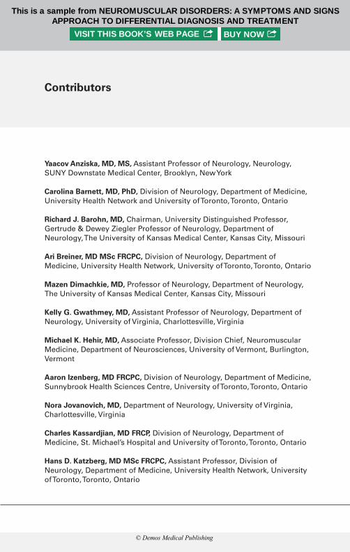
Yaacov Anziska, MD, MS, Assistant Professor of Neurology, Neurology, SUNY Downstate Medical Center, Brooklyn, New York
Carolina Barnett, MD, PhD, Division of Neurology, Department of Medicine, University Health Network and University of Toronto, Toronto, Ontario
Richard J. Barohn, MD, Chairman, University Distinguished Professor, Gertrude & Dewey Ziegler Professor of Neurology, Department of Neurology, The University of Kansas Medical Center, Kansas City, Missouri
Ari Breiner, MD MSc FRCPC, Division of Neurology, Department of Medicine, University Health Network, University of Toronto, Toronto, Ontario
Mazen Dimachkie, MD, Professor of Neurology, Department of Neurology, The University of Kansas Medical Center, Kansas City, Missouri
Kelly G. Gwathmey, MD, Assistant Professor of Neurology, Department of Neurology, University of Virginia, Charlottesville, Virginia
Michael K. Hehir, MD, Associate Professor, Division Chief, Neuromuscular Medicine, Department of Neurosciences, University of Vermont, Burlington, Vermont
Aaron izenberg, MD FRCPC, Division of Neurology, Department of Medicine, Sunnybrook Health Sciences Centre, University of Toronto, Toronto, Ontario
Nora Jovanovich, MD, Department of Neurology, University of Virginia, Charlottesville, Virginia
Charles Kassardjian, MD FRCP, Division of Neurology, Department of Medicine, St. Michael’s Hospital and University of Toronto, Toronto, Ontario
Hans D. Katzberg, MD MSc FRCPC, Assistant Professor, Division of Neurology, Department of Medicine, University Health Network, University of Toronto, Toronto, Ontario
Contributors
© Demos Medical Publishing
This is a sample from NEUROMUSCULAR DISORDERS: A SYMPTOMS AND SIGNSAPPROACH TO DIFFERENTIAL DIAGNOSIS AND TREATMENT
VISIT THIS BOOK’S WEB PAGE BUY NOW

David Lacomis, MD, Professor of Neurology and Pathology; Chief, Division of Neuromuscular Diseases, Departments of Neurology and Pathology (Neuropathology), University of Pittsburgh School of Medicine, Pittsburgh, Pennsylvania
Diana Mnatsakanova, MD, Departments of Neurology, University of Pittsburgh School of Medicine, Pittsburgh, Pennsylvania
Mamatha Pasnoor, MD, Associate Professor of Neurology, Department of Neurology, The University of Kansas Medical Center, Kansas City, Missouri
Karin Provost, DO, PhD, Assistant Professor of Medicine, Department of Internal Medicine, Pulmonary Division, Buffalo, New York
Daniel W. Sheehan, PhD, MD, Associate Professor of Pediatrics, Department of Pediatrics, Pulmonary Division Buffalo, New York
Nicholas J. Silvestri, MD, Associate Professor of Neurology, Department of Neurology, Buffalo, New York
Andrew W. Tarulli, MD, Department of Neurology, Atlantic Neuroscience Institute, Overlook Medical Center, Summit, New Jersey; Clinical Associate Professor, Sidney Kimmel Medical College of Thomas Jefferson University, Philadelphia, Pennsylvania
Simona Treidler, MD, Clinical Neurophysiology Fellow, Department of Neurology, SUNY Downstate Medical Center, Brooklyn, New York
© Demos Medical Publishing
This is a sample from NEUROMUSCULAR DISORDERS: A SYMPTOMS AND SIGNSAPPROACH TO DIFFERENTIAL DIAGNOSIS AND TREATMENT
VISIT THIS BOOK’S WEB PAGE BUY NOW
With Neuromuscular Disorders: A Symptoms and Signs Approach to Differential Diagnosis and Treatment, Nick Silvestri and his co-au-thors have created a novel and valuable resource for both neurol-ogists and generalists. In contrast to the disease classification or diagnostic testing that provides an organizational framework for most texts on neuromuscular disorders, Silvestri and his colleagues have returned to the basics, framing the book around the initial interaction we have with patients—how and what are they feeling and what do we find on their neuromuscular examination.
Chapters of the book are divided into global presentations that should be familiar to neurologists and other health care providers. For instance, limb weakness is covered in separate chapters depend-ing on whether the presentation is acute, subacute, or chronic. The topic of episodic weakness and exercise intolerance, perhaps less familiar to non-neuromuscular practitioners, merits its own chapter, as does ocular and bulbar weakness and respiratory dysfunction. There are two chapters that concentrate on sensory impairment and pain, and an initial chapter devoted to the interpretation of diagnos-tic testing.
Indeed the text provides guidance well beyond the spectrum of neuromuscular disorders. Signs, for instance, are examined from the perspective of both central and peripheral nervous system causes, with etiologies succinctly outlined for quick review by the provider. Clinical pearls are offered to refine both history-taking and exam-ination skills. Key features of neuromuscular disorders that present with the symptoms and signs discussed earlier in each chapter are summarized in tables, at times with guidance on laboratory testing to help narrow the differential diagnosis. Finally, there are brief over-views of treatment and management.
This granular approach, beginning with key elements from the history and physical examination, should be particularly useful for general neurologists and other practitioners who encounter
Foreword
© Demos Medical Publishing
This is a sample from NEUROMUSCULAR DISORDERS: A SYMPTOMS AND SIGNSAPPROACH TO DIFFERENTIAL DIAGNOSIS AND TREATMENT
VISIT THIS BOOK’S WEB PAGE BUY NOW

neuromuscular disease outside of specialty, referral clinics. But its expanded sections that offer further detail I believe will be of use for neuromuscular medicine fellows and their faculty mentors. As I was reviewing it, I felt the book would make a wonderful companion to Aziz Shaibani’s A Video Atlas of Neuromuscular Disorders, a video and photographic resource that, too, follows a symptoms and signs framework.
The pattern-recognition approach that courses through these recent works, of course, lies at the foundation of teaching neurology, whether the focus is neuromuscular disease or not. But in this age of technological advances and ever more complex diagnostic tech-niques, such a foundation is often forgotten or taken for granted. It is refreshing to have this new neuromuscular textbook which reminds us of the value and rationality of returning to the basics.
Gil I. Wolfe, MDIrvin and Rosemary Smith Professor and Chairman,
Department of NeurologyJacobs School of Medicine and Biomedical Sciences,
University at Buffalo/SUNY
© Demos Medical Publishing
This is a sample from NEUROMUSCULAR DISORDERS: A SYMPTOMS AND SIGNSAPPROACH TO DIFFERENTIAL DIAGNOSIS AND TREATMENT
VISIT THIS BOOK’S WEB PAGE BUY NOW

The field of neuromuscular medicine has grown considerably even since I graduated from fellowship just a few years ago with signifi-cant advances in diagnostic testing and treatment. While this book was being written the first drugs were approved for Duchenne mus-cular dystrophy and spinal muscular atrophy, as was only the sec-ond drug in almost 20 years to treat amyotrophic lateral sclerosis. Laboratories around the world are working diligently to determine the cause of and treatment for these and many other neurological disorders. In this context, it is now more important than ever to make firm and accurate diagnoses in our patients.
As asserted in each and every chapter of this book, the history and examination are of paramount importance to guide a rational work-up leading to a diagnosis and directed treatment in the patients we care for. This was the rationale for writing Neuromuscular Disorders: A Symptoms and Signs Approach to Differential Diagnosis and Treatment. There are already a number of excellent textbooks in this field, but most focus on the diagnosis and work back toward the signs and symptoms. The goal of this book was to take the oppo-site approach—to start with presenting complaints and findings and guide the reader along the diagnostic pathway. This book is intended for medical students, mid-level practitioners, residents, and fellows in neuromuscular medicine. I chose the authors of each chapter knowing that they are all great clinician educators and writers and I am extremely grateful for their contributions. I hope that by read-ing Neuromuscular Disorders: A Symptoms and Signs Approach to Differential Diagnosis and Treatment you develop a greater enthusi-asm and confidence in caring for your patients with neuromuscular disorders.
Nicholas J. Silvestri, MD
Preface
© Demos Medical Publishing
This is a sample from NEUROMUSCULAR DISORDERS: A SYMPTOMS AND SIGNSAPPROACH TO DIFFERENTIAL DIAGNOSIS AND TREATMENT
VISIT THIS BOOK’S WEB PAGE BUY NOW

ShareNeuromuscular Disorders: A Symptoms and Signs Approach to Differential Diagnosis and Treatment
atment
© Demos Medical Publishing
This is a sample from NEUROMUSCULAR DISORDERS: A SYMPTOMS AND SIGNSAPPROACH TO DIFFERENTIAL DIAGNOSIS AND TREATMENT
VISIT THIS BOOK’S WEB PAGE BUY NOW

2
INTRODUCTIONWeakness is a common and nonspecific complaint that encom-passes a broad differential diagnosis, including both neurologic and non-neurologic conditions. Weakness is broadly defined as the inability to perform a desired movement with normal force because of a reduction in muscle strength.
Normal motor function involves integrated muscle activity that is modulated by the entire neuro-axis, including the cerebral cortex, basal ganglia, cerebellum, spinal cord, nerve roots, peripheral nerves, neuromuscular junction, and muscle. The tempo of weakness onset, its distribution, and any accompanying symptoms, help to determine its cause.
Weakness can be broadly categorized as focal or generalized.
• Generalized weakness, or quadriparesis, affects all extremities and results either from systemic disease (sepsis, cardiac failure, dehydration, severe hypokalemia, and adrenal insufficiency), high spinal cord lesions, or certain neuromuscular disorders (myasthe-nia gravis [MG], Guillain-Barré syndrome [GBS], periodic paral-ysis). In generalized weakness, certain bulbar motor functions, such as facial movements, articulation, chewing, and swallowing may also be involved.
• Focal weakness, on the other hand, can result from spinal cord involvement or dysfunction of the peripheral nervous system. In spinal cord disease, the weakness is incomplete, with more severe involvement of muscles preferentially innervated by corticospinal tracts. Peripheral nerve disease tends to predominantly involve distal muscles, although there are exceptions. There is no pref-erential involvement of corticospinal tract-innervated muscles. In neuromuscular junction disorders and certain myopathies, the weakness is likely to be worse proximally, sensation is spared, reflexes are normal, and there is frequently involvement of bulbar muscles, especially with ptosis and ophthalmoplegia.
Acute Generalized Weakness
Simona Treidler and Yaacov Anziska
© Demos Medical Publishing
This is a sample from NEUROMUSCULAR DISORDERS: A SYMPTOMS AND SIGNSAPPROACH TO DIFFERENTIAL DIAGNOSIS AND TREATMENT
VISIT THIS BOOK’S WEB PAGE BUY NOW

HISTORYThe most important element of history is weakness onset; acute in minutes to hours (i.e., a vascular event), subacute over weeks (e.g., GBS) or chronic in years (e.g., muscular dystrophy). Fluctuation of the weakness or presence of fatigability with sustained muscle effort suggests MG or a metabolic myopathy.
• Acute attacks of generalized weakness lasting for a few hours and spontaneously resolving are seen in periodic paralysis, as well as in transient vascular pathology. Repeated attacks of weakness suggest neuromuscular junction disorders, periodic paralysis, and metabolic myopathies.
• Another key element is the presence of an underlying neuromus-cular disorder, which may worsen transiently, such as MG.
• A thorough history addresses certain medical comorbidities, such as renal failure, thyroid disease, and adrenal insufficiency.
• It is important to inquire about the patient’s dietary habits and exposure to medications known to be myotoxic, such as statins.
• The examiner should not overlook use of illicit substances, such as cocaine, which can cause overwhelming rhabdomyolysis.
• The presence of preceding viral illnesses and vaccinations sug-gest a viral myositis or GBS, and history of recent travel could implicate a tick paralysis or an acute generalized neuropathy (acute motor axonal neuropathy, GBS).
• Family history is typically more important when diagnosing chronic weakness, such as muscular dystrophy, although in rare cases, a positive history can aid in diagnosing subtler causes of weakness, including metabolic myopathy and periodic paralysis.
SIGNSQuadriparesis
• Quadriparesis or weakness of all limbs, associated with impair-ment of mental status, is a sign of a diffuse cerebral process such as anoxia, severe hypotension, cerebral edema from head trauma, or systemic metabolic abnormalities.
• Quadriparesis with upper motor neuron signs (weakness of upper limb extensors and lower limbs flexors, hyperreflexia, and extensor plantar reflex) and cranial nerve deficits (dysarthria, dys-phagia, ophthalmoplegia) is usually associated with a brainstem lesion, and a rapid diagnosis is essential for possible surgical management. Association of quadriparesis with a sensory level and bowel or bladder abnormalities localizes the lesion to the cer-vical spine. This etiology can be life-threatening, as it may impair the diaphragmatic ventilatory function and disrupt the autonomic nervous system, leading to hypotension and tachycardia.
© Demos Medical Publishing
This is a sample from NEUROMUSCULAR DISORDERS: A SYMPTOMS AND SIGNSAPPROACH TO DIFFERENTIAL DIAGNOSIS AND TREATMENT
VISIT THIS BOOK’S WEB PAGE BUY NOW

• The presence of quadriparesis with lower motor neuron signs (flaccid tone, diminished or absent reflexes, +/− fasciculations) with sensory abnormalities is most consistent with an acute polyneuropathy, such as GBS, also leading to ventilatory failure. The absence of sensory symptoms points to a myositis, as most myopathies do not present acutely.
• Quadripareis presenting with ocular (diplopia, ptosis) and bulbar weakness (dysarthria and dysphagia) usually occurs in a neuro-muscular junction disorder (MG, Lambert Eaton myasthenic syn-drome [LEMS]) as this can also cause respiratory failure.
• Quadriparesis can be associated with other patterns of weakness, including disproportionate proximal or distal weakness, axial weakness, and bulbar, ocular, or facial dysfunction.
Proximal Weakness
• Proximal weakness, including bibrachial palsy and flail arm syn-drome, can be seen in the early stages of GBS, or motor neu-ropathies resulting from amyloid or porphyria. Yet, “man in the barrel syndrome” can also be caused by bilateral watershed brain infarcts. A central cervical spinal cord lesion produces the same pattern of weakness, but there is usually loss of pain and tem-perature sensation in the arms. While proximal weakness is more characteristic in autoimmune myositis, other myopathies, such as metabolic, endocrine, and toxic, can be responsible, rarely in the acute setting. MG can first present with acute proximal weakness
TABLE 2.1 Upper versus Lower Motor Neuron Weakness
Feature Upper motor neuron Lower motor neuron
Weakness distribution
Corticospinal distribution (extensors in upper limbs and flexors lower limbs)
Generalized proximal/distal with no preferential involvement
Sensory involvement
Central pattern None/root/ stocking-glove/nerve distributions
Deep tendon reflexes
Decreased/increased Normal/decreased
Pathologic reflexes
Present Absent
Sphincter function
Could be impaired Normal/impaired in cauda equina lesion
Muscle tone Increased/normal or decreased in acute process
Normal/decreased
Muscle bulk Normal/slight atrophy due to disuse
Atrophy may be marked
Fasciculations No Possible
© Demos Medical Publishing
This is a sample from NEUROMUSCULAR DISORDERS: A SYMPTOMS AND SIGNSAPPROACH TO DIFFERENTIAL DIAGNOSIS AND TREATMENT
VISIT THIS BOOK’S WEB PAGE BUY NOW

and progress to generalized weakness, usually over days. An adult form of acid maltase deficiency and familial periodic paraly-sis may affect only proximal muscles.
Distal Weakness
• Distal weakness rarely generalizes acutely, except in GBS and cen-tral cord syndrome. Patients will complain of difficulty climbing onto a sidewalk, due to foot drop, and as the disease progresses, they will also have difficulty buttoning shirts, using a zipper, or opening a jar. If the physical exam reveals weakness and disso-ciated loss of pain and temperature, then a central cord process is more likely. If there is weakness and areflexia, then GBS is the usual culprit.
Facial and Bulbar Weakness
• Facial and bulbar weakness can be associated with generalized weakness in neuromuscular junction disorders (botulism, MG), GBS, and brainstem strokes. Patients will complain of difficulty drinking through a straw, and lower facial weakness will produce drooling and difficulty handling saliva. Weakness of masticatory muscles, as in MG, may cause fatigue and difficulty chewing. Pharyngeal, palatal, and tongue weakness disturb speech and swallowing. A flaccid palate is associated with nasal regurgita-tion, choking, and aspiration of liquids. Speech becomes slurred or acquires a nasal or hoarse quality.
Neck Weakness
• Neck weakness occurs in many neuromuscular conditions, includ-ing polymyositis, motor neuron disease (MND), and MG. Patients can suffer either from neck flexor or extensor weakness, and in the latter, will have the “dropped head” syndrome. Examination of neck strength in acute muscle weakness is a crude test of respi-ratory impairment, as these muscles and the diaphragm may be simultaneously affected by similar processes. Shortness of breath is typically reported when prone. Since diaphragmatic weakness leads to hypoventilation and carbon dioxide retention, patients may complain of morning headaches, and in the later stages of hypercapnia, may become lethargic and/or confused. Acute onset of shortness of breath occurs in acute motor neuropathies, MG, myositis, and high cervical transverse myelitis.
Paraparesis/Paraplegia
• Paraparesis, paraplegia, or symmetric weakness of the legs, with or without an accompanying sensory level, is generally caused by a spinal cord lesion at or below the upper thoracic spinal cord.
• Paraparesis can also result from lesions at other locations that disturb both upper motor neurons (i.e., parasagittal tumors and
© Demos Medical Publishing
This is a sample from NEUROMUSCULAR DISORDERS: A SYMPTOMS AND SIGNSAPPROACH TO DIFFERENTIAL DIAGNOSIS AND TREATMENT
VISIT THIS BOOK’S WEB PAGE BUY NOW

strokes) and lower motor neurons (such as MND, cauda equina syndrome, peripheral neuropathies).
• Acute paraplegia at an early stage can be associated with flaccid tone and absent reflexes, making localization to the spinal cord challenging, but could alternatively be consistent with a rapidly progressive MND (polio, West Nile virus), or GBS.
• The presence of a thoracic sensory level and increased reflexes in the lower limbs should guide the examiner to consideration of a thoracic cord process, such as an epidural tumor, abscess, hema-toma, prolapsed disc, spinal cord infarction (sparing propriocep-tion), arteriovenous fistula, and transverse myelitis.
PHYSICAL EXAMINATION OF A WEAK PATIENTGeneral Physical Examination
• A general examination should be performed before focusing on the neurologic exam. Special attention should be directed to any signs of chronic systemic illness, occult neoplasms, chronic infections, or endocrine dysfunction. An important component of this examination is the recognition of life-threatening respiratory failure or cardiac arrhythmias, so as to decide on admission to a monitored setting, possible intubation, and initiation of anti-ar-rhythmic medications.
• Unlike those with respiratory failure in primary cardiac or respi-ratory disorders, neuromuscular patients with significant respira-tory dysfunction can appear asymptomatic, without typical signs (such as use of accessory muscles, shallow breathing, or wheez-ing) until they finally decompensate with disease progression. Respiratory function should always be evaluated in all neuromus-cular patients with generalized weakness.
Respiratory Pattern
• The pattern of respiratory muscle function is helpful for making a diagnosis. At rest, diaphragmatic movement performs the major-ity of ventilatory function. Patients with isolated diaphragmatic weakness compensate during inspiration by using the accessory muscles of respiration (pectoral, scalene, intercostal, and sterno-cleidomastoid muscles).
• Diaphragmatic paralysis can be observed by “paradoxical” abdominal movement, with inward movement of the abdominal wall on inspiration. In contrast, patients with impaired intercostal muscle function and preserved diaphragmatic function exhibit inward movement of the upper ribcage and intercostal spaces during inspiration. Auscultation of the chest is often unremarkable in these patients, although crepitations may be heard, reflecting atelectasis, and rarely, transmitted upper airway sounds may be present in patients with bulbar dysfunction.
© Demos Medical Publishing
This is a sample from NEUROMUSCULAR DISORDERS: A SYMPTOMS AND SIGNSAPPROACH TO DIFFERENTIAL DIAGNOSIS AND TREATMENT
VISIT THIS BOOK’S WEB PAGE BUY NOW

Bedside Testing
• The “single breath” counting test performed at bedside tests the ability to count after a maximal inspiration. Patients with a normal respiratory function can reach up to 50, and anything less than 15 cor-relates with severe impairment of vital capacity. Laborious breath-ing while lying flat is another important indication of diaphragmatic weakness and reduced vital capacity, causing orthopnea.
• Bedside spirometry is critical for measurement of ventilatory function in those with neuromuscular disorders, especially in the acute setting. Respiratory thresholds are remembered best through the “20/30/40 rule,” applicable to standard spirometry assessment. Significant ventilatory dysfunction is reflected by forced vital capacity (FVC) <20 mL/kg, maximal inspiratory pres-sure (MIP) <-30 cm H2O, and maximal expiratory pressure (MEP) <40 cm H2O; any abnormal values should trigger critical care sup-port. The objective of bedside spirometry is to detect emerging hypoventilation prior to life-threatening decompensation culmi-nating in hypoxemia, which is detectable by pulse oximetry or arterial blood gas measurement. However, checking pulse oxime-try and/or blood gas earlier in disease course may falsely reassure the examiner, as these will remain normal until there is significant disease progression.
• Dysarthria, presence of a weak cough with difficulty clearing secretions, “staccato speech,” and hypophonia are other import-ant signs of bulbar and respiratory dysfunction that are noted at the bedside. Difficulty lifting the head against gravity correlates with significant diaphragmatic weakness, as both share similar nerve root innervation.
• Autonomic dysfunction is common in patients with acute neu-romuscular diseases, occurring in up to 60% of cases of GBS. Clinical manifestations include orthostatic hypotension, diabe-tes insipidus, ileus, and cardiac arrhythmias, and these may all require medical treatment, occasionally even emergency pacing. Bedside assessment of blood pressure (supine and sitting) and a standard EkG are part of the initial evaluation.
The Neurologic ExaminationThe neurologic examination, with careful attention to strength test-ing, sensory modalities, and deep tendon reflexes, is necessary in all patients who complain of acute weakness. In situations that suggest a central process or an ascending paralysis, a detailed evaluation of the cranial nerves should be performed, in addition to the motor and sensory examination.
The goals of the neurological examination are:
1. Clarify the exact nature of the clinical symptoms. Is the patient truly weak?
© Demos Medical Publishing
This is a sample from NEUROMUSCULAR DISORDERS: A SYMPTOMS AND SIGNSAPPROACH TO DIFFERENTIAL DIAGNOSIS AND TREATMENT
VISIT THIS BOOK’S WEB PAGE BUY NOW

2. Identify clinical symptoms not recognized by the patient. For example, is weakness of the legs accompanied by mild, but less evident, weakness in the arms? Is the weakness accompanied by sensory loss not recognized by the patient?
3. Clarify the anatomic basis for the symptoms. Is the weakness accompanied by changes in the deep tendon reflexes, or by mus-cle atrophy and fasciculations?
TABLE 2.2 Lesion Localization
Location Weakness Sensory loss
Deep tendon reflexes Other features
Cerebral cortex
UMN pattern
Higher order Increased Loss of higher cognitive functions, seizures
Cerebral white matter and subcortical structures
UMN pattern
Higher order (cerebrum)
Increased Loss of higher cognitive functions, psychiatric disturbances, movement disorders
Lower order (thalamus)
Brainstem UMN pattern
Lower order Increased Cranial nerve dysfunction contralateral to long-tract abnormality
Spinal cord UMN below lesionLMN at the lesion level
Lower order Increased Sensory level at or below lesion; bowel/bladder dysfunction
Peripheral nerve
LMN pattern
Lower order Decreased Root or nerve distribution of the deficit
Neuro- muscular junction
Myopathic None Normal Fatigable weakness of proximal or bulbar muscles
Muscle Myopathic None Normal Nonfatigable weakness of proximal muscles
LMN: lower motor neuron; UMN: upper motor neuron; Myopathic (diffuse, proximal weakness); Higher order (stereognosia, two-point discrimination); Lower order (modalities such as light touch, temperature).
© Demos Medical Publishing
This is a sample from NEUROMUSCULAR DISORDERS: A SYMPTOMS AND SIGNSAPPROACH TO DIFFERENTIAL DIAGNOSIS AND TREATMENT
VISIT THIS BOOK’S WEB PAGE BUY NOW

• First, the examiner must inspect the various muscles, looking for atrophy or hypertrophy, as well as spontaneous involuntary movements, including fasciculations, muscle “rippling,” and myo-kymias. While muscle strength evaluation is commonly regarded as “objective,” as it is rated on a numerical scale, this assessment is actually quite subjective and is highly dependent on the skill of the physician. It is also important that the patient exerts a max-imum effort. It can be difficult to assess strength reliably in an individual with severe pain or who is unwilling to fully cooperate with the examination. Patients with anxiety or depression may not be motivated to make a full effort on strength testing.
• Peripheral nerve hypertrophy is seen and/or felt in certain demy-elinating polyneuropathies, including hereditary motor-sensory neuropathy, leprosy, and neurofibromatosis.
• In the acute setting, the most important causes of acute general-ized weakness without localizing signs are disorders affecting the peripheral nerves, the neuromuscular junction, and the muscles. There are clinical features that will help differentiate them.
TABLE 2.3 Generalized Weakness: Peripheral Localization
Clinical signs NeuropathyNeuromuscular junction Myopathy
Weakness pattern
Distal>proximal Variable Proximal>distal
Weakness severity
Constant, progressive
Variable Constant, progressive
Sensory loss Distal>proximal Absent Absent
Pain Variable, distal Absent Usually absent
Muscle atrophy Variable, often early
Absent Variable, usually late
Reflexes Decreased or absent
Normal Normal or slightly decreased
Fasciculations Sometimes Absent Absent
CSF Normal or increased
Normal Normal
Ck level Normal or minimally increase
Normal Increased
Edophronium test
No response May respond No response
Ck: creatine kinase; CSF: cerebrospinal fluid.
© Demos Medical Publishing
This is a sample from NEUROMUSCULAR DISORDERS: A SYMPTOMS AND SIGNSAPPROACH TO DIFFERENTIAL DIAGNOSIS AND TREATMENT
VISIT THIS BOOK’S WEB PAGE BUY NOW

DIAGNOSTIC INVESTIGATIONS • The extent of diagnostic testing performed on a weak patient var-
ies depending upon the differential diagnosis and the potential rate of clinical deterioration. A patient with severe sepsis will need a full work-up to identify the source and subsequent treatment. If one suspects an acute stroke or spinal cord lesion, the following tests should be performed emergently.
Imaging1. CT of head and/or CT angiography head and neck for evaluation
of a central lesion.2. MRI of the spine (or CT myelography when MRI is contraindi-
cated) for evaluation of spine lesions.
Routine Laboratory Tests1. Comprehensive metabolic panel and electrolytes. Elevated liver
enzymes (AST, ALT) could indicate acute muscle destruction, such as in a myositis, but would not be accompanied by an elevated GGT, as in liver failure.
• Hypo- or hyperkalemia are frequently seen in periodic paral-ysis patients with transient and recurrent generalized muscle weakness. Hypokalemia, which can cause a myopathy, is also present in patients with renal disorders, such as renal tubular acidosis types 1 and 2, as well as in those using diuretics, laxa-tives, and glucocorticoids.
• Various metabolic derangements can cause generalized weak-ness, including hypo- or hypernatremia (seen in SIADH, diabe-tes insipidus), hypercalcemia, and uremia.
2. Serum creatine kinase (Ck) is valuable for evaluation of patients with neuromuscular disease. Although it is a marker of muscle disease, increased serum Ck can be seen in MND) and other neu-rogenic disorders, and conversely, elevated Ck levels do not occur in all myopathies. The level of Ck does not appear to correlate with the degree of muscle destruction or weakness. In normal individuals, levels may reach 5 times the upper limit of normal in certain situations, such as after an electrodiagnostic studies, and can be as high as 50 times the upper limit of normal in extreme and prolonged exercise. Other muscle enzymes, including LDH and aldolase, should be also sent.
3. TSH, PTH, vitamin D, and cortisol levels for treatable causes of generalized weakness.
4. Vitamin B levels (B1, B6, B12), E, and folic acid, which if abnormal, can cause neuropathy, myelopathy, or myopathy.
5. In the setting of a suspected myositis, one should order ANA, ds-DNA, ANCA, cryoglobulins, rheumatoid factor, anti-SSA, anti-SSB, and ACE.
© Demos Medical Publishing
This is a sample from NEUROMUSCULAR DISORDERS: A SYMPTOMS AND SIGNSAPPROACH TO DIFFERENTIAL DIAGNOSIS AND TREATMENT
VISIT THIS BOOK’S WEB PAGE BUY NOW

6. Infectious tests for generalized weakness include HIV, HTLV-1/2, and Hepatitis B and C serologies, along with Lyme (Western blot-ting). Clostridium botulinum toxin can be detected in blood or stool, especially in the presentation of acute descending weak-ness accompanied by cranial nerve and parasympathetic dys-function and rapid respiratory failure.
7. Acetylcholine receptor, (binding, blocking, modulating), MUSk, LRP4, agrin, and anti-voltage gated calcium channel antibodies to diagnose a neuromuscular junction disease.
8. Urine porphyrin (uroporphyrin, coproporphyrin), delta amino- levulinic acid, and porphobilinogen (sensitive and specific in an acute attack) could be obtained from urine for porphyric neuropathies.
9. Serum heavy metal testing, including arsenic, lead, mercury, and thalium, in the setting of a motor neuropathy.
Other Monitoring1. EkG/cardiac monitoring, as generalized weakness can be a
feature of acute myocardial infarction or exacerbation of car-diac failure. Cardiac arrhythmias are present in electrolyte abnormalities associated with generalized weakness and in channelopathies.
2. Chest x-ray can show a low-seated diaphragm, cardiomegaly, or pleural effusions.
3. Cerebrospinal fluid (CSF) analysis is indicated in suspected GBS, focusing on the albumino-cytologic dissociation, with elevated protein level and absent white blood cell count. However, detec-tion of an elevated protein level is dependent on the timing of the lumbar puncture. The presence of lymphocytosis may be indica-tive of HIV infection or lymphoma and needs further evaluation. Bacterial, viral, and fungal cultures, along with tuberculosis PCR, are important in the initial evaluation of an acute neuropathy, myopathy, or myelopathy.
4. Nerve conduction studies (NCS)/electromyography (EMG) are rarely used in the acute evaluation of generalized weakness.
In GBS, NCS may have prolonged or absent F waves, and rarely con-duction block. EMG showing spontaneous activity suggests an acute inflammatory myositis.
DIFFERENTIAL DIAGNOSISThere is a myriad of medical and neurological disorders that mani-fest with generalized weakness. This section will focus on the most common disorders of the peripheral nervous system presenting as acute generalized weakness.
© Demos Medical Publishing
This is a sample from NEUROMUSCULAR DISORDERS: A SYMPTOMS AND SIGNSAPPROACH TO DIFFERENTIAL DIAGNOSIS AND TREATMENT
VISIT THIS BOOK’S WEB PAGE BUY NOW

Table 2.4 Differential Diagnosis of Acute Weakness
Systemic CausesSepsisSevere anaemiaHeart failureHepatic failureAcute kidney injuryHypoxemia/anoxic injury/hypercarbiaHypoglycemia/hyperglycemia/hyperosmolar stateElectrolytes abnormalities (hypo/hyperkalemia, hypo/hypernatremia, hypo/hypercalcemia, hypermagnesemia, hypophosphatemia)Thyroid dysfunction (hyperthyroid storm, myxedema)Adrenal dysfunction (hyperaldosteronism/hypercortisolism/adrenal insufficiency)Hypothermia/hyperthermiaNeuroleptic malignant syndromeMalignant hyperthermiaIntoxications (alcohol/opiates/barbiturates/antidepressants/antipsychotic/anticonvulsants/illicit drugs)Central Nervous System DisordersAcute ischemic strokes (bilateral ACA strokes, watershed infarcts, thalamic or brainstem)Hemorrhagic strokes (large with midline shift or herniation)Parasagittal frontal tumorsSyringobulbiaInfections: Meningitis/encephalitisNonconvulsive status epilepticusPost-ictal paralysisMigraine with brainstem auraNeuroinflammatory disorders (multiple sclerosis/neuromyelitis optica/acute disseminated encephalomyelitis, sarcoidosis, rheumatoid arthritis, SLE)Severe Parkinson disease or Parkinsonian syndromeStiff person syndromeSpinal cord DisordersTransverse myelitisSyringomyeliaNeuroinflammatory disorders (multiple sclerosis/neuromyelitis optica/acute disseminated encephalomyelitis, sarcoidosis, rheumatoid arthritis, SLE)HIV, HTLV 1/2 West Nile myelopathySubacute combined degenerationTabes dorsalisSpinal cord infarctionSpinal cord compression (trauma/tumor/abscess/hemorrhage)
(continued)
Guillain-Barré Syndrome
• GBS is an immune-mediated neuropathy presenting with acute generalized weakness. Typically demyelinating, GBS is frequently preceded by infections, such as Campylobacter jejuni, or even
© Demos Medical Publishing
This is a sample from NEUROMUSCULAR DISORDERS: A SYMPTOMS AND SIGNSAPPROACH TO DIFFERENTIAL DIAGNOSIS AND TREATMENT
VISIT THIS BOOK’S WEB PAGE BUY NOW

Table 2.4 Differential Diagnosis of Acute Weakness (continued )
Peripheral Nerve DisordersToxic neuropathies (Heavy metals, drugs, alcohol, and organophosphates)Infectious Neuropathies (HIV, CMV, EBV, Lyme disease, HIV)Acute Inflammatory Demyelinating polyneuropathy (GBS and variants)Thiamine deficiencyVitamin B12 deficiencyHypothyroid neuropathyDiabetes neuropathiesUremic neuropathyPorphyriaCritical illness polyneuropathyNeuromuscular Junction DisordersMyasthenia gravisEaton-Lambert syndromeTetanusBotulismTick paralysisInsect/marine toxinsMyopathiesPolymyositisDermatomyositisNecrotizing myopathyPeriodic paralysisAlcohol myopathyMedication-induced myopathiesOther Causes of WeaknessMotor neuron diseasePolyomyelitis
by vaccinations, such as influenza. The suspected pathophysiol-ogy is molecular mimicry between C. jejuni lipooligosaccharides and GM1 or GD1a gangliosides at the nodes of Ranvier; GM1 and GD1a antibodies bind to these saccharides and activate the myelin-destroying complement apparatus.
• GBS progresses within 1 to 2 weeks, first characterized by severe back pain and limb paresthesias, followed by ascending weakness, proximal>distal, and depressed/absent deep tendon reflexes. Facial and oropharyngeal muscles are affected in 50% of the cases and can sometimes be the initial manifestation.
• Variants of GBS include Miller Fisher syndrome (ophthalmople-gia, ataxia, and areflexia), isolated paraparesis, pharyngeal-cervi-cal-brachial weakness +/- ataxia, and bifacial weakness +/- distal paresthesias.
• Respiratory failure is the most dangerous complication of GBS, as there can be low tidal volume and poor gas exchange resulting in hypercapnia. Another worrisome complication is dysautonomia,
© Demos Medical Publishing
This is a sample from NEUROMUSCULAR DISORDERS: A SYMPTOMS AND SIGNSAPPROACH TO DIFFERENTIAL DIAGNOSIS AND TREATMENT
VISIT THIS BOOK’S WEB PAGE BUY NOW
characterized by blood pressure fluctuations, cardiac arrhythmias, and gastrointestinal/bladder dysfunction. A few disorders mimic GBS, including transverse myelitis, MG, botulism, and carcino-matous or lymphomatous meningitis.
• CSF analysis typically reveals elevated protein with a normal white blood cell count. More than 10 Lymphocytes/mm3 could be seen in Lyme disease, HIV, poliomyelitis, West Nile, sarcoid, and leptomeningeal disease. CSF could be normal in the early pre-sentation, as can the NCS, and there may not be absent F waves, prolonged distal latencies, decreased conduction velocities, or conduction block; there is typically sural nerve sparing.
• Bedside pulmonary function tests (MEP and MIP) are invaluable, as those with diaphragmatic weakness will have a decrease in the force vital capacity (FVC) when supine, with a 25% reduction from the baseline.
• Immunotherapy treatment hastens recovery and decreases the risk of intubation by 50%. Intravenous immunoglobulins (IVIg) 0.4 g/kg/day for 5 days is first line of treatment, due to availabil-ity and ease of administration. Potential mechanisms of action include modulation of pathogenic autoantibody production, inhibition of antibodies, Fc receptor blockade, and immuno-regulation of B and T cells. Plasma exchange is another treat-ment that removes antibodies and treats GBS, usually after five courses of plasma exchange.
• Overall mortality due to adult respiratory distress syndrome, sep-sis, or dysautonomia is 3% to 5%, and those requiring mechanical ventilation have a 20% mortality rate. Only 79% of survivors can walk independently at 1 year, with severe disability occurs in 10% to 15% of GBS cases.
Myasthenia Gravis
• MG is an autoimmune disease in which antibodies bind to ace-tylcholine receptors or to similar molecules in the postsynaptic membrane at the neuromuscular junction. The antibody attack on the neuromuscular junction leads to weakness of skeletal mus-cles, whether generalized or ocular, and is more proximal than distal in presentation.
• Ocular MG (OMG) presents with fluctuating diplopia and ptosis. Bulbar muscles may also be involved, causing dysarthria or dys-phagia. There may be weakness of neck flexors and/or extensors, and all weakness (generalized or ocular) typically worsens with exercise and fluctuates over the course of the day. MG should be considered in any patient who present with sudden weakness, especially if it fluctuates.
• The diagnosis of MG is confirmed by a combination of relevant symptoms and signs and a positive test for specific autoantibodies,
© Demos Medical Publishing
This is a sample from NEUROMUSCULAR DISORDERS: A SYMPTOMS AND SIGNSAPPROACH TO DIFFERENTIAL DIAGNOSIS AND TREATMENT
VISIT THIS BOOK’S WEB PAGE BUY NOW

including acetylcholine receptors, muscle specific kinase (MuSk), and lipoprotein receptor related protein 4 (LRP4). These antibodies are quite sensitive and specific. In seronegative cases, neurophys-iological testing, with repetitive nerve stimulation and single-fi-bre EMG, may be necessary for disease confirmation. While some have used the “edrophonium test” or infusion of short-acting edrophonium chloride, it lacks both sensitivity and specificity, as well as requiring cardiac monitoring.
• Myasthenia crisis is a life-threatening situation defined as respira-tory failure due to severe diaphragmatic weakness, requiring intu-bation, especially if the vital capacity is <15 mL/kg or MIP is <15 to 20 mm H2O. Severe bulbar weakness with inability to adequately protect the airways is another presentation of a crisis. Mortality in myasthenic crises ranges from 4% to 10%.
• Treatment of crisis first requires addressesing the underlying cause for exacerbation, such as infection or medications wors-ening neuromuscular transmission (i.e., aminoglycosides, macro-lides, and anti-arrhythmics).
• In the acute setting, one can use either plasmapheresis (which directly removes antibodies from the peripheral circulation) or intravenous immunoglobulin, at 2 g/kg total over 5 days. Main-tenance therapy with an immunomodulator should be started, such as steroids, as it works fastest, but there are numerous other possibilities.
Botulism
• Botulism is a rare cause of acute muscle weakness. It is caused by an exotoxin of Clostridium botulinum, an anaerobic bacterium that may contaminate improperly preserved or prepared food. The toxin acts at the neuromuscular junction to prevent the release of acetylcholine, thus blocking neuromuscular transmission.
• Clinical symptoms begin typically in 12 to 36 hours after the ingestion of contaminated food. The initial symptoms are typi-cally related to impaired gastrointestinal motility (constipation, emesis, and abdominal cramps), followed by neurological symp-toms involving the extraocular, facial, and pharyngeal muscles. There may be ptosis, “blurred” vision, diplopia, facial weakness, dysarthria, dysphagia, and hoarseness. These early symptoms are similar to those seen in MG, except that in botulism, the pupillary reflexes are affected early. The weakness spreads rap-idly to the muscles of the trunk and extremities and may prog-ress to complete quadriplegia. As the weakness progresses, it can mimic GBS, but there are no sensory symptoms, and deep tendon reflexes are preserved. Careful observation suggests that ventilatory muscle weakness precedes the recognition of limb weakness.
© Demos Medical Publishing
This is a sample from NEUROMUSCULAR DISORDERS: A SYMPTOMS AND SIGNSAPPROACH TO DIFFERENTIAL DIAGNOSIS AND TREATMENT
VISIT THIS BOOK’S WEB PAGE BUY NOW

• Respiratory failure is the most common cause of death and can occur within 6 to 8 hours of the onset of symptoms. Diagnosis includes demonstration of botulinum toxin in serum, stool, or in food that patient has recently consumed. Treatment is with admin-istration of heptavalent antitoxin as soon as possible. Pyridostig-mine and 3,4-diaminopyridine may be used for symptomatic relief.
Organophosphate Compounds
• Organophosphate compounds are widely used as insecticides and more rarely in chemical warfare. These agents act by inhibiting ace-tylcholinesterase, acting as cholinergic agents. Symptoms of acute poisoning develop immediately after the exposure and include nausea, vomiting, abdominal cramps, diarrhea, increased saliva-tion, lacrimation, bronchospasm, and diffuse muscle weakness. Death results from respiratory failure, and treatment with pralidox-ime can reverse the effect if administered within 24 to 48 hours. Atropine will only reverse the effects of excessive cholinergic stim-ulation on muscarinic receptors but will not reverse the muscle weakness that results from overstimulation of nicotinic receptors.Other neurotoxins are presented in Table 2.6.
Disorders of Muscle
• Acute necrosis of muscle cells (rhabdomyolysis) liberates myo-globin, and high concentrations can cause acute renal failure. The most common causes are either toxic/drug-induced or physical
Table 2.5 Causes of Neuromuscular Transmission Disorders Other Than Myasthenia Gravis
PresynapticLEMSBotulismTick paralysisCongenital myasthenia gravisToxins: elapid snakes species, arhropodes, marine speciesDrugs: aminoglycosides, calcium channel blockers, aminopyridines, corticosteroidsSynapticCongenital myasthenic syndromesDrugs: reversible/irreversible cholinesterase inhibitors (edrophonium, pyridostigmine, organophosphates and carbamates)PostsynapticDrug induced myasthenia gravis: Penicillamine, alpha interferonCongenital myasthenic syndromesDrugs and toxins: nondepolarizing/depolarizing blocking agentsAntibiotics: tetracycline
LEMS, Lambert Eaton myasthenic syndrome.
© Demos Medical Publishing
This is a sample from NEUROMUSCULAR DISORDERS: A SYMPTOMS AND SIGNSAPPROACH TO DIFFERENTIAL DIAGNOSIS AND TREATMENT
VISIT THIS BOOK’S WEB PAGE BUY NOW

(status epilepticus, trauma, or prolonged immobility), but rarely, rhabdomyolysis can be the presenting sign of underlying muscle diseases, such as glycogen storage disorders.
Rhabdomyolysis
• Rhabdomyolysis can also result from myositis, or inflammation of the muscles. Myositis can be caused by infections (viral, bac-terial, fungal, or parasitic), drugs/toxins, electrolyte abnormalities (potassium, calcium), or can be idiopathic (polymyositis, derma-tomyositis, necrotizing myopathy).
Table 2.6 Neurotoxic Substances
Heavy MetalsArsenicLeadGoldThalliumMercuryChemical CompoundsOrganophosphatesPlants and Animal ToxinsCiguateraParalytic shellfish poisoningPuffer fishMedicationsIsoniazidEthambutolChloramphenicolLinezolideDapsoneFluoroquinolonePyridoxineNitrous oxideTacrolimusColchicineDisulfiramProcainamideAmiodaroneAntiretroviral agentsAntineoplastic AgentsVinka alkaloidsPlatinium containing drugsEtoposideSuraminIfosfamideThalidomideBortezomib
© Demos Medical Publishing
This is a sample from NEUROMUSCULAR DISORDERS: A SYMPTOMS AND SIGNSAPPROACH TO DIFFERENTIAL DIAGNOSIS AND TREATMENT
VISIT THIS BOOK’S WEB PAGE BUY NOW

• Clinical presentation consists of proximal muscle weakness with or without myalgias, and in the idiopathic forms, there may be associated lung, skin, and cardiac involvement. Laboratory test-ing demonstrates elevated muscle enzymes, presence of specific autoantibodies, and characteristic muscle biopsy changes.
• Treatment is directed at stopping the toxic agent, providing venti-lator support, and avoiding metabolic disturbances by aggressive hydration and electrolyte correction. Immediate treatment with pulse steroids is required for the idiopathic myositis entities.
Periodic Paralysis
• Periodic paralysis and other metabolic abnormalities can manifest clinically as generalized weakness. Periodic paralysis disorders are channelopathies caused by mutations of the genes controlling the structure of various channels (sodium, calcium, and chloride). The symptoms are triggered by stress, high carbohydrate meals, extreme temperature, and heavy exercise.
• Hypokalemic periodic paralysis has two presentations, the auto-somal dominant disorder due to a defect in the muscle mem-brane’s calcium or sodium channels and the thyrotoxic periodic paralysis that occurs in association with hyperthyroidism.■■ Clinical presentation most often first occurs in the teenage
years, with generalized weakness and preserved conscious-ness lasting hours to days. Patients may awaken paralyzed after strenuous exercise or having eaten a meal rich in carbohy-drates the night before. There is sparing of muscles of the face and trunk, and deep tendon reflexes are diminished or lost, but sensory examination is normal.
■■ Diagnosis is based on suspicion, presence of repeated similar episodes in patient history, positive family history and abnor-mal potassium noted on testing. As untreated attacks last
Table 2.7 Medications Causing of Myopathy
SteroidsColchicinezidovudine, other anti-HIV-related retroviral medicationsChloroquine/hydroxychloroquineAmiodaroneCholesterol-lowering (statins>fibrates, niacin, ezetimibe)ProprofolCyclosporineTacrolimusImatinibAmphotericin
© Demos Medical Publishing
This is a sample from NEUROMUSCULAR DISORDERS: A SYMPTOMS AND SIGNSAPPROACH TO DIFFERENTIAL DIAGNOSIS AND TREATMENT
VISIT THIS BOOK’S WEB PAGE BUY NOW

longer, potassium should be administered in the acute setting. Thyroid function testing should also be performed in evalua-tion of a patient with such acute and diffuse muscle weakness.
• Hyperkalemic periodic paralysis is rarer, and the genetic defect is in the sodium channel. The clinical attacks are briefer and occur after exercise or ingestion of meals rich in potassium. The cra-nial and trunk muscles are not usually involved, and the attacks mostly resolve before patients arrived in emergency rooms. In severe situations, administration of intravenous calcium gluco-nate, glucose, insulin, or beta-2 agonists can alleviate the attacks.
ICU Acquired Weakness
• Flaccid paralysis of the limbs may develop in patients in critical care units, typically within a week, but at times, after only a few days. Weakness is noted when the patient fails ventilator wean-ing. A careful search should exclude brainstem disorders (central pontine myelinolysis), acute spinal disorders, toxic myopathies, and GBS. In general, many patients have a combination of criti-cal illness polyneuropathy (CIP) and critical weakness myopathy (CIM).
• Both CIP and CIM are associated with similar risk factors, includ-ing intubation for more than a week, prolonged neuromuscular blockade, sepsis, use of corticosteroids, and elevated blood glu-cose. CIP often spares the cranial nerves; deep tendon reflexes are absent, and there may be sensory loss on clinical exam. For CIP, electrophysiologic evaluation reveals an axonal motor and sensory neuropathy with low amplitudes. In CIM, NCS are nor-mal, but myopathic units may be seen on EMG testing. CIM is the most common cause of ICU-acquired myopathy and it is difficult to differentiate from CIP, as they overlap clinically, so the term ICU-acquired weakness has been used.
• There is no specific treatment known for ICU-acquired weakness. Half of the patients recover fully after a long and rigorous rehabil-itation program, but the other half remains with significant long-term disability.
SUGGESTED READINGS
Barohn RJ, Dimachkie MM, Jackson CE. A pattern recognition approach to the patient with a suspected myopathy. Neurol Clin. 2014;32(3):569–593.
Dhand Uk. Clinical approach to the weak patient in the intensive care unit. Respiratory Care. 2006;51(9):1024–1041.
Gihus, NE. Myasthenia gravis. NEJM, 2016;375:2570–2581.katirji B, kaminski HJ, Ruff RL. Neuromuscular disorders in clinical
practice; 2014.
© Demos Medical Publishing
This is a sample from NEUROMUSCULAR DISORDERS: A SYMPTOMS AND SIGNSAPPROACH TO DIFFERENTIAL DIAGNOSIS AND TREATMENT
VISIT THIS BOOK’S WEB PAGE BUY NOW

Latronico N, Bolton CF. Critical illness polyneuropathy and myop-athy: a major cause of muscle weakness and paralysis. Lancet Neurol. 2011;10:931–941.
Saguil A. Evaluation of the patient with muscle weakness. Am Fam Physician. 2005;71(7):1327–1336.
Wijdicks EFM, klein CJ. Guillain-Barré syndrome. Mayo Clin Proc. 2017;92(3):467–479.
© Demos Medical Publishing
This is a sample from NEUROMUSCULAR DISORDERS: A SYMPTOMS AND SIGNSAPPROACH TO DIFFERENTIAL DIAGNOSIS AND TREATMENT
VISIT THIS BOOK’S WEB PAGE BUY NOW