NEUROLOGY I. INTRODUCTION A. OUTLINE OF HIGHLIGHTED ...
24
NEUROLOGY I. INTRODUCTION A. OUTLINE OF HIGHLIGHTED CONDITIONS 1) Cranial Defect 2) History of Seizure 3) Head Trauma Without History of Seizure 4) Primary Headache Disorders B. IMPLICATIONS FOR JOB PERFORMANCE Abnormalities in neurological functioning can be quite diverse, and therefore may potentially limit the ability to perform virtually all of the numerous physical tasks required of patrol officers. Beyond simple physical performance impairments, neurological abnormalities also have the potential to cause sudden incapacitation as well as impairment of cognitive functioning. II. MEDICAL EXAMINATION AND EVALUATION GUIDELINES A. GENERAL SCREENING RECOMMENDATIONS 1) History: The general questions found on the Medical History Statement do not require elaboration if answered negatively. 2) Examination: A thorough neurological examination on every candidate, regardless of history, would be quite time-consuming. Alternatively, an adequate screening exam for candidates with a negative history could consist of the following components: • Eyes: Examine fundus for papilledema, oculomotor function, and nystagmus. IX-1
Transcript of NEUROLOGY I. INTRODUCTION A. OUTLINE OF HIGHLIGHTED ...

NEUROLOGY
I. INTRODUCTION
A. OUTLINE OF HIGHLIGHTED CONDITIONS
1) Cranial Defect
2) History of Seizure
3) Head Trauma Without History of Seizure
4) Primary Headache Disorders
B. IMPLICATIONS FOR JOB PERFORMANCE
Abnormalities in neurological functioning can be quite diverse, and therefore may potentially limit the ability to perform virtually all of the numerous physical tasks required of patrol officers. Beyond simple physical performance impairments, neurological abnormalities also have the potential to cause sudden incapacitation as well as impairment of cognitive functioning.
II. MEDICAL EXAMINATION AND EVALUATION GUIDELINES
A. GENERAL SCREENING RECOMMENDATIONS
1) History:
The general questions found on the Medical History Statement do not require elaboration if answered negatively.
2) Examination:
A thorough neurological examination on every candidate, regardless of history, would be quite time-consuming. Alternatively, an adequate screening exam for candidates with a negative history could consist of the following components:
• Eyes: Examine fundus for papilledema, oculomotor function, and nystagmus.
IX-1

• Cerebellar: Have candidates raise their arms in front of them with palms up. In this position, observe any drift or tremor; have candidates perform the fingerto-nose and Rhomberg test.
• Reflexes: Examine biceps, triceps, knee, ankle, and Babinski's.
• Gait: Note any abnormality of arm swing, leg swing, heel strike, or foot strike. Distance between medial malleoli should not exceed 1' (see additional tests in Musculoskeletal chapter).
• Sensory: Use 128 cps. tuning fork to detect absence of vibratory sensation on big toe bilaterally. Record result as present or absent, rather than duration of sensation.
B. EVALUATION OF COMMON CLINICAL SYNDROMES
1) CRANIAL DEFECT
a. GENERAL CONSIDERATIONS:
Cranial defects commonly occur as the result of surgical burr holes or craniotomies which have healed with non-union. Both of these lesions substantially increase the risk of sudden incapacitation during combative situations. A burr hole of approximately 2 em in diameter could allow penetration by a finger or other narrow object. The resulting pressure on the galea could cause sudden and severely excruciating pain. "Floating" bone islands do not protect the brain against blunt trauma.
In most cases, this risk could be eliminated by filling the defect with methyl methacrylate or similar surgical polymer. This procedure is commonly performed by neurosurgeons and has a low morbidity rate.
Note: Cranial defects due to evacuation of childhood epidural hematomas will sometimes develop secondary calcification. These may have the appearance of a depressed skull fracture on radiographs, but are often well-fused to the cranium and provide adequate protection.
b. RECOMMENDED EVALUATION PROTOCOL:
The physician should obtain skull radiographs on any candidate with a history suggesting the possibility of cranial defect. Candidates with floating bone islands or burr holes >2 em should be restricted from combative situations. This restriction could be rescinded if the skull defect is repaired. Candidates who have calcified defects should be referred to a neurosurgeon for an opinion regarding whether the calcium deposit has adequate strength to resist pressure.
IX-2

2) HISTORY OF SEIZURE
a. GENERAL CONSIDERATIONS:
Seizure disorders may create a risk of substantial harm to the candidate and the public in several ways:
(1) A seizure triggered by stimuli that occur during a critical incident could result in a failure to perform in a life-threatening situation;
(2) A seizure at other times also threatens the safety of both the officer and others (e.g., a suddenly incapacitating seizure while driving or directing traffic). Even a seizure during routine questioning of a suspect can turn into a critical situation if it causes an officer to lose control of his/her weapon;
(3) Chronic or intermittent impairment in psychomotor ability (due to the condition itself or to medication taken to control symptoms) can interfere with functioning in both critical incidents and during routine activities.
The following discussion is intended to assist screening physicians in addressing each of these concerns on an individualized basis. An outline of the relevant considerations is presented in Table IX-1.
Table IX-1: Evaluation Outline for Assessin·g Candidates at Increased Risk of Seizure
NON-RANDOM SEIZURE • Susceptibility to environmental stimuli, emotional stress, reflex triggers
RANDOM SEIZURE • Clinical Subcategories
~ Single unprovoked idiopathic seizure .. Epilepsy, controlled on medication .. Epilepsy, presently off medication .. Seizure after head trau rna
• Job Related Considerations .. Percentage of time on duty .. Sleep deprivation .. Pattern and timing of seizures ._ Impact of auras
ASSESSING EFFECT OF PSYCHOMOTOR IMPAIRMENT • Medical side-effects • Transient cognitive impairments
IX-3

ASSESSING ON-DUTY SEIZURE RISK
Non-Random Seizures In general, only 20°/o of seizures occur due to precipitating factors, and not all persons with epilepsy are susceptible. Therefore, the physician must determine, on an individual basis, whether a candidate is at risk of having a seizure induced by stimuli that are encountered during either routine patrol officer activities (e.g., a light flickering between trees may cause reflex seizures), or during critical incidents (e.g., emotional stress, physical stress, or reflex stimuli such as flashing lights or sudden sounds). The physician will be able to identify the few candidates who are susceptible on the basis of their medical history. Unless these candidates can demonstrate excellent control with medication, their risk of a seizure during routine patrol officer duties would clearly constitute a direct threat.
Random Seizures There are numerous risk factors to consider when determining an individual's probability of randomly suffering a recurrent seizure. The relative importance of these risk factors varies by clinical subcategory. Therefore, an individualized risk assessment can be best accomplished by considering risk factors within each clinical subcategory. The clinical subcategories discussed below include:
A. SINGLE UNPROVOKED IDIOPATHIC SEIZURE
B. EPILEPSY, CONTROLLED ON MEDICATION
C. EPILEPSY, PRESENTLY OFF MEDICATION
D. SEIZURE AFTER HEAD TRAUMA
A. SINGLE UNPROVOKED IDIOPATHIC SEIZURE
An unprovoked idiopathic seizure is one, which occurs in the absence of an identifiable alteration of systemic metabolic function or insult to the structural integrity of the brain. EEG's and CAT scans are often negative. Seizures associated with sleep deprivation are considered idiopathic.
There are numerous studies that evaluate the risk of seizure recurrence. Metaanalysis is difficult due to differences in definitions and prevalence of risk factors. The usefulness of many studies is also limited by short follow-up periods. · However, one major study (Hauser, et al., 1990) has both adequate follow-up (average four years) and a large subset of patients (78) that are clearly defined as "idiopathic" with no risk factors for recurrence. Therefore, this study provides useful baseline recurrence rates (Table IX-2). As would be expected, these rates are lower than those found in any other study, since other studies include patients with positive risk factors for recurrence.
IX-4
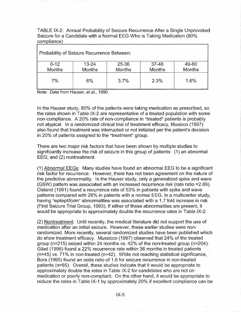
TABLE IX-2: Annual Probability of Seizure Recurrence After a Single Unprovoked Seizure for a Candidate with a Normal EEG Who is Taking Medication (80o/o compliance)
Probability of Seizure Recurrence Between:
0-12 13-24 25-36 37-48 49-60 Months Months Months Months Months
70/o 6% 3.7% 2.3°/o 1.6o/o
Note: Data from Hauser, et al., 1990.
In the Hauser study, 80°/o of the patients were taking medication as prescribed, so the rates shown in Table IX-2 are representative of a treated population with some non-compliance. A 20°/o rate of non-compliance in "treated" patients is probably not atypical. In a randomized clinical trial of treatment efficacy, Musicco (1997) also found that treatment was interrupted or not initiated per the patient's decision in 20o/o of patients assigned to the "treatment" group.
There are two major risk factors that have been shown by multiple studies to significantly increase the risk of seizure in this group of patients: (1) an abnormal EEG; and (2) nontreatment.
(1) Abnormal EEGs: Many studies have found an abnormal EEG to be a significant risk factor for recurrence. However, there has not been agreement on the nature of the predictive abnormality. In the Hauser study, only a generalized spike and wave (GSW) pattern was associated with an increased recurrence risk (rate ratio =2.69). Cleland (1981) found a recurrence rate of 53°/o in patients with spike and wave patterns compared with 26°/o in patients with a normal EEG. In a multicenter study, having "epileptiform" abnormalities was associated with a 1. 7 fold increase in risk (First Seizure Trial Group, 1993). If either of these abnormalities are present, it would be appropriate to approximately double the recurrence rates in Table IX-2.
(2) Nontreatment: Until recently, the medical literature did not support the use of medication after an initial seizure. However, these earlier studies were nonrandomized. More recently, several randomized studies have been published which do show treatment efficacy. Mussicco (1997) observed that 24°/o of the treated group (n=215) seized within 24 months vs. 42o/o of the non-treated group (n=204 ). Gilad (1996) found a 22o/o recurrence rate within 36 months in treated patients (n=45) vs. 71 °/o in non-treated (n=42). While not reaching statistical significance, Bora (1995) found an odds ratio of 1.6 for seizure recurrence in non-treated patients (n=85). Overall, these studies indicate that it would be appropriate to approximately double the rates in Table IX-2 for candidates who are not on medication or poorly non-compliant. On the other hand, it would be appropriate to reduce the rates in Table IX-1 by approximately 20o/o if excellent compliance can be
IX-5
documented, since the Hauser rates were influenced by non-compliance to this degree.
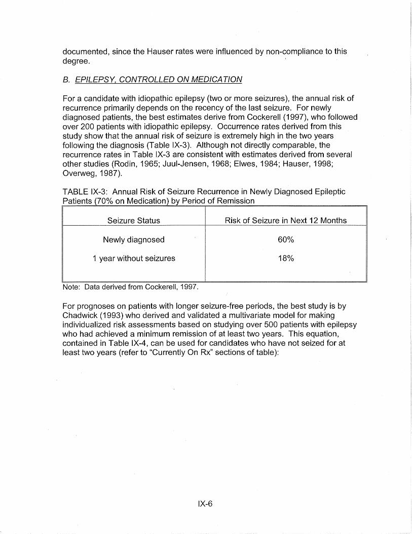
B. EPILEPSY, CONTROLLED ON MEDICATION
For a candidate with idiopathic epilepsy (two or more seizures), the annual risk of recurrence primarily depends on the recency of the last seizure. For newly diagnosed patients, the best estimates derive from Cockerell (1997), who followed over 200 patients with idiopathic epilepsy. Occurrence rates derived from this study show that the annual risk of seizure is extremely high in the two years following the diagnosis (Table IX-3). Although not directly comparable, the recurrence rates in Table IX-3 are consistent with estimates derived from several other studies (Rodin, 1965; Juui-Jensen, 1968; Elwes, 1984; Hauser, 1998; Overweg, 1987).
TABLE IX-3: Annual Risk of Seizure Recurrence in Newly Diagnosed Epileptic Patients (70% on Medication) by Period of Remission
Seizure Status Risk of Seizure in Next 12 Months
Newly diagnosed 60°/o
1 year without seizures 18°/o
Note: Data derived from Cockerell, 1997.
F<?r prognoses on pati~nts with longer seizure-free periods, the best study is by Chadwick (1993) who derived and validated a multivariate model for making individualized risk assessments based on studying over 500 patients with epilepsy who had achieved a minimum remission of at least two years. This equation, contained in Table IX-4, can be used for candidates who have not seized for at least two years (refer to "Currently On Rx" sections of table):
IX-6

TABLE IX-4: Multivariate Model for Making Risk Assessments on Candidates Who are 2+ Years Seizure-Free
Recurrence Medication Status Risk
Currently On Rx Currently Off Rx
Within Next 12 Months
Within Next 24 Months
1- 0.89z
1 - 0.79z
Where z = eT (e = 2.718)
T = (-1.3) + 0.50 if taking two or more medications
1 - 0.69z
1- 0.60z
+ 0.35 if seizures continued after the start of medication + 0.35 if patient has tonic-clonic seizures + 0.50 if patient has myoclonic seizures + 0.20 if EEG is abnormal + (2.0 I number of years since last seizure)
Note: Data derived from Chadwick, 1993.
While medication can reduce the risk of recurrent seizure in patients with idiopathic epilepsy, it does not eliminate it, even in highly compliant patients (Cramer, 1989). In the study by Cockerell (1997) used in Table IX-3, approximately 70o/o of the subjects were on medication. Therefore, to factor in the use of medication in an applicant for whom compliance can be documented, it would be reasonable to reduce the rates cited in Table IX-4 by approximately 30o/o. In the Chadwick study, however, serum levels of medications were measured but not found to be a significant factor in the multivariate analysis.
C. EPILEPSY, PRESENTLY OFF MEDICATION
Due to concerns regarding the toxicity of long-term anti-epileptic medication, many neurologists will attempt to gradually withdraw medication if a patient has been seizure free for 2-5 years. Among adults, the cumulative probability of relapse is 20-70o/o (Overweg 1987, Callaghan 1988, Oller-Daurella 1977, Juui-Jensen 1968) depending on a number of risk factors such as an abnormal EEG at the time of drug withdrawal, the age of onset, and the type of epilepsy.
However, the primary determinant of the annual risk of seizure is the time that has elapsed since withdrawal was initiated. In a meta-analysis of 25 studies (Berg & Shinnar 1994) found that the risk of recurrence was 29o/o (95o/o Cl, 24-34o/o) in the first two years following withdrawal initiation. While this study examined the strength of the three risk factors mentioned above, the data cannot be used for individualized risk assessments. For this purpose, the authors suggested use of the Chadwick 1993,
IX-7

study.
However, using the Chadwick, 1993, study, even individuals with no risk factors have a substantial risk of recurrence during the first two years. During the first post-withdrawal year, the risk of recurrence is 14-24°/o depending on whether the subject was seizurefree for 5 or 2 years before discontinuation, respectively. If no seizure occurs in the first year post-withdrawal, the risk of seizure is 6-1 Oo/o during the second year.
After two years following withdrawal, Chadwick (1996) concludes based on his data and that of Berg & Shinnar (1994) that the added risk of recurrence caused by drug withdrawal is only slight. Therefore, for risk estimates in candidates who are two years or more post-withdrawal, the reader should use the section above which gives estimates for applicants with epilepsy on medication.
D. SEIZURE AFTER HEAD TRAUMA
Depending on its severity, head trauma can be a major risk factor for seizures. The risk of an initial seizure after trauma is discussed later in HEAD TRAUMA WITHOUT HISTORY OF SEIZURE. The purpose of this section is to evaluate the risk of recurrence after an initial seizure. The primary risk factors are the timing of the seizure in relationship to the trauma, and the severity of the trauma.
(1) Early Post-Traumatic Seizure(s): Seizures occurring within the first week post-injury are generally considered to be due to the direct effects of the trauma and are rare in mild injuries. A major study of civilian head injuries found that the occurrence of early seizures was not a risk factor for late seizures after statistical adjustment for other risk factors such as injury severity in a multivariate model (Annegers, 1998). For further consideration, see the section below on HEAD TRAUMA WITHOUT HISTORY OF SEIZURE.
(2) Single Late Post-Traumatic Seizure: There is a high probability that a late posttraumatic seizure will reoccur. The best study of civilian head injuries is a two-year follow-up study of 63 patients by Haltiner (1997). In this study, 82°/o suffered another seizure within one year. Among those seizure-free for one year, approximately 20°/o seized in the following year. Multilinear regression analysis indicated that depressed skull fracture and subdural hematoma were significant risk factors (odds ratios equal approximately 2). However, the one-year recurrence rate was 65°/o even in the lowest risk group. Medication had only a small beneficial effect. Among the 4 7 patients who were compliant, 68°/o seized within two years.
(3) Post-Traumatic Epilepsy (PTE): The occurrence of two or more late posttraumatic seizures fulfills the diagnosis of epilepsy. In the largest long-term (mean follow-up= 8 years) study of 57 patients (Pohlmann-Eden, 1997), only 35% (20) became seizure-free (no seizures within the last 3 years) after the diagnosis. The major risk factors for poor seizure control were a history of missile injury (OR > 1 0), combined seizure types (OR = 2.5), having >1 seizure/month (OR =2), and non-compliance with medication (OR = 9). About half of the 40 medication-
IX-8
compliant patients became seizure-free; only 1/17 of the non-compliant patients were so fortunate. While this represents the largest study of PTE, it does not allow one to estimate annual risks of recurrence. It also is based on patients who were referred to a specialized epilepsy clinic in Germany, and therefore may not be representative of the typical PTE patient. With these limitations in mind, it is recommended that the police physician use the risk estimates found above in the sections on epilepsy as a first approximation.
OTHER FACTORS AFFECTING SEIZURE RECURRENCE RISK ASSESSMENTS
After estimating the annual seizure recurrence risk, the physician should consider the likelihood that a recurrent seizure would in fact create a direct threat of harm to the officer or others while performing routine patrol duties. Relevant factors can include the percentage of time that the officer will be on duty, the impact of sleepdeprivation, the pattern or timing of seizures, and the impact of auras:
(1) The percentage of time that the individual will be on-duty: Full-time employment typically involves working approximately 2000 hours out of 6000 waking hours per year. Therefore, if seizures occurred only while awake, and were truly random, the probability of seizure while on-duty would be about 1/3 of the annual total probability.
(2) Sleep-deprivation: This is the most common seizure threshold-lowering factor and affects approximately 30-40o/o of patients with seizure disorders (Janz, 1974; Broughton, et al., 1984; Mattson, et al., 1965). As little as 24-26 hours without sleep may trigger a seizure. Therefore, if a law enforcement agency requires its officers to work 24-hour shifts, this could substantially increase the risk of seizure while on duty for susceptible individuals. Susceptibility can be ascertained by history, or by a sleep-deprived EEG. However, since working 24-hour shifts is not common, this factor should only be considered by those agencies who can document the need for such work schedules.
(3) Nocturnal or first awakening seizures: Some individuals will report having seizures only while at sleep or upon awakening. This pattern would substantially reduce the risk of a seizure while on-duty. However, the pattern should be well established for a number of years before it is considered. The few existing studies of prognosis in sleep epilepsy found that 33% of patients eventually developed daytime seizures when followed for two years (Gibberd & Bateson, 197 4; Okuma & Kumashiro, 1981 ).
(4) Auras. All seizures are not alike in terms of the risk that they create for the prospective patrol officer. The physician must consider the functional significance of the individual's seizure activity, and to what extent warning "auras" may reduce the likelihood of consequences from this impairment. For example, one study found that only 27°/o (n=11) of simple partial seizures occurring while driving resulted in an accident, compared to 76°/o (n=55) of complex partial seizures
IX-9

(Gastaut & Zifkin, 1987). However, a history of auras reduced the risk of accident during a complex partial seizure to 33o/o (n=33). (Note: the individuals studied were not patrol officers or otherwise driving as part of their jobs.)
Although auras may provide a warning period before a seizure, this warning period may be very brief. Many auras themselves can also cause significant impairment of the senses and judgment. Additionally, although auras generally do not go away with time (Kuhl, 1967), the physician should consider the regularity and pattern of occurrence.
NON-SEIZURE RELATED IMPAIRMENTS
The evaluating physician must also consider whether the candidate is subject to chronic or intermittent interictal impairment, which could interfere with functioning during both critical incidents and routine activities. This impairment could be caused either by side effects of anti-epileptic drugs (AEDs) or interictal EEG discharges.
( 1) Drug impairment: About 30°/o of patients will experience moderate or severe side effects from anti-epileptic medication. The spectrum of potential side effects from AEDs is quite broad and includes cognitive impairment, visual effects, and ataxia. The occurrence of many of these side effects will be evident from a careful review of the candidate's medical records and a thorough neurological examination. However, specialized testing is necessary to detect the presence of more subtle
impairment to neuropsychological functioning, such as lengthened reaction times, decreased memory, decreased concentration, and decreased reasoning ability (Dikmen, 1991 ). These deficits appear most markedly in tasks that are demanding and require quick responses (Wilder & Schmidt, 1986). While these effects are largely dose-related, numerous studies in the past few years have documented neuropsychiatric effects even at therapeutic levels (Andrewes, 1986; Reynolds & Trimble, 1985; Thompson & Trimble, 1982).
(2) Transient cognitive impairment: A small percentage of patients with epilepsy may have interictal epileptiform EEG discharges which can cause errors in complex tasks such as choice reaction time (Aarts, 1984; Sellden, 1971; Hutt, 1977), signal . detection (Tizard & Margerison, 1963; Mirsky & Van Buren, 1965), tracking (Goode, 1970), and short-term memory (Hutt, 1972; Hutt & Gilbert, 1980). Kasteleijn-Nolst Trenite (1987) studied six drivers with epilepsy; he found that during actual driving, three had difficulty with lane control equivalent to the effect of 5-10 mg of diazepam. Whether transient cognitive impairment will occur depends to a large extent on the type, frequency, and duration of the discharges. Generalized spike-wave discharges lasting longer than 3 seconds are of most concern (Braathen, 1988). For example, Goode (1970) found impairments in target tracking in nearly all of these patients. The observed impairment began 1-2 seconds after the spike-wave activity began, and ceased 1-2 seconds before the activity ended.
IX-10
b. RECOMMENDED EVALUATION PROTOCOL:
The physician must carefully question the candidate regarding all of the relevant aspects of this condition: seizure description and frequency, time of day, auras (type, duration, consistency), precipitating factors, etiology, medication, compliance, side-effects, and interference with occupational or other activities.
Complete record review is essential and should include past drug levels, if ·applicable. Additionally, the candidate should submit pharmacy and driving records for the past several years. Letters from past employers regarding work performance and seizures on-the-job are also helpful.
If an EEG would provide useful prognostic information, the candidate should submit the results of a recent sleep-deprived EEG. However, the interpretation of the study can vary significantly, depending on the quality of the machine, the skill of the technician, and the training of the physician. To ensure maximum accuracy, the EEG laboratory should be certified by the American EEG Society's Committee on Laboratory Accreditation, and the reviewer should be board-certified by the American Board of Clinical Neurophysiology.
A complete neurological exam should be performed, including tests for ataxia, incoordination, and nystagmus.
If the candidate is currently well controlled on medication, the following additional work-up is necessary:
• Obtain a serum drug level on the day of the examination.
• The candidate should submit results of a complete neuropsychological evaluation, including an assessment of memory, attention, and psychomotor functioning. (Referral centers for such testing can be obtained from a local university.) A serum drug level must be obtained on the day of testing and should be consisfent with the candidate's typical levels.
To be acceptable for full duty without restriction, the candidate should meet .ill! of the following criteria:
1. NO HISTORY OF SEIZURE TRIGGERED BY STIMULI. WHICH OCCUR DURING ROUTINE DUTIES OR CRITICAL INCIDENTS: Triggers of concern would most commonly be psychological and physical stress, and visual stimuli. If the history is positive, the candidate must be restricted from exposure to the relevant stimuli. Alternatively, the candidate could meet this criterion by demonstrating that medication can prevent these non-random seizures and proving a history of medication compliance.
IX-11

2. NO SIGNIFICANT RISK THAT A RANDOM SEIZURE WOULD OCCUR DURING POLICE DUTIES AND RESULT IN A MAJOR INJURY TO OTHERS: The procedure for estimating this risk is described in detail in a preceding section. As discussed in "Pre-Employment Screening and the Law," no state or federal regulation offers a precise definition of "significant" risk. However, as a rule of thumb, restrictions against field duty would appear appropriate if the risk of a seizure causing substantial harm to others was 1 o/o or greater per year.
3. IF THE CANDIDATE IS ON MEDICATION, THERE ARE NO NEUROLOGICAL OR NEUROPSYCHOLOGICAL DEFICITS THAT COULD SIGNIFICANTLY IMPAIR JOB PERFORMANCE: The physician should discuss the clinical significance of any neuropsychological test abnormalities with the clinician ·who performed the test before recommending appropriate restrictions.
4. NO EVIDENCE OF TRANSIENT COGNITIVE IMPAIRMENT: The history should be negative and the current EEG should not have more than occasional bursts of generalized spike and wave activity lasting >3 seconds.
5. IF CONTINUED COMPLIANCE WITH MEDICATION IS NECESSARY, THE CANDIDATE MUST AGREE TO MAINTAIN COMPLIANCE AND TO ALLOW VERIFICATION BY THE HIRING AGENCY: A written pre-placement contract should specify that the agency's medical department will conduct random therapeutic drugs tests on the candidate after hire, and will periodically review both medical and pharmacy records.
Regardless of medication status, all acceptable candidates who are at risk of seizure recurrence should sign a written agreement specifying that any recurrent seizure will immediately be reported to the hiring agency and that the hiring agency has the right to independently verify this. Verification of seizure status would be best accomplished by review of medical and driving records every 12 months.
3) HEAD TRAUMA WITHOUT HISTORY OF SEIZURE
a. GENERAL CONSIDERATIONS:
Following head trauma, many patients will develop a syndrome which may include headache, vertigo, increased reaction time, decreased concentration, impaired memory, easy fatigability, and irritability. Neither the risk of developing this syndrome nor its severity correlates with the severity of the injury (Russell, 1932; Cartlidge & Shaw, 1981 ). Symptoms (especially headache) will usually appear within 24 hours after the injury. However, some patients do not experience symptoms until weeks following the trauma. Symptoms usually last for several
IX-12

months, but sometimes continue for a year or more. Occasionally, the syndrome may have a waxing and waning course, with dramatic worsening of symptoms several months after they had subsided to a considerable extent.
Although EEGs have no diagnostic or prognostic value in these patients (MacFiynn, 1984 ), there are other objective tests which can be used to rule out the presence of the syndrome. If diffuse cerebral dysfunction is present, visual evoked potential (VEP) to light flashes of varying frequency will be abnormal (Om maya & Gennarelli, 1976). Reaction times will also be prolonged (MacFiynn, 1984 ). The presence of vertigo can be evaluated with electronystagmography (Toglia, 1970). Auditory evoked potentials are delayed in half of symptomatic patients following injuries with loss of consciousness, and can be used to rule out a residual brain stem disorder (Montgomery, 1984; Noseworthy, 1981 ).
A separate concern following head trauma with loss of consciousness is the development of seizures. Unlike post-concussional syndrome, this risk is strongly related to the severity of the injury. A recent large study involving civilian injuries found the five-year cumulative incidence of late seizures (i.e. occurring more than 1 week post trauma) to be approximately 0.9o/o, 1.6°/o, and 1 Oo/o following mild, moderate, and severe injuries, respectively (Annegers, 1998). Given the low risks following mild and moderate injuries, concerns regarding seizure risk should be addressed only in cases of severe injury. This was defined in the Annegers study as brain injuries with one or more of the following features: brain contusion (diagnosed on the basis of observation during surgery or focal neurologic symptoms), intracranial hematoma, or loss of consciousness and/or amnesia for more than 24 hours. Among these patients, 6o/o had a late post-traumatic within the first year; 3.6°/o seized cumulatively between years 1 through 4.
It is important to note that prophylactic antiepileptic agents have been shown to be of no benefit in post-traumatic patients (Schierhout, 1998). Furthermore, the EEG is not helpful in predicting whether late seizures will occur (see review by Janz, 1989), except in cases of penetrating head injuries in which all patients with anterior temporal or central spike foci experienced post-traumatic seizures (Jabbari, et al., 1986).
(For candidates who have already experienced a late post-traumatic seizure, use the evaluation protocol described in the above section on History of Seizure.)
b. RECOMMENDED EVALUATION PROTOCOL:
Physicians should thoroughly question candidates regarding the severity of the injury, nature of the resulting symptoms, and their duration. A complete neurological exam should be performed, including tests for nystagmus. Record review is very important to establish the severity of the injury and to confirm that there have been no post-traumatic seizures.
IX-13

GROUP 1: ASYMPTOMATIC, HISTORY OF MILD INJURY, AND NORMAL EXAM
The risk of seizure in this group is too low to be considered. The main concern is whether the candidate is at risk of a significant recurrence of post-traumatic symptoms.
Level 1: History of symptoms lasting less than one year
Since it is not uncommon for symptoms to last for up to a year, this candidate is probably at low risk of recurrence if he/she has been asymptomatic for at least a few months.
Level 2: History of symptoms lasting more than one year
Since the majority of patients do not have symptoms lasting this long, concerns regarding recurrence or underlying brain damage are justified. Therefore, requiring a symptom-free period of at least several months would probably not be unreasonable. The physician may want to consider specialized testing to ensure complete recovery. Depending on the nature of the symptoms, this could include visual evoked potentials, electronystagmography, auditory evoked potentials, and/or neuropsychological testing. Of these tests, the last would be the easiest to interpret on a functional basis.
GROUP II: HISTORY OF MILD INJURY BUT EITHER SYMPTOMATIC OR ABNORMAL EXAM
These candidates should be deferred until the course of their condition is clearly established to be benign and/or any abnormal findings have resolved or been found to be of no clinical significance.
GROUP Ill: HISTORY OF SEVERE INJURY
Evaluate per above protocol. Risk of seizure would warrant restrictions against patrol work and driving for one year post-trauma.
4) PRIMARY HEADACHE DISORDERS
a) GENERAL CONSIDERATIONS:
Chronic headaches are a common problem. Whether these candidates warrant
IX-14
medical restrictions of patrol officer duties depends on the following considerations:
• Will the psychological stress of patrol officer duties place the candidate at high risk of substantial harm?
• Will the candidate require special accommodation for lost time from work?
• Do the medications used in treatment cause chronic impairment of neuropsychiatric function?
Unfortunately, making an individualized assessment of candidates with this disorder must be based almost exclusively on history. Unlike the evaluation of seizure disorders, there are no risk factors or diagnostic tests that can assist the physician in assessing prognosis. However, a basic understanding of the common chronic headache disorders, aggravating factors, and treatment options can be very helpful.
Common chronic headache disorders:
TENSION HEADACHES: Tension headaches are characterized by mild to moderate head pain without the defining features of migraine (nausea, photophobia, or phonophobia). Based on frequency, tension headaches may be classified as "episodic" if there has been more than 1 0 lifetime attacks but fewer than 15 per month, or as "chronic" if occurring 15 or more times per month for at least 6 months. Tension headaches can have a major impact on both attendance and effectiveness at work. A recent survey by Schwartz, 1998, found that while only 8°/o of subjects with episodic tension headaches reported lost workdays (mean 9 days per year), 44°/o reported decreased effectiveness at work, home, or school (mean 5 days per year). Twelve percent of persons with chronic tension headaches reported lost workdays (mean 27 per year) with 47o/o reporting reduced-effectiveness days (mean 20 days per year).
MIGRAINE HEADACHES: Migraine headaches are usually classified as either "common" or "classical." Both are commonly associated with a wide array of neurological symptoms such as nausea, photophobia, lightheadedness, vertigo, and visual disturbances. In classical migraine, these symptoms occur as an aura before the onset of cephalalgia, typically developing over the course of more than 4 minutes and lasting no more than 60 minutes (Gilman, 1992). In common migraine, there is no aura, and neurological symptoms develop at the same time as the cephalalgia. The classical migraine is characterized by a relatively short duration (<12 hours), compared with common migraine which can last up to 4 days (Sachs, 1985). Certain patients experience "complicated" migraine disorders that are associated with severe neurological deficits such as prolonged hemiparesis, partial blindness, dysarthria, ataxia, or diplopia (Gilman 1992). As with tension headaches, migraines can have a major impact on both attendance and effectiveness at work. Legg (1997) found that migraine
IX-15

patients missed an average of 2.8 days of work per month and reported to work an average of 6 days per month when their productivity was significantly impaired.
CLUSTER HEADACHES: While there is probably a continuum between tension headaches and migraines, cluster headaches are very distinct syndrome. A typical bout involves 1-3 short-lived attacks of periorbital pain per day over a 4-8 week period. This is followed by a pain-free interval that averages one year. The pain begins without warning, and rapidly reaches high (often excruciating) intensity within 2-15 minutes. Attacks last from 30-120 minutes in 75o/o of cases. In about 85o/o of cases, attacks tend to recur at the same times each day for the duration of the bout, with additional attacks occurring randomly (Raskin, 1988). Manzoni (1983) found that many attacks occur during non-working hours, such as around 9:00 p.m. and 1 :00 a.m.
Aggravating factors:
While numerous factors have been found to precipitate and aggravate tension headaches and migraines, work as an police officer will expose the candidate to the most important factor: emotional stress. The role of emotional stress in contributing to both the frequency and severity of these headaches has been illustrated in four recent studies, two of which were prospective in design (Tekle, 1995; Scharff, 1995; Marlowe, 1998; Labbe, 1997). Scharff (1995) found that over 72%) of both migraine and tension headache patients reported that stressful situations sometimes triggered their headaches. Curiously, however, migraines do not tend to occur at the peak of stress, but rather during subsequent relaxation periods (Raskin, 1988).
Short intense bursts of exercise may result in a migraine attack in certain patients (Massey, 1982). Typically, focal neurological symptoms appear immediately following activities such as running or heavy lifting, and are followed several minutes later by nausea and a severe headache (Rooke, 1968). However, conditioning can help reduce the frequency of these headaches in many patients.
Cluster headaches are triggered by precipitating factors in only a small percentage of patients (Raskin, 1988). Paradoxically, vigorous physical exertion at the earliest sign of an attack can be remarkably effective in ameliorating or even aborting an attack in some patients (Atkinson, 1977).
Treatment:
Many of the drugs used to treat the various headache syndromes can impair the neuropsychological functioning of a patrol officer. While determining the impact of a particular drug on a particular candidate can be difficult, there are certain drugs which are associated with such a high incidence of impairment (primarily sedation) that all users would warrant restrictions against driving and
IX-16

carrying weapons. These include benzodiazapines, barbiturates, antihistamines (except newer non-sedating varieties), codeine, propoxyphene, narcotics, and phenothiazines. Support for this prohibition can be found in medical guidelines for commercial drivers (Booker, 1988). (Note: the above list of medications is not necessarily the best medications for the treatment of headaches. However, they are listed above to present the range of typical medications, which may be used by non-specialists.)
The newest treatment available for acute migraine is sumatriptan, available in injectable, oral, and nasal formulations. This medication can greatly reduce the length of an acute attack and has been shown to significantly reduce lost time and reduced productivity at work (Legg, 1997). Side-effects are minimal in severity and transient. Unfortunately, the pill form costs $10-12 each, and may not be covered by certain health plans.
When migraines occur 2-3 times per month, preventive .medication is usually indicated. There are numerous drugs which are available each of which has a probability of success of about 60o/o (Raskin, 1988). Although it has not been clearly established that these drugs alter the natural history of the disorder, many patients are able to discontinue medication completely after 6 months and experience fewer and less severe attacks for long periods of time (Diamond, 1982).
b. RECOMMENDED EVALUATION PROTOCOL:
Physicians must thoroughly question candidates with a history of chronic headache to ascertain the severity, frequency, number lost days from work, associated neurological symptoms, exacerbating factors, and the effectiveness and side-effects of medication.
Review of medical and pharmacy records is strongly recommended. Particular attention should be made to references that indicate the severity of the condition and whether it is aggravated by stress. An inquiry to a past employer regarding lost time due to headaches may be appropriate.
As presented above, medical restrictions should be based on the following considerations:
1. THE CANDIDATE WILL REQUIRE MORE THAN THE AMOUNT OF SICK LEAVE ALLOTTED PER YEAR
This can be determined directly by a review of the candidate's medical records and employment history for the past two years. It is reasonable to assume that this pattern will continue into the near future (2-3 years), with two exceptions. Sick leave may increase if the candidate's headaches are aggravated by stress (see next consideration). Sick leave may decrease if a candidate with
IX-17
migraines is willing to change to a new medication regimen, which may include prophylactic medications and sumatriptan for acute attacks. In this case, the candidate should be offered a reevaluation after a period of time to determine if sick leave use has improved to acceptable levels.
2. THE EMOTIONAL STRESS OF PATROL OFFICER DUTIES WILL PLACE THE CANDIDATE AT A HIGH RISK OF SUBSTANTIAL HARM
This assessment must be based on a well established past medical history of stress exacerbation in the particular candidate under consideration. Additionally, there should be a past history of severe headache disorder causing prolonged absence from work, disability, or change of job/career. In these cases, the stress of police work will likely cause a recurrence of the past disability.
3. THERE IS EPISODIC IMPAIRMENT OF NEUROPSYCHIATRIC FUNCTIONING DUE TO SIDE-EFFECTS OF MEDICATION
As discussed above, use of certain medications is not appropriate for persons such as patrol officers who must make split-second life or death decisions, or whose personal safety (and the safety of others) may be compromised by decreases in vigilance or reaction times. These medications include:
• benzodiazapines • barbiturates • antihistamines (except non-sedating varieties) • codeine • propoxyphene • narcotics • phenothiazines
An appropriate restriction would specify that the candidate should not drive or be assigned to critical tasks when using these medications. Of course, frequent use would make accommodation very difficult, and the candidate should be evaluated by his/her private physician for alternate therapies.
IX-18
REFERENCES
Aarts, H.P., et al. 1984. Selective cognitive impairment during focal and generalized epileptiform EEG activity. Brain. 107:293-308.
Andrewes, D. G., et al. 1986. A comparative study of the cognitive effects of phenytoin and carbamazepine in new referrals with epilepsy. Epilepsia. 27:128-134.
Annegers, J..F., et al. 1979. Remission of seizures and relapse in patients with epilepsy. Epilepsia. 20:729-737.
Annegers JF, et al. A population-based study of seizures after traumatic brain injuries. NEJM. 338:20-24, 1998
Annegers, J.F., et al. 1980. Seizures after head trauma: A population study. Neurology. 30:683-689.
Annegers, J.F., et al. 1986. Risk of recurrence after an initial unprovoked seizure. Epilepsia. 27:43-50.
Atkinson, R. 1977. Physical fitness and headache. Headache. 17:189-195.
Berg AT, Shinnar S. Relapse following discontinuation of antiepileptic drugs: a metaanalysis. Neurology. 44:601-608, 1994.
Booker, H.E., et al. 1988. Conference on Neurological Disorders and Commercial Drivers. Report #FHWA-MC-88-042. Washington: U.S. Dept of Transportation.
Bora I, Seckin B, Zarifoglu M, Turan F, Saidkoglu S, Ogul E. (1995). Risk of recurrence after first unprovoked tonic-clonic seizure in adults. J Neural 242:157-163.
Braathen, G., et al. 1988. Valproate in the treatment of absence epilepsy in children. Epilepsia. 29:548-552.
Broughton, R.J., et al. 1984. Comparison of the psychosocial effects of epilepsy and narcolepsy/cataplexy. Epilepsia. 25(4 ):423-433.
Callaghan, N., et al. 1988. Withdrawal of anticonvulsant drugs in patients free of seizures for two years. NEJM. 31,8(15):942-946.
Cartlidge, N.E.F., and Shaw, D.A. 1981. Head Injury. Philadelphia: W.B. Saunders.
Chadwick D, et al. Outcomes after seizure recurrence in people with well-controlled epilepsy and the factors that influence it. Epilepsia. 37(11 ): 1043-1050, 1996.
Chadwick, D. 1993. Prognostic index for recurrence of seizure after remission of epilepsy. BMJ. 306:1374-1378.
IX-19

Cleland PJ, Mosquera I, Steward WP, Foster JB (1981 ). Prognosis of isolated seizure in adult life. BMJ 283:1364.
Cockerell, OC, et al. Prognosis of Epilepsy. Epilepsia, 38(1 ):31-46, 1997.
Cramer, J.A., et al. 1989. How often is medication taken as prescribed? JAMA. 261:3273-3277.
Cramer, J.A., et al. 1990. Compliance declines between clinic visits. Arch lnterm Med. 150:1509-1510.
Diamond, S., et al. 1982. Long-term study of propranolol in the treatment of migraine. · Headache. 22:268-271.
Dikmen, S.S., et al. 1991. Neurobehavioral effects of phenytoin prophylaxis of posttraumatic seizures. JAMA. 10(265):1271-1277.
Elwes, R.D.C., et al. 1984. The prognosis for seizure control in newly diagnosed epilepsy. NEJM. 311:944-947.
Feeney, D.M., and Walker, A. E. 1979. The prediction of post-traumatic epilepsy: A mathematical approach. Arch Neural. 36:8-12.
First Seizure Trial Group. Randomized clinical trial on the efficacy of antiepileptic drugs in reducing the risk of relapse after a first unprovoked tonic-clonic seizure. Neurology. 43:4 78-483, 1993.
Gastaut, M., and Tassinari, C.A. 1966. Triggering mechanisms in epilepsy. Epilepsia. 7:85-138.
Gastaut, H., and Zifkin, B.G. 1987. The risk of automobile accidents with seizures occurring while driving. Neurology. 37:1613-1616.
Gilad R, Lampl Y, Gabbay U, Eshel Y, Sarova-Pinhas I (1996). Early treatment of a single generalized tonic-clonic seizure to prevent recurrence. Arch Neural 53:1149-1152.
Gibberd, F.B., and Bateson, M.G. 1974. Sleep epilepsy: Its pattern and prognosis. Br Med J. 2:403-407.
Gilman, S. 1992., Advances in neurology, part I. NEJM. 326(24):1608-1616.
Goode, D.J., et al. 1970. Effects of paroxysmal spike-wave on continuous visual-motor performance. Epilepsia. 11 :241-254.
Hauser, W .A., et al. 1993. The incidence of epilepsy in Rochester Minnesota 1935-1984. Epilepsia. 34:453-68.
IX-20

Hauser, W.A., et al. 1990. Seizure recurrence after a first unprovoked seizure: An extended follow-up. Neurology. 40:1163-1170.
Hauser, W.A., and Hesdorffer, D.C. 1990. Prognosis. Chap. 6 in Epilepsy: Frequency, Causes, and Consequences. 197-243. Landover, Md: Epilepsy Foundation of America; New York: Demos.
Hauser, WA, et al. Risk of recurrent seizures after two unprovoked seizures. NEJM, 338(7):429-434, 1998.
Hutt, S.J. 1972. Experimental analysis of brain activity and behaviour in children with "minor" seizures. Epilepsia. 13:520-534.
Hutt, S.J., et al. 1977. Choice reaction time and EEG activity in children with epilepsy. Neuropsychologia. 15:257-267.
Hutt, S.J., and Gilbert, S. 1980. Effects of evoked spike-wave discharges upon short term memory in patients with epilepsy. Cortex. 16:445-457.
Jabbari, B., et al. 1986. Clinical and radiological correlates of EEG in the late phase of head injury: A study of 515 Vietnam veterans. Electroenceph Clin Neurophysiol. 64:285-293.
Janz, D. 1974. Epilepsy and the sleeping-waking cycle. In Handbook of Clinical Neurology, eds. P.J. Vinken and G.W. Bruyn, 457-490. Amsterdam: Elsevier Science Publishing.
Janz, D. 1989. Prognosis and prophylaxis of traumatic epilepsy. In Chronic Epilepsy; Its Prognosis and Management, ed. M.R. Trimble, 87-102. New York: John Wiley & Sons, Ltd.
Jennett, W.B., and Lewin, W.S. 1960. Traumatic epilepsy after closed head injuries. 4 Neural Neurosurg Psychiat. 23:295-301.
Juui-Jensen, P. 1968. Frequency of recurrence after discontinuance of nati-convulsant therapy in patients with epileptic seizures. Epilepsia. 9:11-16.
Kasteleijn-Nolst Trenite, D. G., et al. 1987. The influence of subclinical epileptiform EEG discharges on driving behaviour. Electroenceph Clin Neurophysiol. 67:167-170.
Kudrow, L. 1986. Muscle contraction headaches. Vol. 48 in Handbook of Clinical Neurology, ed. F.C. Rose, 343-352. Amsterdam: Elsevier Science Publishing.
Kuhl, V., et al. 1967. The prognosis of epilepsy with special reference to traffic security. Epilepsia. 8:195-209.
IX-21

Labbe EE, et al. Psychosocial Factors and prediction of headaches in college adults. Headache. 37:1-5, 1997.
Legg, RF, et al. Cost benefit of sumatriptan to an employer. JOEM. 39(7):652-657, 1997
MacFiynn, G., et al. 1984. Measurement of reaction time following minor head injury . .4 Neural Neurosurg Psychiatry. 47:1326-1331.
Manzoni, G.C., et al. 1983. Cluster headache- clinical findings in 180 patients. Cephalalgia. 3:21-30.
Marlowe N. Stressful events, appraisal, coping, and recurrent headache. J Clin Psych. 54(2):24 7-256, 1998.
Massey, E.W. 1982. Effort headaches in runners. Headache. 22:99-100.
Mattson, R.H., et al. 1965. Electroencephalograms of epileptics following sleep deprivation. Arch Neural. 13:310-315.
Mirsky, A. F., and Van Buren, J.M. 1965. On the nature of the "absence" in centrencephalic epilepsy. Electroenceph Clin Neurophysiol. 18:334-348.
Montgomery, A., et al. 1984. Delayed brainstem conduction time in post-concussional syndrome. Lancet. 1:1011.
Musicco,M, Beghi E, Solari, A, Viani F (1997). Treatment of first tonic-clonic seizure does not improve the prognosis of epilepsy. Neurology 49:991-998.
Noseworthy, J.H., et al. 1981. Auditory brainstem responses in post-concussion syndrome. Arch Neural. 38:275-278.
Okuma, T., and Kumashiro, H. 1981. Natural history and prognosis of epilepsy: Report of a multi-institutional study in Japan. Epilepsia. 22:35-53.
Oller-Daurella, L., et al. 1976. Reduction or discontinuance of antiepileptic drugs in patients seizure-free for more than 5 years. In Epileptology, Proceedings of the Seventh international Symposium on Epilepsy, ed. D. Janz, 218-227. Stuttgart: Thieme/PSG.
Oller-Daurella, L., et al. 1977. Clinical, therapeutic, and social status of epileptic patients without seizures for more than five years. In Epilepsy, The Eighth International Symposium, ed. J.K. Penry, 69-75. New York: Raven Press.
Ommaya, A.K., and Gennarelli, T.A. 1976. A physiopathologic basis for noninvasive diagnosis and prognosis of head injury severity. In Head Injuries, ed. R.L. Mclaurin, 49-75. New York: Grune & Stratton.
Overweg, J., et al. 1987. Clinical and EEG prediction of seizure recurrence following
IX-22

antiepileptic drug withdrawal. Epilepsy Res. 1 :272-283.
Philips, C., and Hunter, M. 1982. Headache in a psychiatric population. J Nerv Ments Dis. 170:34-40.
Raskin, N.H. 1988. Headache, 2nd ed. New York: Churchill Livingston.
Reynolds, E.H., and Trimble, M.R. 1985. Adverse neuropsychiatric effects of anticonvulsant drugs. Drugs. 29(6):570-581.
Rodin, E.A., et al. 1965. The prognosis for patients with epilepsy. JOM. 7:560.
Rooke, E.D. 1968. Benign exertion a I headache. Med Clin NA. 52(4 ):801-808.
Russell, W.R. 1932. Cerebral involvement in head injury. Brain. 55:549-603.
Sachs, 0. 1985. Migraine - Understanding a Common Disorder. Berkeley: Univ of CA Press.
Salazar, A.M., et al. 1985. Epilepsy after penetrating head injury: I. Clinical correlates. Neurology. 35:1406-1414.
Scharff L, et al. Triggers of headache episodes and coping responses of headache diagnostic group. Headache. 35:397-403, 1995.
Schierhout G, Roberts I. Prophylactice antiepileptic agents after head injury: a systematic review. J Neural Neurosurg Psych. 64:108-112,1998.
Schwartz BS, et al. Epidemiology of tension-type headache. JAMA. 279(5):381-383, 1998.
Sellden, U. 1971. Psychotechnical performance related to paroxysmal discharges. In EEG Clin Electroenceph. 2:18-27.
Tekle H, et al. Migraine, chronic tension-type headache, and cluster headache in an Ethiopian rural community. Cephalagia. 15(6):482-8.
Temkin, N.R., et al. 1990. A randomized, double-blind study of phenytoin for the prevention of post-traumatic seizures. NEJM. 323:497-501.
Thompson, P.J., and Trimble, M.R. 1982. Anticonvulsant drugs and cognitive function. Epilepsia. 23:531-544.
IX-23

Tizard, B., and Margerison, J.H. 1963. Psychological functions during wave-spike discharge. Brit J Soc Clin Psychol. 3:6-15.
Walker, A. E., and Jablon, S. 1961. A follow-up study of head wounds in World War II. Washington: Veterans Administration; Bethesda, National Institutes of Health.
Walker, A.E., and Erculei, F. 1969. Head Injured Men, 44-57. Springfield: C.C. Thomas.
Weiss, G.H., and Caveness, W.F. 1972. Prognostic factors in the persistence of posttraumatic epilepsy. J Neurosurg. 37:164-169.
,Weiss, G. H., et al. 1983. Prognostic factors for the occurrence of post-traumatic epilepsy. Arch Neural. 40:7-10.
Weiss, G.H., et al. 1986. Predicting post-traumatic epilepsy in penetrating head injury. Arch Neural. 43:771-773.
Wessely, P. 1981. Zur bedeutung von zeitfaktoren bei posttraumatischen anfallen. In Epilepsie, eds. H. Remschmidt, R. Rentz and J. Jungmann, 138-143. Stuttgart: Thieme.
Wilder, B.J., and Schmidt, R.P. 1986. Management of epilepsy. In Epilepsy: Diagnosis, Management. Quality of Life, ed. J.K. Penry. New York: Raven Press.
IX-24