Neurological Objectives Changes in the Geriatric Patient · Changes in the Geriatric Patient ......
8
2/18/2014 1 Neurological Changes in the Geriatric Patient OhioHealth Grant Medical Center LifeLink Winter Update 2014 Amanda Cramer MSN, RN, FNP-BC, CNRN Family Practice Nurse Practitioner Objectives • Review Neuro Anatomy • Anatomic Changes in the Aging Patient • Neuro Assessment Review Neuro-Geriatric Review • Neurological Disorders – Stroke – Back Pain – Neuro Trauma – Cognitive Disorders – Other Neurological Disorders • Neurologic Considerations in the Elderly 2 Neuro Anatomy • SCALP- Skin, SubCutaneous Tissue, Adipose Tissue, Ligament (Galea), Periosteum • Menningies- Dura Mater Arachnoid Pia Mater Brain Anatomy Menningies- Dura Mater , Arachnoid, Pia Mater • Neurons- Cell body, Axon, Dendrite Myelin sheath 3 Neuro Anatomy • Hemispheres • Corpous callusom • Cerebellum • Diencephalon • Midbrain • Pons • Medulla Oblongata • Spinal Cord 4 Neuro Anatomy • Frontal Lobe – Problem Solving, Emotion, Judgment, Creative Thought – Broca’s Area (left frontal) Expressive language • Parietal Lobes – Tactile Sensation, Proprioception Tactile Sensation, Proprioception – Academic Skills – Sensory Comprehension • Occipital Lobe – Visual perception, Visual input, Reading – The perception and recognition of printed words. 5 • Temporal Lobes – Some hearing and vision – Auditory and Visual Memories – Wernicke’s Area (left temporal) Receptive language • Cerebellum Cerebellum – Balance, Equilibrium, Coordination of voluntary movement 6
Transcript of Neurological Objectives Changes in the Geriatric Patient · Changes in the Geriatric Patient ......

2/18/2014
1
Neurological Changes in the
Geriatric Patient
OhioHealth Grant Medical Center
LifeLink Winter Update 2014
Amanda Cramer MSN, RN, FNP-BC, CNRN
Family Practice Nurse Practitioner
p
Objectives
• Review Neuro Anatomy
• Anatomic Changes in the Aging Patient
• Neuro Assessment Review
Neuro-Geriatric Review
• Neurological Disorders– Stroke
– Back Pain
– Neuro Trauma– Cognitive Disorders
– Other Neurological Disorders
• Neurologic Considerations in the Elderly
2
Neuro Anatomy
• SCALP- Skin, SubCutaneous Tissue, Adipose Tissue, Ligament (Galea), Periosteum
• Menningies- Dura Mater Arachnoid Pia Mater
Brain Anatomy
Menningies- Dura Mater, Arachnoid, Pia Mater
• Neurons- Cell body, Axon, DendriteMyelin sheath
3
Neuro Anatomy • Hemispheres
• Corpous callusom
• Cerebellum
• Diencephalonp
• Midbrain
• Pons
• Medulla Oblongata
• Spinal Cord
4
Neuro Anatomy • Frontal Lobe
– Problem Solving, Emotion, Judgment, Creative Thought
– Broca’s Area (left frontal) Expressive language
• Parietal Lobes
– Tactile Sensation, ProprioceptionTactile Sensation, Proprioception
– Academic Skills
– Sensory Comprehension
• Occipital Lobe– Visual perception, Visual input, Reading
– The perception and recognition of printed words.
5
• Temporal Lobes– Some hearing and vision
– Auditory and Visual Memories– Wernicke’s Area (left temporal)
Receptive language
• CerebellumCerebellum– Balance, Equilibrium,
Coordination of voluntary movement
6

2/18/2014
2
• The Ventricals- Lateral Ventricles, Third and Fourth Ventricle.
• Cerebrospinal fluid
Cerebral Spinal Fluid
Cerebrospinal fluid - CNS protection- Compensation- Nutrition
7
• Circle of Willis
• Vertebrobasilar Arteries– Supply posterior circulation
Cerebral Blood Supply
– Through transverse processesof C5- C1
• Carotid Arteries
8
Cranial Nerves
9
Neuro Anatomy
• Vertebral Column- 33 Vertebrae– 7 Cervical (C1-C7)– 12 Thoracic (T1-T12)
Spine
( )– 5 Lumbar (L1-L5)– 5 Sacral (S1-S5)– 4 Coccygeal
• Anterior Longitudinal Ligament, Posterior Longitudinal Ligament
10
• Extends from base of medulla to conusmedullaris (across from L1–L2)
• Surrounded by menningies
Spinal Cord
Surrounded by menningies
• CSF flows in the subarachnoid space
• Vascular supply Comes from branches of the Vertebral
11 12

2/18/2014
3
• 31 pairs along the length of the cord
• First 7 cervical pairs exit above corresponding
t b C8 it b l C7 t b
Spinal nerves
• vertebrae, C8 exits below C7 vertebrae
• All thoracic, lumbar, and sacral nerves exit below corresponding vertebrae
• Part of the Peripheral Nervous System
• Plexus – bundle of nerves join together
13
Neuro Assessment
• Mental Status
• Cranial Nerves
• Proprioception
• Cerebellar Function
• Motor Function
• Sensory Function
• Reflex function
14
Neuro Assessment
• Start with the least amount of stimuli
• Assess head to toe– Except spine injuries, assess strength/sensation toe-
to-head
• Proceed in a systematic approach• Proceed in a systematic approach
• Utilize standardized scales
15
Mental Status • Level of Consciousness-
– One of the most sensitive parameters to neurologic changes
– Large deferential for mental status changes
• Glasgow Coma Scale (3-15)Glasgow Coma Scale (3 15)– Traditional Terms
• Alert, awake, lethargic, stuporous, semi-comatose, and comatose
• Level of consciousness- Check to Verbal, Visual, Tactile, Noxious, Painful
16
Neuro Assessment
• Is the patient able to follow commands? Are they oriented to person, place, time and situation.
Mental Status
• Memory, intellectual performance, insight, problem solving.
• Affect and mood
• Communication- Aphasia, Receptive, Expressive, Dysarthria
17
Neuro Assessment
Cranial Nerve Check
• Pupils- PERRLA, EOMI, Gaze
• Face symmetry and sensation
• Speech taste smell and hearing
18
• Speech, taste, smell and hearing
• Tongue and palate movement
• Shoulder shrug
2/18/2014
4
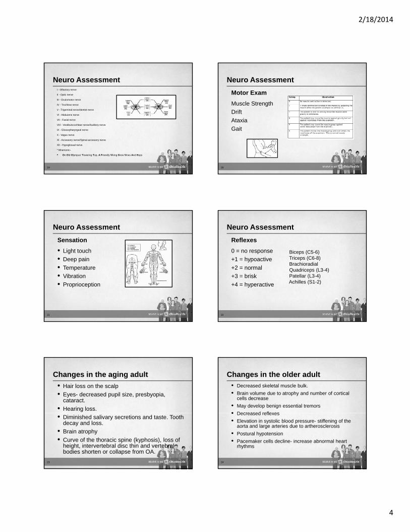
Neuro AssessmentI - Olfactory nerve
II - Optic nerve
III - Oculomotor nerve
IV - Trochlear nerve
V - Trigeminal nerve/dentist nerve
VI - Abducens nerve
VII - Facial nerve
VIII - Vestibulocochlear nerve/Auditory nerve
IX - Glossopharyngeal nerve
X - Vagus nerve
XI - Accessory nerve/Spinal accessory nerve
XII - Hypoglossal nerve
* Mnemonic-
• On Old Olympus' Towering Top, A Friendly Viking Grew Vines And Hops
19
Neuro Assessment
Muscle Strength
Drift
At i
Motor Exam
Ataxia
Gait
20
Neuro Assessment
• Light touch
• Deep pain
T t
Sensation
• Temperature
• Vibration
• Proprioception
21
Neuro Assessment
0 = no response
+1 = hypoactive
2 l
Reflexes
Biceps (C5-6)Triceps (C6-8)Brachioradial
+2 = normal
+3 = brisk
+4 = hyperactive
22
Quadriceps (L3-4)Patellar (L3-4)Achilles (S1-2)
Changes in the aging adult
• Hair loss on the scalp
• Eyes- decreased pupil size, presbyopia, cataract.
• Hearing loss.
• Diminished salivary secretions and taste. Tooth decay and loss.
• Brain atrophy
• Curve of the thoracic spine (kyphosis), loss of height, intervertebral disc thin and vertebral bodies shorten or collapse from OA.
23
Changes in the older adult
• Decreased skeletal muscle bulk.
• Brain volume due to atrophy and number of cortical cells decrease
• May develop benign essential tremors
• Decreased reflexes• Decreased reflexes
• Elevation in systolic blood pressure- stiffening of the aorta and large arteries due to artherosclerosis
• Postural hypotension
• Pacemaker cells decline- increase abnormal heart rhythms
24

2/18/2014
5
Neuro Disorders
• Disruption in normal blood supply to the brain
• New terminology - “Brain Attack”
• Fourth most common cause of death in US
Stroke
Fourth most common cause of death in US
• Primary cause of adult disability in the US
• About 795,000 Americans each year suffer a new or recurrent stroke.
• On average, a stroke occurs every 40 seconds and every 4 minutes someone dies of stroke
25
Stroke
• 80% of strokes
• Diminished blood supply to the brain
Ischemic Stroke
• Most common cause is thromboembolism
26
• Left side stroke– Right visual field deficit– Right Hemiparesis
Ri ht H i
Stroke Symptoms
• Right side stroke– Left Hemi-attention– Left Visual Field Deficit
L ft H i i– Right Hemisensoryloss
– Aphasia
27
– Left Hemiparesis– Left Hemisensory loss
Stroke
• Brief interruption in blood flow by a clot
• Symptoms are the same as stroke but last usually less than five minutes (one minute on average)
Transient Ischemic Attack
less than five minutes (one minute on average).
• No permanent brain injury
• 10% of strokes are preceded by a TIA.
• Also referred to as a “Mini-Stroke” or “Warning Stroke”
28
Stroke
• A weakened vessel ruptures and bleeds into the surrounding brain. The blood accumulates and compresses the surrounding brain tissue.
Hemorrhagic Stroke
g– Intracerebral hemorrhage (within the brain): Most
common cause is chronic hypertension. – Subarachnoid hemorrhage (bleeding around the
brain): Most common cause is Aneurysm rupture
• Symptoms: Headache, N/V, Decreased LOC– SAH- stiff neck, light sensitivity– ICH- Focal Symptoms
29
Stroke Assessment Tools Act FAST
Endorsed By the American Stroke Association
Used by first responders
Q i k A t T l Quick Assessment Tool-Easy to teach patients, families
30

2/18/2014
6
Face Drooping – Does one side of the face droop or is it numb? Ask the person to smile. Is the person's smile uneven?
Arm Weakness – Is one arm weak or numb? Ask the person to raise both arms. Does one arm drift downward?
Speech Difficulty Is speech slurred? Is the person unable toSpeech Difficulty – Is speech slurred? Is the person unable to speak or hard to understand? Ask the person to repeat a simple sentence, like "The sky is blue." Is the sentence repeated correctly?
Time to call 9-1-1 – If someone shows any of these symptoms, even if the symptoms go away, call 9-1-1 and get the person to the hospital immediately. Check the time so you'll know when the first symptoms appeared
31
TIME
32
IS BRAIN
• Restore blood flow to the penumbra
• Stroke Prevention– Antiplatelet agent (ASA, clopidogrel, asa/dipyridamole
XR) Anticoagulant if indicated (warfarin)
Stroke Treatment Goals
XR), Anticoagulant if indicated (warfarin)
• Reduce modifiable risk factors– Hypertension, Smoking, Cholesterol, Glucose control,
CAD, Atrial Fibrillation, Obesity, Diet Control, Hypercoagulability
• Improve function- Physical, Occupational, Speech therapy. Physical Medicine and rehab
33
Neuro Disorders
• Scalp Lacerations
• Subdural Hematoma
Sk ll/ F i l F t
Head Trauma
• Skull/ Facial Fractures
34
Spine injuries
• Dislocation- vertabra overrides another- spinal cord may or may not be involved.
• Subluxation- partial or complete dislocation of one vertebra over another. Damage to the cord and supporting ligaments may be present.
H fl i F d t d i• Hyperflexion- Forward movement- wedge or compression fracture
• Hyperextension- Backward downward movement- whiplash. May see contusionj and ischemia of the cord. Stress at C4 and C5
• Compression Injury- Burst fracture, Wedge
35
Spine Injuries
• Conditions causing back pain– Scoliosis/kyphosis– Neoplasms
Back Pain
p– Infections– Spondylosis– Osteoarthritis– Inflammation
36

2/18/2014
7
• Aging process- Decreased fluid content of nucleus pulposus
• Nucleus pulposus less elastic – tears
Degenerative disc disease
• Less effective shock absorbers
• Degeneration of posterior spinal ligaments
• Symptoms: Back pain with radiation to buttocks or thighs, often positional. Progressive, gradually onset.
37
• Slipping or sliding associated with degenerative changes of the facet joints
• Anterior subluxation most common at L4/5
Lumbar Spondylolithesis
Anterior subluxation most common at L4/5
• Low back pain, vague, dull, achy. Often asymptomatic
• Positive straight leg raising (SLR)
38
• Narrowing of the lumbar spinal canal
• Pain, numbness, or weakness with ambulation
• Walks farther with forward flexion support
Lumbar Stenosis
Walks farther with forward flexion support
• Bowel or bladder dysfunction
39
• Due to bone weakened by osteoporosis, trauma, tumors
• Sudden onset severe pain reproducible on
Vetebral Compression Fracture
Sudden onset, severe pain, reproducible on palpation
40
Neuro Disorders
• Spinal cord injuries-– Concussion- spinal shock or jarring causing
temporary loss of functionContusion bruising of the cord can cause bleeding
Spine Injuries
– Contusion- bruising of the cord, can cause bleeding, edema, necrosis, deficit depends on severity
– Laceration- Tear in the cord, can be complete of incomplete
– Transection: Severing of the cord– Hemorrhage: Bleeding into or around the cord
41
Cognitive Disorders
Confused/perception disorder
Acute onsetReversible with treatment
Delirium
D- drugsE-electrolytesL-Low OxygenReversible with treatment
42
L Low OxygenI- InjuryR- relapsing feverI-infectionU- uremiaM-metabolic

2/18/2014
8
Cognitive Disorders
D- DrugsE- EndocrineM- MetabolicE- EpilepsyN- NutritionT Tumor/Trauma
Dementia
Failing memory, personality changesChronic progressionAffects 7% of the population over the T- Tumor/Trauma
I- InfectionA- Arterial
43
Affects 7% of the population over the age of 65
• Alzheimer disease • Non-Alzheimer-related• Parkinson disease with dementia
D ti ith L b di• Dementia with Lewy bodies• Fluctuating cognition, visual hallucinations, bradykinesia or rigidity, sleep
disturbances, frequent falls, syncope, orthostatic hypotension, urinary incontinence
44
Neuro Disorders
• Movement Disorder
• Neuromuscular disorders
CNS i f ti
Other
• CNS infections
• Tumors
• Migraine
• Seizures
45
Other Geriatric Considerations
• Pharmocokinetic changes – Muscle mass, %of body fat, kidney, liver function, GI
absorption
• Anatomic Changes
• Polypharmacology
• Living Will, Power of Attorney, Code Status
• Social Services
46
Questions
Amanda Cramer MSN, RN, CNP, CNRN
Nurse Practitioner , Medical-Stroke Unit
Ohi H lth G t M di l C tOhioHealth Grant Medical Center
614-566-7606
47