Neurological manifestations and molecular genetics of acute
103
1 Research Program in Molecular Medicine, Biomedicum-Helsinki, Department of Medicine University of Helsinki, Finland Neurological manifestations and molecular genetics of acute intermittent porphyria in North Western Russia Elena Pischik Academic dissertation To be presented with the permission of the Medical Faculty of the University of Helsinki, for public examination in Auditorium of the Department of Dermatology, Helsinki University Central Hospital, Meilahdentie 2, on December 1st, 2006 at 12 noon. Helsinki 2006
Transcript of Neurological manifestations and molecular genetics of acute

1
Research Program in Molecular Medicine, Biomedicum-Helsinki,
Department of Medicine
University of Helsinki,
Finland
Neurological manifestations and molecular genetics of acute
intermittent porphyria in North Western Russia
Elena Pischik
Academic dissertation
To be presented with the permission of the Medical Faculty of the University of Helsinki, for public examination in
Auditorium of the Department of Dermatology, Helsinki University Central Hospital, Meilahdentie 2, on December 1st, 2006 at 12 noon.
Helsinki 2006

2
SUPERVISED BY
Docent Raili Kauppinen, M.D., Ph.D.
Porphyria Research Centre
Department of Medicine
Division of Endocrinology
University of Helsinki
Finland
REVIEWED BY:
Professor George H. Elder, M.D.
Cardiff Porphyria Service
Department of Medical Biochemistry and Immunology
School of Medicine, Cardiff University
Cardiff, United Kingdom
and
Professor Kari Majamaa, M.D., Ph.D.
Department of Neurology
University of Turku
Turku, Finland
OFFICIAL OPPONENT:
Docent Markku Sainio, M.D., Ph.D.
Brain and Work Research Centre
Finnish Institute of Occupational Health
Helsinki, Finland
ISBN 952-92-1340-9 (paperback) ISBN 952-10-3554-4 (PDF)
http://ethesis.helsinki.fi Helsinki 2006
Yliopistonpaino

3
CONTENTS
SUMMARY
ORIGINAL PUBLICATIONS......................................................................................
ABBREVIATIONS.......................................................................................................
1. INTRODUCTION.....................................................................................................
2. REVIEW OF THE LITERATURE...........................................................................
2.1. Porphyrias........................................................................................................ 2.1.1 Porphyrias and haem biosynthesis........................................................... 2.1.2. Molecular genetics of porphyrias............................................................ 2.1.3. Acute porphyrias .................................................................................... 2.1.4. History of acute intermittent porphyria...................................................
2.2. Clinical manifestations of AIP......................................................................... 2.2.1. Prevalence...............................................................................................
2.2.2. Clinical picture of AIP............................................................................ 2.2.3. Neurological manifestations of an acute attack...................................... 2.3. Precipitating factors......................................................................................... 2.4. Diagnosis of AIP.............................................................................................. 2.4.1. Clinical criteria........................................................................................ 2.4.2. Biochemical findings.............................................................................. 2.4.3. Mutation screening in AIP...................................................................... 2.5. The genotype-phenotype correlation in AIP.................................................... 2.5.1. In vitro evidence...................................................................................... 2.5.2. In vivo evidence...................................................................................... 2.6. Pathogenesis of neurological manifestations in AIP........................................ 2.6.1. Main hypotheses......................................................................................
2.6.2. Pathogenesis of autonomic dysfunction.................................................. 2.6.3. Pathogenesis of peripheral neuropathy................................................... 2.6.4. Pathogenesis of acute encephalopathy....................................................
2.7. Treatment of an acute attack........................................................................... 2.7.1. Haem....................................................................................................... 2.7.2. High carbohydrate................................................................................... 2.7.3. Elimination of precipitating factors and supportive treatment................ 2.7.4. Future therapies....................................................................................... 2.8. Prevention of acute attacks.............................................................................. 2.9. Prognosis.........................................................................................................
3. AIMS OF THE PRESENT STUDY.........................................................................
4. MATERIALS AND METHODS.............................................................................
4.1. Patients............................................................................................................. 4.2. Methods............................................................................................................ 4.2.1. Neurological examination....................................................................... 4.2.2. Biochemical analysis............................................................................... 4.2.3. Neurophysiology..................................................................................... 4.2.4. Neuroimaging.......................................................................................... 4.2.5. DNA-analysis.......................................................................................... 4.2.6. Statistical analysis...................................................................................
5
7
8
9
10
10 10 13 15 16 17 17 18 19 23 24 24 24 26 26 26 27 28 28 32 33 36 39 39 40 40 41 41 42 43
44
44 45 45 45 45 46 46 47

4
5. RESULTS.................................................................................................................
5.1. General characteristics of the patients............................................................. 5.1.1. Screening of porphyrin metabolites in the selected patients..................
5.1.2. Characteristics of the patients with AIP ................................................ 5.2. Biochemical analyses......................................................................................
5.2.1. Porphyrins and their precursors............................................................. 5.2.2. Other biochemical findings....................................................................
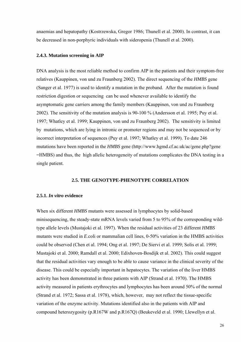
5.3 Clinical manifestations during an acute attack............................................... 5.3.1. A case report of acute porphyria with neurological manifestations.......
5.3.2. Pain during an acute attack........................................................... ........ 5.3.3. Autonomic dysfunction.......................................................................... 5.3.4. Peripheral neuropathy............................................................................ 5.3.5. Rhabdomyolysis..................................................................................... 5.3.6. CNS involvement................................................................................... 5.3.7. Precipitating factors and the course of an acute attack
5.4. Treatment....................................................................................................... 5.5. Survival and recovery during an acute attack................................................. 5.6. Scoring of the symptoms................................................................................ 5.7. Neurophysiological studies............................................................................. 5.8. Neuroimaging..................................................................................................
5.9 Identification and characterisation of the mutations in the HMBS gene......... 5.9.1. Mutations identified among patients with AIP......................................
5.9.2. Novel mutations identified in the HMBS gene....................................... 5.9.3. The effects of the novel mutations.........................................................
5.10. The genotype-phenotype correlation.............................................................. 5.10.1. Correlation between the genotype and clinical symptoms...................
5.10.2. Correlation between the genotype and biochemical characteristics.... 5.10.3. Correlation between urinary PBG excretion and clinical symptoms... 5.10.4. No correlation between erythrocyte HMBS activity, urinary PBG excretion and clinical symptoms.......................................................................
5.11. Prognosis of AIP patients...............................................................................
6. DISCUSSION...........................................................................................................
6.1. Screening of patients with acute polyneuropathy or encephalopathy.............. 6.2. Neurological manifestations in AIP.................................................................
6.2.1. Acute peripheral neuropathy.................................................................. 6.2.2. Neurophysiological findings.................................................................. 6.2.3. Neuroimaging......................................................................................... 6.2.4. Pain during an acute attack.................................................................... 6.2.5. Scaling of an acute attack.......................................................................
6.3. The diagnostics of AIP.................................................................................... 6.4. Differential diagnosis of acute polyneuropathy and encephalopathy............. 6.5. Prognosis of AIP............................................................................................. 6.6. The genotype-phenotype correlation...............................................................
7. CONCLUSIONS.......................................................................................................
8. ACKNOWLEDGEMENTS......................................................................................
9. REFERENCES..........................................................................................................
ORIGINAL PUBLICATIONS I-IV
48
48 48 48 53 53 54 55 55 56 57 57 58 60 61 63 64 65 66 68 70 70 72 73 73 74 75 75 76 77
79
79 80 80 81 82 82 83 83 85 86 87
88
90
92

5
SUMMARY
Acute intermittent porphyria (AIP, MIM #176000) is an inherited metabolic disease due to a
partial deficiency of the third enzyme, hydroxymethylbilane synthase (HMBS, EC: 4.3.1.8), in
the haem biosynthesis. Neurological symptoms during an acute attack, which is the major
manifestation of AIP, are variable and relatively rare, but may endanger a patient’s life.
In the present study, 12 Russian and two Finnish AIP patients with severe neurological
manifestations during an acute attack were studied prospectively from 1995 to 2006.
Autonomic neuropathy manifested as abdominal pain (88%), tachycardia (94%),
hypertension (75%) and constipation (88%). The most common neurological sign was acute
motor peripheral neuropathy (PNP, 81%) often associated with neuropathic sensory loss (54%)
and CNS involvement (85%). Despite heterogeneity of the neurological manifestations in our
patients with acute porphyria, the major pattern of PNP associated with abdominal pain,
dysautonomia, CNS involvement and mild hepatopathy could be demonstrated. If more strict
inclusion criteria for biochemical abnormalities (>10-fold increase in excretion of urinary PBG)
are applied, neurological manifestations in an acute attack are probably more homogeneous
than described previously, which suggests that some of the neurological patients described
previously may not have acute porphyria but rather secondary porphyrinuria. Screening for
acute porphyria using urinary PBG is useful in a selected group of neurological patients with
acute PNP or encephalopathy and seizures associated with pain and dysautonomia.
Clinical manifestations and the outcome of acute attacks were used as a basis for
developing a 30-score scale of the severity of an acute attack. This scale can easily be used in
clinical practice and to standardise the outcome of an attack. Degree of muscle weakness scored
by MRC, prolonged mechanical ventilation, bulbar paralysis, impairment of consciousness and
hyponatraemia were important signs of a poor prognosis. Arrhythmia was less important and
autonomic dysfunction, severity of pain and mental symptoms did not affect the outcome. The
delay in the diagnosis and repeated administrations of precipitating factors were the main cause
of proceeding of an acute attack into pareses and severe CNS involvement and a fatal outcome
in two patients.
Nerve conduction studies and needle EMG were performed in eleven AIP patients
during an acute attack and/or in remission. Nine patients had severe PNP and two patients had
an acute encephalopathy but no clinically evident PNP. In addition to axonopathy, features
suggestive of demyelination could be demonstrated in patients with severe PNP during an acute
attack. PNP with a moderate muscle weakness was mainly pure axonal. Sensory involvement
was common in acute PNP and could be subclinical. Decreased conduction velocities with

6
normal amplitudes of evoked potentials during acute attacks with no clinically evident PNP
indicated subclinical polyneuropathy. Reversible symmetrical lesions comparable with
posterior reversible encephalopathy syndrome (PRES) were revealed in two patients’ brain CT
or MRI during an acute attack. In other five patients brain MRI during or soon after the
symptoms was normal. The frequency of reversible brain oedema in AIP is probably under-
estimated since it may be short-lasting and often indistinguishable on CT or MRI.
In the present study, nine different mutations were identified in the HMBS gene in 11
unrelated Russian AIP patients from North Western Russia and their 32 relatives. AIP was
diagnosed in nine symptom-free relatives. The majority of the mutations were family-specific
and confirmed allelic heterogeneity also among Russian AIP patients. Three mutations,
c.825+5G>C, c.825+3_825+6del and c.770T>C, were novel. Six mutations, c.77G>A
(p.R26H), c.517C>T (p.R173W), c.583C>T (p.R195C), c.673C>T (p.R225X), c.739T>C
(p.C247R) and c.748G>C (p.E250A), have previously been identified in AIP patients from
Western and other Eastern European populations. The effects of novel mutations were studied
by amplification and sequencing of the reverse-transcribed total RNA obtained from the
patients’ lymphoblastoid or fibroblast cell lines. The mutations c.825+5G>C and c.770T>C
resulted in varyable amounts of abnormal transcripts, r.822_825del (p.C275fsX2) and
[r.770u>c, r.652_771del, r.613_771del (p.L257P, p.G218_L257del, p.I205_L257del)]. All
mutations demonstrated low residual activities (0.1-1.3 %) when expressed in COS-1 cells
confirming the causality of the mutations and the enzymatic defect of the disease.
The clinical outcome, prognosis and correlation between the HMBS genotype and
phenotype were studied in 143 Finnish and Russian AIP patients with ten mutations (c.33G>T,
c.97delA, InsAlu333, p.R149X, p.R167W, p.R173W, p.R173Q, p.R225G, p.R225X,
c.1073delA) and more than six patients in each group. The patients were selected from the pool
of 287 Finnish AIP patients presented in a Finnish Porphyria Register (1966-2003) and 23
Russian AIP patients (diagnosed 1995-2003). Patients with the p.R167W and p.R225G
mutations showed lower penetrance (19% and 11%) and the recurrence rate (33% and 0%) in
comparison to the patients with other mutations (range 36 to 67% and 0 to 66%, respectively),
as well as milder biochemical abnormalities [urinary porphobilinogen 47±10 vs. 163±21
μmol/L, p<0.001; uroporphyrin 130±40 vs. 942±183 nmol/L, p<0.001] suggesting a milder
form of AIP in these patients. Erythrocyte HMBS activity did not correlate with the
porphobilinogen excretion in remission or the clinical of the disease. In all AIP severity
patients, normal PBG excretion predicted freedom from acute attacks. Urinary PBG excretion
together with gender, age at the time of diagnosis and mutation type could predict the
likelihood of acute attacks in AIP patients.

7
ORIGINAL PUBLICATIONS
This thesis is based on the following publications, which will be referred to in the text by the
Roman numerals I to IV I. Pischik E, Bulyanitsa A, Kazakov V, Kauppinen R. Clinical features predictive of a poor
prognosis in acute porphyria. J Neurol. 2004; 251:1538-1541
II. Pischik E., Posokhina O., Kazakov V., Kauppinen R. Peripheral nerve demyelination in
acute intermittent porphyria. Submitted.
III. Pischik E, Mehtälä S, Kauppinen R. Nine mutations including three novel mutations among
Russian patients with acute intermittent porphyria. Human Mutat. 2005; 26: 496.
IV. von und zu Fraunberg M, Pischik E, Udd. L, Kauppinen R. Clinical and biochemical
characteristics and genotype-phenotype correlation in 143 Finnish and Russian patients with
acute intermittent porphyria. Medicine. 2005; 84: 35-47.
In addition, some unpublished data are presented

8
ABBREVIATIONS
AIP acute intermittent porphyria ALA δ-aminolaevulinic acid ALAD δ-aminolaevulinic acid dehydratase ALAS δ-aminolaevulinic acid synthase ALT alanine aminotransferase bp base pair cDNA complementary deoxyribonucleic acid COS-1 simian virus 40-transformed monkey kidney cell line CMAP compound muscle action potential CNS central nervous system CSF cerebrospinal fluid CT computed tomography CV conduction velocity CYP cytochrome P-450 DL distal latency DNA deoxyribonucleic acid DW diffusion-weighted (magnetic resonance imaging) EEG electroencephalography EMG electromyography Erc-HMBS HMBS activity in erythrocytes FL minimal F-wave latency FLAIR fluid-attenuated inversion recover (magnetic resonance imaging) GABA gamma aminobutyric acid HMBS hydroxymethylbilane synthase HCP hereditary coproporphyria kb kilobase kD kilodalton MCV motor conduction velocity MRC Medical Research Council scale MRI magnetic resonance imaging mRNA messenger ribonucleic acid PBG porphobilinogen PCR polymerase chain reaction RNA ribonucleic acid SCV sensory conduction velocity SD standard deviation SIADH syndrome of inadequate secretion of antidiuretic hormone VAS visual analogue scale VP variegate porphyria Mutation nomenclature meets the criteria of the Human Genome Variation Society
(http://www.hgvs.org/mutnomen/).
The mutation numbering was based on the cDNA sequence (c., cDNA).
The first base of the ATG initiation Met codon was reported as nucleotide +1
(GenBank, Ref Seq NM_000190).
Amino acids and nucleotides are abbreviated by one-letter codes.

9
1. INTRODUCTION Acute intermittent porphyria (AIP, MIM #176000) is an inherited metabolic disease due to a
partial deficiency of the third enzyme, hydroxymethylbilane synthase (HMBS, also named
porphobilinogen deaminase, PBGD, EC: 4.3.1.8), in the haem biosynthesis (Anderson et al.
2001). The major clinical manifestation of AIP is an acute attack, which includes mental
symptoms, abdominal pain and signs of autonomic dysfunction accompanied by increased
excretion of porphyrin precursors originating from the liver and releasing into the circulation
(Kauppinen 2005). Most of the attacks are short and self-limited. In a protracted attack, acute
peripheral neuropathy (PNP) and signs of CNS involvement may occur (Goldberg 1959; Ridley
1969; Stein, Tschudy 1970). Since an acute attack may endanger life, a rapid diagnosis is
essential because an effective treatment is available (Mustajoki, Nordmann 1993). The overall
prevalence of AIP is 1-10:100 000 (Anderson et al. 2001). However, the misdiagnosis is not
rare (McEneaney et al. 1993), and thus, the prevalence may be underestimated. Most of the
studies on neurology in porphyria were published 30 to 50 years ago (Waldenström 1937;
Schwarz, Moulton 1954; Goldberg 1959; Ridley 1969; Stein, Tschudy 1970; Sorensen, With
1971) before the DNA-diagnostics of AIP became available. Neuroimaging has enabled more
precise understanding of encephalopathy in acute porphyria. Because of improved diagnostics
and early and more effective treatment of acute attacks, severe neurological manifestations
have become rare. The pathogenetic mechanisms of neurological manifestations in AIP are still
obscure and unknown at the molecular level (Meyer et al. 1998).
Mutation screening is useful for confirmation of the diagnosis of AIP in symptomatic
patients and for family studies. In remission, mutation screening of the HMBS gene is currently
the most reliable method to diagnose AIP and it has been applied to the routine diagnostics of
AIP in many countries. In contrast, no such systemic studies to identify underlining mutations
in AIP patients have previously been performed in Russia. At the beginning of this study in
1995 a few cases of acute porphyria have been diagnosed clinically in North Western Russia
including St Petersburg.
The aim of this study was to characterise neurological manifestations of AIP,
precipitating factors and the natural course of an acute attack, efficiency of the treatment, and
the short-term and long-term outcomes of patients with AIP. Based on that information, the
scale of the severity of an acute attack could be established. The pathogenesis of acute PNP and
encephalopathy during an attack could be elucidated by neurophysiological studies and
neuroimaging. The characterisation of the HMBS gene defects allowed the precise diagnosis in
these patients. Finally, a genotype-phenotype correlation could be established.

10
2. REVIEW OF THE LITERATURE
2.1. PORPHYRIAS
2.1.1. Porphyrias and haem biosynthesis
Porphyrias constitute a group of inherited metabolic disorders caused by defective functions of
the enzymes in the haem biosynthesis. Each of seven porphyrias results from a partial
deficiency of one of the enzymes in the haem biosynthetic pathway (Figure 1).
Enzyme
Metabolic pathway
Glycine + Succinyl-CoA
Type of porphyria
ALA–dehydratase deficiency porphyria
Hydroxymethylbilane synthase (HMBS)
Acute intermittent porphyria (AIP)
[Hydroxymethylbilane] Uroporphyrinogen III synthase
Congenital erythropoetic porphyria
Uroporphyrinogen III Uroporphyrinogen decarboxylase
Porphyria cutanea tarda
Coproporphyrinogen III Coproporphyrinogen oxidase
Hereditary coproporphyria
Protoporphyrinogen IX Protoporphyrinogen oxidase
Variegate porphyria
Protoporphyrin IX Ferrochelatase Erythropoetic
protoporphyria
Haem
Figure 1. Haem biosynthesis and different porphyrias
Haem is synthesised in every human aerobic cell (Ponka 1999; Ajioka et al. 2006). Most of
the haem is synthesised in erythroid cells and in the liver. Heam is used as a prosthetic group of
haemoproteins, which are responsible for oxidative reactions, electron transfer processes and
delivering molecular oxygen to cells (Ajioka et al. 2006). In erythroid cells haem enhances
transcription and translation of globin and other erythroid-specific proteins. In nonerythroid
cells haem regulates expression or stability of hemoproteins.
Porphobilinogen (PBG)
δ-aminolaevulinic acid dehydratase (ALA–dehydratase)
δ-aminolaevulinic acid (ALA) δ-aminolaevulinic acid synthase (ALAS)

11
Figure 2. Haem byosynthesis in the cell
Ac, -CH2COOH; Pr, -CH2CH2COOH; Vi, -CH=CH2
Heme oxygenase, which catalyses heme degradation, provides biliverdin and thus, plays a role
in an antioxidant system (Ponka 1999).
Coproporphyrinogen III Protoporphyrinogen IX
Protoporphyrin IX
Haem
Succinil CoA
Glycine δ-aminolaevulinic acid (ALA)
Porphobilinogen
Hydroxymethylbilane
Uroporphyrinogen III
Protoporphyrinogen oxydase
Ferrochelatase
Coproporphyrinogen oxidase
Hydroxymethylbilane synthase
ALA dehydratase ALA synthase
Uroporphyrinogen III synthase
Uroporphyrinogen III dexarboxylase

12
Haem biosynthesis is illustrated in Figures 1 and 2. The first and the last three steps take
place in mitochondria and the intermediate steps are cytosolic. δ-Aminolaevulinic acid (ALA)
and porphobilinogen (PBG) are precursors of tetrapyrroles, named porphyrins. The haem
biosynthetic pathway may be presented as four basic processes: formation of the pyrrole,
assembly of the tetrapyrrole, modification of the tetrapyrrole side chains and oxidation of
protoporphyrinogen IX to protoporphyrin IX and insertion of iron (Ajioka et al. 2006). Iron
molecule coordinated within the tetrapyrrole allows haem to have diverse functions as an
electron carrier and a catalyst for redox reactions (Ajioka et al. 2006).
For the first four enzymes in the pathway both housekeeping and erythroid transcripts
are produced (Table 1). The housekeeping aminolaevulinate synthase 1 (ALAS1) and erythroid-
specific ALAS2 are encoded by different genes (Bishop et al. 1990). The different transcripts
for aminolaevulinate dehydratase (ALAD), hydroxymethylbilane synthase (HMBS) and
uroporphyrinogen III synthase are synthesised via alternative splicing of the transcript, and the
corresponding genes contain tissue-specific promoters (Anderson et al. 2001).
ALAS is the rate-limiting enzyme of haem biosynthesis (Anderson et al. 2001). In the
liver, haem represses synthesis of ALAS1 at transcriptional and translational levels (May et al.
1995; Fraser et al. 2002). ALAS1 is induced directly by numerous drugs, chemicals and alcohol
(Fraser et al. 2002) or by peroxisome-proliferator-activated receptor γ coactivator 1α (PGC-1α).
The transcription of PGC-1α is induced under low glucose concentration (Handschin et al.
2005). In addition, some drugs induce the haem-containing cytochrome P450 enzymes (CYP
450) (Anderson et al. 2001); and the stress activates hepatic haem oxygenase (Rodgers,
Stevenson 1990). These factors result in decreased hepatic haem concentrations and
consequently a loss of negative feedback to ALAS1. Induction of ALAS1 leads to
overproduction of haem precursors in the liver, and accumulation of them in other tissues via
circulation (Anderson et al. 2001). In erythroid cells, haem induces synthesis of ALAS2 mRNA
(Smith, Cox 1997). In acute porphyrias, porphyrin precursors do not accumulate in the bone
marrow (Anderson et al. 2001).
Dipyrromethane serves as a cofactor for the reaction catalysed by HMBS (Shoolingin-
Jordan 1995). Dipyrromethane is a product of autocatalytic coupling of PBG, the same
molecule, which is a substrate for the reaction. Recent studies on dipyrromethane cofactor
assembly mechanism have shown that elevated PBG strongly inhibits the formation of holo-
HMBS-deaminase from apo-HMBS-deaminase (Shoolingin-Jordan et al. 2005). It was
suggested, that PBG levels measured during acute attacks of porphyria were high enough to
inhibit the formation of holo-HMBS and to reduce dramatically the activity of hepatic HMBS in
vivo, which could contribute to overproduction of porphyrin precursors during an acute attack

13
(Shoolingin-Jordan et al. 2005). A 3.4-fold decrease in the activity of hepatic HMBS was
observed when an AIP patient during an acute attack was compared with two other AIP patients
in remission (Strand et al. 1970). This finding could indirectly support the hypothesis
mentioned above (Shoolingin-Jordan et al. 2005).
Some chemicals and toxins are known to reduce the activity of other enzymes of the
haem-biosynthesis. Lead can reduce dramatically the activity of ALAD (Godwin 2001), while
arsenic and alcohol reduce the activities of uroporphyrinogen decarboxylase and
coproporphyrinogen oxidase (Woods, Southern 1989; Doss et al. 2000).
2.1.2. Molecular genetics of porphyrias
The genes involved in the haem biosynthesis have been characterised (Table 1). A lot of
mutations in these genes are responsible for different types of porphyria, demonstrating allelic
heterogeneity of each of them (http://www.hgmd.cf.ac.uk). Mutations in ALAS2 gene are
responsible for X-linked inherited sideroblastic anaemia (Shoolingin-Jordan et al. 2003).
Porphyrias are mainly inherited autosomal dominant disorders with incomplete penetrance
(Anderson et al. 2001) (Table 1).
Table 1. Molecular genetics of porphyria
Enzyme Gene Nomenclature Transcripts Symbol Size Location
Muta-tions
Inhe-ritance
Aminolaevulinate synthase
ALAS1 housekeeping ALAS2 erythroid-specific
ALAS1 ALAS2
17 kb 22 kb
3p21.1 Xp11.21
26
X-linked
Aminolaevulinate dehydratase
ALAD housekeeping ALAD erythroid-specific
ALAD 7 kb 9q34 9 AR
Hydroxymethylbilane synthase
HMBS housekeeping HMBS erythroid-specific
HMBS 10 kb 11q23.3 244 AD
Uroporphyrinogen III synthase
UROS housekeeping UROS erythroid-specific
UROS 34 kb 10q25.2-q26.3
36 AR
Uroporphyrinogen decarboxylase
UROD 3 kb 1p34 65 AD/Ac
Coproporphyrinogen oxidase
CPO 14 kb 3q12 37 AD
Protoporphyrinogen oxidase
PPOX 5.5 kb 1q22 129 AD
Ferrochelatase FECH 45 kb 18q21.3 88 AD/AR AR, autosomal recessive; AD, autosomal dominant; Ac, acquired 2.1.2.1. Hydroxymethylbilane synthase (HMBS) gene
The HMBS gene includes 15 exons (Figure 3, GenBank, Ref Seq NC_000011). Two different
mRNAs, one for erythroid and the other for non-erythroid tissues, are transcribed via two
promoters and processed by alternative splicing. Erythroid mRNA includes exons 2-15, and

14
non-erythroid mRNA exons 1 and 3-15 (Grandchamp et al. 1987; Gubin, Miller 2001). Small
amounts of other alternatively spliced transcripts both for erythroid (Gubin, Miller 2001) and
housekeeping mRNAs (Ong et al. 1998) have been found in healthy subjects.
Mutations in the HMBS gene result in a loss of function or instability of a
mutant transcript. Heterozygous patients have approximately 50% of the total HMBS activity
measured in their erythrocytes, lymphocytes, fibroblasts and hepatocytes (Strand et al. 1970;
Meyer et al. 1972; Sassa et al. 1975; Sassa et al. 1978). This is sufficient to maintain the normal
demand for haem (Anderson et al. 2001)
Figure 3. The HMBS gene and two mRNAs transcribed by alternative splicing.
2.1.2.2. Haem metabolism in neural tissues
Many haemoproteins are detected in the brain. While neuroglobin and nitric oxide synthase
(NOS) type I are specific for neural tissues, cytochromes and soluble guanilate cyclase are
found in every cell (Anderson et al. 2001). Astrocyte CYP 450 is essential for degradation of
xenobiotics, and thus, mediates their toxicity (Meyer et al. 2001). Since exogenous radiolabeled
haematin was not detected in the rat brain homogenates after intravenous injection (De Matteis
et al. 1981), it was concluded that haem is not able to cross the blood-brain barrier. Thus, there
is de novo production of the required haem in the brain. (De Matteis et al. 1981). In the rat brain
tissues, distinct staining with anti-HMBS antibodies has demonstrated, that the highest
immunoreactivity occurs in axons of the corpus striatum (Jorgensen et al. 2000). In the brain of
the adult mice and rat, the activity of ALAS1 was only 20-24% of that in the liver (De Matteis,
Zetterlund et al. 1981; Gorchein 1990).
The regulation of the haem biosynthesis in the nervous tissues is not currently clear
(Anderson et al. 2001). Potential inducers of hepatic ALAS1 had no effect on ALAS activity in
HHMMBBSS ggeennee
EErryytthhrrooiidd
HHoouusseekkeeeeppiinngg
mmRRNNAA
IInniittiiaattiioonn ccooddoonn TTeerrmmiinnaattiioonn ccooddoonn
1 15 14 13121110987654 3
15 14 13121110987654 3 2
100 bp
5' 3'
1000 bp
1 10 11 12 13 15
14
2 3 4 5 6 7 8 9

15
the rat brain in one study (Paterniti et al. 1978) suggesting that excess of porphyrin precursors
in neural tissues is due to their production in the liver (Kauppinen 2005). This observation is
supported by the fact that the liver transplantation in an AIP patient with severe recurrent
attacks resulted in a full biochemical and clinical remission (Soonawalla et al. 2004). However,
the recent study has demonstrated 80-240% increase in ALAS1 activity and mRNA expression
in the brain samples of normal mice treated with porphyrinogenic drugs (Rodriguez, Martinez
Mdel et al. 2005). This suggests that intra-neural synthesis of porphyrin precursors may
contribute to neurological manifestation of an acute attack.
2.1.3. Acute porphyrias
Porphyrias are classified as erythropoetic and hepatic depending on the major tissue of
production and accumulation of porphyrins and their precursors (Moore et al. 1987) (Figure 4).
All hepatic porphyrias, except porphyria cutanea tarda, have similar clinical manifestations with
acute neurovisceral crises, so-called acute attacks. This is accompanied by reddish dark urine
representing porphobilin and porphyrin excess (Anderson et al. 2001).
Figure 4. Classification of porphyrias
Acute intermittent porphyria (AIP, MIM #176000) results from a partial deficiency of
HMBS (also known as porphobilinogen deaminase, EC: 4.3.1.8), the third enzyme in the
pathway. AIP has an autosomal dominant pattern of inheritance. It is the most common type of
acute porphyrias (Anderson et al. 2001). Acute attacks of AIP are clinically indistinguishable
from other acute porphyrias, but in AIP they occur more often than in other types of porphyria,
namely in variegate porphyria (VP) (Hift, Meissner 2005). In contrast to AIP, dermatological
Erythropoetic porphyrias
Hepatic porphyrias
Congenital erythropoetic porphyria Erythropoetic protoporphyria
Porphyria cutanea tarda
Acute porphyrias
ALA-dehydratase deficiency porphyria
Acute intermittent porphyria
Hereditary coproporphyria
Variegate porphyria
P O R P H Y R I A S
16
features such as photosensitivity, skin fragility, vesicular rash and hypertrichosis are found in
14%- 80% of patients with VP and hereditary coproporphyria (HCP) (Whatley, Puy et al. 1999;
Kuhnel et al. 2000; von und zu Fraunberg et al. 2002). Those features are usually independent
from acute attacks and are probably caused by free radical damage by porphyrins in the skin
(Poh-Fitzpatrick 2000). 2.1.4. History of acute intermittent porphyria
Some historical figures with paroxysmal aberrant behavior have been suggested to suffer from
acute porphyria. Among them were King George III, Van Gogh, and Alexander the Great
(Moore et al. 1987; Oldach et al. 1998; Arnold 2004). In none of these cases the diagnosis of
acute porphyria has been proven and it is only highly speculative. The varying manifestations of
congenital erythropoetic porphyria have led to legendary descriptions of patients comparing
them with vampires and were-wolfs (Moore et al. 1987).
The first description of a drug induced acute porphyria was published in 1889. The
patient was an elderly woman who had red urine and died of ingestion of sulphonal, a
barbiturate hypnotic (Stokvis 1889). One year later Ranking and Pardington described two
similar cases who in addition suffered from generalised muscle weakness (Ranking, Pardington
1890). This probably represents the first description of neurological manifestations of acute
porphyria. In 1934, Fisher was the first to show, that porphyria is caused by the impaired haem
biosynthesis (Fisher, Orth 1934). Later, Watson together with Schwartz developed a qualitative
screening-test for PBG in the urine, which could be used to confirm the diagnosis of an acute
porphyric attack (Watson, Schwartz 1941). Waldenström, who worked in Fisher's laboratory,
reported in 1937 a series of 100 northern Swedish patients with acute porphyria (Waldenström
1937; Waldenström 1939). The diagnosis was confirmed later by mutation screening. The
major mutation in the HMBS gene was found to be responsible for AIP with high prevalence in
the northern part of Sweden (Lee, Anvert 1991).
The only systematic neurological evaluation in a sufficiently large group of patients
(n=25) with acute PNP during an acute attack has been performed by Ridley (Ridley 1969). In
this study, the characteristics of PNP in acute porphyria were outlined and this study has been
the main source of knowledge for neurological manifestations in acute porphyria for the
following 30 years. Detailed neurophysiological studies and neuroimaging are lacking in his
description, which are currently the key elements in neurological diagnostics, and quantitative
assessment of urinary PBG was available only in seven cases.

17
Cloning of the HMBS gene and its complete sequencing (Raich et al. 1986; Chreitien et
al. 1988; Lee 1991) resulted in detection of various mutations in the HMBS gene. This has
demonstrated allelic heterogeneity of AIP in different populations (Kauppinen et al. 1995;
Rosipal et al. 1997; Floderus et al. 2002; Gregor et al. 2002; Parera et al. 2003).
Porphyrias in Russia have been studied by Idelson since 1960s (Idel'son et al. 1992), and
currently, the clinical and biochemical studies are conducted in the Center of Haemotology of
Russian Academy of Science in Moscow (Surin et al. 2001; Pustovoit et al. 2003)
2.2. CLINICAL MANIFESTATIONS OF AIP 2.2.1. Prevalence AIP is the most common type of acute porphyria in all countries except South Africa and Chile
where VP is the most common one (Armas et al. 1992; Hift, Meissner 2005). AIP is found in all
ethnic groups and the prevalence varies from 1 to 10:100 000 (Table 2). The misdiagnosis is
frequent, and thus, these numbers may not represent the true frequency. The founder effect is
responsible for higher prevalence of AIP in some regions (Table 2).
Table 2. Prevalence of AIP in different countries Country Prevalence* Reference Finland 3/100 000 Mustajoki, Koskelo 1976 Sweden (whole) 10/100 000 Floderus et al. 2002 Sweden (province Arjeplog) 2 000/100 000 Andersson et al. 1995 Sweden (province Arvidsjaur) 500/100 000 Andersson et al. 1995 Norway (whole) 10/100 000 Tjensvoll, Bruland et al. 2003 Norway (Saltdal region) 600/100 000 Tollali et al. 2002 Western Australia 3/100 000 Saint and Curnow 1962 USA 5-10/100 000 Tschudy et al. 1975 Japan 1.5/100 000 Sugimura 1995 Argentina 0.8/100 000 Parera et al. 2003
* Including symptom-free patients
When 2234 Finnish and 3350 French healthy donors (Mustajoki et al. 1992a; Nordmann
et al. 1997) were assessed for the low erythrocyte HMBS activity, 1:500 to 1:1500 prevalence
values was observed suggesting that the prevalence of the low HMBS might be much higher
than previously estimated.
There is no statistical data available for the prevalence of AIP in Russia with a
population of ~140 million, since only single cases were reported which were mainly
concentrated around Moscow and St Petersburg. In Russia, Belarus and Uzbekistan (Pustovoit
et al. 2003), a total of 42 AIP patients have been reported to date. However, only in four cases,
AIP was confirmed by mutation analysis (Surin et al. 2001).

18
2.2.2. Clinical picture of AIP
2.2.2.1. Acute attacks
The main clinical manifestation of AIP is an acute attack, which is a neurovisceral crisis
manifesting as a combination of signs and symptoms listed in Table 3.
Table 3. Clinical manifestation during an acute attack in different patient series Signs and symptoms 1
n=252 2 n=50
3 n=40(34)‡
4* n=88
5† n=51/22¶
6* n=112/24¶
% % % % % % I. Autonomic dysfunction Abdominal pain 85 94 95 95 96 97 Tachycardia (>80 per min) 28 64 80 85 79 38 Hypertension 40 54 36 55 57 74 Constipation 48 84 48 80 78 27 Vomiting and nausea 59 88 43 80 84 79 Bladder paresis n.a. n.a. 12 n.a. n.a. n.a. II. Peripheral neuropathy or/and encephalopathy Pain in the back and limbs n.a. 52 50 70 25 n.a. Mental symptoms 55 58 40 40 19 1§ “Pareses” / ”Muscle weakness” 42 68 60 50 8 46/10¶ Epileptic seizures 10 16 20 20 2 5 III. Metabolic changes Hyponatraemia n.a. 25 26 61 32 31 Transaminases increased n.a. n.a. 13 n.a. n.a. n.a. Red/ dark urine n.a. n.a. 74 90 90 n.a.
1 Waldenström, 1957; 2 Goldberg, 1959; 3 Stein, Tchudy; 4 Mustajoki, 1976; 5 Mustajoki, Nordmann, 1993; 6 Hift, Meissner, 2005 *AIP and VP. † Cases early treated with haem arginate. ‡40 cases with full and 34 with partial information available. ¶ Attacks/patients. § Only psychosis is included. n.a., not applicable
In the majority of the series, motor deficit was described as “paresis” or “muscle weakness”. In
those cases, retrospective categorisation of this neurological sign was not possible, since from
the data published it was not possible to determine precisely the cause of muscle weakness.
Autonomic dysfunction Pain in the back and limbs Hyponatraemia Mental symptoms Muscle weakness Dark urine
Figure 5. Common combination of symptoms during an acute attack
The signs and symptoms appear usually in combination. The severity of each symptom
and the combination of the symptoms vary among patients. None of the symptoms and/or signs
Abdominal pain +

19
are pathognomonic, but their co-occurrence together with abdominal pain should suggest acute
porphyria (Mustajoki, Koskelo 1976) (Figure 5).
2.2.3. Neurological manifestations of an acute attack
The majority of acute attacks manifest only as a combination of abdominal pain, mild mental
symptoms and autonomic dysfunction and without PNP or focal CNS impairment (Mustajoki,
Nordmann 1993). Only if an attack proceeds, various signs of neurological deficits may develop
(Table 4).
Table 4. The common signs of neurological impairment during an acute attack in different series
1 Goldberg, 1959; 2 Ridley, 1969: 3 Stein, Tchudy; 4 Mustajoki, 1976; 5 Mustajoki, Nordmann, 1993 * All patients with PNP; † Attacks/patients; ‡40 cases with full and 35 with partial information available; §The origin of optic neuritis was suggested. n.a., not applicable
Neurological manifestations of AIP are great imitators leading to potential misdiagnosis
in these patients (Waldenström 1939; Goldberg 1959; Ridley 1969; Crimlisk 1997) (Table 5).
The paroxysmal nature of the attacks and the almost uniform and specific temporal association
of neurological features with acute abdominal pain is characteristic for acute porphyria
(Goldberg 1959). When progressive muscle weakness manifests, the intensity of abdominal
pain may decline (Goldberg 1959; Stein, Tschudy 1970; Hift, Meissner 2005), which may lead
to misdiagnosis.
Signs and symptoms
1 n=50
2* n=29/25†
3 n=40(35)‡
4 n=88
5 n=51/22†
% % % % % I Peripheral neuropathy Muscle weakness 68 100 60 50 8 Low/absent tendon reflexes 54 97 29 n.a. n.a. Respiratory paresis 10 55 9 20 0 Cranial neuropathy 28 69 54 15 0 Neuropathic sensory loss 38 59 26 25 n.a. II. Encephalopathy Seizures 16 21 20 20 1 Mental symptoms 58 86 40 40 19 Coma n.a. n.a. 10 n.a. 0 Blurred vision 6§ 7§ 6§ n.a. 2 Babinski signs 10 3 3 n.a. n.a. Nystagmus
Cerebellar ataxia 2 2
7 0
0 0
n.a. n.a.
n.a. 0

20
Table 5. The common neurological misdiagnoses in patients with acute porphyria* Diseases with predominant CNS involvement
Diseases with predominant involvement of peripheral nerves
Diseases with predominant involvement of autonomic nerves
Viral/ toxic encephalopathy Seizures
Acute inflammatory PNP (=Guillain-Barré syndrome) Acute PNP of other origin
Panic disorder (“crises vasculares”) Rheumatic fever
Psychosis Polymyositis Fatigue Poliomyelitis Hysteria *by Waldenström 1939; Goldberg 1959; Ridley 1969; Crimlisk 1997
The incidence of neurological manifestations has decreased over the years mainly
because of greater awareness and early treatment of the attacks. In the series of 50 AIP patients
published in 1959, 68% of them had paresis and 16% had epileptic seizures (Goldberg 1959).
30 years later Mustajoki and Nordmann reported 51 attacks in 22 patients, who have been
treated with haem arginate at the early phase of an attack. Among those, 8% of the patients
experienced mild and moderate pareses without respiratory involvement and only one patient
had seizures (Mustajoki, Nordmann 1993).
2.2.3.1. Autonomic neuropathy
Autonomic neuropathy is responsible for the majority of symptoms in an acute attack (Table 3).
Tachycardia is usually associated with the activity of the disease (Ridley et al. 1968).
Orthostatic hypotension (Stein, Tschudy 1970), diastolic hypertension (Goldberg 1959; Stein,
Tschudy 1970), diarrhoea (Stein, Tschudy 1970; Mustajoki, Koskelo 1976) and erectile
dysfunction (Goren, Chen 1991) have been occasionally observed.
During an acute attack abnormal parasympathetic cardiac reflexes were demonstrated in
eight AIP patients using the battery of standard Ewing’s tests (Ewing et al. 1985; Laiwah et al.
1985). Of two sympathetic tests, the postural drop of blood pressure was normal and the blood
pressure response to sustained hand-grip was slightly decreased (Laiwah et al. 1985). These
results are questionable, since six of the patients had motor PNP during the acute attack. The
vagal insufficiency rather than sympathetic activation is more likely explanation for cardiac
dysautonomia in AIP. The other signs of cholinergic insufficiency are constipation and bladder
paresis (Freeman 2005). The mechanisms of these phenomena are currently quite speculative,
since no appropriate bowel emptying or urodynamic tests have been reported in AIP patients. A
patient with HCP and chronic intestinal pseudo-obstruction was described (Vassallo et al.
1992), suggesting severe vagal insufficiency, but the diagnosis of HCP was questionable
(Pierach 1992).

21
Cholinergic insufficiency does not explain postural hypotension, diarrhea and excessive
sweating (Stein, Tschudy 1970). These symptoms are probably due to sympathetic insufficiency
(Freeman 2005).
The origin of nausea and vomiting in acute porphyria is unknown (Meyer et al. 1998).
Both abnormal bowel motility and central mechanisms may be involved.
Reduced heart rate variability found in 23 patients with AIP in remission, (Blom et al.
1996) and abnormal response to Valsalva maneuver in 10 asymptomatic patients with AIP
(Laiwah et al. 1985) suggested chronic cardiac dysautonomia with cholinergic insufficiency
(Blom et al. 1996). The tests of sympathetic function in those patients were normal (Laiwah et
al. 1985; Blom et al. 1996).
In summary, autonomic neuropathy in acute porphyria manifests as pandysautonomia,
(Laiwah et al. 1985; Freeman 2005) with parasympathetic insufficiency predominance.
2.2.3.2. Acute peripheral neuropathy
Acute PNP is the most common neurological complication of an acute attack (Goldberg 1959;
Ridley 1969; Stein, Tschudy 1970). It manifests as diffuse muscle weakness, symmetrically
distributed hyporeflexia and sensory loss (McLeod 1995), which appear during an acute attack.
Similar to classical polyneuropathies, the distribution of muscle weakness is symmetrical and
evenly distributed in the proximal and distal muscle groups of the upper extremities. In the
lower extremities the weakness is pronounced in the proximal muscles (Ridley 1969).
Another outstanding feature of PNP in acute porphyria is the preservation of the ankle
jerks in about half of patients while otherwise there is global areflexia (Ridley 1969).
Respiratory failure, the most severe complication of muscle weakness because of diaphragm
paresis, was common in the earlier series (10%-64%) (Goldberg 1959; Ridley 1969; Stein,
Tschudy 1970; Mustajoki, Koskelo 1976) but was not seen in AIP patients treated in the early
phase of an acute attack (Mustajoki, Nordmann 1993).
The sensory symptoms of PNP are usually due to irritation (painful paresthesia,
hyperesthesia) (Tschudy et al. 1975) which may be misdiagnosed as conversion. Sensory loss is
less common. Both “glove-and-stocking” or patchy proximal distribution of sensory loss
atypical for length-dependant polyneuropathies have been described (Ridley 1969). Cranial
neuropathy affecting mainly III, VI, IX and X cranial nerves is present in 35-55% of acute
attacks with PNP (Ridley 1969; Stein, Tschudy 1970).
Motor polyneuropathy without abdominal pain was reported only in a few cases with
AIP (Ridley 1969; Goren, Chen 1991; Niznikievicz, Jablonska-Kaszewska 1996; Cohen et al.

22
1997; Andersson et al. 2002). A predominantly motor type of PNP accompanied by abdominal
pain has been described in the majority cases with AIP. Single cases of mononeuritis multiplex
(Ridley 1969; King et al. 2002), focal motor polyneuropathy (Ridley 1969), predominantly
sensory polyneuropathy (Goren, Chen 1991), retrobulbar neuritis (Wolter et al. 1972), "pseudo
myopathy" ( Cohen et al. 1997; Poersch 1998), "pseudo myasthenia" (Goldberg 1959) and
severe ptosis without ophathalmoplegia (Tan et al. 1990) during an acute attack of AIP have
been published. This suggests that patients with AIP may have variable neurological
manifestations during an acute attack. In some of the cases, however, the diagnosis of acute
porphyria was based on inadequate biochemical diagnostics or their interpretation, and thus,
acute porphyria might not be the cause of those symptoms (Tan et al. 1990; Goren, Chen 1991;
Cohen et al. 1997).
The routine examination of cerebro-spinal fluid (CSF) in patients with AIP and PNP is
usually normal (Goldberg 1959; Ridley 1969; Nagler 1971; McEneaney et al. 1993), indicating
the absence of neuroinflammatory process in the CNS and the proximal nerves.
2.2.3.3. Acute encephalopathy
Single cases of transient cortical blindness (Lai et al. 1977; Kupferschmidt et al. 1995; Garg et
al. 1999; Yen et al. 2002; Wessels et al. 2005), hemianospia (Martin, Heck 1956), cerebellar
ataxia (Gibson, Goldberg 1956; Crimlisk 1997), parkinsonism (Ridley 1969; Crimlisk 1997),
finger tremor (Ridley 1969), dysphasia (Aggarwal et al. 1994) and central pontine myelinolysis
(Susa et al. 1999) have been published, but in general, focal CNS involvement is rare during an
acute attack. Of 25 patients with PNP during an attack, only one manifested extensor plantar
response without other clinical signs of CNS involvement (Ridley 1969). In this case, the focus
of haemorrhagic infarction in the contralateral internal capsule was revealed on autopsy.
Of 224 patients, 4-61 % had hyponatraemia during an acute attack (Goldberg 1959;
Ridley 1969; Stein, Tschudy 1970; Mustajoki, Koskelo 1976; Hift, Meissner 2005), which in
the absence of diarrhea, vomiting and polyuria, is usually a manifestation of the syndrome of
inappropriate secretion of antidiuretic hormone (SIADH) (Suarez et al. 1997). Generalised or
focal epileptic seizures, which accompany 2% to 20% of acute attacks (Stein, Tschudy 1970;
Mustajoki, Nordmann 1993; Bylesjo et al. 1996), have mainly been attributed to hyponatraemia.
Aberrant behavior and various psychiatric manifestations, so-called mental syndrome of
acute porphyria (Wettenberg 1967), appear transiently during an acute attack (Table 3).
Frequencies of these manifestations vary from series to series (19% - 56%). This may be due to
underestimation of mild mental symptoms or different severity of the attacks in these series.

23
In remission no segregation of acute porphyria and schizophrenia or bipolar disorder was found
in a large series of 344 AIP patients in Glasgow (Patience et al. 1994), however anxiety was
more common among AIP patients than in general population (Patience et al. 1994; Millward et
al. 2005).
CSF in patients with encephalopathy during an acute attacks was normal (Aggarwal et
al. 1994; Kupferschmidt et al. 1995; Utz et al. 2001; Celik et al. 2002; Engelhardt et al. 2004;
Maramattom et al. 2005; Wessels et al. 2005).
2.3. PRECIPITATING FACTORS
Both endogenous and exogenous precipitating factors, such as certain medications, alcohol,
infections, low caloric intake or changes in sex hormone balance during the menstrual cycle or
pregnancy, can provoke clinical manifestations in AIP (Goldberg 1959; Stein, Tschudy 1970;
Kauppinen, Mustajoki 1992). Information about potentially safe and unsafe drugs can be found
on the Internet (e.g. www.porphyria-europe.com, www.uq.edu.au/porphyria,
www.uct.ac.za/depts/porphyria).
The major endogenous factor is the hormonal cycle. In women with diagnosed AIP the
menstrual cycle is the most common (30%) precipitating factor of an acute attack manifesting
usually with one to three months interval in premenstrum (Herrick et al. 1990; Kauppinen,
Mustajoki 1992; Anderson et al. 2001). Previously, pregnancies were commonly complicated
by acute attacks mainly during the first trimester and postpartum (Shapiro et al. 1969; Hunter
1971; Wenger et al. 1998). Currently, overall prognosis for pregnancy in AIP is good,
especially, if the diagnosis is known in advance and no porphyrinogenic drugs are used
(Kauppinen, Mustajoki 1992; Andersson et al. 2003).
The exact mechanism of endogenous hormonal fluctuations causing acute attacks is
unknown. The direct role of sex hormones as precipitating factors is unlikely, since attacks
during the second and third trimester of pregnancy are rare, when the level of sex hormones is
at the highest (Pischik, Kauppinen 2006). Moreover, cyclical attacks occur usually in
premenstrum, when the level of oestrogen and progesterone is fluctuating the most. The central
role of the hypothalamic suprachiasmatic clock area in maintaining recurrent attacks of AIP has
been suggested (Pischik, Kauppinen 2006). Gamma amino butyric acid (GABA) mediated
neurons from the clock area have been shown to control directly liver metabolism via
autonomic nerves (Kalsbeek et al. 2004), which could link circadian rhythms and cyclic attacks
of AIP. The high affinity of ALA to GABA receptors has been demonstrated in vitro (Brennan,
Cantrill 1981), and thus, overproduction of ALA can affect synchronisation and the threshold of

24
the GABA-ergic clock cells. This may activate abnormal liver metabolism and, consequently,
precipitate acute attacks by the central mechanism, making AIP a CNS disorder in addition to a
liver disease (Pischik, Kauppinen 2006).
2.4. DIAGNOSIS OF AIP
Diagnosis of AIP is based on the combination of clinical and biochemical findings at the
symptomatic phase. During the asymptomatic phase the diagnosis can be established by
biochemical findings or DNA analysis (Kauppinen, von und zu Fraunberg 2002).
2.4.1. Clinical criteria
The clinical criteria of an acute attack include the paroxysmal nature of the symptoms with
abdominal or back pain associated with one or more signs of autonomic dysfunction,
hyponatraemia, muscle weakness or mental symptoms (Mustajoki, Nordmann 1993) (Figure 5). 2.4.2. Biochemical findings 2.4.2.1. Urine analysis of porphyrins and their precursors
The biochemical criteria of an acute attack include more than a four-fold increase of urinary
PBG excretion, which can be detected by a simple Watson-Schwartz or Hoesch qualitative test
(Watson, Schwartz 1941; Lamon et al. 1974; Bonkovsky, Barnard 1998). The results should
always be confirmed by a quantitative measurement of urinary PBG assayed by ion exchange
chromatography (Mauzerall, Granick 1956), since false positive results in these screening tests
are possible, especially, if perchloric acid instead of amyl alcohol is used as an exract
(Bonkovsky, Barnard 1998; Thunell et al. 2000; Deacon and Elder 2001). False negative results
are also possible, if the urine samples are not sheltered from the light or taken late during an
acute attack (Bonkovsky, Barnard 1998; Thunell et al. 2000).
In 85% of the patients with AIP urinary PBG is elevated in remission as well, but at
least a two-to four-fold increase in PBG excretion is usually observed in patients during an
acute attack (Kauppinen, von und zu Fraunberg 2002). Urinary ALA is always increased during
an acute attack and remains increased in 61% of AIP patients in remission (Kauppinen, von und
zu Fraunberg 2002). Urinary porphyrins are elevated during an acute attack (Lim, Peters 1984).
In AIP, urinary excretion of uroporphyrins include both I and III isomers. Their levels are
higher than those of coproporphyrins I and III (Kauppinen, von und zu Fraunberg 2002).

25
2.4.2.2. Plasma porphyrins
In AIP, plasma emission spectrum test with excitation wavelength of 405 nm shows a peak at
615-620 nm during an acute attack similarly to HCP, which corresponds to elevated plasma
porphyrins (Deacon and Elder 2001). This is due to porphyrins ability to absorb light at
wavelength around 400 nm and their emission as red fluorescence at around 600 nm (Moore et
al. 1987). Of note, it is usually negative in remission (Kauppinen, von und zu Fraunberg 2002).
Plasma emission spectrum test is used mainly to exclude VP, since it has an unique 624-627 nm
spectrum because of protein-associated plasma porphyrins (Poh-Fitzpatrick, Lamola 1976;
Deacon and Elder 2001). A high-performance liquid chromatography (HPLC)-mass
spectrometry can be used to measure porphyrin precursors in plasma (Floderus et al. 2006).
PBG and ALA levels were 3.1±1.0 and 1.7±0.7 µmol/L in the plasma samples taken from the
AIP patients in remission (normal range for P-PBG <0.12 µmol/L and for P-ALA 0.38±0.3
µmol/L) (Floderus et al. 2006), which is one fiftieth to hundredth part of the levels found in
urine samples (Kauppinen, von und zu Fraunberg 2002).
2.4.2.3. Faecal porphyrins
Faecal protoporphyrin excretion was increased in 47% of 13 patients with AIP during an acute
attack and in 19 % of 49 patients during remission in a Finnish series (Kauppinen, von und zu
Fraunberg 2002), but the level of excretion was lower than found in patients with VP (von und
zu Fraunberg et al. 2002). In two Canadian patients with AIP faecal protoporphyrin was normal
during remission (Hindmarsh et al. 1999). Faecal coproporphyrin is usually normal both during
an acute attack and in remission (Kauppinen, von und zu Fraunberg 2002). The high level of
coproporphyrin III isomer in faeces which vastly exceeds that of coproporphyrin I isomer and
protoporphyrin, suggests HCP (Kuhnel et al. 2000).
2.4.2.4. Erythrocyte HMBS activity assay Decreased erythrocyte HMBS activity (Erc-HMBS) has been found in 84 % of patients with
AIP (Kauppinen, von und zu Fraunberg 2002). In the variant form of AIP (5-16% of all
patients) (Whatley et al. 2000; Kauppinen, von und zu Fraunberg 2002), Erc-HMBS is normal
due to an alternative splicing of the HMBS gene in erythroid cells (Grandchamp et al. 1989). Of
note, Erc-HMBS should be assayed in remission, since it may be transiently elevated during an
attack (Kostrzewska, Gregor 1986). Occasionally Erc-HMBS is normal in patients with non-
variant form AIP because of enhanced erythropoesis, e.g. in hypochromic or hemolytic
26
anaemias and hepatopathy (Kostrzewska, Gregor 1986; Thunell et al. 2000). In contrast, it can
be decreased in non-porphyric individuals with sideropenia (Thunell et al. 2000).
2.4.3. Mutation screening in AIP
DNA analysis is the most reliable method to confirm AIP in the patients and their symptom-free
relatives (Kauppinen, von und zu Fraunberg 2002). The direct sequencing of the HMBS gene
(Sanger et al. 1977) is used to identify a mutation in the proband. After the mutation is found
restriction digestion or sequencing can be used whenever available to identify the
asymptomatic gene carriers among the family members (Kauppinen, von und zu Fraunberg
2002). The sensitivity of the mutation analysis is 90-100 % (Andersson et al. 1995; Puy et al.
1997; Whatley et al. 1999; Kauppinen, von und zu Fraunberg 2002). The sensitivity is limited
by mutations, which are lying in intronic or promoter regions and may not be sequenced or by
incorrect interpretation of sequences (Puy et al. 1997; Whatley et al. 1999). To date 246
mutations have been reported in the HMBS gene (http://www.hgmd.cf.ac.uk/ac/gene.php?gene
=HMBS) and thus, the high allelic heterogeneity of mutations complicates the DNA testing in a
single patient.
2.5. THE GENOTYPE-PHENOTYPE CORRELATION
2.5.1. In vitro evidence
When six different HMBS mutants were assessed in lymphocytes by solid-based
minisequencing, the steady-state mRNA levels varied from 5 to 95% of the corresponding wild-
type allele levels (Mustajoki et al. 1997). When the residual activities of 23 different HMBS
mutants were studied in E.coli or mammalian cell lines, 0-50% variation in the HMBS activities
could be observed (Chen et al. 1994; Ong et al. 1997; De Siervi et al. 1999; Solis et al. 1999;
Mustajoki et al. 2000; Ramdall et al. 2000; Edixhoven-Bosdijk et al. 2002). This could suggest
that the residual activities vary enough to be able to cause variance in the clinical severity of the
disease. This could be especially important in hepatocytes. The variation of the liver HMBS
activity has been demonstrated in three patients with AIP (Strand et al. 1970). The HMBS
activity measured in patients erythrocytes and lymphocytes has been around 50% of the normal
(Strand et al. 1972; Sassa et al. 1978), which, however, may not reflect the tissue-specific
variation of the enzyme activity. Mutations identified also in the patients with AIP and
compound heterozygosity (p.R167W and p.R167Q) (Beukeveld et al. 1990; Llewellyn et al.

27
1992; Solis et al. 2004) had a relatively high residual activity in COS cells (Edixhoven-Bosdijk
et al. 2002).
2.5.2. In vivo evidence
In a population-based study which included both symptomatic and symptom-free AIP patients,
a genotype-phenotype correlation for the mutations p.W198X, p.R173W and p.R167W was
demonstrated (Andersson et al. 2000). In this study the p.W198X and p.R173W mutations
caused a more severe disease in comparison with the mutation p.R167W phenotype (Table 6). Table 6. Comparison of clinical presentation of AIP patients with p.W198X, p.R173W and p.R167W mutations in the Norrland study (Andersson et al. 2000) Mutation p.W198X p.R173W p.R167W Manifest AIP, n 147 5 3 Total AIP, n 338 10 24 Penetrance, % 44 50 13 Mean number (range) of hospital admission 8 (0-140) 4 (1-10) 0.3 (0-1) Attacks during the last year, n (%) 35(30) 1 0 Mean (range) age of the first attack 27 (7-65) 20 (16-27) 35 (19-50) Decreased creatinin clearance, n 29 3 1 Chronic hypertension, n no 3 no
Excretion levels of porphyrin precursors in two patient groups (p.W198X and p.R167W)
including both latent and manifest patients did not differ significantly. This study included no
information on Erc-HMBS activities among the patients.
The results of a genotype-phenotype correlation in other acute porphyrias have been
controversial (Whatley et al. 1999; Lamoril et al. 2001; von und zu Fraunberg et al. 2002). The
series of VP and HCP patients suggested the absence of a genotype-phenotype correlation
(Whatley et al. 1999; Lamoril et al. 2001) but they included mainly symptomatic probands.
Thus, the penetrance rate in these families could not be calculated. Moreover, all the missence
mutations and splice defects were pooled independent of the affected residues and the
biochemical findings were not analysed.
In the Finnish series of VP patients a milder clinical and biochemical disease could be
demonstrated in the patients with the mutation p.I12T when they were compared to the patients
with the mutation p.R152C or c.338G>C (von und zu Fraunberg et al. 2002). The mutation
p.I12T was originally identified in a sample from a homozygous patient (I12T/I12T). Of 12
heterozygous family members with this mutation, only one experienced acute attacks during her
life span. The attacks were triggered by sulphonamides and barbiturates. This mutation and
other ones identified in patients with VP and compound heterozygosity, had residual enzymatic
activities in vitro (Roberts et al. 1998), which explains the survival of the patients and supports

28
an idea of a milder phenotype in some of the mutations in acute porphyrias.
2.6. PATHOGENESIS OF NEUROLOGICAL MANIFESTATIONS IN AIP
2.6.1. Main hypotheses
Despite the progress in understanding of the molecular genetics and biochemistry of AIP, the
pathogenesis of transient neurological impairment is still obscure (Meyer et al. 1998).
Currently, two main hypotheses have been raised (Figure 6). Neurological manifestations of
acute porphyria could be precipitated either by direct neurotoxicity of porphyrin precursors or
by deficiency of neural haem-containing enzymes or by both factors together.
2.6.1.1. Neurotoxicity of porphyrin precursors in vitro
The results from the experimental data support the direct neurotoxicity of ALA. In vitro
conditions, ALA concentrations of 10-5 -10-2 M have been toxic to neuromuscular, muscular and
spinal cord preparations in animals and cultured neural and glial cells of chicken embryos
(Loots et al. 1975; Percy et al. 1981; Cutler et al. 1990; Meyer et al. 1998), but not to human
spinal cord neurons (Gorchein 1989) (Figures 5, 6). In rat brain cortex membrane preparations,
substantial decrease in Na+K+ATPase activity has been demonstrated after exposure to ALA
(Russel et al. 1983). Neurotoxicity of PBG has not been tested in vitro (Meyer et al. 1998).
In rat brain preparation of purified synaptosomes, GABA and glutamate release was
affected by ALA in a dose dependent manner (Brennan, Cantrill 1981). This could be explained
by the structural similarity of ALA and inhibitory neurotransmitters GABA and glutamate
(Brennan, Cantrill 1981). The loss of inhibitory effect of those neurotransmitters due to
increased ALA levels could be responsible for seizures during an acute attack (Meyer et al.
1998).
Increased free radical formation due to enolisation and auto-oxidation of ALA have
caused damage to proteins and lipids in isolated liver mitochondria and rat brain tissues
(Demasi et al. 1996). The pathogenetic application of free radicals originating form circulating
porphyrins has previously been suggested for cutaneous porphyrias (Poh-Fitzpatrick 2000) and
other neuropathies (Smith et al. 1999; Pop-Busui et al. 2006).

29
Excess of porphyrins and their precursors (ALA, PBG) from the liver into circulation 2
Critical deficiency of haem and haemoproteins in the liver2 or/and neural tissues 3
• Direct neurotoxicity of ALA
(abnormal functioning or cell death)1
• Structural similarity of ALA and GABA/glutamate
(partial GABA/glutamate receptor antagonism)1
• Inhibition of Na+/K+ ATPase1
• Formation of free radicals and reactive oxygen species from
ALA1.2
• Deficiency of oxidative chain enzymes (cytochromes type a, b and c) 3
• Deficiency of nitric oxide synthase 3 (vasospasm)
• Deficiency of hepatic tryptophan dioxygenase (excess of serotonin 2)
• Deficiency of cytochrome p450, main inactivator of different
metabolites, including neuroactive mediators in CNS 4
• Deficiency of haem-dependent soluble guanilatcyclase 3 (abnormal
cellular signaling)
Precipitating factors
Partial deficiency of HMBS
1) Direct induction of ALAS12 2) Increased haem utilisation, induction of haem oxygenase2 (Indirect induction of ALAS1)
1 Evidence in vitro 2 Evidence in vivo 3 Experimental data does not prove this hypothesis 4 Evidence in liver, but not assayed in neural tissues
NEUROLOGICAL MANIFESTATIONS OF AN ACUTE ATTACK
Protracted critical deficiency of hepatic HMBS 2
Disruption of blood-brain 2 and blood-nerve barriers
Figure 6. Pathogenesis of neurological impairment during an acute attack of AIP (Main hypotheses)

30
2.6.1.2. Porphyrin precursors in vivo
Excess of ALA itself is not sufficient to explain neurological manifestation in acute porphyrias.
An injection of ALA at concentrations comparable with the levels measured in
patients during an acute attack has caused no clinical manifestations in a healthy volunteer,
“healthy” mice, or patients with neuroblastoma, who have received ALA as an agent for
photodynamic therapy (Dowdle et al. 1968; Edwards et al. 1984; Mustajoki et al. 1992b; Fukuda
et al. 2005). Moreover, 61% of AIP patients in remission have increased levels of ALA with no
symptoms of neuropathy (Kauppinen, von und zu Fraunberg 2002), although mild subclinical
neuropathy in those patients cannot be excluded.
2.6.1.3. ALA neurotoxicity and physiological barriers
Because blood-brain barrier protects the brain (Bradbury 1979), concentration of ALA in CSF
is significantly lower (10-100 fold) than measured in serum, which has been shown in the
healthy individuals and in compound heterozygous HMBS(-/-) mice (Figure 7) (Percy, Shanley
1977; Gorchein, Webber 1987; Meyer et al. 1998).
In the concentration of ALA lower than 10-6 M only concurrent agonism of GABA
autoreceptors could be demonstrated in rat brain synaptosomes (Brennan, Cantrill 1981; Meyer
et al. 1998).
concentration ALA (M)
10-2
10-3
Increased parameters of oxidative damage in nervous tissues
10-4
2 x 10⁻⁵
Direct neurotoxicity in animal preparations and cultured neural and glial cells
Modification of uptake and efflux of GABA and glutamate
10-5 in serum 3 x 10⁻⁶
No symptoms in a healthy volunteer
in serum,
in peripheral nerves
10-6
Inhibition Na+/K+ ATPase in rat brain cortex membrane
in peripheral nerves in CSF
10-7 in CSF Remission Attack Experimental data AIP patients, HMBS (-/-) mice
Figure 7. Comparison of ALA concentration detected in experimental models and AIP patients during an acute attack
A blood-nerve barrier, which protects peripheral nerves, is less resistant to toxins than
blood-brain barrier (Bradbury 1979). In mice ALA concentration in the perineural fluid is about
30% of that found in the serum (Meyer et al. 1998), which favours ALA neurotoxicity also for

31
peripheral nerves. The concentration of ALA in CSF has been measured in only three AIP
patients during an acute attack: 192 nmol/L (Gorchein, Webber 1987), 2.1 μmol/L (Sweeney et
al. 1970) or 35 μg/L (Percy, Shanley 1977). Disruption of the blood-brain barrier, which can be
seen as reversible multifocal oedema on brain MRI (FLAIR, DW) in patients with AIP during
an acute attack (Utz et al. 2001; Celik et al. 2002; Yen et al. 2002; Engelhardt et al. 2004;
Wessels et al. 2005) could result from the permeability failure caused by a transient increase of
ALA or other currently unknown factors in the circulation and the actual concentration of
porphyrin precursors in the brain tissue could be higher.
2.6.1.4. Deficiency of haemoproteins
An established mouse model for AIP (Lindberg et al. 1996), resembles the human disease to
some extent. Compound heterozygous HMBS (-/-) mice have ~30% of the normal HMBS
activity and develop increased levels of plasma and urinary ALA and PBG only after induction
of the hepatic ALAS by phenobarbital. Clinical manifestations such as acute attacks do not
develop in mice. In contrast, chronic neurological deficits could be observed in mice even in
the absence of biochemical abnormalities (Lindberg et al. 1996). This suggests that haem-
deficiency could play a role in chronic neuropathy in these mice.
In the brain homogenates of the HMBS (-/-) mice, which have been decapitated after
induction of ALAS with Phenobarbital, a CYP2A5 inhibitor, the activity and mRNA expression
of haem-containing enzymes such as nitric oxide synthase I and soluble guanylate cyclase was
normal (Jover et al. 2000). After infusion of β-naphtoflavone, a CYP 1A1 inhibitor, the haem-
content in the mice brain was not affected, but the CYP 1A1 enzyme has partly retained in the
cytosol instead of endoplasmic reticulum (Meyer et al. 2005). The potential application of these
findings in humans is unclear. The activity of other haemoproteins in neural tissues has neither
been studied in rodents nor in humans.
There have been few as well as controversial reports of mitochondrial respiratory chain
enzymes in muscle tissues. In 15 patients’ samples with “active” acute porphyria, cytochrome c
oxidase activity was decreased (Goldberg et al. 1985), but in a homozygous AIP patient
activities of “mitochondrial respiratory chain enzymes” were normal (Solis et al. 2004).
In 12 patients with AIP during an acute attack, a two-fold increase in the levels of the
whole blood serotonin (5-hydroxytryptamine, 5-HT) and total plasma tryptophan was found (Puy
et al. 1993). This could be attributed to the decreased activity of haem-containing hepatic
tryptophan pyrrolase, as it has been decreased in experimental model of normal rats after
simultaneous treatment with phenobarbital which stimulates hepatic ALAS1 and dicarbethoxy-
dimethyl-ethyl-dihydropyridine which is a suicide inhibitor of hepatic CYP450 (Litman, Correia

32
1985). Since the clinical picture of carcinoid or serotonin syndrome (Alam 1994) has only little
similarities with acute porphyria, it cannot explain all symptoms of porphyria (Meyer et al.
1998). Deficiency of other hepatic haem enzymes (mainly CYP450) has been demonstrated both
in patients with acute porphyria and HMBS(-/-) mice (Mustajoki et al. 1994; Jover et al. 2000),
but their direct role in neurological manifestations of acute porphyria is unclear.
2.6.2. Pathogenesis of autonomic dysfunction
The course of an acute attack is probably directly related to the permeability of blood-brain and
blood-nerve barriers to porphyrin precursors, which are small size molecules (Kauppinen
2005). Absence of a barrier for autonomic nerves and a sparse blood-brain barrier in
hypothalamus and limbic areas may explain initial manifestations of an attack such as
dysautonomia and mild mental changes. Vagus nerve demyelination, axonal loss and
chromatolysis of sympathetic ganglion cell in autopsies (Baker, Watson 1945; Gibson,
Goldberg 1956; Suarez et al. 1997) support the direct involvement of autonomic fibers and
explain some of dysautonomic features.
The growing knowledge of diverse signaling molecules and their receptors in
autonomic nervous system (Grundy, Schemann 2006) suggests that the mechanisms underlying
autonomic dysfunction are very complicated. Direct gut-spasmodic effect of ALA (Cutler et al.
1991) could be mediated through some of the recently recognised receptors. Elevated urinary
and blood tryptophan metabolites in AIP patients during an acute attack (Puy et al. 1993) and in
remission (Bonkovsky et al. 1991) and decreased melatonin production in women with
recurrent acute attacks (Puy et al. 1993) suggest the alteration of tryptophan metabolism in
acute porphyria. The role of peripheral 5-HT3, 5-HT4 and 5-HT7 and 5-HT1A receptors in gut
(Grundy, Schemann 2006) and bladder motility (D'Agostino et al. 2006) has been reported, and
thus, the abnormal tryptophan metabolism could partly contribute to the symptoms of AIP
(Meyer et al. 1998).
Abdominal pain is also considered to be the sign of autonomic dysfunction (Meyer et al.
1998) due to splanchnic dysfunction (Laiwah et al. 1985) such as intestinal dilatation (Berlin,
Cotton 1950) or spasm (Cutler et al. 1991). The alternative mechanisms such as local
vasoconstriction and intestinal ischemia have also been suggested (Denny-Brown, Sciarra
1945; Lithner 2000). The exact mechanism of pain is still obscure (Laiwah et al. 1985) and
even enteric ganglionitis/ganglionopathy (Grundy, Schemann 2006) or sensory neuropathy
could play a role.

33
2.6.3. Pathogenesis of peripheral neuropathy
2.6.3.1. Neurophysiological studies
Traditionally porphyric neuropathy is considered to be axonal (Kasper et al. 2005).
Surprisingly, this generally accepted view is based on scarce evidence. Electrodiagnostic studies
have been reported in a total of 39 patients during an acute attack, and they have been mainly
single case reports (Table 7). In only half of the 39 patients quantitative measurement of
porphyrin precursors was performed (Flugel, Druschky 1977; Albers et al. 1978; Wochnik-Dyjas
et al. 1978; Barohn et al. 1994; King et al. 2002) and only one had AIP confirmed by DNA
analysis (King et al. 2002) suggesting that some of these cases may not have had acute
porphyria. In Ridley's classical work (Ridley 1969), only two of 25 patients underwent
"electromyographic" studies. In one of the cases nerve conduction studies had been performed
but no numerical values were mentioned (Ridley 1969).
Eighty one patients have been studied in remission (Maytham, Eales 1971; Mustajoki,
Seppalainen 1975; Flugel, Druschky 1977; Wochnik-Dyjas et al. 1978; Albers et al. 1978;
Kochar et al. 2000), and of those, three also at the symptomatic phase (Table 7).
Axonal abnormalities confined to motor nerves with sparing of sensory fibers were
common (Albers et al. 1978; Albers, Fink 2004). In addition, mononeuritis multiplex (King et al.
2002) and proximal predilection for denervation (Albers et al. 1978; Wochnik-Dyjas et al. 1978)
have been mentioned. The higher vulnerability of radial, tibial and peroneal nerves has been
reported (Flugel, Druschky 1977; Albers et al. 1978; King et al. 2002). Of the five patients
studied repeatedly every two weeks during the 1st to 7th week, the lowest motor conduction
velocities (MCV) was found during the 5th week from the onset of the porphyric symptoms
(Nagler 1971; Albers et al. 1978).
In several studies, in which axonal motor neuropathy was suggested, the values of
velocities were significantly decreased (range 25-50 m/s, mean 40 m/s based on numerically
available data) (Table 7). Prolonged distal latency (DL) and minimal F-wave latency (FL)
(Maytham, Eales 1971; Barohn et al. 1994; King et al. 2002), temporal dispersion and
conduction block (Barohn et al. 1994) have been evident. All above are the features related to
demyelinating neuropathies. In other series (Ridley 1969; Stein, Tschudy 1970; Albers et al.
1978; Wochnik-Dyjas et al. 1978), no demyelinating features were present or no detailed
information was provided.
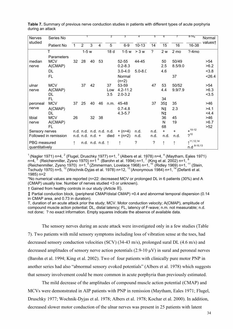
34
Table 7. Summary of previous nerve conduction studies in patients with different types of acute porphyria during an attack
Series No 1 2 3 4 5 6 7 8-14* Nerves studied
Patient No 1 2 3 4 5 6-9 10-13 14 15 16 16-38
Normal values†
T 1-5 w 18 d 1-5 w > 3 w ? 2 w 2 mo ?-4mo Parameters
MCV 32 28 40 53 52-55 44-45 50 50/49 >54 А(CMAP) 0.2-8.3 2.5 8.5/9.0 >6.2 DL 3.0-4.0 5.0-8.0 4.6 <3.8
median nerve
FL Normal (n=2)
37 <26.4
MCV 37 42 37 53-59 47 53 50/52 >54 А(CMAP) Low 4.2-11.2 4.4 9.9/7.9 >6.3 DL 3.5 2.0-3.2 <3.5
ulnar nerve
FL 34 MCV 37 25 40 46 n.m. 45-48 37 35‡ 35 >46 А(CMAP) 0.7-4.8 N‡ 2.3 >4.1
peroneal nerve
DL 4.3-5.7 N‡ <4.4 MCV 26 32 38 36 45 >46 А(CMAP) N 19 >6.7
tibial nerve
FL 68
>52 Sensory nerves n.d. n.d. n.d. n.d. n.d. + (n=4) n.d. n.d. + + +10-12 Followed in remission n.d. n.d. n.d. + died + (n=2) n.d. n.d. n.d. n.d. ?10
PBG measured quantitatively
↑ n.d. n.d. n.d. ↑ ↑ ? ? ↑ ↑ ↑11,12,14
n.d 8-10,13
1 (Nagler 1971) n=4, 2 (Flugel, Druschky 1977) n=1, 3 (Albers et al. 1978) n=4, 4 (Maytham, Eales 1971) n=4, 5 (Reichenmiller, Zysno 1970) n=1 6 (Barohn et al. 1994) n=1, 7 (King et al. 2002) n=1, 8
(Reichenmiller, Zysno 1970) n=1, 9 (Zimmerman, Lovelace 1968) n=1, 10 (Ridley 1969) n=1, 11 (Stein, Tschudy 1970) n=5, 12 (Wochnik-Dyjas et al. 1978) n=12, 13 (Anonymous 1984) n=1, 14 (Defanti et al. 1985) n=2 *No numerical values are reported (n=22: decreased MCV or prolonged DL in 6 patients (30%) and A (CMAP) usually low. Number of nerves studied <3 or unknown). † Gained from healthy controls in our study (Article II). ‡ Partial conduction block, (peripheral CMAP/distal CMAP) =0.4 and abnormal temporal dispersion (0.14 in CMAP area, and 0.73 in duration). T, duration of an acute attack prior the study; MCV. Motor conduction velocity; А(CMAP), amplitude of compound muscle action potential; DL, distal latency; FL, latency of F-wave; n.m. not measurable; n.d. not done; ? no exact information. Empty squares indicate the absence of available data.
The sensory nerves during an acute attack were investigated only in a few studies (Table
7). Two patients with mild sensory symptoms including loss of vibration sense at the toes, had
decreased sensory conduction velocities (SCV) (34-43 m/s), prolonged sural DL (4.6 m/s) and
decreased amplitudes of sensory nerve action potentials (2.9-10 μV) in sural and peroneal nerves
(Barohn et al. 1994; King et al. 2002). Two of four patients with clinically pure motor PNP in
another series had also “abnormal sensory evoked potentials” (Albers et al. 1978) which suggests
that sensory involvement could be more common in acute porphyria than previously estimated.
The mild decrease of the amplitudes of compound muscle action potential (CMAP) and
MCVs were demonstrated in AIP patients with PNP in remission (Maytham, Eales 1971; Flugel,
Druschky 1977; Wochnik-Dyjas et al. 1978; Albers et al. 1978; Kochar et al. 2000). In addition,
decreased slower motor conduction of the ulnar nerves was present in 25 patients with latent

35
AIP, including two patients who excreted normal amounts of porphyrin precursors (Mustajoki,
Seppalainen 1975). This might indicate that other factors besides porphyrin precursors may also
play a role in porphyric neuropathy.
2.6.3.2. Neuropathological studies in peripheral nerves
Neuropathological studies done from sural biopsies or autopsy material have demonstrated
demyelination in addition to axonal changes especially in short motor axons. The histological
changes of axonopathy consisted of axonal swelling, fragmentation and axonal loss (Baker,
Watson 1945; Cavanagh, Mellick 1965; Perlroth et al. 1966; Ridley et al. 1968; Sweeney et al.
1970; Wakayama et al. 1975; Anzil, Dozhic 1978; Suarez et al. 1997). Demyelination manifested
as swelling, beading and patchy irregularity of myelin sheath (Denny-Brown, Sciarra 1945;
Schwarz, Moulton 1954; Drury 1956; Gibson, Goldberg 1956; Hierons 1957; Tschudy 1968;
Stein, Tschudy 1970), the gaps of myelin at the nodes of Ranvier (Denny-Brown, Sciarra 1945),
perineural lymphocytic infiltration (Denny-Brown, Sciarra 1945; Gibson, Goldberg 1956;
Hierons 1957), and segmental remyelination (Hierons 1957). In many cases those findings co-
existed in the same nerve (Wakayama et al. 1975; Anzil, Dozhic 1978; Thorner et al. 1981;
Barohn et al. 1994). Some authors have suggested independent axonal and myelin damage
during an acute attack (Anzil, Dozhic 1978; Barohn et al. 1994), while others have proposed
either primarily axonal (Cavanagh, Mellick 1965; Di Trapani et al. 1984; Yamada et al. 1984;
Suarez et al. 1997) or myelin involvement (Denny-Brown, Sciarra 1945; Gibson, Goldberg 1956;
Hierons 1957) (Table 8). Perivascular exudation has been reported occasionally (Baker, Watson
1945; Denny-Brown, Sciarra 1945). The presence of chromatolysis in the spinal anterior horn
cells is considered as the result of antegrade axonal degeneration, better known as "the dying
back phenomenon" (Baker, Watson 1945; Gibson, Goldberg 1956; Yamada et al. 1984).
Table 8. Findings in peripheral nerve pathological studies in AIP patients with clinical PNP Demyelination
n=17
Axonopathy and demyelination n=13
Axonopathy n=9
Normal n=3
Motor and sensory nerves autopsy
Mason et al. 1933 Schwarz, Moulton 1954 Hierons 1957 Ten Eyck et al. 1961 Campbell 1963 Stein, Tschudy 1970
Baker, Watson 1945 Denny-Brown, Sciarra 1945 Gibson, Goldberg 1956 Yamada et al. 1984 Perlroth et al. 1966
Cavanagh, Mellick 1965 Sweeney et al. 1970 Suarez et al. 1997 Defanti et al. 1985
Hymans Van den Bergh et al. 1937 Löffler 1919 Hierons 1957
Sural nerve biopsy
Drury 1956 Anzil, Dozhic 1978 Wakayama et al. 1975 Thorner et al. 1981 Barohn et al. 1994
Trapani et al. 1984
In several reports of patients who died of an acute attack with PNP the autopsied

36
peripheral nerves were considered normal (Hymans Van den Bergh et al. 1937; Hierons 1957).
In contrast to the HMBS (-/-) mouse model in which chronic axonal PNP has developed
even in the absence of elevated ALA and PBG levels in urine (Meyer et al. 1998), the
homozygous patient with AIP have had evidence of demyelinating PNP, which was confirmed
both in the neurophysiological and neuropathological studies (Solis et al. 2004).
2.6.4 Pathogenesis of acute encephalopathy
2.6.4.1. Neuroimaging during an acute attack
Brain neuroimaging (MRI or CT) performed in 13 cases during an acute attack (Table 9) showed
multi-focal symmetrical lesions, which varied in their location (parietal-occipital or frontal-
temporal). They disappear completely or partially within a few months (King, Bragdon 1991;
Aggarwal et al. 1994; Black et al. 1995; Kupferschmidt et al. 1995; Garg et al. 1999; Susa et al.
1999; Utz et al. 2001; Celik et al. 2002; Yen et al. 2002; Engelhardt et al. 2004; Maramattom et
al. 2005; Wessels et al. 2005). These nonspecific findings correspond at best to vasogenic
oedema resembling posterior reversible encephalopathy syndrome (PRES) (Hinchey et al. 1996;
Casey et al. 2000). It is believed to be the result of transient failure of cerebral vascular
autoregulation causing leakage of fluid into the interstitium (Eichler et al. 2002).
In two cases of porphyric encephalopathy, diffuse cerebral vasospasm has been
documented by angiography (Black et al. 1995; Maramattom et al. 2005). It resulted in
irreversible ischemic lesions in the patients. No vasospasm could be detected by conventional or
MR-angiography in four additional cases (Celik et al. 2002; Yen et al. 2002; Engelhardt et al.
2004; Wessels et al. 2005). Ischemic foci described in patients with AIP (Lai et al. 1977;
Kupferschmidt et al. 1995) could be an outcome of irreversibly impaired microcirculation as
reported previously in other PRES patients (Casey et al. 2000).
In a patient with AIP diffusion weighted (DW) MRI imaging with apparent diffusion
coefficient (ADC) mapping supported PRES as a cause of porphyric encephalopathy (Yen et al.
2002). In contrast to the patients with PRES, location of foci varies in patients with AIP. The
appearance of foci in frontal and temporal areas could be explained by disruption of sympathetic
protection during an acute attack in the area supplied by carotid arteries (Beausang-Linder, Bill
1981; Sze 1996). Pre-neuroimaging blood pressure was usually increased (Table 9), which
suggests that PRES could be secondary to hypertension (Hinchey et al. 1996). It is currently
unknown, what causes vasogenic oedema during an acute attack. The absence of malignant
hypertension (>180/100 mm) in the majority of cases (Table 9) favours endothelial toxicity in
AIP, as it has been demonstrated in patients with eclampsia (Neal et al. 2004).

37
Table 9. Neuroimaging during an acute attack of AIP
Signs and symptoms Blood pressure
Na Main location of foci at MRI (T2)
DW CA/MRA/ Follow-up (d/w/mo after previous MRI)
1* Seizures, hallucinations, Babinski signs, PNP, AP
110 _170 70 120
128 mmol/L
Frontal, parietal (8 d)
C 10 d: Normal MRI
Seizures, confusion, PNP, AP n.d. n.d. No foci, diffuse cortical contrast enhancement
2
18 d later: expressive aphasia n.d. n.d. Parietal (18 d) Sc
7 w: Normal MRI
3* Seizures, amaurosis, PNP Normal n.d Occipital
Sc 6 mo: Mild residual MRI lesions; 14 d: CT normal
4 Seizures, amaurosis, visual anosognosia, PNP, AP, AD
170 100
n.d Occipital, right frontal
Sc 4 w: MRI minimal improvement, no clinical sequels
5 Seizures, apraxia, confusion, AP
n.d. 121 mEq/L
Parietal C Vasospasm (CA and MRA)
4 d: MRI parietal foci (contrast enhancement), normal MRA
6* Seizures, lethargy, PNP, AP, AD
135 75
94 mEq/L
Basal ganglia, thalamus, pons (35 d)
Sc, Brainstem
10 mo: Residual MRI lesions, clinical sequels (dysarthria)
7† Seizures, amaurosis, hallucinations, lethargy, AP
180 100
130 mEq/L
Occipital, parietal (1 d)
C, Sc Normal Doppler sonography
26 d: Normal MRI (FLAIR)
1 attack: Amaurosis, cognitive dysfunction, headache, AP, AD
180 100
n.d Occipital C, Sc Normal Doppler sonography
Died, brain autopsy normal
8†
2 attack: Seizures, amaurosis, AP, AD
170 110
n.d Frontal, parie-tal, occipital
C, Sc ↑(↑ADC) Normal (CA)
9 Seizures, stupor, hallucinations, PNP, AD
210 140
121 mEq/L
Frontal C Normal (MRA) 3 mo: Normal MRI, clinical sequels (PNP)
10† Epileptic state, hallucinations n.d. n.d Frontal, para-sagittal cortex
C ↑ Normal (MRA) 6 w: Residual MRI lesions, no clinical sequels
11 Seizures, hallucination, confusion, headache, AP, AD
110 _160 95 115
Low Frontal, temporal
Sc Normal Mild vasospasm (CA)
1 w: Residual MRI lesions
12 Seizures, amaurosis, lethargy, PNP, AP, AD
170 110
n.d. Frontal, occipital
C ↑ Normal (MRA) 10 d: Residual MRI lesions, clinical sequels (PNP)
1 (King, Bragdon 1991), 2 (Aggarwal et al. 1994), 3-4 (Kupferschmidt et al. 1995), 5 (Black et al. 1995), 6 (Susa et al. 1999), 7 (Utz et al. 2001), 8 (Yen et al. 2002) 9 (Celik et al. 2002), 10 (Engelhardt et al. 2004), 11 (Maramattom et al. 2005), 12 (Wessels et al. 2005). *Diagnosis of AIP was confirmed by low Erc-HMBS activity. †No information of elevated U-PBG during an acute attack DW, diffusion-weighted imaging; ADC, apparent diffusion coefficient; CA, cerebral angiography; MRA, MR-angiography; AP, abdominal pain; AD, autonomic dysfunction; PNP, peripheral neuropathy; C, cortical; Sc, subcortical; ↑ signal hyperintensity (MRI), ↓ hypodensive foci (CT)

38
Pontine myelinolysis was described in a patient with AIP after a rapid correction of
hyponatraemia (Susa et al. 1999). Diffuse demyelination has been revealed in the brain MRI of a
homozygous AIP patient in early infancy. The findings are compatible with those of
neurodegeneration (Solis et al. 2004) which underlines the difference in pathogenesis of AIP in
the heterozygous and homozygous states.
2.6.4.2. Brain autopsies
Brain autopsies, done in the patients with AIP who have died during an acute attack, have
revealed diffuse changes such as loss of neurons and chromatolysis in the brain cortex and/or in
the brainstem (Baker, Watson 1945; Schwarz, Moulton 1954; Gibson, Goldberg 1956; Hierons
1957; Stein, Tschudy 1970; Lai et al. 1977). The chromatolysis is an unspecific finding for acute
porphyria and reflects mainly neurodegeneration and axonal loss (McIlwain, Hoke 2005). In a
patient with severe SIADH (Suarez et al. 1997) and a few others (Stein, Tschudy 1970), axonal
loss could be demonstrated in the suprachiasmal and preoptic nuclei of hypothalamus.
Focal perivascular demyelination in the cerebral white matter or cerebellum has been
described in several patients (Baker, Watson 1945; Denny-Brown, Sciarra 1945; Gibson,
Goldberg 1956; Hierons, 1957; Ten Eyck et al. 1961). Demyelination in CNS has not been as
severe or extensive as in peripheral nerves. In one case, the patient had multiple lesions on the
brain MRI before her death, but her brain necropsy was macroscopically normal and on
microscopic examination mild gliosis was the only finding (Yen et al. 2002).
Focal lesions in brain autopsies have been rare. In one case with acute blindness, focal
lesions could be demonstrated in both occipital lobes (Lai et al. 1977). In a patient with severe
hypertension during an acute attack, a haemorrhagic lesion in the internal capsule has been
revealed (Ridley 1969). In a 14-year old boy, who died of an epileptic state during an acute
attack, multiple cortical ischemic lesions, subcortical haemorrhage and neuroglial and
endothelial proliferation could be demonstrated in the brain autopsy (Hierons 1957).
2.6.4.3. Electroencephalography
In EEG diffuse wave slowing has been identified in 39 patients during an acute attack (Goldberg
1959; Ridley 1969; Stein, Tschudy 1970; Aggarwal et al. 1994; Susa et al. 1999; Celik et al.
2002; Engelhardt et al. 2004). This finding is abnormal but not specific for acute porphyria and
commonly seen in other metabolic encephalopathies (Moe, Sprague 1994). Twenty-nine patients
have experienced also epileptic seizures, but no focal or generalised epileptic activity could be
demonstrated in 16 of them.

39
2.7. TREATMENT OF AN ACUTE ATTACK
2.7.1. Haem
Haem preparations were proposed for the treatment of acute attacks as early as 1978 (Watson et
al. 1978). Exogenous haem down-regulates ALAS in the liver (ALAS1) (Jover et al. 2000).
Haem infusions result in biochemical remission, i.e. decreased excretion of porphyrin precursors.
Biochemical remission lasts usually for a week, which is enough to abolish symptoms of an
acute attack, if precipitating factors are eliminated (Mustajoki, Nordmann 1993).
Haemin is isolated and purified from human red cell concentrates. In Europe and South
Africa, haemin is commercially available as haem arginate (Normosang®, Orphan Europe). In
Nothern America it is available as lyophylised haematin (Panhaematin®, Ovation
Pharmaceuticals Inc), which reconstituted with sterile water before infusion. In Russia, neither
haem arginate nor haematin are included in the algorithm of the standard treatment because of
their cost (Idel'son et al. 1992).
The only placebo-controlled series found insignificant benefit of haem arginate for the
analgesic requirement, pain score and duration of the hospital stay (Herrick et al. 1989).
However, the validity of the trial have been questioned (Mustajoki, Nordmann 1993) because of
the small number of the patients (11 patients treated with haem and 10 with a placebo), a high
proportion of the patients with PNP in this series (43%), delayed administration of preparation
(>2 days after admission) and difficulties in arranging a placebo, which resembles haem arginate.
In an open series of 22 patients (Mustajoki, Nordmann 1993), the patients treated with haem
arginate recovered more rapidly in comparison with those treated with glucose infusions in
earlier studies, however, this comparison is lacking the specificity (Goldberg 1959; Ridley 1969;
Stein, Tschudy 1970; Mustajoki, Koskelo 1976). Safety and efficiency of lyophylised haematin
was also supported in six open-labeled studies involving over 200 AIP patients (Anderson and
Collins 2006)
Haem arginate and lyophylised haematin are usually infused daily for four consequent
days but the longer course may be required if the symptoms are severe and progressing. The
therapy should be started without delay (Mustajoki, Nordmann 1993; Anderson and Collins
2006)
The reported side effects include mild coagulopathy (Simionatto et al. 1988),
trombophlebitis (Mustajoki, Nordmann 1993) and anaphylactic shock in one case (Daimon et al.
2001). Tolerance to haem preparations after repeated infusions has not been reported.

40
2.7.2. High carbohydrate
The treatment of acute attacks with high carbohydrate diet or infusions (300-500g/day)
was proposed in 1964, since dose dependent administration of glucose was shown to down-
regulate ALAS1 in rodents in vivo (Tschudy et al. 1964) and in vitro in chick embryo liver cell
culture (Doss et al. 1985). The underlying mechanism has been discovered recently: the glucose
down-regulates peroxisome-proliferator-activated receptor γ coactivator 1α (PGC-1α), a protein
which directly induces transcription of ALAS1 (Handschin et al. 2005). The use of glucose is
limited only to mild attacks (e.g. mild pain, no paresis, seizures or hyponatraemia) according to
the guidelines for the treatment of an acute attack in the U.S. and South Africa (Anderson et al.
2005; Hift, Meissner 2005) or if haem arginate or haematin are not available locally. If glucose
infusions do not result in clinical remission or if an acute attack is severe at the onset, haem
preparations should be used. In Finland and France all acute attacks severe enough to require
hospitalisation are treated with haem arginate (Mustajoki, Nordmann 1993). Mild or clearly
resolving attacks with minor pain or anxiety may be treated symptomatically (Elder and Hift
2001).
2.7.3. Elimination of precipitating factors and supportive treatment
During an acute attack, all potentially precipitating factors such as drugs and alcohol should be
eliminated, infections should be treated promptly and (parenteral) nutrition of patients should be
adequate to avoid fasting (Kauppinen 2005) Table 10. Treatment of an acute attack Signs and symptoms Medications I: Specific therapy Haem preparations (3mg/kg/d for 3-4 consequent days) II.Symptomatic therapy Abdominal and/or back pain Opiates iv/im bolus or in severe cases as infusion Tachycardia, arrhythmia Beta blockers Hypertension Beta blockers, clonidine, Constipation Lactulose Vomiting Phenothiazines*, butyrophenones*, lorazepam Urinary retention Urethral catheter Respiratory muscle paresis Mechanical ventilation Pneumonia Antibiotics* except sulphonamides Mental changes Insomnia and/or anxiety Most benzodiazepines* including zopiclone Hallucinations Phenothiazines*, droperidol, olanzapine Convulsions Correction of hyponatraemia and hypertension, diazepam.
Gabapentin, levetiracetam if prolonged treatment is needed Hyponatraemia >125 mmol/L: water restriction due to inappropriate
antidiuretic hormone syndrome (SIADH) ≤125 mmol/L or patients with seizures or unconscious: infusion of saline. Correction ≤12mmol/L/day. Be aware of pontine myelinolysis
* The most of medications in the group are well tolerated

41
Symptomatic therapy for pain, hypertension, nausea, and severe hyponatraemia is
commonly required (Crimlisk 1997; Johnson, Criddle 2004; Anderson et al. 2005). If a
hypertensive crisis develops it should be treated as regular (Patel, Mitsnefes 2005). Epileptic
seizures may be treated with intravenous diazepam, gabapentin or levetiracetam (Crimlisk 1997;
Zadra et al. 1998; Anderson et al. 2005; Zaatreh 2005).
2.7.4. Future therapies
Recombinant human HMBS preparation has been shown to lower plasma PBG levels in
symptom-free patients with increased excretion of urinary PBG (Sardh et al. 2003). However, no
decrease in plasma ALA levels could be demonstrated in these patients. The similar biochemical
effect of rh-HMBS therapy was demonstrated in the HMBS(-/-) mouse model (Johansson et al.
2003a). The efficacy evaluation of rh-HMBS-enzyme replacement therapy for acute attacks of
AIP is currently ongoing (http://www.zymenex.com).
Both adenoviral mediated and non-viral HMBS gene transfers corrected the metabolic
defect in the cell lines of AIP patients’ and HMBS (-/-) mice (Johansson et al. 2002; Johansson
et al. 2003b; Johansson et al. 2004a). Only virus-mediated DNA transfer into the liver was
successful and corrected metabolic defect in HMBS (-/-) mice at least for a month
(Johansson et al. 2004a; Johansson et al. 2004b). Adenoviral cytotoxicity especially for
hepatocytes, the immunological response to viral antigens and only transient transduction of the
therapeutic gene limit potential applications of this method (Tarner et al. 2004).
2.8. PREVENTION OF ACUTE ATTACKS
Early diagnosis with mutation screening among a patient’s relatives and information about
precipitating factors can prevent further attacks among patients with acute porphyria
(Kauppinen, Mustajoki 1992). In remission, patients may tolerate medications classified as
potentially safe and the small amounts of alcohol but if porphyric symptoms appear their use
should be restricted (Kauppinen, Mustajoki 1992; Doss et al. 2000).
The exogenous hormonal therapy such as contraceptive pills was introduced for the
profilactical treatment of recurrent acute attacks more than 40 years ago (Perlroth et al. 1966) but
contraceptive pills may provoke acute attacks in 5% of women with AIP (Kauppinen and
Mustajoki 1992). GnRH analogues are not porphyrinogenic and prevent attacks in a half of the
women with cyclical symptoms (Anderson et al. 1981; Herrick et al. 1990; De Block et al.
1999), The responders more frequently had regular menstrual cycles and pronounce decrease in

42
the oestradiol level after the treatment with GnRH analogues (Anderson et al. 1990; Herrick et
al. 1990; De Block et al. 1999) when compared to the women, who were non-responders or
responded only to a high dose of the preparation (Anderson et al. 1990; Herrick et al. 1990;
Soonawalla et al. 2004; Hift, Meissner 2005; Pischik, Kauppinen 2006). This therapy causing
menopause decreased excretion of porphyrin precursors to 60% of the previous values in both
groups (Anderson et al. 1990; Herrick et al. 1990; De Block et al. 1999). Cyclical attacks do not
predict attacks during pregnancy, and pregnancy nowadays seldom precipitates acute attacks
(Kauppinen, Mustajoki 1992; Andersson et al. 2003). Haem preparations have been used during
pregnancy without complications (Gorchein, Webber 1987; Hift, Meissner 2005; Pischik,
Kauppinen 2006). Weekly haem infusions may alleviate the severity of recurrent attacks
(Kauppinen 2005). The liver transplantation can be an option for a severely affected patient with
severe recurrent attacks, if the standard treatment was unsuccessful (Soonawalla et al. 2004), but
to date no long-term follow-up exists.
2.9. PROGNOSIS
Before the glucose and haematin treatment of an acute attack has been introduced, the mortality
rate was very high (Goldberg 1959; Ridley 1969; Mustajoki, Koskelo 1976). In Waldenström
series it was 66% among symptomatic patients (Waldenström 1937).
Currently, because of the improved diagnostics, the early and effective treatment with
haem preparations and the prevention of acute attacks (Kauppinen, Mustajoki 1992) the
mortality has decreased to 5-20% of the cases including the cases which were not diagnosed
previously or in the early phase of an acute attack (Kauppinen, Mustajoki 1992; Jeans et al.
1996; Hift, Meissner 2005).

43
3. AIMS OF THE PRESENT STUDY
At the beginning of this study in 1995 a few cases only of acute porphyria have been suspected
clinically over the previous decades in the north western part of Russia including St Petersburg
area. No systematic clinical and genetical studies have been performed. At that time, very little
was known about the mechanisms underlying the neurological manifestations during an acute
porphyric attack.
The aims of this study:
1. To identify patients with AIP admitted to the neurological wards in North Western Russia
2. To study neurological manifestations and a natural course of an acute attack.
3. To study acute polyneuropathy and central nervous system involvement in patients with AIP
using electrophysiological techniques and neuroimaging.
4. To identify gene defects resulting in AIP among Russian patients.
5. To characterise the outcome of mutations at mRNA and protein level.
6. To investigate a genotype-phenotype correlation in AIP.

44
4. MATERIALS AND METHODS
4.1. PATIENTS
During 1995–2006, 108 patients with signs and symptoms suggestive of acute porphyria were
studied prospectively, both clinically and biochemically to exclude or diagnose acute porphyria,
when they were admitted to the neurological wards in North Western Russia because of
acute/subacute muscle weakness, seizures or confusion. The patients were selected from the City
Hospital #2 (the main place of this study), Regional St Petersburg Hospital, Departments of
Neurology of the Military Medical Academy and Postgraduate Medical Academy, three local
hospitals in St Petersburg and Pskov Regional Hospital. Most of the patients were admitted as
acute cases to those hospitals. If acute PNP or other signs suggestive of porphyria were found, a
neuromuscular specialist from the City Hospital #2 was informed and continued the subsequent
examination. Data from the medical history was obtained from the hospital records and by
interviewing each patient. The patients were unrelated and pedigrees were traced three to four
generations back. Twelve Russian patients were diagnosed with AIP.
In addition the clinical, biochemical and genetic data of 291 Finnish AIP patients
diagnosed in 1966-2006 was analysed. Of the Finnish patients, two individuals were diagnosed
in 2003-2006 at the Central University Hospital of Helsinki and had neurological manifestations.
Their detailed clinical and neuroradiological data was also included in the study.
The diagnosis of symptomatic AIP was confirmed by biochemical analyses of haem
precursors accompanied by typical clinical manifestations (n=14). DNA testing was performed
later at the symptom-free phase (n=13). After diagnosis of AIP in index case was confirmed, the
family members were enrolled in the study group in 2000-2005.
An acute attack was defined as acutely developed typical symptoms of porphyria such as
abdominal pain and/or autonomic dysfunction in patients with at least four-fold increase of
urinary PBG excretion (Mustajoki, Nordmann 1993). Acute PNP manifested as diffuse muscle
weakness, symmetrically distributed hyporeflexia and sensory loss (McLeod 1995), and was
attributed to AIP only if evolved during an acute attack.
Study protocol followed principles outlined in the Declaration of Helsinki and ethical
issues were approved by the City Hospital #2, St Petersburg, Russia and Central University
Hospital of Helsinki, Finland.

45
4.2. METHODS
4.2.1. Neurological examination
Neurological examination included quantitative assessment of muscle strength from 0 to 5 in
upper arm abductors, elbow flexors, wrist extensors, hip flexors, knee extensors and foot dorsal
flexors according to Medical Research Council (MRC, 1943). Then a MRC sum score was
calculated with a maximal value of 60 (Kleyweg et al. 1991). Reflexes, tone, sensation, co-
ordination and mental state were evaluated. The intensity of pain was assessed using linear
Visual Analogue Scale (VAS, 0 to 10 cm, Jensen, Karoly 1992).
4.2.2. Biochemical analysis
During an acute attack the qualitative Watson- Schwartz test detecting urinary PBG (Watson,
Schwartz 1941), quantitative measurement of urinary ALA (Mauzerall, Granick 1956) and/or
coproporphyrin (Rimington 1958) were performed in Russia. For the ALA measurement,
chromatographic columns were manually filled with activated charcoal (Mauzerall, Granick
1956; Kuznetsova et al. 1981).
In remission, urinary ALA and PBG were measured using Bio-Rad columns (Bio-Rad
Laboratories, USA)(Mauzerall, Granick 1956), and urinary and faecal porphyrins using the high
performance liquid chromatography (HPLC, Varian 9100, Varian Inc., USA)(Li et al. 1986) in
Porphyria Research Laboratory in Finland. Plasma porphyrin spectrum was assayed using
spectrofluorometry (Poh-Fitzpatrick, Lamola 1976). The erythrocyte HMBS activity was studied
using spectrophotometry by measuring the conversion of PBG to uroporphyrin (Ford et al.
1980).
4.2.3. Neurophysiology The motor nerve conduction and F-wave studies were performed using standard techniques and
surface recording (Viking IV, Nicolet, USA)(Livenson, Ma 1992). The sensory response was
recorded antidromically for sural and orthodromically for ulnar and median nerves using plate-
shaped electrodes. The skin temperature was maintained at > 320. Recorder values were
considered normal if they were within the mean±2SD of those obtained from control subjects.
Demyelination was considered if MCV was <70% of the low normal level (LNL), or DL or FL
were ≥150% of the upper normal level (UNL) (Meulstee, van der Meche 1995). Axonal
neuropathy was considered if the amplitude of CMAP was ≤50% of LNL in two or more nerves
(Rees et al. 1995). If the signs of axonal and demyelinating neuropathy co-existed mixed PNP

46
was diagnosed. Electromyography was performed with concentric needle electrodes. Details of
individuals examined are provided in article II.
4.2.4. Neuroimaging
Computed tomography (CT) of the brain was performed using a single-detector CT (Somaton
AR.T., Siemens, Germany) in Russia and multidetector CT (LightSpeed 8, GE Medical Systems,
Wis, USA) in Finland with a contiguous 5 mm slice thickness.
Magnetic Resonance Imaging (MRI) was performed using standard head coil (T1 and T2
weighted, Magneton Impact, Siemens in Russia and T1, T2, fluid-attenuated inversion recovery
(FLAIR) and diffusion weighted (DW) imaging, Siemens Sonata in Finland).
4.2.5. DNA-analysis
4.2.5.1. Mutation screening
Genomic DNA was extracted from leukocytes using the QIAamp DNA Blood mini Kit (Qiagen
Inc, Valencia, CA, USA) (Higuchi 1989) and amplified using polymerase chain reaction (PCR)
(Mullis, Faloona 1987). The primers and PCR conditions are described in the article III. PCR
fragments were purified using QIAquick PCR purification Kit (Qiagen Inc, Valencia, CA, USA;
http://www1.qiagen.com) before sequencing (Sanger et al. 1977) with Amplicycle Sequencing
Kit (Perkin Elmer, Wellesley, MA, USA), or when an ABI3730 Automatic DNA Sequencer was
used with Sequencing Analysis 5.0 BigDye Terminator version 3.1 Cycle Sequencing Kit
(Applied Biosystems, Foster City, CA, USA). Direct sequencing of the HMBS gene was
performed for all exons and exon-intron boundary regions, except for erythroid-specific exon 2,
in both directions. The mutation was confirmed by repeated analyses in the presence of negative
and positive controls if available. Digestion with restriction enzymes was used as an additional
method to confirm the mutation whenever the appropriate enzyme was commercially available
(The New England Biolabs, Beverly, MA, USA).
4.2.5.2. mRNA isolation, cDNA synthesis and sequencing
To verify the transcripts of the novel mutations at mRNA level, total mRNA was extracted from
patients’ Epstein-Barr virus transfected lymphoblastoid cell lines or fibroblasts (Chirgwin et al.
1979). Complementary DNA was synthesised from 5 μg of total RNA using Superscript II Rnase
H-Reverse Transcriptase (Gibco BRL Life Technologies, Gaithersburg, MD, USA) and random
hexamers and directly sequenced as described above.

47
4.2.5.3. Eukaryotic expression of HMBS mutations
The expression of a mutant polypeptide was studied in COS-1 eukaryotic cell lines using SVpoly
vector (Stacey and Schnieke, 1990). The mutations were inserted in the human HMBS-SV-poly
(Mustajoki, et al. 2000) using Chameleon double-stranded site-directed mutagenesis kit
(Stratagene Cloning System, La Jolla, CA, USA) or by ligating mutated RT-PCR fragments into
the corresponding sites of the HMBS-SV-poly using T4 DNA ligase (New England Biolabs,
Beverly, MA, USA). The details of mutagenesis primers and plasmid construction are provided
in article III. Forty eight hours post-transfection crude cell extracts of plasmid-containing
cultures were lysed with 300 μL of incubation buffer 150 mM Tris-HCl, 1 mM EDTA, 1% [v/v]
Tween 20 and sonicated three times for 10 sec. The supernatant was assayed for HMBS activity
(Ford et al. 1980). The enzyme activities obtained were compared to that of the background level
from COS-1 cells transfected with SVpoly alone.
4.2.6. Statistical analysis
Continuous variables were expressed as the mean ± standard deviation (SD) and analysed with
Mann-Whitney U-test when two groups were compared or Kruskal-Wallis one-way ANOVA
when more than two groups were compared simultaneously. The mean ± standard error (SEM)
was used for a genotype-phenotype comparison.
To develop a scale for the severity of an acute attack regression analysis with maximum group
discrimination as optimization criteria (Kendall and Stuart 1968) was employed to evaluate
association with outcome variables (the long-term prognosis of a patient) and covariates
(symptoms and signs of an acute attack). The initial values of covariates were normalised from 0
to 1, and an equation of α2 =1 resulted in one solution only for each regression coefficient (α).
Thereafter the scores were adjusted according to αi from 1 to 6.
For a genotype-phenotype comparison logistic regression with the maximum likelihood
estimation as optimization criteria was employed to evaluate the association between
dichotomous outcome variables (the occurrence of acute attacks) and covariates (the mutation,
gender, age at the time of diagnosis, and the results of biochemical tests). Statistical calculations
were performed with SPSS version 10.04 and NCSS 2000

48
5. RESULTS
5.1. GENERAL CHARACTERISTICS OF THE PATIENTS
(Articles I, II and additional data)
5.1.1. Screening of porphyrin metabolites in the selected patients Porphyrin metabolites were studied during an acute phase of 108 Russian patients, who had signs
and symptoms suggestive of acute porphyria. The levels of urinary porphyrins or their precursors
were normal in 85 cases (79%) (Figure 8).
Of the 23 patients who had a 1.5-10 fold excretion of urinary porphyrins or their
precursors 11 individuals were excluded. Exclusion criteria were as follows: less than two-fold
increase in urinary PBG assayed by the qualitative method, ALA and coproporphyrin, and
neurological symptoms, which could be attributed to other diseases (Figure 8). In seven of them
porphyrin metabolism was normal by the time of discharge. In two cases, acute porphyria could
not be excluded during an acute phase because of constant coproporphyrinuria (two to three-
fold). During the six months follow-up urinary excretion of coproporphyrin normalised and other
diagnoses could be established (Figure 8). In summary, of the selected patient group attending
acute neurological wards, 12 cases (11 %) were diagnosed with acute porphyria indicating that
screening for porphyrin metabolites, especially urinary PBG is essential.
5.1.2. Characteristics of the patients with AIP
All Russian patients with acute porphyria were diagnosed to have AIP (Table 10). They were
female patients and had signs of autonomic neuropathy and mental symptoms before developing
paresis or seizures. By the time of admission, ten patients experienced severe acute PNP, one
patient had generalised seizures and mild fever, and the other had hypertensive crisis with
vertigo and mild cognitive impairment. PNP was recurrent with 13 and 20 months interval in two
cases.
None of the patients were diagnosed with AIP before they developed signs of
neurological deficits. Of the 12 patients, seven experienced numerous short episodes of mental
lability, abdominal pain, palpitations and constipation, which varied from mild to moderate. This
symptomatic but undiagnosed period lasted up to 19 years (mean four years). Before acute
porphyria was confirmed, the patients were misdiagnosed with various diseases (Table 12).

49
108 patients with signs and symptoms suggestive of acute porphyria Inclusion criteria: acute/subacute PNP and/or seizures and/or acute/subacute cognitive impairment + autonomic dysfunction and/or abdominal/back pain Urinary PBG and coproporphyrins measured during acute symptoms:
Elevated Normal
23 patients 85 patients Patients with Guillain-Barré syndrome (GBS), who manifested with pain >5 VAS and /or dysautonomia axonal PNP according to the nerve conduction studies PNP of other origin (alcoholic, non specified) Acute thallium intoxication (PNP, autonomic dysfunction, abdominal pain, coma) Polymyositis Systemic lupus erythematosus (mild PNP, dysautonomic features and myalgia) Hyperthyroidism (+ PNP) Botulism (vomiting, acute flaccid tetraparesis, blurred vision, diplopia, bulbar signs) Multiple compression neuropathy Epilepsy Somatoform disorder Schizophrenia The diagnosis was not verified Not followed
N 34
45121111
15536
10
Urinary excretion of ALA and PBG, urinary and faecal porphyrins, plasma porphyrin spectrum, and Erc-HMBS in remission
Abnormal Normal* n=11
11 patients (5 men, 6 women)
The cause of porphyrinuria Neurological manifestation N
12 patients with acute porphyria
(12 women)
12 patients with AIP
Primary biliary cirrhosis Autoimmune hepatitis. Hemolytic anaemia. Systemic vasculitis. Acute hepatitis B Alcohol intoxication + and hepatitis C Alcohol abuse and liver cirrhosis +suprarenal insufficiency A drug-induced hepatitis in a patient with schizophrenia The cause of porphyrinuria and neurological manifestations remained unknown
Chronic autonomic dysfunction, acute PNP Autonomic dysfunction, subacute PNP, abdominal pain Recurrent PNP, dysautonomia, pain Subacute PNP, myalgia Acute lumbosacral plexopathy and abdominal pain due to trauma Acute PNP, encephalopathy, rhabdomyolysis Subacute/recurrent PNP, autonomic dysfunction, mental symptoms, muscle pain Chronic psychosis, muscle pain and weakness Subacute PNP, encephalopathy, psychosis (n=1), subacute PNP with pain (n=1), acute idiopathic PNP + dysautonomia (GBS?)
1 1 1 1 1 1 1 1 3
* Except for mild elevation of urinary coproporphyrin in two cases (primary biliary cirrhosis and hemolytic anaemia) Figure 8. Graphic presentation of the patients screened for urinary porphyrins and their precursors to exclude the diagnosis of acute porphyria during the period of 1995-2005 years

50
Table 11. Clinical characteristics of AIP patients Pt* Sex Age at the
onset of porphyria (years)
Number of attacks before the diagnosis
Age at the time of the diagnosis (years)
Duration of autonomic impairment and mental symptoms before the appearance of neurological manifestations (d)
Neurological manifestations at presentation
1 F 35 1 36 6/14† PNP¶ 2 F 23 9 34 10 PNP 3# F 36 - 36 21 PNP¶ 4# F 20 7 22 29 PNP 5 F 17§ >20 29 15 PNP¶ 6 F 57 - 57 7 PNP¶ 7 F 18 - 18 18 PNP¶ 8 F 23 - 23 3 Seizures and lethargy 9 F 23 4 26 5 PNP 10 F 53§ - 53 15 PNP¶ 11 F 24 3 24 12‡/10 PNP 12 F 25 3 25 3 Vertigo 13 F 17 >10 36 10 Rhabdomyolysis, SIADH 14 F 23 1 24 6 Seizures and lethargy Mean 28 32 11.5 F, female; PNP, peripheral neuropathy *Cases 1-12 were diagnosed in Russia, cases 13 and 14, in Finland. † Diagnosis was confirmed during the patient’s second attack with PNP ‡ Although the diagnosis of AIP was confirmed during the patient’s first attack with PNP, the patient experienced a second attack with PNP § Family history of a deceased relative resembling acute porphyria ¶ PNP confirmed by nerve conduction studies # Died during an acute attack Table 12. Misdiagnoses in patients with AIP before the diagnosis of acute porphyria was suggested I. Before the appearance of neurological deficits A. Psychiatric diseases Hysteria Schizophrenia B. Gastroenterological and nephrological conditions Acute gastritis Acute hepatitis Irritable bowel syndrome Acute pyelonephritis Acute interstitial nephritis Bartter’s syndrome Primary SIADH C. Surgical emergencies Bowel obstruction
Paralytic ileus Renal colic Acute cholecystitis
D. Miscellaneous Hypertensive crisis
Acute poisoning Acute adnexitis Familial Mediterranean fever Hyperthyroidism
II . After the neurological deficits manifested Guillain-Barré syndrome Acute polymyositis Hypokalaemic paralysis Acute viral/toxic encephalitis Epilepsy Hypertensive encephalopathy

51
Family 1
Family 2 Family 3 Family 4 Family 5
Family 6 Family 7 Family 8 Family 9

52
Family 10 Family 11 Family 12
Family 13 Family 14
Figure 9. AIP pedigrees. Black circle and square = affected female and male with clinically symptomatic disease. Grey circle/square = asymptomatic female and male carrying the affected gene. Open circle/square =clinically unaffected family members. M+ = mutation-positive; M- = mutation-negative. Open circle/square without M -=not tested with DNA-analysis, but no suggestive information of AIP according to a medical history. ? = acute porphyria is possible according to a medical history. Arrow = proband

53
5.2. BIOCHEMICAL ANALYSES
(Article I, II and additional data)
5.2.1. Porphyrins and their precursors
An acute attack was diagnosed using a qualitative test of PBG in all Russian patients, but
increased excretion was confirmed by a quantitative test in remission performed in Finland
(Tables 13 and 14). Urinary ALA and coproporphyrin were increased during an acute attack.
The quantitative measurements for urinary coproporphyrins and ALA are not standardised in
North Western Russia, and thus, the levels of ALA remained at the low range compared to
those in Finland due to methodological differences. The quantitative analysis for urinary
PBG was not available in Russia. Table 13. Biochemical findings of urinary porphyrins and their precursors during an acute attack Pt ALA PBG Coproporphyrin
Day of the measurement from the onset of an acute attack
<9.9 μmol/L*
qualitatively -/+/++/+++‡
< 64 nmol/g creat#
<320 nmol/L#
1 17 19.6 +++ 282 31 n.d. +++ 216 2 11 n.d. +++ 3000 3 46 n.d. +++ n.d. 4 41 22 +++ 157 5 25 60 +++ 879 6 11 n.d. +++ 360 28 10.3 +++ 94 7 25 n.d. +++ 583 31 n.d. +++ 3262 8 8 n.d. +++ n.d. 13 10.3 +++ 110.4 9 8 26.1 +++ 183 10 50 42 +++ 431 57 101 +++ 141 11 7 n.d. +++ 1154 12 6 14.4 +++ 24.5 Performed in Finland <34
μmol/L† <9 μmol/L§ <230 nmol /L**
13 16 n.d. 790 730 23 340 630 n.d. 14 12 310 310 170 *Chromatographic columns were manually filled with activated charcoal (Kuznetsova et al. 1981). †Measured using Bio-Rad columns (Bio-Rad Laboratories, USA) (Mauzerall, Granick 1956). ‡(Watson, Schwartz 1941). §Measured using Bio-Rad columns (Mauzerall, Granick 1956). #(Rimington 1958). **(HPLC, Li et al. 1986).
In remission, the mean urinary level of PBG was 40-fold compared to the upper normal
level, ALA 6-fold, coproporphyrin 3-fold and uroporphyrin 70-fold, respectively (Table 14).
Faecal excretions of porphyrins were within the normal range but plasma porphyrin spectrum
was usually positive. Two patients (cases 3 and 4) died during an acute attack and no

54
measurements of PBG or ALA in remission could be done. The erythrocyte HMBS activity
was decreased in the children of case 3 (40 and 39 pmol uroporphyrin/mg prot) and the
mutation, which was found in the HMBS gene, confirmed the diagnosis of AIP in this family.
In case 4 excretion of PBG, ALA and coproporphyrin were increased even after 41 days
from the onset of an acute attack. The diagnosis of AIP in her case could not be confirmed
because the enzyme activity was not measured and her relatives were not available. Severe
symptoms of acute porphyria, absence of the skin lesions and markedly elevated urinary
PBG excretion even two weeks from the onset of an acute attack made the diagnosis of AIP
the most likely.
Table 14. Biochemical findings of abnormal haem metabolism among patients with AIP in remission Erythrocyte*
Urine* Faeces* Plasma
Pt
Erc-HMBS (55-100 uroporphyrin/mg prot)
ALA (<34 μmol/L)
PBG (<9 μmol/L)
Copro-porphyrin(<230 nmol /L)
Uro-porphyrin(<36 nmol /L)
Copro-porphyrin (<100 nmol/g)
Proto-porphyrin(<130 nmol/g)
Emission max 619 nm
1 37 124 293 287 2904 n.d. n.d. + 2 39 122 205 701 1111 18 22 + 5 58 326 523 812 5485 n.d. n.d. + 6 42 173 297 915 1573 n.d. n.d. + 7 32 511 632 258 10955 11 22 + 8 45 148 258 977 1498 26 35 + 9 42 209 795 986 2754 n.d. n.d. + 10 47 97 187 480 1198 n.d. n.d. + 11 53 305 1012 526 3093 34 18 + 12 43 110 114 246 1418 n.d. n.d. + 13 77-81† 150 300 270 1900 1 2 + 14 39 130 250 130 1200 n.d. n.d. - Mean 46 200 406 549 2924 18 20 *Based on one analysis in each patient. †Range based on three measurements. n.d., not done
5.2.2. Other biochemical findings
The elevation of serum liver transaminases was the most common finding (Table15). If a
patient had paresis or seizures (i.e. a severe attack) hepatopathy was commonly present.
Leukocytosis, elevation of serum creatinine and hyponatraemia were also frequent during an
acute attack (Table 15). CSF analyses were within the normal range in 13 patients examined
during an acute attack.
During the subsequent acute attacks in eight patients the elevation of transaminases
was uncommon since they were treated in the early phase of an acute attack. In remission,
mild elevation of transaminases persisted in one patient only who had chronic hepatitis C.
During the follow-up serum creatinine values were constantly elevated in one patient
with monthly recurrent attacks (up to 136 μmol/L, Table 15) and in another patient with

55
interstitial nephritis. Her renal failure was multifactorial (recurrent acute attacks,
hypertensive crises during an acute attack and use of gentamycin). Currently she needs
weekly haemodialysis similar to her sister. In her sister’s case renal insufficiency developed
after an acute attack three years before the diagnosis of AIP was established by a pedigree-
analysis and DNA testing. Their mother is a symptom-free patient with AIP. Father had
several hypertensive crises and died of myocardial infarction at the age of 47. Thus, the
siblings have probably inherited other, currently unknown factors, which made patients
vulnerable for hypertension and renal failure. Table 15. Biochemical findings among AIP patients with neurological manifestations during an acute attack Abnormal values of 16
attacks* %
attacksRange of
abnormal values
Normal range
I. Blood smear Leucocytosis
(with no signs of infection) 7 44 10.6-19.2 > 10x106/L
Hypochromic anaemia, Hb 5 31 9.2-11.9 g/dL 12-14.5 g/dL II. Serum biochemistry ALT 16 100 46-269 U/L < 40 U/L AST 14 88 41-601† U/L < 35 U/L Creatinine 5 31 113-152 µmol/L 55-110 µmol/L Urea 5 31 8.9-18.3 mmol/L 1.7-8.3 mmol/L Bilirubin 3 19 42-70 µmol/L 8.5-20.5 µmol/L Creatine kinase 5‡ 31 254-21806 U/L <190 U/L Sodium 9 56 low 8 50 108-129 mmol/L 135-155 mmol/L high 1 6 156 mmol/L 135-155 mmol/L Potassium 5 31 low 4 25 2.6-3.4 mmol/L 3.5-5.6 mmol/L high 1 6 5.8 mmol/L 3.5-5.6 mmol/L Calcium ionised 1 6 0.88 mmol/L 1.05-1.3 mmol/L Creatine glomerular filtration 3 19 29-77 mL/min 100-160 mL/min III. Urinalysis Leucocytes 6 38 negative Erythrocytes 3 19 negative * Here and below 16 acute attacks are analysed, since two of the Russian patients experienced acute attacks with severe neurological manifestations twice † Range 41-103 U/L, if a patient with rhabdomyolysis is excluded ‡ Not measured in four acute attacks, of those one patient had severe myalgia
5.3. CLINICAL MANIFESTATIONS DURING AN ACUTE ATTACK
(Articles I,II and additional data)
5.3.1 A case report of acute porphyria with neurological symptoms
A 26-year-old Russian woman (case 5) who was admitted to a neurological ward because of
acute sensorimotor PNP accompanied by abdominal and back pain, tachycardia,
constipation, hallucinations and depression (Pischik, Kauppinen 2006). She had had

56
recurrent attacks of severe abdominal pain in premenstrum during two years, which started
after her first delivery. She was treated as a hysteric patient receiving anti-depressive drugs
and tranquilizers with no effect.
The acute attack started 27 days before the admission to the neurological ward with
abdominal and back pain, constipation and tachycardia. Surgical aetiology was excluded.
Twelve days later muscle weakness manifested as monoparesis in the right arm, mainly as
radial nerve palsy, but pareses became diffuse within a week. By the time of admission, her
muscle weakness was symmetrical affecting both the upper and lower extremities (MRC
sum-score 40). Facial diplegia, hyporeflexia and neuropathic sensory loss were evident.
The blood tests demonstrated mild elevation of serum transaminases and creatinine
levels. Her urine was pink and became dark when exposed to the light. Haem arginate was
started for four days together with tramadol, chlorpromazine and propranolol. Her pain and
autonomic dysfunction were alleviated within six days. A clear regress of muscle weakness
began a few days later. Four months later her muscle strength was normal.
5.3.2. Pain during an acute attack
Abdominal pain was common and severe lasting usually for a few weeks (Table 16). All
patients with acute PNP experienced abdominal pain. One third of the patients experienced
pain in the back, which was less severe than abdominal pain.
Table 16. Pain scoring during an acute attack
Type of pain 16 % attacks
VAS (0-10)
Abdominal pain*† 14 88 9.4±0.9 Pain in the back 5 31 7.8±1.7 Pain in the extremities 11 67 9.1±1.1 Myalgia‡ 7 44 9.2±1.0 Dysesthetic 3 19 9.7±0.6 Arthralgia* 1 6 7 Headache 4 25 8.0±1.4 Migraine-like 1 6 10 Non-migraine-like§ 3 19 7.3±0.6 *First symptom during an acute attack. †In four cases laparotomy was performed. ‡Including two patients with mild to moderate rhabdomyolysis §Associated with moderate hypertension in three cases.
Headache was less common but severe when it appeared. In one case, it was the
major symptom of pain, which appeared together with abdominal pain in the majority of her
acute attacks and met the criteria for migraine (Nappi et al. 1989). Pain in the limbs was a
common symptom manifesting as severe myalgia-type, deep and burning sensation which

57
lasted 1–2 weeks after the onset of muscle weakness (Figure 12). Opiates relieved pain and
were used for 4 to 11 days. In four of our patients, laparotomy was performed to exclude
abdominal crisis.
5.3.3. Autonomic dysfunction
Autonomic symptoms varied and developed before but occasionally simultaneously with
muscle weakness or seizures (Table 17). Based on the 16 acute attacks analysed, constipation
preceded abdominal pain. The tachycardia was the most common sign of autonomic
neuropathy. It was often accompanied by systolic hypertension, constipation and abdominal
pain. In case 4 supraventricular arrhythmia proceeded to a cardiac arrest. Vomiting lasted 1-2
days and was accompanied by abdominal pain except one case. In case 8 autonomic
symptoms such as hypotension and constipation were very mild. No intestinal dilatation
could be found in four patients, who underwent diagnostic laparotomy.
Table 17. Clinical manifestations caused by autonomic dysfunction during an acute attack Signs and symptoms 16 %
attacks Pain in abdomen 14 88 Tachycardia ≥100/min 15 94 Arrhythmia 2 13 Supraventricular tachycardia or fibrillation 1 6 Ventricular tachycardia or fibrillation 0 0 Cardiac arrest 1 6 Systolic hypertension >145 mm Hg 12 75 >180 mm Hg 4 25 Diastolic hypertension >95 mm Hg 9 56 Hypotension <80/<50 mm Hg 3 19 Constipation 14 88 Vomiting 6 38 Pollacisuria 2 13 Urinary retention 5 31 Excessive sweating 1 6
5.3.4. Peripheral neuropathy
Acute motor polyneuropathy was the most common neurological sign (Table 18). Muscle
weakness accompanied by rapid atrophy was usually severe (Table 18) affecting both arms
and legs. It was symmetrical at the onset of weakness except case 5, where asymmetrical
weakness in the arms preceded severe symmetrical polyneuropathy. The presence of tendon
reflexes varied: absent in eight attacks and reduced in four, but in three of them ankle jerks
were preserved. Pattern of muscle weakness varied in each attack including intra-individual
variation in patients with recurrent PNP, but usually proximal muscles in the legs were

58
affected more severely than distal. In the upper limbs no difference could be observed.
Respiratory failure developed due to paresis of respiratory muscles, which required
prolonged mechanical ventilation for 3 to 56 days.
In approximately 50% of acute attacks complain of numbness preceded sensory
loss. One patient complained of numbness and pinprick sensation, but sensory deficits were
not detected. Combination of PNP and focal CNS involvement was observed in half of the
cases.
Table 18. Manifestations and scaling of peripheral neuropathy during an acute attack Signs and symptoms No of
attacks % from attacks with PNP
% from all acute attacks
Motor polyneuropathy Severe (MRC-sum-score 0-25) Moderate (MRC-sum-score 26-50) Mild (MRC-sum-score > 50)
13 10 2 1
100 78 15
8
81 62 13 6
Respiratory failure 10 78 62 Mechanical ventilation 8 62 50 Cranial neuropathy 6 46 38 Bulbar palsy 4 31 25 Bilateral facial nerve pareses 4 31 25 Oculomotor nerve paresis 2 15 13 Hypalgesia with glove-and-stocking distribution 6 46 38 + vibration sense loss 4 31 25 Hypo/hyperalgesia with ”old-fashioned bathing costume ” distribution
2 15 13
Areas of paresthesia or dysesthesia and numbness 8 62 50 Total number of attacks 16 13 16
5.3.5. Rhabdomyolysis
5.3.5.1. Case report
A 36-year-old Estonian female (case 13) was admitted to the hospital because of severe
abdominal, back and muscle pain, proximal muscle weakness, confusion and vomiting.
Abdominal pain (VAS 10) and constipation developed 10 days before muscle weakness and
confusion. She had used a combination of paracetamol and codeine and spasmolytic
metamizole in a low dose for the abdominal pain. Previously she had had several
premenstrual episodes of severe abdominal pain and constipation. Irritable bowel syndrome
had been suggested. She had mild proximal muscle weakness, severe myalgia, lethargy,
tachycardia and moderate hypertension. Increased plasma creatinine kinase and myoglobin
levels confirmed rhabdomyolysis. Decreased serum sodium together with 2:1 urinary/serum
osmolarity ratio confirmed SIADH, which is one of the aetiological factors for
rhabdomyolysis. The brain and whole body CT were normal. Thyroid-stimulating hormone,
antinuclear and different viral antibodies including hepatitis B and C, human

59
immunodeficiency virus, influenza and Epstein-Barr virus, Chlamydia, Mycoplasma and
Toxoplasma antibodies, toxins and drugs measured in the urine except opiates were negative.
Normal range 11.d 12.d 14.d 17.d 23.d
S-Na 137-146 mmol/L 128 133 108 121 135 U-Na 80-240 mmol/24 h 151 mmol/L S-osmolarity 285-300 mosm/kg H2O 222 279 U-osmolarity 50-1200 mosm/kg H2O 459 P-K 3.3-4.9 mmol/L 3.3 2.8 3.0 3.5 3.9 S-Ca ionised 1.16-1.3 mmol/L 1.13 1.24 P-CK <150 U/L 21806 1857 83 S-myoglobin < 50 µg/L 2144 Creatinine 40-90 µmol/L 37 39 56
Figure 10. A course of an acute attack in a patient with rhabdomyolysis and SIADH
The patient was treated with hypertonic sodium chloride and sodium bicarbonate
solution and water restriction. Her sodium and CK levels normalised within 10 days. Her
muscle weakness and hypotrophy progressed slowly. Tendon reflexes became low and
proximal hyperalgesia could be detected. Slurred speech, bilateral positive Babinski signs,
dysmetria in coordination tests and Romberg’s instability developed gradually within 10
days. Abdominal pain and tachycardia persisted during that period. Hyporeflexia and
proximal hyperalgesia could be explained by mild PNP, which manifested after
rhabdomyolysis while an attack was proceeding. Treatment with haem-arginate for four
consequent days together with opiates and beta-blockers alleviated her symptoms and she
had rapidly recovered from autonomic neuropathy. Her muscle weakness and tone became
completely normal in six months only.
Premenstrum days
Abdominal pain Restlessness Constipation Weight loss
Autonomic neuropathy Rhabdomyolysis and PNP and mental disorder
Muscle weakness
Rhabdomyolysis Hyporeflexia, hyperalgesia Confusion Dysarthria Babinski, dysmetria
Paracetamol, codeine, Metamizole (low doses)
Severity of the symptoms
10 days 11. 14. 17. 23. 35. day
Admission to the hospital
Diagnosis of AIP Haem arginate
Pulse, per min 111 120 96
Blood pressure, mmHg 176/105 138/85
60
5.3.6. CNS involvement
Mild mental symptoms were common (Table 19) and present early at the beginning of an
acute attack. When an acute attack proceeded extensor plantar was the most common sign of
focal CNS involvement (Table 19). In two attacks, symmetrical muscle weakness caused by
PNP proceeded to asymmetrical weakness with homolateral Babinski sign resembling
hemiparesis. Sensory loss due to CNS impairment was suggested in three attacks (cases 1
and 6). In case 1 during her first attack, focal spinal cord involvement could explain all
symptoms additional to acute PNP (Table 22).
Table 19. Clinical manifestations and scaling of CNS involvement during an acute attack
16 attacks 13 attacks with PNP
Signs and symptoms
N % N % Signs of corticospinal tracts involvement 7 44 6 46 Babinski signs 7 44 6 46 Hemiparesis 2 13 2 15 Sensory loss due to CNS impairment 3 19 3 23 Hemianesthesia combined with hemiparesis 2 13 2 15 Paraanesthesia below Th4 level 1 6 1 8 Mild cerebellar ataxia 2 13 2 15 Nystagmus 2 13 1 8 Epileptic seizures 5 31 3 23 Associated with hyponatraemia
129-134 mmol/L, soon after seizures
3 19
2
15
Mental symptoms 14 88 11 85 Visual hallucinations 3 19 3 23 Anxiety 5 31 3 23 Depression 3 19 2 15 Mild dementia 2 13 2 15 Agitation 6 38 5 38 Euphoria
Confusion 2 5
13 31
2 3
15 23
Anomic dysphasia Blurred vision
1 1
6 6
1 1
8 8
Level of consciousness Normal 11 69 9 69 Lethargy 2 13 1 8 Stupor 0 0 0 0 Coma 3 19 3 23 Signs of hypothalamus dysfunction Fever (>37.2 o C) 9 56 7 54 Hyponatraemia <133 nmol/L 8 50 7 54 <120 mmol/L +hypokalaemia 4 25 4 31
Seizures were either grand mal or secondarily generalised. In cases 8 and 14,
recurrent seizures without or with mild hyponatraemia (Na 129-139 mmol/L) were a
prominent sign of an acute attack, and together with fever, lethargy and Babinski signs led to
misdiagnosis of acute encephalitis. In case 1, seizures were accompanied by hemiparesis
with Babinski sign and low normal serum sodium (137 mmol/L) level suggesting other
61
possible mechanisms, not hyponatraemia for the seizures. In contrast, the case 13 with
SIADH and sodium as low as 108 mmol/L experienced no seizures. The focal epileptiform
discharges in the frontal area could be demonstrated during the serial generalised epileptic
seizures in case 14. EEG was normal in remission in four cases with seizures during an acute
attack.
Mild ataxia together with bilateral Babinski sign manifested in two cases and vertigo
due to hypertensive crisis in one case. One patient experienced short-lasting episode of
blurred vision with normal fundoscopic examination before PNP manifested.
Severe mental symptoms were present in one third of the attacks. They were short-
lasting and resolved completely within one month. Three of the most severely affected
patients were in coma including two fatal cases, but only after the attacks had lasted more
than a month. Autopsy of two patients revealed no focal brain abnormalities, but a diffuse
loss of the cortical neurons similar to those reported previously.
5.3.7. Precipitating factors and the course of an acute attack
5.3.7.1. Precipitating factors
Precipitating factors, responsible for an early phase of an acute attack or provoking more
severe neurological symptoms later, varied and appeared usually in combination (Figure 11).
The initial provocation, either endogenous (n=9 attacks) or exogenous (n=2 attacks) and in
some cases combined (n=5 attacks), was followed by exogenous provocation such as drugs.
Weight loss due to inappropriate nutrition was responsible for proceeding an acute attack in
two cases.
5.3.7.2. Clinical course of an acute attack
Signs of autonomic dysfunction and mental symptoms were prominent in the early phase of
an acute attack lasting for 3 to 29 days (mean 11 days). Paresis or seizures were never the
first sign of an acute attack but appeared after administration of an additional precipitating
factor. If precipitating factors were removed pareses developed fully within 30 days (mean
13.3 days, range 3-27 days) even without a specific treatment and remained in a plateau
phase for less than a month before recovering. If paresis proceeded, an additional
precipitating factor could be identified. Mechanical lung ventilation, bulbar paresis, severe

62
consciousness impairment and arrhythmia developed only at the late stage of an acute attack
(mean 45 days, range 13-83 days).
Figure 11. Staging of an acute attack and connection with precipitating factors in 16 attacks.+ indicates combination of factors/drugs in individual patients
The abdominal pain intensity usually declined by the time paresis developed, however, pain
in the muscles manifested together with progressing muscle weakness (Figure 12).
Figure 12. Onset and duration of symptoms in 14 acute attacks. Abdominal pain (dotted line), pain in the back (dash line), pain in extremities (thick solid line), muscle weakness (thin solid line). 5.3.7.3. Hepatopathy
In a retrospective evaluation of 99 acute attacks with and without neurological
manifestations in 12 Russian and 35 Finnish patients, alanine aminotransferase (ALT) was
elevated in 49 (50%) of acute attacks especially if it was prolonged. In case 1 with severe
PNP, the highest level of ALT (269 U/L) was observed. ALT was normal until the 2nd -27th
clinical severity of the symptoms
duration of an attack

63
days of an acute attack (mean 9.7 days). Maximal elevation of ALT was observed on the 4th
to 60th day of an acute attack (Mean 27.4 days). It was normal if an acute attack lasted less
than six days (except of a few cases with chronic hepatitis). Fluctuations of ALT followed
the clinical course and ALT-peaks followed a clinical nadir with a few days delay. ALT rise
could continue even after the clinical plateau phase. ALT returned to the normal range within
1-1.5 months depending on the previous elevation range.
Case 13 with rhabdomyolysis had the highest aspartate aminotransferase (AST) level
(601 U/L). The peak of AST was observed on the same day with the clinical manifestation of
rhabdomyolysis (Figure 10). The peak co-segregated with normal GTP (25 U/L) and
elevated ALT (128 U/L) suggesting muscular origin of AST elevation. Eight days later,
when rhabdomyolysis subsided but an acute attack proceeded, the values of AST decreased
(116 U/L), at the same time the peak of ALT was observed (233 U/L).
5.4. TREATMENT
Acute attacks were treated with 20% glucose-infusions (300-500 g/day, 5-12 days, n=7)
(Tschudy et al. 1964) or haem arginate (Normosang ®, 2.5-3 mg/kg, 4 days, Orphan Europe,
France, n=7) (Mustajoki, Nordmann 1993) after the diagnosis of AIP was confirmed. The
choice of treatment was based on availability of haem arginate solely. Table 20. The results of the treatment Patient Treatment
The day of initiation of treatment from the onset of an acute attack
The day of initiation of recovery from the start of treatment*
Complete recovery
I. Haem arginate 1, 2nd attack 21. +7 Mild residual paresis 3 48. +10 Deceased 5 26. +3 4 mo 6 19. +7 12 mo 10 52. +8 11 mo 13 23. +9 6 mo 14 12. +5 1 mo Mean±SD 28.7 ±15.2 +7.0±2.4 6.8±4.6 mo II. Glucose‡ 2 13 +9 7 mo 4 42. +15 Deceased 7 25. +12 6 mo 8 8. +7 1 mo 9 9. +10 7 mo 11, 1st attack 7. +13 Mild residual paresis 11, 2nd attack 4. +14 Mild residual paresis 12 6. +7 1 mo Mean±SD 14.3 ±13.0 +10.8±3.1 4.4±3.1 p 0.04 0.035 0.52 *The criteria included decline of the pain VAS<3, blood pressure <140/95 mm Hg, normal consciousness, absence of new symptoms. ‡Only >300g/day considered as the treatment

64
Although the treatment with haem arginate was started late, the plateau phase was
detected significantly earlier in the patients treated with haem than those treated
conventionally (Table 20). Minimal improvement of muscle weakness could be recorded a
week after the last infusion. In cases 3 and 6, progression of neurological deficit required
four more infusions of haem arginate resulting in a second plateau phase in case 6, but not in
case 3, who experienced septicemia leading to a fatal outcome.
Of the eight patients experiencing acute attacks during the follow-up period (range 4-
9 years), five cases (1, 5, 8, 12 and 13) were treated with haem arginate for 1-4 days after the
onset of an acute attack and no neurological deficits was observed. In contrast, case 11, who
was one of the three cases (7, 9 and 11) with recurrent attacks and treated conventionally
with 20% glucose 2-4 days after the onset of acute attack, had her earlier distal pareses
worsened as a sign of acute motor polyneuropathy (Table 20).
In 14 attacks opiates were used for a week or longer with no serious side effects or
risk of abuse. Beta-blockers were used in all cases to correct tachycardia and mild or
moderate hypertension. In two cases of hypertensive crisis (blood pressure > 220/120 mm
Hg), the drugs were inefficient and other medications such as clonidine, pentamine and
calcium blockers were used. No additional anticonvulsants except diazepam for a few days
were administered to the patients with seizures after hypertension and hyponatraemia were
corrected.
5.5. SURVIVAL AND RECOVERY FROM AN ACUTE ATTACK
(Article I and additional data)
When duration of an acute attack before the diagnosis of AIP was compared to the long-term
prognosis of the patients, a clear difference was observed between the patients who survived
(18.3±12.1 days, n=12) and the patients who died of an acute attack (44.0±4.2 days, n=2).
This suggestes that misdiagnosis and malpractice were the main causes for a poor prognosis.
If the patients who recovered within six months (20.6±14.2 days, n=5) were compared to
those who needed a longer period of more than 6 months to recover completely (15.2±19.2
days, n=7), no such correlation could be demonstrated suggesting that additional endogenous
mechanisms can be involved in maintaining the neurological deficits.
The pneumonia following mechanical ventilation and accompanied by septicaemia
and coagulopathy was the cause of death in one patient. Recurrent episodes of ventricular
fibrillation resulted in a cardiac arrest in the second case. The full recovery from muscle
weakness was observed in one year in all but two patients who experienced mild residual
distal pareses (Table 20).

65
5.6. SCORING OF SYMPTOMS
(Article I)
Twelve Russian patients were grouped according to the short- and long- term outcome: 1) a
complete recovery within ≤6 months. 2) a complete recovery within >6 months or with
residual signs (pareses) and 3) a death during an acute attack (Article I, Table 1).
Fifteen covariates were initially evaluated for scoring the symptoms and scored (0-10)
according to their clinical importance (Article I, Table 1). 0-60 MRC-sum score (Kleyweg et
al. 1991) was transformed to a motor score ranging from 0 to 6. Of these, a significant step-
wise increase between prognostic groups I-III could be identified in five of them (X1-X5:
muscle weakness, mechanical ventilation, bulbar palsy, unconsciousness and hyponatraemia)
and a non-significant step-wise increase for arrhythmia and seizures. The symptoms and
signs which showed no significant step-wise increase between three prognostic groups were
excluded from the further calculations and scored 0. For the covariates X1-X5 (Article I,
Table 1) the coefficients α had approximately the same values (range 0.39-0.51) and thus,
they were assigned similar points in our scoring system. Each of them was graded from 0 to
6 comparable to the 0-6 scale initially chosen for the muscle strength. Table 21. The long-term follow-up and scores of the 12 Russian patients during an acute attack
Complete recovery ≤6 months
Complete recovery > 6 months or residual signs*
Deceased during an acute attack
Patients n=4 n=6 n=2 Group (scores) I (<5) II (5-25) III (>25) Scale Total
mean scores†
Mean Range Mean Range Mean Range
Scores from MRC (60-MRC/10) 0-6 3.6 1.25 0-3 4.8 4-6 5.5 5-6 MRC score sum 0-60 25.5 48.5 34-60 14.5 0-24 6 0-12 Scores from mechanical ventilation 0-6 2.1 0 2.6 0-6 6 6 Duration of mechanical
ventilation. d 0-56 21.3 0 10.2 0-31 44 32-56
Bulbar palsy 0-6 2.1 0 2 0-6 6 6 Impairment of consciousness 0-6 1.4 0.25 0-2 0.5 0-6 6 6 Hyponatraemia 0-6 1.9 0 0 2.5 0-6 6 6 Total scores ‡ 30 11.1 1.5 0-3 12.4 6-24 29.5 29-30 Duration of an acute attack before diagnosis of AIP was confirmed, d
20 6-25 16 9-51 44 41-47
*Mild not progressing weakness in wrist and feet in cases 1 and 11. †Total mean scores are evaluated from 12 attacks with neurological symptoms, in subgroups from the most severe attack in each patient. ‡ANOVA, p=0.0006.
The total score (maximum 30) for each patient is a sum of scores including muscle
strength (0-6), ventilation time (0-6), bulbar palsy (0/6), impaired consciousness (0-6) and

66
hyponatraemia (0-6). The total mean scores were calculated for each patient groups and
compared to the clinical outcome of the patients within the group (Table 21) during the
follow-up period of 24 months. A significant difference among the groups I-III was
demonstrated (p=0.0006. Table 21) and the scale of an acute attack could be established
(Article I, Table 2).
The classification of acute attacks by their severity was established (Article I): mild 0
scores; moderate 1-4 scores; severe 5-25 scores; critical >25 scores. According to this
classification one Finnish patient (case 13) had a severe acute attack (severe hyponatraemia,
muscle weakness, lethargy, 9 scores) and another Finnish patient (case 14) had a moderate
acute attack (mild hyponatraemia, 3 scores), which corresponded to a complete recovery
within 6 and 1 month, respectively.
5.7. NEUROPHYSIOLOGICAL STUDIES
(Article II)
Features suggestive of demyelination in at least one motor nerve could be demonstrated in
five of the six patients with PNP studied during an acute attack (Article II, Figure 1). In four
cases more than two nerves were affected. In 80% of the nerves studied, MCVs were
decreased ranging from 15 to 50 m/s (32% to 97% of LNL). Slowing of MCV within the
range of demyelination could be demonstrated in four patients.
Decreased MCV with normal DL was not rare. However, DL was prolonged in at
least two nerves in all patients, which was more commonly observed in the lower extremities
than in the upper extremities. In one case dramatic prolongation of DL reaching the level of
demyelination (9.2-9.8 s) was observed. Prolongation of DL was always associated with the
decrease in MCV of the same nerve.
F-waves could not be evoked in three patients (Article II, Figure 1) who had the
lowest MCVs. In other patients FL was prolonged in all nerves studied, and it was always
within the range of demyelination in the median nerve (44.3-56.4 s). In one patient it was the
only demyelinating sign, and thus, did not fulfill the criteria of demyelinating PNP.
All six patients had also signs of axonopathy: CMAP amplitudes were <50% of LNL
in 86% of the nerves studied (range 0.1-10.4 mV). Of note, CMAP amplitude <10%LNL or
undetectable CMAPs indicating severe axonal loss could be demonstrated in one to four
nerves only in three patients. Two of three nerves, where CMAP could not be evoked, were
radial. The amplitudes of CMAP in eight additional nerves (20%) were within 10 to 30% of
LNL.

67
In summary, a mixed pattern of PNP was identified in four patients. They had usually
more severe muscle weakness: in three patients the MRC sum of score was 0-4, and it was
40 in one patient with a few demyelinating features..
A demonstrative case 3 is a 36-year-old previously healthy pregnant woman with
severe acute neuropathy. During her first and fatal attack, MCV as low as 15-27 m/s in seven
nerves was detected on the 8th day from the clinical onset of PNP (Article II, Figure 1)
suggesting that demyelination is an early phenomenon in acute porphyria. Two patients
classified with pure “axonal“ PNP had moderate muscle weakness (MRC sum of score 24-
34).
Sensory conduction velocities (SCV) were decreased in all nerves studied (range 17-
48 m/s) including a patient without sensory loss, which suggests that sensory neuropathy
may be subclinical in AIP. SCV was <70% LNL (17-25 m/s) in two patients probably
indicating demyelination.
Needle EMG revealed signs of denervation such as fibrillation potential and positive
sharp waves both in the proximal and distal muscles. Signs of reinnervation such as
prolonged large amplitude polyphasic motor unit potentials were detected in five patients.
Two of them were examined within two weeks from the onset of muscle weakness
suggesting that those findings could result from previous, although asymptomatic, episodes
of denervation. One patient with respiratory failure had positive waves and prolonged MUPs
in diaphragm muscle, prolonged DL (11.3 ms) and decreased CMAP amplitude (0.12 mV) in
phrenic nerve suggesting neurogenic origin of respiratory failure.
Most of the electrophysiological abnormalities improved in remission with an
exception of one patient with mild monthly attacks. MCV and SCV values decreased in 50%
of the nerves studied suggesting new episodes of subclinical PNP. Two patients, who
experienced residual distal paresis, showed similar MCV and DL in the median nerve as the
others, but CMAP amplitudes were more decreased when compared to the patients without
neurological deficits in remission (0.1-0.3 mV vs. 3.2-8.5 mV). This suggested that axonal
loss was responsible for residual pareses. Levels of porphyrin precursors in urine did not
correlate with the residual neurophysiological abnormalities.
Subclinical sensorimotor PNP was confirmed in two patients with no clinically
evident PNP during an acute attack since at least seven abnormal findings (MCV and/or FL,
and SCV) could be detected in both patients. In contrast, the amplitudes of motor and
sensory evoked potentials were normal. Since the abnormalities were mild (±20% of the
reference values) and fulfilled neither the criteria for demyelination nor axonopathy, it was
classified as unspecific PNP (Hadden et al. 1998).

68
5.8. NEUROIMAGING
In seven cases brain CT or MRI was performed during clinical manifestations of focal CNS
involvement. In two cases, focal lesions could be identified during an acute attack (Figure
13, Table 22) when they were performed within 24 h of the nadir of encephalopathy. In case
13, a reduced bright signal at was T1 detected in the posterior pituitary ten days after severe
hyponatraemia due to SIADH (Figure 14). This finding favours inappropriate release rather
than inappropriate secretion of ADH, which might be a sign of transient pituitary
dysfunction.
Figure 13. PRES in a case 14. T2 weighted MR imaging (TR 3528 ms/TE 90 ms) reveals wide areas of increased signal intensity (A). In axial fluid-attenuated-inversion-recovery (FLAIR, TR 9999 ms/TE 105 ms) image sequences mild hyperintensity is found in left occipital lobe (B, arrowhead). Diffusion weighted MRI imaging (TR 0 ms/TE:137 ms) reveals an increased diffusion coefficient in the parasagittal cortex within occipital lobes as a sign of focal oedema. A combination with decreased ADC values within the same area suggesting secondary cytotoxic edema (C, arrowhead).
Figure 14. Case 13 MRI (T1): reduced bright signal from posterior pituitary (A, arrowhead). The normal posterior hypophysis can be demonstrated as a bright spot within the sella turcica on T1-weighted MRI (B, arrowhead).
(A) (B) (C)
(A) (B)

69
Table 22. Clinical manifestations and neuroimaging data of nine patients during an acute attack and in remission Case Clinical manifestations* Neuroimaging
1 1st attack Acute neuropathy, paraanesthesia below Th4, urinary retention, Babinski signs
Not performed
1 2nd attack Acute PNP, hemiparesis, Babinski sign, mental symptoms, secondarily generalised seizures (last episode 16 h before CT), hypertension 180/100 mm Hg during seizures, Na 137-140 mmol/L
CT (no contrast enhancement): two symmetrical reversible hypodensive foci (20 and 21 HU) in subcortical white matter of fronto-temporal lobes, 13 and 22 mm
1 1.5 mo later
Residual signs of PNP MRI (T1, T2): normal
3 attack Acute PNP, lethargy, mental symptoms during MRI, blurred vision 10 days before MRI
MRI (T1, T2): normal
5 1st attack Hallucinations, depression MRI (T1, T2): normal 5 2nd attack Acute PNP, mental symptoms Not performed 6 attack Acute PNP, Babinski sign,
hemiparesis, hemianesthesia Not performed
6 4 mo later
Muscle weakness, Babinski sign, hemianesthesia
MRI (T1, T2): normal
8 attack Lethargy, seizures, Babinski signs 3 days before MRI
MRI (T1, T2): normal
10 attack Acute PNP, Babinski signs, mild cerebellar ataxia, nystagmus
Not performed
10 1.5 mo later
Muscle weakness, Babinski signs MRI (T1, T2): liquor cysts 3 and 4 mm in left cerebellar hemisphere, and left temporal lobe, small paraventricular liquor density foci
10 2 y later Symptom-free MRI (T1, T2): no regression 12 attack Vertigo, nausea, nystagmus, mental
symptoms 10 days before MRI MRI (T1, T2): normal
13 attack Severe SIADH (Na 108 mmol/L), rhabdomyolysis, confusion, MRI after correction of hyponatraemia
MRI (T1, T2): Decreased bright spot signal from neurohypophysis at T1
CT (+contrast enhancement):small unspecific hyperdensive area in the left frontal cortex
14 attack Seizures, confusion, Na 129 mmol/L during MRI
MRI (T1, T2, FLAIR, DW): ↑T2 signal: wide occipital, frontoparietal and temporal cortical areas ↓T2: signal: small area (5 mm) in left cerebellum ↑DW/↓ADC: left occipital and parasagittal area (2 mm) PC MRA: normal
14 10 days later
Symptom-free MRI (T1, T2, FLAIR, DW): ↑T2 areas diminished ↓T2: signal in left cerebellum disappeared
14 1.5 mo Symptom-free MRI (T1, T2, FLAIR, DW): normal * Severe abdominal pain in all cases except 8 and 12.. FLAIR, fluid-attenuated-inversion-recovery imaging;. DW, diffusion weighted imaging; PC MRA, phase contrast magnetic resonance angiography

70
5.9. IDENTIFICATION AND CHARACTERISATION OF THE MUTATIONS IN
THE HMBS GENE
(Article III and additional data)
5.9.1. Mutations identified among patients with AIP
Mutations in the HMBS gene were revelead in all AIP patients studied by DNA analysis. Of
the 13 families with AIP studied, 11 mutations could be identified in the HMBS gene (Tables
23 and 24). The mutations were family-specific and only two mutations, (p.R26H and
p.R173W) could be identified in two unrelated families.
Of the 33 family members studied by mutation analysis, ten additional patients with
AIP could be identified (Table 23, Figure 8). Only two of the latter group had increased
urinary porphyrin precursors. Table 23. Mutations identified in families with AIP and biochemical characteristics of the symptom-free AIP patients
Urine samples in remission
Family (=case) No/ members/ AIP patients
Origin Mutation Pt* Age† y/ Sex ALA
(<34 μmol/L)
PBG (<9 μmol/L)
Erc-HMBS activity in remission (55-100 uroporphyrin/mg prot)
1/3/2 Russian c.77G>A 1 7/F 13 5 25 2/3/2 Russian c.770T>C 1 18/F 1 6 37 3/3/3 Russian c.825+5G>C 1
2 17/F 20/M
42 17
11 4
39 40
4/1/1 Kazakhian 5/5/1 Russian/
Polish c.517С>Т
6/1/1 German c.748G>С 7/5/1 Russian c.77G>A 8/3/2 Russian/
Chuvashian c.673C>T 1 51/M No sample available
9/3/1 Russian c.739Т>С 10/6/2 Russian c.825+3_825+6del 1 54/F 53 47 68 11/8/2 Russian c.517С>Т 1 5/F Not informative in
childhood 41
12/4/3 Russian c.583C>T 1 2‡
56/F 30/F
24 3
5 3
43 44
13/2/2 Estonian c.1000_1018del 1 9/F Not informative in childhood
31
14/1/1 Indian c.652-2del *Asymptomatic patients with AIP identified by a mutation analysis among the family members. †By the time of examination. ‡Chronic renal insufficiency and haemodialysis after an acute attack.

71
Of the three RT-PCR fragments (611 bp ∼60%. 492 bp ∼30% and 453 bp ∼10%). which
Figure 15. Identification of novel mutations using manual (A-B) and automated (C) direct sequencing of HMBS gene in the presence of mutation-negative control. The mutations 770T>C and c.825+5G>C are seen as an extra band in PAGE electrophoresis (A-B) or as a double wave in automated sequencing (C) for a heterozygous patients. Mutations c.825+3_6del (B) and1000_1018del (C) are seen as a double sequencing from the point of deletion.
C
CG AT
Normal
CG AT
770T C
5'5'
3'
AGGCACCTGgt
Exon 12
Intron 12
5'5'
3'
AGGCACCT/C*Ggt
Exon 12
Intron 12
C G A T
IVS13del+3-6aagt
Normal
Exon 13Exon 13
Intron 13Intron 13
5'5'CAAgtaag/c*tggggggaa
CAAgtaagtggggggaa
3'3'
C G A T C G A T
5'CAAgtggggggaaatgg3'
IVS13+5g c
A B
770 T>C Normal
Normal c.825+ c.825+ 3_6del 5G>C
c.825 +5C T A T G A A G G A T G G G C A A g t a a g t g g g g g g a a a t g g g c g g C T A T G A A G G A T G G G C A A g t a a c t g g g g g g a a a t g g g c g g
c.1000 c.1018C T T G G G C A T C A G C C T G G C C A A C T T G T T G C T G A G C A A C C C A G T T G G C T G C C C A G A A C A C T T G T T G C T G A G C A A
Exon 13 Intron 14

72
Table 24. Mutations identified in patients with AIP
In vitro expression Mutation
Restric-tion enzyme
mRNA
cDNA*
Outcome Erc-HMBS
activity† (nmol/mg prot/h) Mean (range)
%
Ex 3 c.77G>A1 AciI r.77g>a +1 p.R26H 3.9‡ Ex 10 c.517С>Т2 MspI r.517c>u +2 p.R173W 59 (31-86) 1 (1§) Ex 10 c.583C>T3 HhaI r.583c>u n.d. p.R195C 34 (23-49) 0.2. Int 11 c.652-2del - [r.652_771del,
r.613_771del + p.G218_L257del
p.I205_L257del 40 (32-49) 30 (30-30)
0.3 0.1
Ex 12 c.673C>T3 Cac8I r.673c>u -3 p.R225X 57 (36-78) 0.9 (0§)Ex 12 c.739Т>С4 AciI r.739u>c +4 p.С247R 27 (17-37) 0.1 Ex 12 c.748G>С5 - r.748g>c n.d. p.Е250А 35 (23-48) 0.3 Ex 12 c.770T>C MspI [r.770u>c,
r.652_771del, r.613_771del]
+ + +
[p.L257P, p.G218_L257del, p.I205_L257del]
38 (34-43) 40 (32-49) 30 (30-30)
0.2 0.3 0.1
Int 13 c.825+5G>C BsrI r.822_825del + p.C275fsX2 29 (16-46) 0.1 Int 13 c.825+3_6del - r.spl? - p.? Ex 15 1000_1018del BalI r.1000_1018del - N333fsX3 84 (50-249) 1.3
2519 (861-4786)
100 SVpoly-HMBS-wild type SVpoly expressed in COS-1 cells 26 (18-43) 0
Novel mutations are indicated in bold. 1(Llewellyn et al.. 1993); 2(Lee. Anvert. 1991); 3(Kauppinen et al.. 1995); 4(Mgone et al.. 1993); 5(Lundin et al.. 1995). * + mutation identified in cDNA sample; - no mutation identified in cDNA. n.d. not done. †in four independent transfections. ‡(Ong et al.. 1997) in E.coli; §(Mustajoki et al.. 2000) in COS-1
5.9.2 Novel mutations identified in the HMBS gene
In Family 2 a patient and her daughter were heterozygous for the point mutation c.770T>C,
which is the second last nucleotide in the exon 12 (Table 24, Figure 15). In Family 3 direct
sequencing of genomic DNA samples from two symptom-free children of a patient who died
during an acute attack demonstrated a point mutation, c.825+5G>C, in the boundary region
of exon and intron 13 (Figure 15).
In Family10 direct sequencing of genomic DNA samples of a patient and her
symptom-free sister demonstrated a deletion of 4 bp in the position 3-6 of intron 13,
c.825+3_825+6del, suggesting an alteration of the splice site (Figure 15). The mutations
c.770T>C, c.825+5G>C and c.825+3_825+6del co-segregated with the low erythrocyte
HMBS activity in these family (Tables 13 and 23).
A novel mutation 1000_1018del (N333fsX3) was identified in Family 13 (Figure 15).
Repeated studied of Erc-HMBS activity showed normal results in her case (77-81, normal
55-100 uroporphyrin/mg prot) despite the absence of any obvious iron deficiency or
enhanced erythropoesis. However, decreased Erc-HMBS activity (31 uroporphyrin/mg prot)
detected in her son indicated that this mutation caused a non-variant form of AIP.

73
5.9.3. The effects of the novel mutations
In two of four novel mutations c.825+5G>C and c.770T>C abnormal cDNA sequences could
be identified (Table 24). Sequencing of cDNA from c.825+5G>C mutation revealed
del822_825 transcript (Figure 16). The mutation c.770G>C resulted in missense mutation
r.770u>c (p.L257P) or deletion of exon 12 (r.652_771del. p.G218_L257del) or deletion of
exons 11 and 12 (r.613_771del. p.I205_L257del).
Figure 16. Identification of abnormal cDNA (r.822_825del) resulted from the mutation c.825+5G>C by direct sequencing of E.coli clone containing the mutation No abnormal cDNA could be revealed in patients with mutation c.825+3_6del and
1000_1018del (suggested polypeptide N333fsX3). Both patients with mutation c.825+3_6del
were heterozygous for the intraexonic SNP in exon 10: c.606 G>T which confirmed that
even after subcloning of RT-PCR-products no abnormal allele with a 606T polymorphism
could be demonstrated. This suggests too low stability of abnormal mRNA species to be
identified using RT-PCR technique or too low transcription rate of mutant transcript (<30%)
(Kauppinen et al. 1995; Mustajoki et al. 1997).
In expression studies, the residual enzyme activities of all ten abnormal transcripts
expressed in COS-1 cells were reduced close to the background level (Table 24) confirming
the loss of function of the mutants.
5.10. THE GENOTYPE-PHENOTYPE CORRELATION
(Article IV)
To investigate a genotype-phenotype correlation of different mutations, Finnish and Russian
AIP families sharing a mutation were pooled, with the material limited to those mutations

74
where data was available from at least 6 patients for each mutation. Thus, 10 different
mutations and 23 AIP families including 190 patients were selected. The patients who were
younger than 15 years at the time of the study or died before 1966 or patients whose clinical
data were not available were excluded. The study population consisted of 143 heterozygous
AIP patients with ten common mutations (c.33G>T, c.97delA, InsAlu333, p.R149X,
p.R167W, p.R173W, p.R173Q, p.R225G, p.R225X, c.1073delA) (Figure 17). Eighty eight
patients were asymptomatic individuals who had never experienced an acute attack requiring
hospitalization and 55 patients had experienced one or several attacks (Article IV, Figure 2).
23 Russian AIP patients 287 Finnish AIP patients 12 symptomatic (1995-2003) 116 symptomatic (1966-2003) 11 asymptomatic 171 asymptomatic 12 families 45 families 9 mutations in the HMBS gene 26 mutations in the HMBS gene
143 patients ≥6 patients in each mutation group
23 families 10 selected mutations
Figure 17. Study group for an analysis of a genotype-phenotype correlation
5.10.1. Correlation between the genotype and clinical symptoms
Patients with the p.R167W and p.R225G mutations showed lower penetrance (19% and
11%) than patients with other mutations (range 36 to 67%, Article IV, Table 1). Recurrent
attacks were rare for patients with the p.R167W and p.R225G mutations occurring in only
two of six symptomatic patients, none of whom experienced paresis. In contrast, 17 (50%) of
34 patients with the p.R149X, p.R173W,p.R173Q, and p.R225X mutations experienced
subsequent attacks after diagnosis of AIP resulting in severe neurological symptoms and
paresis in six of them (36%). The high percentage of subjects diagnosed late in their life
spans with mutations p.R167W or p.R225G (9 patients) suggested that these patients were
more likely to be symptom-free and to live longer. Median age at the onset of symptoms was
23 years but was remarkably higher, 45 years (range 40 to 46 years), among patients with the
p.R167W mutation.
The patients with variant form of AIP (c.33G>T) were chosen as a reference group
and a multivariate logistic regression analysis was performed, adjusted for the age, gender,
and the year of the diagnosis (Article IV, Table 2). The analysis revealed that the risk for
acute attacks was highest for patients with the c.97delA, p.R149X and p.R173Q mutations,

75
whereas the risk of attacks was lowest for patients with the p.R167W and p.R225G
mutations. No significant differences existed between the other mutation groups. In a model
including only mutation type as an explanatory variable, Cox & Snell R2 and Nagelkerke
R2, which are estimates of the R2 statistics in linear regression summarising how much of
the variability in the data could be explained by the model, were 0.135 and 0.183,
respectively (the maximum value for Cox & Snell R2 is less than one, for Nagelkerke R2 is
one). That indicates that the genotype determines less than 20% of the variation in clinical
penetrance.
The cases with hepatoma were distributed evenly among the different mutation
groups (Article IV, Table 1) including milder mutations p.R167W and p.R225G.
5.10.2. Correlation between the genotype and biochemical characteristics
The patients with the mutations p.R167W and p.R225G showed significantly lower urinary
PBG excretion in remission than the others (PBG pooled: 47±10 vs. 163±21 μmol/L,
p<0.001, Article IV, Table 4, Figure 3). Similarly, urinary ALA, uroporphyrin and
coproporphyrin excretions were significantly lower in the patients with these two mutations
than in the others (ALA pooled: 40±6 vs. 104±11 μmol/L, p<0.001, uroporphyrin pooled:
130±40 vs. 942±183 nmol/L, p<0.001, coproporphyrin pooled: 399±77 vs. 610±54 nmol/d,
p=0.01).
5.10.3. Correlation between urinary PBG excretion and clinical symptoms
In order to study the correlation between the urinary excretions, erythrocyte HMBS activity
and occurrence of clinical symptoms the patients were divided into two groups: group A with
low PBG excretion including the patients with the mutations p.R167W and p.R225G and
group B with high PBG excretion including the patients with the mutations c.33G>T,
c.97delA, InsAlu333, p.R149X, p.R173W, p.R173Q, p.R225X and c.1073delA (Article IV,
Figure 3). Low PBG excretion correlated with a less severe disease, since only six out of 36
patients (17%) experienced acute attacks in the group A (Article IV, Table 1), whereas 49
(46%) of 107 patients were symptomatic (p=0.003) in the group B. Furthermore, subsequent
attacks were less frequent in the group A occurring only in two of six symptomatic patients
(33%) and none of them experienced paresis. In contrast, 22 (45%) of 49 symptomatic
patients experienced subsequent attacks in the group B resulting in paresis in eight patients
(36%). The patients with the c.97delA mutation had the highest clinical penetrance (67%)
accompanied by a three-fold level of excretion of urinary PBG. Regardless of the mutation

76
type, all patients with normal urinary PBG excretion (<9 μmol/l, n=13) were symptom-free
(Article IV, Table 5).
5.10.4. No correlation between erythrocyte HMBS activity, urinary PBG excretion and
clinical symptoms A significant positive correlation existed pairwise between all urinary and some faecal tests,
mainly for faecal coproporphyrin, but not between any of these tests and erythrocyte HMBS
activity. The correlation coefficients varied from 0.60 to 0.83 (p<0.0001) for all urinary tests
and from 0.06 to 0.85 (p=0.0001-0.7) for faecal tests.
When the patients with the c.33G>T mutation causing the normal erythrocyte HMBS
activity were excluded from the comparison the mean erythrocyte HMBS activities for
groups A (n=29) and B (n=49) were 39±2 and 36±1 nmol/mg prot/h, respectively (p=0.3).
Among the patients with the classical form of AIP, 19 (36%) of 53 patients with the
erythrocyte HMBS activity < 40 nmol/mg prot/h experienced acute symptoms, whereas 11
(44%) of 25 patients with the erythrocyte HMBS activity ≥ 40 nmol/mg prot/h were
symptomatic. This indicates that the high expression level of the normal allele in
erythrocytes alone could not predict freedom from the clinical manifestations.
We have compared the clinical manifestations between the group including the
mutations c.97delA and p.R173Q with the lowest erythrocyte HMBS activities and another
group including the other mutations. In the former group 15 of 34 patients (44%)
experienced acute attacks, whereas in the latter group 40 of 109 patients (37%) were
symptomatic. This indicates that in the patients with the classical form of AIP, the
erythrocyte HMBS activity does not correlate with the PBG excretion in remission or with
the severity of the disease.
Acute attacks were less frequent among the patients with the variant form of AIP and
the c.33G>T mutation occurring in only 2 of 12 (17%) patients, whereas 53 of 131 (40%)
patients with the classical form of AIP were symptomatic.
Table 25. Comparisons of the patient’ groups based on cross-reacting immunological material class CRIM +
p.R149X, p.R167W, p.R173W, p.R173Q,
CRIM – c.33G>T, c.97delA, InsAlu333, p.R225G, p.R225X, c.1073delA
CRIM- with residual HMBS activity in vitro p.R225G, c.1073delA
U-PBG, µmol/L 127±22* 106±20 52±21 Erc-HMBS, nmol/mg prot/h 39±2 37±1† 35±2 Penetrance, % 39 38 27 *Mean ± SEM. †Variant form (c.33G>T) is excluded

77
Cross-reacting immunological material (CRIM) positive mutations detectable by
HMBS-specific antibodies (Anderson et al. 1981) were pooled and compared with CRIM
negative mutations but no significant differences in porphyrin excretions or enzyme
activities could be observed (Table 25).
The mutations p.R225G and c.1073delA that maintain 16% and 50% residual HMBS
activity in vitro (Mustajoki et al. 2000) showed significantly lower PBG (52±21 vs.166±16
µmol/L, p< 0.009) and ALA (33±7 vs. 106±11 µmol/L, p< 0.017) excretions than other
CRIM- mutations, but in Erc-HMBS activities no significant differences existed (Table 25
and Article IV, Figure 4). Table 26. Clinical and biochemical findings in patients with milder phenotype for p.R167W and p.R225G mutations vs. AIP patients with other eight selected mutations Comparison with another mutations
Clinical findings Biochemical findings
Lower, compare with another mutations
Penetrance (occurrence of acute attacks) Recurrence of acute attacks after diagnosis of AIP Severity of recurrent attacks (neurological manifestations during recurrent acute attacks) Mortality due to acute attacks
Urinary ALA Urinary PBG Urinary uroporphyrin
No difference Mortality due to hepatoma Erc-HMBS
5.11. PROGNOSIS OF AIP PATIENTS (Article IV)
According to the analysis of data of 143 Finnish and Russian AIP patients, the proportion of
symptomatic patients decreased dramatically from 49% to 17% among patients diagnosed
before and after 1980. The decrease in acute symptoms was even more prominent in male
patients, since only two out of 21 patients (10%) diagnosed after 1980 had experienced acute
attacks requiring hospitalisation.
Thirty-seven patients deceased during the follow-up (1966-2002) (Kauppinen and
Mustajoki 1992). Of the seven patients who died during an acute attack (before 1980), three
had died during a subsequent attack following diagnosis of AIP but had not received proper
counseling, three other patients had died during their first severe attack at which they were at
first misdiagnosed, and one patient had experienced recurrent attacks with no diagnosis and
AIP was confirmed retrospectively by pedigree analysis and typical clinical symptoms. Of
note, seven additional patients died due to hepatoma.
Of the 46 patients who were symptomatic at the time of diagnosis 18 (39%) subjects
experienced recurrent attacks during the follow-up. In contrast, of the 93 patients who were
symptom-free at the time of diagnosis only five (5%) subjects experienced one or more

78
attacks during the follow-up (p<0.0001), indicating the role of an early diagnosis and a
proper counseling in AIP.
Neurological deficits accompanied acute attacks in 13 cases (28 %) before AIP was
diagnosed and eight of these patients (61%) experienced subsequent acute attacks after
diagnosis, resulting in paresis in three cases (38%). Three additional patients experienced
paresis after diagnosis was confirmed, one of whom was symptom-free at the time of the
diagnosis and two patients who had previously experienced acute attacks without paresis
(Article IV, Figure II). The high number of patients with previous and subsequent paresis
suggests a higher risk, although not significant, of neurological manifestations in those
patients. All recurrent attacks complicated by paresis except one occurred before 1980 and
none of them was treated with haem arginate.
In all AIP patients normal PBG excretion predicted freedom from acute attacks. The
risk of symptoms was highest for female patients with markedly increased PBG excretion
(>100 μmol/l, Article IV, Table 5).

79
6. DISCUSSION
6.1. SCREENING OF PATIENTS WITH ACUTE POLYNEUROPATHY OR ENCEPHALOPATHY
Since acute porphyria is a great imitator (Waldenström 1939; Crimlisk 1997) the
majority of patients with clinical signs and symptoms suggestive of acute porphyria have no
inherited porphyria. These patients may have transient abnormalities in haem metabolism
which usually manifests as mild coproporphyrinuria (Daniell et al. 1997; Bonkovsky,
Barnard 1998). Unfortunately, misinterpretation of such findings leads commonly to
overestimation of inherited porphyrias. In our series more than 100 patients with symptoms
suggestive of acute porphyria were screened for urinary coproporphyrin and porphyrin
precursors. Of those, 21% of patients had elevated levels in urinalysis and 12 patients (11%)
were diagnosed to have acute porphyria and the rest of them had mainly hepatopathy of
various origins. Our results suggest that screening for acute porphyria using urinary PBG is
useful in a selected neurological group including patients with acute PNP or encephalopathy,
seizures associated with pain and dysautonomia.
Our study represents the first prospective study of the patients with various neurological
manifestations and screening for acute porphyria. According to the design of the study, only
patients with neurological manifestations were screened, and thus, patients with mild attacks
were excluded from this study group. Moreover, our Russian patient material represents a
currently rare severe form of acute attacks in comparison with other attacks of porphyria
(Mustajoki, Nordmann 1993; Hift, Meissner 2005), which is could be probably explained by
design of the study and commonly delayed diagnosis of porphyria.
Since the neurological manifestations of porphyria are polymorphic and most of the
patients have not been examined by a neurologist, the estimate that only 5-17% of
symptomatic patients have neurological impairment (Mustajoki, Nordmann 1993; Albers et
al. 1978) may be too low. In South African series 57% of AIP patients experienced paresis,
usually before admission to the hospital (Hift and Meissner 2005). In our study, of 143
patients, 27% of symptomatic patients have experienced paresis during an acute attack, but
more mild neurological symptoms may have been overlooked.

80
6.2. NEUROLOGICAL MANIFESTATIONS IN AIP
6.2.1. Acute peripheral neuropathy
Acute PNP was the most common neurological sign during an acute attack in our series as
well as in the other ones reported earlier (Goldberg 1959; Ridley 1969; Stein, Tschudy 1970;
Mustajoki, Koskelo 1976; Suarez et al. 1997). In our series of 12 patients, the association of
abdominal pain, dysautonomia, common preservation of ankle jerks and a more pronounced
weakness in the proximal leg muscles and mild hepatopathy were the hallmarks of acute
PNP. Similar findings were described in the series of 25 patients from the United Kingdom,
which is the only series including patients with acute porphyria and polyneuropathy solely
(Ridley 1969). PNP in acute porphyria has been considered pure motor (Albers, Fink 2004;
Kasper et al. 2005), but in our patients sensory symptoms were also frequent in addition to
muscle weakness. In contrast to the previous series (Ridley 1969), a combination of PNS
and CNS involvement was present in the majority of our cases indicating that neurological
manifestations may vary individually and mild changes especially mental symptoms may be
missed in the clinical examination.
Based on our findings of elevated serum creatinine kinase values rhabdomyolysis is
probably a more common aetiological factor of muscle weakness during an acute attack than
previously reported (Marsden, Peters 2004) and it could probably attribute to the proximal
predilection of muscle weakness in some of the cases. It is also important for physicians to
notice, that if muscle weakness appear during an acute attack or proceed after a period of
stabilisation of a patient’s neurological condition, the use of drugs or inadequate nutrition
can be a cause for a patient’s deterioration. This could be demonstrated in our patients who
were commonly affected by both drugs and fasting resulting in progression of muscle
weakness or seizures. Unfortunately, neurological manifestations of acute porphyria are still
mainly iatrogenic in origin (Waldenström 1939; Mustajoki, Koskelo 1976).
Currently, there is no good explanation for the neurological manifestations at the
molecular level in AIP (Meyer et al. 1998). The reversible nature of the symptoms during an
acute attacks favours neurotoxicity of porphyrin precursors to haem deficiency (Kauppinen
2005). A full clinical remission in a severely affected patient after liver transplantation
(Soonawalla et al. 2004) supports the role of porphyrin metabolites in the pathogenesis of
neurological symptoms. Vulnerability of neuronal and vascular structures to toxic effects of
porphyrins and their precursors vary inter-individually (Article IV), and this may be affected
by other genes related to neuronal resistance (Huang et al. 1999) or to efflux systems of
porphyrin metabolism (von und zu Fraunberg et al. 2002).

81
6.2.2. Neurophysiological findings
Despite porphyric neuropathy being considered axonal (Albers, Fink 2004), distinct
demyelinating features were revealed in nerve conduction studies even at the onset of PNP in
our patient. Decreased MCVs, which have often been reported also in previous series (Table
7) (Maytham, Eales 1971; Flugel, Druschky 1977; Wochnik-Dyjas et al. 1978), have been
considered to be secondary to the primary axonal damage, and thus, a late complication in
AIP. Severe demyelination, which was found in our patient’s early phase of PNP, raises a
question of a possible role of primary demyelination in some cases of acute PNP in AIP. In
three patients of ours, demyelination associated with the clinical severity of neuropathy
rather than with duration of it. Abnormal sensory evoked potentials which were found in our
patients and described earlier in at least ten additional patients tested with sensory nerve
conduction studies (Table 7) (Albers et al. 1978; Wochnik-Dyjas et al. 1978; Barohn et al.
1994; King et al. 2002), support our clinical findings that sensorimotor PNP is common in
AIP and should be found during the physical examination of a patient.
The reinnervation pattern which was demonstrated early during the first
episode of acute PNP and the mild transient conduction slowing in patients with no evident
PNP during acute attack suggest that peripheral nerves may be affected preceding or even
without clinical PNP in AIP. The similar reinnervation pattern found in an additional patient
(Nagler 1971) supports this finding.
The mechanism of primary demyelination in AIP could be related to an increased
free radical formation because of ALA auto-oxidation (Demasi et al. 1996; Smith et al.
1999), occurring, however, only at the high concentrations (Demasi et al. 1996). This could
explain our finding that demyelination was present mainly in severe cases of acute PNP,
who had at least 3-fold excretion of ALA even in remission. Inhibition of Na+/K+ ATPase
and a high affinity to glutamate receptors by ALA has been demonstrated in CNS (Brennan,
Cantrill 1981; Russel et al. 1983), which could be attributed to the peripheral nerves causing
slowing of conduction (Cleland 1996; Djemli-Shipkolye et al. 2001) (Article II, Figure 3).
Previous findings of autopsies (Denny-Brown, Sciarra 1945; Drury 1956;
Gibson, Goldberg 1956; Cavanagh, Mellick 1965) are in agreement with our
electrophysiological data suggesting that demyelination of peripheral nerves may appear
primarily in combination with primary axonopathy.

82
6.2.3. Neuroimaging
Posterior reversible encephalopathy syndrome (PRES) is the main finding in the brain MRI
during an acute attack (King, Bragdon 1991; Aggarwal et al. 1994; Black et al. 1995;
Kupferschmidt et al. 1995; Garg et al. 1999; Susa et al. 1999; Utz et al. 2001; Celik et al.
2002; Yen et al. 2002; Engelhardt et al. 2004; Maramattom et al. 2005; Wessels et al. 2005).
This was clearly demonstrated also in our two patients with encephalopathy. PRES suggests
breakage of blood-brain barrier (Lamy et al. 2004) in AIP patients, which permits access of
neurotoxins such as ALA to neurons. The exact mechanism of permeability impairment has
not been studied in AIP.
The pattern of encephalopathy in AIP is usually related to localisation of oedema. For
example blurred vision has been reported in the cases with occipital oedema (Kupferschmidt
et al. 1995; Yen et al. 2002; Wessels et al. 2005). Of the seven patients who had signs of
CNS involvement such as seizures and confusion during an acute attack and were studied by
neuroimaging in our series, no lesions could be demonstrated in five cases. The possible
explanations include delayed neuroimaging and short-lasting oedema in those patients, as
well as not sufficient resolution to detect small oedema without special techniques such as
FLAIR, DW and contrast enhancement. However, the possibility that not all the patients with
acute encephalopathy during an acute attack have vasogenic oedema could not be ruled out.
PRES could explain seizures in AIP. The previous hypothesis of hyponatraemia as the main
cause of seizures in AIP (Solinas, Vajda 2004) is unlikely, since severe hyponatraemia of
less than 125 mmol/L (Wijdicks, Sharbrough 1993) occurs less frequently than seizures
(Bylesjo et al. 1996).
6.2.4. Pain during an acute attack
The intensity of pain by VAS was scored in our patients with AIP during an acute attack.
The adequate evaluative and discriminative properties of scaling have been demonstrated in
other diseases (Villanueva et al. 2004). According to our results, patients with AIP and acute
PNP had severe abdominal pain scored as 7-10 accompanied by muscle weakness or
preceding it.
Acute PNP without abdominal pain is a rare phenomenon (Goren, Chen 1991;
Niznikievicz, Jablonska-Kaszewska 1996; Cohen et al. 1997; Andersson et al. 2002) and
none of our patients experienced it. In two cases of acute porphyric encephalopathy without
PNP abdominal pain was absent or very mild. Few such cases have been reported previously
(Kupferschmidt et al. 1995; Engelhardt et al. 2004) suggesting that acute encephalopathy and

83
polyneuropathy may have different mechanisms in AIP.
Myalgic or dysesthetic pain in the extremities were common and lasted usually for 1-
2 weeks following muscle weakness. This temporal association has not been described
previously and could suggest that this type of pain is related to muscle weakness similar to
myalgias described in Guillain-Barré syndrome (Moulin et al. 1997).
6.2.5. Scaling of an acute attack
Based on our patient data we have developed a scale in order to evaluate the severity and the
prognosis of an acute porphyric attack. Scoring can easily be applied to the bed-side follow-
up of a patient, and certain symptoms such as severe muscle weakness, bulbar palsy,
impairment of consciousness and severe hyponatraemia should alert a medical personnel to
transfer a patient into an intensive care unit. Although arrhythmia was the cause of death in
one of our patients, the number of the patients was too small to draw final conclusions of the
severity of arrhythmias for the prognosis. Thus, arrhythmia could not be in the list of alerting
signs of an acute attack, but is most likely a sign of more severe dysautonomia as reported in
Guillain-Barré syndrome (Flachenecker et al. 2001). Many other dramatic symptoms such as
pain, vomiting, constipation and mental symptoms had no effect on the prognosis. During an
acute attack the follow-up of pulse (Ridley et al. 1968) and scaling of pain using VAS are the
useful bed-side tools to evaluate the activity and recovery from an acute attack.
6.3. THE DIAGNOSTICS OF AIP
The combination of clinical symptoms and biochemical findings is mandatory in order to
diagnose acute attack of AIP (Kauppinen 2005). Misdiagnosis is possible if the analysis is
based only on qualitative methods of PBG detection. False positive results are common
(33%) (Thunell et al. 2000) because of increased urobilinogen, presence of pH indicators,
such as phenazopyridinium chloride, methyl red or urosein, in the urine, or medications
which produce red colour of urine in the presence of Ehrlich’s reagent (Bonkovsky, Barnard
1998). False negative results may appear especially if samples are not shielded from the light
properly or analysis is delayed.
The algorithm of diagnostics of AIP is presented in Table 27.
In the earlier series of patients with AIP and neurological manifestations, the
diagnosis of AIP was based on reliable quantitative urinalyses of porphyrin precursors in the
several series (Stein, Tschudy 1970; Mustajoki, Koskelo 1976; Flugel, Druschky 1977;
Albers et al. 1978; Wochnik-Dyjas et al. 1978). Thus, the diagnosis of neurological

84
manifestations resulted from an acute attack was correct. In the other studies, only qualitative
screening test was used to screen urinary PBG (Goldberg 1959; Ridley 1969) or excretions
were normal (Tan et al. 1990; Goren, Chen 1991; Crimlisk 1997; Engelhardt et al. 2004) or
not measured (Cohen et al. 1997; Utz et al. 2001; Yen et al. 2002). Thus, the diagnosis of
acute porphyria was not always confirmed according to the current criteria (Kauppinen
2005). In our series the diagnosis of AIP was established by modern biochemical and
genetical testing in remission and increased urinary PBG excretion and typical clinical
manifestations could be demonstrated during an acute attack. All of them are mandatory
before the final diagnosis of acute porphyria as a cause of polyneuropathy or
encephalopathy can be made. If more strict inclusion criteria for biochemical abnormalities
are required (Thunell et al. 2000; Kauppinen 2005), neurological manifestations of acute
porphyria are probably more homogeneous than described previously.
Table 27. The algorithm of diagnostics of AIP
I. When to screen for acute porphyria
Symptoms or signs
Acute abdominal or back pain and PNP or encephalopathy or dysautonomia or seizures or SIADH. Usually more than one symptom
II. What to screen U-PBG: if >5-fold, the diagnosis of acute porphyria is likely A. During an acute attack U-PBG is normal but U-ALA is increased, look for lead intoxication
P-porphyrin spectrum: positive (AIP, HCP and porphyria cutanea tarda: 615-620 nm; VP: 624-627 nm)
Additional tests if precursors are positive
F-coproporphyrin (AIP normal*, HCP↑↑↑ I << III †, VP ↑↑† ‡) F-protoporphyrin (AIP normal or < 2-fold*, VP ↑↑↑, HCP < 2-fold‡)
U-uroporphyrin ↑↑↑ >> U-coproporphyrin ↑ in AIP* if increased but U-ALA or U-PBG are normal, exclude secondary
porphyrinuria
B. During remission Repeat biochemical tests (abnormal in 88% of patients in remission*) Erc-HMBS activity low, but in the variant form (5-16%) is normal*§ DNA analysis and family screening if a specific mutation is found *(Kauppinen, von und zu Fraunberg 2002), †(Kuhnel et al. 2000), ‡ (Mustajoki 1980; von und zu Fraunberg et al. 2002), § (Whatley et al. 2000).
Mutation analysis is currently used in routine analysis of AIP in remission and in the
family studies (Kauppinen, von und zu Fraunberg 2002) despite high allelic heterogeneity of
the mutations in the HMBS gene. In our patients series nine different mutations including
five amino acid substitutions, nonsense mutation and three novel splicing defects
(c.825+5G>C, c.825+3_6del, c.770T>C) were identified in 11 Russian AIP patients.
Screening of them can be used in the genetic counseling of these families in order to find the
individuals at risk. Most of the mutations were family-specific, although the majority of

85
them has been found also in the Western and other Eastern European populations (Lee,
Anvert 1991; Llewellyn et al. 1993; Mgone et al. 1993; Kauppinen et al. 1995; Lundin et al.
1995; Rosipal et al. 1997; Gregor et al. 2002). This suggests that mutations may have
originated from other parts of Europe or they may have appeared de novo. The diversity of
the mutations may reflect an old international history of St Petersburg where immigration of
various professionals was common in the 18 and 19th centuries (Chesnokova 2003)
The causality of the novel mutations should always be proven in expression studies
(Mustajoki et al. 2000). In our study enzyme activities of the mutants have been decreased to
0.1-1.3% of the wild type activity which confirms the causality of the mutations in these
families.
6.4.DIFFERENTIAL DIAGNOSIS OF ACUTE POLYNEUROPATHY AND ENCEPHALOPATHY
The combination of typical signs and symptoms (Ridley 1969) is pathognomonic for AIP,
but pain, dysautonomia, hyponatraemia and even mental symptoms may also be present in
other polyneuropathies (Ferraro-Herrera et al. 1997; Moulin et al. 1997; Colls 2003; Cochen
et al. 2005, http://www.neuro.wustl.edu/neuromuscular/). Thus, no clinical features can
ultimately exclude AIP in a patient with acute PNP. Thus, screening of urinary PBG should
be performed in any patient with acute PNP. In addition, patients with a combination of
acute encephalopathy, dysautonomia or pain should be screened for urinary PBG during a
symptomatic phase (Table 27).
Abnormal metabolism of porphyrin and their precursors may be also detected in
patients with non-inherited conditions, so-called secondary porphyrinuria (Daniell et al.
1997; Bonkovsky, Barnard 1998) (Table 28). They usually manifest as mild to moderate
transient coproporphyrinuria, but porphyrin precursors are not markedly elevated (Daniell et
al. 1997). Lead intoxication is an exception, because urinary ALA is usually significantly
elevated (Godwin 2001).
Secondary porphyrinuria has been suggested to cause neurological manifestations
(Oberndorfer et al. 2002; Ahle et al. 2005), but this is very unlikely, since porphyrins per se
do not cause neurological manifestations in patients with cutaneous symptoms of porphyria
and high amounts of porphyrins in the circulation (Schmidt et al. 1974; Anderson et al.
2001). In the majority of the cases of secondary porphyrinuria, neurological manifestations
could be explained by other reasons (Table 28) (Khella, Souayah 2002; Kucera et al. 2002).

86
Table 28. Secondary porphyrinuria and their neurological manifestations* Diagnosis Common features with
porphyria Specific features Pathogenesis
Lead intoxication
Acute PNP + autonomic features ± pain ± brain oedema U-ALA ↑↑↑ + U-coproporphyrin ↑↑
B-Pb ↑↑ Zinc protoporphyrin ↑↑ Erc-ALAD activity ↓↓↓ Erythrocyte punctation Haemoglobin ↓
1) ALAD activity ↓↓↓ causing accumulation of ALA 2) Neurological manifestations are mainly due to ALA ↑↑↑, but inhibition of several Ca-binding proteins is also important
Thallium. arsenic intoxication
Acute PNP + autonomic features ± pain ± brain oedema U-coproporphyrin ↑↑ ± U-uroporphyrin ↑↑
Urinary metals ↑↑. Alopecia in chronic thallium intoxication (absent in acute intoxication)
1) Coproporphyrinogen oxidase or/and uroporphyrinogen deaminase activities ↓↓↓. 2) Neurological impairment independent of porphyrins
Alcohol intoxication
Autonomic features ± acute PNP ± pain ± brain oedema U-coproporphyrin ↑↑
Elevated levels of alcohol in plasma
1) Coproporphyrinogen oxidase or/and uroporphyrinogen decarboxylase activities ↓↓↓. 2) Direct neurotoxicity
Liver diseases (cirrhosis. hepatitis etc)
Autonomic features ± acute PNP ± pain ± brain oedema U-coproporphyrin ↑↑
Specific signs of the liver disease
1) Defect of elimination of porphyrins from the liver 2) Neurological impairment independent of porphyrins
Anaemias (hemolytic, aplastic, pernicious), Leukemias/ lymphomas
Autonomic features ± acute PNP ± pain ± brain oedema + P-protoporphyrin ↑↑ ± ↑ U-PBG/ALA
Specific signs of anaemia
1) Defect in terminal stage of haem synthesis in the bone marrow 2) Neurological impairment independent of porphyrins
*(Prato et al. 1968; Campbell et al. 1978; Daniell et al. 1997; Bonkovsky, Barnard 1998; Godwin 2001; Khella, Souayah 2002; Kucera et al. 2002).
6.5. PROGNOSIS OF AIP
Our patients represent severe cases of AIP who were undiagnosed at the onset of the disease.
The duration of diagnostic delay of an acute attack correlated with a fatal outcome in two
cases mainly due to the administration of drugs known to precipitate AIP for misdiagnosed
attack. The causes of death were related to complications of prolonged ventilation and
cardiac dysautonomia similarly to the previous series (Goldberg 1959; Ridley 1969;
Mustajoki, Koskelo 1976). The majority of our patients (83%) had full functional recovery
even after a severe attack Both mortality and neurological sequels have been lower in our
series (14% and 17%, respectively), than in previous series (35-48% and 24-50%,
respectively) when patients with neurological manifestations solely have been included
(Goldberg 1959; Ridley 1969; Sorensen, With 1971; Mustajoki, Koskelo 1976).
Currently, the overall prognosis of AIP is good. The mortality during an acute attack
has decreased dramatically during the last decades among diagnosed AIP patients
(Kauppinen, Mustajoki 1992) (Article IV) and thus, DNA diagnostics among family

87
members is recommended before the adulthood since it decreases the likelihood of a
manifest disease to 5% in patients diagnosed at the presymptomatic phase. However, the
mortality of AIP patients is around 5-20% during an acute attack (Kauppinen, Mustajoki
1992; Jeans et al. 1996; Hift and Meissner 2005) and physicians should be aware of a
potentially fatal outcome of a patient.
6.6. THE GENOTYPE-PHENOTYPE CORRELATION
The recent characterization of genotypes in the families with AIP has raised a question
whether the genotype affects the phenotype in these individuals. We have demonstrated, that
two mutations p.R167W and p.R225G resulted in a milder phenotype in terms of both
clinical manifestations and biochemical abnormalities when compared with other eight
mutations studied (c.33G>T, c.97delA, InsAlu333, p.R149X, p.R173W, p.R173Q, p.R225X
and c.1073delA). Our results support the previous conclusion that patients with the mutation
p.R167W have milder clinical manifestations than those with the p.R173W and p.W198X
mutations (Andersson et al. 2000).
Some parameters such as low urinary excretion of porphyrin precursors (PBG<100
µmol/L) and milder mutations, have predictive values and can be applied directly for clinical
use. Normal urinary PBG excretion predicted freedom from acute attacks (Kauppinen,
Mustajoki 1992), in a current series also supporting the theory that porphyrin precursors are
crucial in the pathogenesis of clinical manifestations (Meyer et al. 1998).

88
7. CONCLUSIONS
The study describes neurological manifestations and molecular genetics of AIP in North
Western Russia. The main results are summarised as follows:
7.1. Clinical manifestations of an acute attack
• Screening for acute porphyria using urinary PBG is useful in a selected neurological
group including patients with acute peripheral neuropathy (PNP) or encephalopathy
and seizures associated with pain and dysautonomia.
• The most common neurological symptom of an acute attack is PNP. Despite
heterogeneity of the manifestations of PNP in acute porphyria, the major pattern of
PNP associated with abdominal pain, dysautonomia, CNS involvement and mild
hepatopathy could be demonstrated. If more strict inclusion criteria for biochemical
abnormalities are applied (>10-fold increase in excretion of urinary PBG) are applied,
neurological manifestations in an acute attack are probably more homogeneous than
described previously, which suggests that some of the neurological patients described
previously may not have acute porphyria but rather secondary porphyrinuria. 7.2. Scaling and prognosis of an acute attack
• A 30-score scale was proposed for the severity of an acute attack. This scale can
easily be used in clinical practice and can be used internationally in order to
standardise the outcome of an attack.
• Degree of muscle weakness, prolonged mechanical ventilation, bulbar paralysis,
impairment of consciousness and hyponatraemia were important signs of a poor
prognosis. Arrhythmia was less important and autonomic dysfunction and mental
symptoms did not affect the outcome.
• The delay in the diagnosis and repeated administrations of precipitating factors were
the main cause of proceeding of an acute attack into pareses and severe CNS
involvement.
7.3. Neurophysiological studies and neuroimaging
• Features suggestive of demyelination could be demonstrated in addition to
axonopathy in patients with severe PNP during an acute attack. PNP with a moderate
muscle weakness was mainly pure axonal. Sensory involvement was common in
acute PNP and could be subclinical. Decreased conduction velocities with normal

89
amplitudes of evoked potentials during acute attacks with no clinically evident PNP
indicate subclinical polyneuropathy.
• Reversible symmetrical lesions comparable with posterior reversible encephalopathy
syndrome (PRES) were revealed in two patients’ brain CT or MRI during an acute
attack. In other five patients brain MRI during or soon after the symptoms was
normal. The frequency of reversible brain oedema in AIP is probably under-estimated
since it may be short-lasting and often indistinguishable on CT or MRI.
7.4. Mutation spectrum of AIP patients in North Western Russia
• Nine different mutations, including five amino acid substitutions, nonsense mutation
and three novel splicing defects (c.825+5G>C, c.825+3_6del and c.770T>C) were
identified among Russian AIP patients. In two of three novel mutations [c.885+5G>C
and c.770T>C] abnormal cDNA sequences could be identified.
• The normal and nine mutant polypeptides were expressed in COS-1 cells. Enzyme
activities of all mutated alleles were decreased to 0.1-1.3% of the wild type activity,
confirming the causality of the mutations.
7.5. The genotype-phenotype correlation of mutation type and biochemical and clinical
findings
• Two mutations, p.R167W and p.R225G, resulted in a milder phenotype in terms of
both clinical manifestations and biochemical abnormalities in comparison with the
eight mutations studied (c.33G>T. C.97delA. InsAlu333. p.R149X. p.R173W.
p.R173Q. p.R225X. c.1073delA).
• Erythrocyte HMBS activity did not correlate with the porphobilinogen excretion in
remission or with clinical severity of the disease.
• Urinary PBG excretion together with gender, age at the time of diagnosis and
mutation type may predict the likelihood of acute attacks in AIP patients.

90
8. ACKNOWLEDGEMENTS This study was carried out during 2000-2006 at the Department of Medicine and Division of Endocrinology in the University of Helsinki. Professor Kimmo Kontula, who is the Head of the Department of Medicine, is thanked for providing an inspiring work atmosphere and excellent research facilities in Biomedicum-Helsinki, where this work was done as a part of the Research Program for Molecular Medicine.
I express my heartfelt gratitude to my supervisor Docent Raili Kauppinen, who has introduced me to the world of science and porphyria. She taught me scientific way of thinking, and all the laboratory skills. She has showed me the beauty of science and pointed out that science should always be fun because otherwise it is not worth while. She was the most brilliant teacher that I have ever seen. She became my best friend and a part of my family, which is a great honor for me. The format of this paper is limited to the number of heartfelt words, which I would like to express to her, but not everything can be expressed even in words.
Professors George Elder and Kari Majamaa, the official reviewers of this thesis, are thanked for their careful and on-time examination of my manuscript and criticism and valuable comments. Dr. Yevgeny Pavlushkov is thanked for correction of my English in the manuscript, which was finalised by George Elder, who removed all unnecessary articles.
I would like to express my deepest gratitude to my teacher in neurology, Professor Valery Kazakov from the Department of Neurology of Pavlov State Medical University in Russia. I had a great opportunity to have a teacher, who is a prominent clinical neurologist, and at the same time, very modest, gentle and kind as a person. He has been very enthusiastic about my work, even though he was skeptical at the beginning about finding any patients with neurological manifestations of acute porphyria in Russia.
I would like to express my gratitude to all members in the Porphyria Research Group, who made my work very pleasant and productive in Finland. Especially to Dr Mikael Fraunberg, who is my friend, colleague and the most educated person I have met. I thank him for his support, friendship and unlimited help with the statistics. Professor Pertti Mustajoki has given me valuable advices and support throughout these years and possibility to use his exceptionally large patient database to be analysed. Kaisa Timonen is thanked because of her very nice and heartfelt personality and for introducing me to the world of dermatology in porphyrias. Professor Raimo Tenhunen is thanked because of providing me the opportunity to have our Russian patients’ samples to be analysed in his excellent laboratory, which is specialised in biochemical analyses of haem biosynthesis. Tarja Andersson is thanked because she taught me how to use spectrophoto- and -fluorometry and all the other tricks that you will need, in case I will get enough money to put up my own Porphyria Laboratory in Saint Petersburg. She is also very much acknowledged to have been able to identify all patients’ names in my poorly written tags in blood and urine samples, which were sometimes marked only in Russian or just with numbers. Sirpa Ranta, who is a chemist and took later care of the biochemical measurements together with Tarja, is acknowledged because of her enthusiasm to porphyria. Helena Ahola did excellent laboratory work and found the first two mutations in Russian patients before I decided to learn how to use a pipette myself. Susanna Mehtälä, who was my tutor in the laboratory, taught me the first basics about PCR, sequencing and later at the more advanced level about in vitro expression. When my nights became too long and it could be seen that I slept too little, she helped me in expressing my hundreds of clones. Lina Udd is thanked for making the database of the Finnish patients with AIP into the electronic version.
I would like to thank my other teacher in neurology, Docent Dimitry Rudenko, for showing me high standards of clinical thinking and life-long learning in medicine. I am grateful to my colleague and friend, Tima Stuchevskaya, with whom we have shared the clinical work on acute polyneuropathies and thoughts of “how to improve the health care in

91
Russia”. Discussions with her have been very fruitful, and she has always had enough patience to listen me. I am also grateful to our neurophysiologist Oksana Posokhina in the City Hospital #2 of St Petersburg in Russia for providing all neurophysiologic studies to my patients, whenever I have wished. In addition, she provided me her excellent guidance in neurophysiology. I would like to thank the Head of the Neuromuscular Unit, Semyon Perfilyev, in City Hospital #2 in St Petersburg, for his positive attitude to me. He was really patient and kind with me to let me work in my scientific project in Helsinki as much as I needed. Anton Bulyanitsa is thanked for solving statistical problems for the scaling.
I would like to express my gratitude and admiration to Professor Natan Gadoth, who visited our City Hospital #2 in St Petersburg from the Department of Neurology in Meir General Hospital in Kfar Saba in Israel. He is an absolutely outstanding person, whose knowledge of neurology, medicine and IQ just amazed me. I thank him for teaching me and discussing with me about the topics of my research work and life in general.
I would like to thank all my Russian friends, who have been supporting me during all these years of hard working and shuttling between Helsinki and St Petersburg. Despite busy life style of all of us, we have had a chance to meet each other and have a social life, too.
I would like to thank Marc, Katrin and Ralf Baumann for taking me as their family member even in their Christmas party, which has extended my family roots also to Finland. I would like to thank Finland, because to me it has been the most peaceful place in the world. It has always been friendly to me, and where I have felt happy and comfortable. I have noticed that lack of bureaucracy and corruption makes one’s life much easier.
I would like to express my love and thanks to my parents, Inessa and Grigorij Pekin, and to my grandparents for their love, high expectations from me and everything, which they have given to me all my life. Special thanks is for taking care of my daughter Marina that she could have spent many sunny summer days in Lithuania and practice her dancing skills daily. I would like to thank my sister Olga for her sincere love to me and my family. Her medical studies in Pavlov’s University gave us an opportunity to make her a full member of our family and she became like a “vice-mother” to Marina.
And finally, I would like to thank my husband, Vadim and my daughter, Marina for sharing their lives with me. Vadim has helped me with our home computer when ever I needed urgently pedigrees or figures for the next morning. Marina has kept me going in our ordinary life and her laughter and jolly character has reminded me of importance of relaxing. Both of them I want to hug and thank of being patient with me when I have wanted to fulfill my needs to grow up as a medical doctor, scientist and a person. I feel that I have become wiser, stronger and more experienced in many ways after all these years. All that experience has made my life more international than it used to be (my father’s inheritance?) and I want to share that feeling with all of you.
This work has been supported by grants from the Academy of Finland and Research Founds and the Clinical Research Institute of the Helsinki University Central Hospital.
Helsinki, November 2006
Elena Pischik

92
REFERENCES Aggarwal A, Quint DJ, Lynch JP. MR imaging of porphyric encephalopathy. Am J Roentgenol.
162:1218-1220, 1994. Ahle G, Wierzba S, Haupts M, Konig M, Gehlen W. Acute left hemispheric syndrome with
cortical lesions in a patient with secondary porphyrinuria. J Neurol 252:983-4, 2005. Ajioka RS, Phillips JD, Kushner JP. Biosynthesis of heme in mammals. Biochim Biophys Acta
1763:723-36, 2006. Alam MJ. Chronic refractory diarrhoea: a manifestation of endocrine disorders. Dig Dis 12:46-61,
1994. Albers JW, Robertson WC, Daube JR. Electrodiagnostic findings in acute porphyric neuropathy.
Muscle Nerve 1:292-296, 1978. Albers JW. Clinical neurophysiology of generalized polyneuropathy. J Clin Neurophysiol 10:149-
166, 1993. Albers JW, Fink JK. Porphyric neuropathy. Muscle Nerve 30:410-22, 2004. Anderson KE, Spitz IM, Bardin CW, Kappas A. A gonadotropin releasing hormone analogue
prevents cyclical attacks of porphyria. Arch Intern Med 150:1469-74, 1990. Anderson KE, Sassa S, Bishop DF, Desnick RJ. Disorders of Heme Biosynthesis: X-Linked
Sideroblastic Anemia and the Porphyrias. In: Scriver CR, Beaudet A, Sly WS,Valle D, eds. The Metabolic and Molecular Bases of Inherited Disease. Vol. 2 (ed 8th). New York: McGraw-Hill; pp 2991-3062, 2001.
Anderson KE, Bloomer JR, Bonkovsky HL, Kushner JP, Pierach CA, Pimstone NR, Desnick RJ. Recommendations for the diagnosis and treatment of the acute porphyrias. Ann Intern Med 142:439-50, 2005.
Anderson KE, Collins S. Open-label study of hemin for acute porphyria: clinical practice implications. Am J Med 119:801, 19-24, 2006.
Anderson PM, Reddy RM, Anderson KE, Desnick RJ. Characterization of the porphobilinogen deaminase deficiency in acute intermittent porphyria. Immunologic evidence for heterogeneity of the genetic defect. J Clin Invest 68:1-12, 1981.
Andersson C, Thunell S, Floderus Y, Forsell C, Lundin G, Anvret M, Lannfelt L, Wetterberg L, Lithner F. Diagnosis of acute intermittent porphyria in northern Sweden: an evaluation of mutation analysis and biochemical methods. J Intern Med 237:301-308, 1995.
Andersson C, Floderus Y, Wikberg A, Lithner F. The W198X and R173W mutations in the porphobilinogen deaminase gene in acute intermittent porphyria have higher clinical penetrance than R167W. A population-based study. Scand J Clin Lab Invest 60:643-648, 2000.
Andersson C, Nilsson A, Backstrom T. Atypical attack of acute intermittent porphyria--paresis but no abdominal pain. J Intern Med 252:267-270, 2002.
Andersson C, Innala E, Backstrom T. Acute intermittent porphyria in women: clinical expression, use and experience of exogenous sex hormones. A population-based study in northern Sweden. J Intern Med 254:176-83, 2003.
Anonymous. Case records of the Massachusetts General Hospital. Weekly clinicopathological exercises. Case 39-1984. A 29-year-old woman with abdominal pain, myalgia, and muscle weakness. N Engl J Med 311:839-47, 1984.
Anzil AP, Dozhic S. Peripheral nerve changes in porphyric neuropathy: findings in sural nerve biopsy. Acta Neuropathol. 42:121-126, 1978.
Armas R, Wolff C, Krause P, Chana P, Parraguez A, Soto J. The hepatic porphyrias: experience with 105 cases. Rev Med Chil 120:259-66, 1992.
Arnold WN. The illness of Vincent van Gogh. J Hist Neurosci 13:22-43, 2004. Baker AB, Watson CJ. The central nervous system in porphyria. J Neuropathol Exp Neurol 4:68-
76, 1945. Barohn RJ, Sanchez JA, Anderson KE. Acute peripheral neuropathy due to hereditary
coproporphyria. Muscle Nerve 17:793-799, 1994. Beausang-Linder M, Bill A. Cerebral circulation in acute arterial hypertension - protective effects
of sympathetic nervous activity. Acta Physiol Scand 111:193-199, 1981. Berlin L, Cotton R. Gastro-intestinal manifestations of porphyria. Am J Digest Dis 17:110-114,
1950. Beukeveld GJ, Wolthers BG, Nordmann Y, Deybach JC, Grandchamp B, Wadman SK. A
retrospective study of a patient with homozygous form of acute intermittent porphyria. J Inherit

93
Metab Dis 13:673-683, 1990. Bishop DF, Henderson AS, Astrin KH. Human delta-aminolevulinate synthase: assignment of the
housekeeping gene to 3p21 and the erythroid-specific gene to the X chromosome. Genomics 7:207-14, 1990.
Black KS, Mirsky P, Kalina P, Greenberg RW, Drehobl KE, Sapan M, Meikle E. Angiographic demonstration of reversible cerebral vasospasm in porphyric encephalopathy. Am J Neuroradiol 16:1650-1652, 1995.
Blom H, Andersson C, Olofsson B-O, Bjerle P, Wiklund U, Lithner F. Assessment of autonomic nerve function in acute intermittent porphyria; a study based on spectral analysis of heart rate variability. J Intern Med 240:73-79, 1996.
Bonkovsky HL, Healey JF, Lourie AN, Gerron GG. Intravenous heme-albumin in acute intermittent porphyria: evidence for repletion of hepatic hemoproteins and regulatory heme pools. Am J Gastroenterol 86:1050-6, 1991.
Bonkovsky HL, Barnard GF. Diagnosis of porphyric syndromes: a practical approach in the era of molecular biology. Semin Liver Dis 18:57-65, 1998.
Bradbury M. The concept of a blood-brain barrier. Chichester: John Wiley&Sons Ltd; 1979. Brennan MJ, Cantrill RC. Delta-Aminolaevulinic acid and amino acid neurotransmitters. Mol Cell
Biochem 38 Spec No:49-58, 1981. Bylesjo I, Forsgren L, Lithner F, Boman K. Epidemiology and clinical characterisctics of seizures
in patients with acute intermittent porphyria. Epilepsia. 37:230-235, 1996. Campbell JAH. The pathology of South African genetic porphyria. S Afr J Lab Clin Med 9:197,
1963. Campbell BC, Meredith PA, Moore MR, Goldberg A. Erythrocyte delta-aminolaevulinic acid
dehydratase activity and changes in delta-aminolaevulinic acid concentration in various forms of anaemia. Br J Haematol 40:397-400, 1978.
Casey SO, Sampaio RC, Michel E, Truwit CL. Posterior reversible encephalopathy syndrome: utility of fluid-attenuated inversion recovery MR imaging in the detection of cortical and subcortical lesions. Am J Roentgenol 21:1199-1206, 2000.
Cavanagh JB, Mellick RS. On the nature of peripheral nerve lesions associated with acute intermittent porphyria. J Neurol Neurosurg Psychiatry 28:320-327, 1965.
Celik M, Forta H, Dalkilic T, Babacan G. MRI reveals reversible lesions resembling posterior reversible encephalopathy in porphyria. Neuroradiology 44:839-41, 2002.
Chen CH, Astrin KH, Lee G, Anderson KE, Desnick RJ. Acute intermittent porphyria: identification and expression of exonic mutations in the hydroxymethylbilane synthase gene. An initiation codon missense mutation in the housekeeping transcript causes "variant acute intermittent porphyria" with normal expression of the erythroid-specific enzyme. J Clin Invest 94:1927-1934, 1994.
Chesnokova AN. Foreigners and their progeny in St Petersburg. The Germans. The French. The Britons. 1703 - 1917 [Russian]. St Petersburg, Satis. 306p, 2003.
Chirgwin JM, Przybyla AE, MacDonald RJ, Rutter WJ. Isolation of biologically active ribonucleic acid from sources enriched in ribonuclease. Biochemistry 18:5294-9, 1979.
Chreitien S, Dubart A, Beaupain D, Raich N, Grandchamp B, Rosa J, Goosens M, Romeo PH. Alternative transcription and splicing of the human porphobilinogen deaminase results either in tissue-specific of in house-keeping expression. Proc Natl Acad Sci USA 85:6-9, 1988.
Cleland TA. Inhibitory glutamate receptor channels. Mol Neurobiol 13:97-136, 1996. Cochen V, Arnulf I, Demeret S, Neulat ML, Gourlet V, Drouot X, Moutereau S, Derenne JP,
Similowski T, Willer JC, Pierrot-Deseiligny C, Bolgert F. Vivid dreams, hallucinations, psychosis and REM sleep in Guillain-Barre syndrome. Brain 128:2535-45, 2005.
Cohen PL, Hadler NM, Starkenburg R. Acute intermittent porphyria presenting as acute muscle pain, fever, and weakness. Arthritis Rheum 40:586-587, 1997.
Colls BM. Guillain-Barre syndrome and hyponatraemia. Intern Med J 33:5-9, 2003. Crimlisk HL. The little imitator -porphyria: a neuropsychiatric disorder. J Neurol Neurosurg
Psychiatry 62:319-328, 1997. Cutler MG, Mair J, Moore MR. Pharmacological activities of delta-aminolaevulinic acid,
protoporphyrin IX and haemin in isolated preparations of rabbit gastric fundus and jejunum. J Auton Pharmacol 10:119-26, 1990.
Cutler MG, Turner JM, Moore MR. A comparative study of the effects of delta-aminolaevulinic acid and the GABAA agonist, muscimol, in rat jejunal preparations. Pharmacol Toxicol 69:52-5,

94
1991. D'Agostino G, Condino AM, Gallinari P, Franceschetti GP, Tonini M. Characterization of
prejunctional serotonin receptors modulating [3H]acetylcholine release in the human detrusor. J Pharmacol Exp Ther 316:129-35, 2006.
Daimon M, Susa S, Igarashi M, Kato T, Kameda W. Administration of heme arginate, but not hematin, caused anaphylactic shock. Am J Med 110:240, 2001.
Daniell WE, Stockbridge HL, Labbe RF, Woods JS, Anderson KE, Bissell DM, Bloomer JR, Ellefson RD, Moore MR, Pierach CA, Schreiber WE, Tefferi A, Franklin GM. Environmental chemical exposures and disturbances of heme synthesis. Environ Health Perspect 105 Suppl 1:37-53, 1997.
Deacon AC, Elder GH. ACP Best Practice No 165: front line tests for the investigation of suspected porphyria. J Clin Pathol 54:500-7, 2001.
De Block CE, Leeuw IH, Gaal LF. Premenstrual attacks of acute intermittent porphyria: hormonal and metabolic aspects - a case report. Eur J Endocrinol 141:50-4, 1999.
De Matteis F, Zetterlund P, Wetterberg L. Brain 5-aminolaevulinate synthase. Developmental aspects and evidence for regulatory role. Biochem J 196:811-7, 1981.
De Siervi A, Rossetti MV, Parera VE, Astrin KH, Aizencang GI, Glass IA, Batlle AM, Desnick RJ. Identification and characterization of hydroxymethylbilane synthase mutations causing acute intermittent porphyria: evidence for an ancestral founder of the common G111R mutation. Am J Med Genet 86:366-75, 1999.
Defanti CA, Sghirlanzoni A, Bottacchi E, Peluchetti D. Porphyric neuropathy: a clinical, neurophysiological and morphological study. Ital J Neurol Sci 6:521-526, 1985.
Demasi M, Penatti CA, DeLuca R, Bechara EJ. The prooxidant effect of 5-aminolevulinic acid in the brain tissue of rats: implication in neuropsychiatric manifestation in porphyrias. Free Radic Biol Med 20:291-299, 1996.
Denny-Brown D, Sciarra D. Changes in the nervous system in acute porphyria. Brain 68:1-16, 1945.
Di Trapani G, Casali C, Tonali P, Topi GC. Peripheral nerve findings in hereditary coproporphyria. Light and ultrastructural studies in two sural nerve biopsies. Acta Neuropathol 63:96-107, 1984.
Djemli-Shipkolye A, Coste T, Raccah D, Vague P, Pieroni G, Gerbi A. Na,K-atpase alterations in diabetic rats: relationship with lipid metabolism and nerve physiological parameters. Cell Mol Biol (Noisy-le-grand) 47:297-304, 2001.
Doss M, Sixel-Dietrich F, Verspohl F. "Glucose effect" and rate limiting function of uroporphyrinogen synthase on porphyrin metabolism in hepatocyte culture: relationship with human acute hepatic porphyrias. J Clin Chem Clin Biochem 23:505-13, 1985.
Doss M, Kuhnel A, Gross U. Alcohol and porphyrin metabolism. Alcohol Alcohol 35:109-25, 2000.
Dowdle E, Mustard P, Spong N, Eales L. The metabolism of (5-14C) delta -aminolevulinic acid in normal and porphyric human subjects. Clin Sci 34:233-251, 1968.
Drury RA. A nerve biopsy in acute intermittent porphyria. J Pathol Bacteriol 71:511, 1956. Edixhoven-Bosdijk A, de Rooij FW, de Baar-Heesakkers E, Wilson JH. Residual activity of
human porphobilinogen deaminase with R167Q or R167W mutations: an explanation for survival of homozygous and compound heterozygous acute intermittent porphyrics. Cell Mol Biol (Noisy-le-grand) 48:861-6, 2002.
Edwards S, Jacson D, Reynoldson J, Shanley B. Neuropharmacology of delta-aminolaevulinic acid. II. Effect of chronic administration in mice. Neurosci Lett 50:169-173, 1984.
Eichler FS, Wang P, Wityk RJ, Beauchamp NJ, Jr., Barker PB. Diffuse metabolic abnormalities in reversible posterior leukoencephalopathy syndrome. Am J Neuroradiol 23:833-7, 2002.
Elder GH, Hift RJ. Treatment of acute porphyria. Hosp Med 62:422-5, 2001. Engelhardt K, Trinka E, Franz G, Unterberger I, Spiegel M, Beer R, Pfausler B, Kampfl A,
Schmutzhard E. Refractory status epilepticus due to acute hepatic porphyria in a pregnant woman: induced abortion as the sole therapeutic option? Eur J Neurol 11:693-7, 2004.
Ewing DJ, Martyn CN, Young RJ, Clarke BF. The value of cardiovascular autonomic function tests: 10 years experience in diabetes. Diabetes Care 8:491-8, 1985.
Ferraro-Herrera AS, Kern HB, Nagler W. Autonomic dysfunction as the presenting feature of Guillain-Barre syndrome. Arch Phys Med Rehabil 78:777-9, 1997.
Fisher H, Orth H. Die Chemie des Pyrrols. Johnson Reprint Lepzig: Akademische Verlag

95
Gueleschaft mbh; 1934. Flachenecker P, Toyka KV, Reiners K. Cardiac arrhythmias in Guillain-Barre syndrome. An
overview of the diagnosis of a rare but potentially life-threatening complication. [German] Nervenarzt 72:610-7, 2001.
Floderus Y, Shoolingin-Jordan PM, Harper P. Acute intermittent porphyria in Sweden. Molecular, functional and clinical consequences of some new mutations found in the porphobilinogen deaminase gene. Clinical Genetics 62:288-97, 2002.
Floderus Y, Sardh E, Molle C, Andersson C, Rejkjaer L, Andersson DE, Harper P. Variations in Porphobilinogen and 5-Aminolevulinic Acid Concentrations in Plasma and Urine from Asymptomatic Carriers of the Acute Intermittent Porphyria Gene with Increased Porphyrin Precursor Excretion. Clin Chem, 2006.
Flugel KA, Druschky K-F. Electromyogram and Nerve Conduction in Patients with Acute intermittent Porphyria. J Neurol 214:265-279, 1977.
Ford RE, Ou C-N, Ellefson RD. Assay for erythrocyte uroporphyrinogen I synthase activity, with porphobilinogen as a substrate. Clin Chem 26:1182-1185, 1980.
Fraser DJ, Podvinec M, Kaufmann MR, Meyer UA. Drugs mediate the transcriptional activation of the 5-aminolevulinic acid synthase (ALAS1) gene via the chicken xenobiotic-sensing nuclear receptor (CXR). J Biol Chem 277:34717-34726, 2002.
Freeman R. Autonomic peripheral neuropathy. Lancet 365:1259-70, 2005. Fukuda H, Casas A, Batlle A. Aminolevulinic acid: from its unique biological function to its star
role in photodynamic therapy. Int J Biochem Cell Biol 37:272-6, 2005. Garg MK, Mohaparto AK, Dugal JS, Singh AP. Cortical blindness in acute intermittent porphyria.
J Assoc Physicians India 47:727-729, 1999. Gibson JB, Goldberg A. The neuropathology of acute porphyria. J Pathol Bacteriol 71:495-506,
1956. Godwin HA. The biological chemistry of lead. Curr Opin Chem Biol 5:223-7, 2001. Goldberg A. Acute intermittent porphyria. A study of 50 cases. QJM 28:183-209, 1959. Goldberg A, Doyle D, Yeung-Laiwah AC. Relevance of cytochrome C-oxydase deficiency to the
pathogenesis of acute porphyria. QJM 57:799, 1985. Gorchein A, Webber R. d-aminolaevulinic acid in plasma, cerebrospinal fluid, saliva and
erythrocytes: studies in normal, uraemic and porphyric subjects. Clin Sci 72:103-112, 1987. Gorchein A. d-aminolaevulinic acid is not directly toxic to human spinal cord neurons in culture.
Biochem Soc Trans 17:577-578, 1989. Goren MB, Chen C. Acute intermittent porphyria with atypical neuropathy. South Med J 84:668-
669, 1991. Grandchamp B, de Verneuil H, Beaumont C, Chretien S, Walter O, Nordmann Y. Tissue-specific
expression of porphobilinogen deaminase: two isoenzymes from a single gen. Eur J Biochem 162:105-110, 1987.
Grandchamp B, Picat C, Mignotte V, Wilson JH, Te Velde K, Sandkuyl L, Romeo PH, Goossens M, Nordmann Y. Tissue-specific splicing mutation in acute intermittent porphyria. Proc Natl Acad Sci USA 86:661-664, 1989.
Gregor A, Schneider-Yin X, Szlendak U, Wettstein A, Lipniacka A, Rufenacht UB, Minder EI. Molecular study of the hydroxymethylbilane synthase gene (HMBS) among Polish patients with acute intermittent porphyria. Hum Mutat 19:310, 2002.
Grundy D, Schemann M. Enteric nervous system. Curr Opin Gastroenterol 22:102-10, 2006. Gubin AN, Miller JL. Human erythroid porphobilinogen deaminase exists in 2 splice variants.
Blood 97:815-7, 2001. Hadden RD, Cornblath DR, Hughes RA, Zielasek J, Hartung HP, Toyka KV, Swan AV.
Electrophysiological classification of Guillain-Barre syndrome: clinical associations and outcome. Plasma Exchange. Ann Neurol 44:780-8, 1998.
Handschin C, Lin J, Rhee J, Peyer AK, Chin S, Wu PH, Meyer UA, Spiegelman BM. Nutritional regulation of hepatic heme biosynthesis and porphyria through PGC-1alpha. Cell 122:505-15, 2005.
Herrick AL, McColl KE, Moore MR, Cook A, Goldberg A. Controlled trial of haem arginate in acute hepatic porphyria. Lancet 1:1295-7, 1989.
Herrick AL, McColl KE, Wallace AM, Moore MR, Goldberg A. LHRH analogue treatment for the prevention of premenstrual attacks of acute porphyria. QJM 75:355-63, 1990.
Hierons R. Changes in the nervous system in acute porphyria. Brain 80:176-192, 1957. Hift RJ, Meissner PN, Todd G, Kirby P, Bilsland D, Collins P, Ferguson J, Moore MR.

96
Homozygous variegate porphyria: an evolving clinical syndrome. Postgrad Med J 69:781-6, 1993. Hift RJ, Meissner PN. An analysis of 112 acute porphyric attacks in Cape Town, South Africa:
Evidence that acute intermittent porphyria and variegate porphyria differ in susceptibility and severity. Medicine 84:48-60, 2005.
Higuchi R. Simple and rapid preparation of samples for PCR. In: Ehrlich HA, eds. PCR Technology. Principles and Applications for DNA Amplification. New York: Stockton Press; pp 31-8, 1989.
Hinchey J, Chaves C, Appignani B, Breen J, Pao L, Wang A, Pessin MS, Lamy C, Mas JL, Caplan LR. A reversible posterior leukoencephalopathy syndrome. N Engl J Med 334:494-500, 1996.
Hindmarsh JT, Oliveras L, Greenway DC. Biochemical differentiation of the porphyrias. Clin Biochem 32:609-19, 1999.
Huang WC, Juang SW, Liu IM, Chi TC, Cheng JT. Changes of superoxide dismutase gene expression and activity in the brain of streptozotocin-induced diabetic rats. Neurosci Lett 275:28-5, 1999.
Hunter DJ. Acute intermittent porphyria and pregnancy. J Obstet Gynaecol Br Commonw 78:746-750, 1971.
Hymans Van den Bergh AA, Dellaert R, Grotepass Nyssen R, Van Bogaer L. Etude clinique, anatomique et chimique d'un cas de porphyrie aigue idiopathique. Ann Med 42:510-526, 1937.
Idel'son LI, Aziz GA, Vasil'ev SA, Goncharova NB. Effectiveness of comprehensive treatment with Normosang and plasmapheresis in acute intermittent porphyria.[Russian] Gematol Transfuziol 37:15-7, 1992.
Jeans JB, Savik K, Gross CR, M.K. W, Bossenmaier IC, Pierach CA, Bloomer JR. Mortality in patients with acute intermittent porphyria requiring hospitalization: a United States case series. Am J Med Genet 65:269-273, 1996.
Jensen MP, Karoly P. Self-report scales and procedures for assessing pain in adults. In: Melzack R. eds. Handbook of pain assessment. New York: Guildford Press; pp 135-151, 1992.
Johansson A, Moller C, Gellerfors P, Harper P. Non-viral mediated gene transfer of porphobilinogen deaminase into mammalian cells. Scand J Clin Lab Invest 62:105-13, 2002.
Johansson A, Moller C, Fogh J, Harper P. Biochemical characterization of porphobilinogen deaminase-deficient mice during phenobarbital induction of heme synthesis and the effect of enzyme replacement. Mol Med 9:193-9, 2003a.
Johansson A, Moller C, Harper P. Correction of the biochemical defect in porphobilinogen deaminase deficient cells by non-viral gene delivery. Mol Cell Biochem 250:65-71, 2003b.
Johansson A, Nowak G, Moller C, Blomberg P, Harper P. Adenoviral-mediated expression of porphobilinogen deaminase in liver restores the metabolic defect in a mouse model of acute intermittent porphyria. Mol Ther 10:337-43, 2004a.
Johansson A, Nowak G, Moller C, Harper P. Non-viral delivery of the porphobilinogen deaminase cDNA into a mouse model of acute intermittent porphyria. Mol Genet Metab 82:20-6, 2004b.
Johnson AL, Criddle LM. Pass the salt: indications for and implications of using hypertonic saline. Crit Care Nurse 24:36-8, 2004.
Jorgensen PE, Erlandsen EJ, Poulsen SS, Markussen S, Koch C, Brock A. Activity and immunohistochemical localization of porphobilinogen deaminase in rat tissues. Scand J Clin Lab Invest 60:635-41, 2000.
Jover R, Hoffmann F, Scheffler-Koch V, Lindberg RL. Limited heme synthesis in porphobilinogen deaminase-deficient mice impairs transcriptional activation of specific cytochrome P450 genes by phenobarbital. Eur J Biochem 267:7128-37, 2000.
Kalsbeek A, La Fleur S, Van Heijningen C, Buijs RM. Suprachiasmatic GABAergic inputs to the paraventricular nucleus control plasma glucose concentrations in the rat via sympathetic innervation of the liver. Neurosci 24:7604-13, 2004.
Kasper LD, Braunwald E, Fauci A, Hauser SL, Longo D, Jameson JL. Harrison's Principles of Internal Medicine. ed 16th. New York: McGraw-Hill; 2005.
Kauppinen R, Mustajoki P. Prognosis of acute porphyria: occurrence of acute attacks, precipitating factors, and associated diseases. Medicine 71:1-13, 1992.
Kauppinen R, Mustajoki S, Pihlaja H, Peltonen L, Mustajoki P. Acute intermittent porphyria in Finland: 19 mutations in the porphobilinogen deaminase gene. Hum Mol Genet 4:215-222, 1995.
Kauppinen R, Timonen K, von und zu Fraunberg M, Laitinen E, Ahola H, Tenhunen R, Taketani S, Mustajoki P. Homozygous variegate porphyria: 20 y follow-up and characterization of molecular defect. J Invest Dermatol 116:610-3, 2001.

97
Kauppinen R, von und zu Fraunberg M. Molecular and biochemical studies of acute intermittent porphyria in 196 patients and their families. Clin Chem 48:1891-1900, 2002.
Kauppinen R. Porphyrias. Lancet 365:241-52, 2005. Khella SL, Souayah N. Hepatitis C: a review of its neurologic complications. Neurologist 8:101-6,
2002. King PH, Bragdon AC. MRI reveals multiple reversible cerebral lesions in an attack of acute
intermittent porphyria. Neurology. 41:1300-1302, 1991. King PH, Petersen NE, Rakhra R, Schreiber WE. Porphyria presenting with bilateral radial motor
neuropathy: evidence of a novel gene mutation. Neurology 58:1118-21, 2002. Kleyweg RP, van der Meche FGA, Schmitz PIM. Interobserver agreement in the assessment of
muscle strength and functional abilities in Guillain-Barre syndrome. Muscle Nerve 14:1103-1109, 1991.
Kochar DK, Poonia A, Kumawat BL, Shubhakaran, Gupta BK. Study of motor and sensory conduction velocities, late responses (F-wave and H-reflex) and somatosensory evoked potential in latent phase of intermittent acute porphyria. Electromyogr Clin Neurophysiol 40:73-79, 2000.
Kostrzewska E, Gregor A. Increased activity of porphobilinogen deaminase in erythrocytes during attacks of acute intermittent porphyria. Ann Clin Res 18:195-8, 1986.
Kucera P, Balaz M, Varsik P, Kurca E. Pathogenesis of alcoholic neuropathy. Bratisl Lek Listy 103:26-9, 2002.
Kuhnel A, Gross U, Doss MO. Hereditary coproporphyria in Germany: clinical-biochemical studies in 53 patients. Clin Biochem 33:465-73, 2000.
Kupferschmidt H, Bont A, Schnorf H, Landis T, Walter E, Peter J, Krahenbuhl S, Meier PJ. Transient cortical blindness and bioccipital brain lesions in two patients with acute intermittent porphyria. Ann Intern Med 123:598-600, 1995.
Kuznetsova NP, Pankov BS, Chubarova AS, Krivosheev BN, Kapralov IK. Porphyrias [Russian]. Moscow, Medicina, 1981
Lai CW, Hung TP, Lin WS. Blindness of cerebral origin in acute intermittent porphyria. Report of a case and postmortem examination. Arch Neurol 34:310-312, 1977.
Laiwah AC, Macphee GJ, Boyle P, Moore MR, Goldberg A. Autonomic neuropathy in acute intermittent porphyria. J Neurol Neurosurg Psychiatry 48:1025-1030, 1985.
Lamon J, With TK, Redeker AG. The Hoesch test: bedside screening for urinary porphobilinogen in patients with suspected porphyria. Clin Chem 20:1438-40, 1974.
Lamoril J, Puy H, Whatley SD, Martin C, Woolf JR, Da Silva V, Deybach JC, Elder GH. Characterization of mutations in the CPO gene in British patients demonstrates absence of genotype-phenotype correlation and identifies relationship between hereditary coproporphyria and harderoporphyria. Am J Hum Genet 68:1130-8, 2001.
Lamy C, Oppenheim C, Meder JF, Mas JL. Neuroimaging in posterior reversible encephalopathy syndrome. J Neuroimaging 14:89-96, 2004.
Lee J-C. Molecular genetic investigation of the human porphobilinogen deaminase gene in acute intermittent porphyria. Academic Dissertation. Department of Clinical Genetics, Karolinska Hospital and Department of Psychiatry, St Göran's Hospital, Stockholm, University of Stockholm, Sweden. 1991
Lee JC, Anvret M. Identification of the most common mutation within the porphobilinogen deaminase gene in Swedish patients with acute porphyria. Proc Natl Acad Sci USA 88:10912-10915, 1991.
Li F, Lim CK, Peters TJ. Analysis of urine and faecal porphyrins by HPLC coupled to an advanced automated sample processor. Biomed Chromatogr 1:93-94, 1986.
Lim CK, Peters TJ. Urine and faecal profiles by reverse-phase high performance liquid chromatography in the porphyrias. Clin Chem Acta 139:55-63, 1984.
Lindberg RL, Porcher C, Grandchamp B, Ledermann B, Burki K, Brandner S, Aguzzi A, Meyer UA. Porphobilinogen deaminase deficiency in mice causes a neuropathy resembling that human hepatic porphyria. Nat Genet 12:195-199., 1996.
Lindberg RL, Martini R, Baumgatner M, Erne B, Borg J, Zielasek J, Ricker K, Steck A, Toyka KV, Meyer UA. Motor neuropathy in porphobilinogen deaminase-deficient mice imitates the peripheral neuropathy of human acute porphyria. J Clin Invest 103:1127-1134, 1999.
Lithner F. Could attacks of abdominal pain in cases of acute intermittent porphyria be due to intestinal angina? J Intern Med 2000 Mar;247(3):407-9 247:407-409, 2000.
Litman DA, Correia MA. Elevated brain tryptophan and enhanced 5-hydroxytryptamine turnover

98
in acute hepatic heme deficiency: clinical implications. J Pharmacol Exp Ther 232:337-45, 1985. Livenson JA, Ma DM. Laboratory Reference for Clinical Neurophysiology. Philadelphia: F.A.
Davis Company; 1992. Llewellyn DH, Smyth SJ, Elder GH, Hutchesson AC, Rattenbury JM, Smith MF. Homozygous
acute intermittent porphyria: compound heterozygosity for adjacent base transitions in the same codon of the porphobilinogen deaminase gene. Hum Genet 89:97-8, 1992.
Llewellyn DH, Whatley S, Elder GH. Acute intermittent porphyria caused by an arginine to histidine substitution (R26H) in the cofactor-binding cleft of porphobilinogen deaminase. Hum Mol Genet 2:1315-1316, 1993.
Löffler W. Ueber Porphyrinurie mit akuter aufsteigender paralyse. Cor Bl Schweiz Aertze 49:1871-8, 1919.
Loots JM, Becker DM, Meyer BJ, Goldstuck N, Kramer S. The effect of porphyrin precursors on monosynaptic reflex activity in the isolated hemisected frog spinal cord. J Neural Transm 36:71-81, 1975.
Lundin G, Hashemi J, Floderus Y, Thunell S, Sagen E, Lagreid A, Wassif W, Peters T, Anvret M. Four mutations in the porphobilinogen deaminase gene in patients with acute intermittent porphyria. J Med Genet 32:979-981, 1995.
Maramattom BV, Zaldivar RA, Glynn SM, Eggers SD, Wijdicks EF. Acute intermittent porphyria presenting as a diffuse encephalopathy. Ann Neurol 57:581-4, 2005.
Marsden JT, Peters TJ. Rhabdomyolysis in a patient with acute intermittent porphyria. Ann Clin Biochem 41:341-3, 2004.
Martin WJ, Heck FJ. The porphyrins and porphyria. Am J Med 20:239-250, 1956. Mason VR, Courville C, Ziskind E. The porphyrins in human disease. Medicine 12:355-439,
1933. Mauzerall D, Granick S. Occurrence and determination of 5-aminolevulinic acid and
porphobilinogen in urine. J Biol Chem 219:435-446, 1956. May BK, Dogra SC, Sadlon TJ, Bhasker CR, Cox TC, Bottomley SS. Molecular regulation of
heme biosynthesis in higher vertebrates. Prog Nucleic Acid Res Mol Biol 51:1-51, 1995. Maytham DV, Eales L. Electrodiagnostic findings in porphyria. J Lab Clin Med 17:99-100, 1971. McEneaney D, Hawkins S, Trimble E, Smye M. Porphyric neuropathy -a rare and often neglected
differential diagnosis of Gullian-Barre syndrome. J Neurol Sci 114:231-232, 1993. McIlwain DL, Hoke VB. The role of the cytoskeleton in cell body enlargement, increased nuclear
eccentricity and chromatolysis in axotomized spinal motor neurons. BMC Neurosci 6:19, 2005. McLeod JG. Investigation of peripheral neuropathy. J Neurol Neurosurg Psychiatry 58:274-283,
1995. Medical Research Council. Aids to the investigations of the peripheral nervous system. London:
HMSO; 1943. Meulstee J, van der Meche FG. Electrodiagnostic criteria for polyneuropathy and demyelination:
application in 135 patients with Guillain-Barre syndrome. Dutch Guillain-Barre Study Group. J Neurol Neurosurg Psychiatry 59:482-486, 1995.
Meyer RP, Knoth R, Schiltz E, Volk B. Possible function of astrocyte cytochrome P450 in control of xenobiotic phenytoin in the brain: in vitro studies on murine astrocyte primary cultures. Exp Neurol 167:376-84, 2001.
Meyer RP, Lindberg RL, Hoffmann F, Meyer UA. Cytosolic persistence of mouse brain CYP1A1 in chronic heme deficiency. Biol Chem 386:1157-1164, 2005.
Meyer UA, Strand LJ, Doss M, Rees C, Marver HS. Intermittent acute porphyria - demonstration of genetic defect in porphobilinogen metabolism. N Engl J Med 286:1277-1282, 1972.
Meyer UA, Schuurmans MM, Lindberg RL. Acute porphyrias: pathogenesis of neurological manifestations. Semin Liver Dis 18:43-52, 1998.
Mgone C, Lanyon W, Moore M, Louie G, Connor J. Detection of high mutation frequency in exon 12 of the p porphobilinogen deaminase gene in patients with acute intermittent porphyria. Hum Genet 92:619-622, 1993.
Millward LM, Kelly P, King A, Peters TJ. Anxiety and depression in the acute porphyrias. J Inherit Metab Dis 28:1099-107, 2005.
Moe SM, Sprague SM. Uremic encephalopathy. Clin Nephrol 42:251-6, 1994. Moore MR, McColl KE, Rimington C, Goldberg A. Disorders of porphyrin metabolism. New
York: Plenum Publishing Corporation; 1987. Moulin DE, Hagen N, Feasby TE, Amireh R, Hahn A. Pain in Guillain-Barre syndrome.

99
Neurology 48:328-31, 1997a. Mullis KB, Faloona F. Specific synthesis of DNA in vitro via polymerase-catalyzed chain
reaction. Methods Enzymol 155:335-350, 1987. MustajokiP, Seppalainen AM. Neuropathy in latent hereditary porphyria. BMJ 2:310-312, 1975. Mustajoki P, Koskelo P. Hereditary hepatic porphyrias in Finland. Acta Med Scand 200:171-178,
1976. Mustajoki P. Variegate porphyria. Twelve years' experience in Finland. QJM 49:191-203, 1980. Mustajoki P, Kauppinen R, Lannfelt L, Lilius L, Koistinen J. Frequency of low erythrocyte
porphobilinogen deaminase activity in Finland. J Intern Med 232:389-395, 1992a. Mustajoki P, Timonen K, Gorchein A, Seppalainen AM, Matikainen E, Tenhunen R. Sustained
high plasma 5-aminolevulinic acid concentration in a volunteer: no porphyric symptoms. Eur J Clin Invest 22:407-411, 1992b.
Mustajoki P, Nordmann Y. Early administration of Heme Arginate for acute porphyric attacks. Arch Intern Med 153:2004-2008, 1993.
Mustajoki P, Mustajoki S, Rautio A, Arvela P, Pelkonen O. Effects of heme arginate on cytochrome P450-mediated metabolism of drugs in patients with variegate porphyria and in healthy men. Clin Pharmacol Ther 56:9-13, 1994.
Mustajoki S, Kauppinen R, Mustajoki P, Suomalainen A, Peltonen L. Steady-state transcript levels of the porphobilinogen deaminase gene in patients with acute intermittent porphyria. Genome Res 7:1054-60, 1997.
Mustajoki S, Laine M, Lahtela M, Mustajoki P, Peltonen L, Kauppinen R. Acute intermittent porphyria: expression of mutant and wild-type porphobilinogen deaminase in COS-1 cells. Mol Med 6:670-679, 2000.
Nagler W. Peripheral neuropathy in acute intermittent porphyrias. Arch Phys Med Rehabil 52:426-431, 1971.
Nappi G, Agnoli A, Manzoni GC, Nattero G, Sicuteri F. Classification and diagnostic criteria for primary headache disorders: Ad Hoc Committee IHS, 1988. Funct Neurol 4:65-71, 1989.
Neal CR, Hunter AJ, Harper SJ, Soothill PW, Bates DO. Plasma from women with severe pre-eclampsia increases microvascular permeability in an animal model in vivo. Clin Sci 107:399-405, 2004.
Niznikievicz J, Jablonska-Kaszewska I. Diagnostic difficulties in acute intermittent porphyria. Report of two cases. [Polish]. Polskie Archiwum Medycyny Wewnetrznej. 95:561-564, 1996.
Nordmann Y, Puy H, Da Silva V, Siminin S, Robreau AM, Bonaiti C, Phung LN, Deybach JC. Acute Intermittent Porphyria: prevalence of mutations in the porphobilinogen deaminase gene in blood donors in Franse. J Intern Med 242:213-217, 1997.
Oberndorfer S, Hitzenberger P, Gruber W, Seidel J, Urbanits S, Doss M, Grisold W. Secondary coproporphyrinuria in a patient with the full clinical picture of a hereditary acute hepatic porphyria. A misleading clinical and biochemical course. J Neurol 249:1325-6, 2002.
Oldach DW, Richard RE, Borza EN, Benitez RM. A mysterious death. N Engl J Med 338:1764-9, 1998.
Ong PM, Lanyon WG, Graham G, Hift RJ, Halkett J, Moore MR, Connor JM. Acute intermittent porphyria: the in vitro expression of mutant hydroxymethylbilane synthase. Mol Cell Probes 11:293-6, 1997.
Ong PM, Lanyon WG, Moore MR, Connor JM. Acute intermittent porphyria: alternative splicing of hydroxymethylbilane synthase mRNA excludes exons 3 and 12. Mol Cell Probes 12:63-70, 1998.
Parera VE, De Siervi A, Varela L, Rossetti MV, Batlle AM. Acute porphyrias in the Argentinean population: a review. Cell Mol Biol (Noisy-le-grand) 49:493-500, 2003.
Patel HP, Mitsnefes M. Advances in the pathogenesis and management of hypertensive crisis. Curr Opin Pediatr 17:210-4, 2005.
Paterniti JR, Jr., Simone JJ, Beattie DS. Detection and regulation of delta-aminolevulinic acid synthetase activity in the rat brain. Arch Biochem Biophys 189:86-91, 1978.
Patience DA, Blackwood DH, McColl KE, Moore MR. Acute intermittent porphyria and mental illness--a family study. Acta Psychiatr Scand 89:262-7, 1994.
Percy VA, Shanley BC. Porphyrin precursors in blood, urine and cerebrospinal fluid in acute porphyria. South African Medical Journal 52:219, 1977.
Percy VA, Lamm MC, Taljaard JJ. delta-Aminolaevulinic acid uptake, toxicity, and effect on [14C]gamma-aminobutyric acid uptake into neurons and glia in culture. J Neurochem 36:69-76, 1981.
100
Perlroth MG, Tschudy DP, Marver HS, Berard CW, Zeigel RF, Rechcigl M, Collins A. Acute intermittent porphyria. New morphologic and biochemical findings. Am J Med 41:149-62, 1966.
Pierach CA. Gut transit in coproporphyria? J Clin Gastroenterol 15:352, 1992. Pischik E, Kauppinen R. Can pregnancy stop cyclical attacks of porphyria? Am J Med 119:88-90,
2006. Rodriguez JA, Martinez Mdel C, Gerez E, Batlle A, Buzaleh AM. Heme oxygenase,
aminolevulinate acid synthetase and the antioxidant system in the brain of mice treated with porphyrinogenic drugs. Cell Mol Biol (Noisy-le-grand) 51:487-94, 2005.
Poersch M. Acute intermittent porphyria with transient paresis and contracture. Evidence for initial myopathic dysfunction? [German]. Nervenarzt 69:171-173, 1998.
Poh-Fitzpatrick MB, Lamola AA. Direct spectrofluorometry of diluted erythrocytes and plasma: a rapid diagnostic method in primary and secondary porphyrinemias. J Lab Clin Med 87:362-370, 1976.
Poh-Fitzpatrick MB. Porphyrias: photosensitivity and phototherapy. Methods Enzymol 319:485-93, 2000.
Ponka P. Cell biology of heme. Am J Med Sci 318:241-56, 1999. Pop-Busui R, Sima A, Stevens M. Diabetic neuropathy and oxidative stress. Diabetes Metab Res
Rev, 2006. Prato V, Mazza U, Massaro Al, Bianco G, Battistini V. Porphyrin synthesis and metabolism in
iron deficiency anaemia. II. In vivo studies. Blut 16:333-41, 1968. Pustovoit IS, Karpova IV, Pivnik AV, Surin VL, Luk'ianenko AV, Luchinina IA. Clinical
manifestations of porphyrin metabolism disorders. [Russian]. Ter Arkh 75:68-73, 2003. Puy H, Deybach JC, Baudry P, Callebert J, Touitou Y, Nordmann Y. Decreased nocturnal plasma
melatonin levels in patients with recurrent acute intermittent porphyria attacks. Life Sci 53:621-7, 1993.
Puy H, Deybach JC, Lamoril J, Robreau AM, da Silva V, Gouya L, Grandchamp B, Nordmann Y. Molecular epidemiology and diagnosis of PBG deaminase gene defects in acute intermittent porphyria. Am J Hum Genet 60:1373-83, 1997.
Raich N, Romeo PH, Dubart A, Beaupain D, Cohen-Solal M, Goosens M. Molecular cloning and complete primary sequence of human erythrocyte PBG deaminase. Nucleic Acids Res 14:5955-5968, 1986.
Ramdall RB, Cunha L, Astrin KH, Katz DR, Anderson KE, Glucksman M, Bottomley SS, Desnick RJ. Acute intermittent porphyria: novel missense mutations in the human hydroxymethylbilane synthase gene. Genet Med 2:290-5, 2000.
Ranking JE, Pardington GL. Two cases of haematoporphyrin in the urine. Lancet 2:607-609, 1890.
Rees JH, Soudain SE, Gregson NA, Hughes RA. Campylobacter jejuni infection and Guillain-Barre syndrome. N Engl J Med 333:1374-9, 1995.
Reichenmiller HE, Zysno EA. Neuropsychiatrische Strörungen bei vier Fällen von akuter intermittierender Porphyrie. Verhandl Dtsch Gessellsch Inn Med:748-752, 1970.
Ridley A, Hierons R, Cavangh JB. Tachycardia and the neuropathy of porphyria. Lancet 2:708, 1968.
Ridley A. The neuropathy of acute intermittent porphyria. QJM 38:307-333, 1969. Rimington C. Quantitative determination of porphobilinogen and porphyrin in urine and faeces.
Assoc Clin Path Broadsheet No. 21, 1958. Roberts AG, Puy H, Dailey TA, Morgan RR, Whatley SD, Dailey HA, Martasek P, Nordmann Y,
Deybach JC, Elder GH. Molecular characterization of homozygous variegate porphyria. Hum Mol Genet 7:1921-5, 1998.
Rodgers PA, Stevenson DK. Developmental biology of heme oxygenase. Clin Perinatol 17:275-91, 1990.
Rodriguez JA, Martinez Mdel C, Gerez E, Batlle A, Buzaleh AM. Heme oxygenase, aminolevulinate acid synthetase and the antioxidant system in the brain of mice treated with porphyrinogenic drugs. Cell Mol Biol (Noisy-le-grand) 51:487-94, 2005.
Rosipal R, Puy H, Lamoril J, Martasek P, Nordmann Y, Deybach JC. Molecular analysis of porphobilinogen (PBG) deaminase gene mutations in acute intermittent porphyria: first study in patients of Slavic orygin. Scand J Clin Lab Invest 57:217-224, 1997.
Russel VA, Lamm MC, Taljarrd JJ. Inhibition of Na+, K+ -ATPase activity by delta-aminolaevulinic acid. Neurochem Res 8:1407-1415, 1983.

101
Saint EG, Curnow DH. Porphyria in Western Australia. Lancet 1:133-6, 1962. Sanger F, Nicklen S, Coulson AR. DNA sequencing with chain-termination inhibitors. Proc Natl
Acad Sci USA 74:5463-5467, 1977. Sardh E, Rejkjaer L, Harper P, Andersson DEH. First clinical trial of i.v. rhPBGD in healthy
subjects with and without diagnosed manifest acute intermittent porphyria (AIP). Physiol Res 52:1S-33S, 2003 (http://www.porphyria-europe.com/02-for-healthcare/Abstracts/abstracts_V52_2003.pdf)
Sassa S, Solish G, Levere RD, Kappas A. Studies in porphyria. IV. Expression of the gene defect of acute intermittent porphyria in cultured human skin fibroblasts and amniotic cells: prenatal diagnosis of the porphyric trait. J Exp Med 142:722-31, 1975.
Sassa S, Zalar GL, Kappas A. Studies in porphyria. VII. Induction of uroporphyrinogen-I synthase and expression of the gene defect of acute intermittent porphyria in mitogen-stimulated human lymphocytes. J Clin Invest 61:499-508, 1978.
Schmidt H, Snitker G, Thomsen K, Lintrup J. Erythropoietic protoporphyria. A clinical study based on 29 cases in 14 families. Arch Dermatol 110:58-64, 1974.
Schwarz GA, Moulton JA. Porphyria; a clinical and neuropathologic report. Arch Intern Med 94:221-47, 1954.
Shapiro HI, Mazur JR, Emich JR. Acute intermittent porphyria and pregnancy. Obstet Gynecol 34:189-193, 1969.
Shoolingin-Jordan P, McNeill L, Al Dbass A, Gill R, Wood S, Mohammed F, Mosley J, Butler D. Structural aspects of human PBG deaminase in relation to acute intermittent porphyria. In Porphyrins and Porphyrias, Cape Town; 2005 (http://www.porphyria-europe.com/02-for-healthcare/Abstracts/ AbstractList.asp?intNb)
Shoolingin-Jordan PM. Porphobilinogen deaminase and uroporphyrinogen III synthase: structure, molecular biology, and mechanism. J Bioenerg Biomembr 27:181-95, 1995.
Shoolingin-Jordan PM, Al-Daihan S, Alexeev D, Baxter RL, Bottomley SS, Kahari ID, Roy I, Sarwar M, Sawyer L, Wang SF. 5-Aminolevulinic acid synthase: mechanism, mutations and medicine. Biochim Biophys Acta 1647:361-6, 2003.
Simionatto CS, Cabal R, Jones RL, Galbraith RA. Thrombophlebitis and disturbed hemostasis following administration of intravenous hematin in normal volunteers. Am J Med 85:538-40, 1988.
Smith KJ, Kapoor R, Felts PA. Demyelination: the role of reactive oxygen and nitrogen species. Brain Pathol 9:69-92, 1999.
Smith SJ, Cox TM. Translational control of erythroid delta-aminolevulinate synthase in immature human erythroid cells by heme. Cell Mol Biol (Noisy-le-grand) 43:103-14, 1997.
Solinas C, Vajda FJ. Epilepsy and porphyria: new perspectives. J Clin Neurosci 11:356-61, 2004. Solis C, Lopez-Echaniz I, Sefarty-Graneda D, Astrin KH, Desnick RJ. Identification and
expression of mutations in the hydroxymethylbilane synthase gene causing acute intermittent porphyria (AIP). Mol Med 5:664-71, 1999.
Solis C, Martinez-Bermejo A, Naidich TP, Kaufmann WE, Astrin KH, Bishop DF, Desnick RJ. Acute intermittent porphyria: studies of the severe homozygous dominant disease provides insights into the neurologic attacks in acute porphyrias. Arch Neurol 61:1764-70, 2004.
Soonawalla ZF, Orug T, Badminton MN, Elder GH, Rhodes JM, Bramhall SR, Elias E. Liver transplantation as a cure for acute intermittent porphyria. Lancet 363:705-6, 2004.
Sorensen AW, With TK. Persistent pareses after porphyric attack. Acta Med Scand 190:219-222, 1971.
Stein JA, Tschudy DP. Acute intermittent porphyria: a clinical and biochemical study of 46 patients. Medicine 49:1-16, 1970.
Stokvis BJ. Over twee zeldsame kleurstoffen in urine van zicken. Nederlands tijdschr Geneeskunde 13, 1889.
Strand LJ, Felsher BF, Redeker AG, Marver HS. Heme biosynthesis in intermittent acute prophyria: decreased hepatic conversion of porphobilinogen to porphyrins and increased delta aminolevulinic acid synthetase activity. Proc Natl Acad Sci U S A 67:1315-20, 1970.
Strand LJ, Meyer UA, Felsher BF, Redeker AG, Marver HS. Decreased red cell uroporphyrinogen I synthetase activity in intermittent acute porphyria. J Clin Invest 51:2530-6, 1972.
Suarez JI, Cohen ML, Larkin J, Kernich CA, Hricik DE, Daroff RB. Acute intermittent porphyria: clinicopathological correlation. Report of a case and review of the literature. Neurology 48:1678-1683, 1997.
Sugimura K. Acute intermittent porphyria [Japanese]. Nippon Risho 53:1518-1521, 1995. Surin VL, Luk'ianenko AV, Karpova IV, Misiurin AV, Pustovot IS, Pivnik AV. Three new

102
mutations in the porphobilinogen deaminase gene, detected in acute intermittent porphyria patients from Russia. [Russian]. Genetika 37:690-7, 2001.
Susa S, Daimon M, Morita Y, Kitagawa M, Hirata A, Manaka H, Sasaki H, Kato T. Acute intermittent porphyria with central pontine myelinolysis and cortical laminar necrosis. Neuroradiology 41:835-839, 1999.
Sweeney VP, Pathak MA, Asbury AK. Acute intermittent porphyria. Increased ALA-synthetase activity during an acute attack. Brain 93:369-80, 1970.
Sze G. Cortical brain lesions in acute intermittent porphyria Ann Intern Med 125:422-423, 1996. Tan E, Kansu T, Zileli T. Severe ptosis without ophtalmoplegia due to porphyric neuropathy. Clin
Neurol Neurosurg 92:287-288, 1990. Tarner IH, Muller-Ladner U, Fathman CG. Targeted gene therapy: frontiers in the development of
'smart drugs'. Trends Biotechnol 22:304-10, 2004. Ten Eyck FW, Martin WJ, Kernohan JW. Acute porphyria: necropsy studies in nine cases. Mayo
Clin Proc 36:409-22, 1961. Thorner PS, Bilbao JM, Sima AA, Briggs S. Porphyric neuropathy: an ultrastructural and
quantative case study. Canadian J Neurol Sci 8:281-287, 1981. Thunell S, Harper P, Brock A, Petersen NE. Porphyrins, porphyrin metabolism and porphyrias. II.
Diagnosis and monitoring in the acute porphyrias. Scand J Clin Lab Invest 60:541-59, 2000. Tishler PV, Woodward B, O'Connor J, Holbrook DA, Seidman LJ, Hallett M, Knighton DJ. High
prevalence of acute intermittent porphyria in a psychiatric patient population. Am J Psychiatry 41:1430-1436, 1995.
Tjensvoll K, Bruland O, Floderus Y, Skadberg O, Sandberg S, Apold J. Haplotype analysis of Norwegian and Swedish patients with acute intermittent porphyria (AIP): Extreme haplotype heterogeneity for the mutation R116W. Dis Markers 19: 41-46, 2003.
Tollali G, Nielsen EW, Brekke OL. Acute intermittent porphyria. [Norwegian]. Tidsskr Nor Laegeforen 30:11, 2002.
Trapani GD, Casali C, Tonali P, Topi GC. Peripheral nerve findings in hereditary coproporphyria. Acta Neuropathol 63:96-107, 1984.
Tschudy DP, Welland FH, Collins A. The effect of carbohydrate feeding on the induction of delta-aminolevulinic acid synthetase. Metabolism 13:396-406, 1964.
Tschudy DP. Acute intermittent porphyria. Semin Hematol 5:370, 1968. Tschudy DP, Valsamis M, Magnussen CR. Acute intermittent porphyria: Clinical and selected
research aspects. Ann Intern Med 83:851-864, 1975. Utz N, Kinkel B, Hedde JP, Bewermeyer H. MR imaging of acute intermittent porphyria
mimicking reversible posterior leukoencephalopathy syndrome. Neuroradiology 43:1059-1062, 2001. Vassallo MJ, Camilleri M, Sullivan SN, Thomforde GM. Effects of erythromycin on gut transit in
pseudo-obstruction due to hereditary coproporphyria. J Clin Gastroenterol 14:255-9, 1992. Villanueva I, del Mar Guzman M, Javier Toyos F, Ariza-Ariza R, Navarro F. Relative efficiency
and validity properties of a visual analogue vs a categorical scaled version of the Western Ontario and McMaster Universities Osteoarthritis (WOMAC) Index: Spanish versions. Osteoarthritis Cartilage 12:225-31, 2004.
Wakayama Y, Muroga T, Hibino R, Matsui T, Utsumi M. Peripheral nerve lesions in acute intermittent porphyria. [Japanese]. Rinsho Shinkeigaku 15:333-9, 1975.
Waldenström J. Studien uber porphyrie. Acta Med Scand Suppl 82:1-254, 1937. Waldenström J. Neurological symptoms caused by so-called acute porphyria. Acta Psychiatr
Neurol Scand 14:375-379, 1939. Watson CJ, Pierach CA, Bossenmaier I. Use of hematin in the acute attack of the "inducible"
hepatic porphyrias. Adv Intern Med 23:265-285, 1978. Watson CW, Schwartz S. Simple test for urinary porphobilinogen. Exp Biol Med 47:393-394,
1941. Wenger S, Meisinger V, Brucke T, Deecke L. Acute porphyric neuropathy during pregnancy -
effect of haematin therapy. Eur Neurol 39:187-188, 1998. Wessels T, Blaes F, Rottger C, Hugens M, Huge S, Jauss M. Cortical amaurosis and status
epilepticus with acute porphyria. [German]. Nervenarzt 76:992-5, 997-8, 2005. Wettenberg LA. A neuropsychiatric and genetic investigation of acute intermittent porphyria.
Academic dissertation. Psychiatric Research Centre, Ulleråker Hospital and the Institute for Medical Genetics, University of Uppsala, Sweden. 1967
Whatley SD, Puy H, Morgan RR, Robreau AM, Roberts AG, Nordmann Y, Elder GH, Deybach

103
JC. Variegate porphyria in Western Europe: identification of PPOX gene mutations in 104 families, extent of allelic heterogeneity, and absence of correlation between phenotype and type of mutation. Am J Hum Genet 65:984-94, 1999.
Whatley SD, Woolf JR, Elder GH. Comparison of complementary and genomic DNA sequencing for the detection of mutations in the HMBS gene in British patients with acute intermittent porphyria: identification of 25 novel mutations. Hum Genet 104:505-10, 1999.
Whatley SD, Roberts AG, Llewellyn DH, Bennett CP, Garrett C, Elder GH. Non-erythroid form of acute intermittent porphyria caused by promoter and frameshift mutations distant from the coding sequence of exon 1 of the HMBS gene. Hum Genet 107:243-8, 2000.
Wijdicks EF, Sharbrough FW. New-onset seizures in critically ill patients. Neurology 43:1042-4, 1993.
Wochnik-Dyjas D, Niewiadomska M, Kostrzewska E. Porphyric polyneuropathy and its pathogenesis in the light of electrophysiological investigations. J Neurol Sci 35:243-256, 1978.
Wolter JR, Clark RL, Kallet HA. Ocular involvement in acute intermittent porphyria. Am J Ophatalmol 74:666-674, 1972.
von und zu Fraunberg M, Timonen K, Mustajoki P, Kauppinen R. Clinical and biochemical characteristics and genotype-phenotype correlation in Finnish variegate porphyria patients. Eur J Hum Genet 10:649-57, 2002.
Woods JS, Southern MR. Studies on the etiology of trace metal-induced porphyria: effects of porphyrinogenic metals on coproporphyrinogen oxidase in rat liver and kidney. Toxicol Appl Pharmacol 97:183-90, 1989.
Yamada M, Kondo M, Tanaka M, Okeda R, Hatakeyama S, Fukui T, Tsukagoshi H. An autopsy case of acute porphyria with a decrease of both uroporphyrinogen I sythetase and ferrochelatase activities. Acta Neuropathol 64:6-11, 1984.
Yen PS, Chen CJ, Lui CC, Wai YY, Wan YL. Diffusion-weighted magnetic resonance imaging of porphyric encephalopathy: a case report. Eur Neurol 48:119-121, 2002.
Zaatreh MM. Levetiracetam in porphyric status epilepticus: a case report. Clin Neuropharmacol 28:243-4, 2005.
Zadra M, Grandi R, Erli LC, Mirabile D, Brambilla A. Treatment of seizures in acute intermittent porphyria: safety and efficasy of gabapentin. Seizure. 7:415-416, 1998.
Zimmerman EA, Lovelace R. The etiology of the neuropathy in acute intermittent porphyria. Trans Am Neurol Assoc 93: 294-296, 1968. ELECTRONIC-DATABASE INFORMATION
Human Gene Mutation Database, http://www.hgmd.cf.ac.uk (for mutations in the HMBS gene, http://www.hgmd.cf.ac.uk/ac/gene.php?gene=HMBS) Human Genome Variation Society, http://www.hgvs.org/mutnomen/ European Porphyria Initiative, www.porphyria-europe.com University of Cape Town, Porphyria Service, www.uct.ac.za/depts/porphyria