Neuroinflammation: Imaging and targeted therapy - Johns Hopkins
34
Neuroinflammation: Imaging and targeted therapy Sujatha Kannan, MD Anesthesiology and Critical Care Medicine Division of Pediatric Anesthesiology and Critical Care Medicine Co-Director, Pediatric Neurocritical Care Program Johns Hopkins University SOM Research Scientist, Hugo-Moser Research Center, KKI
Transcript of Neuroinflammation: Imaging and targeted therapy - Johns Hopkins

Neuroinflammation: Imaging and targeted therapy
Sujatha Kannan, MD Anesthesiology and Critical Care Medicine
Division of Pediatric Anesthesiology and Critical Care Medicine
Co-Director, Pediatric Neurocritical Care Program
Johns Hopkins University SOM
Research Scientist, Hugo-Moser Research Center, KKI

Magnitude of the problem
Perinatal brain injury-a major cause of morbidity and mortality
1 in 303 children have cerebral palsy or 3.3 per 1,000 8-yr old children have cerebral palsy (CDC) 1 in 110 children have autism spectrum disorders (CDC, 2009) Lifetime costs for an individual with cerebral palsy is about $921,000.
Unmeasurable social and emotional costs.
KKI
Autism Speaks

Magnitude of the problem
• Wide spectrum of clinical presentations with perinatal brain injury
• Injury to the developing brain is unique: different responses based on
the timing of injury • Injury involves both grey matter and white matter.
• Motor, somatosensory and cognitive deficits noted in CP
• Difficult to study, animal models not representative
• Diagnoses is clinical and often late

Inflammation and brain injury
• Immune dysregulation of the brain implicated in autism and cerebral palsy
• Significant correlation between chorioamnionitis and PVL/ cerebral palsy (Dammann, Wu, Yoon)
• Increased incidence of autism in patients with cerebral palsy (Kirby, 2011)
• Autism may be related to maternal immune activation,
infection/inflammation (Patterson, Fatemi, Meyer, Shi) • Immune activation may be mediated by microglia in the
fetal/newborn brain

KALA PHARMACEUTICALS | CONFIDENTIAL
Glia: The neglected brain cells?
Glial cells are more than “nerve cement” Glial cells make up ~90% of the brain cells and more than half the volume! As we go up in the evolutionary cycle, more of the brain is made of glia Fruit Fly: 25% Mouse : 65% Human: 90% For every neuron there are 9 glia!
http://stanmed.stanford.edu/2009fall/article6.html [article by Bruce Goldman (Stanford)]

KALA PHARMACEUTICALS | CONFIDENTIAL
“Neuro” Science versus “Glia” Science
Santiago Ramon y Cajal
Camillo Golgi
Shared Nobel Prize in
Physiology
in 1906; ‘Fathers’ of the field of
neuroscience
Over the last several years the focus has shifted to microglia and astrocytes
for preservation of neurons; Ben Barres, JD Rothstein etc.

Microglial Cells: Unique Role in the developing brain
• Resident macrophages in the brain; “Surveillance Cells”
• Undergo dynamic changes in the developing brain
• Present in the white matter tracts in the developing brain in high density (Monier, 2007)
• Decrease in numbers and move to the cortex from the white matter tracts by 1-2 years of age (Billiards, 2006)
• Increased presence of microglia noted in the brain of patients with PVL and autism (Haynes, 2003; Vargas)
• Play a role in remodeling • Activated in the presence of inflammation

I
II
III
Fetal Inflammatory Response Syndrome
IV
IL6
Courtesy Dr. Roberto Romero, Perinatology Research Branch, NICHD

Maternal Inflammation and CP Mechanism of brain injury?
S. Kannan, 2009, Journal of Child Neurology.

Animal Model
Pregnant New Zealand White rabbits (28 days) Laparotomy and intrauterine injection
Saline LPS (20µg/Kg) from E. Coli
(Born spontaneously at term-31 days)
Endotoxin kits Control kits
Neurobehavioral scoring, PET scan, MRI, and/or immunohistochemistry

Rabbit model of cerebral palsy
S. Kannan group et.al. AJOG; 2008
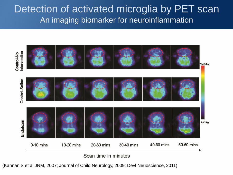
Detection of activated microglia by PET scan An imaging biomarker for neuroinflammation
(Kannan S et al JNM, 2007; Journal of Child Neurology, 2009; Devl Neuoscience, 2011)

Activated Microglial Cells
Change in microglial morphology from ramified to more amoeboid and rounded form with endotoxin exposure.
Increased activated microglia in white matter tracts in endotoxin kits.
(Kannan S group et al JNM, 2007; Journal of Child Neurology, 2009)
100 mm
CC LV IC FH
Co
ntr
ol
End
oto
xin

White matter injury

Activated microglia and oligodendrocytes
Control
Endotoxin
Microglia Oligodendrocytes
A decrease in the number of mature oligodendrocytes (MBP staining) is noted with an increase in the presence of activated microglia in endotoxin kits ; IC=Internal Capsule
IC
IC

EC
EC
IC
IC
CR
CR
CC
CC
200mm
Co
ntr
ol
End
oto
xin
Myelination on Postnatal Day 5
Decrease in Myelin basic protein staining noticed on postnatal day 5 in the corpus callosum, corona radiata and internal capsule

50µm
CONTROL ENDOTOXIN
Day1 Injury to neurons
Impairment in dendritic branching, organization and decreased spines seen in endotoxin kits upon Golgi staining.
Associated with learning deficits and memory impairment
Seen in brains of patients with mental retardation
Determine if there is impairment in learning associated with this injury
Molecular markers responsible for synaptogenesis, dendrite formation and axon guidance

Corresponds with epidemiological studies where inflammatory cytokines are seen in the postnatal serum up to at least 2 weeks of age in neonates who later develop CP.
ELGAN Studies: 2011; 2012
There may be window of opportunity to treat even after birth
Microglial activation persists long after birth
Inflammation persists postnatally
Duration of inflammation in the developing brain: Treatment window?
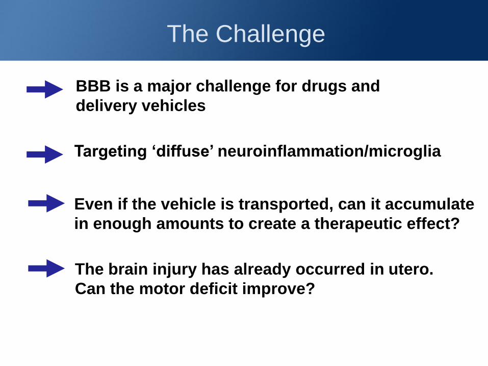
The Challenge
BBB is a major challenge for drugs and
delivery vehicles
Targeting ‘diffuse’ neuroinflammation/microglia
Even if the vehicle is transported, can it accumulate
in enough amounts to create a therapeutic effect?
The brain injury has already occurred in utero.
Can the motor deficit improve?

Dendrimers: ‘Tree-like polymers’ In collaboration with Kannan Rangaramanujam
Co-Director, Center for Nanomedicine, Wilmer Eye Institute
Dendrimers are well-defined, tree-like polymers made synthetically, with a size of ~ 4 – 20 nm.
Flexible, open structure, where each component of the tree can be manipulated
Biocompatible, can be made biodegradable Multifunctionality (therapy, imaging, targeting)
Strategy: use the intrinsic targeting and release properties of dendrimers as building blocks and tailor the nanodevice to the specific clinical application

KALA PHARMACEUTICALS | CONFIDENTIAL
Biodistribution of dendrimer in newborns with
CP (Subarachnoid)
Dendrimer localizes to activated microglial cells in the brain of kits with
neuroinflammation, far removed from the site of injection.
Nuclei
DAPI
Dendrimer
localization
FITC
Microglia
Lectin
Colocalization
of dendrimer
in microglia
A C B D
E G F H
I K J L
63X x 3 zoom
63X
40X

Dendrimer localizes in activated microglia and
astrocytes even upon IV administration
Kannan S et al, Science Translational Medicine, 2012

But can it release the drug specifically where we want it to?
Reach target cells
Release drug inside cells
Menjoge, Kannan, Tomalia, Drug discovery Today (2010)
• N-acetyl cysteine has anti-inflammatory and anti-oxidant effects; GSH precursor
• Has been shown to reduce infarct volume and inflammation in animal models of stroke and cerebral ischemia
• NAC conjugated to dendrimer such that it will not release in plasma but will release intracellularly in a sustained manner
• Validated in vitro
Kannan RM and Kannan S group et al, Biomaterials 2009, International Journal of
Pharmaceutics 2009, Bioconj Chem. 2008.
Dendrimer-drug conjugates

Menjoge, Kannan, Tomalia, Drug discovery Today (2010)
Internalization

Menjoge, Kannan, Tomalia, Drug discovery Today (2010)
Expulsion
PAMAM Dendrimer
S-S- NAC
Cleavable disulfide bond
NAC released
GSH
GSH
GSH
GSH
GSH
Delivery of the Drug

Menjoge, Kannan, Tomalia, Drug discovery Today (2010)
Kannan et al US patents filed
(3)
Expulsion
PAMAM Dendrimer
PAMAM-S-S-NAC
NAC linked by disulfide bond
Internalization
= S-S- NAC
S-S- NAC
—SH NAC
Cleavable disulfide bond
(1) (2) GSH
(2)
NAC released
GSH
GSH
—SH NAC

Neurobehavioral Assessment CP Kit-PBS treatment on day 1
Day 1 Day 5
Kannan S et al, Science Translational Medicine, 2012

Neurobehavioral Assessment CP Kit- D-NAC 10mg/kg Day 1
Day 1 Day 5
Kannan S et al, Science Translational Medicine, 2012

Improvement in Motor Function
Dendrimer-NAC
Dramatic Improvement in motor function seen by Day 5,
upon dendrimer-NAC treatment
Kannan S et al, Science Translational Medicine, 2012

Decrease in activated microglia

Myelination and neuronal injury
Kannan S et al, SciTM 2012
Associated with decrease in markers of oxidative injury
Increase in glutathione levels
Decrease in inflammation at day 5 of age
Decrease in neuronal injury

Kannan et al; Science TM., 2012 CREDIT FOR PICTURE: BRICELYN STRAUCH/SCIENCE
Feature Article in Science TM
Nanomedicine for Brain Injury
Patents Pending (2009/2010);
Highlighted in Nature, Science, Nature
Review Drug Discov., C& EN (2012);
PediatricsYear in Review SCCM (2013)
Summary
• Postnatal therapy for prenatal injury
• Targeted therapy can prevent
or arrest fetal neuro-inflammation
• Platform for delivering drugs in a targeted, sustained manner for brain injury: implications in other neurodegenerative diseases

Ongoing Studies
• Can we switch the microglial phenotype to prevent ongoing
injury and promote resolution?
• Effect of neuroinflammation in altering serotonin and
kynurenine metabolism in the developing brain
• Targeting the kynurenine pathway in microglia to decrease
injury and facilitate cortical development in the perinatal age
• Targeting specific enzyme pathways that are seen primarily
in activated microglia
• In vitro slice studies to evaluate microglial function and
action and uptake of nanoparticles/ dendrimers with change
in phenotype
• Evaluation of neuroinflammation and dendrimer therapies in
models of TBI, glioblastoma, EAE etc.

Collaborators & Acknowledgements
• Center for Nanomedicine-Wilmer Eye Institute (Kannan Rangaramanujam/Justin Hanes groups)
• PET Center and SAIRP (Marty Pomper and Dean Wong groups) • KKI (Mike Johnston/Ali Fatemi Group) • fMRI (Galit Pelled) • TBI group/Brain Injury (Courtney Robertson; Ray Koehler groups,
ACCM, JHU) • MFM (Irina Burd, Integrated Research Center for Fetal Medicine) • BSi (Barbara Slusher group) • Biomarker group (Allen Everett; Mela Bembea) • Hypothermic Cardiac Arrest (Baumgartner group)
Funding Sources: R01-NICHD R01 HD069562 (Current) Pediatric Critical Care Scientist Development Program-K12; K08-NICHD; Perinatology Research Branch-NICHD (Previous)
S.Kannan lab group: Current members- Bindu Balakrishnan (post-doc); Elizabeth Nance (Post-doc on Hartwell fellowship); Fan Zhang (Graduate student); Monica Williams (ACCM junior faculty)