Neuroimaging Pharmacopoeia - download.e-bookshelf.de · preter. Thus, the goal of Neuroimaging...
30
Neuroimaging Pharmacopoeia 123 Daniel Thomas Ginat Juan E. Small Pamela Whitney Schaefer Editors
Transcript of Neuroimaging Pharmacopoeia - download.e-bookshelf.de · preter. Thus, the goal of Neuroimaging...

Neuroimaging Pharmacopoeia
123
Daniel Thomas GinatJuan E. SmallPamela Whitney Schaefer Editors

Neuroimaging Pharmacopoeia

Daniel Thomas Ginat • Juan E. Small Pamela Whitney Schaefer Editors
Neuroimaging Pharmacopoeia
ISBN 978-3-319-12714-9 ISBN 978-3-319-12715-6 (eBook) DOI 10.1007/978-3-319-12715-6 Springer Cham Heidelberg NewYork Dordrecht London
Library of Congress Control Number: 2015932672
© Springer International Publishing Switzerland 2015 This work is subject to copyright. All rights are reserved by the Publisher, whether the whole or part of the material is concerned, specifi cally the rights of translation, reprinting, reuse of illustrations, recitation, broadcasting, reproduction on microfi lms or in any other physical way, and transmission or information storage and retrieval, electronic adaptation, computer software, or by similar or dissimilar methodology now known or hereafter developed. Exempted from this legal reservation are brief excerpts in connection with reviews or scholarly analysis or material supplied specifi cally for the purpose of being entered and executed on a computer system, for exclusive use by the purchaser of the work. Duplication of this publication or parts thereof is permitted only under the provisions of the Copyright Law of the Publisher’s location, in its current version, and permission for use must always be obtained from Springer. Permissions for use may be obtained through RightsLink at the Copyright Clearance Center. Violations are liable to prosecution under the respective Copyright Law. The use of general descriptive names, registered names, trademarks, service marks, etc. in this publication does not imply, even in the absence of a specifi c statement, that such names are exempt from the relevant protective laws and regulations and therefore free for general use. While the advice and information in this book are believed to be true and accurate at the date of publication, neither the authors nor the editors nor the publisher can accept any legal responsibility for any errors or omissions that may be made. The publisher makes no warranty, express or implied, with respect to the material contained herein.
Printed on acid-free paper
Springer is part of Springer Science+Business Media (www.springer.com)
Editors Daniel Thomas Ginat, MD, MS Department of Radiology University of ChicagoPritzker Medical School Chicago , IL USA
Juan E. Small, MD Department of Radiology Lahey Clinic Burlington , MA USA
Pamela Whitney Schaefer, MD Department of Radiology Massachusetts General HospitalHarvard Medical School Boston , MA USA
The authors dedicate this book to the wonderful fi eld or neuroradiology and head and neck imaging, the great advances and discoveries in pharmacology, and to a healthy and (illicit) drug-free world.


vii
What you have in front of you is a highly original, different, and useful pub-lication and the only one of its kind in all of diagnostic radiology. The classic defi nition of “pharmacopoeia” is that of a book containing directions for the identifi cation and preparation of drugs or a list of drugs and their uses. So, in a classic sense, this book is not a pharmacopoeia but something that goes beyond one. Each chapter describes one medication or drug and its indica-tions and action mechanisms, and although in many instances this would suf-fi ce to be considered a pharmacopoeia, the authors go further and present a unique and complete series of high-quality neuroimaging studies that refl ect the side effects and complications of the drugs discussed. It is, in my opinion, the best and most complete collection of this sort of images that I have even come across, and there is no question that it will be very helpful to neurora-diologists, radiologists, emergency physicians, and many others. Although are shorter than others, all 55 chapters are excellent. Just perusing through the images is fun, eye opening, and informative.
Dr. Schaefer, Small, and Ginat join many of the great, Avicenna, Galen, Vesalius and others who in the past have discussed the use of drugs in phar-macopeias. My recommendations: buy the book- you will not be disap-pointed, look at the images- you will be entertained, read the book- you will be a better radiologist, and keep the book handy- you will help your patients.
Mauricio Castillo, MD, FACR Professor of Radiology and Chief of Neuroradiology University of North Carolina at Chapel Hill Editor in Chief, American Journal of Neuroradiology President, American Society of Neuroradiology
Foreword

ix
Various medicinal and illicit drugs can result in serious complications affecting the brain, head and neck, and spine. Many of these complications are visible on imaging. In addition, less serious complications or innocuous effects can result in diagnostic conundrums for the imaging interpreter unaware of the patient’s drug exposures or their imaging manifestations, while others may produce rather characteristic changes on imaging that should be readily recognized as such. Until now, imaging interpreters have been unable to turn to a dedicated source of information relating to this subject. In this work, we have compiled a fairly comprehensive review of the imaging features of the effects of drugs and pharmaceuticals, from A to Z.
Considering the widespread use of medicinal and illicit drug use through-out the world, familiarity with this subject is critical for the imaging inter-preter. Thus, the goal of Neuroimaging Pharmacopoeia is to serve as a resource for recognizing these effects and formulating an appropriate differ-ential diagnosis. The term pharmacopoeia is derived from the two Greek words pharmakon for medicine and poiein to make. As such, a pharmacopeia is a compilation of medicinal or pharmacological drugs with their formulas, methods of preparation, effects, and directions of use.
This text represents an adaptation of the classical pharmacopeia, in which each chapter essentially comprises an illustrated pharmacological vignette comprising the indications, mechanism of action, discussion, differential diagnosis, and relevant fi gures for general classes or specifi c drugs. Although pharmacology is a dynamic fi eld with perpetual development of new agents, this text provides a fundamental approach for understanding and interpreting neuroimaging studies in patients with drug-induced changes. This knowledge can in turn be used to help optimize patient management in certain cases.
Chicago , IL , USA Daniel Thomas Ginat , MD, MS Burlington , MA , USA Juan E. Small , MD Boston , MA , USA Pamela Whitney Schaefer , MD
Pref ace


xi
Alaa Jaly, Lancashire Teaching Hospitals
Chris Coutinho, MB, FRCR, Lancashire Teaching Hospitals
Jenny Hoang, MBBS, Duke University
Sachin Mathur, MRCP, FRCR, Lancashire Teaching Hospitals
Gregory Christoforidis, MD, University of Chicago
Alisa Gean, MD, University of California, San Francisco
Christine Glastonbury, MBBS, University of California, San Francisco
Vesna Petronic-Rosic, MD, University of Chicago
Caroline Robson, MBChB, Boston Children’s Hospital
Acknowledgement

xiii
1 Tobacco Cigarette Smoking . . . . . . . . . . . . . . . . . . . . . . . . . . . . . 1 Michael C. Veronesi and Daniel Thomas Ginat
2 Alcohol . . . . . . . . . . . . . . . . . . . . . . . . . . . . . . . . . . . . . . . . . . . . . . 15 Michael C. Veronesi and Daniel Thomas Ginat
3 Methanol . . . . . . . . . . . . . . . . . . . . . . . . . . . . . . . . . . . . . . . . . . . . 35 Allan Lee , Dee Nandurkar , and Ronil V. Chandra
4 Cannabis (Marijuana) . . . . . . . . . . . . . . . . . . . . . . . . . . . . . . . . . 41 Eileen C. Ang , Stephen L. Stuckey , Daniel Thomas Ginat , and Ronil V. Chandra
5 Crack and Cocaine . . . . . . . . . . . . . . . . . . . . . . . . . . . . . . . . . . . . 49 Rania Hito and Daniel Thomas Ginat
6 Amphetamines . . . . . . . . . . . . . . . . . . . . . . . . . . . . . . . . . . . . . . . . 59 Ronil V. Chandra , Daniel Thomas Ginat , and Juan E. Small
7 Opioids . . . . . . . . . . . . . . . . . . . . . . . . . . . . . . . . . . . . . . . . . . . . . . 69 Daniel Thomas Ginat
8 Betel Nuts . . . . . . . . . . . . . . . . . . . . . . . . . . . . . . . . . . . . . . . . . . . . 79 Grayson W. Hooper , Timothy Biega , and Daniel Thomas Ginat
9 Licorice . . . . . . . . . . . . . . . . . . . . . . . . . . . . . . . . . . . . . . . . . . . . . . 83 Juan E. Small and Daniel Thomas Ginat
10 Centella asiatica . . . . . . . . . . . . . . . . . . . . . . . . . . . . . . . . . . . . . . . 87 Janu Pirakalathanan , Stephen L. Stuckey , and Ronil V. Chandra
11 Nitrous Oxide (N 2 O) . . . . . . . . . . . . . . . . . . . . . . . . . . . . . . . . . . . 91 Daniel Thomas Ginat
12 Iodinated Contrast Agents . . . . . . . . . . . . . . . . . . . . . . . . . . . . . . 95 Harut Haroyan and Daniel Thomas Ginat
13 Gadolinium-Based Contrast Agents . . . . . . . . . . . . . . . . . . . . . . 105 Harut Haroyan and Daniel Thomas Ginat
14 Pantopaque (Myodil, Iodophenylundecylic Acid) . . . . . . . . . . . 111 Daniel Thomas Ginat
Contents
xiv
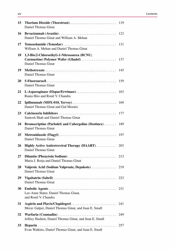
15 Thorium Dioxide (Thorotrast) . . . . . . . . . . . . . . . . . . . . . . . . . . . 119 Daniel Thomas Ginat
16 Bevacizumab (Avastin) . . . . . . . . . . . . . . . . . . . . . . . . . . . . . . . . . 123 Daniel Thomas Ginat and William A. Mehan
17 Temozolamide (Temodar) . . . . . . . . . . . . . . . . . . . . . . . . . . . . . . . 131 William A. Mehan and Daniel Thomas Ginat
18 1,3-Bis(2-Chloroethyl)-1- Nitrosourea (BCNU; Carmustine) Polymer Wafer (Gliadel) . . . . . . . . . . . . . . . . . . . . 137 Daniel Thomas Ginat
19 Methotrexate . . . . . . . . . . . . . . . . . . . . . . . . . . . . . . . . . . . . . . . . . 145 Daniel Thomas Ginat
20 5-Fluorouracil . . . . . . . . . . . . . . . . . . . . . . . . . . . . . . . . . . . . . . . . 159 Daniel Thomas Ginat
21 L-Asparaginase (Elspar/Erwinase) . . . . . . . . . . . . . . . . . . . . . . . 163 Rania Hito and Ronil V. Chandra
22 Ipilimumab (MDX-010, Yervoy) . . . . . . . . . . . . . . . . . . . . . . . . . 169 Daniel Thomas Ginat and Gul Moonis
23 Calcineurin Inhibitors . . . . . . . . . . . . . . . . . . . . . . . . . . . . . . . . . 177 Santosh Shah and Daniel Thomas Ginat
24 Bromocriptine (Parlodel) and Cabergoline (Dostinex) . . . . . . . 189 Daniel Thomas Ginat
25 Metronidazole (Flagyl) . . . . . . . . . . . . . . . . . . . . . . . . . . . . . . . . . 197 Daniel Thomas Ginat
26 Highly Active Antiretroviral Therapy (HAART). . . . . . . . . . . . 203 Daniel Thomas Ginat
27 Dilantin (Phenytoin Sodium) . . . . . . . . . . . . . . . . . . . . . . . . . . . . 213 Maria J. Borja and Daniel Thomas Ginat
28 Valproic Acid (Sodium Valproate, Depakote) . . . . . . . . . . . . . . 219 Daniel Thomas Ginat
29 Vigabatrin (Sabril) . . . . . . . . . . . . . . . . . . . . . . . . . . . . . . . . . . . . 223 Daniel Thomas Ginat
30 Embolic Agents . . . . . . . . . . . . . . . . . . . . . . . . . . . . . . . . . . . . . . . 231 Lee-Anne Slater , Daniel Thomas Ginat , and Ronil V. Chandra
31 Aspirin and Plavix/Clopidogrel . . . . . . . . . . . . . . . . . . . . . . . . . . 241 Merav Galper , Daniel Thomas Ginat , and Juan E. Small
32 Warfarin (Coumadin) . . . . . . . . . . . . . . . . . . . . . . . . . . . . . . . . . . 249 Jeffrey Hashim , Daniel Thomas Ginat , and Juan E. Small
33 Heparin . . . . . . . . . . . . . . . . . . . . . . . . . . . . . . . . . . . . . . . . . . . . . 257 Evan Watkins , Daniel Thomas Ginat , and Juan E. Small
Contents
xv
34 Tissue Plasminogen Activator (tPA) . . . . . . . . . . . . . . . . . . . . . . 261 Evan Watkins , Juan E. Small , and Daniel Thomas Ginat
35 Supplemental Oxygen . . . . . . . . . . . . . . . . . . . . . . . . . . . . . . . . . . 263 Daniel Thomas Ginat
36 Hypertonic Saline . . . . . . . . . . . . . . . . . . . . . . . . . . . . . . . . . . . . . 271 Daniel Thomas Ginat
37 Mannitol (1,2,3,4,5,6-Hexanehexol) . . . . . . . . . . . . . . . . . . . . . . . 277 Daniel Thomas Ginat
38 HiDAC (High-Dose Ara-C; Cytarabine; Cytosine Arabinoside; Cytosar-U; Depocyt) . . . . . . . . . . . . . . . . . . . . . . . 281 Daniel Thomas Ginat
39 Triple H Therapy . . . . . . . . . . . . . . . . . . . . . . . . . . . . . . . . . . . . . 285 Daniel Thomas Ginat
40 Insulin. . . . . . . . . . . . . . . . . . . . . . . . . . . . . . . . . . . . . . . . . . . . . . . 289 Lee-Anne Slater , Stephen L. Stuckey , and Ronil V. Chandra
41 Manganese in Total Parenteral Nutrition . . . . . . . . . . . . . . . . . . 293 Daniel Thomas Ginat
42 Zinc Oxide (ZnO) . . . . . . . . . . . . . . . . . . . . . . . . . . . . . . . . . . . . . 299 Daniel Thomas Ginat and Juan E. Small
43 Angiotensin Converting Enzyme (ACE) Inhibitors . . . . . . . . . . 303 Daniel Thomas Ginat and Jason M. Johnson
44 Acetazolamide (Diamox) . . . . . . . . . . . . . . . . . . . . . . . . . . . . . . . 309 Daniel Lopes Noujaim , Juan E. Small , and Daniel Thomas Ginat
45 Facial Fillers . . . . . . . . . . . . . . . . . . . . . . . . . . . . . . . . . . . . . . . . . 313 Daniel Thomas Ginat and Charles J. Schatz
46 Synthetic Corticosteroids . . . . . . . . . . . . . . . . . . . . . . . . . . . . . . . 319 Jason M. Johnson , Yi Li , and Daniel Thomas Ginat
47 Oral Contraceptives (Estrogen and Progestin) . . . . . . . . . . . . . 329 Kimberly Kallianos , Daniel Thomas Ginat , and Jason M. Johnson
48 Vaccines . . . . . . . . . . . . . . . . . . . . . . . . . . . . . . . . . . . . . . . . . . . . . 335 Daniel Thomas Ginat
49 Acetominophen (Tylenol, Paracetamol) . . . . . . . . . . . . . . . . . . . 341 Daniel Thomas Ginat
50 Propofol . . . . . . . . . . . . . . . . . . . . . . . . . . . . . . . . . . . . . . . . . . . . . 347 Mariam Aboian , Jason M. Johnson , and Daniel Thomas Ginat
51 Bisphosphonates . . . . . . . . . . . . . . . . . . . . . . . . . . . . . . . . . . . . . . 351 Ana M. Franceschi , Daniel Thomas Ginat , and Jason M. Johnson
Contents
xvi
52 Recombinant Human Bone Morphogenetic Protein . . . . . . . . . 359 Marianne S. Reed , Jason M. Johnson , and Daniel Thomas Ginat
53 Retinoids (13-cis-Retinoic Acid, Isotretinoin, Accutane, All-trans-retinoic acid) . . . . . . . . . . . . . . . . . . . . . . . . 363 Daniel Thomas Ginat
54 Topical Prostaglandin Analogues . . . . . . . . . . . . . . . . . . . . . . . . 367 Daniel Thomas Ginat and Nurhan Torun
55 Nasal Decongestants . . . . . . . . . . . . . . . . . . . . . . . . . . . . . . . . . . . 371 Daniel Thomas Ginat
Index . . . . . . . . . . . . . . . . . . . . . . . . . . . . . . . . . . . . . . . . . . . . . . . . . . . 375
Contents

xvii
Contributors
Mariam Aboian, MD , Department of Radiology, University of California, San Francisco, CA, USA
Eileen C. Ang, MBBS , Department of Diagnostic Imaging, Monash Medical Center, Monash Health, Melbourne , VIC, Australia
Timothy Biega, MD , Department of Radiology, Tripler Army Medical Center, Honolulu, HI, USA
Maria J. Borja, MD , Department of Radiology, Massachusetts General Hospital, Boston, MA, USA
Ronil V. Chandra, MBBS, MMed, FRANZCR , Department of Diagnostic Imaging, Monash Medical Center, Monash Health, Melbourne, VIC, Australia
Ana M. Franceschi, MD , Diagnostic Radiology, New York University Medical Center, New York, NY, USA
Merav Galper, MD , Department of Diagnostic Radiology, Lahey Clinic, Burlington, MA, USA
Daniel Thomas Ginat, MD, MS , Department of Radiology, University of Chicago, Pritzker Medical School, Chicago, IL, USA
Harut Haroyan, MD , Department of Radiology, University of Chicago, Chicago, IL, USA
Jeffrey Hashim, MD , Department of Diagnostic Radiology, Lahey Clinic, Burlington, MA, USA
Rania Hito, MD , Department of Radiology, University of Massachusetts Memorial Medical Center, Worcester, MA, USA
Grayson W. Hooper, DO, Department of Radiology, Tripler Army Medical Center, Honolulu, HI, USA
Jason M. Johnson, MD , Department of Diagnostic Radiology, MD Anderson Cancer Center, Houston, TX, USA
Kimberly Kallianos, MD , Department of Radiology, University of California, San Francisco, CA, USA

xviii
Allan Lee, MBBS, FRANZCR , Department of Diagnostic Imaging, Monash Medical Center, Monash Health, Melbourne, VIC, Australia
Yi Li, MD , Department of Radiology, University of California, San Francisco, CA, USA
William A. Mehan, MD , Department of Radiology, Tufts Medical Center, Boston, MA, USA
Gul Moonis, MD , Department of Radiology, Columbia University, New York, NY, USA
Dee Nandurkar, MBBS, FRANZCR , Department of Diagnostic Imaging, Monash Medical Center, Monash Health, Melbourne, VIC, Australia
Daniel Lopes Noujaim, MD , Department of Diagnostic Radiology, Lahey Clinic, Burlington, MA, USA
Janu Pirakalathanan, MBBS , Department of Diagnostic Imaging, Monash Medical Center, Monash Health, Melbourne, VIC, Australia
Marianne S. Reed, MD , Diagnostic Radiology, Yale University, New Haven, CT, USA
Pamela W. Schaefer, MD , Department of Radiology, Massachussetts General Hospital, Harvard Medical School, Boston, MA, USA
Charles J. Schatz, MD, FACR , Department of Radiology, Beverly Tower Wilshire Advanced Imaging, University of Southern California Keck School of Medicine, Los Angeles, CA, USA
Santosh Shah, MD , Department of Radiology, Massachusetts General Hospital, Boston, MA, USA
Juan E. Small, MD , Department of Diagnostic Radiology, Lahey Clinic, Burlington, MA, USA
Lee-Anne Slater, MBBS, MMed, FRANZCR , Department of Diagnostic Imaging, Monash Medical Center, Monash Health, Melbourne, VIC, Australia
Stephen L. Stuckey, MBBS, MMed, MD, FRANZCR , Department of Diagnostic Imaging, Monash Medical Center, Monash Health, Melbourne, VIC, Australia
Nurhan Torun, MD , Department of Ophthalmology, Beth Israel Deaconess Medical Center, Boston, MA, USA
Michael C. Veronesi, MD, PhD , Department of Radiology, University of Chicago, Chicago, IL, USA
Evan Watkins, MD , Department of Diagnostic Radiology , Lahey Clinic , Burlington , MA , USA
Contributors

1D.T. Ginat et al. (eds.), Neuroimaging Pharmacopoeia,DOI 10.1007/978-3-319-12715-6_1, © Springer International Publishing Switzerland 2015
1.1 Uses
Although tobacco use is legal, it is not recommended or prescribed for any therapeutic or medicinal purposes. It is smoked or chewed for recreational use as a potent temporary stimu-lant and mood elevator.
1.2 Mechanism
Nicotine, the primary active compound in cigarettes, is considered one of the most addictive of all sub-stances because of its rapid onset and offset effects on the brain’s dopamine reward systems. Inhaled nicotine can distribute in the brain within 10 s of inhalation but lasts mere seconds, causing a power-ful urge for more. Of note, the long-term devastating sequela from smoking as it relates to neurologic dis-ease are only partially explained by nicotine. Aside from nicotine, tobacco contains over six thousand other chemical compounds. In particular, polycyclic aromatic hydrocarbons and the tobacco-specifi c nitrosamine 4-(methylnitrosamino)-1-(3-pyridyl)-1- butanone have been implicated as the major
carcinogens associated with cigarette smoking. Additional details regarding the mechanisms of action for specifi c conditions are discussed in subse-quent sections.
1.3 Discussion
Tobacco use is the leading cause of preventable death in the United States. An estimated 400,000 deaths or nearly one of every fi ve deaths in the United States is associated with adverse condi-tions caused by smoking each year. Nearly every organ system is affected by cigarette smoking. The associated manifestations of cigarette smok-ing from a neuroimaging standpoint include the direct link between smoking and large vessels and lacunar infarcts, chronic small vessel isch-emic disease, cerebral aneurysms, cerebral venous thrombosis, head and neck cancer of the squamous type, lung cancer metastases to the brain, and Warthin tumors.
1.3.1 Stroke
Compared with nonsmokers, cigarette smoking is estimated to increase the risk of stroke by 2–4- fold. Of note, stroke is the third leading cause of death in the United States with signifi cant comor-bidity in survivors. Mechanisms by which pri-mary and second-hand tobacco smoke exposure increase the risk of stroke and heart disease
M. C. Veronesi , MD, PhD Department of Radiology , University of Chicago , Chicago , IL , USA
D. T. Ginat , MD, MS (*) Department of Radiology , University of Chicago, Pritzker Medical School , Chicago , IL , USA e-mail: [email protected]
1 Tobacco
Michael C. Veronesi and Daniel Thomas Ginat

2
include carboxyhemoglobinemia, increased platelet aggregation, increased fi brinogen levels, reduced HDL cholesterol, and direct toxic effects of compounds such as 1,3-butadiene, a vapor phase constituent of environmental tobacco smoke that has been shown to accelerate athero-sclerosis. Indeed, atherosclerosis and formation of both occlusive and embolic thrombi are the major causes of cerebrovascular accidents. Smoking is also associated with lacunar infarcts and chronic small vessel ischemic disease. Smoking cessation results in a considerable reduction in stroke risk.
On conventional angiography, CTA or MRA, atherosclerosis manifests as luminal narrowing that may be associated with calcifi cations. Acute embolic thrombus is suggested by intraluminal high attenuation on non-contrast CT, such as the hyperdense MCA sign, and manifests as an abrupt termination of the artery with absence of fl ow distally on CTA, MRA, or conventional angiography (Figs. 1.1 ). Hyperacute infarcts are often unapparent on non-contrast CT, but acute and early subacute infarcts can appear as areas
of hypoattenuation with loss of gray white mat-ter differentiation and swelling. MRI with diffu-sion-weighted imaging is more sensitive for detecting early infarcts, which appear as areas of high T2 signal and restricted diffusion (Fig. 1.2 ). Besides smoking, other causes and risk factors for stroke include hypertension, hypercholesterolemia, diabetes, use of other drugs, such as cocaine and amphetamines (refer to Chaps. 5 and 6 ), dissection (Fig. 1.3 ), and vasculitis, such as lupus or Takayasu arteritis (Fig. 1.4 ). In addition to large territorial infarcts, smokers are prone to more extensive small ves-sel ischemic disease, which can manifest as areas of high T2 signal in the cerebral white matter (Fig. 1.5 ).
1.3.2 Cerebral Aneurysm
Besides smoking, additional risk factors that predis-pose to aneurysm formation include fenestrated arteries (Fig. 1.6 ), fi bromuscular dysplasia, neurofi -bromatosis, alpha-1-antytripsyn defi ciency, and
a b
Fig. 1.1 Embolic infarct. This smoker presented with acute left hemiplegia. Non-contrast axial CT image ( a ) shows a hypoattenuation within the right MCA territory
( arrow ). 3D CTA image ( b ) shows occlusion of the right MCA (expected location encircled )
M.C. Veronesi and D.T. Ginat
3
a b
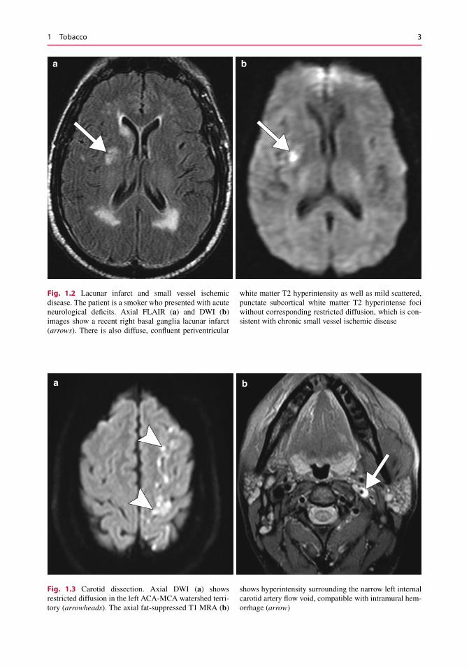
Fig. 1.2 Lacunar infarct and small vessel ischemic disease. The patient is a smoker who presented with acute neurological defi cits. Axial FLAIR ( a ) and DWI ( b ) images show a recent right basal ganglia lacunar infarct ( arrows ). There is also diffuse, confl uent periventricular
white matter T2 hyperintensity as well as mild scattered, punctate subcortical white matter T2 hyperintense foci without corresponding restricted diffusion, which is con-sistent with chronic small vessel ischemic disease
a b
Fig. 1.3 Carotid dissection. Axial DWI ( a ) shows restricted diffusion in the left ACA-MCA watershed terri-tory ( arrowheads ). The axial fat-suppressed T1 MRA ( b )
shows hyperintensity surrounding the narrow left internal carotid artery fl ow void, compatible with intramural hem-orrhage ( arrow )
1 Tobacco
4
connective tissue disorders, such as Ehlers-Danlos syndrome and Marfan’s syndrome and polycystic kidney disease. Otherwise, the differential diagno-sis of a cerebral aneurysm on radiologic imaging
includes an infundibulum (usually manifests as a triangular dilatation with the vessel arising from the apex that measures less than 2 mm), pseudoaneu-rysm, and mycotic aneurysm (Fig. 1.7 ).
a b
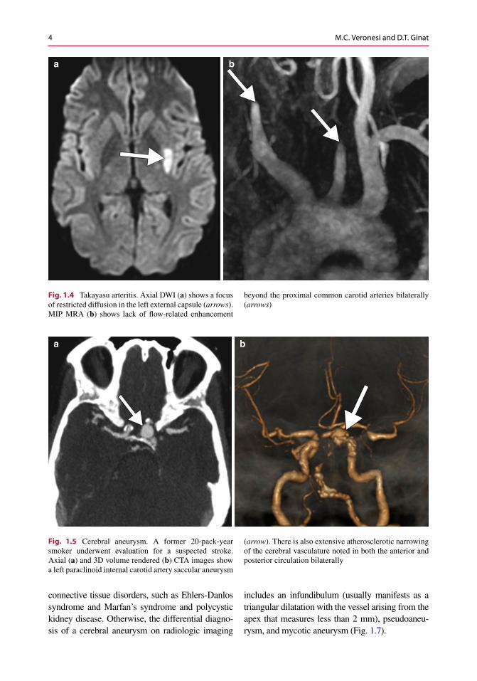
Fig. 1.4 Takayasu arteritis. Axial DWI ( a ) shows a focus of restricted diffusion in the left external capsule ( arrows ). MIP MRA ( b ) shows lack of fl ow-related enhancement
beyond the proximal common carotid arteries bilaterally ( arrows )
a b
Fig. 1.5 Cerebral aneurysm. A former 20-pack-year smoker underwent evaluation for a suspected stroke. Axial ( a ) and 3D volume rendered ( b ) CTA images show a left paraclinoid internal carotid artery saccular aneurysm
( arrow ). There is also extensive atherosclerotic narrowing of the cerebral vasculature noted in both the anterior and posterior circulation bilaterally
M.C. Veronesi and D.T. Ginat

5
1.3.3 Cerebral Venous Thrombosis
Secondary polycythemia due to chronic smok-ing is a risk factor developing cerebral venous thrombosis. Refer to the L-asparaginase and oral contraceptives chapters (Chaps. 21 and 47 ) for examples of venous thrombosis on imaging.
1.3.4 Head and Neck Cancer, Squamous Cell Carcinoma
Tobacco smoking, along with alcohol, is well established as the dominant risk factor for head and neck squamous cell carcinoma (HNSCC). This risk is correlated with the intensity and duration of tobacco use and is synergistic with concomitant alcohol consumption. There are more than sixty recognized compounds in tobacco that have a specifi c carcinogenic potential. In particular, nitrosamines and
polycyclic hydrocarbons can alter the molecu-lar profi le of an individual and causes muta-tions. Nicotine, originally thought only to be responsible for tobacco addiction, is also involved in tumor promotion and progression with antiapoptotic and indirect mitogenic prop-erties. Other factors that can increase the risk of HNSCC include HPV infection and certain occupational exposures. Imaging with contrast- enhanced CT and MRI allows depiction of the anatomy of the larynx and submucosal tumor extension.
Dual-energy CT improves the diagnostic per-formance and interobserver reproducibility of evaluations of laryngeal cartilage invasion by squamous cell carcinoma. CT, MRI, and PET-CT also provide information regarding cervical nodal disease, systemic metastases, and synchronous malignancies.
On CT or MRI, head and neck squamous cell carcinomas often manifest as ill-defi ned enhancing masses. Aggressive features of
a b
Fig. 1.6 Arterial fenestration. Frontal projection 3D vol-ume rendered CTA image ( a ) shows a fenestration of the proximal basilar artery ( arrow ). Lateral projection 3D
volume rendered CTA image ( b ) shows a dorsally ori-ented aneurysm arising from the caudal aspect of the fen-estration ( arrowhead )
1 Tobacco
6
HNSCC include tissue invasion and necrosis (Fig. 1.8 ). The most common sites of head and neck squamous cell cancer are the fl oor of the mouth, tongue, soft palate, anterior tonsillar pillar, and retromolar trigone. In addition to locoregional spread, HNSCC can metastasize to distant organs hematogenously, most com-monly to the lungs and bones. Metastasis to the brain is an infrequent, but carries a poor prog-nostic outcome (Fig. 1.9 ). Besides smoking, alcohol (refer to Chap. 2 ), betel nuts (refer to Chap. 8 ), and HPV are the other major risk fac-tors for head and neck squamous cell carci-noma. Interestingly, HPV-positive squamous cell carcinomas generally have a more favor-able prognosis and tend to have tumors that are relatively well-defi ned and large cystic nodal metastases (Fig. 1.10 ). Otherwise, the differen-tial for other malignant cancers in the head and neck include thyroid cancer, lymphoma, sali-vary gland cancer, and sarcoma. Furthermore, head and neck abscesses can sometimes resem-ble necrotic or ulcerated head and neck
squamous cell carcinomas and nodal metasta-ses (Fig. 1.11 ).
1.3.5 Brain Metastases
In addition to HNSCC, smoking has been shown to be a risk factor for the formation of several other types of cancers, including lung cancer, acute myeloid leukemia, bladder cancer, cervical cancer, renal cancer, esophageal cancer, gastric cancer, pancreatic cancer, and colorectal cancer. The majority of these cancers have at least some potential for metastasizing to the brain, which is the most feared complication of systemic malig-nancies. If no one smoked, one of every three cancer deaths in the United States would be avoided. Lung cancer has the highest association with smoking, causing an estimated 90 % of all lung cancer deaths in men and 80 % of all lung cancer deaths in women, and is the most com-mon cancer that metastasizes to the brain. Nicotine- derived nitrosamine ketone (NNK) has
a b
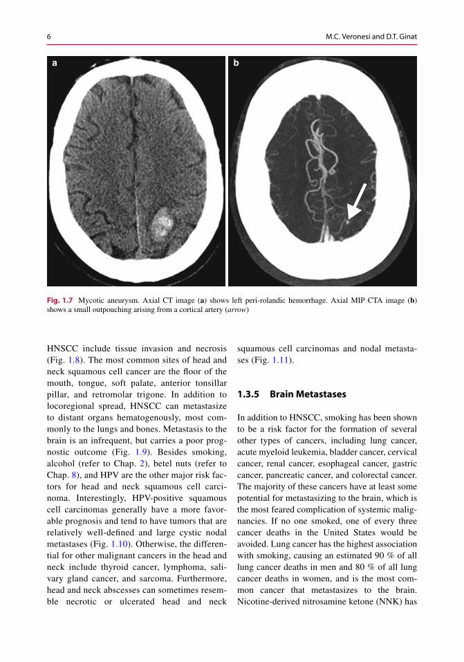
Fig. 1.7 Mycotic aneurysm. Axial CT image ( a ) shows left peri-rolandic hemorrhage. Axial MIP CTA image ( b ) shows a small outpouching arising from a cortical artery ( arrow )
M.C. Veronesi and D.T. Ginat

7
a b
Fig. 1.8 Oral squamous cell carcinoma in a 30-pack-year male smoker. Axial ( a ) and coronal ( b ) post-contrast CT images show an ulcerating mass in the left fl oor of the
mouth and oral tongue ( arrows ) with associated submen-tal necrotic cervical lymphadenopathy ( arrowhead )
a b
Fig. 1.9 Laryngeal squamous cell carcinoma with brain metastasis. This 25-pack-year smoker had been recently diagnosed with laryngeal squamous cell carcinoma when he developed generalized seizures. Axial CT image ( a ) shows a
left vocal cord mass ( arrow ) that also involves the paraglot-tic fat and anterior commissure. Post-contrast T1-weighted MRI image ( b ) demonstrates a heterogeneous metastatic lesion in the right frontal lobe ( arrowheads )
1 Tobacco
8
a
c
b
d
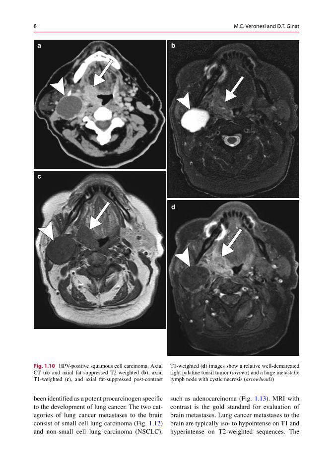
Fig. 1.10 HPV-positive squamous cell carcinoma. Axial CT ( a ) and axial fat-suppressed T2-weighted ( b ), axial T1-weighted ( c ), and axial fat-suppressed post-contrast
T1-weighted ( d ) images show a relative well-demarcated right palatine tonsil tumor ( arrows ) and a large metastatic lymph node with cystic necrosis ( arrowheads )
been identifi ed as a potent procarcinogen specifi c to the development of lung cancer. The two cat-egories of lung cancer metastases to the brain consist of small cell lung carcinoma (Fig. 1.12 ) and non- small cell lung carcinoma (NSCLC),
such as adenocarcinoma (Fig. 1.13 ). MRI with contrast is the gold standard for evaluation of brain metastases. Lung cancer metastases to the brain are typically iso- to hypointense on T1 and hyperintense on T2-weighted sequences. The
M.C. Veronesi and D.T. Ginat
9
a b
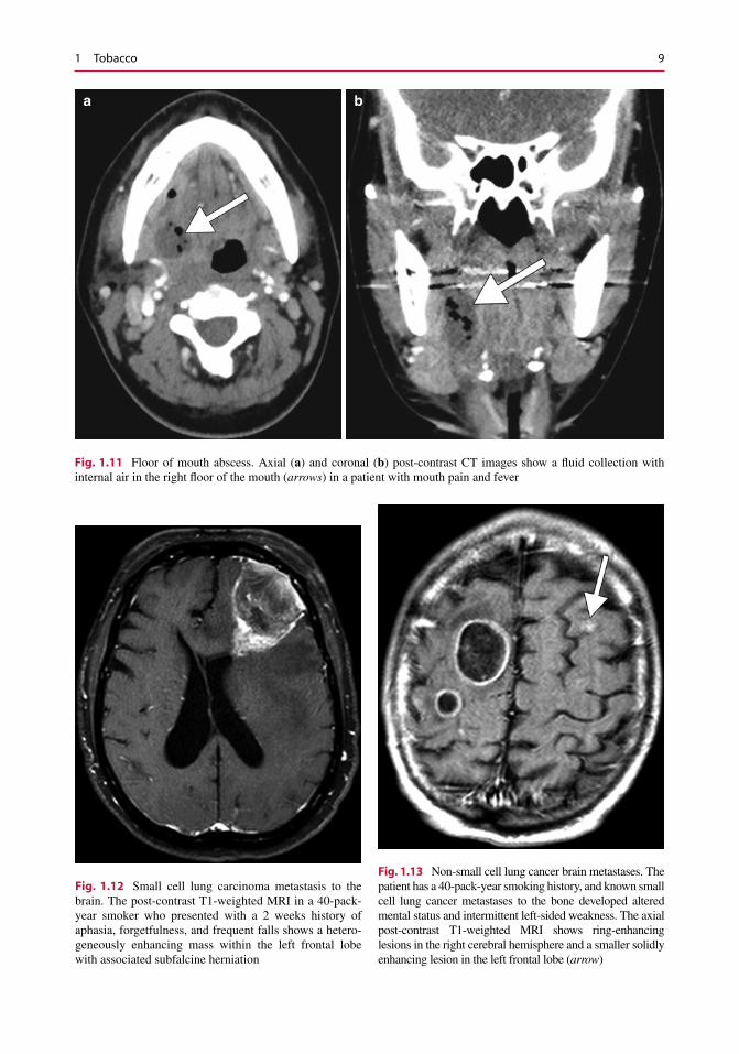
Fig. 1.11 Floor of mouth abscess. Axial ( a ) and coronal ( b ) post-contrast CT images show a fl uid collection with internal air in the right fl oor of the mouth ( arrows ) in a patient with mouth pain and fever
Fig. 1.12 Small cell lung carcinoma metastasis to the brain. The post-contrast T1-weighted MRI in a 40-pack- year smoker who presented with a 2 weeks history of aphasia, forgetfulness, and frequent falls shows a hetero-geneously enhancing mass within the left frontal lobe with associated subfalcine herniation
Fig. 1.13 Non-small cell lung cancer brain metastases. The patient has a 40-pack-year smoking history, and known small cell lung cancer metastases to the bone developed altered mental status and intermittent left-sided weakness. The axial post-contrast T1-weighted MRI shows ring-enhancing lesions in the right cerebral hemisphere and a smaller solidly enhancing lesion in the left frontal lobe ( arrow )
1 Tobacco

10
enhancement pattern is usually intense whether uniform, punctate, or ring enhancing. Hemorrhagic lung metastases may display high intrinsic T1 signal. On T2-weighted sequences, metastatic lesions are typically hyperintense with hyperintense peritumoral edema. On MR spectroscopy, there is often an intratumoral cho-line peak without choline elevation in the peritu-moral edema and depletion of NAA. CT may be the initial exam obtained when metastatic dis-ease is not yet known. On precontrast imaging, the mass may be iso- to hypoattenuating, sur-rounded by variable amounts of vasogenic edema. Similar to MRI, following administra-tion of contrast in CT, enhancement is variable and can be intense, punctate, nodular, or ring enhancing. It is also important to evaluate the skull, soft tissues, and head and neck lymph nodes for additional metastases.
The differential diagnosis for brain metastases includes primary brain tumors (Fig. 1.14 ), cere-bral abscess (refer to Chaps. 7 , 16 , 19 , and 46 ), focal subacute stroke, and tumefactive demyelin-ating lesions.
1.3.6 Warthin Tumor
Papillary lymphomatous cystadenoma of the parotid gland, or Warthin tumor, is signifi cantly associated with a history of cigarette smoking. Indeed, over 90 % of Warthin tumors occur in tobacco smokers. The tumor arises almost exclusively in the parotid gland and has a predi-lection for male Caucasians. These lesions most commonly present as asymptomatic masses, but may cause facial nerve paralysis or tinnitus. Warthin tumors consists of oncocytic cells con-taining numerous mitochondria frequently showing structural abnormalities and reduced metabolic function. Smoking can lead to dam-age to mitochondrial DNA due to the develop-ment of numerous reactive oxygen species. In this context, a high rate of deleted mitochondrial DNA has been detected in the oncocytic cells of Warthin tumor. Typical imaging features consist
of well-defi ned solid and/or cystic masses usually within the inferior pole (tail) of the parotid gland, not infrequently multifocal and bilateral (Fig. 1.15 ).
The differential diagnosis of Warthin tumor includes non-neoplastic conditions, such as benign lymphoepithelial lesions, Sjogren syn-drome, and sarcoidosis. For example, benign lymphoepithelial lesions are typically associ-ated with HIV and classically appear as mul-tiple bilateral cystic parotid masses that may contain solid components (Fig. 1.16 ). Warthin tumors can also resemble pleomorphic adeno-mas on imaging. However, pleomorphic adeno-mas are less likely to occur in the parotid tail and display a bosselated appearance and high T2 signal on MRI (Fig. 1.17 ). While clinical history may be helpful in differentiating some of these possibilities, predictive features of malignancy on imaging include T2 hypointen-sity of the parotid, ill- defi ned margins, diffuse growth, infi ltration of subcutaneous tissue,
Fig. 1.14 Glioblastoma. Axial post-contrast T1-weighted MR image shows a ring-enhancing mass centered in the left posterior cingulate gyrus ( arrow )
M.C. Veronesi and D.T. Ginat

11
lymphadenopathy, and perineural spread (Fig. 1.18 ). However, Warthin tumors often demonstrate T2 hypointense foci and low ADC
values and thus cannot be reliably differenti-ated from many malignant tumors on diffusion-weighted imaging. In addition, Warthin tumors typically display marked hypermetabolism on 18 FDG-PET.
Fig. 1.15 Warthin tumor. This smoker with COPD pre-sented with growing right-sided neck mass. The coronal post-contrast CT image shows a well-defi ned hyperatten-uating mass within the right parotid tail ( arrow )
Fig. 1.16 Benign lymphoepithelial lesions in a patient with HIV. The coronal CT image shows cystic lesions in the bilateral parotid glands ( arrows ). The lesion on the left side contains a solid nodule
Fig. 1.17 Parotid pleomorphic adenoma. Axial fat- suppressed T2-weighted MRI shows a bosselated T2 hyperintense mass arising from the deep lobe of the right parotid gland ( arrow )
Fig. 1.18 Parotid pleomorphic adenocarcinoma. The coronal STIR MRI shows a heterogeneous mass arising from the deep lobe of the parotid gland (*) with perineural extension along the mandibular nerve across the foramen ovale ( arrow )
1 Tobacco