Neurocognitive Function
12
Neurocognitive Function in Dopamine-b-Hydroxylase Deficiency Marieke Jepma* ,1,2 , Jaap Deinum 3 , Christopher L Asplund 4 , Serge ARB Rombouts 1,2,5 , Jouke T Tamsma 6 , Nathanja Tjeerdema 6 , Michiel M Spape ´ 7 , Emily M Garland 8 , David Robertson 8 , Jacques WM Lenders 3,9 and Sander Nieuwenhuis 1,2 1 Leiden University, Institute of Psychology, Leiden, The Netherlands; 2 Leiden Institute for Brain and Cognition (LIBC), Leiden, The Netherlands; 3 Division of Vascular Medicine, Department of Internal Medicine, Radboud University Nijmegen Medical Center, Nijmegen, The Netherlands; 4 Department of Psychology, Vanderbilt Vision Research Center, Vanderbilt University, Nashville, TN, USA; 5 Department of Radiology, Leiden University Medical Center, Leiden, The Netherlands; 6 Department of General Internal Medicine & Endocrinology, Leiden University Medical Centre, Leiden, The Netherlands; 7 School of Psychology, University of Nottingham, Nottingham, UK; 8 Autonomic Dysfunction Center and Department of Clinical Pharmacology, Vanderbilt University School of Medicine, Nashville, TN, USA; 9 Department of Medicine III, Carl Gustav Carus University Medical Center, Dresden, Germany Dopamine-b-hydroxylase (DbH) deficiency is a rare genetic syndrome characterized by the complete absence of norepinephrine in the peripheral and the central nervous system. DbH-deficient patients suffer from several physical symptoms, which can be treated successfully with L-threo-3,4-dihydroxyphenylserine, a synthetic precursor of norepinephrine. Informal clinical observations suggest that DbH-deficient patients do not have obvious cognitive impairments, even when they are not medicated, which is remarkable given the important role of norepinephrine in normal neurocognitive function. This study provided the first systematic investigation of neurocognitive function in human DbH deficiency. We tested 5 DbH-deficient patients and 10 matched healthy control participants on a comprehensive cognitive task battery, and examined their pupil dynamics, brain structure, and the P3 component of the electroencephalogram. All participants were tested twice; the patients were tested once ON and once OFF medication. Magnetic resonance imaging scans of the brain revealed that the patients had a smaller total brain volume than the control group, which is in line with the recent hypothesis that norepinephrine has a neurotrophic effect. In addition, the patients showed an abnormally small or absent task-evoked pupil dilation. However, we found no substantial differences in cognitive performance or P3 amplitude between the patients and the control participants, with the exception of a temporal-attention deficit in the patients OFF medication. The largely spared neurocognitive function in DbH-deficient patients suggests that other neuromodulators have taken over the function of norepinephrine in the brains of these patients. Neuropsychopharmacology (2011) 36, 1608–1619; doi:10.1038/npp.2011.42; published online 6 April 2011 Keywords: dopamine-b-hydroxylase deficiency; norepinephrine; cognition; brain; DOPS INTRODUCTION The locus coeruleus–norepinephrine (LC–NE) system is one of the major neuromodulatory systems in the brain. For a long time, investigators have associated this system with basic functions such as arousal and the sleep–wake cycle (Aston- Jones et al , 1984; Jouvet, 1969), and with various neuropsy- chiatric disorders such as depression and attention-deficit hyperactivity disorder (Ressler and Nemeroff, 2001; Siever and Davis, 1985). In addition, recent studies have shown that the LC–NE system is involved in more specific cognitive functions, such as memory, attention, perception, and decision making (Aston-Jones and Cohen, 2005; Robbins, 1997; Sara, 2009). These findings suggest that NE is essential for normal cognitive function in humans. Dopamine-b-hydroxylase (DbH) deficiency is a rare genetic syndrome that is characterized by the congenital absence of the enzyme DbH, which is responsible for the conversion of dopamine (DA) to NE (Man in ’t Veld et al, 1987a; Robertson et al, 1986). As a result, DbH deficiency is characterized by a complete lack of NE and epinephrine in both the central and the peripheral nervous system (Man in ‘t Veld et al, 1987a). There are currently approximately 15 patients with DbH deficiency known worldwide. These patients suffer from several physical symptoms, including severe orthostatic hypotension, fatigue, and impaired exercise tolerance (Robertson and Garland, 2010). The only Received 5 November 2010; revised 27 January 2011; accepted 24 February 2011 *Correspondence: M Jepma, Department of Psychology, Cognitive Psychology Unit, Leiden University, Wassenaarseweg 52, 2333 AK Leiden, The Netherlands, Tel: + 31-71-5273646, Fax: + 31-71-5273619, E-mail: [email protected] Neuropsychopharmacology (2011) 36, 1608–1619 & 2011 American College of Neuropsychopharmacology. All rights reserved 0893-133X/11 www.neuropsychopharmacology.org
Transcript of Neurocognitive Function
Neurocognitive Function in Dopamine-b-HydroxylaseDeficiency
Marieke Jepma*,1,2, Jaap Deinum3, Christopher L Asplund4, Serge ARB Rombouts1,2,5, Jouke T Tamsma6,Nathanja Tjeerdema6, Michiel M Spape7, Emily M Garland8, David Robertson8, Jacques WM Lenders3,9
and Sander Nieuwenhuis1,2
1Leiden University, Institute of Psychology, Leiden, The Netherlands; 2Leiden Institute for Brain and Cognition (LIBC), Leiden, The Netherlands;3Division of Vascular Medicine, Department of Internal Medicine, Radboud University Nijmegen Medical Center, Nijmegen, The Netherlands;4Department of Psychology, Vanderbilt Vision Research Center, Vanderbilt University, Nashville, TN, USA; 5Department of Radiology, Leiden
University Medical Center, Leiden, The Netherlands; 6Department of General Internal Medicine & Endocrinology, Leiden University Medical
Centre, Leiden, The Netherlands; 7School of Psychology, University of Nottingham, Nottingham, UK; 8Autonomic Dysfunction Center and
Department of Clinical Pharmacology, Vanderbilt University School of Medicine, Nashville, TN, USA; 9Department of Medicine III, Carl Gustav
Carus University Medical Center, Dresden, Germany
Dopamine-b-hydroxylase (DbH) deficiency is a rare genetic syndrome characterized by the complete absence of norepinephrine in the
peripheral and the central nervous system. DbH-deficient patients suffer from several physical symptoms, which can be treated
successfully with L-threo-3,4-dihydroxyphenylserine, a synthetic precursor of norepinephrine. Informal clinical observations suggest that
DbH-deficient patients do not have obvious cognitive impairments, even when they are not medicated, which is remarkable given the
important role of norepinephrine in normal neurocognitive function. This study provided the first systematic investigation of
neurocognitive function in human DbH deficiency. We tested 5 DbH-deficient patients and 10 matched healthy control participants on a
comprehensive cognitive task battery, and examined their pupil dynamics, brain structure, and the P3 component of the
electroencephalogram. All participants were tested twice; the patients were tested once ON and once OFF medication. Magnetic
resonance imaging scans of the brain revealed that the patients had a smaller total brain volume than the control group, which is in line
with the recent hypothesis that norepinephrine has a neurotrophic effect. In addition, the patients showed an abnormally small or absent
task-evoked pupil dilation. However, we found no substantial differences in cognitive performance or P3 amplitude between the patients
and the control participants, with the exception of a temporal-attention deficit in the patients OFF medication. The largely spared
neurocognitive function in DbH-deficient patients suggests that other neuromodulators have taken over the function of norepinephrine
in the brains of these patients.
Neuropsychopharmacology (2011) 36, 1608–1619; doi:10.1038/npp.2011.42; published online 6 April 2011
Keywords: dopamine-b-hydroxylase deficiency; norepinephrine; cognition; brain; DOPS
��������������������������������������������������������������
INTRODUCTION
The locus coeruleus–norepinephrine (LC–NE) system is one ofthe major neuromodulatory systems in the brain. For a longtime, investigators have associated this system with basicfunctions such as arousal and the sleep–wake cycle (Aston-Jones et al, 1984; Jouvet, 1969), and with various neuropsy-chiatric disorders such as depression and attention-deficithyperactivity disorder (Ressler and Nemeroff, 2001; Siever andDavis, 1985). In addition, recent studies have shown that the
LC–NE system is involved in more specific cognitive functions,such as memory, attention, perception, and decision making(Aston-Jones and Cohen, 2005; Robbins, 1997; Sara, 2009).These findings suggest that NE is essential for normal cognitivefunction in humans.
Dopamine-b-hydroxylase (DbH) deficiency is a raregenetic syndrome that is characterized by the congenitalabsence of the enzyme DbH, which is responsible for theconversion of dopamine (DA) to NE (Man in ’t Veld et al,1987a; Robertson et al, 1986). As a result, DbH deficiency ischaracterized by a complete lack of NE and epinephrine inboth the central and the peripheral nervous system (Man in‘t Veld et al, 1987a). There are currently approximately 15patients with DbH deficiency known worldwide. Thesepatients suffer from several physical symptoms, includingsevere orthostatic hypotension, fatigue, and impairedexercise tolerance (Robertson and Garland, 2010). The only
Received 5 November 2010; revised 27 January 2011; accepted 24February 2011
*Correspondence: M Jepma, Department of Psychology, CognitivePsychology Unit, Leiden University, Wassenaarseweg 52, 2333 AKLeiden, The Netherlands, Tel: + 31-71-5273646, Fax: + 31-71-5273619,E-mail: [email protected]
Neuropsychopharmacology (2011) 36, 1608–1619
& 2011 American College of Neuropsychopharmacology. All rights reserved 0893-133X/11
www.neuropsychopharmacology.org
effective treatment of DbH deficiency involves administra-tion of the drug L-threo-3,4-dihydroxyphenylserine (DOPS,droxidopa), which is converted directly into NE via L-aromatic-amino-acid decarboxylase, thereby bypassingDbH (Biaggioni and Robertson, 1987; Goldstein, 2006;Man in ‘t Veld et al, 1987b). Studies in rats and mice haveshown that DOPS crosses the blood–brain barrier, andactivates the production of NE in the central nervous systemas well as the peripheral nervous system (Ishikawa et al,1987; Kato et al, 1987a, b; Semba and Takahashi, 1985;Thomas et al, 1998). Treatment with DOPS results in adramatic relief of physical symptoms and a substantialimprovement in the quality of life of DbH-deficient patients.
The biochemical features, autonomic physiology, andphysical symptoms associated with human DbH deficiencyhave already been described in several studies (see, eg,Mathias et al, 1990; Robertson et al, 1991; Thompson et al,1995; Timmers et al, 2004). In addition, a post-mortemmicroscopic examination of the brain of one DbH-deficientpatient has revealed no histological abnormalities and noevidence for neuronal loss (Cheshire et al, 2006). However,to date there have been no systematic studies on thecognitive and brain function in DbH deficiency. Informalclinical observations suggest that even before startingtreatment, DbH-deficient patients do not have obviouscognitive impairments, which is striking given the largeamount of evidence that NE plays an important role innormal cognitive function (Sara, 2009). This suggests thatmore carefully controlled laboratory tests may reveal subtleneurocognitive deficits in DbH-deficient patients that haveremained unnoticed in informal observations.
This study provides the first systematic evaluation ofneurocognitive function in DbH deficiency. We tested fivepatients with DbH deficiency on a battery of cognitive tasksthat have been proposed to depend on normal noradrener-gic function, including an emotional working-memory task(Chamberlain et al, 2006; Oei et al, 2010) and a temporal-attention task (attentional-blink task; De Martino et al,2008; Nieuwenhuis et al, 2005a; Warren et al, 2009),expecting that these tasks would reveal possible abnormal-ities in the DbH-deficient patients. In addition, weexamined task-evoked changes in pupil diameter, andrecorded the electroencephalogram (EEG) during a target-detection task to examine event-related potential (ERP)correlates of noradrenergic activity (Liu et al, 2009;Nieuwenhuis et al, 2005b; Pineda et al, 1989). To assesswhether potential abnormalities in performance wererestricted to NE-mediated tasks, we also tested the patientson a spatial-attention task that does not probe noradrener-gic function (Greenwood et al, 2005; Nieuwenhuis et al,2007). Finally, we acquired an MRI scan of the patients’brain to assess possible abnormalities in brain volume andstructure. We tested the patients once ON and once OFFDOPS medication, and compared their results with those ofa matched healthy control group.
MATERIALS AND METHODS
Participants
We tested 5 DbH-deficient patients (two Dutch, twoAmerican, and one Canadian) and 10 healthy controls (all
Dutch). The two American patients were brothers, and theother patients were unrelated (see Supplementary Table 1for the demographic and clinical details of the patients).The genetic mutations in the DbH gene have been identifiedfor all patients. Patient 1 is homozygous for the IVS1 +2T4C mutation, a mutation of the 50 splice site in thefirst intron that leads to abnormal splicing and hence adysfunctional protein. Patient 2 is homozygous for amissense mutation in 764G4T (C255F; Deinum et al,2004). Patients 3 and 4 are heterozygous for both the IVS1+ 2T4C mutation and the 991G4A (D331N) missensemutation. Patient 5 is homozygous for two missensemutations in 259G4A (V87M) and 991G4A (D331N).Patient 5 also has a rare mosaic deletion at chromosome11p13 (46,XX,del(11)(p12p14)/46,XX), which is unrelated toher DbH deficiency (Erez et al, 2010).
The patient and control group were matched for age, sex,and IQ (Table 1). We used the Vocabulary subtest of theWechsler Adult Intelligence Scale (WAIS III, Wechsler,1997) and the Raven’s Standard Progressive Matrices test(SPM; Raven et al, 1988) to estimate IQ. The Dutch patientsand their controls were matched for educational level aswell. Given the different educational systems in the UnitedStates and the Netherlands, it was not possible to match theAmerican patients and their Dutch control participants interms of educational level; hence, we matched for estimatedIQ instead of educational level. Participants gave writteninformed consent before participation, and the study wasapproved by the medical ethics committee of the LeidenUniversity Medical Center and the institutional reviewboard of Vanderbilt University.
General Procedure
All participants were tested twice on the same cognitive-taskbattery, with an intervening period of 6 to 13 days.Participants were seated in a chair during performance ofall tests. The patient and control groups had similarintervening periods (Table 1). Two patients were testedON medication on the first test day and OFF medication onthe second test day, and the other three patients were testedin the opposite order. Two of these patients had never beenon DOPS medication before and started taking medicationat least 2 days before the second test day. The other patientsstopped taking their daily medication 4 to 13 days before
Table 1 Demographic Details of the Control Group and thePatient Group (Means±SD)
Control group(N¼ 10)
Patient group(N¼ 5)
Age (years) 24.6±11.0 24.4±10.0
Sex (proportion female) 6/10 3/5
Interval between test sessions (days) 7.5±3.2 7.6±2.7
Scaled WAIS-III vocabulary score 8.6±2.3 11.4±3.4
Raven’s SPM score 44.5±6.9 45.6±4.6
Estimated IQ (based on SPM score) 106.5±11.1 107.2±8.6
Abbreviations: SPM, standard progressive matrices, highest possible score¼ 66;WAIS, Wechsler adult intelligence scale, highest possible scaled vocabularyscore¼ 19.
DbH deficiency and cognitionM Jepma et al
1609
Neuropsychopharmacology
the OFF-medication test day and stayed off medicationup to and including this day. Preceding and during theON-medication test day, the patients took their DOPSmedication as usual.
The task battery included five cognitive tasks, describedbelow and, in more detail, in the Supplementary Methods.At the beginning and end of each test day, participantscompleted the Positive Affect and Negative Affect Schedule(PANAS; Watson et al, 1988; translated into Dutch byPeeters et al, 1996). To measure catecholamine levels, wecollected blood and 24-h urine samples from the patients,before each test session (Table 1). Blood samples were takenafter 15 min of supine rest. We also collected blood samplesfrom most control participants. As we expected nodifferences in catecholamine levels between the two sessionsfor the control participants, their blood samples werecollected only once. Finally, on one of the test days astructural T1-weighted MRI brain scan was acquired (seeSupplementary Methods for details of acquisition andanalysis).
Emotional Working-Memory Task
NE plays an important role in emotional memory (see, eg,Chamberlain et al, 2006). The well-known phenomenon thatemotional events are memorized better than neutral events(see, eg, Cahill and McGaugh, 1998), for example, isassociated with b-adrenergic-dependent modulations ofamygdala–hippocampus interactions (Strange et al, 2003;Strange and Dolan, 2004). In addition, emotional distractorstimuli impair working-memory performance to a higherdegree than neutral distractor stimuli (see, eg, Buchner et al,2004; Dolcos and McCarthy, 2006; Oei et al, 2009, 2010), aneffect that is reduced by administration of the b-adrenergicantagonist propranolol (Oei et al, 2010). We examined theeffects of emotional and neutral distractor stimuli onperformance in the working-memory task used by Oeiet al (2009, 2010).
Each trial of this task started with the presentation ofeither one or four letters (the target set), which had to beheld in memory for later recognition. The target set wasfollowed by a 1500 ms delay period during which either aneutral picture or a negatively arousing picture waspresented. After this, four letters (the probe set) werepresented and participants had to indicate, as quickly andaccurately as possible, whether or not the probe setcontained a letter from the preceding target set.
Attentional-Blink Task
The attentional-blink paradigm is the most commonly usedparadigm for investigating attentional selection in thetemporal domain (for a review, see Martens and Wyble,2010). The attentional blink refers to a deficit in processingthe second of two target stimuli that are presented in closetemporal succession. This deficit is most severe when thesecond target is presented within 200–400 ms after the firsttarget (Raymond et al, 1992), and is thought to result fromcompetition between the two target stimuli for limitedattentional resources (Shapiro et al, 1997). When the twotargets are presented within B200 ms, performance is often
spared (see, eg, Hommel and Akyurek, 2005), a phenom-enon termed ‘lag-1 sparing’.
The temporal dynamics of the LC–NE system suggest thatthe LC–NE system mediates attentional selection in thetemporal domain (Cohen et al, 2004; Dayan and Yu, 2006;Usher et al, 1999). LC neurons exhibit a phasic increase inactivity shortly following task-relevant or otherwise moti-vationally significant stimuli (Aston-Jones et al, 2000). Theresulting transient release of NE in cortical areas tempora-rily increases the responsivity of these areas to their input,which selectively facilitates the processing of the elicitingstimulus (Berridge and Waterhouse, 2003; Servan-Schreiberet al, 1990). Phasic increases in LC activity are followed by abrief refractory period during which LC–NE-mediatedfacilitation of information processing is temporarily un-available (see, eg, Aghajanian et al, 1977). These temporaldynamics of the LC–NE system suggest that the attentionalblink may be mediated by the LC–NE system (Nieuwenhuiset al, 2005a; Warren et al, 2009). Consistent with this idea,b-adrenergic blockade impaired detection of the secondtarget in an attentional-blink task (De Martino et al, 2008).
On each trial of this task, participants viewed a rapidserial visual presentation (RSVP) stream consisting of twotarget stimuli (T1 and T2; digits) and multiple distractorstimuli (letters), presented for B100 ms each. The temporaldistance between T1 and T2 was 1, 2, 3, or 7 items. Followingeach stream, participants were asked to report T1 and T2.
Visual-Search Task
This task examined attentional selection in the spatialdomain. The spatially nonspecific pattern of LC projectionsto the cortex suggests that the LC–NE system does notmediate spatial attention (Cohen et al, 2004; Greenwoodet al, 2005; Nieuwenhuis et al, 2007). This task was includedto assess whether possible performance abnormalities of theDbH-deficient patients were restricted to NE-mediatedtasks. On each trial of this task, participants searched fora target stimulus (a red vertical bar) among a variablenumber of distractor stimuli (green vertical bars and redhorizontal bars) in a visual-search array, and indicated asquickly as possible whether the target stimulus was presentor absent.
Oddball Tasks Combined with EEG Measurement
We examined the P3, a prominent component of the scalp-recorded event-related brain potential. The P3 component isa broad, positive, large-amplitude potential that peaksbetween 300 and 400 ms following presentation of stimuliin any sensory modality (Sutton et al, 1965), and is largestover central-parietal midline electrodes. The amplitude ofthe P3 is strongly affected by the subjective probability andmotivational significance of the eliciting stimulus: P3amplitude increases with decreasing probability and withincreasing motivational significance of the eliciting stimu-lus. In contrast, with the exception of tone intensity (Rothet al, 1984), P3 amplitude is relatively insensitive to physicalstimulus properties. Several lines of evidence suggest thatthe P3 reflects the phasic response of the LC–NE systemto the outcome of stimulus evaluation and decisionmaking, and the consequent effects of the noradrenergic
DbH deficiency and cognitionM Jepma et al
1610
Neuropsychopharmacology
potentiation of information processing (reviewed inNieuwenhuis et al, 2005b; see also Liu et al, 2009; Pinedaet al, 1989).
The most common paradigm for studying the P3 is theoddball task, in which infrequent target stimuli areembedded in a series of frequently presented non-targetstimuli (standards), and participants have to respond toeach target stimulus but not to the standard stimuli. Wemeasured participants’ EEG while they performed visualand auditory versions of the oddball task, and assessed theP3 elicited by target stimuli.
Pitch-Discrimination Task Combined with Pupillometry
We examined participants’ pupil diameter during perfor-mance of a pitch-discrimination task. Although theluminance level is the most important determinant ofpupil diameter, there are also small but reliable changesin pupil diameter related to cognitive processing (Beattyand Wagoner, 1978; Kahneman, 1973). A large number ofstudies have shown that task processing is accompanied bya rapid increase in pupil diameter, and that the size of thispupil dilation reflects the information-processing load (see,eg, Hess and Polt, 1964).
Several studies have reported that DbH-deficient patientshave small pupils, but a normal pupillary light reflex andaccommodation response (Biaggioni et al, 1990; Man in‘t Veld et al, 1987a; Robertson et al, 1986). In addition, onestudy reported a prolonged redilation time following thelight reflex in a sibling pair with DbH deficiency (Smith andSmith, 1999). The light reflex and accommodation responseboth produce pupil constrictions, which are subserved bythe iris sphincter muscles. These muscles are innervated bycholinergic input from the parasympathetic nervous system.In contrast, pupil dilation is controlled by the iris dilatormuscles that are activated primarily via noradrenergicinnervation of a-1 adrenoceptors (Hoffman and Taylor,2001). This suggests that task-evoked pupil dilations inDbH-deficient patients might be abnormal.
On each trial of this task, a sequence of two tones waspresented, and participants had to indicate whether thesecond tone was higher or lower in pitch than the first. Weanalyzed the baseline pupil diameter of the participants andtheir pupil dilation in response to the second tone.
RESULTS
The behavioral, EEG, and pupil data of the controlparticipants were analyzed using repeated-measures ANO-VAs, with session (session 1 vs session 2) and theindependent task variables as within-subject factors. Wetested whether the critical measures/effects in each patientOFF medication deviated from those in the control groupusing a modified t-test developed specifically to compareindividual patients with a small control group (Crawfordand Howell, 1998). In addition, we examined the effects ofmedication on the patients’ scores, using the regression-based method developed by Crawford and Garthwaite(2006; see Supplementary Methods for details of theseanalyses).
We focus our description of the results on the criticalmeasures/effects of each task. The full factorial analyses of
the data, the PANAS (ie, subjective state) data, and resultsof the individual participants are reported in the Supple-mentary Results.
Catecholamine Concentrations
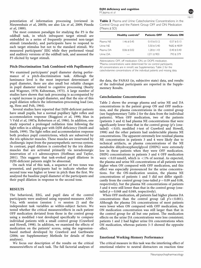
Table 2 shows the average plasma and urine NE and DAconcentrations in the patient group ON and OFF medica-tion, and the plasma concentrations in the control group(see Supplementary Table 2 for the data from the individualpatients). When OFF medication, two of the patients(patients 3 and 4) had plasma NE concentrations that weresignificantly lower than that in the control group (p’s (one-tailed) o0.03; modified t-test of Crawford and Howell,1998) and the other patients had undetectable plasma NEconcentrations. The apparent extremely low residual plasmaNE concentration in patients 3 and 4 were likely due totechnical artifacts, as plasma concentrations of the NEmetabolite dihydroxyphenylglycol (DHPG) were extremelylow in these patients when they were OFF medication.DHPG concentrations in patients 3 and 4 OFF medicationwere o0.03 nmol/l, which is o1% of normal. As expected,the plasma and urine NE concentrations of all patients werehigher when ON compared with OFF medication, and thiseffect was especially pronounced for the urine concentra-tions. For the ON-medication session, the plasma NEconcentrations of patients 1 and 5 did not differ signifi-cantly from the control group (one-tailed p¼ 0.09 and 0.08,respectively), but the plasma NE concentrations of patients3 and 4 were still lower than that in the control group (one-tailed p¼ 0.048 and 0.049, respectively).
When OFF medication, all patients had higher plasma DAconcentrations than the control group (all p’so0.001).Although the plasma DA concentrations of most patientswere lower when ON compared with OFF medication, theON medication concentration was still larger than that inthe control group for all but one patient. The medicationeffects on the urine DA concentrations were less consistent;patients 1 and 2 had higher urine DA concentrations whenON medication, whereas patients 3–5 showed the oppositeeffect.
Emotional Working-Memory Performance
The critical measure in this task was the interfering effect ofemotional relative to neutral distractors on reaction time
Table 2 Plasma and Urine Catecholamine Concentrations in theControl Group and the Patient Group OFF and ON Medication(Means±SD)
Healthy controlsa Patients OFF Patients ON
Plasma NE 1.46±0.45 0.10±0.12 0.57±0.13
Urine NE F 5.50±5.40 9682±4839
Plasma DA 0.06±0.02 1.28±1.43 0.40±0.40
Urine DA F 1271±903 793±379
Abbreviations: OFF, off medication; ON, on DOPS medication.aPlasma concentrations were determined for six control participants.All concentrations are in nmol/l; see Supplementary Table 2 for thecatecholamine concentrations of the individual patients and missing data.
DbH deficiency and cognitionM Jepma et al
1611
Neuropsychopharmacology

(RT). As expected, the control participants responded moreslowly on trials with emotional compared to neutraldistractors (F(1, 7)¼ 14.7, p¼ 0.006). In addition, consistentwith previous studies (Oei et al, 2009, 2010), distractor typeinteracted with target presence (F(1, 7)¼ 16.3, p¼ 0.005),
indicating that the emotional-interference effect on RT wassignificant on target-present trials (F(1, 7)¼ 43.9, po0.001;effect range¼ 80–299 ms) but not on target-absent trials(F(1, 7)¼ 0.75, p¼ 0.42).
Figure 1 shows the average increase in correct RT on trialswith emotional relative to neutral distractors as a functionof target presence, in the control group and in the patientgroup OFF and ON medication. When OFF medication, allpatients showed an emotion-related slowing of responses ontarget-present trials that did not differ from the effect in thecontrol group (effect range¼ 72–226 ms; all t’s (7) o0.8; p’s40.24; Table 3; see Supplementary Figure 2 for theindividual effects). In addition, all patients showed asmaller emotional interference effect when they were ONcompared to OFF medication, but this medication effect didnot differ significantly from the control group’s practiceeffect in any of the patients (all p’s 40.08; Table 3). Thenormal emotional-interference effect in the patients OFFmedication, and the finding that this interference effect wasless pronounced when the patients were ON medication areboth remarkable given the evidence that emotional-inter-ference effects are normally mediated by NE.
Figure 1 Average emotional-interference effect (ie, RT on trials withemotional relative to neutral distractors) for the control group and thepatient group OFF and ON medication, as a function of target presence(error bars are SEM). Because session did not interact with distractor typeor target presence in the control group, the results from the control groupare averaged across the two sessions.
Table 3 For Each Critical Effect/Measure, the P-Value Reflecting the Significance of the Difference Between Each Patient’s OFF MedicationScore and the Average Score of the Control Group (Crawford and Howell, 1998), and the P-Value Indicating the Significance of theDeviation of Each Patient’s Medication Effect from the Control Group’s Practice Effect (Crawford and Garthwaite, 2006)
Patient
1 2 3 4 5
Patient OFF medication vs control group
Emotional-interference effect on RT in target-present trials F 0.44 0.25 0.29 0.41
Attentional-blink size 0.051 F 0.19 0.10 0.38
Visual search efficiency in target-present trials 0.36 0.15 0.14 0.15 0.41
Visual search efficiency in target-absent trials 0.41 0.23 0.36 0.24 0.50
P3 amplitude auditory oddball task 0.10 0.34 0.16 0.09 0.04
P3 amplitude visual oddball task 0.27 0.32 0.13 0.048 0.01
Baseline pupil diameter F 0.03 0.45 0.22 0.002a
Pupil dilation response F 0.03 0.003 0.21 0.001
Brain volume (dm3) 0.29 0.006 0.03 0.01 0.02
% Gray matter 0.46 0.16 0.16 0.10 0.053
% White matter 0.33 0.39 0.33 0.46 0.12
% Cerebrospinal fluid 0.39 0.30 0.45 0.30 0.35
Patient’s medication effect vs control group’s practice effect
Emotional-interference effect on RT in target-present trials F 0.19 0.09 0.18 0.19
Attentional-blink size 0.045 F 0.003 0.049 0.24
Visual search efficiency in target-present trials 0.45 0.28 0.28 0.28 0.39
Visual search efficiency in target-absent trials 0.22 0.11 0.18 0.50 0.16
P3 amplitude auditory oddball task 0.19 0.44 0.41 0.38 0.19
P3 amplitude visual oddball task 0.21 0.27 0.29 0.36 0.35
Baseline pupil diameter F 0.25 0.20 0.003 0.21
Pupil dilation response F 0.34 0.03 0.04 0.08
aThis patient had significantly larger pupils than the control group, which was due to a genetic defect unrelated to DBH deficiency: a mosaic deletion at chromosome11p13 (Erez et al, 2010).The p-values o0.05, which indicate that the estimated percentage of the normal population that would show a more extreme effect is o5%, are bold-faced.(F) Indicates that no data were collected.
DbH deficiency and cognitionM Jepma et al
1612
Neuropsychopharmacology
The full factorial analysis of the effects of target presence,working-memory load, distractor type, and session oncorrect RT and accuracy in the control group is reported inthe Supplementary Results and in Supplementary Figure 1.
Attentional-Blink Performance
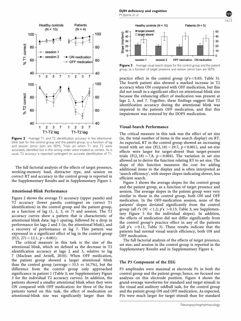
Figure 2 shows the average T1 accuracy (upper panels) andT2 accuracy (lower panels; contingent on correct T1identification) in the control group and the patient group,as a function of lag (1, 2, 3, or 7) and session. The T2accuracy curves show a pattern that is characteristic ofattentional blink data: lag-1 sparing, followed by a drop inperformance for lags 2 and 3 (ie, the attentional blink), anda recovery of performance at lag 7. This pattern wasexpressed in a significant effect of lag in the control group(F(3, 27)¼ 12.1, p¼ 0.001).
The critical measure in this task is the size of theattentional blink, which we defined as the decrease in T2identification accuracy at lags 2 and 3, relative to lag7 (Maclean and Arnell, 2010). When OFF medication,the patient group showed a larger attentional blinkthan the control group (average¼ 33.5 vs 16.7%), but thedifference from the control group only approachedsignificance in patient 1 (Table 3; see Supplementary Figure3 for the individual T2 accuracy curves). In addition, thepatients showed a smaller attentional blink when they wereON compared with OFF medication: for three of the fourpatients tested on this task, the effect of medication onattentional-blink size was significantly larger than the
practice effect in the control group (p’so0.05; Table 3).The fourth patient also showed a marked increase in T2accuracy when ON compared with OFF medication, but thisdid not result in a significant effect on attentional-blink sizebecause the enhancing effect of medication was present atlags 2, 3, and 7. Together, these findings suggest that T2identification accuracy during the attentional blink wasimpaired in the patients OFF medication, and that thisimpairment was restored by the DOPS medication.
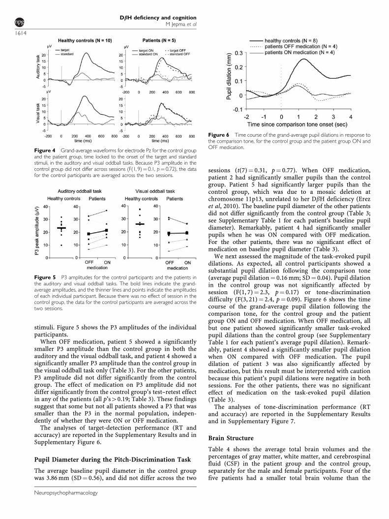
Visual-Search Performance
The critical measure in this task was the effect of set size(ie, the total number of items in the search display) on RT.As expected, RT in the control group showed an increasingtrend with set size (F(2, 18)¼ 29.7, po0.001), and set-sizeeffects were larger for target-absent than target-presenttrials (F(2, 18)¼ 7.8, p¼ 0.004). The variation in set sizeallowed us to derive the function relating RT to set size. Theslope of this function measures the cost for addingadditional items to the display and is often interpreted as‘search efficiency’, with steeper slopes indicating slower, lessefficient search.
Figure 3 shows the average slopes for the control groupand the patient group, as a function of target presence andsession. The average slopes in the patient group were verysimilar to those in the control group, both ON and OFFmedication. In the OFF-medication session, none of thepatients’ slopes deviated significantly from the controlgroup (all t’s (9) o1.2; p’s 40.13; Table 3; see Supplemen-tary Figure 5 for the individual slopes). In addition,the effects of medication did not differ significantly fromthe control group’s practice effect in any of the patients(all p’s 40.11; Table 3). These results indicate that thepatients had normal visual search efficiency, both ON andOFF medication.
The full factorial analysis of the effects of target presence,set size, and session in the control group is reported in theSupplementary Results and in Supplementary Figure 4.
The P3 Component of the EEG
P3 amplitudes were maximal at electrode Pz in both thecontrol group and the patient group; hence, we focused ouranalyses on this electrode position. Figure 4 shows thegrand-average waveforms for standard and target stimuli inthe visual and auditory oddball task, for the control groupand the patient group ON and OFF medication. As expected,P3s were much larger for target stimuli than for standard
Figure 2 Average T1 and T2 identification accuracy in the attentional-blink task for the control group and the patient group, as a function of lagand session (error bars are SEM). Trials on which T1 and T2 wereaccurately identified but in the wrong order were treated as correct. As isusual, T2 accuracy is reported contingent on accurate identification of T1.
Figure 3 Average visual-search slopes for the control group and the patientgroup, as a function of target presence and session (error bars are SEM).
DbH deficiency and cognitionM Jepma et al
1613
Neuropsychopharmacology
stimuli. Figure 5 shows the P3 amplitudes of the individualparticipants.
When OFF medication, patient 5 showed a significantlysmaller P3 amplitude than the control group in both theauditory and the visual oddball task, and patient 4 showed asignificantly smaller P3 amplitude than the control group inthe visual oddball task only (Table 3). For the other patients,P3 amplitude did not differ significantly from the controlgroup. The effect of medication on P3 amplitude did notdiffer significantly from the control group’s test–retest effectin any of the patients (all p’s40.19; Table 3). These findingssuggest that some but not all patients showed a P3 that wassmaller than the P3 in the normal population, indepen-dently of whether they were ON or OFF medication.
The analyses of target-detection performance (RT andaccuracy) are reported in the Supplementary Results and inSupplementary Figure 6.
Pupil Diameter during the Pitch-Discrimination Task
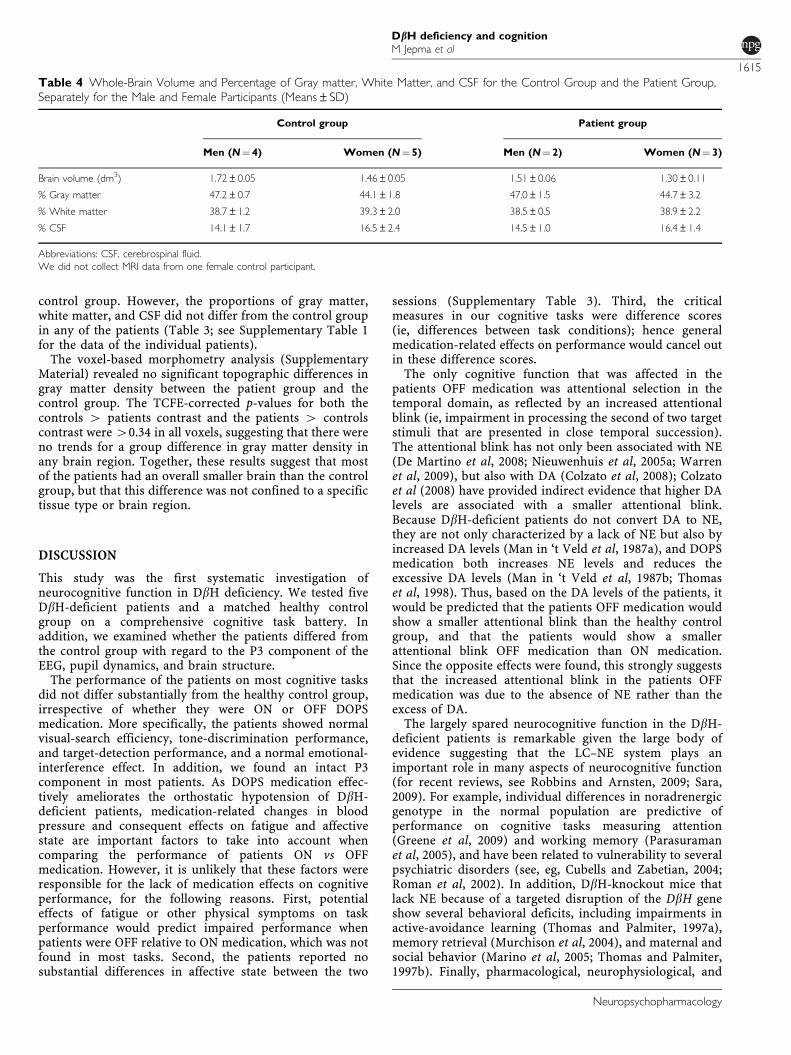
The average baseline pupil diameter in the control groupwas 3.86 mm (SD¼ 0.56), and did not differ across the two
sessions (t(7)¼ 0.31, p¼ 0.77). When OFF medication,patient 2 had significantly smaller pupils than the controlgroup. Patient 5 had significantly larger pupils than thecontrol group, which was due to a mosaic deletion atchromosome 11p13, unrelated to her DbH deficiency (Erezet al, 2010). The baseline pupil diameter of the other patientsdid not differ significantly from the control group (Table 3;see Supplementary Table 1 for each patient’s baseline pupildiameter). Remarkably, patient 4 had significantly smallerpupils when he was ON compared with OFF medication.For the other patients, there was no significant effect ofmedication on baseline pupil diameter (Table 3).
We next assessed the magnitude of the task-evoked pupildilations. As expected, all control participants showed asubstantial pupil dilation following the comparison tone(average pupil dilation¼ 0.16 mm; SD¼ 0.04). Pupil dilationin the control group was not significantly affected bysession (F(1, 7)¼ 2.3, p¼ 0.17) or tone-discriminationdifficulty (F(3, 21)¼ 2.4, p¼ 0.09). Figure 6 shows the timecourse of the grand-average pupil dilation following thecomparison tone, for the control group and the patientgroup ON and OFF medication. When OFF medication, allbut one patient showed significantly smaller task-evokedpupil dilations than the control group (see SupplementaryTable 1 for each patient’s average pupil dilation). Remark-ably, patient 4 showed a significantly smaller pupil dilationwhen ON compared with OFF medication. The pupildilation of patient 3 was also significantly affected bymedication, but this result must be interpreted with cautionbecause this patient’s pupil dilations were negative in bothsessions. For the other patients, there was no significanteffect of medication on the task-evoked pupil dilation(Table 3).
The analyses of tone-discrimination performance (RTand accuracy) are reported in the Supplementary Resultsand in Supplementary Figure 7.
Brain Structure
Table 4 shows the average total brain volumes and thepercentages of gray matter, white matter, and cerebrospinalfluid (CSF) in the patient group and the control group,separately for the male and female participants. Four of thefive patients had a smaller total brain volume than the
Figure 4 Grand-average waveforms for electrode Pz for the control groupand the patient group, time locked to the onset of the target and standardstimuli, in the auditory and visual oddball tasks. Because P3 amplitude in thecontrol group did not differ across sessions (F(1, 9)¼ 0.1, p¼ 0.72), the datafor the control participants are averaged across the two sessions.
Figure 6 Time course of the grand-average pupil dilations in response tothe comparison tone, for the control group and the patient group ON andOFF medication.
Figure 5 P3 amplitudes for the control participants and the patients inthe auditory and visual oddball tasks. The bold lines indicate the grand-average amplitudes, and the thinner lines and points indicate the amplitudesof each individual participant. Because there was no effect of session in thecontrol group, the data for the control participants are averaged across thetwo sessions.
DbH deficiency and cognitionM Jepma et al
1614
Neuropsychopharmacology
control group. However, the proportions of gray matter,white matter, and CSF did not differ from the control groupin any of the patients (Table 3; see Supplementary Table 1for the data of the individual patients).
The voxel-based morphometry analysis (SupplementaryMaterial) revealed no significant topographic differences ingray matter density between the patient group and thecontrol group. The TCFE-corrected p-values for both thecontrols 4 patients contrast and the patients 4 controlscontrast were 40.34 in all voxels, suggesting that there wereno trends for a group difference in gray matter density inany brain region. Together, these results suggest that mostof the patients had an overall smaller brain than the controlgroup, but that this difference was not confined to a specifictissue type or brain region.
DISCUSSION
This study was the first systematic investigation ofneurocognitive function in DbH deficiency. We tested fiveDbH-deficient patients and a matched healthy controlgroup on a comprehensive cognitive task battery. Inaddition, we examined whether the patients differed fromthe control group with regard to the P3 component of theEEG, pupil dynamics, and brain structure.
The performance of the patients on most cognitive tasksdid not differ substantially from the healthy control group,irrespective of whether they were ON or OFF DOPSmedication. More specifically, the patients showed normalvisual-search efficiency, tone-discrimination performance,and target-detection performance, and a normal emotional-interference effect. In addition, we found an intact P3component in most patients. As DOPS medication effec-tively ameliorates the orthostatic hypotension of DbH-deficient patients, medication-related changes in bloodpressure and consequent effects on fatigue and affectivestate are important factors to take into account whencomparing the performance of patients ON vs OFFmedication. However, it is unlikely that these factors wereresponsible for the lack of medication effects on cognitiveperformance, for the following reasons. First, potentialeffects of fatigue or other physical symptoms on taskperformance would predict impaired performance whenpatients were OFF relative to ON medication, which was notfound in most tasks. Second, the patients reported nosubstantial differences in affective state between the two
sessions (Supplementary Table 3). Third, the criticalmeasures in our cognitive tasks were difference scores(ie, differences between task conditions); hence generalmedication-related effects on performance would cancel outin these difference scores.
The only cognitive function that was affected in thepatients OFF medication was attentional selection in thetemporal domain, as reflected by an increased attentionalblink (ie, impairment in processing the second of two targetstimuli that are presented in close temporal succession).The attentional blink has not only been associated with NE(De Martino et al, 2008; Nieuwenhuis et al, 2005a; Warrenet al, 2009), but also with DA (Colzato et al, 2008); Colzatoet al (2008) have provided indirect evidence that higher DAlevels are associated with a smaller attentional blink.Because DbH-deficient patients do not convert DA to NE,they are not only characterized by a lack of NE but also byincreased DA levels (Man in ‘t Veld et al, 1987a), and DOPSmedication both increases NE levels and reduces theexcessive DA levels (Man in ‘t Veld et al, 1987b; Thomaset al, 1998). Thus, based on the DA levels of the patients, itwould be predicted that the patients OFF medication wouldshow a smaller attentional blink than the healthy controlgroup, and that the patients would show a smallerattentional blink OFF medication than ON medication.Since the opposite effects were found, this strongly suggeststhat the increased attentional blink in the patients OFFmedication was due to the absence of NE rather than theexcess of DA.
The largely spared neurocognitive function in the DbH-deficient patients is remarkable given the large body ofevidence suggesting that the LC–NE system plays animportant role in many aspects of neurocognitive function(for recent reviews, see Robbins and Arnsten, 2009; Sara,2009). For example, individual differences in noradrenergicgenotype in the normal population are predictive ofperformance on cognitive tasks measuring attention(Greene et al, 2009) and working memory (Parasuramanet al, 2005), and have been related to vulnerability to severalpsychiatric disorders (see, eg, Cubells and Zabetian, 2004;Roman et al, 2002). In addition, DbH-knockout mice thatlack NE because of a targeted disruption of the DbH geneshow several behavioral deficits, including impairments inactive-avoidance learning (Thomas and Palmiter, 1997a),memory retrieval (Murchison et al, 2004), and maternal andsocial behavior (Marino et al, 2005; Thomas and Palmiter,1997b). Finally, pharmacological, neurophysiological, and
Table 4 Whole-Brain Volume and Percentage of Gray matter, White Matter, and CSF for the Control Group and the Patient Group,Separately for the Male and Female Participants (Means±SD)
Control group Patient group
Men (N¼4) Women (N¼5) Men (N¼2) Women (N¼ 3)
Brain volume (dm3) 1.72±0.05 1.46±0.05 1.51±0.06 1.30±0.11
% Gray matter 47.2±0.7 44.1±1.8 47.0±1.5 44.7±3.2
% White matter 38.7±1.2 39.3±2.0 38.5±0.5 38.9±2.2
% CSF 14.1±1.7 16.5±2.4 14.5±1.0 16.4±1.4
Abbreviations: CSF, cerebrospinal fluid.We did not collect MRI data from one female control participant.
DbH deficiency and cognitionM Jepma et al
1615
Neuropsychopharmacology
lesion studies in animals suggest that the LC–NE systemplays a crucial role in regulating the optimization ofbehavioral performance (see, eg, Aston-Jones and Cohen,2005; Bouret and Sara, 2005). However, it must be notedthat our task battery did not address all aspects of cognitivefunction. For example, we did not assess higher-levelcognitive functions such as executive control and explora-tory behavior. Therefore, our results leave open thepossibility that the patients have subtle cognitive deficitsthat were not revealed by our task battery. In addition,although our data clearly indicate that there were nosubstantial abnormalities in the performance of the patientson our test battery, it cannot be excluded that there weresome subtle differences that failed to reach significancebecause of a lack of power of our experimental design.
Although the relatively normal performance of thepatients on our cognitive task battery is striking, it isconsistent with informal clinical observations that DbH-deficient patients do not have obvious cognitive impair-ments or psychiatric disorders. Indeed, the absence ofmental problems in most DbH-deficient patients who havebeen encountered so far has intrigued investigators in theareas of depression and schizophrenia (Cubells andZabetian, 2004). It is especially remarkable that the patientsOFF medication did not show impaired performance oncognitive tasks that are normally mediated by the LC–NEsystem (eg, the emotional working-memory task), andshowed a relatively intact P3 component, which is thoughtto reflect the noradrenergic potentiation of informationprocessing (Liu et al, 2009; Nieuwenhuis et al, 2005b;Pineda et al, 1989). These findings suggest that alternativeneural mechanisms and/or neuromodulatory systems com-pensate for the absence of NE in DbH-deficient patients.Previous findings that DbH-deficient patients have arelatively normal sleep pattern (Tulen et al, 1990, 1991),although the sleep–wake cycle is normally mediated by theLC–NE system (Hobson et al, 1986; Jouvet, 1969), areconsistent with this idea.
Since DbH is responsible for the conversion of DA to NE,it is thought that DA rather than NE is stored and releasedby noradrenergic neurons in DbH-deficient patients.Indeed, plasma DA levels in DBH-deficient patients respondto various physiological and pharmacological manipula-tions that normally affect plasma NE levels (Man in ‘t Veldet al, 1987a; Robertson et al, 1986), although it remains tobe determined whether this also applies to DA levels in thecentral nervous system. Thus, a possible explanation for thespared neurocognitive function in DbH deficiency is thatDA has, to some extent, taken over the function of NE in thebrains of DbH-deficient patients. Obviously, a functionalreplacement of NE by DA would require the presence ofpostsynaptic receptors with DA affinity in noradrenergicsynapses. Studies in mice suggest that some a2-adrenergicreceptor subtypes have a comparable affinity for DA and NE(Zhang et al, 1999), whereas a1- and b-adrenergic receptorshave a much lower affinity for DA than for NE (Zhang et al,2004). However, as the congenital absence of NE may havealtered the affinity of adrenergic receptors, it is unknownwhether the same receptor characteristics apply to DbH-deficient patients. Another possible explanation for afunctional replacement of NE by DA is that DbH-deficientpatients have an increased density of postsynaptic DA
receptors on noradrenergic synapses. A recent positronemission tomography (PET) study in mice suggests thatDbH-knockout mice have a normal density of D2 dopaminereceptors in the high-affinity state (Skinbjerg et al, 2010),which does not support this hypothesis. However, as resultsfrom DbH-knockout mice might not be generalizable tohuman DbH-deficient patients, the assessment of DAreceptor densities in human DbH-deficient patients, forexample using PET scanning, remains an importantobjective for future studies.
It is interesting to note that the first study that used genetargeting to produce DbH-deficient mice found that themajority of DbH-deficient embryos died in mid-gestationand only 5% reached adulthood (Thomas et al, 1995). Toprevent embryonic lethality, subsequent studies using DbH-knockout mice have supplied the embryos with adrenergicagonists (isoproterenol and phenylephrine) and DOPS viathe maternal drinking water, such that NE is present in theDbH-knockout mice until birth. The results of Thomas et al(1995) suggest that the human DbH-deficient patients mayrepresent the minority of DbH-deficiency cases who havesurvived this condition. If this is true, an interestingspeculation is that these patients were able to survivebecause they happened to have optimal dopaminergic ornoradrenergic genotypes to compensate for the absence ofNE. Future studies might assess this possibility by examin-ing whether the frequency of occurrence of specific alleles ofdopaminergic and noradrenergic genes (eg, the COMT,DAT, and the dopamine and noradrenergic receptor genes)in DbH-deficient patients deviates from those in the normalpopulation.
In contrast to the generally normal neurocognitivefunction in the DbH-deficient patients, we did find clearabnormalities in their task-evoked pupil dilation response.The task-evoked pupil dilation was very small or absent inmost of the patients, which might be due to a decreasednoradrenergic innervation of the iris dilator muscle.However, it is also possible that the abnormal pupildynamics in some of the patients resulted from ocularabnormalities unrelated to their DBH deficiency; this mightexplain why the pupil-dilation response was not restored byDOPS medication. Importantly, the small or absent task-evoked pupil dilations of the patients did not reflect adecreased processing of the task-related stimuli, as theirperformance on the tone-discrimination task, during whichtheir pupils were measured, was not impaired.
The patient group also differed from the control groupwith regard to total brain volume: all but one patient had asignificantly smaller brain volume than the control group, butthe relative proportions of gray matter, white matter, and CSF,and the distribution of gray matter volume across the braindid not deviate from those in the control group. The smallerbrain volume in most of the DBH-deficient patients is in linewith recent findings suggesting that NE has a neurotrophiceffect on cortical neurons (see, eg, Counts and Mufson, 2010;Kalinin et al, 2007; Madrigal et al, 2007, 2009). Apparently, thedecreased brain volume of the patients did not result incognitive impairments; this suggests that although thepatients have a smaller number of neurons, their neuronsare intact and make proper connections.
To conclude, our findings suggest that neurocognitivefunction in human DbH-deficient patients is largely spared,
DbH deficiency and cognitionM Jepma et al
1616
Neuropsychopharmacology
even when they are OFF medication, but that their totalbrain volume is smaller than that of the normal population.The normal neurocognitive function in DbH-deficientpatients is striking given the important role of NE innormal cognition, but corroborates informal clinicalobservations that most patients do not have obviouscognitive impairments. Our findings suggest that DbH-deficient patients have developed alternative mechanisms tocompensate for the absence of NE in the brain, possiblythrough a functional replacement of NE by DA; the natureof these compensatory mechanisms remains to be exploredby future studies.
ACKNOWLEDGEMENTS
This work was supported by the Netherlands Organizationfor Scientific Research. We thank all patients for participat-ing in the study, and Argho Ray, Rachel van der Ham,Andre Keizer, Sasha Key, Bonnie K Black, and SusanWilliams for their technical assistance.
DISCLOSURE
The authors declare no conflict of interest.
REFERENCES
Aghajanian GK, Cedarbaum JM, Wang RY (1977). Evidence fornorepinephrine-mediated collateral inhibition of locus coeruleusneurons. Brain Res 136: 570–577.
Aston-Jones G, Cohen JD (2005). An integrative theory of locuscoeruleus-norepinephrine function: adaptive gain and optimalperformance (Review). Annu Rev Neurosci 28: 403–450.
Aston-Jones G, Foote SL, Bloom FE (1984). Anatomy andphysiology of locus coeruleus neurons: functional implications.In: Ziegler M, Lake CR (eds). Norepinephrine. Frontiers ofClinical Neuroscience, Vol 2. Williams and Wilkins: Baltimore,MD. pp 92–116.
Aston-Jones G, Rajkowski J, Cohen J (2000). Locus coeruleus andregulation of behavioral flexibility and attention. Prog Brain Res126: 165–182.
Beatty J, Wagoner BL (1978). Pupillometric signs of brainactivation vary with level of cognitive processing. Science 199:1216–1218.
Berridge CW, Waterhouse BD (2003). The locus coeruleus-noradrenergic system: modulation of behavioral state andstate-dependent cognitive processes. Brain Res Brain Res Rev42: 33–84.
Biaggioni I, Goldstein DS, Atkinson T, Robertson D (1990).Dopamine-beta-hydroxylase deficiency in humans. Neurology40: 370–373.
Biaggioni I, Robertson D (1987). Endogenous restoration ofnoradrenaline by precursor therapy in dopamine beta-hydro-xylase deficiency. Lancet 2: 1170–1172.
Bouret S, Sara SJ (2005). Network reset: a simplified overarchingtheory of locus coeruleus noradrenaline function (Review).Trends Neurosci 28: 574–582.
Buchner A, Rothermund K, Wentura D, Mehl B (2004). Valence ofdistractor words increases the effects of irrelevant speech onserial recall. Mem Cognit 32: 722–731.
Cahill L, McGaugh JL (1998). Mechanisms of emotional arousaland lasting declarative memory (Review). Trends Neurosci 21:294–299.
Chamberlain SR, Muller U, Blackwell AD, Robbins TW, SahakianBJ (2006). Noradrenergic modulation of working memory andemotional memory in humans (Review). Psychopharmacology(Berl) 188: 397–407.
Cheshire Jr WP, Dickson DW, Nahm KF, Kaufmann HC,Benarroch EE (2006). Dopamine beta-hydroxylase deficiencyinvolves the central autonomic network. Acta Neuropathol 112:227–229.
Cohen JD, Aston-Jones G, Gilzenrat MS (2004). A systems-leveltheory on attention and cognitive control: guided activation,adaptive gating, conflict monitoring, and exploitation versusexploration. In: Posner MI (ed). Cognitive Neuroscience ofAttention. Guilford Press: New York. pp 71–90.
Colzato LS, Slagter HA, Spape MM, Hommel B (2008). Blinks of theeye predict blinks of the mind. Neuropsychologia 46: 3179–3183.
Counts SE, Mufson EJ (2010). Noradrenaline activation ofneurotrophic pathways protects against neuronal amyloidtoxicity. J Neurochem 113: 649–660.
Crawford JR, Garthwaite PH (2006). Comparing patients’ predictedtest scores from a regression equation with their obtained scores:a significance test and point estimate of abnormality withaccompanying confidence limits. Neuropsychology 20: 259–271.
Crawford JR, Howell DC (1998). Comparing an individual’s testscore against norms derived from small samples. Clin Neurop-sychol 12: 482–486.
Cubells JF, Zabetian CP (2004). Human genetics of plasmadopamine beta-hydroxylase activity: applications to research inpsychiatry and neurology. Psychopharmacology (Berl) 174: 463–476.
Dayan P, Yu AJ (2006). Phasic norepinephrine: a neural interruptsignal for unexpected events. Network 17: 335–350.
De Martino B, Strange BA, Dolan RJ (2008). Noradrenergicneuromodulation of human attention for emotional and neutralstimuli. Psychopharmacology (Berl) 197: 127–136.
Deinum J, Steenbergen-Spanjers GC, Jansen M, Boomsma F,Lenders JW, van Ittersum FJ et al (2004). DBH gene variants thatcause low plasma dopamine beta hydroxylase with or without asevere orthostatic syndrome. J Med Genet 41: e38.
Dolcos F, McCarthy G (2006). Brain systems mediating cognitiveinterference by emotional distraction. J Neurosci 26: 2072–2079.
Erez A, Li J, Geraghty MT, Ben-Shachar S, Cooper ML, Mensing DEet al (2010). Mosaic deletion 11p13 in a child with dopaminebeta-hydroxylase deficiencyFcase report and review of theliterature (Review). Am J Med Genet A 152A: 732–736.
Goldstein DS (2006). L-Dihydroxyphenylserine (L-DOPS): anorepinephrine prodrug (Review). Cardiovasc Drug Rev 24:189–203.
Greene CM, Bellgrove MA, Gill M, Robertson IH (2009).Noradrenergic genotype predicts lapses in sustained attention.Neuropsychologia 47: 591–594.
Greenwood PM, Fossella JA, Parasuraman R (2005). Specificityof the effect of a nicotinic receptor polymorphism on individualdifferences in visuospatial attention. J Cogn Neurosci 17:1611–1620.
Hess EH, Polt JM (1964). Pupil size in relation to mental activityduring simple problem-solving. Science 143: 1190–1192.
Hobson JA, Lydic R, Bahdoyan HA (1986). Evolving concepts ofsleep cycle generation: from brain centers to neuronal popula-tion. Behav Brain Sci 9: 371–448.
Hoffman BB, Taylor P (2001). Neurotransmission: the autonomicand somatic motor nervous system. In: Hardman JG, LimbirdLE, Molinoff PB, Gilman AG (eds). Goodman & Gilman’s ThePharmacological Basis of Therapeutics. McGraw-Hill: New Yorkpp 115–153.
Hommel B, Akyurek EG (2005). Lag-1 sparing in the attentionalblink: benefits and costs of integrating two events into a singleepisode. Q J Exp Psychol A 58: 1415–1433.
Ishikawa Y, Kato Y, Murakami Y, Inoue T, Koshiyama H, Imura H(1987). Effect of L-threo-3,4-dihydroxyphenylserine (L-DOPS)
DbH deficiency and cognitionM Jepma et al
1617
Neuropsychopharmacology
on catecholamine levels in plasma and cerebrospinal fluid (CSF)in anesthetized rats. Proc Soc Exp Biol Med 184: 197–200.
Jouvet M (1969). Biogenic amines and the states of sleep. Science163: 32–41.
Kahneman D (1973). Attention and Effort. Prentice-Hall:Englewood Cliffs, NJ.
Kalinin S, Gavrilyuk V, Polak PE, Vasser R, Zhao J, Heneka MTet al (2007). Noradrenaline deficiency in brain increases beta-amyloid plaque burden in an animal model of Alzheimer’sdisease. Neurobiol Aging 28: 1206–1214.
Kato T, Karai N, Katsuyama M, Nakamura M, Katsube J Studies onthe activity of L-threo-3,4-dihydroxyphenylserine (L-DOPS) as acatecholamine precursor in the brain. Comparison with that ofL-dopa. Biochem Pharmacol, (1987a) 36: 3051–3057.
Kato T, Katsuyama M, Karai N, Nakamura M, Katsube J Studies onthe central action of L-threo-3,4-dihydroxyphenyl-serine(L-threo-DOPS) in FLA-63-treated mice. Pharmacol BiochemBehav, (1987b) 26: 407–411.
Liu J, Kiehl KA, Pearlson G, Perrone-Bizzozero NI, Eichele T,Calhoun VD (2009). Genetic determinants of target and novelty-related event-related potentials in the auditory oddball response.Neuroimage 46: 809–816.
Maclean MH, Arnell KM (2010). Personality predicts temporalattention costs in the attentional blink paradigm. Psychon BullRev 17: 556–562.
Madrigal JL, Kalinin S, Richardson JC, Feinstein DL (2007).Neuroprotective actions of noradrenaline: effects on glutathionesynthesis and activation of peroxisome proliferator activatedreceptor delta. J Neurochem 103: 2092–2101.
Madrigal JL, Leza JC, Polak P, Kalinin S, Feinstein DL (2009).Astrocyte-derived MCP-1 mediates neuroprotective effects ofnoradrenaline. J Neurosci 29: 263–267.
Man in ‘t Veld AJ, Boomsma F, Moleman P, Schalekamp MACongenital dopamine-beta hydroxylase deficiency. A novelorthostatic syndrome. Lancet, (1987a) 1: 183–188.
Man in ‘t Veld AJ, Boomsma F, van den Meiracker AH,Schalekamp MA Effect of unnatural noradrenaline precursoron sympathetic control and orthostatic hypotension in dopa-mine-beta-hydroxylase deficiency. Lancet, (1987b) 2: 1172–1175.
Marino MD, Bourdelat-Parks BN, Cameron Liles L, Weinshenker D(2005). Genetic reduction of noradrenergic function alters socialmemory and reduces aggression in mice. Behav Brain Res 161:197–203.
Martens S, Wyble B (2010). The attentional blink: past, present,and future of a blind spot in perceptual awareness (Review).Neurosci Biobehav Rev 34: 947–957.
Mathias CJ, Bannister RB, Cortelli P, Heslop K, Polak JM,Raimbach S et al (1990). Clinical, autonomic and therapeuticobservations in two siblings with postural hypotension andsympathetic failure due to an inability to synthesize noradrena-line from dopamine because of a deficiency of dopamine betahydroxylase. Q J Med 75: 617–633.
Murchison CF, Zhang XY, Zhang WP, Ouyang M, Lee A, ThomasSA (2004). A distinct role for norepinephrine in memoryretrieval. Cell 117: 131–143.
Nieuwenhuis S, Gilzenrat MS, Holmes BD, Cohen JD The role of thelocus coeruleus in mediating the attentional blink: a neurocompu-tational theory. J Exp Psychol Gen, (2005a) 134: 291–307.
Nieuwenhuis S, Aston-Jones G, Cohen JD Decision making, the P3,and the locus coeruleus norepinephrine system (Review).Psychol Bull, (2005b) 131: 510–532.
Nieuwenhuis S, van Nieuwpoort IC, Veltman DJ, Drent ML (2007).Effects of the noradrenergic agonist clonidine on temporal andspatial attention. Psychopharmacology (Berl) 193: 261–269.
Oei NY, Tollenaar MS, Elzinga BM, Spinhoven P (2010).Propranolol reduces emotional distraction in working memory:a partial mediating role of propranolol-induced cortisolincreases? Neurobiol Learn Mem 93: 388–395.
Oei NY, Tollenaar MS, Spinhoven P, Elzinga BM (2009).Hydrocortisone reduces emotional distracter interference inworking memory. Psychoneuroendocrinology 34: 1284–1293.
Parasuraman R, Greenwood PM, Kumar R, Fossella J (2005).Beyond heritability: neurotransmitter genes differentially mod-ulate visuospatial attention and working memory. Psychol Sci 16:200–207.
Peeters FPML, Ponds RWHM, Vermeeren MTG (1996). Affectivityand self-report of depression and anxiety (Dutch). Tijdschr Psych38: 240–250.
Pineda JA, Foote SL, Neville HJ (1989). Effects of locus coeruleuslesions on auditory, long-latency, event-related potentials inmonkey. J Neurosci 9: 81–93.
Raven JC, Court JH, Raven J (1988). Manual for Raven’sProgressive Matrices and Vocabulary Scales: Section 3 StandardProgressive Matrices. Lewis: London.
Raymond JE, Shapiro KL, Arnell KM (1992). Temporary suppres-sion of visual processing in an RSVP task: an attentional blink?J Exp Psychol Hum Percept Perform 18: 849–860.
Ressler KJ, Nemeroff CB (2001). Role of norepinephrine in thepathophysiology of neuropsychiatric disorders. CNS Spectr 6:663–666.
Robbins TW (1997). Arousal systems and attentional processes(Review). Biol Psychol 45: 57–71.
Robbins TW, Arnsten AF (2009). The neuropsychopharmacologyof fronto-executive function: monoaminergic modulation(Review). Annu Rev Neurosci 32: 267–287.
Robertson D, Garland EM (2010). Dopamine Beta-HydroxylaseDeficiency. GeneReviews http://www.ncbi.nlm.nih.gov/books/NBK1474/.
Robertson D, Goldberg MR, Onrot J, Hollister AS, Wiley R,Thompson Jr JG et al (1986). Isolated failure of autonomicnoradrenergic neurotransmission. Evidence for impaired beta-hydroxylation of dopamine. N Engl J Med 314: 1494–1497.
Robertson D, Haile V, Perry SE, Robertson RM, Phillips III JA,Biaggioni I (1991). Dopamine beta-hydroxylase deficiency. Agenetic disorder of cardiovascular regulation. Hypertension 18:1–8.
Roman T, Schmitz M, Polanczyk GV, Eizirik M, Rohde LA, HutzMH (2002). Further evidence for the association betweenattention-deficit/hyperactivity disorder and the dopamine-beta-hydroxylase gene. Am J Med Genet 114: 154–158.
Roth WT, Dorato KH, Kopell BS (1984). Intensity and task effectson evoked physiological responses to noise bursts. Psychophy-siology 21: 466–481.
Sara SJ (2009). The locus coeruleus and noradrenergic modulationof cognition (Review). Nat Rev Neurosci 10: 211–223.
Semba J, Takahashi R (1985). The effects of L-threo-dihydrox-yphenylserine on norepinephrine metabolism in rat brain.Psychiatry Res 15: 319–326.
Servan-Schreiber D, Printz H, Cohen JD (1990). A network modelof catecholamine effects: gain, signal-to-noise ratio, andbehavior. Science 249: 892–895.
Shapiro KL, Caldwell J, Sorensen RE (1997). Personal names andthe attentional blink: a visual ‘cocktail party’ effect. J Exp PsycholHum Percept Perform 23: 504–514.
Siever LJ, Davis KL (1985). Overview: toward a dysregulationhypothesis of depression. Am J Psychiatry 142: 1017–1031.
Skinbjerg M, Seneca N, Liow JS, Hong J, Weinshenker D, Pike VWet al (2010). Dopamine beta-hydroxylase-deficient mice havenormal densities of D(2) dopamine receptors in the high-affinitystate based on in vivo PET imaging and in vitro radioligandbinding. Synapse 64: 699–703.
Smith SA, Smith SE (1999). Bilateral Horner’s syndrome: detectionand occurrence. J Neurol Neurosurg Psychiatry 66: 48–51.
Strange BA, Dolan RJ (2004). Beta-adrenergic modulation ofemotional memory-evoked human amygdala and hippocampalresponses. Proc Natl Acad Sci USA 101: 11454–11458.
DbH deficiency and cognitionM Jepma et al
1618
Neuropsychopharmacology
Strange BA, Hurlemann R, Dolan RJ (2003). An emotion-inducedretrograde amnesia in humans is amygdala- and beta-adrener-gic-dependent. Proc Natl Acad Sci USA 100: 13626–13631.
Sutton S, Braren M, Zubin J, John ER (1965). Evoked-potentialcorrelates of stimulus uncertainty. Science 150: 1187–1188.
Thomas SA, Marck BT, Palmiter RD, Matsumoto AM (1998).Restoration of norepinephrine and reversal of phenotypesin mice lacking dopamine beta-hydroxylase. J Neurochem 70:2468–2476.
Thomas SA, Matsumoto AM, Palmiter RD (1995). Noradrenaline isessential for mouse fetal development. Nature 374: 643–646.
Thomas SA, Palmiter RD Disruption of the dopamine beta-hydroxylase gene in mice suggests roles for norepinephrine inmotor function, learning, and memory. Behav Neurosci, (1997a)111: 579–589.
Thomas SA, Palmiter RD Impaired maternal behavior in micelacking norepinephrine and epinephrine. Cell, (1997b) 91:583–592.
Thompson JM, O’Callaghan CJ, Kingwell BA, Lambert GW,Jennings GL, Esler MD (1995). Total norepinephrine spillover,muscle sympathetic nerve activity and heart-rate spectralanalysis in a patient with dopamine beta-hydroxylase deficiency.J Auton Nerv Syst 55: 198–206.
Timmers HJ, Deinum J, Wevers RA, Lenders JW (2004). Congenitaldopamine-beta-hydroxylase deficiency in humans (Review). AnnNY Acad Sci 1018: 520–523.
Tulen JH, Man in ’t Veld AJ, Dzoljic MR, Mechelse K, Moleman P(1991). Sleeping with and without norepinephrine: effects ofmetoclopramide and D,L-threo-3,4-dihydroxyphenylserine onsleep in dopamine beta-hydroxylase deficiency. Sleep 14: 32–38.
Tulen JH, Man in’t Veld AJ, Mechelse K, Boomsma F (1990). Sleeppatterns in congenital dopamine beta-hydroxylase deficiency.J Neurol 237: 98–102.
Usher M, Cohen JD, Servan-Schreiber D, Rajkowski J, Aston-JonesG (1999). The role of locus coeruleus in the regulation ofcognitive performance. Science 283: 549–554.
Warren CM, Breuer AT, Kantner J, Fiset D, Blais C, Masson ME(2009). Target-distractor interference in the attentional blinkimplicates the locus coeruleus-norepinephrine system. PsychonBull Rev 16: 1106–1111.
Watson D, Clark LA, Tellegen A (1988). Development andvalidation of brief measures of positive and negative affect: thePANAS scales. J Pers Soc Psychol 54: 1063–1070.
Wechsler D. Weschsler Adult Intelligence Scale-III. The Psycho-logical Corporation: San Antonio, TX; (1997).
Zhang W, Klimek V, Farley JT, Zhu MY, Ordway GA (1999).Alpha2C adrenoceptors inhibit adenylyl cyclase in mousestriatum: potential activation by dopamine. J Pharmacol ExpTher 289: 1286–1292.
Zhang WP, Ouyang M, Thomas SA (2004). Potency of catechola-mines and other L-tyrosine derivatives at the cloned mouseadrenergic receptors. Neuropharmacology 47: 438–449.
Supplementary Information accompanies the paper on the Neuropsychopharmacology website (http://www.nature.com/npp)
DbH deficiency and cognitionM Jepma et al
1619
Neuropsychopharmacology