Neuro-ophthalmology sjtu ophthalmology 樊莹. Neuro-ophthalmology an important sensory end organ an...
70
Neuro-ophthalmology Neuro-ophthalmology sjtu ophthalmology sjtu ophthalmology 樊樊 樊樊
-
Upload
alvin-daniel -
Category
Documents
-
view
227 -
download
1
Transcript of Neuro-ophthalmology sjtu ophthalmology 樊莹. Neuro-ophthalmology an important sensory end organ an...

Neuro-ophthalmologyNeuro-ophthalmology
sjtu ophthalmology sjtu ophthalmology 樊莹樊莹

Neuro-ophthalmologyNeuro-ophthalmology an important sensory end organan important sensory end organ intimatedly related to the brainintimatedly related to the brain
cranial nerves: cranial nerves: 5050 %% IIII 、、 IIIIII 、、 IVIV 、、 VV 、、 VIVI 、、 VIVIII
cranial nerve fiber: cranial nerve fiber: 3838 %% intracranial diseases: intracranial diseases: 6565 %%
vision loss blindness diediagnosis including etiology and location

optic neuropathyoptic neuropathy
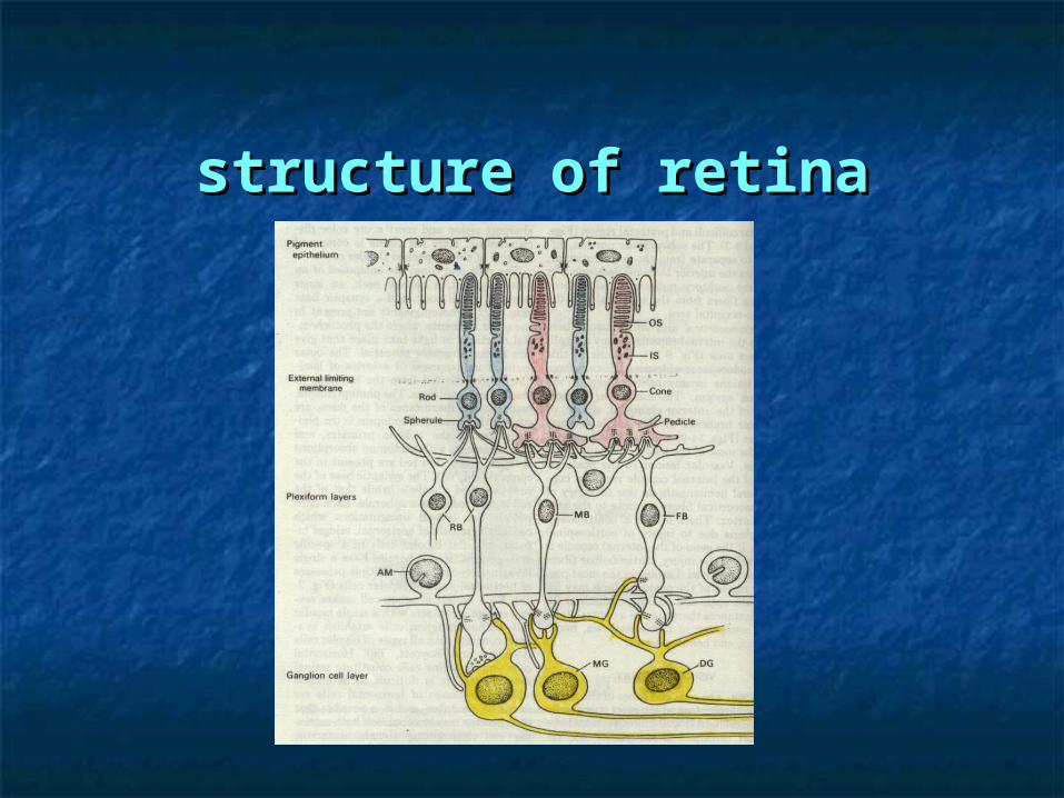
structure of retinastructure of retina

nerve fiber layer nerve fiber layer
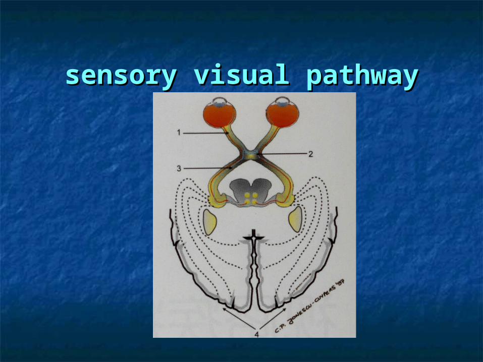
sensory visual pathwaysensory visual pathway


anatomy of the optic nerveanatomy of the optic nerve
from optic disc to optic chiasmfrom optic disc to optic chiasm lengthlength :: 45~50mm45~50mm includingincluding :: intraglobeintraglobe 11 mm mm
intraorbitintraorbit 25~30 25~30 mmmm optic canaloptic canal 55 mmmm intracrania intracrania 1010 mmmm

histology of the optic nervehistology of the optic nerve
comprised by ganglion cell axonscomprised by ganglion cell axons covered by meninges (3 layers)covered by meninges (3 layers) lack of Schwann's cellslack of Schwann's cells hard to rebuildhard to rebuild


to valuate optic nerveto valuate optic nerve
morphologymorphology :: ophthalmoscope ultrasonicophthalmoscope ultrasonic X-ray CT MRIX-ray CT MRI
functionfunction :: vision acuity visual field vision acuity visual field pupil test VEP+ERGpupil test VEP+ERG dark adaption colour perceptiondark adaption colour perception
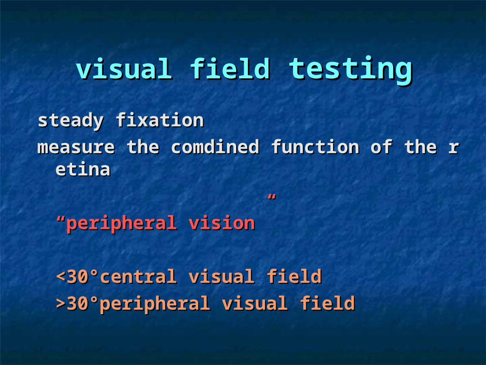
visual fieldvisual field testing testing
steady fixation steady fixation measure the comdined function of the retinameasure the comdined function of the retina
““peripheral vision”peripheral vision”
<30°central visual field<30°central visual field>30°peripheral visual field>30°peripheral visual field

kinetic perimetrykinetic perimetry

static perimetry static perimetry

electrophysiologic testingelectrophysiologic testing electrooculogram EOGelectrooculogram EOG electroretinogram ERGelectroretinogram ERG visual evoked potential VEPvisual evoked potential VEP


pupilpupil

light reflexlight reflex
normal
abnormal

optic neuropathyoptic neuropathy
inflammatoryinflammatory :: optic neuritisoptic neuritis vascularvascular : : anterior ischemic optic neuropathy (AION)anterior ischemic optic neuropathy (AION) neoplastic infiltrationneoplastic infiltration :: glioma, leukaemia, lymphoma, glioma, leukaemia, lymphoma, meningeal carcinomatosismeningeal carcinomatosis othersothers : : optic atrophyoptic atrophy 、、 papilledemapapilledema

Optic neuritisOptic neuritis
duo to a variety of causesduo to a variety of causesthe most common is the most common is demyelinationdemyelinationelse:else: immune-mediatedimmune-mediated direct infectionsdirect infections granulomatous optic nueropathygranulomatous optic nueropathy
contiguous inflammatory diseasecontiguous inflammatory disease

demyelinative optic neuritisdemyelinative optic neuritis clinical features:clinical features: chiefly in women(3:1) chiefly in whitechiefly in women(3:1) chiefly in white associated with multiple sclerosisassociated with multiple sclerosis subacute(2~7days)subacute(2~7days) color vision and contrast sensitivity impaired color vision and contrast sensitivity impaired pain(in eye 90%)(by eye movement 50%)pain(in eye 90%)(by eye movement 50%) abmormal in pupil testingabmormal in pupil testing change in VEP change in VEP a central scotoma in most casesa central scotoma in most cases brain MRI (necesssary)brain MRI (necesssary) including:including: papillitis papillitis
retrobulbar neuritisretrobulbar neuritis

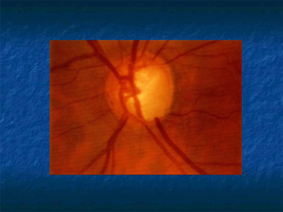
PapillitisPapillitis in 35% casesin 35% cases hyperemia of the optic disk and large veins(ehyperemia of the optic disk and large veins(e
arly signs)arly signs) edema (nearly more than 3D) (common)edema (nearly more than 3D) (common) blurring of the disk margins (common)blurring of the disk margins (common) filling of the physiologic cup (common)filling of the physiologic cup (common) retinal exudates and edema (uncommon)retinal exudates and edema (uncommon) hemorrhage (uncommon)hemorrhage (uncommon) vitreous cells (uncommon)vitreous cells (uncommon)
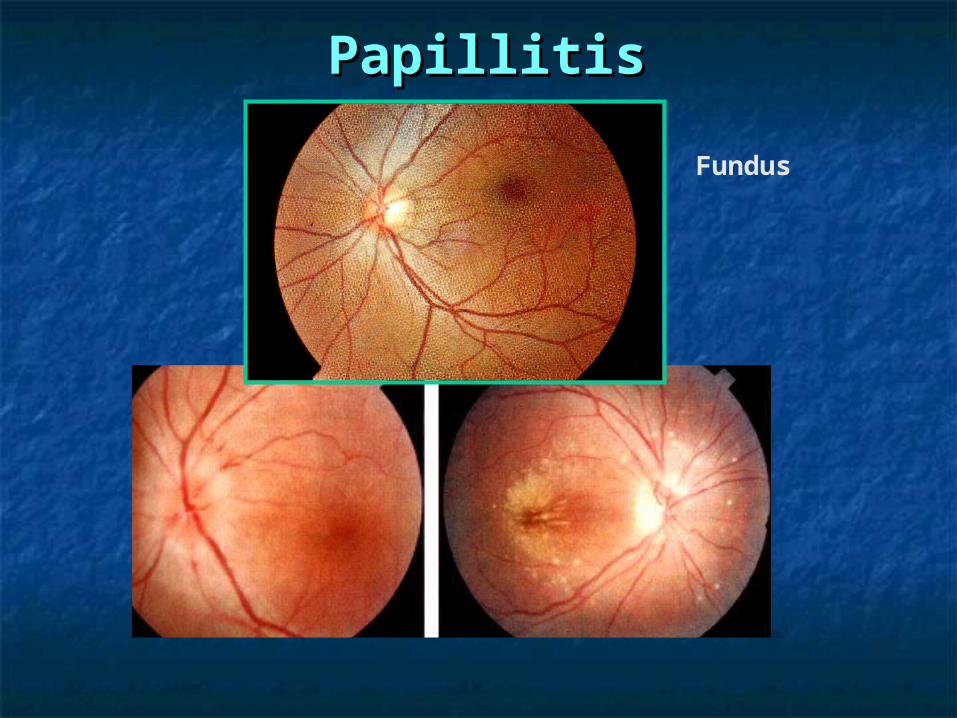
PapillitisPapillitis
Fundus

PapillitisPapillitis
FFA

PapillitisPapillitis
Visual field
CT

Retrobulbar neuritisRetrobulbar neuritis
vision lossvision loss no obvious changes in the optic disk (at eno obvious changes in the optic disk (at e
arlier stage)arlier stage) secondary optic atrophysecondary optic atrophy

demyelinative optic neuritisdemyelinative optic neuritis
differential diagnosis:differential diagnosis: compressive optic neuropathycompressive optic neuropathy anterior ischemic optic neuropathyanterior ischemic optic neuropathy autoimmune optic neuropathyautoimmune optic neuropathy toxic amblyopiatoxic amblyopia Leber's hereditary optic neuropathyLeber's hereditary optic neuropathy vitamin Bvitamin B1212 deficiency deficiency papilledemapapilledema

demyelinative optic neuritisdemyelinative optic neuritis
treatmenttreatment:: steroid therapysteroid therapy can accelerate recovery of visioncan accelerate recovery of vision can not influence the ultimate visual outcomecan not influence the ultimate visual outcome may increase the risk of recurrencymay increase the risk of recurrency intravenous steroid therapyintravenous steroid therapy useful for multiple sclerosisuseful for multiple sclerosis

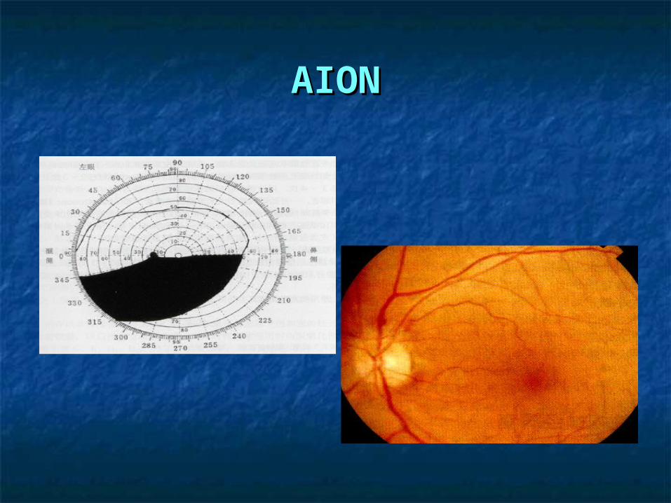
anterior ischemic optic anterior ischemic optic neuropathy (AION)neuropathy (AION)
due to infarction of the retrolaminar opdue to infarction of the retrolaminar optic nerve from occlusion or decreased tic nerve from occlusion or decreased perfusion of the short posterior ciliary perfusion of the short posterior ciliary arteriesarteries

vessels of the optic nervevessels of the optic nerve

AIONAION
causes:causes: arteriosclerosisarteriosclerosis diabetesdiabetes hypertensionhypertension hyperlipidemiahyperlipidemia intracranial strokeintracranial stroke vasculitis,migraine,inherited prothrombotivasculitis,migraine,inherited prothromboti
c states(in younger patients)c states(in younger patients) reduced cup,optic nerve head drusen,increreduced cup,optic nerve head drusen,incre
ased intraocular pressureased intraocular pressure

AIONAIONclinical features:clinical features:
in the sixth or seventh decadein the sixth or seventh decade vision loss without painvision loss without pain generally abrupt,progressivegenerally abrupt,progressive altitudinal vision field defects()altitudinal vision field defects() FFA:decreased perfusionFFA:decreased perfusion segmented the nonarteritic formsegmented the nonarteritic form diffuse the arteritic formdiffuse the arteritic form disk leakage the late phasedisk leakage the late phase
AIONAION

AIONAION

AIONAION
treatment:treatment: No treatment has been shown to provide long-teNo treatment has been shown to provide long-te
rm benefit.rm benefit.Low-dose aspirin may reduce the risk of involvemLow-dose aspirin may reduce the risk of involvem
ent of the fellow eye.ent of the fellow eye.identify the arteritic AION (high-dose systemic stidentify the arteritic AION (high-dose systemic st
eroids)eroids)

PapilloedemaPapilloedema
noninflammatory congestion due to raised inoninflammatory congestion due to raised intracranial pressurentracranial pressure
causescauses intracranial massintracranial mass abscessesabscesses subdural hematomasubdural hematoma arteriovenous malformationarteriovenous malformation subarachnoid hemorrhagesubarachnoid hemorrhage meningitis or encephalitismeningitis or encephalitis acquired hydrocephalitisacquired hydrocephalitis elseelse
PapilledemaPapilledema
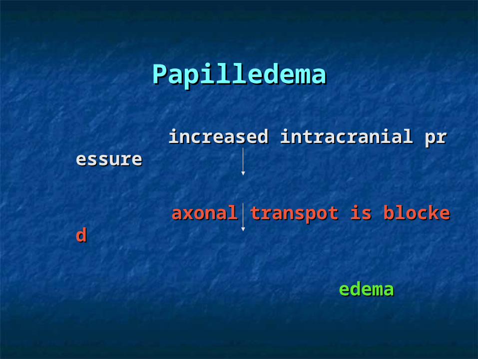
increased intracranial pressureincreased intracranial pressure
axonal transpot is blockedaxonal transpot is blocked
edemaedema

PapilledemaPapilledema
clinical features:clinical features: normal vision in most cases for early papillenormal vision in most cases for early papille
demadema 1~2days to occur and 1 week to develop full1~2days to occur and 1 week to develop full
yy severe vision loss for late papilledemasevere vision loss for late papilledema

PapilledemaPapilledema
the early stage

papilledemapapilledema
the middle stage

papilledemapapilledemathe late stage

papilledemapapilledema
FFAFFA

papilledemapapilledemadifferential diagnosis:differential diagnosis:
burid drusen of the optic nerveburid drusen of the optic nerve small hyperopic diskssmall hyperopic disks AIONAION myelinated nerve fibersmyelinated nerve fibers

papilledemapapilledema
treatment:treatment:deal with the underlying causedeal with the underlying cause

Optic atrophyOptic atrophy primary optic atrophyprimary optic atrophy secondary optic atrophysecondary optic atrophy

primaryprimary secondarysecondary
axonal fiber axonal fiber atrophyatrophy
++ ++
proliferation of gliaproliferation of glia -- ++
pale diskpale disk ++ ++
papilledemapapilledema -- ++
blurring of blurring of marginmargin
-- ++
retinal vesselsretinal vessels normalnormal abnormalabnormal
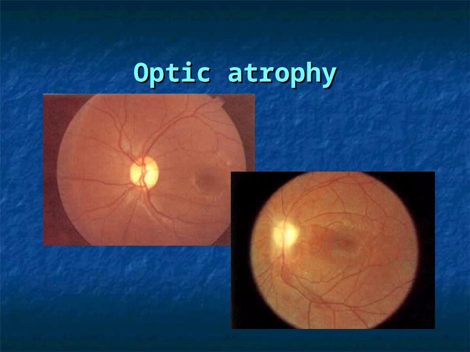
Optic atrophyOptic atrophy

Optic atrophyOptic atrophy
treatment: treatment: to the underlying causeto the underlying cause

optic nerve tumoroptic nerve tumor
glioma of optic nerveglioma of optic nerve
meningioma of optic nervemeningioma of optic nerve

Glioma of optic nerveGlioma of optic nerve

Glioma of optic nerveGlioma of optic nerve
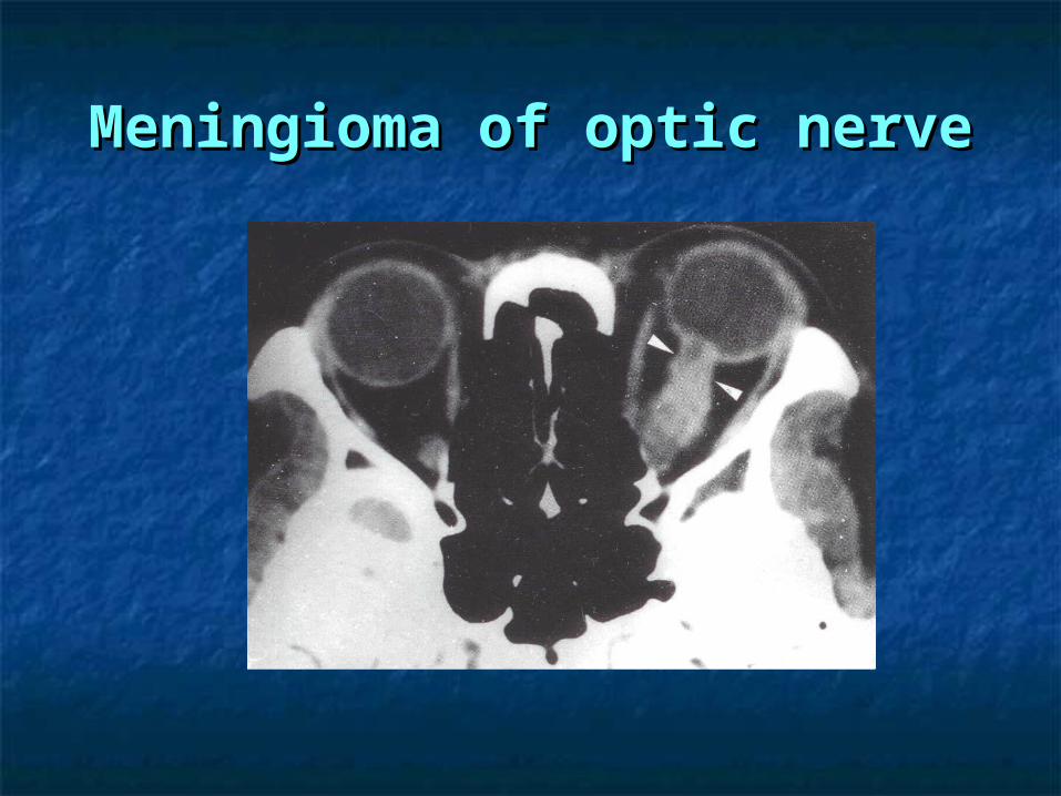
Meningioma of optic nerveMeningioma of optic nerve

Meningioma of optic nerveMeningioma of optic nerve

optic nerve tumoroptic nerve tumor
treatment:treatment: operationoperation

the optic chiasmthe optic chiasm

视交叉病变视交叉病变


the optic chiasm diseasesthe optic chiasm diseases
pituitary adenomapituitary adenoma craniopharyngiomacraniopharyngioma tuberculum sellae meningiomatuberculum sellae meningioma chiasmatic and optic nerve gliomaschiasmatic and optic nerve gliomas

pituitary adenopituitary adenomama

the retrochiasmatic visual paththe retrochiasmatic visual pathwaysways
cortical blindnesscortical blindness macular sparingmacular sparing macular splittingmacular splitting


casescases