Neuro Behcet · Cerebral vasculitis Example: ... Bilateral Papilledema for which he had several LP...
38
3/16/2018 1 NEURO BEHCET Mohamed Abdel Azim FRCOphth Visual impairment in Behcet’s disease is not always caused by uveitis
Transcript of Neuro Behcet · Cerebral vasculitis Example: ... Bilateral Papilledema for which he had several LP...

3/16/2018
1
NEURO BEHCET
Mohamed Abdel Azim FRCOphth
Visual impairment in Behcet’s disease is not
always caused by uveitis

3/16/2018
2
Neurologic manifestations of Behcet’s disease
Central nervous system :
(1) parenchymal involvement, which includes brainstem involvement, hemispheric manifestations, spinal cord lesions, and meningoencephalitic presentations
(2) nonparenchymal involvement, including dural sinus thrombosis, arterial occlusion, and/or aneurysms.
Peripheral neuropathy and myopathy are relatively rare.
Cerebral vasculitis
Example: Foci of
hyperintensity in the
pons and midbrain
bilaterally

3/16/2018
3
Venous sinus thrombosis
Example: irregular filling
defects in the straight sinus
Venous sinus thrombosis
Example: Cerebral
angiography confirms
non-filling of the posterior
half of the sagittal sinus

3/16/2018
4
Arterial complications
Stenosis, occlusions and
aneurysms (most serious).
(very rare-exceptional)

3/16/2018
5
Cerebral vasculitis affecting the
visual centers / visual pathway.
History
PC: 25 years old engineer presented to my clinic with a 2
years history of poor vision more in the right eye.

3/16/2018
6
History
He also complained of constant headache for which he had
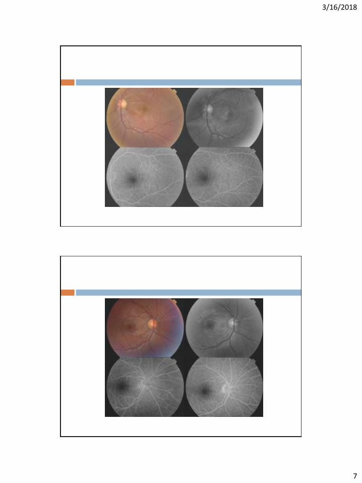
several brain MRIs revealing brain vasculitis.
Erroneous diagnosis of uveitis as the cause of his poor vision.
He received several doses of methyl prednisolone pulse
therapy and several doses of peri-ocular steroids with limited
improvement.
Methyl prednisolone pulse therapy X 3 was repeated in
addition to oral cyclophosphamide
Ophthalmic examination
RVA: CF 1 meter with LVA: 6/24-
central scotoma
Q&D AC Q&D
RRR pupils RRR
NAD Fundus NAD
pale disc?
3/16/2018
7

3/16/2018
8
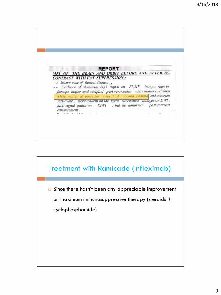
RVA too poor to allow
VF testing however it could
be obtained by confrotation
3/16/2018
9
Treatment with Ramicade (Infleximab)
Since there hasn’t been any appreciable improvement
on maximum immunosuppressive therapy (steroids +
cyclophosphamide).

3/16/2018
10
Post-treatment
Almost immediately the patient noticed marked
improvement of his symptoms.
I examined him one month following his initiation doses
and found his RVA 6/24 and LVA of 6/18+.
FA was normal (unchanged).
His VF revealed great improvement.
Post treatment R & L VF

3/16/2018
11
Pre- vs. Post- treatment LVF
Completely rehabilitated and back to his job as an engineer.
He is maintained on a low dose methotrexate (10 mg / week) to reduce the possibility of the production of anti-(anti-TNF) antibodies.
This patient is currently controlled on 2-3 monthly Ramicade and low dose prednisolone since more than 9 years.

3/16/2018
12
Present
Venous sinus thrombosis causing
intracranial hypertension

3/16/2018
13
History
Date: (12/1/09) 22 year old male student
PC:
2 months history of headache and diplopia of acute onset.
POH:
Bilateral Hypermetropia with mild amblyopia.
Bilateral Papilledema for which he had several LP (++ opening pressure but of normal cytology)
PMH:
Behcet’s disease (recurrent mouth and genital ulcers, superficial thromophlebitis).
DH:
Several courses of systemic steroids.
Examination
Appearance:

3/16/2018
14
Examination
RVA: 6/18 LVA 6/12
Normal Anterior segment bilaterally
Bilateral established papilledema.
R incomittent esotropia due to R 6 th N palsy.

3/16/2018
15

3/16/2018
16
Management
Relation between papilledema and Behcet’s disease,
Coincidental ?
Treated with diuretics, repeated LP and steroids
however with little response.
Bilateral ON sheath decompression was scheduled to
relief the unresponsive ICP.
Managment
MRV in patients diagnosed with IIH revealed more 10% were
have cerebral venous sinus thrombosis (CVST)in presence of
normal MRI. [Occurrence of Cerebral Venous Sinus Thrombosis
in Patients with Presumed Idiopathic Intracranial Hypertension
Ophthalmology (December 2006)]
Finding venous sinus thrombosis ;a potentially fatal condition;
means the patient needs anti-coagulants , systemic
immunosuppression and closer monitoring.

3/16/2018
17
Management
The patient was started on anti-coagulants aiming at an INR >2<3.
Started on Infliximab (Remacade), Azathioprin(Immurane) and prednisolone since the venous occlusion is probably secondary to Behcet’s related vasculitis.
In addition; he continued on Lasix to control the raised ICP.

3/16/2018
18
Managment
Only few weeks later his headache started to resolve.
Fundus exam revealed complete resolution of the papilledema.
VF testing revealed no further deterioration over the next 4 months.
MRV was repeated reporting resolution of the venous thrombosis.
3/16/2018
19
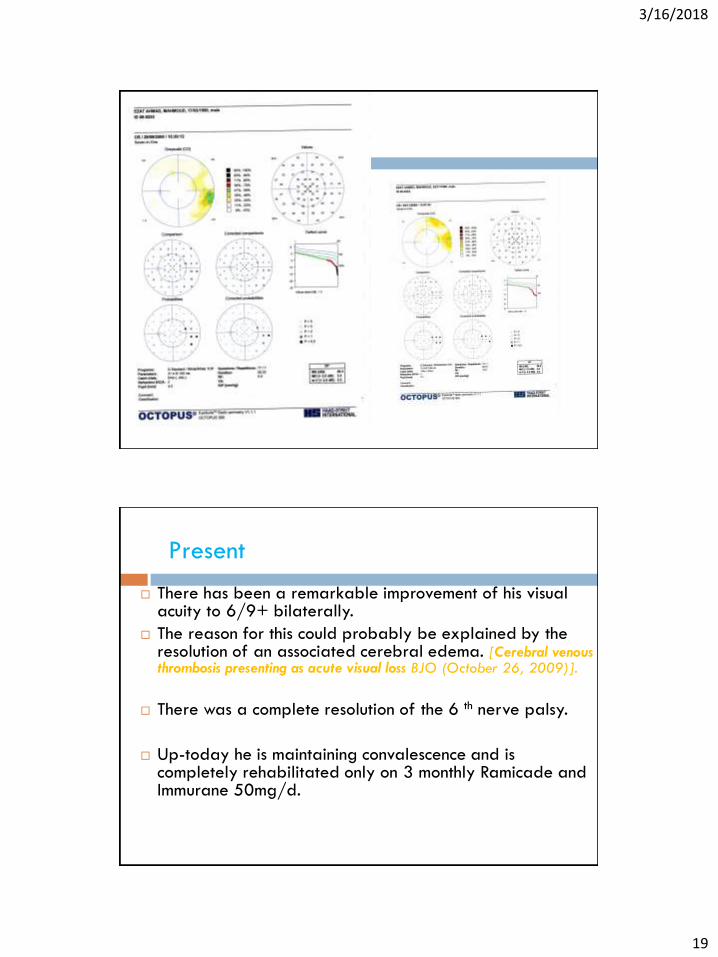
There has been a remarkable improvement of his visual acuity to 6/9+ bilaterally.
The reason for this could probably be explained by the resolution of an associated cerebral edema. [Cerebral venous thrombosis presenting as acute visual loss BJO (October 26, 2009)].
There was a complete resolution of the 6 th nerve palsy.
Up-today he is maintaining convalescence and is completely rehabilitated only on 3 monthly Ramicade and Immurane 50mg/d.
Present

3/16/2018
20

3/16/2018
21
History
29 years old male Behcet’s disease
Presented 9/2014 blurred vision L>R severe headache
LP done +++ opening pressure no cells or growth
MRI and MRA –free
MRV—occluded extra-cranial dural sinus
Tuberculin, HCV, HIV –ve
Examination
RVA 6/9 LVA NPL
Fundus examination :
-Bilateral markedly elevated discs with hges.
-Left optic disc was “choked” and extremely elevated
with totally empty retinal vessels, retinal hemorrhage
and associated vitritis

3/16/2018
22

3/16/2018
23
Managment
FA 11/2014 Right disc edema with splinter hges
delayed venous filling
VEP markedly delayed in L eye
TT: Clexan 60mg X2, pred 60mg, Marevan 6mg
Endoxane (cyclophosphamide) 850mg and Cidamex
250mg X4
Hemorrhages partially resolved leaving an established
papilledema.
We had to repeat the lumbar puncture twice to reduce
the IC pressure for fear of optic atrophy.
He was then started on cyclopsporin A 200 mg instead
of endoxan (which I think is not very effective)
gradually stopping the anticoagulants.
3/16/2018
24
Present
Last visit 8/2017 papilledema has completely
resolved maintaining 6/6 vision.
curently he is on CSA 200mg and pred 10mg

3/16/2018
25
27 years old Behcet patient.
Presented with severe headache, ocular redness
and ?diplopia.
Past history of retinal phlebitis 3 years ago.

3/16/2018
26
examination
RVA =LVA = 6/6
AC: quiet and deep
Fundus exam: no evidence of active uveitis however
there was bilateral established disc edema.
? R 6th nerve palsy.
MRV revealed venous sinus thrombosis.
3/16/2018
27
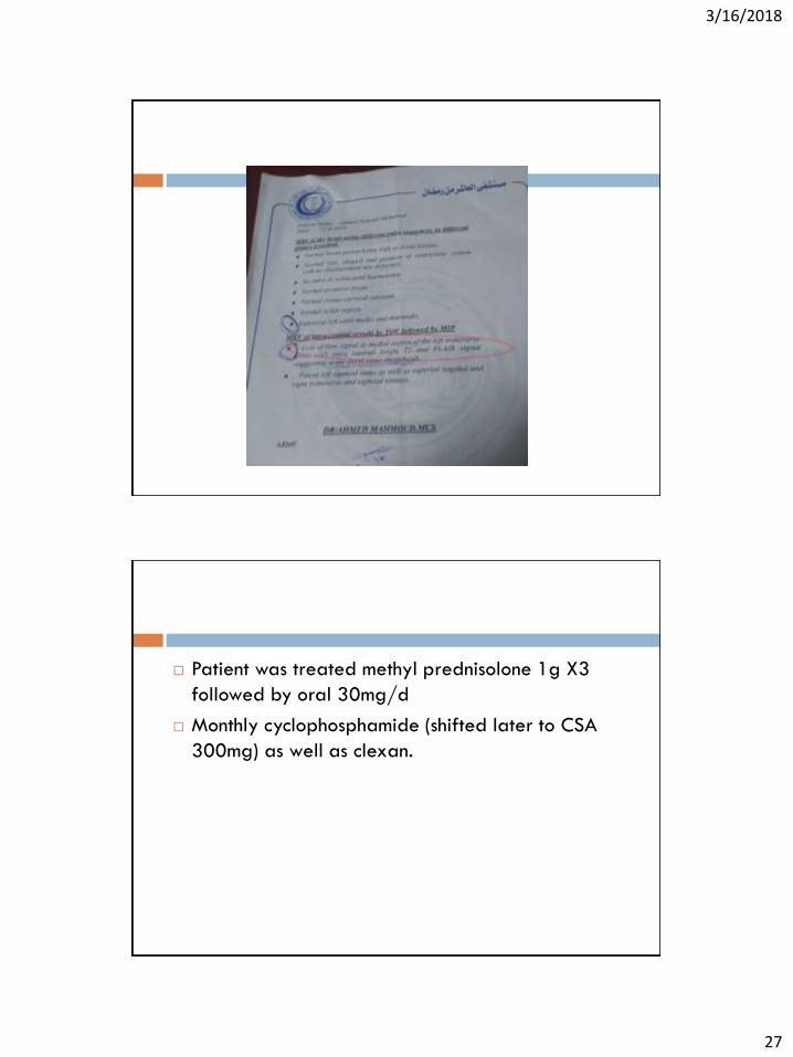
Patient was treated methyl prednisolone 1g X3
followed by oral 30mg/d
Monthly cyclophosphamide (shifted later to CSA
300mg) as well as clexan.

3/16/2018
28
Arterial complications including
aneurysms

3/16/2018
29
Young fit male.
One morning went into coma! (2006)
Brain CT scan revealed intracerebral hemorrhage–
ruptured aneurysm.
Visiting interventional radiology expert– titanium clip.
Recovered with no residues.
Routine ophthalmic exam—6/6 vision
slightly swollen discs ?/ within normal.
History

3/16/2018
30
3/16/2018
31
More than 3 years later (Late 2009) he re-
presented with reduced vision (very gradually
progressive)
O/E : VA: 6/12 bilaterally
AC: Q&D
Fundus: bil macular edema and
slight disc swelling
Cause???

3/16/2018
32

3/16/2018
33

3/16/2018
34
What is going on?!
What is the relation between his ruptured aneurysm
and the macular edema?!

3/16/2018
35

3/16/2018
36
Diagnosis was revised as retinal vasculitis including cerebral vasculitis (cause of ruptured aneurysm).
The diagnosis of—Behcet’s disease was made?
Systemic immunosuppression started : prednisolone 40mg + CSA 200mg.
One month latter he reported great improvement in quality of vision.
RVA 6/9, LVA 6/6.
3/16/2018
37
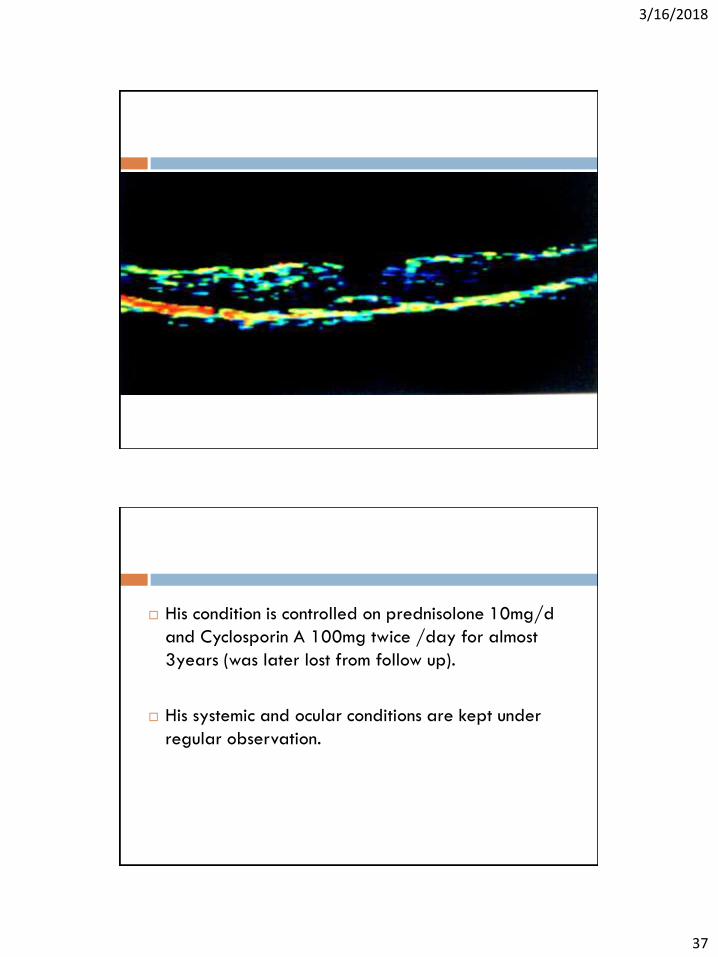
His condition is controlled on prednisolone 10mg/d
and Cyclosporin A 100mg twice /day for almost
3years (was later lost from follow up).
His systemic and ocular conditions are kept under
regular observation.

3/16/2018
38
Conclusion
I wanted to high-light the entity “neuro-Behcet” as a
cause of ophthalmic complaint.
Proper management of these conditions can reverse
the pathology allowing rehabilitation of the
patients.
Importance of MRV in all cases of intra-cranial
hypertension for the presence of venous thrombosis.