Chapter 40 Sleep, Dreaming, and Wakefulness Copyright © 2014 Elsevier Inc. All rights reserved.

Copyright © 2016, the American Society of Anesthesiologists, Inc. Wolters Kluwer Health, Inc. Unauthorized reproduction of this article is prohibited.<zdoi;10.1097/ALN.0000000000001342>
Anesthesiology, V 125 • No 5 929 November 2016
T he neural correlates of anesthetic- and sleep-induced unconsciousness have yet to be fully clarified, but
growing evidence suggests that fragmentation of corti-cal networks consistently occurs during pharmacologic, physiologic, and pathologic states of unconsciousness. Anesthetics with diverse molecular profiles and neuro-physiologic effects have been shown to fragment functional networks in the cortex,1–5 and both sleep and anesthe-sia reduce surrogates of cortical information transfer.4–12 Several recent studies encourage further investigation of the role of cortex in anesthetic-induced unconsciousness. First, in rats, cross-modal cortical interactions are more sensitive to anesthetic effects than first-order thalamocor-tical circuits.13 Second, in humans, disrupted corticocor-tical connectivity has been reported to better distinguish propofol-induced unconsciousness from wakefulness and sedation compared with changes in thalamocorti-cal connectivity.14,15 Although correlative, these findings
are mechanistically relevant given the apparent require-ment for integration of cortical networks during normal
What We Already Know about This Topic
• Accumulatingevidencesuggeststhatfragmentationofcorti-calnetworksoccursduringphysiologic,pharmacologic,andpathologicstatesofunconsciousness
• Corticalconnectivityandacetylcholine levelswereexaminedinrelationtochangesinbehavioralarousalduetopropofolorsevofluraneanesthesiaandnormalsleepinrat
What This Article Tells Us That Is New
• Disruption of cortical connectivity in high γ band correlatedwith anesthetic- and sleep-induced unconsciousness,whileθ connectivity correlated with cholinergic tone and corticalactivation
• Functionalfragmentationofhigh-frequencyactivityinthecor-texmaybeacommonnetwork-levelmechanismofuncon-sciousnessduringgeneralanesthesiaandsleep
Copyright © 2016, the American Society of Anesthesiologists, Inc. Wolters Kluwer Health, Inc. All Rights Reserved. Anesthesiology 2016; 125:929-42
ABSTRACT
Background: Significant advances have been made in our understanding of subcortical processes related to anesthetic- and sleep-induced unconsciousness, but the associated changes in cortical connectivity and cortical neurochemistry have yet to be fully clarified.Methods: Male Sprague–Dawley rats were instrumented for simultaneous measurement of cortical acetylcholine and elec-troencephalographic indices of corticocortical connectivity—coherence and symbolic transfer entropy—before, during, and after general anesthesia (propofol, n = 11; sevoflurane, n = 13). In another group of rats (n = 7), these electroencephalographic indices were analyzed during wakefulness, slow wave sleep (SWS), and rapid eye movement (ReM) sleep.Results: Compared to wakefulness, anesthetic-induced unconsciousness was characterized by a significant decrease in cortical acetylcholine that recovered to preanesthesia levels during recovery wakefulness. Corticocortical coherence and frontal–pari-etal symbolic transfer entropy in high γ band (85 to 155 hz) were decreased during anesthetic-induced unconsciousness and returned to preanesthesia levels during recovery wakefulness. Sleep-wake states showed a state-dependent change in coherence and transfer entropy in high γ bandwidth, which correlated with behavioral arousal: high during wakefulness, low during SWS, and lowest during ReM sleep. By contrast, frontal–parietal θ connectivity during sleep-wake states was not correlated with behavioral arousal but showed an association with well-established changes in cortical acetylcholine: high during wake-fulness and ReM sleep and low during SWS.Conclusions: Corticocortical coherence and frontal–parietal connectivity in high γ bandwidth correlates with behavioral arousal and is not mediated by cholinergic mechanisms, while θ connectivity correlates with cortical acetylcholine levels. (Anesthesiology 2016; 125:929-42)
This article is featured in “This Month in Anesthesiology,” page 1A. Corresponding article on page 832. Supplemental Digital Content is avail-able for this article. Direct URL citations appear in the printed text and are available in both the HTML and PDF versions of this article. Links to the digital files are provided in the HTML text of this article on the Journal’s Web site (www.anesthesiology.org). This article has an audio podcast.
Submitted for publication November 12, 2015. Accepted for publication July 18, 2016. From the Department of Anesthesiology (D.P., H.L., G.A.M.), Center for Consciousness Science (D.P., B.H.S., G.A.M.), and Neuroscience Graduate Program (G.A.M.), University of Michigan, Ann Arbor, Michigan.
Neural Correlates of Wakefulness, Sleep, and General Anesthesia
An Experimental Study in Rat
DineshPal,Ph.D.,BrianH.Silverstein,B.A.,HeonsooLee,Ph.D.,GeorgeA.Mashour,M.D.,Ph.D.
PeRIOPeRATIVe MeDICINe
Downloaded From: http://anesthesiology.pubs.asahq.org/pdfaccess.ashx?url=/data/Journals/JASA/935792/ on 11/01/2016

Copyright © 2016, the American Society of Anesthesiologists, Inc. Wolters Kluwer Health, Inc. Unauthorized reproduction of this article is prohibited.
Anesthesiology 2016; 125:929-42 930 Pal et al.
Wakefulness and Unconsciousness
conscious experience.16,17 Furthermore, the relationship of anesthetic- and sleep-induced unconsciousness is a sub-ject of active exploration,18–25 but systematic comparisons of brain connectivity changes during anesthetic-induced (intravenous and inhaled) and sleep-induced uncon-sciousness have been lacking. Finally, despite the focus on functional, directed, and effective connectivity changes during general anesthesia,26 there is little understanding of the underlying neurochemical control of connectivity patterns, which might provide an explanatory link to the molecular actions of general anesthetics.
In this study, we investigated the relationship of cortical acetylcholine and measures of brain connectivity—coherence and symbolic transfer entropy—derived from the electroen-cephalogram under multiple conditions: (1) before, during, and after unconsciousness induced by propofol or sevoflu-rane, (2) during spontaneous wakefulness, (3) during slow wave sleep (SWS), and (4) during rapid eye movement (ReM) sleep. We report that electroencephalographic coher-ence and frontal-parietal directed connectivity in high γ (85 to 155 hz) bandwidth are present during wakefulness and disrupted during physiologic (SWS and ReM sleep) and pharmacologic (propofol and sevoflurane) states of uncon-sciousness. By contrast, coherence and bidirectional frontal–parietal connectivity in θ (4 to 10 hz) bandwidth are present during states of cortical activation (low-amplitude fast-wave electroencephalogram) with (wakefulness) or without (ReM sleep) behavioral arousal and correlate with cortical acetyl-choline levels. We conclude that coherence and frontal-parietal directed connectivity in high γ band are neural correlates of wakefulness that are not mediated by cholinergic mechanisms.
Materials and MethodsAll experiments were conducted on adult male Sprague–Dawley rats (n = 31; 300 to 350 g; Charles River Laboratories Inc., USA). The rats were housed in a temperature-con-trolled facility with 12-h light:12-h dark cycle (lights on at 6:00 AM) and had ad libitum access to food and water. The experimental procedures were approved by the Institutional Animal Care and Use Committee at the University of Michi-gan (Ann Arbor, Michigan) and were in compliance with the Guide for the Care and Use of Laboratory Animals (eighth edition; The National Academies Press, Washington, D.C.) and Animals in Research: Reporting In Vivo experiments (ARRIVe) guidelines.
Surgical ProceduresRats were anesthetized with 3 to 4% isoflurane in 100% oxygen and positioned in a stereotaxic frame (model 963; David Kopf Instruments, USA) using blunt ear bars. Iso-flurane (1 to 2%) was delivered through a rat anesthesia mask (Kopf model 906) and was titrated to effect during surgery. General anesthesia was assessed by the absence of pedal and palpebral reflex. An anesthetic agent analyzer
(Datex Medical Instrumentation, Inc., USA) was used for continuous monitoring of delivered isoflurane concentra-tion. Core body temperature was monitored using a rectal probe (model 7001h; Physitemp Instruments, Inc., USA). A small animal far-infrared heating pad (Kent Scientific Co., USA) was used for the maintenance of body temperature at 37.0° ± 1.0°C. All rats (n = 31) were implanted with stain-less steel screw electrodes to record electroencephalogram from the (1) frontal cortex: anterior–posterior = +3.0 mm and medial–lateral = 2.5 mm, (2) parietal cortex: anterior–posterior = −4.0 mm and medial–lateral = ±2.5 mm, and (3) occipital cortex: anterior–posterior = −8.0 mm and medial–lateral = ±2.5 mm, and a screw electrode over the nasal sinus served as the reference electrode; all coordinates were with respect to bregma. In a subgroup of these rats (n = 7), multistranded insulated (except at the tip) wires (AS636; Cooner Wires, Inc., USA) were implanted in the dorsal neck muscles for recording electromyogram. These rats were used for recording sleep-wake states. All other rats (n = 24) were implanted with a CMA/11 microdialy-sis guide cannula (CMA Microdialysis; harvard Apparatus, USA) aimed at 1.0 mm above the prefrontal cortex (PFC) (anterior–posterior = +3.0 mm, medial–lateral = 0.5 mm, and ventral = 4.0 mm)27 for microdialysis measurement of acetylcholine levels. The electrodes were interfaced with six-pin pedestal(s) (MS363; Plastics One, USA), and the entire assembly along with a microdialysis guide cannula was secured with dental cement (Cat No. 51459; Stoelting Co, USA). A subgroup of rats (n = 11) with the electrodes for electroencephalographic recording and a microdialy-sis guide tube was implanted with an indwelling catheter (Micro-Renathane tubing, MRe-040; Braintree Scientific, USA) in the internal jugular vein to provide access for pro-pofol infusion. The catheter was tunneled under the skin and mated with a port (313-000BM-10; Plastics One) sutured on the back muscles between the scapulae. The remaining rats (n = 13) with the electrodes for electroencephalographic recording and a microdialysis guide tube but without an intravenous catheter were used for sevoflurane experiments. Buprenorphine hydrochloride (Buprenex®; Reckitt Benck-iser Pharmaceuticals Inc., USA) was used for presurgical (0.01 mg/kg body weight, subcutaneous) and postsurgical (0.03 mg/kg body weight, subcutaneous, every 8 to 12 h for 24 h) analgesia, and all rats received a single presurgical dose (20 mg/kg body weight, subcutaneous) of antibiotic cefazolin (West-Ward-Pharmaceutical Corp., USA). The rats implanted with catheters received an additional dose of antibiotic gen-tamicin (5.0 mg/kg body weight, intravenous) during the surgery. Animals were kept under observation until ambula-tory and then returned to their home cage for postsurgical recovery. All rats were provided at least 7 to 10 days of post-surgical recovery during which they were conditioned to the recording chamber and cables. The jugular venous catheter was flushed with 0.3 ml heparinized saline (1 U/ml; Sagent Pharmaceuticals, USA) every other day.
Downloaded From: http://anesthesiology.pubs.asahq.org/pdfaccess.ashx?url=/data/Journals/JASA/935792/ on 11/01/2016
Copyright © 2016, the American Society of Anesthesiologists, Inc. Wolters Kluwer Health, Inc. Unauthorized reproduction of this article is prohibited.
Anesthesiology 2016; 125:929-42 931 Pal et al.
PERIOPERATIVE MEDICINE
Microdialysis Quantification of Acetylcholine Using High-performance Liquid Chromatography Coupled with Electrochemical DetectionMicrodialysis samples were collected every 12.5 min (25 µl) with CMA/11 microdialysis probes (1-mm cuprophane membrane, 0.24-mm diameter, 6 kDa), which were per-fused continuously with Ringer’s solution (147 mM NaCl, 2.4 mM CaCl2, 4.0 mM KCl, 10 µM neostigmine; ph 6.0 ± 0.2) at 2.0 µl/min using a CMA/400 syringe pump (CMA Microdialysis; harvard Apparatus). From each microdialysis sample, 22 µl was injected into a high-perfor-mance liquid chromatography paired with an electrochemi-cal detector (Bioanalytical Systems, USA) for acetylcholine quantification. An ion-exchange (mobile phase: 50 mM Na2hPO4, ph 8.5) analytical column (MF-6150; Bioana-lytical Systems) separated acetylcholine and choline in the dialysis samples, which were proportionately catalyzed into h2O2 by an immobilized enzyme reactor column (MF-6151; Bioanalytical Systems). h2O2 was detected by oxi-dation (applied potential at 500 mV, Ag+/AgCl reference electrode) at a platinum working electrode. The chromato-grams were digitized to quantify acetylcholine levels using ChromGraph software (Bioanalytical Systems) and a seven-point standard curve (0.05 to 1.0 pmol).
Electrophysiologic Data AcquisitionThe electrophysiologic signals were amplified with a Grass Model 15 LT bipolar portable physiodata amplifier system (15A54 Quad Amplifier; Natus Neurology Inc., USA). An MP150 data acquisition unit along with AcqKnowledge soft-ware (version 4.1.1; Biopac Systems, Inc., USA) was used for digitizing and storing the data. The electrode implanted over nasal sinus served as a reference for recording monopolar electroencephalogram (0.1 to 300 hz, 1-khz sampling rate) from the frontal, parietal, and occipital cortices, which were used for connectivity analyses. Bipolar electroencephalo-gram (frontal–parietal and parietal–parietal, 0.1 to 100 hz, 250-hz sampling rate) and electromyogram (1 to 100 hz, 250-hz sampling rate) were recorded for quantification of sleep-wake states. electroencephalogram signals were ampli-fied 5,000 times, while electromyogram signals were ampli-fied 10,000 times.
Corticocortical Coherence and Directed Connectivity AnalysisThe data were first downsampled to 500 hz to reduce com-putation time, and an IIR notch filter was applied to remove 60 hz line noise. Magnitude squared coherence was calcu-lated at individual frequencies in 0.5-hz intervals from 0.5 to 155 hz between all electrode pairs in 2-s moving win-dows using the “mscohere.m” function in the MATLAB Signal Processing Toolbox (MathWorks Inc., USA). Using the approach demonstrated in previous publications from our28,29 and other30,31 laboratories, the coherence values were averaged over the following frequency bands: δ (0.5 to 4 hz),
θ (4 to 10 hz), α (10 to 15 hz), β (15 to 25 hz), low γ (γ1: 25 to 55 hz), medium γ (γ2: 85 to 125 hz), and high γ (γ3: 125 to 155 hz). The mean global coherence was obtained by averaging the coherence for individual channel pairs for each animal. To control for the possibility of spurious coher-ence affecting the analysis, the empirical data were statisti-cally compared with a surrogate data set in which the phase relationships between the electroencephalogram signals were disrupted without altering the spectral content of each sig-nal.32 Paired Student’s t test showed a significant difference (P < 0.000001) in the coherence between the empirical and surrogate data sets, confirming that the measured coherence was not accounted for merely by spectral shifts of the signal across states.
We used normalized symbolic transfer entropy (NSTe) to assess directed connectivity. NSTe is an information theo-retic measure and serves as a surrogate for directed cortical communication. Our previous studies have validated the use of NSTe to measure connectivity changes in humans7,8,11 and rats,28 and other laboratories have supported our findings with NSTe using different approaches: functional connec-tivity and dynamic causal modeling.9,14 Transfer entropy, the basis of NSTe, was also used in a previous study of evoked potentials in low γ range (less than or equal to 50 hz) in rats33 and head-restrained ferrets.34 We selected θ and γ bands for NSTe analysis because these have been linked to electroen-cephalographic activation, changes in cortical acetylcholine, and changes in states of behavioral arousal and conscious-ness.29,35 NSTe was analyzed between frontal and parietal areas because studies from human volunteers, patients, and animals suggest the importance of frontal–parietal networks in consciousness of the environment.7–9,11,14,26,33,36 NSTe was calculated between left frontal and left parietal areas in θ (4 to 10 hz), low γ (γ1: 25 to 55 hz), medium γ (γ2: 85 to 125 hz), and high γ (γ3: 125 to 155 hz) bands as has been described previously.11,28 In brief, the sampling rate was kept at 1 khz to improve the stability of the analysis. The continu-ous raw electroencephalographic data were first filtered for the frequency band of interest (θ, low γ, medium γ, and high γ) with a zero-phase FIR filter of order 400 designed using firls2.m (MATLAB Signal Processing Toolbox; MathWorks Inc.) and implemented using filtfilt.m (MATLAB Signal Pro-cessing Toolbox; MathWorks Inc.). Data were then extracted from time periods of interest and analyzed in 10-s bins. Note that all electroencephalographic analyses were performed on the same data epochs. NSTe requires three parameters: embedding dimension (dE), time delay (τ), and prediction time (δ). These parameters were selected such that the transfer entropy from the source signal to the target signal was maxi-mized for a given data window, as described previously.11,28 In brief, dE was kept fixed at 3, δ was set by selecting the lag that maximized the cross-correlation between the two signals, and τ was varied from 1 to 30. The potential bias of symbolic transfer entropy was removed using a shuffled data set, after which the unbiased data set was normalized.11,28
Downloaded From: http://anesthesiology.pubs.asahq.org/pdfaccess.ashx?url=/data/Journals/JASA/935792/ on 11/01/2016

Copyright © 2016, the American Society of Anesthesiologists, Inc. Wolters Kluwer Health, Inc. Unauthorized reproduction of this article is prohibited.
Anesthesiology 2016; 125:929-42 932 Pal et al.
Wakefulness and Unconsciousness
Simultaneous Electroencephalographic Recordings and Microdialysis Measurement of Changes in Acetylcholine Levels before, during, and after Propofol- and Sevoflurane-induced UnconsciousnessThe rats were connected to the electroencephalogram record-ing system at least 30 min before the start of an experimen-tal session (9:30 AM to 10:00 AM), and a microdialysis probe being infused with Ringer’s solution at 2.0 µl/min was low-ered into the PFC. The electroencephalogram was recorded continuously while the microdialysis samples were collected every 12.5 min throughout the experiment. Previous reports from our29 and other37 laboratories have shown that it takes up to 37.5 min for the in vivo baseline acetylcholine levels to stabilize after probe insertion. Therefore, the first three microdialysis samples were excluded and six preanesthesia baseline microdialysis samples were collected. In order to hold the behavioral state constant during the baseline condi-tion, the rats were kept awake using gentle tapping on the recording chamber. At the completion of six wake microdi-alysis samples, the rats were either connected to a microsy-ringe pump (SP 101I; WPI Inc., USA) through the venous catheter for continuous propofol infusion (800 µg ⋅ kg−1 ⋅ min−1) or exposed to sevoflurane anesthesia (2.0 to 2.2%), and six microdialysis samples were collected during the administration of the anesthetic. Sevoflurane experiments were conducted in a custom-made clear round recording chamber, which could be rotated in a direction opposite to that of the rat’s movement. This apparatus allows the rat to stay in the center of the chamber, thereby preventing any kinks in the microdialysis tubing. The recording chamber can be sealed during anesthesia and was fitted with a door to allow access to the animal during anesthetic exposure. The chamber also had ports for inlet and outlet of anesthetic vapors, which were monitored using two anesthetic agent analyzers (Datex Medical Instrumentation, Inc.). The anes-thetic concentration was based on preliminary experiments conducted in our laboratory and was titrated to produce loss of righting reflex along with slow wave (high-amplitude δ wave) electroencephalogram, both of which are used as a surrogate for loss of consciousness.38,39 Propofol and sevoflurane produced loss of righting reflex within about 20 min, after which the rats were maintained in a recumbent position for the rest of the anesthetic administration. The core body temperature was monitored and maintained at 37.0° ± 1.0°C using a far-infrared heating pad. At the com-pletion of six microdialysis samples during the adminis-tration of anesthetic, the propofol infusion or sevoflurane exposure was stopped. Thereafter, six microdialysis samples were collected in the postsevoflurane recovery period. Initial experiments with propofol (n = 3) showed that acetylcholine levels during the postpropofol recovery period (six micro-dialysis samples) remained below baseline wake levels. The data from these initial propofol experiments, which only had six postpropofol recovery samples, were excluded from the analysis. In addition, it took much longer for the rats
to recover righting reflex after propofol anesthesia (approxi-mately 29 min) as compared to sevoflurane (less than 10 min), which likely reflects pharmacokinetics. Therefore, in order to ensure a complete recovery from the effects of propofol anesthesia, microdialysis samples were collected for an extended recovery period (12 microdialysis samples) in the remaining eight rats. In the propofol group, we could not collect microdialysis samples in one of the eight rats with extended recovery period because of a microdialysis probe malfunction. In the sevoflurane group, two rats did not have good electroencephalogram signals and the microdialysis probe failed in one of the rats. The last epoch during wake, anesthetic-induced unconsciousness, and postanesthetic recovery period was taken as the most stable and representa-tive of the corresponding states, and therefore the electro-encephalogram and acetylcholine analysis was restricted to these 12.5-min epochs.
Polysomnographic Recordings and Quantification of Sleep-Wake StatesMonopolar (frontal, parietal, and occipital cortices) and bipolar electroencephalogram (frontal–parietal and pari-etal–parietal) along with electromyogram were recorded for 12 h between 6:00 AM and 6:00 PM during the light phase. Bipolar electroencephalogram and electromyogram in 10-s bins were used for the characterization and quantification of wake (low-amplitude fast-wave electroencephalogram with high muscle tone), SWS (high-amplitude slow wave electroencephalogram with low muscle tone), and ReM sleep (low-amplitude fast-wave electroencephalogram along with muscle atonia). Only pure sleep or wake epochs were selected for the analysis, i.e., wakefulness epoch was characterized by the presence of low-amplitude fast-wave electroencephalogram and high muscle tone for 100% of the time in the 10-s epoch. Similarly, for ReM sleep, the selected epochs were characterized by low-amplitude fast-wave electroencephalogram and muscle atonia for 100% of the time in 10-s epoch. Selection of these pure epochs ensured that there was no potential confound in the con-nectivity analysis arising from mixed states within a single epoch. Of a total of 4,320 10-s epochs, we selected a subset of 99 pure epochs for connectivity analysis. Note that the state of wakefulness in these experiments was spontaneous, while the state of baseline wakefulness in the study of pro-pofol- and sevoflurane-induced unconsciousness was main-tained by the experimenters.
Histologic Confirmation of the Microdialysis SitesAfter 3 to 7 days of microdialysis and electroencephalogram data collection, rats were deeply anesthetized (ketamine and xylazine, 80 and 10 mg/kg body weight, intraperitoneal) and transcardially perfused with 100 ml of phosphate buff-ered saline (0.1 M, ph 7.2; 1219SK; eM Sciences, USA) followed by 250 ml fixative (4% paraformaldehyde, 4% sucrose, 0.1 M phosphate buffer, ph 7.2; 1224SK; eM
Downloaded From: http://anesthesiology.pubs.asahq.org/pdfaccess.ashx?url=/data/Journals/JASA/935792/ on 11/01/2016

Copyright © 2016, the American Society of Anesthesiologists, Inc. Wolters Kluwer Health, Inc. Unauthorized reproduction of this article is prohibited.
Anesthesiology 2016; 125:929-42 933 Pal et al.
PERIOPERATIVE MEDICINE
Sciences) solution. The brains were extracted and stored in a fixative solution for at least 24 h and then allowed to equili-brate in 30% sucrose in phosphate buffer. Forty-micron coronal sections were cut through the PFC on a cryostat (Leica Microsystems Nussloch Gmbh, Germany). The sec-tions were mounted on slides and stained with cresyl violet to confirm the site of microdialysis.
Statistical AnalysesStatistical analyses were conducted in consultation with the Center for Statistical Consultation and Research at the Uni-versity of Michigan. A priori power analysis (nQuery Advisor + nTerim; Statistical Solutions Ltd, USA) using the acetyl-choline pilot data indicated that a minimum sample size of 3 would have 80% power (effect size = 4.0) to detect a dif-ference in means of 0.8 pmole (SD of difference = 0.2) with Student’s paired t test with one-sided α = 0.05/3 (Bonfer-roni correction for three pairwise tests). All statistical com-parisons were conducted in a within-group design, and P < 0.05 was considered statistically significant. each group of rats in this study had different surgical implants: jugu-lar venous catheter and microdialysis probe in the propofol group, microdialysis probe without jugular venous catheter in the sevoflurane group, and neither catheter nor micro-dialysis probe in the sleep-wake group. Therefore, random allocation of rats to different experimental conditions was not possible. Additionally, the experimenters could not be blinded to the experiments because the experimental inter-ventions (propofol infusion, sevoflurane exposure, and sleep-wake recordings) were clearly different and could not be masked. Repeated-measures ANOVA (RMANOVA) with Tukey multiple comparisons test was used for the compari-son of acetylcholine levels and the electroencephalographic coherence in each frequency band between the last epochs from the periods of (1) wakefulness, (2) anesthetic-induced unconsciousness (propofol and sevoflurane), and (3) postan-esthetic recovery wakefulness. These epochs were also used for comparison of directed connectivity between the frontal and parietal areas for θ and γ (low, medium, and high) bands. Similarly, RMANOVA with Tukey post hoc test was used for the comparison of coherence and directed connectivity dur-ing sleep-wake states. The data are reported as mean ± SeM along with 95% CI. To enhance readability, only P values are provided in the Results section, while mean ± SeM, 95% CI, and F statistic are provided in a tabular format (tables 1 to 5, Supplemental Digital Content 1, http://links.lww.com/ALN/B316, which show descriptive and inferential sta-tistics). Statistical comparisons were performed with Graph Pad Prism 6.05 (Graph Pad Software, Inc., USA).
Results
Cortical Acetylcholine across Multiple States of ArousalThe temporal course of the changes in PFC acetylcho-line levels before, during, and after anesthetic-induced
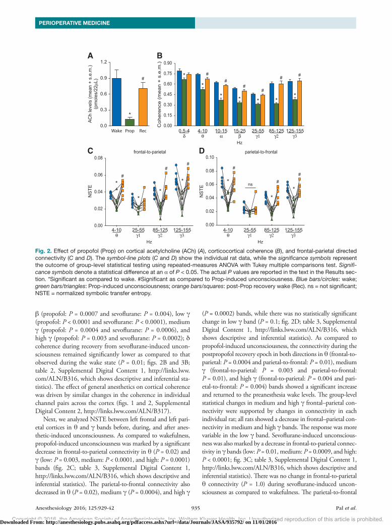
unconsciousness is illustrated in figure 1A. histologic analysis confirmed the probe placement in the PFC for all rats in the propofol and sevoflurane groups (fig. 1, B and C). The effect of propofol and sevoflurane on cortical ace-tylcholine is shown in figures 2 and 3, respectively. There was a statistically significant decrease in acetylcholine lev-els during propofol-induced (P = 0.005) and sevoflurane-induced (P = 0.0006) unconsciousness as compared to the levels observed in the waking state (figs. 2A and 3A; table 1, Supplemental Digital Content 1, http://links.lww.com/ALN/B316, which shows descriptive and inferential statis-tics). The decrease in acetylcholine was comparable between the propofol (approximately 90%) and sevoflurane (approx-imately 85%) groups. Compared to anesthetic-induced unconsciousness, the recovery wake epoch for both pro-pofol (P = 0.004) and sevoflurane (P = 0.002) groups was characterized by a significant increase in acetylcholine levels that was not statistically different from preanesthesia wake acetylcholine levels (figs. 2A and 3A; table 1, Supplemental Digital Content 1, http://links.lww.com/ALN/B316, which shows descriptive and inferential statistics). Previous reports have demonstrated that cortical acetylcholine levels are high during wakefulness, low during SWS, and high again dur-ing ReM sleep, approximating the levels observed during the waking state.35,40 These changes in cortical acetylcho-line across sleep-wake states have been well described and accepted. Therefore, we did not measure changes in cortical acetylcholine during sleep-wake states in the current study.
Electroencephalographic Coherence and Directed Connectivity before, during, and after Anesthetic-induced UnconsciousnessThe decrease in acetylcholine levels in PFC during anesthetic-induced unconsciousness was associated with a reduction in long-range corticocortical coherence (figs. 2B and 3B; table 2, Supplemental Digital Content 1, http://links.lww.com/ALN/B316, which shows descriptive and inferential statistics). As compared to wakefulness, anesthetic-induced unconsciousness produced a significant decrease in electro-encephalographic coherence in δ (propofol: P = 0.001 and sevoflurane: P = 0.004), θ (propofol: P = 0.0004 and sevo-flurane: P = 0.003), α (propofol: P = 0.0004 and sevoflu-rane: P = 0.006), β (propofol: P = 0.0003 and sevoflurane: P = 0.0006), low γ (propofol: P < 0.0001 and sevoflurane: P < 0.0001), medium γ (propofol: P < 0.0001 and sevoflurane: P < 0.0001), and high γ (propofol: P = 0.0002 and sevoflu-rane: P < 0.0001) bands (figs. 2B and 3B; table 2, Supple-mental Digital Content 1, http://links.lww.com/ALN/B316, which shows descriptive and inferential statistics). Compared to anesthetic-induced unconsciousness, coherence during the recovery wake epoch showed a significant increase and returned to wake levels in all bands except the δ band in the propofol and sevoflurane groups: δ (propofol: P = 0.08 and sevoflurane: P = 0.05), θ (propofol: P = 0.001 and sevoflurane: P = 0.03), α (propofol: P = 0.0002 and sevoflurane: P = 0.02),
Downloaded From: http://anesthesiology.pubs.asahq.org/pdfaccess.ashx?url=/data/Journals/JASA/935792/ on 11/01/2016

Copyright © 2016, the American Society of Anesthesiologists, Inc. Wolters Kluwer Health, Inc. Unauthorized reproduction of this article is prohibited.
Anesthesiology 2016; 125:929-42 934 Pal et al.
Wakefulness and Unconsciousness
350Seconds
ACh peak
nA
0
0.2
0.4
0.6
0.8
1.0
1.2
140 210 280
3.72 mm
3.24 mm
3.00 mm
fmi fmi
fmi fmi
fmi fmi
PrL
PrL
PrL
IL
IL
IL
B
C
A Continuous microdialysis sample collection and electroencephalographic recording
6 samples 6 samples
Continuous propofol(800 µg kg-1min-1)
6 samples
Recovery
12 samples6 samples
Freely moving wake
Continuous sevoflurane(2.0-2.2%) Recovery
Sequential microdialysis samplesWake Propofol Recovery
AC
h le
vels
(m
ean
+ s.
e.m
.)(p
mol
es/2
2µL)
1 32 4 5 6 1 32 4 5 6 1 32 4 5 6 7 98 101112
0.8
0.0
0.4
0.2
0.6
1.2
1.0LORR RORR
Sequential microdialysis samplesWake Sevoflurane Recovery
1 32 4 5 6 1 32 4 5 6 1 32 4 5 6AC
h le
vels
(m
ean
+ s.
e.m
.)(p
mol
es/2
2µL)
0.8
0.0
0.4
1.6
1.2 LORR RORR
6 samples
Freely moving wake
Fig. 1. (A) Schematic illustrating the design for microdialysis experiments and temporal course of electroencephalogram and acetylcholine (ACh) data collection. Each ACh sample epoch represents microdialysate collected during a period of 12.5 min. The electroencephalographic data were analyzed in the corresponding 12.5-min epochs. Blue, green, and orange bars represent wake, anesthetic-induced unconsciousness (propofol or sevoflurane), and postanesthetic recovery wake epochs, respectively. The epochs highlighted with broken lines were selected for statistical comparisons. Arrows indicate the approximate time points at which rats showed loss of righting reflex (LORR) and recovery of righting reflex (RORR). (B) Reconstruction diagram shows a cascade of coronal brain section drawings from the rat brain atlas27 to illustrate the location (vertical cylinders) of microdialysis probes (1 mm) within the prefrontal cortex. Green cylinders represent the microdialysis probes from the propofol experiments, while the red cylinders represent those from the sevoflurane experiments. Numbers above the coronal brain section drawings are the anterior–posterior stereotaxic coordinates relative to bregma. The chromatogram above the reconstruction diagram shows signal/noise ratio and the retention time for ACh. (C) Cresyl violet–stained representative coronal brain section through prefrontal cortex shows the dialysis probe track and the site of microdialysis. Arrow indicates the location of the ventral tip of the microdialysis membrane. fmi = forceps minor corpus callosum; IL = infralimbic area; PrL = prelimbic area. Scale bar = 1 mm.
Downloaded From: http://anesthesiology.pubs.asahq.org/pdfaccess.ashx?url=/data/Journals/JASA/935792/ on 11/01/2016
Copyright © 2016, the American Society of Anesthesiologists, Inc. Wolters Kluwer Health, Inc. Unauthorized reproduction of this article is prohibited.
Anesthesiology 2016; 125:929-42 935 Pal et al.
PERIOPERATIVE MEDICINE
β (propofol: P = 0.0007 and sevoflurane: P = 0.004), low γ (propofol: P < 0.0001 and sevoflurane: P < 0.0001), medium γ (propofol: P = 0.0004 and sevoflurane: P = 0.0006), and high γ (propofol: P = 0.003 and sevoflurane: P = 0.0002); δ coherence during recovery from sevoflurane-induced uncon-sciousness remained significantly lower as compared to that observed during the wake state (P = 0.01; figs. 2B and 3B; table 2, Supplemental Digital Content 1, http://links.lww.com/ALN/B316, which shows descriptive and inferential sta-tistics). The effect of general anesthetics on cortical coherence was driven by similar changes in the coherence in individual channel pairs across the cortex (figs. 1 and 2, Supplemental Digital Content 2, http://links.lww.com/ALN/B317).
Next, we analyzed NSTe between left frontal and left pari-etal cortices in θ and γ bands before, during, and after anes-thetic-induced unconsciousness. As compared to wakefulness, propofol-induced unconsciousness was marked by a significant decrease in frontal-to-parietal connectivity in θ (P = 0.02) and γ (low: P = 0.003, medium: P < 0.0001, and high: P = 0.0001) bands (fig. 2C; table 3, Supplemental Digital Content 1, http://links.lww.com/ALN/B316, which shows descriptive and inferential statistics). The parietal-to-frontal connectivity also decreased in θ (P = 0.02), medium γ (P = 0.0004), and high γ
(P = 0.0002) bands, while there was no statistically significant change in low γ band (P = 0.1; fig. 2D; table 3, Supplemental Digital Content 1, http://links.lww.com/ALN/B316, which shows descriptive and inferential statistics). As compared to propofol-induced unconsciousness, the connectivity during the postpropofol recovery epoch in both directions in θ (frontal-to-parietal: P = 0.0004 and parietal-to-frontal: P = 0.01), medium γ (frontal-to-parietal: P = 0.003 and parietal-to-frontal: P = 0.01), and high γ (frontal-to-parietal: P = 0.004 and pari-etal-to-frontal: P = 0.004) bands showed a significant increase and returned to the preanesthesia wake levels. The group-level statistical changes in medium and high γ frontal–parietal con-nectivity were supported by changes in connectivity in each individual rat; all rats showed a decrease in frontal–parietal con-nectivity in medium and high γ bands. The response was more variable in the low γ band. Sevoflurane-induced unconscious-ness was also marked by a decrease in frontal-to-parietal connec-tivity in γ bands (low: P = 0.01, medium: P = 0.0009, and high: P < 0.0001; fig. 3C; table 3, Supplemental Digital Content 1, http://links.lww.com/ALN/B316, which shows descriptive and inferential statistics). There was no change in frontal-to-parietal θ connectivity (P = 1.0) during sevoflurane-induced uncon-sciousness as compared to wakefulness. The parietal-to-frontal
Coh
eren
ce (
mea
n +
s.e.
m.)
0.15
0.30
0.45
0.60
0.00
0.75
0.90
Hz
0.5-4δ
4-10θ
10-15α
15-25β
25-55γ1
85-125γ2
125-155γ3
B
*
#
#
*
*
##
#
#
** **
A
AC
h le
vels
(mea
n +
s.e.
m.)
(pm
oles
/22µ
L)
0.0
0.3
0.6
1.2
0.9
Wake Prop Rec
#
*
NS
TE
parietal-to-frontal
0.00
0.06
0.02
0.08
0.10
4-10θ
25-55γ1
85-125γ2
125-155γ3
Hz
D
*#
#
**
#
0.04
ns
*NS
TE
frontal-to-parietal
0.00
0.04
0.02
0.06
0.08
Hz
4-10θ
25-55γ1
85-125γ2
125-155γ3
C#
#
#
** *
Fig. 2. Effect of propofol (Prop) on cortical acetylcholine (ACh) (A), corticocortical coherence (B), and frontal-parietal directed connectivity (C and D). The symbol-line plots (C and D) show the individual rat data, while the significance symbols represent the outcome of group-level statistical testing using repeated-measures ANOVA with Tukey multiple comparisons test. Signifi-cance symbols denote a statistical difference at an α of P < 0.05. The actual P values are reported in the text in the Results sec-tion. *Significant as compared to wake. #Significant as compared to Prop-induced unconsciousness. Blue bars/circles: wake; green bars/triangles: Prop-induced unconsciousness; orange bars/squares: post-Prop recovery wake (Rec). ns = not significant; NSTE = normalized symbolic transfer entropy.
Downloaded From: http://anesthesiology.pubs.asahq.org/pdfaccess.ashx?url=/data/Journals/JASA/935792/ on 11/01/2016

Copyright © 2016, the American Society of Anesthesiologists, Inc. Wolters Kluwer Health, Inc. Unauthorized reproduction of this article is prohibited.
Anesthesiology 2016; 125:929-42 936 Pal et al.
Wakefulness and Unconsciousness
connectivity decreased in θ (P = 0.003) and higher γ (medium: P = 0.0004 and high: P < 0.0001) bands, while there was no statistically significant change in low γ band (P = 0.4; fig. 3D; table 3, Supplemental Digital Content 1, http://links.lww.com/ALN/B316, which shows descriptive and inferential statistics). Compared to sevoflurane-induced unconsciousness, bidirec-tional connectivity increased in medium (frontal-to-parietal: P = 0.01 and parietal-to- frontal: P = 0.01) and high (frontal-to-parietal: P = 0.0003 and parietal-to-frontal: P = 0.0005) γ bands during the postsevoflurane recovery wake epoch and returned to the presevoflurane wake levels. Frontal-to-parietal θ connec-tivity was significantly higher during post-sevoflurane recovery wake epoch than during presevoflurane wake (P = 0.04) and the unconscious state (P = 0.02), while the parietal-to-frontal θ connectivity showed an increase (P < 0.0001) during postsevo-flurane recovery wake epoch and returned to the presevoflurane wake levels.
Electroencephalographic Coherence and Directed Connectivity during Sleep-Wake StatesAs compared to wakefulness, SWS was characterized by a significant decrease in corticocortical coherence levels in δ (P < 0.0001), θ (P = 0.0001), α (P = 0.007), medium γ
(P = 0.0002), and high γ (P = 0.0001) bands; there was no statis-tical difference in coherence in β (P = 0.07) and low γ (P = 0.2) bands (fig. 4A; table 4, Supplemental Digital Content 1, http://links.lww.com/ALN/B316, which shows descriptive and infer-ential statistics). Compared to SWS, corticocortical coherence decreased during ReM sleep in β (P = 0.0004) and γ range (low: P = 0.001, medium: P = 0.0004, and high: P = 0.008), while it increased in θ (P < 0.0001) and α (P = 0.0001) bands (fig. 4A; table 4, Supplemental Digital Content 1, http://links.lww.com/ALN/B316, which shows descriptive and inferential statistics). There was no significant difference (P = 0.2) in δ coherence between SWS and ReM sleep. Compared to wakefulness, ReM sleep was characterized by reduced coherence in δ (P < 0.0001), β (P = 0.004), and the entire γ range (low: P = 0.0001, medium: P < 0.0001, and high: P < 0.0001); there was no sig-nificant difference in θ (P = 0.2) and α (P = 0.6) bands between wakefulness and ReM sleep (fig. 4A; table 4, Supplemental Digital Content 1, http://links.lww.com/ALN/B316, which shows descriptive and inferential statistics). Global changes in cortical coherence during sleep-wake states were also evident in the changes in coherence observed in individual channel pairs across the cortical areas (fig. 3, Supplemental Digital Content 2, http://links.lww.com/ALN/B317).
AC
h le
vels
(mea
n +
s.e.
m.)
(pm
oles
/22µ
L)
0.0
0.4
0.8
1.6
1.2
Wake Sevo Rec
A
*
#
Hz
Coh
eren
ce (
mea
n +
s.e.
m.)
0.15
0.30
0.45
0.60
0.00
0.75
0.90
0.5-4δ
4-10θ
10-15α
15-25β
25-55γ1
85-125γ2
125-155γ3
B
**
**
#
##
#
**
#
*
#
*
#*
C frontal-to-parietal
NS
TE
0.00
0.04
0.02
0.06
0.08
Hz
4-10θ
25-55γ1
85-125γ2
125-155γ3
#*
#
*
#
*
parietal-to-frontal
NS
TE
0.00
0.04
0.02
0.08
0.10
4-10θ
25-55γ1
85-125γ2
125-155γ3
Hz
D#
*#
*
#
*0.06
ns
*
Fig. 3. Effect of sevoflurane (Sevo) on cortical acetylcholine (ACh) (A), corticocortical coherence (B), and frontal-parietal directed connectivity (C and D). The symbol-line plots (C and D) show the individual rat data, while the significance symbols represent the outcome of group-level statistical testing using repeated-measures ANOVA with Tukey multiple comparisons test. Signifi-cance symbols denote a statistical difference at an α of P < 0.05. The actual P values are reported in the text in the Results sec-tion. *Significant as compared to wake. #Significant as compared to Sevo-induced unconsciousness. Blue bars/circles: wake; green bars/triangles: Sevo-induced unconsciousness; orange bars/squares: post-Sevo recovery wake (Rec). ns = not significant; NSTE = normalized symbolic transfer entropy.
Downloaded From: http://anesthesiology.pubs.asahq.org/pdfaccess.ashx?url=/data/Journals/JASA/935792/ on 11/01/2016

Copyright © 2016, the American Society of Anesthesiologists, Inc. Wolters Kluwer Health, Inc. Unauthorized reproduction of this article is prohibited.
Anesthesiology 2016; 125:929-42 937 Pal et al.
PERIOPERATIVE MEDICINE
Bidirectional frontal–parietal connectivity in θ band (fig. 4, B and C) followed a pattern that closely mirrored the corti-cal acetylcholine levels reported across sleep-wake states.35,40 Compared to the wake state, frontal–parietal θ connectivity decreased during SWS (frontal-to- parietal: P = 0.0006 and pari-etal-to-frontal: P < 0.0001) and increased during ReM sleep
(frontal-to-parietal: P = 0.003 and parietal-to-frontal: P = 0.007; fig. 4, B and C; table 5, Supplemental Digital Content 1, http://links.lww.com/ALN/B316, which shows descriptive and infer-ential statistics). As opposed to frontal–parietal θ connectivity, frontal–parietal connectivity in the γ range showed a progres-sive reduction from wake to ReM sleep and did not mirror the known changes in cortical cholinergic tone (fig. 4, B and C). The frontal-to-parietal connectivity in medium (P = 0.006) and high γ (P = 0.002) bands showed a significant decrease from wake to SWS, and all three γ bands were significantly reduced during ReM sleep as compared to both wake (low: P = 0.009, medium: P = 0.0007, and high: P = 0.0002) and SWS (low: P = 0.01, medium: P = 0.003, and high: P = 0.04). Frontal-to-parietal connectivity in the low γ band was not significantly different between wake and SWS (P = 0.12; fig. 4B; table 5, Supplemental Digital Content 1, http://links.lww.com/ALN/B316, which shows descriptive and inferential statistics). Simi-larly, as compared to wakefulness, the parietal-to-frontal con-nectivity in γ bands decreased during SWS (low γ: P = 0.04, medium γ: P = 0.003, and high γ: P = 0.0005) and during ReM sleep (low γ: P = 0.009, medium γ: P = 0.0004, and high γ: P = 0.0001). As compared to SWS, the parietal-to-frontal connectivity in medium γ (P = 0.002) and high γ (P = 0.009) bands decreased further during ReM sleep; there was no statisti-cal difference in low γ between SWS and ReM sleep (P = 0.05; fig. 4C; table 5, Supplemental Digital Content 1, http://links.lww.com/ALN/B316, descriptive and inferential statistics). The group-level statistical changes in medium and high γ frontal–parietal connectivity were supported by changes in connectivity in each individual rat; all rats showed a decrease in frontal–pari-etal connectivity in medium and high γ bands. The response was more variable in the low γ band.
DiscussionIn this study of multiple states of arousal, we demonstrate that corticocortical coherence and frontal-parietal directed con-nectivity in high γ range (85 to 155 hz) is a consistent neural correlate of wakefulness. Reduction of high γ frontal-parietal directed connectivity was not simply found at a group level but also in each animal under the state of anesthetic- or sleep-induced unconsciousness. Frontal–parietal connectivity in low γ range (25 to 55 hz) showed similar patterns, but the response was not consistent across all animals studied. In con-trast to frontal–parietal high γ connectivity, frontal–parietal connectivity in θ bandwidth was correlated with cholinergic tone in the presence (wakefulness) or absence (ReM sleep) of behavioral arousal. The relationship among different elec-troencephalographic indices, cortical acetylcholine, and the behavioral states is summarized in table 1.
Breakdown of High γ Corticocortical Coherence during Anesthesia and SleepThe emergence of cortical γ oscillations has been posited to be a result of local interactions between inhibitory and excitatory pro-cesses—a reciprocal interaction between inhibitory fast-spiking
Coh
eren
ce (
mea
n +
s.e.
m.)
0.15
0.30
0.45
0.60
0.00
0.75
0.90A
B
C
Hz
0.5-4δ
4-10θ
10-15α
15-25β
25-55γ1
85-125γ2
125-155γ3
*#
***
#
#
#
******
##
NS
TE
0.00
0.05
0.15
0.20
0.10
4-10θ
25-55γ1
85-125γ2
125-155γ3
*
*
*
*#
#*
#*
#*
0.00
0.01
0.04
0.05
0.02
0.03
NS
TE
frontal-to-parietal
Hz
4-10θ
25-55γ1
85-125γ2
125-155γ3
**
** #
#*
#*
*
0.00
0.04
0.05
0.02
parietal-to-frontal
Hz
0.00
0.05
0.15
0.20
0.10
NST
E
NST
E 0.03
0.01
*
Fig. 4. Corticocortical coherence (A) and frontal-parietal directed connectivity (B and C) during wakefulness, slow wave sleep, and rapid eye movement sleep. The symbol-line plots (B and C) show the individual rat data, while the sig-nificance symbols represent the outcome of group-level sta-tistical testing using repeated-measures ANOVA with Tukey multiple comparisons test. The right axis in B and C applies only to the gray shaded area. Significance symbols denote a statistical difference at an α of P < 0.05. The actual P values are reported in the text in the Results section. *Significant as compared to wake. #Significant as compared to slow wave sleep. Blue bars/circles: wake; yellow bars/triangles: slow wave sleep; red bars/squares: rapid eye movement sleep. NSTE = normalized symbolic transfer entropy.
Downloaded From: http://anesthesiology.pubs.asahq.org/pdfaccess.ashx?url=/data/Journals/JASA/935792/ on 11/01/2016

Copyright © 2016, the American Society of Anesthesiologists, Inc. Wolters Kluwer Health, Inc. Unauthorized reproduction of this article is prohibited.
Anesthesiology 2016; 125:929-42 938 Pal et al.
Wakefulness and Unconsciousness
parvalbumin-positive γ-aminobutyric acid neurons (interneu-ron–interneuron γ or the ING model) or the interaction of excitatory pyramidal neurons with these inhibitory neurons (pyramidal–interneuron γ or the PING model).41–43 human and animal studies have demonstrated both local and long-range γ synchronization across neuronal networks.44–47 Previous stud-ies of low γ oscillations (less than or equal to 50 hz) showed that anterior–posterior corticocortical phase synchronization in rats,48 and coherence in surgical patients,49 decreased under anes-thetic-induced unconsciousness. In a recent study conducted in surgical patients, Nicolaou and Georgiou30 reported that global field synchrony in the γ band was significantly reduced during surgical anesthesia with propofol, sevoflurane, and desflurane. γ coherence was also found to be disrupted during ReM sleep in animals.50–52 In this study, we confirmed these previous findings, and in addition we demonstrated a decrease in high γ (125 to 155 hz) coherence during propofol- and sevoflurane-induced anesthesia as well as during SWS and ReM sleep.
Disruption of High γ Frontal-Parietal Directed Connectivity during Anesthesia and SleepThe changes in coherence and similar phase-based measures of functional connectivity are indicative of statistical covariation in neural oscillations across the brain but do not provide any information on the influence of one brain region over another. Therefore, in order to gain insights into the information exchange between different cortical areas, we studied the effect of anesthetic- and sleep-induced unconsciousness on cortico-cortical directed connectivity. Past work in humans and animals supports a role for directed connectivity from frontal cortex to more posterior cortical areas in consciousness. In human vol-unteers and surgical patients, we have demonstrated that three distinct general anesthetics selectively suppress anterior-to-pos-terior directed connectivity,7,8,11 and related studies in human volunteers show that diverse anesthetics reduce the complexity of cortical response to perturbation with transcranial magnetic
stimulation.2,4 however, these studies were either restricted to a lower γ range (because of the use of scalp electroencephalo-gram) or utilized a perturbational approach involving transcra-nial magnetic stimulation of cortex and measuring the spread of response as an index of brain connectivity. The current study in rats is the first study on spontaneous frontal–parietal con-nectivity across multiple states of arousal with an emphasis on high γ bandwidth. Our data confirm previous reports from human and animal studies7,8,11,14,33,34 that show a preferential inhibition of frontal-to-parietal connectivity during anesthesia in the low γ range, but in addition demonstrate for the first time that high γ (85 to 155 hz) frontal-parietal directed con-nectivity appears to be most consistently affected and closely correlated to loss of wakefulness during general anesthesia, SWS, and ReM sleep. There are two major possibilities that account for this finding. The first is that there is a fragmenta-tion of cortical networks, which confines information process-ing in the high γ bandwidth to a modular network that cannot effectively communicate. The second possibility—not mutually exclusive—is that information processing itself is diminished even within the local modules. Discriminating among these possibilities depends, in part, on the direction of the connectiv-ity and the oscillation bandwidths. For example, the parietal-to-frontal connectivity is preserved in low γ bandwidth during anesthetic-induced unconsciousness and correlates with visual sensory processing, suggesting that feedforward information transfer in this frequency range is preserved.33 Importantly, as opposed to the effective connectivity approach based on per-turbation of cortical networks through transcranial magnetic stimulation,2,4,6 the information theoretic approach used in the current study reliably correlated with the behavioral quiescence during ReM sleep, which is often associated with disconnected consciousness, i.e., dreams.53,54 The consistent finding of dis-rupted frontal–parietal connectivity in high γ bandwidth across sleep and propofol- or sevoflurane-induced unconsciousness provides further evidence that unconsciousness due to sleep and
Table 1. Relationship between Electroencephalogram, Cortical Acetylcholine, States of Arousal, and Frontal-Parietal Directed Connectivity in θ and High γ (85 to 155 Hz) Bandwidth
Unconscious States Wakefulness
Propofol SevofluraneSlow Wave
Sleep
Rapid Eye Movement
SleepSpontaneous Wakefulness
Postpropofol Recovery
WakePostsevoflurane Recovery Wake
Electroencephalogram Slow Slow Slow Activated/fast Activated/fast Activated/fast Activated/fastCortical acetylcholine Low Low Low High High High Highθ Coherence Low Low Low High High High High
θ Frontal-to-parietal connectivity Low Not affected Low High High High High
θ Parietal-to-frontal connectivity Low Low Low High High High High
γ Coherence Low Low Low Low High High High
γ Frontal-to-parietal connectivity Low Low Low Low High High High
γ Parietal-to-frontal connectivity Low Low Low Low High High High
Coherence and frontal–parietal high γ connectivity were disrupted during all states of behavioral unconsciousness, i.e., propofol- or sevoflurane-induced unconsciousness, slow wave sleep, and rapid eye movement sleep. Slow electroencephalogram indicates high amplitude–slow wave electroencepha-logram, activated/fast electroencephalogram indicates low amplitude–fast wave electroencephalogram. Boldface type indicates signatures of rapid eye movement sleep consistent with other states of behavioral unconsciousness.
Downloaded From: http://anesthesiology.pubs.asahq.org/pdfaccess.ashx?url=/data/Journals/JASA/935792/ on 11/01/2016

Copyright © 2016, the American Society of Anesthesiologists, Inc. Wolters Kluwer Health, Inc. Unauthorized reproduction of this article is prohibited.
Anesthesiology 2016; 125:929-42 939 Pal et al.
PERIOPERATIVE MEDICINE
anesthesia might have shared cortical mechanisms.55–57 Frontal–parietal high γ connectivity consistently correlated with behav-ioral arousal and may therefore be involved in what has been referred to as “connected consciousness,” i.e., consciousness of environmental stimuli during wakefulness. Based on our past work relating network hub structure to directionality in brain and nonbiologic networks,12 we propose that these changes in directed connectivity reflect shifts in network topology.
Relationship of Cortical Acetylcholine and Corticocortical ConnectivityCholinergic neurons in the basal forebrain, laterodorsal teg-mentum, or pedunculopontine tegmentum are part of the arousal-promoting circuitry.35,58 Our data show a decrease in acetylcholine in PFC during propofol- and sevoflurane-induced unconsciousness, which recovered back to baseline levels during recovery from anesthesia. Similar findings have been reported by previous studies showing a decrease in acetylcholine across the frontal cortex and in hippocampus during isoflurane, sevo-flurane, and propofol anesthesia.59–61 Cortical acetylcholine levels are also known to be high during ReM sleep, a state in which consciousness can occur in the absence of behavioral arousal.35,40,53 As opposed to high γ coherence and frontal–parietal connectivity, which correlated with the state of behav-ioral arousal, our data reveal that coherence and frontal–parietal connectivity in the θ bandwidth parallel levels of cortical ace-tylcholine. Therefore, cortical acetylcholine and θ connectivity correlate with an active cortex during wakefulness and ReM sleep, rather than being associated with behavioral arousal. The dissociation between cortical acetylcholine and behavioral arousal is also supported by previous reports that ablation of cholinergic neurons in basal forebrain either did not affect total wakefulness62 or had a transient effect on wakefulness,63 while systemic atropine produced a dissociated state with waking behavior in the presence of slow wave electroencephalogram.64 Conversely, cortical acetylcholine is increased after ketamine administration, despite the behavioral phenotype of general anesthesia.29 Interestingly, the observed stepwise decrease in high γ frontal–parietal directed connectivity across wakeful-ness, SWS, and ReM sleep parallels progressive reduction in levels of cortical norepinephrine across the sleep-wake cycle.65 Furthermore, infusion of norepinephrine into basal forebrain under desflurane anesthesia in rats has been shown to produce microarousals along with increased cross-approximate entropy and decrease in the frontal delta power,66 while antagonism of norepinephrine in rat barrel cortex produced synaptic quies-cence.67 however, there has been no direct study of the role of cortical norepinephrine in frontal–parietal brain connectivity.
LimitationsA limitation of the current study is that we cannot draw causal conclusions regarding the relationships of cortical acetylcholine, behavioral arousal, and changes in electroencephalographic indices of connectivity. Furthermore, anesthetic induction and emergence are inherently unstable states and are particularly
difficult to characterize in rodents, which makes it difficult to reliably quantify the neurochemical and neurophysiologic changes during state transitions. Therefore, in order to identify correlates of clearly defined states of consciousness, we focused on the most stable and representative epochs for anesthetic-induced unconsciousness. We acknowledge that the epochs of wakefulness we evaluated were states of complete wakeful-ness, and thus the neural correlates identified might relate to cognition rather than to consciousness, per se. As such, this investigation does not provide direct information regarding the transition points for states of wakefulness. however, unlike the many gradations along the continuum of sedation and general anesthesia that could have occurred between our data points, sleep and wakefulness are posited to discretely alternate or “flip-flop” across states.68 Thus, the finding of impaired frontal–parietal connectivity during sleep states is supportive of observa-tions during wakefulness and general anesthesia, despite the fact that the transitions were not studied directly.
It is also important to note that, although our study assessed changes in corticocortical connectivity as correlates of wakefulness and anesthetic or sleep-induced unconscious-ness, we cannot exclude important influences of the thala-mus and/or basal forebrain through the ascending reticular activation system21,55,58 on cortical activation and intracorti-cal connectivity. Furthermore, in order to minimize the use of research animals and to avoid redundancy, we relied on the previously published literature35,40 and did not measure cortical acetylcholine during sleep-wake states.
Bioelectric potentials originating in muscle tissue are a known source of artifacts in scalp electroencephalographic data. In the current study, electroencephalogram was recorded using stainless steel electrodes directly implanted into the cranium. The electrodes were neither in physical contact with muscle tissues nor in close proximity (approxi-mately 3 to 4 mm away from nearest muscle tissue), and to our knowledge there is no preclinical rodent study showing interference from muscle tissues in the electroencephalo-graphic data recorded using transcranial stainless steel elec-trodes. This issue was also addressed in a previous rat study (same electrodes, montage, and recording parameters) from our laboratory28 and found no significant correlation of brain and muscle activity over a wide frequency range (0.1 to 250 hz). however, a focused analysis to exclude completely the possibility of interference from muscle tissues in electro-encephalographic data was not conducted in this study.
ConclusionThis is the first study to report suppression of electroen-cephalographic coherence and frontal–parietal directed connectivity in high γ bandwidth across multiple states of unconsciousness (propofol anesthesia, sevoflurane anesthesia, SWS, ReM sleep) compared with spontaneously occurring wakefulness. These data suggest that coherence and frontal–parietal connectivity in high γ range are neural correlates of
Downloaded From: http://anesthesiology.pubs.asahq.org/pdfaccess.ashx?url=/data/Journals/JASA/935792/ on 11/01/2016

Copyright © 2016, the American Society of Anesthesiologists, Inc. Wolters Kluwer Health, Inc. Unauthorized reproduction of this article is prohibited.
Anesthesiology 2016; 125:929-42 940 Pal et al.
Wakefulness and Unconsciousness
wakefulness and that the fragmentation of these oscillations might contribute to the loss of behavioral arousal.
AcknowledgmentsThe authors thank Ralph Lydic, Ph.D. (Robert H. Cole Pro-fessor of Neuroscience, Department of Anesthesiology, Uni-versity of Tennessee, Knoxville, Tennessee), for training and assistance with acetylcholine quantification. The authors also thank Chris Andrews, Ph.D. (Statistical Consultant, Center for Statistical Consultation and Research, University of Michigan, Ann Arbor, Michigan), for help with statistical analysis and Stella Wisidagamage, M.S. (Laboratory Techni-cian, Department of Anesthesiology, University of Michigan, Ann Arbor, Michigan), for help with data collection.
Research SupportSupported by grant No. R01 GM098578 from the National Institutes of Health (Bethesda, Maryland) to Dr. Mashour. Additional funding support received from the Department of Anesthesiology, University of Michigan Medical School, Ann Arbor, Michigan.
Competing InterestsThe authors declare no competing interests.
CorrespondenceAddress correspondence to Dr. Mashour: Department of An-esthesiology, University of Michigan Medical School, 1H247 University Hospital, SPC-5048, 1500 E., Medical Center Drive, Ann Arbor, Michigan 48109. [email protected]. This article may be accessed for personal use at no charge through the Journal Web site, www.anesthesiology.org.
References 1. Boveroux P, Vanhaudenhuyse A, Bruno MA, Noirhomme Q,
Lauwick S, Luxen A, Degueldre C, Plenevaux A, Schnakers C, Phillips C, Brichant JF, Bonhomme V, Maquet P, Greicius MD, Laureys S, Boly M: Breakdown of within- and between-network resting state functional magnetic resonance imaging connectivity during propofol-induced loss of consciousness. Anesthesiology 2010; 113:1038–53
2. Ferrarelli F, Massimini M, Sarasso S, Casali A, Riedner BA, Angelini G, Tononi G, Pearce RA: Breakdown in cortical effective connectivity during midazolam-induced loss of con-sciousness. Proc Natl Acad Sci USA 2010; 107:2681–6
3. Lewis LD, Weiner VS, Mukamel EA, Donoghue JA, Eskandar EN, Madsen JR, Anderson WS, Hochberg LR, Cash SS, Brown EN, Purdon PL: Rapid fragmentation of neuronal networks at the onset of propofol-induced unconsciousness. Proc Natl Acad Sci USA 2012; 109:E3377–86
4. Casali AG, Gosseries O, Rosanova M, Boly M, Sarasso S, Casali KR, Casarotto S, Bruno MA, Laureys S, Tononi G, Massimini M: A theoretically based index of consciousness independent of sensory processing and behavior. Sci Transl Med 2013; 5:198ra105
5. Schroeder KE, Irwin ZT, Gaidica M, Bentley JN, Patil PG, Mashour GA, Chestek CA: Disruption of corticocortical infor-mation transfer during ketamine anesthesia in the primate brain. Neuroimage 2016; 134:459–65
6. Massimini M, Ferrarelli F, Huber R, Esser SK, Singh H, Tononi G: Breakdown of cortical effective connectivity during sleep. Science 2005; 309:2228–32
7. Lee U, Kim S, Noh GJ, Choi BM, Hwang E, Mashour GA: The directionality and functional organization of frontopa-rietal connectivity during consciousness and anesthesia in humans. Conscious Cogn 2009; 18:1069–78
8. Ku SW, Lee U, Noh GJ, Jun IG, Mashour GA: Preferential inhibition of frontal-to-parietal feedback connectivity is a neurophysiologic correlate of general anesthesia in surgical patients. PLoS One 2011; 6:e25155
9. Jordan D, Ilg R, Riedl V, Schorer A, Grimberg S, Neufang S, Omerovic A, Berger S, Untergehrer G, Preibisch C, Schulz E, Schuster T, Schröter M, Spoormaker V, Zimmer C, Hemmer B, Wohlschläger A, Kochs EF, Schneider G: Simultaneous electroencephalographic and functional magnetic resonance imaging indicate impaired cortical top-down processing in association with anesthetic-induced unconsciousness. A nesthesiology 2013; 119:1031–42
10. Lee H, Mashour GA, Noh GJ, Kim S, Lee U: Reconfiguration of network hub structure after propofol-induced uncon-sciousness. Anesthesiology 2013; 119:1347–59
11. Lee U, Ku S, Noh G, Baek S, Choi B, Mashour GA: Disruption of frontal-parietal communication by ketamine, propofol, and sevoflurane. Anesthesiology 2013; 118:1264–75
12. Moon JY, Lee U, Blain-Moraes S, Mashour GA: General rela-tionship of global topology, local dynamics, and directional-ity in large-scale brain networks. PLoS Comput Biol 2015; 11:e1004225
13. Raz A, Grady SM, Krause BM, Uhlrich DJ, Manning KA, Banks MI: Preferential effect of isoflurane on top-down vs. bottom-up pathways in sensory cortex. Front Syst Neurosci 2014; 8:191
14. Boly M, Moran R, Murphy M, Boveroux P, Bruno MA, Noirhomme Q, Ledoux D, Bonhomme V, Brichant JF, Tononi G, Laureys S, Friston K: Connectivity changes underlying spectral EEG changes during propofol-induced loss of con-sciousness. J Neurosci 2012; 32:7082–90
15. Monti MM, Lutkenhoff ES, Rubinov M, Boveroux P, Vanhaudenhuyse A, Gosseries O, Bruno MA, Noirhomme Q, Boly M, Laureys S: Dynamic change of global and local infor-mation processing in propofol-induced loss and recovery of consciousness. PLoS Comput Biol 2013; 9:e1003271
16. Oizumi M, Albantakis L, Tononi G: From the phenomenology to the mechanisms of consciousness: Integrated Information Theory 3.0. PLoS Comput Biol 2014; 10:e1003588
17. Godwin D, Barry RL, Marois R: Breakdown of the brain’s functional network modularity with awareness. Proc Natl Acad Sci USA 2015; 112:3799–804
18. Murphy M, Bruno MA, Riedner BA, Boveroux P, Noirhomme Q, Landsness EC, Brichant JF, Phillips C, Massimini M, Laureys S, Tononi G, Boly M: Propofol anesthesia and sleep: A high-density EEG study. Sleep 2011; 34:283–91A
19. Moore JT, Chen J, Han B, Meng QC, Veasey SC, Beck SG, Kelz MB: Direct activation of sleep-promoting VLPO neurons by volatile anesthetics contributes to anesthetic hypnosis. Curr Biol 2012; 22:2008–16
20. Kottler B, Bao H, Zalucki O, Imlach W, Troup M, van Alphen B, Paulk A, Zhang B, van Swinderen B: A sleep/wake circuit controls isoflurane sensitivity in Drosophila. Curr Biol 2013; 23:594–8
21. Baker R, Gent TC, Yang Q, Parker S, Vyssotski AL, Wisden W, Brickley SG, Franks NP: Altered activity in the central medial thalamus precedes changes in the neocortex during transi-tions into both sleep and propofol anesthesia. J Neurosci 2014; 34:13326–35
22. Han B, McCarren HS, O’Neill D, Kelz MB: Distinctive recruit-ment of endogenous sleep-promoting neurons by volatile anesthetics and a nonimmobilizer. Anesthesiology 2014; 121:999–1009
23. Liu X, Yanagawa T, Leopold DA, Chang C, Ishida H, Fujii N, Duyn JH: Arousal transitions in sleep, wakefulness, and anes-thesia are characterized by an orderly sequence of cortical events. Neuroimage 2015; 116:222–31
Downloaded From: http://anesthesiology.pubs.asahq.org/pdfaccess.ashx?url=/data/Journals/JASA/935792/ on 11/01/2016

Copyright © 2016, the American Society of Anesthesiologists, Inc. Wolters Kluwer Health, Inc. Unauthorized reproduction of this article is prohibited.
Anesthesiology 2016; 125:929-42 941 Pal et al.
PERIOPERATIVE MEDICINE
24. Zalucki O, Day R, Kottler B, Karunanithi S, van Swinderen B: Behavioral and electrophysiological analysis of general anes-thesia in 3 background strains of Drosophila melanogaster. Fly (Austin) 2015; 9:7–15
25. Zhang Z, Ferretti V, Güntan İ, Moro A, Steinberg EA, Ye Z, Zecharia AY, Yu X, Vyssotski AL, Brickley SG, Yustos R, Pillidge ZE, Harding EC, Wisden W, Franks NP: Neuronal ensembles sufficient for recovery sleep and the sedative actions of α2 adrenergic agonists. Nat Neurosci 2015; 18:553–61
26. Hudetz AG: General anesthesia and human brain connectiv-ity. Brain Connect 2012; 2:291–302
27. Paxinos G, Watson C: The Rat Brain in Sterotaxic Coordinates, 6th edition. London, Academic Press, 2007
28. Borjigin J, Lee U, Liu T, Pal D, Huff S, Klarr D, Sloboda J, Hernandez J, Wang MM, Mashour GA: Surge of neurophysi-ological coherence and connectivity in the dying brain. Proc Natl Acad Sci USA 2013; 110:14432–7
29. Pal D, Hambrecht-Wiedbusch VS, Silverstein BH, Mashour GA: Electroencephalographic coherence and cortical ace-tylcholine during ketamine-induced unconsciousness. Br J Anaesth 2015; 114:979–89
30. Nicolaou N, Georgiou J: Global field synchrony during gen-eral anaesthesia. Br J Anaesth 2014; 112:529–39
31. Cavinato M, Genna C, Manganotti P, Formaggio E, Storti SF, Campostrini S, Arcaro C, Casanova E, Petrone V, Piperno R, Piccione F: Coherence and consciousness: Study of fronto-parietal gamma synchrony in patients with disorders of con-sciousness. Brain Topogr 2015; 28:570–9
32. Schreiber T, Schmitz A: Surrogate time series. Physica D Nonlinear Phenomena 2000; 142:346–82
33. Imas OA, Ropella KM, Ward BD, Wood JD, Hudetz AG: Volatile anesthetics disrupt frontal-posterior recurrent infor-mation transfer at gamma frequencies in rat. Neurosci Lett 2005; 387:145–50
34. Wollstadt P, Sellers KK, Hutt A, Frohlich F, Wibral M: Anesthesia-related changes in information transfer may be caused by reduction in local information generation. Conf Proc IEEE Eng Med Biol Soc 2015; 2015:4045–8
35. Lydic R, Baghdoyan HA: Sleep, anesthesiology, and the neurobiology of arousal state control. Anesthesiology 2005; 103:1268–95
36. Naghavi HR, Nyberg L: Common fronto-parietal activity in attention, memory, and consciousness: Shared demands on integration? Conscious Cogn 2005; 14:390–425
37. Osman NI, Baghdoyan HA, Lydic R: Morphine inhibits acetylcholine release in rat prefrontal cortex when deliv-ered systemically or by microdialysis to basal forebrain. A nesthesiology 2005; 103:779–87
38. Brown EN, Lydic R, Schiff ND: General anesthesia, sleep, and coma. N Engl J Med 2010; 363:2638–50
39. Vanini G, Nemanis K, Baghdoyan HA, Lydic R: GABAergic transmission in rat pontine reticular formation regulates the induction phase of anesthesia and modulates hyper-algesia caused by sleep deprivation. Eur J Neurosci 2014; 40:2264–73
40. Marrosu F, Portas C, Mascia MS, Casu MA, Fà M, Giagheddu M, Imperato A, Gessa GL: Microdialysis measurement of cor-tical and hippocampal acetylcholine release during sleep-wake cycle in freely moving cats. Brain Res 1995; 671:329–32
41. Buzsáki G, Wang XJ: Mechanisms of gamma oscillations. Annu Rev Neurosci 2012; 35:203–25
42. Sohal VS: Insights into cortical oscillations arising from opto-genetic studies. Biol Psychiatry 2012; 71:1039–45
43. Bosman CA, Lansink CS, Pennartz CM: Functions of gamma-band synchronization in cognition: From single circuits to functional diversity across cortical and subcortical systems. Eur J Neurosci 2014; 39:1982–99
44. Desmedt JE, Tomberg C: Transient phase-locking of 40 Hz electrical oscillations in prefrontal and parietal human
cortex reflects the process of conscious somatic perception. Neurosci Lett 1994; 168:126–9
45. Rodriguez E, George N, Lachaux JP, Martinerie J, Renault B, Varela FJ: Perception’s shadow: Long-distance synchroniza-tion of human brain activity. Nature 1999; 397:430–3
46. Melloni L, Molina C, Pena M, Torres D, Singer W, Rodriguez E: Synchronization of neural activity across cortical areas corre-lates with conscious perception. J Neurosci 2007; 27:2858–65
47. Uhlhaas PJ, Pipa G, Neuenschwander S, Wibral M, Singer W: A new look at gamma? High- (>60 Hz) γ-band activity in cor-tical networks: Function, mechanisms and impairment. Prog Biophys Mol Biol 2011; 105:14–28
48. Imas OA, Ropella KM, Wood JD, Hudetz AG: Isoflurane disrupts anterio-posterior phase synchronization of flash-induced field potentials in the rat. Neurosci Lett 2006; 402:216–21
49. John ER, Prichep LS, Kox W, Valdés-Sosa P, Bosch-Bayard J, Aubert E, Tom M, di Michele F, Gugino LD, diMichele F: Invariant reversible QEEG effects of anesthetics. Conscious Cogn 2001; 10:165–83
50. Castro S, Falconi A, Chase MH, Torterolo P: Coherent neocor-tical 40-Hz oscillations are not present during REM sleep. Eur J Neurosci 2013; 37:1330–9
51. Castro S, Cavelli M, Vollono P, Chase MH, Falconi A, Torterolo P: Inter-hemispheric coherence of neocortical gamma oscil-lations during sleep and wakefulness. Neurosci Lett 2014; 578:197–202
52. Cavelli M, Castro S, Schwarzkopf N, Chase MH, Falconi A, Torterolo P: Coherent neocortical gamma oscillations decrease during REM sleep in the rat. Behav Brain Res 2015; 281:318–25
53. Hobson JA: REM sleep and dreaming: Towards a theory of protoconsciousness. Nat Rev Neurosci 2009; 10:803–13
54. Voss U, Holzmann R, Tuin I, Hobson JA: Lucid dreaming: A state of consciousness with features of both waking and non-lucid dreaming. Sleep 2009; 32:1191–200
55. Franks NP: General anaesthesia: From molecular targets to neuronal pathways of sleep and arousal. Nat Rev Neurosci 2008; 9:370–86
56. Pal D, Lipinski WJ, Walker AJ, Turner AM, Mashour GA: State-specific effects of sevoflurane anesthesia on sleep homeo-stasis: Selective recovery of slow wave but not rapid eye movement sleep. Anesthesiology 2011; 114:302–10
57. Tung A, Bergmann BM, Herrera S, Cao D, Mendelson WB: Recovery from sleep deprivation occurs during propofol anesthesia. Anesthesiology 2004; 100:1419–26
58. Jones BE: From waking to sleeping: Neuronal and chemical substrates. Trends Pharmacol Sci 2005; 26:578–86
59. Shichino T, Murakawa M, Adachi T, Arai T, Miyazaki Y, Mori K: Effects of inhalation anaesthetics on the release of acetyl-choline in the rat cerebral cortex in vivo. Br J Anaesth 1998; 80:365–70
60. Kikuchi T, Wang Y, Sato K, Okumura F: In vivo effects of propofol on acetylcholine release from the frontal cortex, hippocampus and striatum studied by intracerebral microdi-alysis in freely moving rats. Br J Anaesth 1998; 80:644–8
61. Nemoto C, Murakawa M, Hakozaki T, Imaizumi T, Isosu T, Obara S: Effects of dexmedetomidine, midazolam, and pro-pofol on acetylcholine release in the rat cerebral cortex in vivo. J Anesth 2013; 27:771–4
62. Blanco-Centurion C, Gerashchenko D, Shiromani PJ: Effects of saporin-induced lesions of three arousal populations on daily levels of sleep and wake. J Neurosci 2007; 27:14041–8
63. Kaur S, Junek A, Black MA, Semba K: Effects of ibotenate and 192IgG-saporin lesions of the nucleus basalis magnocellu-laris/substantia innominata on spontaneous sleep and wake states and on recovery sleep after sleep deprivation in rats. J Neurosci 2008; 28:491–504
Downloaded From: http://anesthesiology.pubs.asahq.org/pdfaccess.ashx?url=/data/Journals/JASA/935792/ on 11/01/2016

Copyright © 2016, the American Society of Anesthesiologists, Inc. Wolters Kluwer Health, Inc. Unauthorized reproduction of this article is prohibited.
Anesthesiology 2016; 125:929-42 942 Pal et al.
Wakefulness and Unconsciousness
64. Qiu MH, Chen MC, Lu J: Cortical neuronal activity does not regulate sleep homeostasis. Neuroscience 2015; 297:211–8
65. Aston-Jones G, Bloom FE: Activity of norepinephrine-contain-ing locus coeruleus neurons in behaving rats anticipates fluc-tuations in the sleep-waking cycle. J Neurosci 1981; 1:876–86
66. Pillay S, Vizuete JA, McCallum JB, Hudetz AG: Norepinephrine infusion into nucleus basalis elicits
microarousal in desflurane-anesthetized rats. Anesthesiol-ogy 2011; 115:733–42
67. Constantinople CM, Bruno RM: Effects and mechanisms of wakefulness on local cortical networks. Neuron 2011; 69:1061–8
68. Lu J, Sherman D, Devor M, Saper CB: A putative flip-flop switch for control of REM sleep. Nature 2006; 441:589–94
From Fairground to Background: Zacheus Rogers, Who Taught Edmund Andrews to Oxygenate Nitrous Oxide
ANESTHESIOLOGY REFLECTIONS FROM THE WOOD LIBRARY-MUSEUM
On May 30, 1865, after the end of the American Civil War, the “Main Building of the Great North Western Sanitary Fair” (right) opened in Chicago. At that fairground, a dentist named Zacheus Rogers demonstrated “vitalized air” for dental anesthesia. By late 1868, for dental anesthesia, Dr. Rogers had reported having “used a mixture containing one-third free oxygen [in nitrous oxide] for several years, and that in his opinion it is far pleasanter than unmixed nitrous oxide.” Since surgical stimulation could be greater and/or more sustained than dental stimulation, Chicago surgeon Edmund Andrews, M.D. (1824 to 1904, left), suggested that “the best proportion of oxygen [in nitrous oxide] will be found to be one-fifth by volume, which is the same as in the atmospheric air.” Although most historians credit surgeon Andrews with oxygenating nitrous oxide for anesthesia, he was actually taught this concept by Dr. Zacheus Rogers, whom history somehow relegated from fairground (or foreground) to historical background. Surgeon Andrews’ original contributions to anesthesia included: 1) increasing Rogers’ nitrous oxide to oxygen ratio from a dental 2:1 to a surgical 4:1 and 2) publishing oxygenated nitrous oxide in the medical literature as “The Oxygen Mixture, A New Anesthetic Combination.” (Copyright © the American Society of Anesthesiologists’ Wood Library-Museum of Anesthesiology.)
George S. Bause, M.D., M.P.H., Honorary Curator, ASA’s Wood Library-Museum of Anesthesiology, Schaumburg, Illinois, and Clinical Associate Professor, Case Western Reserve University, Cleveland, Ohio. [email protected].
Downloaded From: http://anesthesiology.pubs.asahq.org/pdfaccess.ashx?url=/data/Journals/JASA/935792/ on 11/01/2016