Nervous Tissue - Leilehua High School Anatomy &...
15
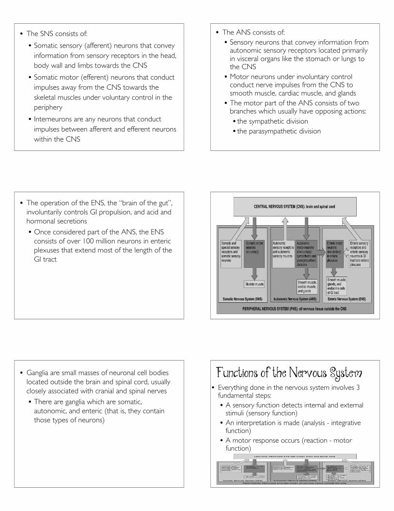
Nervous Tissue Overview of the Nervous System • The nervous system detects environmental changes that impact the body, then works in tandem with the endocrine system to respond to such events • It is responsible for all our behaviors, memories, and movement • It is able to accomplish all these functions because of the excitable characteristic of nervous tissue, which allows for the generation of nerve impulses (called action potentials) Organization of the Nervous System • Over 100 billion neurons and 10–50 times that number of support cells (called neuroglia) are organized into two main subdivisions: • The central nervous system (CNS) • The peripheral nervous system (PNS) • The central nervous system (CNS) consists of the brain and spinal cord • The peripheral nervous system (PNS) consists of all nervous tissue outside the CNS, including nerves, ganglia, enteric plexuses, and sensory receptors • Most signals that stimulate muscles to contract and glands to secrete originate in the CNS • The PNS is further divided into: • A somatic nervous system (SNS) • An autonomic nervous system (ANS) • An enteric nervous system (ENS)
Transcript of Nervous Tissue - Leilehua High School Anatomy &...
1
NervousTissue
Overview of the Nervous System• The nervous system detects environmental changes
that impact the body, then works in tandem with theendocrine system to respond to such events• It is responsible for all our behaviors, memories,
and movement• It is able to accomplish all these functions
because of the excitable characteristic ofnervous tissue, which allows for the generationof nerve impulses (called action potentials)
Organization of the Nervous System• Over 100 billion neurons
and 10–50 times thatnumber of support cells(called neuroglia) areorganized into two mainsubdivisions:• The central nervous
system (CNS)• The peripheral nervous
system (PNS)
• The central nervoussystem (CNS) consistsof the brain and spinalcord
• The peripheral nervoussystem (PNS) consistsof all nervous tissueoutside the CNS,including nerves,ganglia, entericplexuses, and sensoryreceptors
• Most signals that stimulatemuscles to contract andglands to secrete originatein the CNS
• The PNS is further dividedinto:• A somatic nervous
system (SNS)• An autonomic nervous
system (ANS)• An enteric nervous
system (ENS)
2
• The SNS consists of:
• Somatic sensory (afferent) neurons that conveyinformation from sensory receptors in the head,body wall and limbs towards the CNS
• Somatic motor (efferent) neurons that conductimpulses away from the CNS towards theskeletal muscles under voluntary control in theperiphery
• Interneurons are any neurons that conductimpulses between afferent and efferent neuronswithin the CNS
• The ANS consists of:• Sensory neurons that convey information from
autonomic sensory receptors located primarilyin visceral organs like the stomach or lungs tothe CNS• Motor neurons under involuntary control
conduct nerve impulses from the CNS tosmooth muscle, cardiac muscle, and glands• The motor part of the ANS consists of two
branches which usually have opposing actions:• the sympathetic division• the parasympathetic division
• The operation of the ENS, the “brain of the gut”,involuntarily controls GI propulsion, and acid andhormonal secretions
• Once considered part of the ANS, the ENSconsists of over 100 million neurons in entericplexuses that extend most of the length of theGI tract
• Ganglia are small masses of neuronal cell bodieslocated outside the brain and spinal cord, usuallyclosely associated with cranial and spinal nerves
• There are ganglia which are somatic,autonomic, and enteric (that is, they containthose types of neurons)
Functions of the Nervous System• Everything done in the nervous system involves 3
fundamental steps:• A sensory function detects internal and external
stimuli (sensory function)• An interpretation is made (analysis - integrative
function)• A motor response occurs (reaction - motor
function)
3
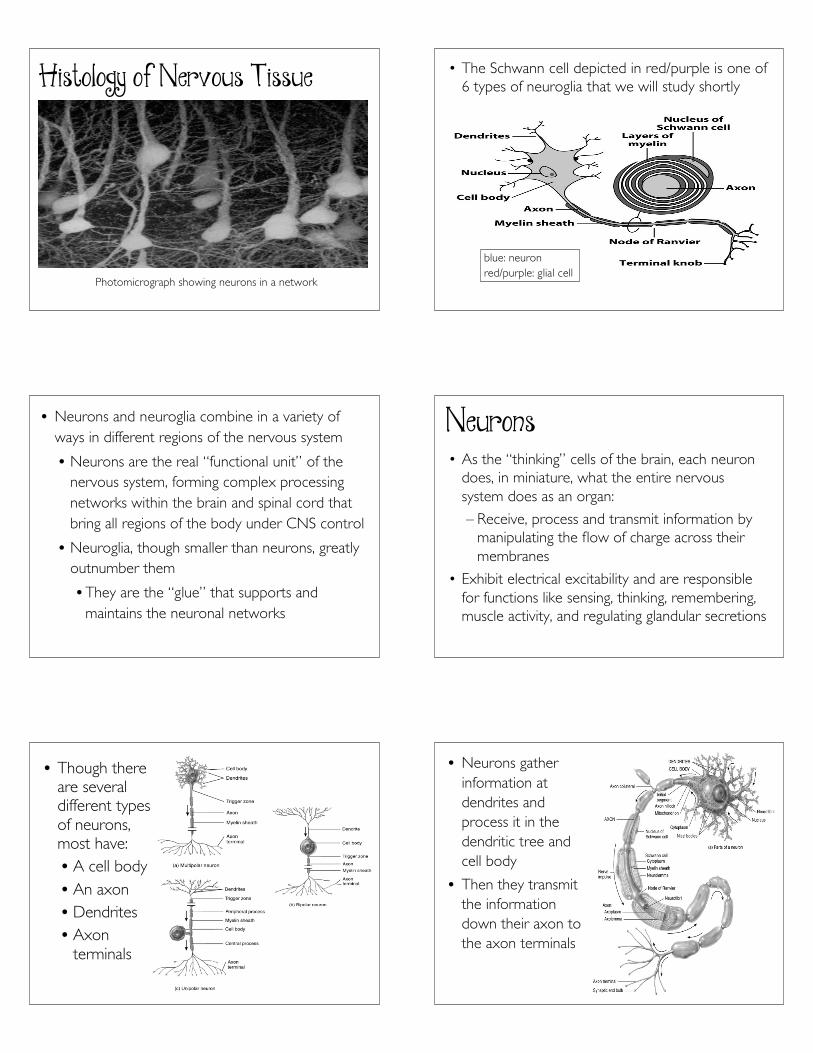
Histology of Nervous Tissue
Photomicrograph showing neurons in a network
• The Schwann cell depicted in red/purple is one of6 types of neuroglia that we will study shortly
blue: neuronred/purple: glial cell
• Neurons and neuroglia combine in a variety ofways in different regions of the nervous system
• Neurons are the real “functional unit” of thenervous system, forming complex processingnetworks within the brain and spinal cord thatbring all regions of the body under CNS control
• Neuroglia, though smaller than neurons, greatlyoutnumber them
• They are the “glue” that supports andmaintains the neuronal networks
Neurons• As the “thinking” cells of the brain, each neuron
does, in miniature, what the entire nervoussystem does as an organ:
– Receive, process and transmit information bymanipulating the flow of charge across theirmembranes
• Exhibit electrical excitability and are responsiblefor functions like sensing, thinking, remembering,muscle activity, and regulating glandular secretions
• Though thereare severaldifferent typesof neurons,most have:• A cell body• An axon• Dendrites• Axon
terminals
• Neurons gatherinformation atdendrites andprocess it in thedendritic tree andcell body
• Then they transmitthe informationdown their axon tothe axon terminals
4
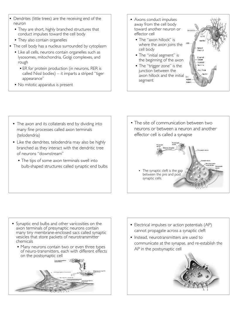
• Dendrites (little trees) are the receiving end of theneuron• They are short, highly branched structures that
conduct impulses toward the cell body• They also contain organelles
• The cell body has a nucleus surrounded by cytoplasm• Like all cells, neurons contain organelles such as
lysosomes, mitochondria, Golgi complexes, andrough• ER for protein production (in neurons, RER is
called Nissl bodies) – it imparts a striped “tigerappearance”
• No mitotic apparatus is present
• Axons conduct impulsesaway from the cell bodytoward another neuron oreffector cell• The “axon hillock” is
where the axon joins thecell body
• The “initial segment” isthe beginning of the axon
• The “trigger zone” is thejunction between theaxon hillock and the initialsegment
• The axon and its collaterals end by dividing intomany fine processes called axon terminals(telodendria)
• Like the dendrites, telodendria may also be highlybranched as they interact with the dendritic treeof neurons “downstream”
• The tips of some axon terminals swell intobulb-shaped structures called synaptic end bulbs
• The site of communication between twoneurons or between a neuron and anothereffector cell is called a synapse
• The synaptic cleft is the gapbetween the pre and postsynaptic cells.
• Synaptic end bulbs and other varicosities on theaxon terminals of presynaptic neurons containmany tiny membrane-enclosed sacs called synapticvesicles that store packets of neurotransmitterchemicals• Many neurons contain two or even three types
of neuro-transmitters, each with different effectson the postsynaptic cell
• Electrical impulses or action potentials (AP)cannot propagate across a synaptic cleft
• Instead, neurotransmitters are used tocommunicate at the synapse, and re-establish theAP in the postsynaptic cell
5
• Substances synthesized or recycled in the neuroncell body are needed in the axon or at the axonterminals
• Two types of transport systems carry materialsfrom the cell body to the axon terminals and back
• Slow axonal transport conveys axoplasm inone direction only – from the cell body towardthe axon terminals
• Fast axonal transport moves materials in bothdirections
• Slow axonal transport supplies new axoplasm (thecytoplasm in axons) to developing or regeneratingaxons and replenishes axoplasm in growing andmature axons
• Fast axonal transport that occurs in an anterograde(forward) direction moves organelles and synapticvesicles from the cell body to the axon terminals
• Fast axonal transport that occurs in a retrograde(backward) direction moves membrane vesiclesand other cellular materials from the axonterminals to the cell body to be degraded orrecycled
• Substances that enter the neuron at the axonterminals are also moved to the cell body by fastretrograde transport• These substances include trophic chemicals such
as nerve growth factor, as well as harmful agentssuch as tetanus toxin and the viruses that causerabies and polio• A deep cut or puncture wound in the head or
neck is a more serious matter than a similarinjury in the leg because of the shorter transittime for the harmful substance to reach thebrain (treatment must begin quickly)
• Neurons display great diversity in size andshape - the longest of them are almost as longas a person is tall, extending from the toes tothe lowest part of the brain• The pattern of dendritic branching is varied
and distinctive for neurons in different partsof the NS• Some have very short axons or lack axons
altogether• Both structural and functional features are used
to classify the various neurons in the body
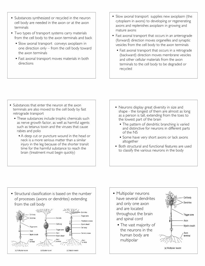
• Structural classification is based on the numberof processes (axons or dendrites) extendingfrom the cell body
• Multipolar neuronshave several dendritesand only one axonand are locatedthroughout the brainand spinal cord• The vast majority of
the neurons in thehuman body aremultipolar
6
• Bipolar neurons have onemain dendrite and oneaxon• They are used to
convey the specialsenses of sight, smell,hearing and balance•As such, they arefound in the retina ofthe eye, the innerear, and the olfactory(olfact = to smell)area of the brain
• Unipolar(pseudounipolar) neuronscontain one processwhich extends from thebody and divides into acentral branch thatfunctions as an axon andas a dendritic root• Unipolar structure is
often employed forsensory neurons thatconvey touch andstretching informationfrom the extremities
• The functional classification of neurons is based onelectrophysiological properties (excitatory orinhibitory) and the direction in which the AP isconveyed with respect to the CNS
• Sensory or afferent neurons convey APs intothe CNS through cranial or spinal nerves
•Most are unipolar
• Motor or efferent neurons convey APs awayfrom the CNS to effectors (muscles and glands)in the periphery through cranial or spinal nerves
•Most are multipolar
• Interneurons or association neurons aremainly located within the CNS betweensensory and motor neurons
• Interneurons integrate (process) incomingsensory information from sensory neuronsand then elicit a motor response byactivating the appropriate motor neurons
•Most interneurons are multipolar instructure
Neuroglia• Neuroglia (glial cells) play a major role in support
and nutrition of the brain, but they do notmanipulate information
• They maintain the internal environment so thatneurons can do their jobs
• Neuroglia do not generate or conduct nerveimpulses
• They support neurons by:
• Forming the Blood Brain Barrier (BBB)
• Forming the myelin sheath (nerve insulation)around neuronal axons
•Making the CSF that circulates around thebrain and spinal cord
• Participating in phagocytosis
7
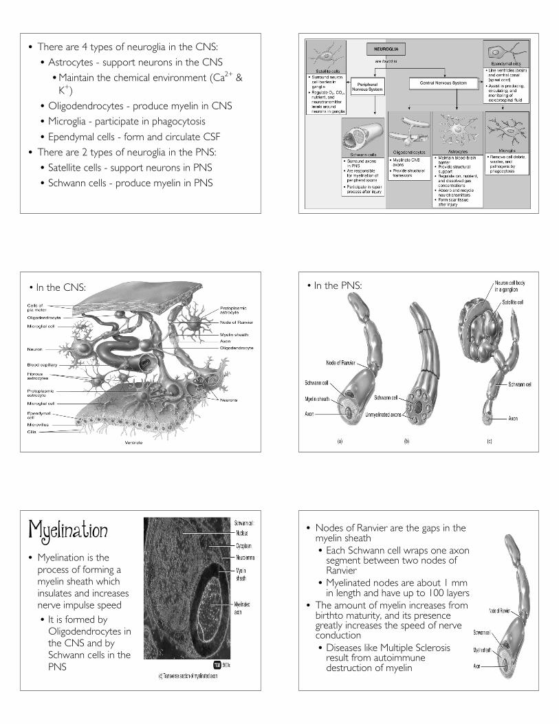
• There are 4 types of neuroglia in the CNS:
• Astrocytes - support neurons in the CNS
•Maintain the chemical environment (Ca2+ &K+)
• Oligodendrocytes - produce myelin in CNS
• Microglia - participate in phagocytosis
• Ependymal cells - form and circulate CSF
• There are 2 types of neuroglia in the PNS:
• Satellite cells - support neurons in PNS
• Schwann cells - produce myelin in PNS
• In the CNS: • In the PNS:
Myelination• Myelination is the
process of forming amyelin sheath whichinsulates and increasesnerve impulse speed• It is formed by
Oligodendrocytes inthe CNS and bySchwann cells in thePNS
• Nodes of Ranvier are the gaps in themyelin sheath• Each Schwann cell wraps one axon
segment between two nodes ofRanvier
• Myelinated nodes are about 1 mmin length and have up to 100 layers
• The amount of myelin increases frombirthto maturity, and its presencegreatly increases the speed of nerveconduction• Diseases like Multiple Sclerosis
result from autoimmunedestruction of myelin
8
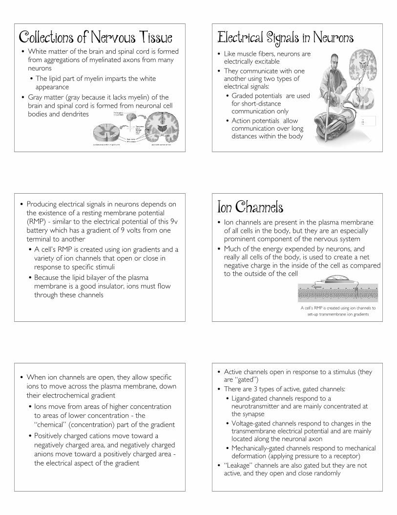
Collections of Nervous Tissue• White matter of the brain and spinal cord is formed
from aggregations of myelinated axons from manyneurons• The lipid part of myelin imparts the white
appearance• Gray matter (gray because it lacks myelin) of the
brain and spinal cord is formed from neuronal cellbodies and dendrites
Electrical Signals in Neurons• Like muscle fibers, neurons are
electrically excitable• They communicate with one
another using two types ofelectrical signals:• Graded potentials are used
for short-distancecommunication only
• Action potentials allowcommunication over longdistances within the body
• Producing electrical signals in neurons depends onthe existence of a resting membrane potential(RMP) - similar to the electrical potential of this 9vbattery which has a gradient of 9 volts from oneterminal to another• A cell’s RMP is created using ion gradients and a
variety of ion channels that open or close inresponse to specific stimuli• Because the lipid bilayer of the plasma
membrane is a good insulator, ions must flowthrough these channels
Ion Channels• Ion channels are present in the plasma membrane
of all cells in the body, but they are an especiallyprominent component of the nervous system
• Much of the energy expended by neurons, andreally all cells of the body, is used to create a netnegative charge in the inside of the cell as comparedto the outside of the cell
A cell’s RMP is created using ion channels to
set-up transmembrane ion gradients
• When ion channels are open, they allow specificions to move across the plasma membrane, downtheir electrochemical gradient
• Ions move from areas of higher concentrationto areas of lower concentration - the“chemical” (concentration) part of the gradient
• Positively charged cations move toward anegatively charged area, and negatively chargedanions move toward a positively charged area -the electrical aspect of the gradient
• Active channels open in response to a stimulus (theyare “gated”)
• There are 3 types of active, gated channels:• Ligand-gated channels respond to a
neurotransmitter and are mainly concentrated atthe synapse
• Voltage-gated channels respond to changes in thetransmembrane electrical potential and are mainlylocated along the neuronal axon
• Mechanically-gated channels respond to mechanicaldeformation (applying pressure to a receptor)
• “Leakage” channels are also gated but they are notactive, and they open and close randomly
9
Resting Membrane Potential• A neuron’s RMP is measured at rest, when it is not
conducting a nerve impulse
• The resting membrane potential exists becauseof a small buildup of negative ions in the cytosolalong the inside of the membrane, and an equalbuildup of positive ions in the extracellular fluidalong the outside surface of the membrane
• The buildup of charge occurs only very close tothe membrane – the cytosol elsewhere in the cellis electrically neutral
• The RMP is slightly negative because leakage channelsfavor a gradient where more K+ leaks out, than Na+
leaks in (there are more K+ channels than Na+
channels)• There are also large negatively charged proteins
that always remain in the cytosol• Left unchecked, inward leakage of Na+ would
eventually destroy the resting membrane potential• The small inward Na+ leak and outward K+ leak are
offset by the Na+/K+ ATPases (sodium-potassiumpumps) which pumps out Na+ as fast as it leaks in
• In neurons, a typical value for the RMP is –70 mV (theminus sign indicates that the inside of the cell isnegative relative to the outside)
• A cell that exhibits an RMP is said to be polarized• In this state, the cell is “primed” - it is ready to
produce an action potential• In order to do so, graded potentials must first be
produced in order to depolarize the cell tothreshold• A graded potential occurs whenever ion flow in
mechanically gated or ligand-gated channelsproduce a current that is localized – it spreads toadjacent regions for a short distance and thendies out within a few millimeters of its point oforigin
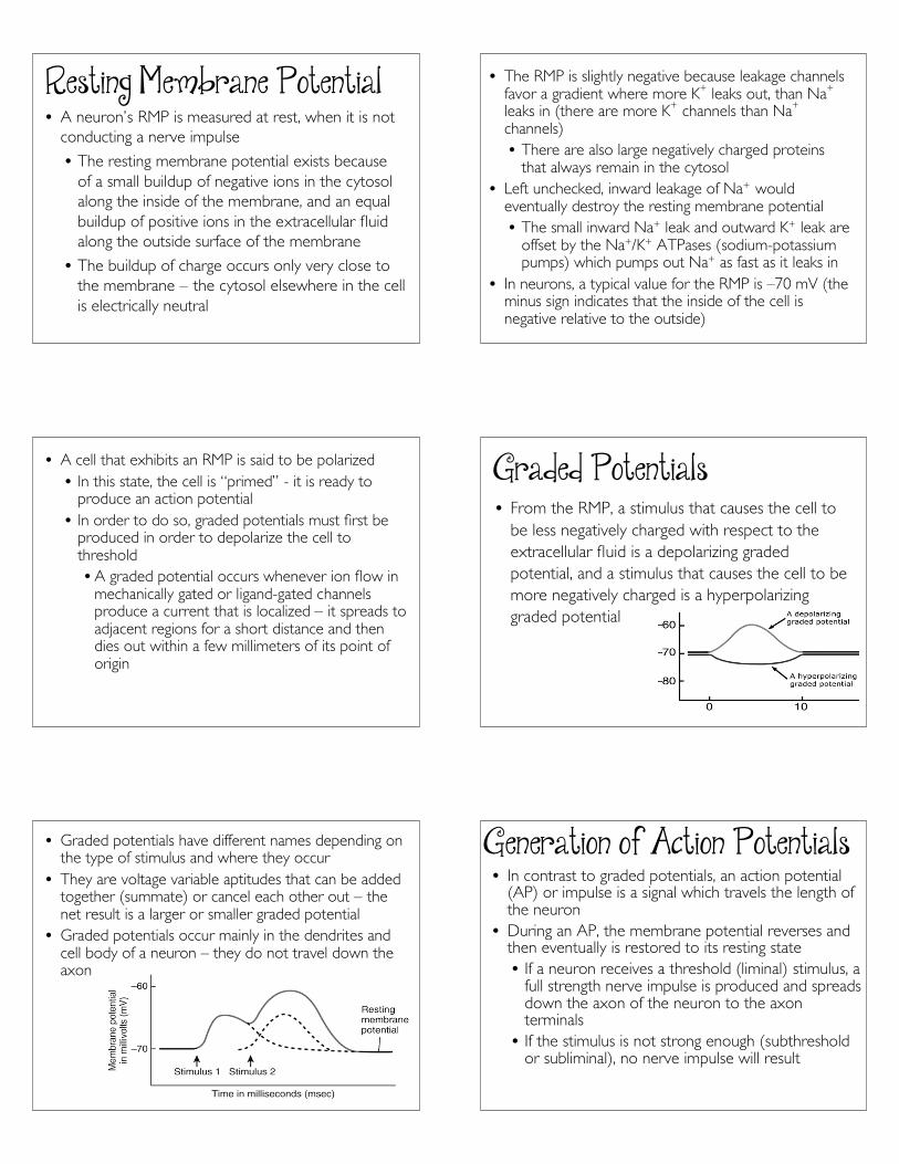
Graded Potentials• From the RMP, a stimulus that causes the cell to
be less negatively charged with respect to theextracellular fluid is a depolarizing gradedpotential, and a stimulus that causes the cell to bemore negatively charged is a hyperpolarizinggraded potential
• Graded potentials have different names depending onthe type of stimulus and where they occur
• They are voltage variable aptitudes that can be addedtogether (summate) or cancel each other out – thenet result is a larger or smaller graded potential
• Graded potentials occur mainly in the dendrites andcell body of a neuron – they do not travel down theaxon
Generation of Action Potentials• In contrast to graded potentials, an action potential
(AP) or impulse is a signal which travels the length ofthe neuron
• During an AP, the membrane potential reverses andthen eventually is restored to its resting state• If a neuron receives a threshold (liminal) stimulus, a
full strength nerve impulse is produced and spreadsdown the axon of the neuron to the axonterminals
• If the stimulus is not strong enough (subthresholdor subliminal), no nerve impulse will result

10
• An AP has two main phases:• a depolarizing phase and• a repolarizing phase
• Graded potentials that result in depolarization of theneuron from –70mV to threshold (about –55 mV inmany neurons) will cause a sequence of events torapidly unfold• Voltage-gated Na+ channels open during the steep
depolarization phase allowing Na+ to rush into thecell and making the inside of the cell progressivelymore positive
• Only a total of 20,000 Na+
actually enter the cell ineach little area of themembrane, but they changethe potential considerably(up to +30mV)]
• During the repolarization,phase K+ channels open andK+ rushes outward• The cell returns to a
progressively morenegative state until theRMP of –70mV is onceagain restored
• While the voltage-gated K+ channels are open,outflow of K+ may be large enough to cause anafter-hyperpolarizing phase of the action potential
• During this phase, the voltage-gated K+ channelsremain open and the membrane potentialbecomes even more negative (about –90 mV)
• As the voltage-gated K+ channels close, themembrane potential returns to the restinglevel of –70 mV
• According to the all-or-none principle, if astimulus reaches threshold, the action potential isalways the same
• A stronger stimulus will not cause a largerimpulse
• After initiating an action potential, there is aperiod of time called the absolute refractoryperiod during which a cell cannot generateanother AP, no matter how strong the stimulus
• This period coincides with the period of Na+
channel activation and inactivation (inactivatedNa+ channels must first return to the restingstate)
• This places an upper limit of 10–1000 nerveimpulses per second, depending on theneuron
11
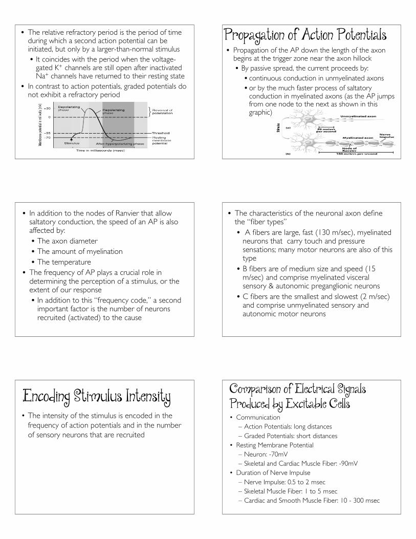
• The relative refractory period is the period of timeduring which a second action potential can beinitiated, but only by a larger-than-normal stimulus• It coincides with the period when the voltage-
gated K+ channels are still open after inactivatedNa+ channels have returned to their resting state
• In contrast to action potentials, graded potentials donot exhibit a refractory period
Propagation of Action Potentials• Propagation of the AP down the length of the axon
begins at the trigger zone near the axon hillock• By passive spread, the current proceeds by:• continuous conduction in unmyelinated axons• or by the much faster process of saltatory
conduction in myelinated axons (as the AP jumpsfrom one node to the next as shown in thisgraphic)
• In addition to the nodes of Ranvier that allowsaltatory conduction, the speed of an AP is alsoaffected by:• The axon diameter• The amount of myelination• The temperature
• The frequency of AP plays a crucial role indetermining the perception of a stimulus, or theextent of our response• In addition to this “frequency code,” a second
important factor is the number of neuronsrecruited (activated) to the cause
• The characteristics of the neuronal axon definethe “fiber types”• A fibers are large, fast (130 m/sec), myelinated
neurons that carry touch and pressuresensations; many motor neurons are also of thistype• B fibers are of medium size and speed (15
m/sec) and comprise myelinated visceralsensory & autonomic preganglionic neurons• C fibers are the smallest and slowest (2 m/sec)
and comprise unmyelinated sensory andautonomic motor neurons
Encoding Stimulus Intensity• The intensity of the stimulus is encoded in the
frequency of action potentials and in the numberof sensory neurons that are recruited
Comparison of Electrical SignalsProduced by Excitable Cells• Communication
– Action Potentials: long distances– Graded Potentials: short distances
• Resting Membrane Potential– Neuron: -70mV– Skeletal and Cardiac Muscle Fiber: -90mV
• Duration of Nerve Impulse– Nerve Impulse: 0.5 to 2 msec– Skeletal Muscle Fiber: 1 to 5 msec– Cardiac and Smooth Muscle Fiber: 10 - 300 msec

12
• Propagation Speed of Action Potentials
– 18 times faster along the largest-diameter,myelinated axons, than the propagation speedalong the sarcolemma of a skeletal musclefiber
Signal Transmission at Synapses• A synapse is the functional junction between
one neuron and another, or between a neuronand an effector such as a muscle or gland
• The two types of synapses are electrical andchemical
Electrical Synapses• Action potentials (impulses) conduct directly
between the plasma membranes of adjacentneurons through gap junctions
• Faster communication because the action potentialpasses directly from the presynaptic cell to thepostsynaptic cell
• Can synchronize (coordinate) the activity of agroup of neurons or muscle fibers
Chemical Synapses• Signal transmission at the synapse is a one-way transfer from
a presynaptic neuron to a postsynaptic neuron
• When an AP reaches the end bulb of axon terminals,voltage-gated Ca2+ channels open and Ca2+ flows inward,triggering release of the neurotransmitter
• The neurotransmitter crosses the synaptic cleft and bindsto ligand-gated receptors on the postsynaptic membrane
• The more neurotransmitter released, the greater thenumber and intensity of graded potentials in thepostsynaptic cell
• In this way, the presynaptic neuron convertsan electrical signal (nerve impulse) into achemical signal (released neurotransmitter)
• The postsynaptic neuron receives thechemical signal and in turn generates anelectrical signal (postsynaptic potential)
• The time required for these processes at achemical synapse produces a synaptic delayof about 0.5 msec
13
Excitatory & Inhibitory PostsynapticPotentials• A neurotransmitter causes either an excitatory or an
inhibitory graded potential:• Excitatory postsynaptic potential (EPSP) causes a
depolarization of the postsynaptic cell, bringing itcloser to threshold. Although a single EPSPnormally does not initiate a nerve impulse, thepostsynaptic cell does become more excitable
• Inhibitory postsynaptic potential (IPSP)hyperpolarizes the postsynaptic cell taking it fartherfrom threshold
• Spatial summation occurs when postsynapticpotentials arrive near the same location
• Temporal summation occurs whenpostsynaptic potentials arrive close to thesame time
• Whether or not the postsynaptic cell reachesthreshold depends on the net effect aftersummation of all the postsynaptic potentials
Structure of NeurotransmitterReceptors
• There are two major types of neurotransmitterreceptors– Ionotropic
•Contains a neurotransmitter binding site andan ion channel
– Metabotropic•Contains a neurotransmitter binding site and is
coupled to a separate ion channel by a Gprotein
Removal of Neurotransmitter• If a neurotransmitter could linger in the synaptic cleft, it
would influence the postsynaptic neuron, muscle fiber, orgland cell indefinitely – removal of the neurotransmitter isessential for normal function• Removal is accomplished by diffusion out of the
synaptic cleft, enzymatic degradation, and re-uptake bycells.• An example of a common neurotransmitter
inactivated through enzymatic degradation isacetylcholine• The enzyme acetylcholinesterase breaks down
acetylcholine in the synaptic cleft
Spatial & Temporal Summation ofPostsynaptic Potentials
• If several presynaptic end bulbs release theirneurotransmitter at about the same time, thecombined effect may generate a nerve impulse dueto summation
– May be spatial or temporal
• The postsynaptic neuron is an integrator
– It receives excitatory and inhibitory signals,integrates them, and responds accordingly
Neurotransmitters• Both excitatory and inhibitory
neurotransmitters are present in the CNS andthe PNS
• A given neurotransmitter may be excitatory insome locations and inhibitory in others
• Neurotransmitters can be divided into twoclasses based on size• Small- Molecule Neurotransmitters• Neuropeptides
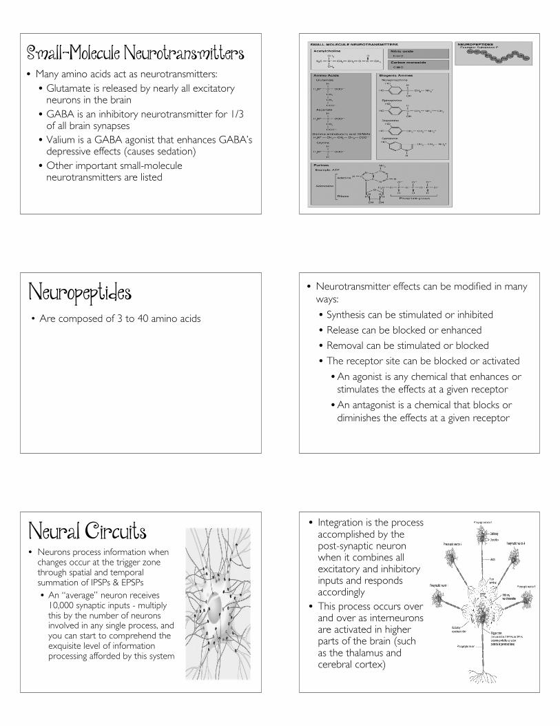
14
Small-Molecule Neurotransmitters• Many amino acids act as neurotransmitters:• Glutamate is released by nearly all excitatory
neurons in the brain• GABA is an inhibitory neurotransmitter for 1/3
of all brain synapses• Valium is a GABA agonist that enhances GABA’s
depressive effects (causes sedation)• Other important small-molecule
neurotransmitters are listed
Neuropeptides• Are composed of 3 to 40 amino acids
• Neurotransmitter effects can be modified in manyways:
• Synthesis can be stimulated or inhibited
• Release can be blocked or enhanced
• Removal can be stimulated or blocked
• The receptor site can be blocked or activated
• An agonist is any chemical that enhances orstimulates the effects at a given receptor
• An antagonist is a chemical that blocks ordiminishes the effects at a given receptor
Neural Circuits• Neurons process information when
changes occur at the trigger zonethrough spatial and temporalsummation of IPSPs & EPSPs• An “average” neuron receives
10,000 synaptic inputs - multiplythis by the number of neuronsinvolved in any single process, andyou can start to comprehend theexquisite level of informationprocessing afforded by this system
• Integration is the processaccomplished by thepost-synaptic neuronwhen it combines allexcitatory and inhibitoryinputs and respondsaccordingly
• This process occurs overand over as interneuronsare activated in higherparts of the brain (suchas the thalamus andcerebral cortex)
15
• A neuronal network may contain thousands oreven millions of neurons• Types of circuits include diverging,
converging, reverberating, and parallel after-discharge
• In a diverging circuit, a small number of neurons inthe brain stimulate a much larger number of neuronsin the spinal cord• A converging circuit is the opposite
• In a reverberating circuit, impulses are sent backthrough the circuit time and time again• Used in breathing, coordinated muscular activities,
waking up, and short-term memory• Parallel after-discharge circuits involve a single
presynaptic cell that stimulates a group of neurons,which then synapse with a common postsynaptic cell• Used in precise activities such as mathematical
calculations
Regeneration & Repair of NervousTissue• The nervous system exhibits plasticity, the
capability to change based on experience• But has very limited powers of regeneration,
the capability to replicate or repair damagedneurons
• Neurogenesis, the birth of new neurons fromundifferentiated stem cells, is normally verylimited
• Damaged axons does not occur in mostregions of the CNS
• The cell bodies of neurons lose their mitoticfeatures at birth and can only be repaired throughregeneration after an injury (they are neverreplaced by daughter cells as occurs with epithelialtissues)
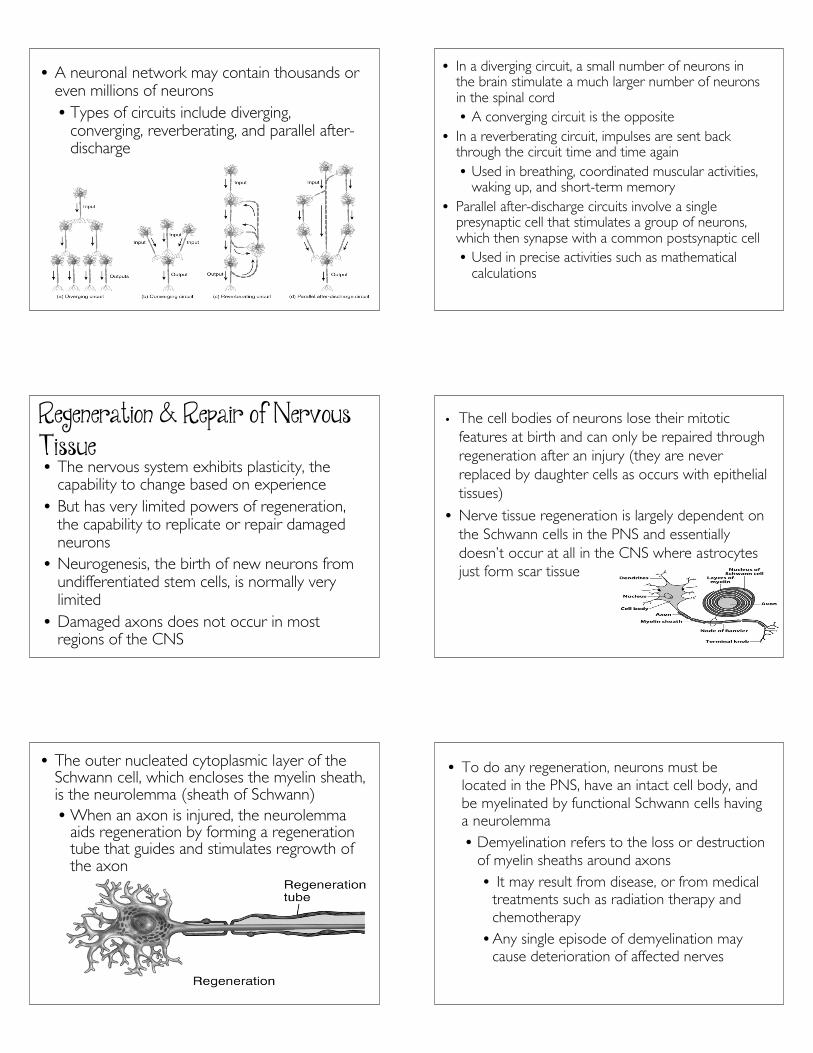
• Nerve tissue regeneration is largely dependent onthe Schwann cells in the PNS and essentiallydoesn’t occur at all in the CNS where astrocytesjust form scar tissue
• The outer nucleated cytoplasmic layer of theSchwann cell, which encloses the myelin sheath,is the neurolemma (sheath of Schwann)• When an axon is injured, the neurolemma
aids regeneration by forming a regenerationtube that guides and stimulates regrowth ofthe axon
• To do any regeneration, neurons must belocated in the PNS, have an intact cell body, andbe myelinated by functional Schwann cells havinga neurolemma• Demyelination refers to the loss or destruction
of myelin sheaths around axons• It may result from disease, or from medical
treatments such as radiation therapy andchemotherapy• Any single episode of demyelination may
cause deterioration of affected nerves