Nerves, Muscles & Movement Notes 09a - PBworks
21
HL Biology Notes for Nerves, Muscles & Movement A. De Jong/TFSS 2007 1 of 21 The notes in this document cover IB topics 11.2 and option E.1, E.2, E.4 and E.5. Organization of the Nervous System The nervous system is divided into the peripheral nervous system (PNS) and central nervous system (CNS). The PNS consists of sensory neurons running from stimulus receptors that inform the CNS of the stimuli motor neurons running from the CNS to the effectors (muscles and glands) that respond to the stimuli The CNS consists of the spinal cord the brain The Peripheral Nervous System The PNS is subdivided into the sensory‐somatic nervous system which connects the external environment and the CNS there are 12 pairs of cranial nerves, which connect directly to the brain (e.g. the optic nerve), and they may be sensory, motor, or mixed nerves there are 31 pairs of spinal nerves, all of which are mixed all our conscious awareness of the external environment and all our motor activity to cope with it operate through the sensory‐somatic division of the PNS actions of the sensory‐somatic nervous system are largely voluntary – skeletal muscle is controlled by this system the autonomic nervous system (ANS) which connects the internal environment and the CNS consists of sensory neurons and motor neurons that run between the CNS and various internal organs it is responsible for monitoring conditions in the internal environment and bringing about appropriate changes in them actions of the autonomic nervous system are largely involuntary ‐ cardiac muscle (heart), blood vessels, digestive system, smooth muscle, and glands are controlled by this system uses two groups of motor neurons to stimulate the effectors CNS External Environment Internal Environment Sensory- Somatic NS Autonomic NS Sensory Neurons Sensory Neurons Motor Neurons Motor Neurons
Transcript of Nerves, Muscles & Movement Notes 09a - PBworks

HLBiology NotesforNerves,Muscles&Movement
A.DeJong/TFSS2007 1of21
ThenotesinthisdocumentcoverIBtopics11.2andoptionE.1,E.2,E.4andE.5.
OrganizationoftheNervousSystem
The nervous system is divided into the peripheral nervous system (PNS) and central nervous system(CNS).
ThePNSconsistsof
sensoryneuronsrunningfromstimulusreceptorsthatinformtheCNSofthestimuli motorneurons running from theCNS to the effectors (muscles and glands) that respond to the
stimuli
TheCNSconsistsof
thespinalcord thebrain
ThePeripheralNervousSystem
ThePNSissubdividedinto
thesensory‐somaticnervoussystemwhichconnectstheexternalenvironmentandtheCNS
thereare12pairsofcranialnerves,whichconnectdirectlytothebrain(e.g.theopticnerve),andtheymaybesensory,motor,ormixednerves
thereare31pairsofspinalnerves,allofwhicharemixed all our conscious awareness of the external environment and all ourmotor activity to cope
withitoperatethroughthesensory‐somaticdivisionofthePNS actions of the sensory‐somatic nervous system are largely voluntary – skeletal muscle is
controlledbythissystem
theautonomicnervoussystem(ANS)whichconnectstheinternalenvironmentandtheCNS
consistsofsensoryneuronsandmotorneuronsthatrunbetweentheCNSandvariousinternalorgans
it is responsible for monitoring conditions in the internal environment and bringing aboutappropriatechangesinthem
actions of the autonomic nervous system are largely involuntary ‐cardiac muscle (heart),bloodvessels,digestivesystem,smoothmuscle,andglandsarecontrolledbythissystem
usestwogroupsofmotorneuronstostimulatetheeffectors
CNS External Environment
Internal Environment
Sensory- Somatic NS
Autonomic NS
Sensory Neurons Sensory Neurons
Motor Neurons Motor Neurons

HLBiology NotesforNerves,Muscles&Movement
A.DeJong/TFSS2007 2of21
■ preganglionicneuronsariseintheCNSandruntoaganglioninthebody■ postganglionicneuronsruntotheeffectororgan(synapseoccursintheganglion
further subdivided into the sympathetic and parasympathetic nervous systems, which arelargelyantagonistictoeachother:
Imagefromhttp://users.rcn.com/jkimball.ma.ultranet/BiologyPages/A/autonomic.gif
TheSympatheticNervousSystem
Theneurotransmitterofthepreganglionicneuronsisacetylcholine(Ach).Itstimulatesactionpotentialsinthepostganglionicneurons.Theneurotransmitterofthepostganglionicneuronsisnoradrenaline.Theactionofnoradrenalineonaparticularglandormusclemaybeexcitatoryorinhibitory.
StimulationofthesympatheticbranchoftheANSpreparesthebodyforemergencies:“fightorflight”.
TheParasympatheticNervousSystem
Themain nerves of the parasympathetic nervous system are the vagus nerves, which originate in themedullaoblongata. Acetylcholine is theneuro‐transmitteratallpre‐andmanypostganglionicneurons.Somepostganglionicneuronsreleasenitricoxideastheirneurotransmitter.
The parasympathetic nervous system returns the body to normal after they have been altered bysympatheticstimulation:“restanddigest”.

HLBiology NotesforNerves,Muscles&Movement
A.DeJong/TFSS2007 3of21
AlthoughtheANSisconsideredtobeinvoluntary,thisisnotentirelytrue.AcertainamountofconsciouscontrolcanbeexertedoveritashaslongbeendemonstratedbypractitionersofyogaandZenBuddhism.During their periods of meditation, these people are able to alter a number of autonomic functionsincludingheart rate and the rateof oxygen consumption. These changes arenot simply a reflectionofdecreasedphysicalactivitybecausetheyare lowerthan levels foundduringsleeporhypnosis. AnotherexampleofconsciouscontroloftheANSisthecontrolofemptyingofthebladderandbowels.
TheCentralNervousSystem
ThespinalcordconductssensoryinformationfromthePNStothebrain,andconductsmotorinformationfromthebraintotheeffectors,includingskeletal,smoothandcardiacmuscle,andglands.Italsoservesasaminorreflexcentre.
The brain receives sensory input from the spinal cord and its own nerves. It devotes most of itscomputationalpowertoprocessingitsvarioussensoryinputsandinitiatingappropriateandcoordinatedmotoroutputs.
Boththespinalcordandbrainconsistofwhitematter(bundlesofaxonscoatedwithmyelinsheaths)andgreymatter(cellbodies&dendrites,coveredinsynapses).Theyarealsocoveredwithconnectivetissuecalledthemeninges.
Anextracellular fluid thatdiffers in its composition from theECF in the restof thebodysurrounds thecells of the CNS. Cerebrospinal fluid (CSF) contains less protein than ECF, and is found within thecerebrospinalcanalofthespinalcordandwithinthefourventriclesofthebrain.
TheSpinalCord
Imagefromhttp://neuro.wehealny.org/images/14_01.jpg
There are 31 pairs of spinal nerves. These are all classed asmixed nerves because they contain bothsensoryandmotoraxons.
sensoryaxonspassintothedorsalrootganglionwheretheircellbodiesarelocatedandthenonintothespinalcorditself
motoraxonspass intopass into theventralrootsbeforeunitingwith thesensoryaxons to formthemixednerves

HLBiology NotesforNerves,Muscles&Movement
A.DeJong/TFSS2007 4of21
Theprimaryfunctionsofthespinalcord:
itconnectsalargepartofthePNStothebrain
it is a minor coordinating centre responsible for some simple reflexes such as the withdrawalreflex
TheBrain
MedullaOblongatacontrolsinvoluntaryandvisceralactivities
Cerebellumcontrolsbodybalance,muscularcoordinationandequilibrium.
Hypothalamusmaintainstheinternalenvironment
regulatesbodytemperature,thirst,hunger,metabolism,pleasure,pain,etc.
Thalamussortsincomingandoutgoingimpulsesandsendstotheappropriatecentre
CerebralCortexcentreofallvoluntarymusclecontrolandmentalactivity
analysis coding info.storage recognition
memory understanding intelligence senseintegration
Imagefromhttp://www.emc.maricopa.edu/faculty/farabee/BIOBK/brain_3.gif
Thepituitarygland,locatedatthebaseofthebrain,isapproximatelythesizeofapea,andiscomposedoftwolobes.
anteriorlobe:stimulatedbythehypothalamusofthebraintosecreteseveralhormones
thyroidstimulatinghormone(TSH) follicle‐stimulatinghormone(FSH) luteinizinghormone(LH)
prolactin growthhormone adrenocorticotropichormone(ACTH)
posteriorlobe:releasestwohormones,synthesizedbythehypothalamus,intothebloodstream
antidiuretichormone(ADH) oxytocin

HLBiology NotesforNerves,Muscles&Movement
A.DeJong/TFSS2007 5of21
Howdoweknowwhatthebraindoes?
Investigatingbrain‐damagedpatients
o Forexample,theexperiencesofsoldierssurvivingbullet‐woundstotherearoftheskullledtothediscoveryoftheroleofthevisualcortexontherearofthecerebralhemispheres.
o Patientswhoarenotimmediatelykilledbyastrokeoftenexperienceparalysisorlossofaspecificbodyfunction–post‐mortemanalysisidentifiestheparticularpartofthebrainaffectedbythestroke.
Animalexperiments
o Wehavelearnedalotaboutbrainfunctionbystudyingmammalsandothervertebrates,removingpartsofahealthybrainorseveringconnectionsbetweenneurons.
o Inoneinvestigationusingcats,severingthefibresthatcrossoverinthecentreofthebrainbelowthetwohalvesofthecerebralhemispheresgavecluestotheinteractionofleftandrighthalvesofthebrain.
fMRI
o FunctionalmagneticresonanceimagingisanadvancedformofMRIthatdetectsthepartsofthebrainthatareactivewhenthebodyperformsspecifictasks.Thereisalwaysademandforoxygenandglucose(foodenergy)inthebrain,buttherearelocalincreasesindemandwhenaparticularareaofthebrainisinuse.fMRIdetectsincreasesinredbloodcelloxygenationatthesiteofneuralactivity.

HLBiology NotesforNerves,Muscles&Movement
A.DeJong/TFSS2007 6of21
Neurons
All neurons are specialized cells that carry anelectrochemicalimpulsecalledanactionpotential.
Sensory neurons run from the stimulus receptors (e.g. fortouch,vision,sound,odourandtaste)totheCNS.
Interneurons, foundonly in theCNS,andarestimulatedbysensoryneurons,other interneurons,orboth. Thebrain isestimated to contain 100 billion interneurons averaging1000synapseseach.
Motor neurons such as the one pictured can have an axonthat is up to onemetre in length. They transmit impulsesfromthebraintotheeffectors–musclesandglands.
Imagefromhttp://www.gonzaga.k12.nf.ca/academics/science/sci_page/biology/neuron1.gif
NerveImpulseTransmissionNeuronssendmessageselectrochemically,whichmeansthatchemicalscauseanelectricsignal.Ionshaveeitherapositive(+)ornegative(‐)charge.Importantionsfornerveimpulsetransmissionare:
sodium(Na+) potassium(K+)
calcium(Ca++) chloride(Cl‐)
Somedefinitions:
Membranepotential:theelectricalpotentialdifference(voltage)acrossacell'smembrane.
Actionpotential:awaveofelectricaldischargethattravelsalongthemembraneofacell.Actionpotentialsareusedbythenervoussystemtotransmitinformationbetweenneurons,andbetweenneuronsandeffectors.
Resting potential: the membrane potential that would be maintained if there were no actionpotentials,synapticpotentialsorotheractivechangesinthemembranepotential.Formostcells,this is anegativenumber. The restingpotential of aneuron isusually ‐70mV.
Atrest,K+caneasilycrossthroughthemembrane,whileCl‐andNa+havemoretroublecrossing.Thenegatively‐chargedproteinmolecules(A‐)cannotcrossthemembrane.Inaddition,thesodium‐potassiumionpumpisactivelypumpingthreeNa+outforeverytwoK+itputsin.
Imagefromhttp://faculty.washington.edu/chudler/gif/ioncon.gif

HLBiology NotesforNerves,Muscles&Movement
A.DeJong/TFSS2007 7of21
Anactionpotentialoccurswhenaneuronsendsanimpulsedownanaxon,awayfromthecellbody.Itisanexplosionofelectricalactivitythatiscreatedbyadepolarizingcurrent.(Thismeansthatastimulushascausedtherestingpotentialtomovetoward0mV.)Whenthedepolarizationreachesabout‐55mV,aneuronwillfireanactionpotential.Thisvalueiscalledthethreshold.Ifthisvalueisnotreached,theactionpotentialwillnotfire.
* The action potential for any given neuron is always the same. There is no “big” or “small” actionpotentialforaneuron.
Actionpotentialsarecausedbyanexchangeofionsacrossthemembraneofaneuron:
Astimuluscausessodiumchannelstoopen,allowingsodiumionstoenterthecell.
Thiscausesdepolarization,becausethesodiumionsarepositivelycharged.
The potassium channels open after depolarization begins, which causes potassium to leave thecell,reversingthedepolarization.
Around this time, sodium channels begin to close, which causes a repolarisation, as the actionpotentialgoesbacktoward‐70mV.
The action potential actually goes past ‐70 mV (a hyperpolarisation) because the potassiumchannelsstayopenabittoolong.
Gradually,theionconcentrationsgobacktorestinglevelsandthecellreturnsto‐70mV.
Imagefromhttp://faculty.washington.edu/chudler/ap3.gif
Synapses
Nerveimpulsesaretransmittedalonganindividualneuronbymeansofanactionpotential.Sincethesesignalsmustbetransmittednotonlyalongasingleneuron,butfromoneneurontoanother,orfromaneurontoaneffector,theremustbeameansofpassingthesignalfromoneneurontoanother.
Ajunctionbetweentwoneuronsiscalledasynapse.Forinformationtopassbetweenneurons,itmustcrossthesynapse.Invertebrates,andsomefishhaveelectricalsynapses,inwhichtheactionpotentialinthepre‐synapticneuroncantriggeranactionpotentialinthepost‐synapticneuronbecausethereisaphysicalconnectionbetweenthetwoneurons.Electricalsynapsesarefasterthanchemicalsynapses.
Mostnervesareconnectedbychemicalsynapses,whichconsistof: apre‐synapticendingthatcontainsneurotransmitters,mitochondriaandothercellorganelles
aneurotransmitterisasubstance(suchasnorepinephrineoracetylcholine)thattransmitsnerveimpulsesacrossasynapse
apost‐synapticendingthatcontainsreceptorsitesforneurotransmitters

HLBiology NotesforNerves,Muscles&Movement
A.DeJong/TFSS2007 8of21
asynapticcleftorspacebetweenthepre‐synapticandpost‐synapticendings
Forcommunicationbetweenneuronstooccur,anelectricalimpulsemusttraveldownanaxontothesynapticterminal.
ActionofNeurotransmitters:
1. Atthepre‐synapticterminal,anelectricalimpulse(actionpotential)causesachangeinmembranepermeabilitytoCa++,whichallowsCa++toflowintothesynapticknob.
Imagefromhttp://users.rcn.com/jkimball.ma.ultranet/BiologyPages/S/Synapse.gif
2. PresenceofCa++willtriggerthemigrationofvesiclescontainingneurotransmitterstowardthepre‐synapticmembrane.
3. Thevesiclemembranewillfusewiththepre‐synapticmembrane,releasingneurotransmittersintothesynapticcleft.(anexampleofexocytosis)
4. Neurotransmittermoleculesdiffuseacrossthesynapticcleftwheretheycanbindwithreceptorsitesonthepost‐synapticendingtoinfluencetheelectricalresponseinthepost‐synapticneuron.
Whenaneurotransmitterbindstoapost‐synapticreceptor,itchangesthepost‐synapticcell'sexcitability,makingiteithermoreorlesslikelytofireanactionpotential.
Ifthenumberofexcitatorypost‐synapticeventsislargeenough,theywilladdtocauseanactionpotentialinthepost‐synapticcellandacontinuationofthe“message”.
Manypsychoactivedrugsandneurotoxinscanchangethepropertiesofneurotransmitterrelease,neurotransmitterreuptakeandtheavailabilityofreceptorbindingsites.
NeurotransmittersandSynapses
SynapsesofthePNSareclassifiedaccordingtotheneurotransmitterused.Eachsynapseusesonlyoneneuro‐transmitter.
Mostsynapsesintheparasympatheticnervoussystemarecholinergicsynapses,anduseacetylcholine.Neuromuscularjunctionsarealsocholinergic.
Mostsynapsesinthesympatheticnervoussystemareadrenergicsynapses,andusenoradrenaline.
Synapsesofthebrainuseamuchwiderrangeofneurotransmitters,includingdopamineandenkephalins.

HLBiology NotesforNerves,Muscles&Movement
A.DeJong/TFSS2007 9of21
Neurotransmittersbindtoreceptorsonthepostsynapticmembrane,causingtemporarychangesinitspermeability.
SomeneurotransmitterscauseNa+orotherpositiveionstoenterthepost‐synapticneuron,helpingtodepolarizeitandcauseanactionpotential.
Thesearecalledexcitatorysynapses.
OtherneurotransmitterscauseCl‐tomoveintothepost‐synapticneuron–thiscauseshyperpolarisation.Hyperpolarisationmakesitmoredifficulttocreateanactionpotential(furtherfromthreshold).
Thesearecalledinhibitorysynapses.
Mostpost‐synapticneuronshavesynapseswithmorethanonepre‐synapticneuron–thesemaybeamixofexcitatoryandinhibitorysynapses,andwhetheranactionpotentialisinitiatedinthepost‐synapticneuronisdeterminedbythesumofallneurotransmittermessages.
Parkinson'sDiseaseiscausedbythedeathofneuronsinapartofthebraincalledthesubstantianigra.Theseneuronsreleasetheneurotransmitterdopamineatinhibitorysynapseswithneuronsthathelptocontrolmusclecontractions.Withoutdopamine,musclecontractionscannotbeproperlycontrolled–thiscausesthesymptomsofParkinson's:
earlysymptomsincludefeelingtiredandshaky,andalossofconcentration eventually,thebodybecomesstiffbecauseantagonisticmusclescannotrelax uncontrollableshakingaffectsthehandsandotherbodypartsandmovementsbecomeveryslow
Painreceptorsarefoundintheskinandotherorgans.Theyconsistoffreenerveendings,whichperceivemechanical,chemicalorthermalstimuli.Painsignalsaresentfromthesenerveendingstothespinalcordvianervefibres,whichcarrythemuptothethalamusorbrainstem.Fromhere,painsignalsmaybepassedontosensoryareasofthecerebralcortex,givingconsciousrecognitionofpain.Sincetherearebothfastandslownerveendings,apainfulstimuluscausesaninitialsharppainsensation,followedbyaslow,burningpain.
Thesensationofpainisnecessarytotellthebodywhenitisbeingdamaged–thisallowsthepainwithdrawalreflexorotherreactionstooccur.Sometimespaininterfereswiththeabilitytoconcentrate.Inthesesituations,paincontrolsystemsinthebrainandspinalcordcanbeusedtoreduceorpreventfeelingsofpain.Thisinvolvestwonaturalpainkillers:
enkephalinsreleasedbythebrainblockcalciumchannelsinthemembraneofthepre‐synapticneurons,blockingsynaptictransmissionsothatpainsignalsdonotreachthebrain
endorphinsproducedbythepituitaryglandarecarriedtothebrainandotherorgansbytheblood,andbindtoreceptorsinthemembranesofneuronsthatsendpainsignalstothebrain–endorphinsaresecretedduringstressfultimes,afterinjuries,andsometimesduringphysicalexercisesuchasrunning
PsychoactiveDrugs
Psychoactivedrugsaffectthebrainandpersonality.Theyeitherincreaseordecreasesynaptictransmission:
theycanbindtothereceptorsiteonpost‐synapticmembranes,mimickingtheneurotransmitterorblockingthebindingoftheneurotransmitter

HLBiology NotesforNerves,Muscles&Movement
A.DeJong/TFSS2007 10of21
theycanalsoreducetheeffectoftheenzymewhichnormallybreaksdowntheneurotransmitter,whichcausesanincreaseintheeffectoftheneurotransmitter
nicotinemimicsacetylcholine,whilecurareblocksacetylcholine
ExcitatoryPsychoactiveDrugsincreasetheactivityofthenervoussystem,andmayhavedifferenteffectsonbehaviour:
Nicotinestimulatessynaptictransmissionatcholinergicsynapsesinmanypartsofthebrain,andcausesreleaseofadrenalinefromtheadrenalgland.Thisresultsinincreasedbloodpressureandcardiacfrequency.Itaffectsmood,actinglikeastimulantandcausingeuphoria.
Cocaineblocksthereabsorptionofdopamineandnoradrenalineatsynapsesinthebrain,causingincreasedenergy,alertnessandtalkativeness.Itgivesanintensefeelingofeuphoria.Physicaleffectsincludeincreasedcardiacfrequencyandbodytemperature,anddilationofthepupils.
Amphetaminesstimulatetransmissionatadrenergicsynapsesandhavesimilareffectstococaine.Usersexperienceincreasedalertnessandreducedappetite.“Ecstasy”isaderivativeofamphetamines.Itcausesfeelingsofempathy,opennessandcaring,loweringaggressionandincreasingsexualbehaviour.
Caffeineincreasesheartrateandurineproduction.Itcausessomemoodelevationandincreasesalertness.
InhibitoryPsychoactiveDrugsdecreasetheactivityofthenervoussystem.
BenzodiazepinessuchasValium®relaxmuscles,decreasecirculation,respirationandbloodpressure.Theyreduceanxietyandelevatemood.Inhighdosestheycausedrowsiness,slurredspeechandlossofmusclecontrol.Doctorsprescribethemforuseastranquillizers.
Cannabiscontainsmanychemicals,includingTHC,whichbindstocannabinoidreceptorsintherain,blockingsynaptictransmission.Itsusersclaimitincreasestheintensityofsensoryperception,givesafeelingofemotionalwell‐beingandallowsclearthinkingaboutcomplexideas.Thereisstrongevidence,though,thattheabilitytoconcentrate,controlmusclecontractionsandjudgetimesanddistancesisdiminished.
Alcoholactsasaninhibitorinatleasttwoways(enhancesGABA,aninhibitoryneurotransmitter,andbydecreasingtheactivityofglutamate,anexcitatoryneurotransmitter.)Insmallquantities,alcoholreducesinhibitions,makingpeoplemoreconfidentandtalkative.Italsoreducesreactiontimesandfinemusclecoordination.Inlargerquantitiesitcausesmemoryloss,slurredspeech,lossofbalanceandpoormusclecoordination,andmaycauseviolentbehaviour.
Addictionisastateoftakingamood‐alteringdrughabituallyandbeingunabletogiveitupwithoutexperiencingunpleasantsideeffects.Ithasmanycauses:
THCinterfereswithdopaminemetabolism–thisproducesastateofdependence,withmore&moreofthedrugbeingrequiredtoproduceitseffect.
Geneticpredispositionmaybeafactorwithsomepeople–insufficientlevelsoftheenzymesrequiredtobreakdownthedrug,forexample,orapersonalitytypethatisinclinedtowardsunnecessaryrisk‐taking.
Socialfactors:poordiet,highunemployment&limitedaccesstoeducation&trainingthatcouldleadtorewardingemployment,combinedwithlittleopportunityforself‐fulfilmentcangenerateasenseofhopelessnessthatcouldleadtoseeingdrugsasanescapemechanism.

HLBiology NotesforNerves,Muscles&Movement
A.DeJong/TFSS2007 11of21
PerceptionofStimuli
Sensoryreceptorsactasenergytransducers.Thismeansthattheyconvertanon‐electricalsignal(e.g.lightorsound)toanelectricalone.Thisresultsinanactionpotentialinasensoryneuronbecausegatedionchannels(forNa+)areopened.
TypesofSensoryReceptors
chemoreceptorshavemembraneproteinswhichbindaparticularsubstance bindingresultsindepolarizationofthemembrane actionpotentialbringsmessagetothebrain e.g.scent,taste,pHofblood
mechanoreceptorsaresensitivetomovement inhumans,semi‐circularcanalsintheinnerearassociatedwithasystemofhaircells achangeinspeedordirection(ofthebody)movesfluidinthecanals,whichbendsthehairs thiscausesactionpotentialstothebrain
thermoreceptorsaresensitivetotemperature coldreceptorsintheskinsendanactionpotentialwhenthetemperaturedrops warm receptors (deeper in the skin than cold receptors) send an action potential when
temperatureincreases the temperature centre in the hypothalamus also contains thermoreceptors, whichmonitor
thetemperatureoftheblood(body)
photoreceptorsaresensitivetolight rodsandconesintheeyecontainphotopigmentswhichbreakdownwhenexposedtolight thiscausesanactionpotentialtothebrain
■ rodscontainrhodopsinandaresensitivetolightintensity■ conescontainiodopsins(red,greenorblue)andareresponsibleforcolourvision
Reflexesareafastresponsetoastimulus.
SpinalReflexesinvolvethespinalcordandnotthebrain.Theyarepartofinnatebehaviour,andinvolveonlytwoorthreenervecells.
KneeJerkReflex thekneeistapped;thisstretchesthetendon stretchreceptorinthemusclesendsanactionpotentialtothespinalcord theactionpotentialispassedtoamotorneuron,whichmakesthemusclecontract thelowerlegmoves
PainWithdrawalReflex youprickyourfinger(orstubyourtoe) apainreceptorneuronsendsandactionpotentialtothespinalcord anassociationneuronpassestheactionpotentialtoamotorneuron thiscausesthebicepstocontract,movingyourfingerawayfromthesourceofthepain
CranialReflexesinvolvethenervesofthebrain: PupilReflex
whenbrightlightisperceived,theiriswillimmediatelycontract thiswillreducetheamountoflightupontheretinasothatitisnotdamaged thebrainstemisresponsibleforthisreflex‐absenceofthepupilreflexcanindicatedamageto
thebrainstem(braindeath)

HLBiology NotesforNerves,Muscles&Movement
A.DeJong/TFSS2007 12of21
BlinkReflex whenanobjectcomesclosetotheeye,youwillblinkorcloseyoureye thishelpspreventdamagetotheeye youcanlearntocontrolyourblinkreflex,forexample,learningtoputincontactlenses
ReflexArc
Imagefromhttp://www.biotopics.co.uk/humans/refarc.gif
StructureoftheEye
Imagefromhttp://www.ai.rug.nl/~lambert/projects/BCI/literature/misc/oog‐retina.gif
fovea

HLBiology NotesforNerves,Muscles&Movement
A.DeJong/TFSS2007 13of21
StructureoftheRetina
Imagefromhttp://www.rhsmpsychology.com/images/retina.jpg
ProcessingVisualStimuli
Theretinacontainsrods,cones,andnervecellsresponsibleforvision.
Rodsareresponsiblefordetectinglightintensity,whileconesareresponsibleforcolourvision.
Thefoveaisa“yellowspot”ontheretinawhichisentirelycomposedofcones–thisisthesiteofmostaccuratevision.Thefoveaisfoundjustabovetheblindspot,wheretheopticnerveconnectsatthebackoftheeye.
Whitelighthittingthefoveatriggersactionpotentialsinallconesandisperceivedaswhitebythebrain.
Bluelighthittingthefoveatriggersactionpotentialsinbluecones,andisperceivedasbluebythebrain.
Thereisacertainamountofoverlapintheabsorptionofcolour,particularlybetweengreenandred–thismeansthatredorgreenlightcouldtriggeractionpotentialsinbothredandgreencones.
Lightenteringtheeyeisrefractedbythecorneaandlens.Itpassesthroughthevitreoushumour(clear)toreachtheretina.
lightmustalsopassthroughgangliaandbipolarneuronstoreachtherodsandcones
conesaremostlylocatedinthefovea rodsarefoundthroughouttheretina(exceptthefovea)
Conesarelinkedindividuallytobipolarneurons.Thismakesthemlesssensitivetolightbutincreasestheiraccuracy.

HLBiology NotesforNerves,Muscles&Movement
A.DeJong/TFSS2007 14of21
Severalrodsareconnectedtoasinglebipolarneuron.Thismakesthemmoresensitivetolightbutreducestheiraccuracy.
Whenlighthascausedanactionpotentialintherodsorcones,itispassedontothebipolarneurons.
actionpotentialsfromothercellsmayinhibitorfurtherexciteabipolarcell actionpotentialsarepassedfrombipolarcellstogangliaandontotheopticnerve
Theopticnerveiscomposedofmanynervefibres,whichareconnectedtodifferentpartsoftheretina.
somefibresconnectintheopticchiasma,whileothersdonot asaresultacompletepictureistransmittedtothebrain
Imagefromhttp://media‐2.web.britannica.com/eb‐media/48/63348‐004‐3D434AC1.gif
Contralateralprocessingisduetotheopticchiasma,wheretherightbrainprocessesinformationfromtheleftvisualfield,andviceversa,asillustratedabove.
Edge enhancement occurs within the retina, and is bestdemonstratedbytheHermanngridillusion(atright):
• dark,greyblobsappearatthe‘crossroads’wherethewhitelinesintersect–unlessyouaredirectlylookingatthatspot
• thishastodowiththereceptivefieldsoftheretina,whicharesmallerwhenlookingdirectlyattheintersectionpoints(seeleft)
Imagesfromhttp://www.michaelbach.de/ot/lum_herGrid/index.html

HLBiology NotesforNerves,Muscles&Movement
A.DeJong/TFSS2007 15of21
ColourBlindnessisaresultofadeficiencyinoneormoretypesofcones.Themostcommontypeisared‐greendeficiency,inwhichitisdifficulttodistinguishbetweencertainshadesofredandgreen.Thosewithred‐greencolour‐blindnesswouldbeunabletoseethenumber“15”intheimagebelow:
Imagefromhttp://www.biologie.uni‐hamburg.de/b‐online/library/falk/vision/colorblind.jpg
ControllingHowLightEnterstheEye
Lightenterstheeyethroughthepupil,andopeninginthecentreintheiris(thecolouredpartoftheeye).
PupilSizechangesinresponsetobrightnessoflight.
Inbrightlight,thecircularmusclesoftheiriscontract,andthepupilbecomessmaller.Thisreducestheamountoflightenteringtheeyetopreventretinadamage.
Indimlight,thesemusclesrelax,openingthepupil.Thisincreasestheamountoflightthatenterstheeye.
Imagefromhttp://www.schools.net.au/edu/lesson_ideas/optics/images/eye_contract.gif
LensThicknesschangesinordertofocuslightontheretina.
Lightreflectedoffadistantobjecthasparallelrays.Refractionthroughthelensfocusesitontheretina.
Lightreflectedoffanearobjecthasdivergentrays.Lighthastoberefractedmoreinordertofocusproperlyontheretina,sothelensthickens.

HLBiology NotesforNerves,Muscles&Movement
A.DeJong/TFSS2007 16of21
BrainDeathandthePupilReflex
Braindeathisdefinedastheirreversiblecessationofallbrainfunctions.Modernmedicaltechnologycankeepapatientalive(heartbeating,lungs‘breathing’)longafterthebrainstopsdirectingthesefunctions–becauseofthis,andthepossibilityofusingbrain‐deadpatients’organsfortransplantsurgery,itisnecessarytohaveindicatorsofbraindeath.
Theagreedcriteriaforbraindeath(absenceofallbrainfunction)are:
absenceofpupilreflex absenceofblinkreflex eyesdonotrotateintheirsocketswhentheheadismoved eyesdonotmovewhenicedwaterisplacedintheouterearcanal nocough(orgagging)whenasuctiontubeisplaceddeepintothetrachea breathingdoesnotcommencewhenthepatientistakenofftheventilator
StructureoftheHumanEar
Imagefromhttp://www.perceptualentropy.com/wiki/images/7/7c/HumanEar.jpg
Themalleus(hammer),incus(anvil)andstapes(stirrups)aretheossicles(bones)oftheinnerear.

HLBiology NotesforNerves,Muscles&Movement
A.DeJong/TFSS2007 17of21
PerceptionofSound
Theearconsistsofthreebasicparts‐theouterear,themiddleear,andtheinnerear.Eachpartoftheearservesaspecificpurposeinthetaskofdetectingandinterpretingsound:
Theouterear(pinnaandearcanal)servestocollectandchannelsoundtothemiddleear.o Soundenteringtheearcanalisapressurewave,withalternatinghighandlowpressure
regions:
o Whenthesoundreachestheeardrum(tympanicmembrane),theenergycausesitto
vibrate.
Themiddleearservestotransformtheenergyofasoundwaveintotheinternalvibrationsofthebonestructureofthemiddleearandultimatelytransformthesevibrationsintoacompressionalwaveintheinnerear.
o Themiddleearisanair‐filledcavity.
o Vibrationofthetympanicmembranecausestheinterconnectedossiclestovibrate,transmittingthesoundwavetothefluidoftheinnerear.
o ThemiddleearisconnectedtothemouthbytheEustachiantube,whichallowsforequalizationofpressurewithinthemiddleear.
Theinnerearservestotransformtheenergyofacompressionalwavewithintheinnerearfluidintonerveimpulsesthatcanbetransmittedtothebrain.
o Theinnerearconsistsofthecochlea,semicircularcanalsandtheauditorynerve.
o Thesemicircularcanalshavenoroleinhearing–theyactasaccelerometersthatassistwithbalance.
o Thecochleaisfluid‐filledandlinedwithhair‐likecells.Whentheossiclesvibrate,theytransmittheenergyofthevibrationtothecochleaviatheovalwindow.
o Becauseeachofthehair‐likenervecellsdiffersinlengthandsensitivitytothefluid’smotion,eachrespondstoadifferentfrequency.Whenstimulatedbyitsnaturalfrequency,thenervecellwillvibrate,triggeringanactionpotentialintheauditorynerve.
HLBiology NotesforNerves,Muscles&Movement
A.DeJong/TFSS2007 18of21
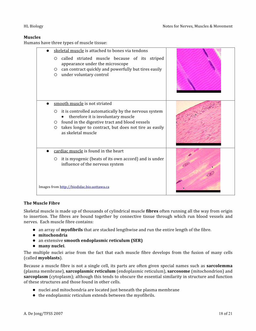
MusclesHumanshavethreetypesofmuscletissue:
skeletalmuscleisattachedtobonesviatendons
called striated muscle because of its stripedappearanceunderthemicroscope
cancontractquicklyandpowerfullybuttireseasily undervoluntarycontrol
smoothmuscleisnotstriated
itiscontrolledautomaticallybythenervoussystem■ thereforeitisinvoluntarymuscle
foundinthedigestivetractandbloodvessels takes longer tocontract,butdoesnot tireaseasily
asskeletalmuscle
cardiacmuscleisfoundintheheart
itismyogenic(beatsofitsownaccord)andisunderinfluenceofthenervoussystem
Imagesfromhttp://biodidac.bio.uottawa.ca
TheMuscleFibre
Skeletalmuscleismadeupofthousandsofcylindricalmusclefibresoftenrunningallthewayfromoriginto insertion. The fibres are bound together by connective tissue throughwhich run blood vessels andnerves.Eachmusclefibrecontains:
anarrayofmyofibrilsthatarestackedlengthwiseandruntheentirelengthofthefibre. mitochondria anextensivesmoothendoplasmicreticulum(SER) manynuclei.
Themultiple nuclei arise from the fact that eachmuscle fibre develops from the fusion of many cells(calledmyoblasts).
Becauseamuscle fibre isnota single cell, itspartsareoftengivenspecialnames suchassarcolemma(plasmamembrane),sarcoplasmicreticulum(endoplasmicreticulum),sarcosome(mitochondrion)andsarcoplasm(cytoplasm);althoughthistendstoobscuretheessentialsimilarityinstructureandfunctionofthesestructuresandthosefoundinothercells.
nucleiandmitochondriaarelocatedjustbeneaththeplasmamembrane theendoplasmicreticulumextendsbetweenthemyofibrils.

HLBiology NotesforNerves,Muscles&Movement
A.DeJong/TFSS2007 19of21
Seenfromthesideunderthemicroscope,skeletalmusclefibresshowapatternofcrossbanding,whichgivesrisetotheothername:striatedmuscle.
Thestriatedappearanceofthemusclefibreiscreatedbyapatternofalternating
darkAbandsandlightIbands.o TheAbandsarebisectedbytheHzoneo TheIbandsarebisectedbytheZline.
Eachmyofibrilismadeupofarraysofparallelfilaments.
Thethickfilamentshaveadiameterofabout15nm.Theyarecomposedoftheproteinmyosin. Thethinfilamentshaveadiameterofabout5nm.Theyarecomposedchieflyoftheproteinactinalongwithsmalleramountsoftwootherproteins:troponinandtropomyosin.
Imagefromhttp://users.rcn.com/jkimball.ma.ultranet/BiologyPages/S/sarcomere.gif
Theanatomyofasarcomere ThethickfilamentsproducethedarkAband. ThethinfilamentsextendineachdirectionfromtheZline.Wheretheydonotoverlapthethickfilaments,theycreatethelightIband.
TheHzoneisthatportionoftheAbandwherethethickandthinfilamentsdonotoverlap.
The entire array of thick and thin filaments between the Z lines is called a sarcomere. Shortening of thesarcomeresinamyofibrilproducestheshorteningofthemyofibriland,inturn,ofthemusclefibreofwhichitisapart.
Imagefromhttp://www.mrothery.co.uk/images/Imag109.gif

HLBiology NotesforNerves,Muscles&Movement
A.DeJong/TFSS2007 20of21
NeuromuscularJunctions
Asynapsebetweenamotorneuronandamuscleiscalledaneuromuscularjunction:
Imagefromhttp://www.emc.maricopa.edu/faculty/farabee/BIOBK/synapse.gif
MuscleContraction&SlidingFilamentTheory
Duringmusclecontractionthemyofilamentsmyosinandactinslidetowardeachotherandoverlap.Thisshortensthesarcomeresandtheentiremuscle.Musclecellsare“shocked”bynerveimpulsesfrommotorneurons. The point of attachment of the nerve to themuscle is called the neuromuscular junction. Amotorneuronanditsmusclecellsarereferredtoasamotorunit.
Thenerveimpulseiscarriedfromthemotorneuronacrossthegaptothesarcolemma(membrane)ofthemuscle cell by aneurotransmitter called acetylcholine (ACh).After the impulsehaspassed, an enzymecalledcholinesterasedeactivatesacetylcholine,readyingthemuscleforthenextnerveimpulse.
Stimulation of the muscle cells causes Ca++ to be released into the cell. The Ca++ binds to the actinfilamentscausingthemtoexposeactivesitestothemyosincross‐bridges.Thecrossbridgesbindtotheactivesites,forminganewmolecularstructure,whichcausesthecross‐bridgetobendtowardthecentre,pullingtheactinfilamentwithit.EnergyfromATPisusedtobreakthebond,straightenthecrossbridge,andallowthecrossbridgetoformanewbondwithanotheractivesitefurtherdowntheactinfilament.
Thiscyclecontinuesuntilthemusclecontractioniscomplete.Then,ATPisusedtocauseactivetransport,movingthecalciumionsoutofthemusclefibre,resultinginrelaxationofthemuscle.
TheNervousSystem&Movement
Nervesstimulatemusclecontraction.Eachdifferentmuscleusedinlocomotionmustcontractatthecorrecttime,sothemovementiscoordinated.Sincemusclesareconnectedtobones(bytendons),contractioncausesthebonestomove.Themovementisusuallyreversedbyanothermuscleontheoppositesideofthebone–anantagonisticpair.Jointsareplaceswherebonesmeet,andareclassifiedbytherangeofmotionatthejointandtypeofconnection:
o fibrous:nomovement(e.g.suturesbetweenbonesofthecranium)o cartilaginous:bonesconnectedbycartilage;limitedrangeofmotion(e.g.betweenvertebrae)o synovial:fluid‐filledcavitiesbetweenthebonesallowgreaterrangeofmotion
elbow:ahingejointallowsforextension&retraction hip:aballandsocketjointwithawiderangeofmotion

HLBiology NotesforNerves,Muscles&Movement
A.DeJong/TFSS2007 21of21
AntagonisticPair:
Imagesfromwww.saburchill.com/chapters/chap0009.htmlTheelbowisatypicalhingejointinvolvingbones,cartilage,ligaments,tendonsandmuscles:
Imagefromhttp://www.botany.uwc.ac.za/sci_ed/grade10/manphys/images/man/hinge.gif
bonessupportthebodyandallowforlocomotion musclesmovethebones nervesstimulatemusclecontraction synovialfluidprotectsthebonesandlubricatesthejointandiscontainedwithinbursa ligamentsconnectbonestobones tendonsconnectmusclestobones