
Nerve Testing - Detecting Upper Extremity Peripheral Neuropathic Pain
12
journal of orthopaedic & sports physical therapy | volume 42 | number 5 | may 2012 | 413 [ CLINICAL COMMENTARY ] U pper-limb neurodynamic tests (ULNTs) (FIGURE 1, ONLINE VIDEOS) use a series of movements to apply mechanical forces to a portion of the nervous system. 12,38 ULNTs also load nonneural tissues. 12,38 Therefore, when central pain mechanisms are not the primary reason for a patient’s pain experience, a ULNT response could be related to neural or nonneural tissue sensitivity. In these situations, a ULNT response is thought to be related to neural tissue sensitiv- T T SYNOPSIS: The validity of upper-limb neurody- namic tests (ULNTs) for detecting peripheral neu- ropathic pain (PNP) was assessed by reviewing the evidence on plausibility, the definition of a positive test, reliability, and concurrent validity. Evidence was identified by a structured search for peer- reviewed articles published in English before May 2011. The quality of concurrent validity studies was assessed with the Quality Assessment of Diagnos- tic Accuracy Studies tool, where appropriate. Bio- mechanical and experimental pain data support the plausibility of ULNTs. Evidence suggests that a positive ULNT should at least partially reproduce the patient’s symptoms and that structural dif- ferentiation should change these symptoms. Data indicate that this definition of a positive ULNT is reliable when used clinically. Limited evidence sug- gests that the median nerve test, but not the radial nerve test, helps determine whether a patient has cervical radiculopathy. The median nerve test does not help diagnose carpal tunnel syndrome. These findings should be interpreted cautiously, because diagnostic accuracy might have been distorted by the investigators’ definitions of a positive ULNT. Furthermore, patients with PNP who presented with increased nerve mechano- sensitivity rather than conduction loss might have been incorrectly classified by electrophysiological reference standards as not having PNP. The only evidence for concurrent validity of the ulnar nerve test was a case study on cubital tunnel syndrome. We recommend that researchers develop more comprehensive reference standards for PNP to accurately assess the concurrent validity of ULNTs and continue investigating the predictive validity of ULNTs for prognosis or treatment response. J Or- thop Sports Phys Ther 2012;42(5):413-424, Epub 8 March 2012. doi:10.2519/jospt.2012.3988 T T KEY WORDS: carpal tunnel syndrome, cervical radiculopathy, cubital tunnel syndrome, reliability 1 PhD candidate, Division of Physiotherapy and NHMRC Centre of Clinical Research Excellence in Spinal Pain, Injury and Health, School of Health and Rehabilitation Sciences, The University of Queensland, Brisbane, Australia. 2 Professor, Division of Physiotherapy and NHMRC Centre of Clinical Research Excellence in Spinal Pain, Injury and Health, School of Health and Rehabilitation Sciences, The University of Queensland, Brisbane, Australia. 3 Associate Professor, Division of Physiotherapy and NHMRC Centre of Clinical Research Excellence in Spinal Pain, Injury and Health, School of Health and Rehabilitation Sciences, The University of Queensland, Brisbane, Australia. Robert J. Nee was funded by an Endeavour International Postgraduate Research Scholarship from the Australian Government and a Research Scholarship from The University of Queensland. Address correspondence to Dr Michel W. Coppieters, Division of Physiotherapy, School of Health and Rehabilitation Sciences, The University of Queensland, Brisbane, Queensland 4072, Australia. E-mail: [email protected] ROBERT J. NEE, PT, MAppSc 1 • GWENDOLEN A. JULL, PT, PhD 2 • BILL VICENZINO, PT, PhD 2 • MICHEL W. COPPIETERS, PT, PhD 3 The Validity of Upper-Limb Neurodynamic Tests for Detecting Peripheral Neuropathic Pain SUPPLEMENTAL VIDEO ONLINE ity when it changes with movement of a distant body part that further loads or unloads the nervous system (eg, con- tralateral neck sidebending in- creases a sensory response in the forearm). 12,38 Moving a distant body part to evaluate a ULNT re- sponse is referred to as structural differentiation. 12,38,45 Peripheral neuropathic pain (PNP) is pain that arises as a direct result of a lesion or disease affecting the somato- sensory component of the peripheral nervous system. 99 Clinicians use ULNTs to help determine whether patients have PNP conditions such as cervical radicu- lopathy, 110 carpal tunnel syndrome, 104,111 and cubital tunnel syndrome. 20,85 The ra- tionale for the use of ULNTs is that they are considered capable of detecting the increased nerve mechanosensitivity as- sociated with these conditions. 12,38,42,45,112 Other clinical tests proposed for detect- ing these conditions, such as the Spurl- ing test, 92 Phalen’s test, 75 and the elbow flexion-pressure test, 68 use the same rationale. While ULNTs can also be used to guide treatment selection, 12,38,45 a specific assessment of their diagnostic validity is important. Guidelines recommend that clinicians use these tests when examin- ing patients with symptoms affecting the neck or upper limb, 1,16 and expert physical
description
Â
Transcript of Nerve Testing - Detecting Upper Extremity Peripheral Neuropathic Pain
journal of orthopaedic & sports physical therapy | volume 42 | number 5 | may 2012 | 413
[ clinical commentary ]
Upper-limb neurodynamic tests (ULNTs) (FIGURE 1, ONLINE VIDEOS) use a series of movements to apply mechanical forces to a portion of the nervous system.12,38 ULNTs also load nonneural tissues.12,38 Therefore, when
central pain mechanisms are not the primary reason for a patient’s pain experience, a ULNT response could be related to neural or
nonneural tissue sensitivity. In these situations, a ULNT response is thought to be related to neural tissue sensitiv-
TT SYNOPSIS: The validity of upper-limb neurody-namic tests (ULNTs) for detecting peripheral neu-ropathic pain (PNP) was assessed by reviewing the evidence on plausibility, the definition of a positive test, reliability, and concurrent validity. Evidence was identified by a structured search for peer-reviewed articles published in English before May 2011. The quality of concurrent validity studies was assessed with the Quality Assessment of Diagnos-tic Accuracy Studies tool, where appropriate. Bio-mechanical and experimental pain data support the plausibility of ULNTs. Evidence suggests that a positive ULNT should at least partially reproduce the patient’s symptoms and that structural dif-ferentiation should change these symptoms. Data indicate that this definition of a positive ULNT is reliable when used clinically. Limited evidence sug-gests that the median nerve test, but not the radial nerve test, helps determine whether a patient has cervical radiculopathy. The median nerve test does not help diagnose carpal tunnel syndrome.
These findings should be interpreted cautiously, because diagnostic accuracy might have been distorted by the investigators’ definitions of a positive ULNT. Furthermore, patients with PNP who presented with increased nerve mechano-sensitivity rather than conduction loss might have been incorrectly classified by electrophysiological reference standards as not having PNP. The only evidence for concurrent validity of the ulnar nerve test was a case study on cubital tunnel syndrome. We recommend that researchers develop more comprehensive reference standards for PNP to accurately assess the concurrent validity of ULNTs and continue investigating the predictive validity of ULNTs for prognosis or treatment response. J Or-thop Sports Phys Ther 2012;42(5):413-424, Epub 8 March 2012. doi:10.2519/jospt.2012.3988
TT KEY WORDS: carpal tunnel syndrome, cervical radiculopathy, cubital tunnel syndrome, reliability
1PhD candidate, Division of Physiotherapy and NHMRC Centre of Clinical Research Excellence in Spinal Pain, Injury and Health, School of Health and Rehabilitation Sciences, The University of Queensland, Brisbane, Australia. 2Professor, Division of Physiotherapy and NHMRC Centre of Clinical Research Excellence in Spinal Pain, Injury and Health, School of Health and Rehabilitation Sciences, The University of Queensland, Brisbane, Australia. 3Associate Professor, Division of Physiotherapy and NHMRC Centre of Clinical Research Excellence in Spinal Pain, Injury and Health, School of Health and Rehabilitation Sciences, The University of Queensland, Brisbane, Australia. Robert J. Nee was funded by an Endeavour International Postgraduate Research Scholarship from the Australian Government and a Research Scholarship from The University of Queensland. Address correspondence to Dr Michel W. Coppieters, Division of Physiotherapy, School of Health and Rehabilitation Sciences, The University of Queensland, Brisbane, Queensland 4072, Australia. E-mail: [email protected]
ROBERT J. NEE, PT, MAppSc1 • GWENDOLEN A. JULL, PT, PhD2 • BILL VICENZINO, PT, PhD2 • MICHEL W. COPPIETERS, PT, PhD3
The Validity of Upper-Limb Neurodynamic Tests for Detecting
Peripheral Neuropathic Pain
SUPPLEMENTAL VIDEO ONLINE
ity when it changes with movement of a distant body part that further loads or unloads the nervous system (eg, con-
tralateral neck sidebending in-creases a sensory response in the forearm).12,38 Moving a distant body part to evaluate a ULNT re-sponse is referred to as structural
differentiation.12,38,45
Peripheral neuropathic pain (PNP) is pain that arises as a direct result of a lesion or disease affecting the somato-sensory component of the peripheral nervous system.99 Clinicians use ULNTs to help determine whether patients have PNP conditions such as cervical radicu-lopathy,110 carpal tunnel syndrome,104,111 and cubital tunnel syndrome.20,85 The ra-tionale for the use of ULNTs is that they are considered capable of detecting the increased nerve mechanosensitivity as-sociated with these conditions.12,38,42,45,112 Other clinical tests proposed for detect-ing these conditions, such as the Spurl-ing test,92 Phalen’s test,75 and the elbow flexion-pressure test,68 use the same rationale.
While ULNTs can also be used to guide treatment selection,12,38,45 a specific assessment of their diagnostic validity is important. Guidelines recommend that clinicians use these tests when examin-ing patients with symptoms affecting the neck or upper limb,1,16 and expert physical
42-05 Nee.indd 413 4/18/2012 7:10:31 PM

414 | may 2012 | volume 42 | number 5 | journal of orthopaedic & sports physical therapy
[ clinical commentary ]therapists and pain consultants rely heav-ily on neurodynamic tests for making a clinical diagnosis of PNP.90 The validity of ULNTs to detect PNP can be assessed by answering 4 questions.41,58,80,87 First, are ULNTs plausible tests for detecting PNP? Second, what criteria should be used to define a positive ULNT? Third, can cli-nicians make reliable decisions about a positive ULNT? Fourth, are ULNTs accu-rate for detecting PNP clinically (concur-rent validity)? This clinical commentary reviews the available evidence to help answer these questions.
SEARCH STRATEGY
Search terms were entered into PubMed, CINAHL, EMBASE, Sco-pus, and Web of Science to find peer-
reviewed articles published in English before May 2011. Titles and abstracts were screened, and full-text articles of all poten-tially relevant publications were retrieved for further assessment. Reference lists of retrieved articles were hand searched for additional publications. Cadaveric stud-ies measuring nerve strain (percent elon-gation) and nerve sliding (longitudinal displacement) during ULNT movements had to use whole-body or transthoracic specimens that maintained the nerve root attachments to the spinal cord.124 Biome-chanical studies focusing only on moving individual digits were excluded, because the hand and wrist are normally moved together during ULNTs.12,38 FIGURE 2 sum-marizes the search.
DIAGNOSING PERIPHERAL NEUROPATHIC PAIN
A reference standard for diag-nosing PNP is needed to interpret results from clinical studies on
ULNT validity. Treede et al99 proposed that their criteria for “probable” neuro-pathic pain would be sufficient for mak-ing a neuropathic pain diagnosis. For PNP, probable means that (1) the pa-tient’s symptoms fit a nerve-related dis-tribution, (2) the history of symptoms is
consistent with a nerve-related problem, and either (3a) a clinical neurological examination shows positive or negative sensory signs that match the innervation territory of the suspected nerve problem, or (3b) diagnostic tests, such as imaging or electrophysiological studies, confirm an injury or disease that explains the dis-tribution of PNP.99
PLAUSIBILITY OF ULNTs
Biomechanical studies on nerve strain, sliding, and compres-sion help answer wheth-
er ULNTs are plausible tests for detecting PNP. Cadaveric studies show that joint movements used in the me-
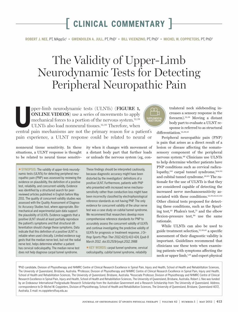
ULNT1MEDIAN
• Shoulder girdle stabilization• Shoulder abduction• Wrist/finger extension• Forearm supination• Shoulder external rotation• Elbow extension• Structural di erentiation - Cervical sidebending - Release wrist extension
ULNT2MEDIAN
• Shoulder girdle depression• Elbow extension• Shoulder external rotation and forearm supination• Wrist/finger extension• Shoulder abduction• Structural di erentiation - Cervical sidebending - Release shoulder girdle depression - Release wrist extension
ULNTRADIAL
• Shoulder girdle depression• Elbow extension• Shoulder internal rotation and forearm pronation• Wrist/finger flexion• Shoulder abduction• Structural di erentiation - Cervical sidebending - Release shoulder girdle depression - Release wrist flexion
ULNTULNAR
• Wrist/finger extension• Forearm pronation• Elbow flexion• Shoulder external rotation• Shoulder girdle depression• Shoulder abduction• Structural di erentiation - Cervical sidebending - Release shoulder girdle depression - Release wrist extension
FIGURE 1. Standard sequence of joint movements and suggested structural differentiation maneuvers for each ULNT.12 Joint movements for each ULNT can be applied in different sequences (ONLINE VIDEOS).12,38 Abbreviation: ULNT, upper-limb neurodynamic test.
42-05 Nee.indd 414 4/18/2012 7:10:32 PM

journal of orthopaedic & sports physical therapy | volume 42 | number 5 | may 2012 | 415
dian (ULNTMEDIAN),13,18,21,56,57,61,66,67,119,124,127 radial (ULNTRADIAL),122 and ulnar (ULN-TULNAR)2,3,13,21,47,66,72,83,98,123 nerve tests in-crease strain in the corresponding nerve. Each test preferentially loads its corre-sponding nerve at the elbow and wrist,13,56 suggesting that mechanosensitivity of a particular nerve near these joints may be most readily assessed by the correspond-ing ULNT. ULNTs cannot selectively test mechanosensitivity of individual nerve roots.55 Shoulder girdle stabiliza-tion/depression and shoulder abduction involved in all ULNTs increase strain
throughout the brachial plexus.55 Cadav-eric13,18,21,67,118,122-124 and in vivo22,29,30,32,33,35,
49,52,65,100,101 studies also show that ULNT movements produce sliding between the nerve and surrounding tissues. A nerve segment slides toward the moving joint as each ULNT movement lengthens the nerve bed.
Strain and sliding produced by a joint movement are greatest in nerve segments closest to the moving joint.18,30,32,52,56,57,122-124 However, when the limb is in the end ULNT position, biome-chanical effects from wrist movement or
neck sidebending spread along the entire nerve.18,21,22,30,52,56,57,61,65,119,122,124 These data support the concept of structural differ-entiation. The spread of biomechanical effects along the nerve is a plausible ex-planation for why movement of a distant body part can change sensory responses at the end of a ULNT.
Transfer of strain through the fascial network in the neck and upper limb91,93 may also explain why moving a distant body part changes sensory responses at the end of a neurodynamic test.5 How-ever, a separate literature search did
PubMed, 932 records CINAHL, 237 records†
EMBASE, 622 records Scopus, 1388 records Web of Science, 293 records
Full-text articles retrieved, 112
Articles included in review, 78
Excluded, 3360 including duplicates
Excluded, 46 • Cadaver limbs only, 13 • Finger motion only, 8 • Validity questions not addressed, 25
Biomechanical studies, 40 Clinical studies, 38
Cadaver studies, 23 In vivo studies, 17
“AND” excursion (English, humans)*
“AND” biomechanics (English, humans)*
“AND” sliding (English, humans)*
Median nerve Radial nerve Ulnar nerve Brachial plexus Spinal nerve roots Spinal nerves Nerve roots
Brachial plexus tension test Brachial plexus provocation test Neural tissue provocation test Upper-limb tension test Upper-limb neural tension test Upper-limb neurodynamic test Neurodynamic(s) “AND” test (English)*
Relevant articles from hand search of reference lists, 12
FIGURE 2. Search strategy and results. *Limits used for each search. †Nerve structure terms were searched in isolation because combining each term with biomechanics, excursion, or sliding revealed no records.
42-05 Nee.indd 415 4/18/2012 7:10:33 PM

416 | may 2012 | volume 42 | number 5 | journal of orthopaedic & sports physical therapy
[ clinical commentary ]
not identify any studies that specifically measured whether strain produced by a structural differentiation maneuver, such as contralateral neck sidebending, might be transferred to distant parts of this fas-cial network.
ULNT movements also compress nerves. For example, wrist extension compresses the median nerve in the car-pal tunnel114,115; the combination of elbow extension, forearm pronation, and wrist flexion compresses the deep branch of the radial nerve in the radial tunnel39,62; and the combination of elbow flexion and wrist extension compresses the ulnar nerve in the cubital tunnel.69
Based on these biomechanical data, ULNTs appear to be plausible tests for detecting PNP. Strain and compression from ULNT movements will likely pro-voke mechanically sensitive neural tis-sues in patients with PNP. Furthermore, the ability for wrist or neck movement in the end ULNT position to produce bio-mechanical effects throughout the nerve supports using structural differentiation to determine whether an ULNT response is related to nerve mechanosensitivity.
An experimental pain model further supports the plausibility of ULNTs. Cop-pieters et al19 induced experimental pain in the thenar muscles of asymptomatic volunteers and showed that ULNT1ME-
DIAN did not change the distribution or
intensity of muscle-related pain. This suggests that ULNT1MEDIAN can poten-tially distinguish pain related to muscle irritation from pain related to increased nerve mechanosensitivity. Although bio-mechanical and experimental pain data support using ULNTs to detect PNP, it must be remembered that plausibility is the lowest level of test validity.87
DEFINING A POSITIVE ULNT
Sensory responses, resistance to movement, and range of motion during a ULNT are assessed to de-
termine whether a patient shows signs of increased nerve mechanosensitiv-ity.12,38 To be useful criteria for defining a positive test, ULNT responses should exhibit 2 properties.80 First, the ULNT responses must discriminate patients with PNP from asymptomatic individu-als. Second, concurrent validity studies must show that these ULNT responses also discriminate patients with PNP from patients who present with compet-ing diagnoses. This section addresses the potential ability of ULNT responses to discriminate patients with PNP from asymptomatic individuals and proposes criteria for defining a positive ULNT. Evidence on the concurrent validity of ULNTs is presented later in this clinical commentary.
Sensory ResponsesFIGURE 3 shows the most common areas in which asymptomatic individuals reported sensory responses at the end of ULNT1ME-
DIAN,23,54,63 ULNT2MEDIAN,63,77 and ULNTRA-
DIAL.73,126 There were no equivalent studies for ULNTULNAR. Sensory responses were predominantly described as stretch, ache, pain, burning, and tingling.23,54,63,73,77,102,126 Structural differentiation with contra-lateral neck sidebending increased limb responses in more than 85% of partici-pants.54,73,77,126 This suggests that asymp-tomatic individuals have a certain level of nerve mechanosensitivity. The variety of responses reported by asymptomatic in-dividuals signifies the need to be specific about the type of sensory response that qualifies as a positive ULNT in symptom-atic populations. To be confident that a sensory response distinguishes a patient with PNP from asymptomatic individu-als and, therefore, potentially discrimi-nates patients with PNP from those with competing diagnoses, the ULNT needs to reproduce at least part of the patient’s symptoms. For example, if a patient re-ports pain in the neck that spreads down 1 limb past the elbow, the ULNT should reproduce this pain at least somewhere in the neck, upper arm, or forearm.
Resistance to MovementResistance to movement during ULNT-1MEDIAN has been quantified by relating shoulder girdle elevation force24,25 and torque resisting passive elbow extension50 to elbow extension range of motion. Only shoulder girdle elevation force has been assessed in a symptomatic population.25 Patients with nerve-related neck and uni-lateral arm pain showed increased shoul-der girdle elevation force at earlier stages of elbow extension in the symptomatic limb. These findings cannot be general-ized to everyday clinical practice, because a load cell was used to measure shoulder girdle elevation force.
Two studies used clinically feasible methods for quantifying resistance to movement during ULNT1MEDIAN.48,105 Ex-aminers identified the onset of resistance
x
x
ULNT1MEDIAN and ULNT2MEDIAN ULNTRADIAL Anterior view ULNTRADIAL Posterior view
FIGURE 3. The most common areas where asymptomatic individuals reported sensory responses at the end of ULNT1MEDIAN,23,54,63 ULNT2MEDIAN,63,77 and ULNTRADIAL.
73,126 Abbreviation: ULNT, upper-limb neurodynamic test.
42-05 Nee.indd 416 4/18/2012 7:10:34 PM

journal of orthopaedic & sports physical therapy | volume 42 | number 5 | may 2012 | 417
during elbow extension in asymptomatic participants and measured this angle with a standard goniometer48 or an elec-trogoniometer.105 Intraclass correlation coefficients (ICC2,1)
86 for interexaminer reliability were 0.42 (calculated from re-ported data)48 and 0.48.105 The standard error of measurement36 for both studies was 10° (calculated from reported data). This translates to a smallest detectable difference at a 95% confidence level (SDD95)
36 of 28°. This amount of mea-surement error suggests that onset of resistance probably cannot be sensitive enough to discriminate patients with PNP from asymptomatic individuals and is, therefore, unlikely to be a useful crite-rion for a positive ULNT1MEDIAN.
Range of MotionULNT range of motion is usually quan-tified by the joint angle at pain onset or pain tolerance (eg, ULNT1MEDIAN el-bow extension or ULNTRADIAL shoulder abduction). Patients with PNP are ex-pected to exhibit less range of motion in their symptomatic limb compared to their asymptomatic limb or asymptom-atic individuals.
Several studies have compared ULNT-1MEDIAN
14,15,17,26,84,94-97,103,121 or ULNTRADI-
AL71,73,106,107,121,125 range of motion between
patients’ symptomatic and asymptom-atic limbs or between patients and a-symptomatic individuals. Only Chien et al15 included patients with PNP who met the diagnostic criteria of Treede et al.99 Patients with cervical radiculopathy had less ULNT1MEDIAN range of motion at pain onset in the symptomatic limb compared to the asymptomatic limb or asymptom-atic individuals (FIGURE 4). However, sig-nificant differences in range of motion for group data do not help determine wheth-er an individual patient has an abnormal deficit in ULNT range of motion.
One strategy for determining whether an individual patient has an abnormal deficit in ULNT range of motion is to identify an absolute cut-off for the symp-tomatic limb. Davis et al28 proposed that, when the neck is in contralateral side-
bending before applying ULNT1MEDIAN, elbow extension deficits greater than 60° at pain onset could be classified as abnormal. Because this proposed cut-off is based on asymptomatic data only, its ability to discriminate patients with PNP from asymptomatic individuals needs to be tested. Despite this proposed cut-off, data suggest that it is very difficult to find an absolute range-of-motion cut-off that successfully identifies patients with PNP. ULNT1MEDIAN range of motion at pain onset is highly variable in asymptomatic individuals17,94,95,105 and patients with cer-vical radiculopathy (FIGURE 4).15 There is also considerable overlap in ULNT1MEDIAN range of motion between these 2 groups. Comparing asymptomatic and cervical radiculopathy data is appropriate, be-cause these studies15,17,94,95,105 used simi-lar methods for applying ULNT1MEDIAN. Range-of-motion variability and overlap highlight the difficulty in distinguishing normal from abnormal range of motion in an individual patient. Measurement error adds to the difficulty in finding an effective absolute range-of-motion cut-off. SDD95 estimates for elbow extension
range of motion at pain onset during ULNT1MEDIAN in asymptomatic17,105 and symptomatic103 individuals range from 14° to 20° (calculated from reported data). In light of the variability and over-lap in range of motion for these popula-tions, this amount of measurement error makes it unlikely that an absolute cut-off can accurately discriminate patients with PNP from asymptomatic individuals. It is, therefore, questionable whether an absolute range-of-motion cut-off could be a meaningful criterion for a positive ULNT1MEDIAN.
Another strategy for detecting abnor-mal deficits in ULNT range of motion is to identify a relative cut-off that requires a certain difference in range of motion between the symptomatic and asymp-tomatic limbs in an individual patient. PNP conditions in which bilateral in-volvement is common are the exception (eg, more than 50% of individuals with carpal tunnel syndrome have the condi-tion bilaterally10). Despite this exception, no data currently exist on the differ-ence in range of motion between limbs that would normally be expected in as-
Chien et al15
Coppieters et al17
Sterling et al94
Sterling et al95
Vanti et al105
0 10 20 30 40 50 60
Deficit in Elbow Extension at Onset of Pain, deg
Symptomatic limb of patients with cervical radiculopathyAsymptomatic limb of patients with cervical radiculopathyAsymptomatic individuals
(n = 38)
(n = 38)
(n = 31)
(n = 10)
(n = 20)
(n = 20)
(n = 36)
FIGURE 4. Average deficit in elbow extension range of motion at the onset of pain during ULNT1MEDIAN (cervical spine in neutral). Error bars represent 1 standard deviation and provide an indication of the variability in range-of-motion deficit among participants. Abbreviation: ULNT, upper-limb neurodynamic test.
42-05 Nee.indd 417 4/18/2012 7:10:35 PM

418 | may 2012 | volume 42 | number 5 | journal of orthopaedic & sports physical therapy
[ clinical commentary ]ymptomatic individuals. It is unknown whether a certain difference in range of motion between limbs may discriminate patients with PNP from asymptomatic individuals.
In summary, due to the measurement error for resistance to movement and the lack of discriminatory cut-offs for range of motion, current evidence does not support these components of the test response to decide whether a patient’s ULNT is positive. At this time, the sug-gested criteria for a positive ULNT are (1) at least partial reproduction of the patient’s symptoms and (2) a change in these symptoms with structural dif-ferentiation. Reproducing the patient’s symptoms is necessary because asymp-tomatic individuals report a wide vari-ety of sensations in response to ULNTs. Changing the patient’s symptoms with structural differentiation is necessary to show that these symptoms are at least partly related to increased nerve mechanosensitivity.
RELIABILITY OF A POSITIVE ULNT
The next question is whether this definition of a positive ULNT is reliable when used clinically.
Most reports of ULNT reliability in as-ymptomatic14,17,27,43,48,63,70,74,77,105,126 and symptomatic17,84,103 populations have focused on measuring range of mo-tion, not whether examiners agreed on a positive test. Four studies assessed reliability for identifying a positive ULNT.9,82,108,110 Only Schmid et al82 re-quired that a positive test reproduce the patient’s symptoms and that structural differentiation change these symptoms. Each ULNT was applied to 31 patients with unilateral arm and/or neck pain that had been present for at least 4 weeks. According to cut-offs proposed by Landis and Koch,59 interexaminer reliability was moderate (κ = 0.41-0.60) for ULNT1MEDIAN, ULNT2MEDIAN, and ULNTRADIAL, and fair (κ = 0.21-0.40) for ULNTULNAR (TABLE 1).82 Kappa values can
be reduced by a high or low proportion of positive tests (prevalence) or inflated by a high level of disagreement between examiners on the proportion of posi-tive tests (bias).88 Prevalence and bias indices88 (calculated from original data obtained from the authors) were low (TABLE 1), indicating that these issues did not affect the kappa values reported by Schmid et al.82 Additional studies are needed to improve the precision of these reliability estimates, because 95% con-fidence intervals for each ULNT’s kap-pa value ranged from less than 0.20 to greater than 0.70 (TABLE 1). Nevertheless, clinical tests with fair to moderate reli-ability can still have sufficient concurrent validity to help make a diagnosis.41,109
CONCURRENT VALIDITY OF ULNTs
Evidence on the concurrent va-lidity of ULNTs came from diag-nostic accuracy studies on cervical
radiculopathy110 and carpal tunnel syn-drome,104,111 and a case study on cubital tunnel syndrome.85 The methodological quality of the diagnostic accuracy stud-ies was assessed with the Quality Assess-ment of Diagnostic Accuracy Studies (QUADAS) tool.116,117
Cervical RadiculopathyWainner and colleagues110 developed a
clinical prediction rule to diagnose cer-vical radiculopathy from 81 patients re-ferred for electrophysiological testing for suspected cervical radiculopathy or carpal tunnel syndrome. ULNT1MEDIAN and ULNTRADIAL were 2 of several clini-cal tests considered as potential predic-tors for the diagnostic prediction rule. Needle electromyography was the ref-erence standard for diagnosing cervical radiculopathy. Only 1 of the following 3 criteria was required for a ULNT to be positive: (1) the ULNT reproduced the patient’s symptoms, (2) the difference be-tween limbs in elbow extension (ULNT-1MEDIAN) or wrist flexion (ULNTRADIAL) was greater than 10°, or (3) contralateral neck sidebending increased symptoms or ipsilateral neck sidebending decreased symptoms. The methodological quality of this study was high (11/14 QUADAS items).116,117
The data from Wainner and col-leagues110 suggest that ULNT1MEDIAN, but not ULNTRADIAL, may help determine whether a patient has cervical radiculop-athy. The negative likelihood ratio (LR) of 0.12 indicated that a negative ULNT-1MEDIAN would essentially rule out cervical radiculopathy.51,110 A positive ULNT1ME-
DIAN combined with positive findings on the 3 other clinical tests in the diagnostic prediction rule (ipsilateral cervical rota-tion less than 60°, reduction of symp-toms with the supine distraction test,
TABLE 1Interexaminer Reliability
for a Positive ULNT82*
Abbreviations: CI, confidence interval; ULNT, upper-limb neurodynamic test.*A positive ULNT required at least partial reproduction of a patient’s symptoms and a change in these symptoms with structural differentiation.†Calculated from original data obtained from Schmid et al,82 according to formulas proposed by Sim and Wright.88
‡Confidence interval calculated as 1.96 times reported standard error. The standard error of the kappa value for each ULNT was 0.18.82
Test Kappa Value (95% CI) Prevalence Index† Bias Index†
ULNT1MEDIAN 0.54 (0.19, 0.89)‡ 0.19 0.10
ULNT2MEDIAN 0.46 (0.11, 0.81)‡ 0.23 0.06
ULNTRADIAL 0.44 (0.09, 0.79)‡ 0.29 0.06
ULNTULNAR 0.36 (0.01, 0.71)‡ 0.32 0.10
All ULNTs combined 0.45 (0.27, 0.63) ... ...
42-05 Nee.indd 418 4/18/2012 7:10:36 PM

journal of orthopaedic & sports physical therapy | volume 42 | number 5 | may 2012 | 419
and provocation of symptoms with the Spurling test) would confirm the pres-ence of cervical radiculopathy (positive LR, 30.30).51,110 These findings should be interpreted cautiously because of the wide 95% confidence intervals (TABLE
2). Additionally, prediction rule perfor-mance should be confirmed in a second patient sample before it is considered ready for widespread clinical applica-tion.64 ULNTRADIAL does not help detect cervical radiculopathy, because LRs were between 0.5 and 2.0 (TABLE 2).51,110 LRs in this range mean that clinical test results do not lead to important shifts in pretest-to-posttest probability of the target con-dition being present.51
Carpal Tunnel SyndromeWainner and colleagues111 used the same sample of patients to develop a diag-nostic prediction rule for carpal tunnel syndrome. Nerve conduction tests were the reference standard for diagnos-ing carpal tunnel syndrome, and the aforementioned criteria were used for a positive ULNT. ULNT1MEDIAN and ULN-TRADIAL were not considered helpful for either making or ruling out a diagnosis of carpal tunnel syndrome, because LRs for each test were between 0.5 and 2.0 (TABLE 3).51,111 Combining ULNT1MEDIAN or ULNTRADIAL with other clinical tests did not improve diagnostic accuracy, be-cause neither test was included in the di-
agnostic prediction rule for carpal tunnel syndrome.111
The diagnostic accuracy of ULNT1ME-
DIAN for detecting carpal tunnel syndrome was also assessed by Vanti et al.104 They studied 44 consecutive patients referred for nerve conduction tests for possible carpal tunnel syndrome. The method-ological quality of this study was also high (12/14 QUADAS items).116,117 Two separate analyses were performed that involved slightly different definitions of a positive ULNT1MEDIAN. First, a posi-tive test required the presence of only 1 of the 3 criteria used by Wainner and colleagues.110,111 Second, the “symptom reproduction” criterion was modified so that symptoms had to be reproduced in the first 3 digits of the hand (typical me-dian nerve distribution), but still only 1 of the 3 criteria was required for a positive test. ULNT1MEDIAN was not considered helpful for either making or ruling out a diagnosis of carpal tunnel syndrome with either definition of a positive test, because LRs were between 0.5 and 2.0 (TABLE 3).51,104
Cubital Tunnel SyndromeThe only evidence on concurrent validity for ULNTULNAR came from a case study of a patient with suspected cubital tunnel syndrome.85 ULNTULNAR was considered positive because it reproduced the pa-tient’s symptoms and structural differen-tiation changed these symptoms. Surgical confirmation of ulnar nerve entrapment at the elbow and alleviation of the pa-tient’s forearm and hand symptoms after surgical release confirmed a diagnosis of PNP. A corresponding improvement in the ULNTULNAR response after surgery supported the concurrent validity of this test.85 However, conclusions about the di-agnostic accuracy of ULNTULNAR cannot be made from a case study.41
Potential for Bias in Diagnostic Accuracy StudiesDespite the high QUADAS scores for the diagnostic accuracy studies on cervical radiculopathy110 and carpal tunnel syn-
TABLE 2Diagnostic Accuracy of ULNT1MEDIAN and ULNTRADIAL for Cervical Radiculopathy110
Abbreviations: CI, confidence interval; CPR, clinical prediction rule; LR, likelihood ratio; ULNT, upper-limb neurodynamic test.*ULNT positive if 1 or more of the following criteria are present: ULNT reproduces the patient’s symptoms, greater than 10° difference between limbs in elbow extension (ULNT1MEDIAN) or wrist flexion (ULNTRADIAL) at the end of the test, contralateral neck sidebending increased symptoms or ipsilateral neck sidebending decreased symptoms when performed at the end position of the ULNT on the symp-tomatic limb.†All 4 of the following variables are present: positive ULNT1MEDIAN, ipsilateral cervical rotation less than 60°, supine cervical distraction test alleviates symptoms, and ipsilateral Spurling test provokes symptoms.‡Negative LR not reported because the focus of the CPR was to identify patients who were most likely to have cervical radiculopathy confirmed by electrophysiological testing.
Test Sensitivity (95% CI) Specificity (95% CI) Positive LR (95% CI) Negative LR (95% CI)
ULNT1MEDIAN* 0.97 (0.90, 1.00) 0.22 (0.12, 0.33) 1.30 (1.10, 1.50) 0.12 (0.01, 1.90)
ULNTRADIAL* 0.72 (0.52, 0.93) 0.33 (0.21, 0.45) 1.10 (0.77, 1.50) 0.85 (0.37, 1.90)
Diagnostic CPR†‡ 0.24 (0.05, 0.43) 0.99 (0.97, 1.00) 30.30 (1.70, 538.20) ...
TABLE 3Diagnostic Accuracy of ULNT1MEDIAN and
ULNTRADIAL for Carpal Tunnel Syndrome104,111
Abbreviations: CI, confidence interval; LR, likelihood ratio; ULNT, upper-limb neurodynamic test.*ULNT positive if 1 or more of the following criteria are present: ULNT reproduces the patient’s symptoms, greater than 10° difference between limbs in elbow extension (ULNT1MEDIAN) or wrist flexion (ULNTRADIAL) at the end of the test, contralateral neck sidebending increased symptoms or ipsilateral neck sidebending decreased symptoms when performed at the end position of the ULNT on the symp-tomatic limb.†Same criteria for a positive ULNT, but symptoms had to be reproduced in the first 3 digits (median nerve distribution).
Test Sensitivity (95% CI) Specificity (95% CI) Positive LR (95% CI) Negative LR (95% CI)
ULNT1MEDIAN111* 0.75 (0.58, 0.92) 0.13 (0.04, 0.22) 0.86 (0.67, 1.10) 1.90 (0.72, 5.10)
ULNT1MEDIAN104* 0.92 (0.74, 0.98) 0.15 (0.05, 0.36) 1.08 (0.38, 3.08) 0.56 (0.19, 1.59)
ULNT1MEDIAN104† 0.54 (0.35, 0.72) 0.70 (0.48, 0.85) 1.81 (1.13, 2.88) 0.65 (0.41, 1.04)
ULNTRADIAL111* 0.64 (0.45, 0.83) 0.30 (0.17, 0.42) 0.91 (0.65, 1.30) 1.20 (0.62, 2.40)
42-05 Nee.indd 419 4/18/2012 7:10:37 PM
420 | may 2012 | volume 42 | number 5 | journal of orthopaedic & sports physical therapy
[ clinical commentary ]drome,104,111 2 significant methodological concerns make it necessary to carefully interpret diagnostic accuracy findings. The first is the definition of a positive ULNT. Only 1 of 3 criteria—symptom reproduction, a greater-than-10° differ-ence in range of motion between limbs, or a change in symptoms with neck side-bending—was required for a positive ULNT.104,110,111 This is a liberal definition of a positive test. No data support a dif-ference greater than 10° in ULNT range of motion between limbs as able to distin-guish symptomatic patients from asymp-tomatic individuals. More importantly, changing symptoms with structural dif-ferentiation (neck sidebending) was not required for a positive ULNT. QUADAS scoring does not address this method-ological issue.116,117 It is unclear whether this liberal definition of a positive ULNT influenced diagnostic accuracy because the number of patients with a positive test whose symptoms did not change with structural differentiation was not reported.
The second concern is the potential limitation of an electrophysiological ref-erence standard of conduction loss. This reference standard assumes that conduc-tion loss is consistently present in PNP. However, clinical studies of cervical radic-ular pain89 and carpal tunnel syndrome120 have demonstrated that increased nerve mechanosensitivity may contribute to PNP even when impulse conduction is normal. The pathophysiology of PNP helps explain the potential discrepancy between increased nerve mechanosensi-tivity and electrophysiological evidence of conduction loss. Increased mechano-sensitivity is related to increased excit-ability of small-diameter afferents,6,11,31,37 central nervous system pathways,6 and nociceptors in the nervi nervorum and sinu-vertebral nerves that innervate the nervous system’s connective tissues.4,53,81 These pathophysiological changes cannot be detected by electrophysiological tests that focus on damage or conduction loss in large-diameter fibers.60 Consequently, patients with PNP who present with in-
creased nerve mechanosensitivity rather than conduction loss may often be incor-rectly classified by needle electromyog-raphy and nerve conduction tests as not having PNP. This potential misclassifica-tion of patients with PNP might have bi-ased estimates of the diagnostic accuracy of ULNT1MEDIAN and ULNTRADIAL.78
Alternate Strategies for a Reference StandardThe potential incorrect classification of patients with PNP who present with in-creased nerve mechanosensitivity rather than conduction loss suggests that an electrophysiological reference standard of conduction loss may not be compre-hensive enough to judge the diagnostic accuracy of clinical tests of nerve mecha-nosensitivity.113 One way to address this problem is to create a composite refer-ence standard by combining needle elec-tromyography and nerve conduction tests with other tests.78 Quantitative sensory testing can assess the function of small-diameter afferents and provide evidence of sensory hypersensitivity.44,79 Magnetic resonance neurography34 and ultrasound imaging7,40 can identify signs of nerve ir-ritation and nerve thickening. Therefore, quantitative sensory testing, magnetic resonance neurography, or ultrasound imaging might be options for a compos-ite reference standard for various PNP conditions.
The composite reference standard approach has its own methodological challenges. The combination(s) of test results necessary to conclude that the target condition is present must be de-termined in advance.78 For example, Beekman et al8 assessed the diagnostic accuracy of provocation tests for ulnar neuropathy at the elbow with a reference standard of positive electrophysiological findings or evidence of nerve thickening on ultrasound imaging. Identifying these combinations also requires that each test within the composite reference standard be labeled as positive or negative in an individual patient. However, deciding whether quantitative sensory testing or
ultrasound imaging on its own is posi-tive in an individual patient can be dif-ficult.40,44 Therefore, more work is needed to develop composite reference standards for different PNP conditions.
When an ideal reference standard for a diagnostic label is unavailable, research on predictive validity for prognosis or treatment response provides alternative information on how ULNT results can be used clinically.41,78 Raney et al76 provided this type of information on ULNT1MEDI-
AN. They developed a clinical prediction rule to identify patients with neck pain who will improve after cervical traction and exercise. The reference standard was whether a patient reported being at least “a great deal better” after 6 treatments. A positive ULNT1MEDIAN was retained as 1 of 5 variables in the rule. A positive test reproduced the patient’s symptoms, and neck sidebending changed these symp-toms. Further studies are needed to de-termine whether this rule may predict a preferential response to cervical traction and exercise, or whether patients who are positive on the rule may respond equally well to other interventions.46 Researchers should continue investigating the predic-tive validity of ULNTs.
CONCLUSION
The available evidence was re-viewed to assess the validity of using ULNTs to detect PNP condi-
tions such as cervical radiculopathy, car-pal tunnel syndrome, and cubital tunnel syndrome. Aspects of validity that were assessed included plausibility, the defi-nition of a positive test, reliability, and concurrent validity (diagnostic accuracy).
ULNTs are plausible tests for detect-ing PNP. A positive ULNT should at least partially reproduce the patient’s symptoms, and structural differentiation should change these symptoms. This definition of a positive ULNT is reliable when used clinically. However, concur-rent validity studies need to determine whether this specific definition of a posi-tive test may improve the diagnostic ac-
42-05 Nee.indd 420 4/18/2012 7:10:38 PM

journal of orthopaedic & sports physical therapy | volume 42 | number 5 | may 2012 | 421
curacy of ULNTs.The minimal evidence available pre-
vents any definitive statements about the diagnostic accuracy of ULNTs for detect-ing PNP. Evidence shows that, when us-ing a liberal definition of a positive test, ULNT1MEDIAN, but not ULNTRADIAL, can help determine whether a patient has cervical radiculopathy. When using simi-lar criteria, ULNT1MEDIAN does not help diagnose carpal tunnel syndrome. Con-trasting results in the diagnostic accuracy of ULNT1MEDIAN for detecting cervical ra-diculopathy and carpal tunnel syndrome suggest that diagnostic accuracy of the same ULNT may be different for differ-ent PNP conditions. Diagnostic accuracy findings should be interpreted cautious-ly, because results may be distorted by the liberal definition of a positive test. Furthermore, patients with PNP who presented with increased nerve mecha-nosensitivity rather than conduction loss might have been incorrectly classified by electrophysiological reference standards as not having PNP. Researchers should try to develop more comprehensive ref-erence standards for PNP to accurately assess the concurrent validity of ULNTs and continue investigating whether ULNTs have predictive validity for prog-nosis or treatment response. t
REFERENCES
1. American Physical Therapy Association. Guide to physical therapist practice. Second edition. Phys Ther. 2001;81:9-746.
2. Aoki M, Takasaki H, Muraki T, Uchiyama E, Mu-rakami G, Yamashita T. Strain on the ulnar nerve at the elbow and wrist during throwing motion. J Bone Joint Surg Am. 2005;87:2508-2514. http://dx.doi.org/10.2106/JBJS.D.02989
3. Apfelberg DB, Larson SJ. Dynamic anatomy of the ulnar nerve at the elbow. Plast Reconstr Surg. 1973;51:79-81.
4. Asbury AK, Fields HL. Pain due to peripheral nerve damage: an hypothesis. Neurology. 1984;34:1587-1590.
5. Barker PJ, Briggs CA. Attachments of the pos-terior layer of lumbar fascia. Spine (Phila Pa 1976). 1999;24:1757-1764.
6. Baron R, Binder A, Wasner G. Neuro-pathic pain: diagnosis, pathophysiological mechanisms, and treatment. Lancet Neurol.
2010;9:807-819. http://dx.doi.org/10.1016/S1474-4422(10)70143-5
7. Beekman R, Schoemaker MC, Van Der Plas JP, et al. Diagnostic value of high-resolution sonog-raphy in ulnar neuropathy at the elbow. Neurol-ogy. 2004;62:767-773.
8. Beekman R, Schreuder AH, Rozeman CA, Koe-hler PJ, Uitdehaag BM. The diagnostic value of provocative clinical tests in ulnar neuropathy at the elbow is marginal. J Neurol Neurosurg Psychiatry. 2009;80:1369-1374. http://dx.doi.org/10.1136/jnnp.2009.180844
9. Bertilson BC, Grunnesjo M, Strender LE. Reli-ability of clinical tests in the assessment of patients with neck/shoulder problems—impact of history. Spine (Phila Pa 1976). 2003;28:2222-2231. http://dx.doi.org/10.1097/01.BRS.0000089685.55629.2E
10. Bland JD, Rudolfer SM. Clinical surveillance of carpal tunnel syndrome in two areas of the United Kingdom, 1991-2001. J Neurol Neurosurg Psychiatry. 2003;74:1674-1679.
11. Bove GM, Ransil BJ, Lin HC, Leem JG. Inflam-mation induces ectopic mechanical sensitiv-ity in axons of nociceptors innervating deep tissues. J Neurophysiol. 2003;90:1949-1955. http://dx.doi.org/10.1152/jn.00175.2003
12. Butler DS. The Sensitive Nervous System. Ad-elaide, Australia: Noigroup Publications; 2000.
13. Byl C, Puttlitz C, Byl N, Lotz J, Topp K. Strain in the median and ulnar nerves during upper-extremity positioning. J Hand Surg Am. 2002;27:1032-1040. http://dx.doi.org/10.1053/jhsu.2002.35886
14. Byng J. Overuse syndromes of the upper limb and the upper limb tension test: a comparison between patients, asymptomatic keyboard workers and asymptomatic non-keyboard work-ers. Man Ther. 1997;2:157-164. http://dx.doi.org/10.1054/math.1997.0296
15. Chien A, Eliav E, Sterling M. Whiplash (grade II) and cervical radiculopathy share a similar sensory presentation: an investigation us-ing quantitative sensory testing. Clin J Pain. 2008;24:595-603. http://dx.doi.org/10.1097/AJP.0b013e31816ed4fc
16. Childs JD, Cleland JA, Elliott JM, et al. Neck pain: clinical practice guidelines linked to the International Classification of Functioning, Dis-ability, and Health from the Orthopaedic Section of the American Physical Therapy Association. J Orthop Sports Phys Ther. 2008;38:A1-A34. http://dx.doi.org/10.2519/jospt.2008.0303
17. Coppieters M, Stappaerts K, Janssens K, Jull G. Reliability of detecting ‘onset of pain’ and ‘submaximal pain’ during neural provocation testing of the upper quadrant. Physiother Res Int. 2002;7:146-156.
18. Coppieters MW, Alshami AM. Longitudinal excursion and strain in the median nerve during novel nerve gliding exercises for carpal tunnel syndrome. J Orthop Res. 2007;25:972-980. http://dx.doi.org/10.1002/jor.20310
19. Coppieters MW, Alshami AM, Hodges PW. An experimental pain model to investigate the
specificity of the neurodynamic test for the median nerve in the differential diagnosis of hand symptoms. Arch Phys Med Rehabil. 2006;87:1412-1417. http://dx.doi.org/10.1016/j.apmr.2006.06.012
20. Coppieters MW, Bartholomeeusen KE, Stap-paerts KH. Incorporating nerve-gliding tech-niques in the conservative treatment of cubital tunnel syndrome. J Manipulative Physiol Ther. 2004;27:560-568. http://dx.doi.org/10.1016/j.jmpt.2004.10.006
21. Coppieters MW, Butler DS. Do ‘sliders’ slide and ‘tensioners’ tension? An analysis of neurody-namic techniques and considerations regarding their application. Man Ther. 2008;13:213-221. http://dx.doi.org/10.1016/j.math.2006.12.008
22. Coppieters MW, Hough AD, Dilley A. Different nerve-gliding exercises induce different magni-tudes of median nerve longitudinal excursion: an in vivo study using dynamic ultrasound im-aging. J Orthop Sports Phys Ther. 2009;39:164-171. http://dx.doi.org/10.2519/jospt.2009.2913
23. Coppieters MW, Stappaerts KH, Everaert DG, Staes FF. Addition of test components during neurodynamic testing: effect on range of motion and sensory responses. J Orthop Sports Phys Ther. 2001;31:226-235; discussion 236-237.
24. Coppieters MW, Stappaerts KH, Staes FF, Everaert DG. Shoulder girdle elevation during neurodynamic testing: an assessable sign? Man Ther. 2001;6:88-96. http://dx.doi.org/10.1054/math.2000.0375
25. Coppieters MW, Stappaerts KH, Wouters LL, Janssens K. Aberrant protective force generation during neural provocation testing and the effect of treatment in patients with neurogenic cervicobrachial pain. J Manipulative Physiol Ther. 2003;26:99-106. http://dx.doi.org/10.1067/mmt.2003.16
26. Coppieters MW, Stappaerts KH, Wouters LL, Janssens K. The immediate effects of a cervical lateral glide treatment technique in patients with neurogenic cervicobrachial pain. J Orthop Sports Phys Ther. 2003;33:369-378.
27. Coppieters MW, Van de Velde M, Stappaerts KH. Positioning in anesthesiology: toward a better understanding of stretch-induced perioperative neuropathies. Anesthesiology. 2002;97:75-81.
28. Davis DS, Anderson IB, Carson MG, Elkins CL, Stuckey LB. Upper limb neural tension and seat-ed slump tests: the false positive rate among healthy young adults without cervical or lumbar symptoms. J Man Manip Ther. 2008;16:136-141.
29. Dilley A, Greening J, Lynn B, Leary R, Morris V. The use of cross-correlation analysis between high-frequency ultrasound images to measure longitudinal median nerve movement. Ultra-sound Med Biol. 2001;27:1211-1218.
30. Dilley A, Lynn B, Greening J, DeLeon N. Quan-titative in vivo studies of median nerve sliding in response to wrist, elbow, shoulder and neck movements. Clin Biomech (Bristol, Avon). 2003;18:899-907.
31. Dilley A, Lynn B, Pang SJ. Pressure and stretch mechanosensitivity of peripheral nerve fibres
42-05 Nee.indd 421 4/18/2012 7:10:39 PM

422 | may 2012 | volume 42 | number 5 | journal of orthopaedic & sports physical therapy
[ clinical commentary ]following local inflammation of the nerve trunk. Pain. 2005;117:462-472. http://dx.doi.org/10.1016/j.pain.2005.08.018
32. Dilley A, Odeyinde S, Greening J, Lynn B. Longitudinal sliding of the median nerve in patients with non-specific arm pain. Man Ther. 2008;13:536-543. http://dx.doi.org/10.1016/j.math.2007.07.004
33. Dilley A, Summerhayes C, Lynn B. An in vivo investigation of ulnar nerve sliding during upper limb movements. Clin Biomech (Bristol, Avon). 2007;22:774-779. http://dx.doi.org/10.1016/j.clinbiomech.2007.04.004
34. Du R, Auguste KI, Chin CT, Engstrom JW, Weinstein PR. Magnetic resonance neurog-raphy for the evaluation of peripheral nerve, brachial plexus, and nerve root disorders. J Neurosurg. 2010;112:362-371. http://dx.doi.org/10.3171/2009.7.JNS09414
35. Echigo A, Aoki M, Ishiai S, Yamaguchi M, Naka-mura M, Sawada Y. The excursion of the median nerve during nerve gliding exercise: an observa-tion with high-resolution ultrasonography. J Hand Ther. 2008;21:221-227; quiz 228. http://dx.doi.org/10.1197/j.jht.2007.11.001
36. Eliasziw M, Young SL, Woodbury MG, Fryday-Field K. Statistical methodology for the concur-rent assessment of interrater and intrarater reliability: using goniometric measurements as an example. Phys Ther. 1994;74:777-788.
37. Eliav E, Benoliel R, Tal M. Inflammation with no axonal damage of the rat saphenous nerve trunk induces ectopic discharge and mechano-sensitivity in myelinated axons. Neurosci Lett. 2001;311:49-52.
38. Elvey RL. Physical evaluation of the peripheral nervous system in disorders of pain and dys-function. J Hand Ther. 1997;10:122-129.
39. Erak S, Day R, Wang A. The role of supinator in the pathogenesis of chronic lateral elbow pain: a biomechanical study. J Hand Surg Br. 2004;29:461-464. http://dx.doi.org/10.1016/j.jhsb.2004.06.001
40. Fowler JR, Gaughan JP, Ilyas AM. The sensitivity and specificity of ultrasound for the diagnosis of carpal tunnel syndrome: a meta-analysis. Clin Orthop Relat Res. 2011;469:1089-1094. http://dx.doi.org/10.1007/s11999-010-1637-5
41. Fritz JM, Wainner RS. Examining diagnostic tests: an evidence-based perspective. Phys Ther. 2001;81:1546-1564.
42. Gifford LS, Butler DS. The integration of pain sciences into clinical practice. J Hand Ther. 1997;10:86-95.
43. Grant R, Forrester C, Hides J. Screen based key-board operation: the adverse effects on the neu-ral system. Aust J Physiother. 1995;41:99-107.
44. Haanpaa M, Attal N, Backonja M, et al. NeuPSIG guidelines on neuropathic pain assessment. Pain. 2011;152:14-27. http://dx.doi.org/10.1016/j.pain.2010.07.031
45. Hall TM, Elvey RL. Nerve trunk pain: physical di-agnosis and treatment. Man Ther. 1999;4:63-73. http://dx.doi.org/10.1054/math.1999.0172
46. Hancock M, Herbert RD, Maher CG. A guide
to interpretation of studies investigating sub-groups of responders to physical therapy inter-ventions. Phys Ther. 2009;89:698-704. http://dx.doi.org/10.2522/ptj.20080351
47. Hicks D, Toby EB. Ulnar nerve strains at the elbow: the effect of in situ decompression and medial epicondylectomy. J Hand Surg Am. 2002;27:1026-1031. http://dx.doi.org/10.1053/jhsu.2002.35870
48. Hines T, Noakes R, Manners B. The upper limb tension test: inter-tester reliability for assessing the onset of passive resistance R1. J Man Manip Ther. 1993;1:95-98.
49. Hough AD, Moore AP, Jones MP. Peripheral nerve motion measurement with spectral Dop-pler sonography: a reliability study. J Hand Surg Br. 2000;25:585-589. http://dx.doi.org/10.1054/jhsb.2000.0453
50. Jaberzadeh S, Scutter S, Nazeran H. Mechano-sensitivity of the median nerve and mechani-cally produced motor responses during upper limb neurodynamic test 1. Physiotherapy. 2005;91:94-100. http://dx.doi.org/10.1016/j.physio.2004.09.021
51. Jaeschke R, Guyatt GH, Sackett DL. Users’ guides to the medical literature. III. How to use an article about a diagnostic test. B. What are the results and will they help me in caring for my patients? The Evidence-Based Medicine Working Group. JAMA. 1994;271:703-707.
52. Julius A, Lees R, Dilley A, Lynn B. Shoulder posture and median nerve sliding. BMC Mus-culoskelet Disord. 2004;5:23. http://dx.doi.org/10.1186/1471-2474-5-23
53. Kallakuri S, Cavanaugh JM, Blagoev DC. An immunohistochemical study of innervation of lumbar spinal dura and longitudinal ligaments. Spine (Phila Pa 1976). 1998;23:403-411.
54. Kenneally M, Rubenach H, Elvey R. The upper limb tension test: the SLR test of the arm. In: Grant R, ed. Physical Therapy of the Cervical and Thoracic Spine. Edinburgh, UK: Churchill Livingstone; 1988:167-194.
55. Kleinrensink GJ, Stoeckart R, Mulder PG, et al. Upper limb tension tests as tools in the diagno-sis of nerve and plexus lesions. Anatomical and biomechanical aspects. Clin Biomech (Bristol, Avon). 2000;15:9-14.
56. Kleinrensink GJ, Stoeckart R, Vleeming A, Sni-jders CJ, Mulder PG. Mechanical tension in the median nerve. The effects of joint positions. Clin Biomech (Bristol, Avon). 1995;10:240-244.
57. Kleinrensink GJ, Stoeckart R, Vleeming A, Snijders CJ, Mulder PG, van Wingerden JP. Peripheral nerve tension due to joint motion. A comparison between embalmed and unem-balmed human bodies. Clin Biomech (Bristol, Avon). 1995;10:235-239.
58. Krebs DE. Measurement theory. Phys Ther. 1987;67:1834-1839.
59. Landis JR, Koch GG. The measurement of ob-server agreement for categorical data. Biomet-rics. 1977;33:159-174.
60. Lee DH, Claussen GC, Oh S. Clinical nerve con-duction and needle electromyography studies. J
Am Acad Orthop Surg. 2004;12:276-287.61. Lewis J, Ramot R, Green A. Changes in me-
chanical tension in the median nerve: possible implications for the upper limb tension test. Physiotherapy. 1998;84:254-261. http://dx.doi.org/10.1016/S0031-9406(05)65524-1
62. Links AC, Graunke KS, Wahl C, Green JR, 3rd, Matsen FA, 3rd. Pronation can increase the pressure on the posterior interosseous nerve under the arcade of Frohse: a possible mecha-nism of palsy after two-incision repair for distal biceps rupture—clinical experience and a cadaveric investigation. J Shoulder Elbow Surg. 2009;18:64-68. http://dx.doi.org/10.1016/j.jse.2008.07.001
63. Lohkamp M, Small K. Normal response to Upper Limb Neurodynamic Test 1 and 2A. Man Ther. 2011;16:125-130. http://dx.doi.org/10.1016/j.math.2010.07.008
64. McGinn TG, Guyatt GH, Wyer PC, Naylor CD, Stiell IG, Richardson WS. Users’ guides to the medical literature: XXII: how to use articles about clinical decision rules. Evidence-Based Medicine Working Group. JAMA. 2000;284:79-84.
65. McLellan DL, Swash M. Longitudinal sliding of the median nerve during movements of the upper limb. J Neurol Neurosurg Psychiatry. 1976;39:566-570.
66. Millesi H, Zoch G, Rath T. The gliding apparatus of peripheral nerve and its clinical significance. Ann Chir Main Memb Super. 1990;9:87-97.
67. Nee RJ, Yang CH, Liang CC, Tseng GF, Coppiet-ers MW. Impact of order of movement on nerve strain and longitudinal excursion: a biomechani-cal study with implications for neurodynamic test sequencing. Man Ther. 2010;15:376-381. http://dx.doi.org/10.1016/j.math.2010.03.001
68. Novak CB, Lee GW, Mackinnon SE, Lay L. Pro-vocative testing for cubital tunnel syndrome. J Hand Surg Am. 1994;19:817-820. http://dx.doi.org/10.1016/0363-5023(94)90193-7
69. Ochi K, Horiuchi Y, Tanabe A, Morita K, Takeda K, Ninomiya K. Comparison of shoulder inter-nal rotation test with the elbow flexion test in the diagnosis of cubital tunnel syndrome. J Hand Surg Am. 2011;36:782-787. http://dx.doi.org/10.1016/j.jhsa.2010.12.019
70. Oliver GS, Rushton A. A study to explore the reliability and precision of intra and inter-rater measures of ULNT1 on an asymptomatic population. Man Ther. 2011;16:203-206. http://dx.doi.org/10.1016/j.math.2010.05.009
71. Paungmali A, O’Leary S, Souvlis T, Vicenzino B. Naloxone fails to antagonize initial hypoalgesic effect of a manual therapy treatment for lateral epicondylalgia. J Manipulative Physiol Ther. 2004;27:180-185. http://dx.doi.org/10.1016/j.jmpt.2003.12.022
72. Pechan J, Julis I. The pressure measurement in the ulnar nerve. A contribution to the patho-physiology of the cubital tunnel syndrome. J Biomech. 1975;8:75-79.
73. Petersen CM, Zimmermann CL, Hall KD, Przechera SJ, Julian JV, Coderre NN. Upper limb
42-05 Nee.indd 422 4/18/2012 7:10:40 PM

journal of orthopaedic & sports physical therapy | volume 42 | number 5 | may 2012 | 423
neurodynamic test of the radial nerve: a study of responses in symptomatic and asymptomatic subjects. J Hand Ther. 2009;22:344-353; quiz 354. http://dx.doi.org/10.1016/j.jht.2009.05.001
74. Petersen SM, Covill LG. Reliability of the ra-dial and ulnar nerve biased upper extremity neural tissue provocation tests. Physiother Theory Pract. 2010;26:476-482. http://dx.doi.org/10.3109/09593981003607629
75. Phalen GS. The carpal-tunnel syndrome. Seven-teen years’ experience in diagnosis and treat-ment of six hundred fifty-four hands. J Bone Joint Surg Am. 1966;48:211-228.
76. Raney NH, Petersen EJ, Smith TA, et al. Devel-opment of a clinical prediction rule to identify patients with neck pain likely to benefit from cervical traction and exercise. Eur Spine J. 2009;18:382-391. http://dx.doi.org/10.1007/s00586-008-0859-7
77. Reisch R, Williams K, Nee RJ, Rutt RA. ULNT2 – median nerve bias: examiner reliability and sensory responses in asymptomatic subjects. J Man Manip Ther. 2005;13:44-55. http://dx.doi.org/10.1179/106698105790835804
78. Reitsma JB, Rutjes AW, Khan KS, Coomarasamy A, Bossuyt PM. A review of solutions for diag-nostic accuracy studies with an imperfect or missing reference standard. J Clin Epidemiol. 2009;62:797-806. http://dx.doi.org/10.1016/j.jclinepi.2009.02.005
79. Rolke R, Magerl W, Campbell KA, et al. Quantita-tive sensory testing: a comprehensive protocol for clinical trials. Eur J Pain. 2006;10:77-88. http://dx.doi.org/10.1016/j.ejpain.2005.02.003
80. Sackett DL, Haynes RB. The architecture of diagnostic research. BMJ. 2002;324:539-541.
81. Sauer SK, Bove GM, Averbeck B, Reeh PW. Rat peripheral nerve components release calcitonin gene-related peptide and prostaglandin E2 in response to noxious stimuli: evidence that nervi nervorum are nociceptors. Neuroscience. 1999;92:319-325.
82. Schmid AB, Brunner F, Luomajoki H, et al. Reliability of clinical tests to evaluate nerve function and mechanosensitivity of the upper limb peripheral nervous system. BMC Mus-culoskelet Disord. 2009;10:11. http://dx.doi.org/10.1186/1471-2474-10-11
83. Schuind FA, Goldschmidt D, Bastin C, Burny F. A biomechanical study of the ulnar nerve at the elbow. J Hand Surg Br. 1995;20:623-627.
84. Selvaratnam PJ, Matyas TA, Glasgow EF. Nonin-vasive discrimination of brachial plexus involve-ment in upper limb pain. Spine (Phila Pa 1976). 1994;19:26-33.
85. Shacklock MO. Positive upper limb tension test in a case of surgically proven neuropathy: analysis and validity. Man Ther. 1996;1:154-161. http://dx.doi.org/10.1054/math.1996.0265
86. Shrout PE, Fleiss JL. Intraclass correlations: uses in assessing rater reliability. Psychol Bull. 1979;86:420-428.
87. Sim J, Arnell P. Measurement validity in physical therapy research. Phys Ther. 1993;73:102-110; discussion 110-115.
88. Sim J, Wright CC. The kappa statistic in reliabil-ity studies: use, interpretation, and sample size requirements. Phys Ther. 2005;85:257-268.
89. Slipman CW, Plastaras CT, Palmitier RA, Huston CW, Sterenfeld EB. Symptom provocation of fluoroscopically guided cervical nerve root stimulation. Are dynatomal maps identical to dermatomal maps? Spine (Phila Pa 1976). 1998;23:2235-2242.
90. Smart KM, Blake C, Staines A, Doody C. Clinical indicators of ‘nociceptive’, ‘peripheral neuropathic’ and ‘central’ mechanisms of musculoskeletal pain. A Delphi survey of expert clinicians. Man Ther. 2010;15:80-87. http://dx.doi.org/10.1016/j.math.2009.07.005
91. Smith DC, Mitchell DA, Peterson GW, Will AD, Mera SS, Smith LL. Medial brachial fascial compartment syndrome: anatomic basis of neuropathy after transaxillary arteriography. Radiology. 1989;173:149-154.
92. Spurling R, Scoville W. Lateral rupture of the cervical intervertebral discs: a common cause of shoulder and arm pain. Surg Gynecol Obstet. 1944;78:350-358.
93. Stecco C, Gagey O, Macchi V, et al. Tendinous muscular insertions onto the deep fascia of the upper limb. First part: anatomical study. Morphologie. 2007;91:29-37. http://dx.doi.org/10.1016/j.morpho.2007.05.001
94. Sterling M, Jull G, Vicenzino B, Kenardy J. Characterization of acute whiplash-associated disorders. Spine (Phila Pa 1976). 2004;29:182-188. http://dx.doi.org/10.1097/01.BRS.0000105535.12598.AE
95. Sterling M, Jull G, Vicenzino B, Kenardy J. Sen-sory hypersensitivity occurs soon after whiplash injury and is associated with poor recovery. Pain. 2003;104:509-517.
96. Sterling M, Treleaven J, Jull G. Responses to a clinical test of mechanical provocation of nerve tissue in whiplash associated disorder. Man Ther. 2002;7:89-94.
97. Sweeney J, Harms A. Persistent mechanical al-lodynia following injury of the hand. Treatment through mobilization of the nervous system. J Hand Ther. 1996;9:328-338.
98. Toby EB, Hanesworth D. Ulnar nerve strains at the elbow. J Hand Surg Am. 1998;23:992-997. http://dx.doi.org/10.1016/S0363-5023(98)80005-1
99. Treede RD, Jensen TS, Campbell JN, et al. Neuropathic pain: redefinition and a grading system for clinical and research purposes. Neurology. 2008;70:1630-1635. http://dx.doi.org/10.1212/01.wnl.0000282763.29778.59
100. Tuzuner S, Inceoglu S, Bilen FE. Median nerve excursion in response to wrist movement after endoscopic and open carpal tunnel release. J Hand Surg Am. 2008;33:1063-1068. http://dx.doi.org/10.1016/j.jhsa.2008.03.007
101. Tuzuner S, Ozkaynak S, Acikbas C, Yildirim A. Median nerve excursion during endoscopic car-pal tunnel release. Neurosurgery. 2004;54:1155-1160; discussion 1160-1161.
102. van der Heide B, Allison GT, Zusman M. Pain
and muscular responses to a neural tissue provocation test in the upper limb. Man Ther. 2001;6:154-162. http://dx.doi.org/10.1054/math.2001.0406
103. van der Heide B, Bourgoin C, Eils G, Garnevall B, Blackmore M. Test-retest reliability and face validity of a modified neural tissue provocation test in patients with cervicobrachial pain syndrome. J Man Manip Ther. 2006;14:30-36. http://dx.doi.org/10.1179/106698106790820863
104. Vanti C, Bonfiglioli R, Calabrese M, et al. Up-per Limb Neurodynamic Test 1 and symptoms reproduction in carpal tunnel syndrome. A va-lidity study. Man Ther. 2011;16:258-263. http://dx.doi.org/10.1016/j.math.2010.11.003
105. Vanti C, Conteddu L, Guccione A, et al. The Upper Limb Neurodynamic Test 1: intra- and intertester reliability and the effect of several repetitions on pain and resistance. J Manipula-tive Physiol Ther. 2010;33:292-299. http://dx.doi.org/10.1016/j.jmpt.2010.03.003
106. Vicenzino B, Collins D, Benson H, Wright A. An investigation of the interrelationship between manipulative therapy-induced hypoalgesia and sympathoexcitation. J Manipulative Physiol Ther. 1998;21:448-453.
107. Vicenzino B, Collins D, Wright A. The initial effects of a cervical spine manipulative physio-therapy treatment on the pain and dysfunction of lateral epicondylalgia. Pain. 1996;68:69-74.
108. Viikari-Juntura E. Interexaminer reliability of observations in physical examinations of the neck. Phys Ther. 1987;67:1526-1532.
109. Wainner RS. Reliability of the clinical examina-tion: how close is “close enough”? J Orthop Sports Phys Ther. 2003;33:488-491.
110. Wainner RS, Fritz JM, Irrgang JJ, Boninger ML, Delitto A, Allison S. Reliability and diagnostic accuracy of the clinical examina-tion and patient self-report measures for cervical radiculopathy. Spine (Phila Pa 1976). 2003;28:52-62. http://dx.doi.org/10.1097/01.BRS.0000038873.01855.50
111. Wainner RS, Fritz JM, Irrgang JJ, Delitto A, Allison S, Boninger ML. Development of a clini-cal prediction rule for the diagnosis of carpal tunnel syndrome. Arch Phys Med Rehabil. 2005;86:609-618. http://dx.doi.org/10.1016/j.apmr.2004.11.008
112. Wainner RS, Gill H. Diagnosis and nonoperative management of cervical radiculopathy. J Orthop Sports Phys Ther. 2000;30:728-744.
113. Walsh J, Hall T. Reliability, validity and diagnos-tic accuracy of palpation of the sciatic, tibial and common peroneal nerves in the examina-tion of low back related leg pain. Man Ther. 2009;14:623-629. http://dx.doi.org/10.1016/j.math.2008.12.007
114. Weiss ND, Gordon L, Bloom T, So Y, Rempel DM. Position of the wrist associated with the lowest carpal-tunnel pressure: implica-tions for splint design. J Bone Joint Surg Am. 1995;77:1695-1699.
115. Werner R, Armstrong TJ, Bir C, Aylard MK.
42-05 Nee.indd 423 4/18/2012 7:10:41 PM

424 | may 2012 | volume 42 | number 5 | journal of orthopaedic & sports physical therapy
[ clinical commentary ]
@ MORE INFORMATIONWWW.JOSPT.ORG
Intracarpal canal pressures: the role of finger, hand, wrist and forearm position. Clin Biomech (Bristol, Avon). 1997;12:44-51.
116. Whiting P, Rutjes AW, Reitsma JB, Bossuyt PM, Kleijnen J. The development of QUADAS: a tool for the quality assessment of studies of diag-nostic accuracy included in systematic reviews. BMC Med Res Methodol. 2003;3:25. http://dx.doi.org/10.1186/1471-2288-3-25
117. Whiting PF, Weswood ME, Rutjes AW, Reitsma JB, Bossuyt PN, Kleijnen J. Evaluation of QUADAS, a tool for the quality assess-ment of diagnostic accuracy studies. BMC Med Res Methodol. 2006;6:9. http://dx.doi.org/10.1186/1471-2288-6-9
118. Wilgis EF, Murphy R. The significance of longitu-dinal excursion in peripheral nerves. Hand Clin. 1986;2:761-766.
119. Wilson S, Selvaratnam P, Briggs C. Strain at the
subclavian artery during the upper limb tension test. Aust J Physiother. 1994;40:243-248.
120. Witt JC, Hentz JG, Stevens JC. Carpal tunnel syndrome with normal nerve conduction stud-ies. Muscle Nerve. 2004;29:515-522. http://dx.doi.org/10.1002/mus.20019
121. Wright A, Thurnwald P, O’Callaghan J, Smith J, Vicenzino B. Hyperalgesia in tennis elbow patients. J Musculoskelet Pain. 1994;2:83-97.
122. Wright TW, Glowczewskie F, Jr., Cowin D, Wheel-er DL. Radial nerve excursion and strain at the elbow and wrist associated with upper-extrem-ity motion. J Hand Surg Am. 2005;30:990-996. http://dx.doi.org/10.1016/j.jhsa.2005.06.008
123. Wright TW, Glowczewskie F, Jr., Cowin D, Wheel-er DL. Ulnar nerve excursion and strain at the elbow and wrist associated with upper extrem-ity motion. J Hand Surg Am. 2001;26:655-662. http://dx.doi.org/10.1053/jhsu.2001.26140
124. Wright TW, Glowczewskie F, Wheeler D, Miller G, Cowin D. Excursion and strain of the median nerve. J Bone Joint Surg Am. 1996;78:1897-1903.
125. Yaxley GA, Jull GA. Adverse tension in the neu-ral system: a preliminary study of tennis elbow. Aust J Physiother. 1993;39:15-22.
126. Yaxley GA, Jull GA. A modified upper limb tension test: an investigation of responses in normal subjects. Aust J Physiother. 1991;37:143-152.
127. Zoech G, Reihsner R, Beer R, Millesi H. Stress and strain in peripheral nerves. Neuro Orthop. 1991;10:73-82.
EARN CEUs With JOSPT’s Read for Credit Program
JOSPT’s Read for Credit (RFC) program invites Journal readers to study and analyze selected JOSPT articles and successfully complete online quizzes about them for continuing education credit. To participate in the program:
1. Go to www.jospt.org and click on “Read for Credit” in the left-hand navigation column that runs throughout the site or on the link in the “Read for Credit” box in the right-hand column of the home page. 2. Choose an article to study and when ready, click “Take Exam” for that article. 3. Login and pay for the quiz by credit card. 4. Take the quiz. 5. Evaluate the RFC experience and receive a personalized certificate of continuing education credits.
The RFC program o�ers you 2 opportunities to pass the quiz. You may review all of your answers—including the questions you missed. You receive 0.2 CEUs, or 2 contact hours, for each quiz passed. The Journal website maintains a history of the quizzes you have taken and the credits and certificates you have been awarded in the “My CEUs” section of your “My JOSPT” account.
42-05 Nee.indd 424 4/18/2012 7:10:41 PM


