Nephron Structure
23
Kidney structure
-
Upload
mustafa-khandagawi -
Category
Documents
-
view
38 -
download
0
description
renal
Transcript of Nephron Structure

Kidney structure

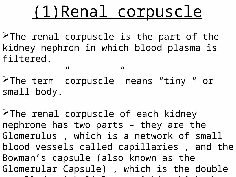
(1)Renal corpuscleThe renal corpuscle is the part of the kidney nephron in which blood plasma is filtered.
The term ”corpuscle” means “tiny “ or small body.
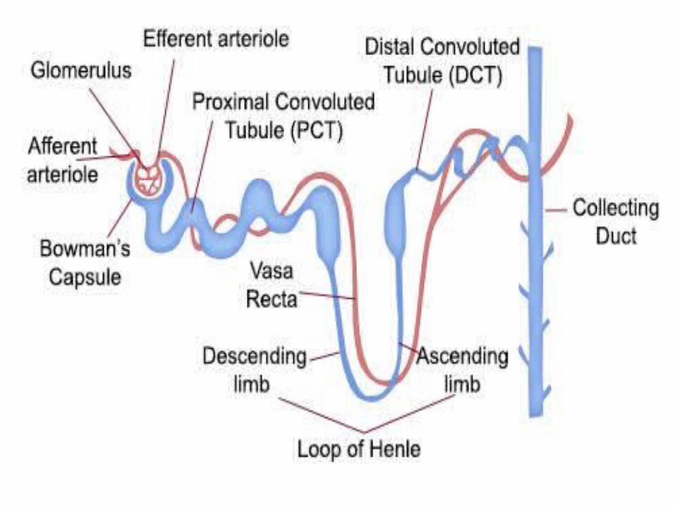
The renal corpuscle of each kidney nephrone has two parts – they are the Glomerulus , which is a network of small blood vessels called capillaries , and the Bowman’s capsule (also known as the Glomerular Capsule) , which is the double – walled epithelial cup within which the glomerulus is contained.

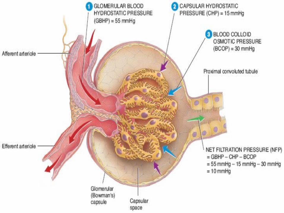
Within the glomerulus are glomerulur capillaries that are located bewteen the afferent arteriole bringing blood into the glomerulus and the efferent arteriole draining blood away from the glomerulus.
The (outgoing) efferent arteriole has a smaller diameter than the (incoming) afferent arteriole.
This difference in arteriole diameters helps to raise the blood pressure in the glomerulus.
The area between the double-walls of the Bowman’s capsule is called capsular space.
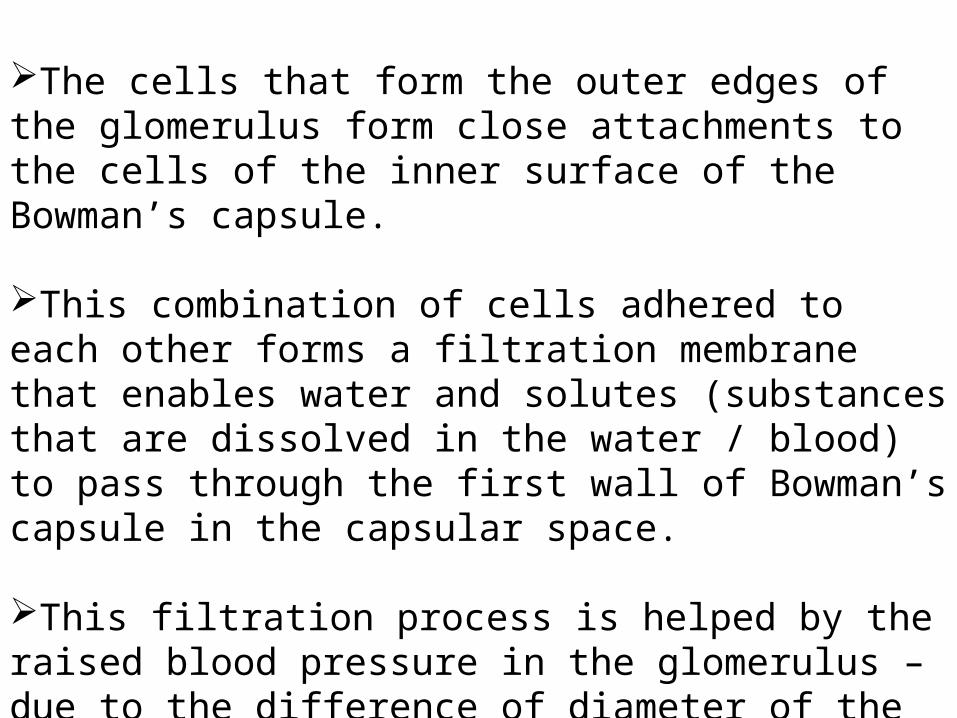
The cells that form the outer edges of the glomerulus form close attachments to the cells of the inner surface of the Bowman’s capsule.
This combination of cells adhered to each other forms a filtration membrane that enables water and solutes (substances that are dissolved in the water / blood) to pass through the first wall of Bowman’s capsule in the capsular space.
This filtration process is helped by the raised blood pressure in the glomerulus – due to the difference of diameter of the afferent and efferent arterioles.
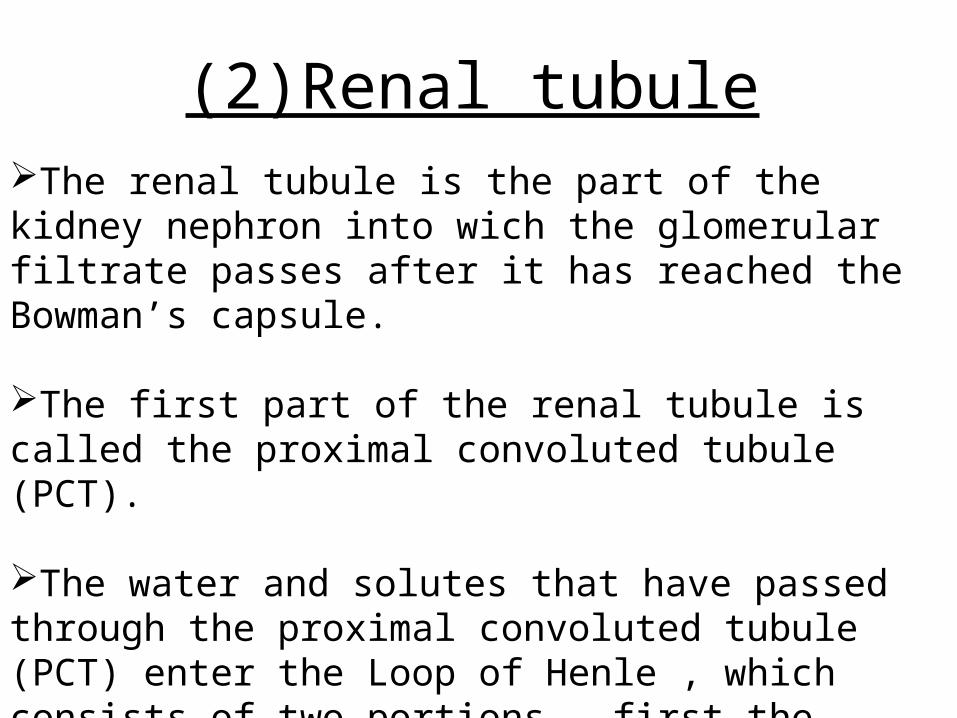
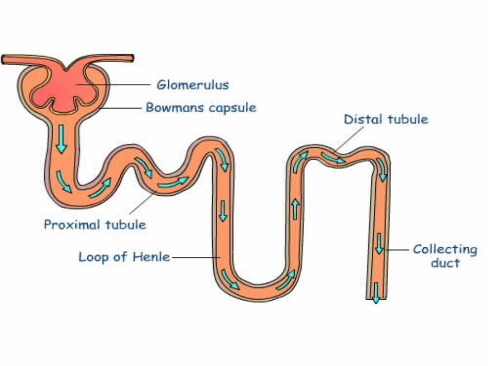
(2)Renal tubuleThe renal tubule is the part of the kidney nephron into wich the glomerular filtrate passes after it has reached the Bowman’s capsule.
The first part of the renal tubule is called the proximal convoluted tubule (PCT).
The water and solutes that have passed through the proximal convoluted tubule (PCT) enter the Loop of Henle , which consists of two portions – first the descending limb of Henle , then the ascending limb of Henle.
In order to pass through the Loop of Henle , the water ( and substances dissolved in it) pass from the renal cortex into renal medulla , then back to the renal cortex.
When this fluid returns to the renal cortex (via the ascending limb of Henle) it passes into the distal convoluted tubule (DCT).
The distal convoluted tubules of many individual kidney nephrons converge onto a single collecting duct.
The fluid that has passed through the distal convoluted tubules is drained into the collecting duct.
Many collecting ducts join together to form several hundred papillary ducts.
There are typically about 30 papillary ducts per renal papilla (the renal papillae being the tips of the renal pyramids – which point towards the centre of the kidney).
At each renal papilla the contents of the papillary ducts drain into the minor calces – the channels through which the fluid passes , via the majot calyx , into the centre of the kidney – called the renal pelvis.

Blood cleaning by the kidneys
This section is about processes performed by the kidneys in order to filter (clean) blood.
These are:-(1)Glomerular filtration also called “Ultra – filtration”.(2)Tubular reabsorption also called “Selective re – absorption”.(3)Tubular secretion.
(1)Glomerular filtration Blood enters the kidney via the renal artery.
This separates many times(Renal artery →Segmental arteries→ Interlobar artery → Afferent arterioles) , eventually forming many afferent arterioles , each of which delivers blood to an individual kidney nephron.

The diameter of the afferent (incoming) arteriole is greater than the diameter of the efferent arteriole ( which blood leaves the glomerulus).
The pressure of the blood inside the glomerulus is increased due to the difference in diameter of the incoming and out – going arterioles.
This increased blood pressure helps to force the following components of the blood out of the glomerular capillaries:-•Most of the water.•Most / all of the salts.•Most / all of the glucose. •Most / all of the urea.
The above are filtered in preference to other components of blood based on particle size (Water and solutes of relative molecular mass less than 68,000 from the filtrate).
Blood cells and plasma proteins are not filtered through the glomerular capillaries because they are relatively larger in physical size.
The water and salts that have been forced out of the glomerular capillaries pass into the Bowman’s capsule and are called the glomerular filtrate.
This glomerular filtrate is formed at a rate above 125 cm3 perminute in humans.

This volume is approx. 20% of the plasma delivered during that time (It contains all the materials present in the blood except blood cells and most proteins – which are too large to cross the basement membrane of the glomerulus).
The glomerular filtrate passes from the renal corpuscle to the renal tubule.

(2)Tubular reabsorption Only about 1% of the glomerular filtrate actually leaves the body because the rest ( the other 99%) is reabsorbed into the blood while it passes through the renal tubules and ducts.
This is called tubular reabsorption and occurs via three mechanisms:-(1)Osmosis.(2)Diffusion.(3)Active transport.
Reabsorption varies according to the body’s needs , enabling the body to retain most of its nutrients.
Most of the volume of the filtrate solution is reabsorbed in the proximal convoluted tubule (PCT).
This includes some water and most / all of the glucose (except in the case of diabetics).
Most of the energy consumed by the kidneys is used in the reabsorption of sodium ions (Na+) , which are solutes – that is , they are dissolved in the water component of the filtrate solution.

As the concentration of Na+ in the filtrate solution is high (about the same as the concentration of Na+ in blood plasma) , Na+ moves from the tubular fluid into the PCT.
In the cases of many Na+ ions this occurs with the help of symprters.
Symporters simultaneously faciliate passage through the PCT membrane of both Na+ and another substances / solutes.
Other such substances that are reabsorbed with Na+ in this way include glucose(and important type of sugar) , amino acid , lactic acid , and bicarbonate ions.
These then move on through cells via diffusion and / or other transport processes.
Solutes are selectively moved from the GF to the plasma by active transport.
Following the movement of solutes (including Na+), water is then also reabsorbed by osmosis.
About 80% of the filtrate volume is reabsorbed in this way.
As this part of the reabsorption process is not controlled by the PCT itself, it is sometimes called obligatory water reabsorption.
In the loop of Henle the remaining water (together with the dissolved salts and urea) passes from the PCT into the descending limb of Henle.
It then passes along the loop of Henle , and up the ascending limb of Henle.
The different permeability