Neonatal alloimmune thrombocytopenia due to HPA-9b incompatibility
3
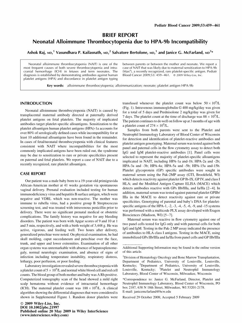
Pediatr Blood Cancer 2009;53:459–461 BRIEF REPORT Neonatal Alloimmune Thrombocytopenia due to HPA-9b Incompatibility Ashok Raj, MD, 1 Vasundhara P. Kailasnath, MD, 2 Salvatore Bertolone, MD, 1 and Janice G. McFarland, MD 3 * INTRODUCTION Neonatal alloimmune thrombocytopenia (NAIT) is caused by transplacental maternal antibody directed at paternally derived platelet antigens on fetal platelets. The majority of implicated antibodies target platelet-specific alloantigens. Sensitization to the platelet alloantigen human platelet antigens (HPA)-1a accounts for over 80% of serologically defined cases while incompatibility for at least 10 additional alloantigens have been found in the remainder. In cases of fetal/neonatal thrombocytopenia with clinical features consistent with NAIT where incompatibilities for the most commonly implicated antigens have been ruled out, the syndrome may be due to sensitization to rare or private specificities present on paternal and fetal platelets. We report a case of NAIT due to a recently recognized, rare platelet alloantigen. CASE REPORT Our patient was a male baby born to a 19-year-old primigravida African-American mother at 41 weeks gestation via spontaneous vaginal delivery. Prenatal evaluation included testing for human immunodeficiency virus and hepatitis B surface antigen, which were negative and VDRL which was non-reactive. The mother was immune to rubella virus, had a positive group B Streptococcus screening test, and was treated with intravenous penicillin prior to delivery. There were no significant prenatal medical or obstetric complications. The family history was negative for any bleeding disorders. The patient was born with Apgar scores of 9 and 10 at 1 and 5 min, respectively, and with a birth weight of 3,448 g. He was active, vigorous, and feeding well. Two hours after delivery, generalized petechiae were noted. On physical examination, he had skull molding, caput succedaneum and petechiae over the face, trunk, and upper and lower extremities. Examination of all other organ systems was unremarkable with absence of hepatosplenome- galy, normal neurologic examination, and absence of signs of infection including temperature instability, respiratory distress, lethargy, poor perfusion, or poor feeding. Laboratory investigation revealed severe thrombocytopenia with a platelet count of 5 10 9 /L and normal white blood cell and red cell counts. The blood group of both mother and baby was A Rh positive. Computerized tomography scan of the head showed a mild right scalp hematoma without evidence of intracranial hemorrhage (ICH). The maternal platelet count was 188 10 9 /L. A clinical algorithm showing the differential diagnoses that were considered is shown in Supplemental Figure 1. Random donor platelets were transfused whenever the platelet count was below 50 10 9 /L (Fig. 1). Intravenous immunoglobulin G 400 mg/kg/day was given for a total of 5 days and Prednisolone 2 mg/kg/day was given for 7 days. The platelet count at the time of discharge was 88 10 9 /L. The patient continues to do well on follow up at 3 months of age with a platelet count of 274 10 9 /L. Samples from both parents were sent to the Platelet and Neutrophil Immunology Laboratory of Blood Center of Wisconsin for detection and identification of platelet-reactive antibodies and platelet antigen genotyping. Maternal serum was tested against both panel and paternal cells in the flow cytometry assay to detect both IgG and IgM platelet-reactive antibodies [1]. Panel cells were selected to represent the majority of platelet-specific alloantigens implicated in NAIT, including HPA-1a and-1b; HPA-2a and -2b; HPA-3a and -3b; HPA-4a; HPA-5a and -5b; HPA-15a and-15b. Platelet glycoprotein (GP) specific antibodies were sought in maternal serum using the Pak-2MP assay (GTI, Brookfield, WI) which detects reactivity against platelet GP Ib-IX, GP IV, and class I HLA; and the Modified Antigen Capture ELISA (MACE) which detects antibodies reactive with GPs IIb/IIIa, and Ia/IIa [2–4]. In addition, maternal serum was tested against paternal platelet GPIIb/ IIIa in the MACE to detect reactivity against rare or private specificities. Genotyping of parental and baby’s DNA for platelet- specific antigens of the HPA-1, -2, -3, -4, -5, -6, -9, and -15 systems was performed with a multicode PLX assay developed with Eragen Biosciences (Madison, WI) [5–7]. Maternal serum was reactive in flow cytometry against one of two panel cells tested for IgG only and with paternal cells for both IgG and IgM. Testing in the Pak-2 MP assay indicated the presence of antibodies to HLA class I antigens. Testing in the MACE, using immobilized GPs IIb/IIIa and Ia/IIa from panel cells and GP IIb/IIIa Neonatal alloimmune thrombocytopenia (NAIT) is one of the most frequent causes of both severe thrombocytopenia and intra- cranial hemorrhage (ICH) in fetuses and term neonates. The diagnosis is established by demonstrating antibodies against human platelet antigens (HPA) and discordance in platelet antigen typing between parents or between the mother and neonate. We report a case of NAIT that was likely due to maternal sensitization to HPA-9b (Max a ), a recently recognized, rare platelet-specific antigen. Pediatr Blood Cancer 2009;53: 459–461. ß 2009 Wiley-Liss, Inc. Key words: alloimmune thrombocytopenia; alloimmunization; neonate; platelet antigen HPA-9b ß 2009 Wiley-Liss, Inc. DOI 10.1002/pbc.21997 Published online 20 May 2009 in Wiley InterScience (www.interscience.wiley.com) —————— Additional Supporting Information may be found in the online version of this article. 1 Division of Hematology Oncology and Bone Marrow Transplantation, Department of Pediatrics, University of Louisville, Louisville, Kentucky; 2 Department of Pediatrics, University of Louisville, Louisville, Kentucky; 3 Platelet and Neutrophil Immunology Laboratory, Blood Center of Wisconsin, Milwaukee, Wisconsin *Correspondence to: Janice G. McFarland, Director, Platelet and Neutrophil Immunology Laboratory, Blood Center of Wisconsin, PO box 2187, 638 N 18th Street, Milwaukee, WI 53201-2178. E-mail: [email protected] Received 29 October 2008; Accepted 5 February 2009
Transcript of Neonatal alloimmune thrombocytopenia due to HPA-9b incompatibility
Pediatr Blood Cancer 2009;53:459–461
BRIEF REPORTNeonatal Alloimmune Thrombocytopenia due to HPA-9b Incompatibility
Ashok Raj, MD,1 Vasundhara P. Kailasnath, MD,2 Salvatore Bertolone, MD,1 and Janice G. McFarland, MD3*
INTRODUCTION
Neonatal alloimmune thrombocytopenia (NAIT) is caused by
transplacental maternal antibody directed at paternally derived
platelet antigens on fetal platelets. The majority of implicated
antibodies target platelet-specific alloantigens. Sensitization to the
platelet alloantigen human platelet antigens (HPA)-1a accounts for
over 80% of serologically defined cases while incompatibility for at
least 10 additional alloantigens have been found in the remainder.
In cases of fetal/neonatal thrombocytopenia with clinical features
consistent with NAIT where incompatibilities for the most
commonly implicated antigens have been ruled out, the syndrome
may be due to sensitization to rare or private specificities present
on paternal and fetal platelets. We report a case of NAIT due to a
recently recognized, rare platelet alloantigen.
CASE REPORT
Our patient was a male baby born to a 19-year-old primigravida
African-American mother at 41 weeks gestation via spontaneous
vaginal delivery. Prenatal evaluation included testing for human
immunodeficiency virus and hepatitis B surface antigen, which were
negative and VDRL which was non-reactive. The mother was
immune to rubella virus, had a positive group B Streptococcus
screening test, and was treated with intravenous penicillin prior to
delivery. There were no significant prenatal medical or obstetric
complications. The family history was negative for any bleeding
disorders. The patient was born with Apgar scores of 9 and 10 at 1
and 5 min, respectively, and with a birth weight of 3,448 g. He was
active, vigorous, and feeding well. Two hours after delivery,
generalized petechiae were noted. On physical examination, he had
skull molding, caput succedaneum and petechiae over the face,
trunk, and upper and lower extremities. Examination of all other
organ systems was unremarkable with absence of hepatosplenome-
galy, normal neurologic examination, and absence of signs of
infection including temperature instability, respiratory distress,
lethargy, poor perfusion, or poor feeding.
Laboratory investigation revealed severe thrombocytopenia with
a platelet count of 5� 109/L and normal white blood cell and red cell
counts. The blood group of both mother and baby was A Rh positive.
Computerized tomography scan of the head showed a mild right
scalp hematoma without evidence of intracranial hemorrhage
(ICH). The maternal platelet count was 188� 109/L. A clinical
algorithm showing the differential diagnoses that were considered is
shown in Supplemental Figure 1. Random donor platelets were
transfused whenever the platelet count was below 50� 109/L
(Fig. 1). Intravenous immunoglobulin G 400 mg/kg/day was given
for a total of 5 days and Prednisolone 2 mg/kg/day was given for
7 days. The platelet count at the time of discharge was 88� 109/L.
The patient continues to do well on follow up at 3 months of age with
a platelet count of 274� 109/L.
Samples from both parents were sent to the Platelet and
Neutrophil Immunology Laboratory of Blood Center of Wisconsin
for detection and identification of platelet-reactive antibodies and
platelet antigen genotyping. Maternal serum was tested against both
panel and paternal cells in the flow cytometry assay to detect both
IgG and IgM platelet-reactive antibodies [1]. Panel cells were
selected to represent the majority of platelet-specific alloantigens
implicated in NAIT, including HPA-1a and-1b; HPA-2a and -2b;
HPA-3a and -3b; HPA-4a; HPA-5a and -5b; HPA-15a and-15b.
Platelet glycoprotein (GP) specific antibodies were sought in
maternal serum using the Pak-2MP assay (GTI, Brookfield, WI)
which detects reactivity against platelet GP Ib-IX, GP IV, and class I
HLA; and the Modified Antigen Capture ELISA (MACE) which
detects antibodies reactive with GPs IIb/IIIa, and Ia/IIa [2–4]. In
addition, maternal serum was tested against paternal platelet GPIIb/
IIIa in the MACE to detect reactivity against rare or private
specificities. Genotyping of parental and baby’s DNA for platelet-
specific antigens of the HPA-1, -2, -3, -4, -5, -6, -9, and -15 systems
was performed with a multicode PLX assay developed with Eragen
Biosciences (Madison, WI) [5–7].
Maternal serum was reactive in flow cytometry against one of
two panel cells tested for IgG only and with paternal cells for both
IgG and IgM. Testing in the Pak-2 MP assay indicated the presence
of antibodies to HLA class I antigens. Testing in the MACE, using
immobilized GPs IIb/IIIa and Ia/IIa from panel cells and GP IIb/IIIa
Neonatal alloimmune thrombocytopenia (NAIT) is one of themost frequent causes of both severe thrombocytopenia and intra-cranial hemorrhage (ICH) in fetuses and term neonates. Thediagnosis is established by demonstrating antibodies against humanplatelet antigens (HPA) and discordance in platelet antigen typing
between parents or between the mother and neonate. We report acase of NAIT that was likely due to maternal sensitization to HPA-9b(Maxa), a recently recognized, rare platelet-specific antigen. PediatrBlood Cancer 2009;53: 459–461. � 2009 Wiley-Liss, Inc.
Key words: alloimmune thrombocytopenia; alloimmunization; neonate; platelet antigen HPA-9b
� 2009 Wiley-Liss, Inc.DOI 10.1002/pbc.21997Published online 20 May 2009 in Wiley InterScience(www.interscience.wiley.com)
——————Additional Supporting Information may be found in the online version
of this article.
1Division of Hematology Oncology and Bone Marrow Transplantation,
Department of Pediatrics, University of Louisville, Louisville,
Kentucky; 2Department of Pediatrics, University of Louisville,
Louisville, Kentucky; 3Platelet and Neutrophil Immunology
Laboratory, Blood Center of Wisconsin, Milwaukee, Wisconsin
*Correspondence to: Janice G. McFarland, Director, Platelet and
Neutrophil Immunology Laboratory, Blood Center of Wisconsin, PO
box 2187, 638 N 18th Street, Milwaukee, WI 53201-2178.
E-mail: [email protected]
Received 29 October 2008; Accepted 5 February 2009

from paternal platelets was negative. Both parents were blood group
type A1. Platelet alloantigen genotyping revealed incompatibilities
for HPA-1b, -3b, and -9b with paternal DNA and for HPA-3b and -9b
with the child’s DNA (Table I). Maternal serum was absorbed with
HPA-3a/a and HPA-9a/a platelets to remove HLA antibody and
retested using flow cytometry against in tact HPA-3b/b and paternal
platelets. The absorption removed all reactivity against the HPA-3b/
b cells but reactivity persisted against the paternal HPA-9a/b
platelets, suggesting the presence of anti-HPA-9b antibody.
DISCUSSION
NAIT is the most common cause of severe fetal/neonatal
thrombocytopenia and one of the most frequent causes of ICH in this
population [8,9]. Testing for this disorder should be performed for
any neonate with unexplained thrombocytopenia, regardless of the
presumed cause. NAIT is caused by alloimmunization due to feto–
maternal platelet antigen incompatibility. Maternal IgG alloanti-
bodies to platelet antigens cross the placenta and bind to fetal
platelets [10]. The diagnosis is established by demonstrating
antibodies against platelet-specific antigens and discordance in
platelet antigen typing between the parents or between the mother
and the neonate. Similar to Rh alloimmunization, this disorder tends
to worsen in subsequent pregnancies and also as an affected
gestation progresses [11,12] (Supplemental Fig. 2).
In Caucasians, HPA-1a and HPA-5b incompatibilities are the most
common causes of NAIT [13,14]. Advances in the understanding of
HPA and improvements in diagnostic techniqueshave made it possible
to identify maternal–fetal incompatibility for other alloantigens in
many NAIT cases, but others go unresolved despite use of the best
available diagnostic techniques [13]. Inconclusive serologic evalua-
tion of clinically compelling cases may be due to the absence of rare
platelet polymorphisms on laboratory control platelets [15–18].
Moreover, if the testing is performed against whole platelets, other
maternal antibodies that are not thought to cause NAIT (e.g., anti-HLA
or -ABO) may be detected. These, in turn, might obscure reactivity to
the more relevant platelet-specific antigens.
The HPA-9b determinant, discovered in the evaluation of an
apparent NAIT case [15], is created by a single nucleotide poly-
morphism (SNP) in the GPIIb gene, a guanine-to-adenine
substitution at position 2602, resulting in a valine-to-methionine
substitution in the protein. The inheritance pattern, as that of
the other HPA antigens, is autosomal co-dominant [15].
Ethnicity may play a role in the incidence of HPA-9b. With the
exception of the current case, the family involved being African-
American; all of the reported HPA-9b positive individuals have been
Caucasian. However, testing of larger numbers of non-Caucasian
individuals will be necessary before any firm conclusions can be
drawn about the differences in the incidence of this marker in
various ethnic groups.
Demonstration of both an incompatibility for a platelet-specific
antigen between the mother and father or neonate and the relevant
antibody in maternal serum is generally required to serologically
confirm a diagnosis of NAIT. Sensitive and reliable platelet
alloantigen genotyping methods are used for platelet typing, and
usually, the antibody can be detected using platelet antibody tests
that utilize isolated platelet GPs as targets, eliminating possible
interfering non-platelet-specific antibodies (e.g., HLA, ABO) that
may obscure relevant platelet-specific antibody. However, in some
instances the relevant antibody is not detected in GP specific assays,
and reactivity in an intact platelet test is used to support the
diagnosis, provided steps are taken to remove possible interfering
non-platelet-specific antibodies. In the present case, no specific anti-
GPIIb/IIIa reactivity was detected in maternal serum against
paternal platelets in the GP-specific assay (MACE), as might be
expected if anti-HPA-9b were present. However, incompatibility
with both the child’s and the father’s DNA for this rare platelet GPIIb
polymorphism was documented, and moreover, testing of maternal
serum after absorption to remove anti-HLA reactivity continued to
demonstrate reactivity against intact paternal (HPA-9a/b) platelets,
but not against HPA-3b/b platelets, suggesting that anti-HPA-9b
was indeed present. Previous studies have also reported difficulty
in detecting anti-HPA-9b using isolated GPIIb/IIIa in clinically
compelling cases of NAIT where other platelet antigen incompa-
tibilities had been excluded [18]. Due to the low-frequency of HPA-
9b in the population (<0.5%) and its presence on both paternal and
baby’s platelets in this case, together with the flow cytometry
reactivity in absorbed maternal serum against the father’s HPA-9b
positive platelets; we believe that the likelihood that this case of
NAIT was due to anti-HPA-9b is quite high.
Maternal immunization against HPA-9b may have serious
clinical consequences. A recent study reported ICH in three of
Pediatr Blood Cancer DOI 10.1002/pbc
Fig. 1. Platelet counts (�109/L) of the baby during first 18 days of life.
[Color figure can be viewed in the online issue, which is available at
www.interscience.wiley.com.]
TABLE I. Platelet Antigen Genotyping of Parents and Baby
HPA-1 HPA-2 HPA-3 HPA-4 HPA-5 HPA-6 HPA-9 HPA-15
Mother 1a/1a 2a/2a 3a /3a 4a/4a 5a/5a 6a/6a 9a/9a 15a/15b
Father 1a/1b 2a/2a 3b/3b 4a/4a 5a/5a 6a/6a 9a/9b 15a/15b
Baby 1a/1a 2a/2a 3a/3b 4a/4a 5a/5a 6a/6a 9a/9b 15a/15b
HPA, human platelet antigen. Shading indicates incompatibilities between mother and baby that are possible causes of NAIT in this case.
460 Raj et al.

five cases in which clinical findings were available, suggesting that
this severe complication is at least as common in NAIT due to anti-
HPA-9b as it is in the more commonly recognized cases due to
sensitization to HPA-1a [18]. In conclusion, maternal sensitization
against HPA-9b is an important cause of NAIT and should be
considered, along with other rarely implicated platelet alloantigens,
in cases of apparent NAIT not explained by maternal–fetal
incompatibility for more commonly recognized platelet markers.
REFERENCES
1. Visentin GP, Wolfmeyer K, Newman PJ, et al. Detection of drug-
dependent, platelet-reactive antibodies by antigen-capture ELISA
and flow cytometry. Transfusion 1990;30:694–700.
2. Gottschall JL, Elliot W, Lianos E, et al. Quinine induced immune
thrombocytopenia associated with hemolytic uremic syndrome: A
new clinical entity. Blood 1991;77:306–310.
3. Kickler TS, Herman JH, Furihata K, et al. Identification of Bak-b, a
new platelet-specific antigen associated with post transfusion
purpura. Blood 1988;71:894–898.
4. Friend PJ, McCarthy LJ, Filo RS, et al. Transmission of idiopathic
(autoimmune) thrombocytopenic purpura by liver transplantation.
NEJM 1990;323:807–811.
5. Johnson SC, Marshall DJ, Harms G, et al. Multiplexed genetic
analysis using an expanded genetic alphabet. Clin Chem 2004;
50:2019–2027.
6. Pietz BC, Warden MB, DuChateau BK, et al. Multiplex genotyping
of human minor histocompatibility antigens. Hum Immunol
2005;66:1174–1182.
7. Curtis RB, Fick A, Lochowicz AJ, et al. Neonatal alloimmune
thrombocytopenia associated with maternal–fetal incompatibility
for blood group B. Transfusion 2008;48:358–364.
8. Bussel JB, Zacharoulis S, Kramer K, et al. Clinical and diagnostic
comparison of neonatal alloimmune thrombocytopenia to non-
immune cases of thrombocytopenia. Pediatr Blood Cancer 2005;
45:176–183.
9. Williamson LM, Hackett G, Rennie J, et al. The natural history of
feto–maternal alloimmunization to the platelet-specific antigen
HPA-1a (PlA1, Zwa) as determined by antenatal screening. Blood
1998;92:2280–2287.
10. Kaplan C, Daffos F, Forestier F, et al. Current trends in neonatal
alloimmune thrombocytopenia: Diagnosis and therapy. In: Kaplan-
Gouet C, Schlegel N, Salmon CH, McGregor J, editors. Platelet
immunology; fundamental and clinical aspects. Paris: John Libby
Eurotext; 1991. pp. 267–278.
11. Bussel JB, Zabusky MR, Berkowitz RL, et al. Fetal alloimmune
thrombocytopenia. N Engl J Med 1997;337:22–26.
12. Bussel JB, Berkowitz RL, McFarland JG, et al. Antenatal treatment
of neonatal alloimmune thrombocytopenia. N Engl J Med 1988;
319:1374–1378.
13. Davoren A, Curtis BR, Aster RH, et al. Human platelet antigen-
specific alloantibodies implicated in 1,162 cases of neonatal
alloimmune thrombocytopenia. Transfusion 2004;44:1220–
1225.
14. Mueller-Eckhardt C, Kiefel V, Grubert A, et al. 348 cases of
suspected neonatal alloimmune thrombocytopenia. Lancet 1989;1:
363–366.
15. Noris P, Simsek S, Bruijne-Admiraal LG, et al. Maxa, a new low-
frequency platelet-specific antigen localized on glycoprotein IIb, is
associated with neonatal alloimmune thrombocytopenia. Blood
1995;86:1019–1026.
16. Santoso S, Kiefel V, Richter IG, et al. A functional platelet
fibrinogen receptor with a deletion in the cysteine-rich repeat
region of the beta (3) integrin: The Oe(a) alloantigen in neonatal
alloimmune thrombocytopenia. Blood 2002;99:1205–1214.
17. Jallu V, Meunier M, Brement M, et al. A new platelet poly-
morphism Duv (aþ), localized within the RGD binding domain of
glycoprotein IIIa, is associated with neonatal thrombocytopenia.
Blood 2002;99:4449–4456.
18. Peterson JA, Balthazor SM, Curtis BR, et al. Maternal alloimmu-
nization against the rare platelet-specific antigen HPA-9b (Maxa) is
an important cause of neonatal alloimmune thrombocytopenia.
Transfusion 2005;45:1487–1495.
Pediatr Blood Cancer DOI 10.1002/pbc
Thrombocytopenia and HPA-9b Incompatibility 461