Neddylation Inhibition Activates the Extrinsic Apoptosis ... · 1 Neddylation Inhibition Activates...
45
1 Neddylation Inhibition Activates the Extrinsic Apoptosis Pathway through ATF4-CHOP-DR5 Axis in Human Esophageal Cancer Cells Ping Chen1,2#*, Tao Hu1#, Yupei Liang2, Pei Li1, Xiaoyu Chen1, Jingyang Zhang1, Yangcheng Ma1, Qianyun Hao1, Jinwu Wang3, Ping Zhang2, Yanmei Zhang4, Hu Zhao4, Shengli Yang1, Jinha Yu5, Lak Shin Jeong5, Hui Qi6, Meng Yang6,7, Robert M. Hoffman6,7, Ziming Dong1*, Lijun Jia2* 1College of Basic Medical Sciences, Zhengzhou University; Collaborative Innovation Center of Henan province for cancer chemoprevention, Zhengzhou, 450001, China 2Cancer Institute, Fudan University Shanghai Cancer Center; Collaborative Innovation Center of Cancer Medicine; Department of Oncology, Shanghai Medical College, Fudan University; Shanghai, 200032, China 3Department of Pathology, Linzhou Cancer Hospital, Linzhou, 456550, China 4Department of Clinical Laboratory, Huadong Hospital; Research Center on Aging and Medicine; Fudan University, Shanghai, 200040, China. 5College of Pharmacy, Seoul National University, Seoul, 151-742, Korea 6AntiCancer Biotech (Beijing) Co., Ltd., Beijing, China 7Department of Surgery, University of California, San Diego, California 92103-82201; and AntiCancer, Inc., San Diego, California 92111-3204, USA. # These authors contributed equally to this work. Running title: MLN4924 induces DR5-mediated apoptosis in ESCC Keywords: esophageal squamous cell carcinoma; neddylation; MLN4924; apoptosis; DR5 Grant Supports: This work was supported by National Basic Research Program of China (973 program, 2012CB910302), National Natural Science Foundation Grant of China (Grant No. 81001102, 81101894, 81172092, 81372196), the Program for Professor of Special Appointment (Eastern Scholar) at Shanghai Institutions of Higher Learning, “Shuguang Program” supported by Shanghai Education Development Foundation (14SG07), Development Foundation for The Excellent Youth Scholars of Zhengzhou University and Research Foundation of Education Bureau of Henan Province, China (Grant No. 15A310024). Corresponding authors: Lijun Jia, Cancer Institute, Fudan University Shanghai Cancer Research. on January 7, 2020. © 2016 American Association for Cancer clincancerres.aacrjournals.org Downloaded from Author manuscripts have been peer reviewed and accepted for publication but have not yet been edited. Author Manuscript Published OnlineFirst on March 16, 2016; DOI: 10.1158/1078-0432.CCR-15-2254
Transcript of Neddylation Inhibition Activates the Extrinsic Apoptosis ... · 1 Neddylation Inhibition Activates...

1
Neddylation Inhibition Activates the Extrinsic Apoptosis Pathway through
ATF4-CHOP-DR5 Axis in Human Esophageal Cancer Cells
Ping Chen1,2#*, Tao Hu1#, Yupei Liang2, Pei Li1, Xiaoyu Chen1, Jingyang Zhang1, Yangcheng Ma1, Qianyun Hao1, Jinwu Wang3, Ping Zhang2, Yanmei Zhang4, Hu Zhao4, Shengli Yang1, Jinha Yu5, Lak Shin Jeong5, Hui Qi6, Meng Yang6,7, Robert M. Hoffman6,7, Ziming Dong1*, Lijun Jia2*
1College of Basic Medical Sciences, Zhengzhou University; Collaborative Innovation Center of Henan province for cancer chemoprevention, Zhengzhou, 450001, China
2Cancer Institute, Fudan University Shanghai Cancer Center; Collaborative Innovation Center of Cancer Medicine; Department of Oncology, Shanghai Medical College, Fudan University; Shanghai, 200032, China
3Department of Pathology, Linzhou Cancer Hospital, Linzhou, 456550, China 4Department of Clinical Laboratory, Huadong Hospital; Research Center on Aging and Medicine; Fudan University, Shanghai, 200040, China.
5College of Pharmacy, Seoul National University, Seoul, 151-742, Korea 6AntiCancer Biotech (Beijing) Co., Ltd., Beijing, China 7Department of Surgery, University of California, San Diego, California 92103-82201;
and AntiCancer, Inc., San Diego, California 92111-3204, USA.
# These authors contributed equally to this work.
Running title: MLN4924 induces DR5-mediated apoptosis in ESCC
Keywords: esophageal squamous cell carcinoma; neddylation; MLN4924; apoptosis;
DR5
Grant Supports: This work was supported by National Basic Research Program of China (973 program, 2012CB910302), National Natural Science Foundation Grant of China (Grant No. 81001102, 81101894, 81172092, 81372196), the Program for Professor of Special Appointment (Eastern Scholar) at Shanghai Institutions of Higher Learning, “Shuguang Program” supported by Shanghai Education Development Foundation (14SG07), Development Foundation for The Excellent Youth Scholars of Zhengzhou University and Research Foundation of Education Bureau of Henan Province, China (Grant No. 15A310024). Corresponding authors: Lijun Jia, Cancer Institute, Fudan University Shanghai Cancer
Research. on January 7, 2020. © 2016 American Association for Cancerclincancerres.aacrjournals.org Downloaded from
Author manuscripts have been peer reviewed and accepted for publication but have not yet been edited. Author Manuscript Published OnlineFirst on March 16, 2016; DOI: 10.1158/1078-0432.CCR-15-2254

2
Center; Collaborative Innovation Center of Cancer Medicine; Department of Oncology, Shanghai Medical College, Fudan University; Shanghai, 200032, China. E-mail: [email protected]; Tel: 86-21-34777621; Ziming Dong, College of Basic Medical Sciences, Zhengzhou University; Collaborative Innovation Center of Henan province for cancer chemoprevention, Zhengzhou, 450001, China. E-mail: [email protected]; Tel: 86-371-67739523; Ping Chen, College of Basic Medical Sciences, Zhengzhou University; Collaborative Innovation Center of Henan province for cancer chemoprevention, Zhengzhou, 450001, China. E-mail: [email protected]; Tel: 86-13460203714.
Conflict of interest: The authors disclose no conflicts.
Abstract
Purpose: Targeting protein neddylation pathway has become an attractive anticancer
strategy; however, the role of death receptor-mediated extrinsic apoptosis during
treatment remained to be determined.
Experimental Design: The activation of extrinsic apoptosis and its role in MLN4924
treatment of human esophageal squamous cell carcinoma (ESCC) were evaluated both
in vitro and in vivo. The expression of the components of extrinsic apoptotic pathway
was determined by immunoblotting analysis and down-regulated by siRNA silencing
for mechanistic studies.
Results: Pharmaceutical or genetic inactivation of neddylation pathway induced Death
Receptor 5 (DR5)-mediated apoptosis and led to the suppression of ESCC in murine
models. Mechanistically, neddylation inhibition stabilized activating transcription
factor 4 (ATF4), a Cullin-Ring E3 ligase (CRL) substrate. Transcription factor CHOP
was subsequently transactivated by ATF4 and further induced the expression of DR5
to activate caspase-8 and induce extrinsic apoptosis. Moreover, the entire neddylation
pathway was hyperactivated in ESCC and was negatively associated with patient
Research. on January 7, 2020. © 2016 American Association for Cancerclincancerres.aacrjournals.org Downloaded from
Author manuscripts have been peer reviewed and accepted for publication but have not yet been edited. Author Manuscript Published OnlineFirst on March 16, 2016; DOI: 10.1158/1078-0432.CCR-15-2254

3
overall survival.
Conclusion: Our findings highlight a critical role of ATF4-CHOP-DR5 axis-mediated
extrinsic apoptosis in neddylation-targeted cancer therapy and support the clinical
investigation of neddylation inhibitors (e.g. MLN4924) for the treatment of ESCC, a
currently treatment-resistant disease with neddylation hyperactivation.
Translational Relevance
Esophageal cancer is one of the most deadly human cancers of the digestive system in
the world. Although great efforts have been made to develop novel anti-ESCC
strategies in the last decades, few achievements have been obtained in the effective
treatment of ESCC. In this study, we report that the entire neddylation pathway is
hyperactivated in ESCC and negatively associated with overall survival of ESCC
patients, implying that neddylation is an attractive anticancer target of ESCC.
Moreover, we find that pharmaceutical or genetic inactivation of neddylation pathway
induces ATF4-CHOP-DR5 axis-mediated extrinsic apoptosis and leads to significant
suppression of human ESCC tumors in murine model. Our findings highlight a critical
role of the overactivated neddylation pathway in the development of esophageal
cancer and, more importantly, support the future clinical investigation of neddylation
inhibitors (e.g. MLN4924) for the treatment of ESCC, a currently treatment-resistant
disease.
Research. on January 7, 2020. © 2016 American Association for Cancerclincancerres.aacrjournals.org Downloaded from
Author manuscripts have been peer reviewed and accepted for publication but have not yet been edited. Author Manuscript Published OnlineFirst on March 16, 2016; DOI: 10.1158/1078-0432.CCR-15-2254

4
Introduction
Protein posttranslational modifications play a crucial role in the regulation of
tumorigenesis and tumor progression. Neddylation is a novel type of posttranslational
modifications that adds the ubiquitin-like molecule NEDD8 to substrate proteins and
thus regulates their function (1-3). This process is catalyzed by a cascade comprising
the NEDD8-activating enzyme E1 (NAE, NAE1 and UBA3 heterodimer),
NEDD8-conjugating enzyme E2, and substrate-specific NEDD8-E3 ligases (1-3). The
best known substrates of neddylation are cullin family proteins, the essential subunits
of multiunit Cullin-RING E3 ubiquitin ligases (CRL) whose dysfunction leads to
tumorigenesis and tumor progression (1-3). NEDD8 conjugation also regulates the
function of several other important cancer-related proteins, including oncoproteins
(e.g. Mdm2(4), HuR(5) and Smurf1(6)) and tumor suppressors (e.g. p53(4), pVHL(7)
and TGF-β type II receptor(8) ). Moreover, the entire neddylation pathway, including
NEDD8-activating enzyme E1, NEDD8-conjugating enzyme E2 and global
neddylation of substrates, is reported to be hyperactivated in several human cancers
and associated with disease progression, such as worse patient overall survival (3, 6, 9,
10). These findings highlight a pivotal role of neddylation in carcinogenesis and
tumor progression and support to develop neddylation as an attractive anticancer
target.
Targeting neddylation pathway has been recently demonstrated as an attractive
anticancer strategy, evidenced by the efficacy of the NEDD8-activating enzyme
Research. on January 7, 2020. © 2016 American Association for Cancerclincancerres.aacrjournals.org Downloaded from
Author manuscripts have been peer reviewed and accepted for publication but have not yet been edited. Author Manuscript Published OnlineFirst on March 16, 2016; DOI: 10.1158/1078-0432.CCR-15-2254

5
(NAE) inhibitor MLN4924, a first-in-class anticancer agent, in a multitude of
preclinical studies(1-3, 11-19). Currently, MLN4924, used as a single agent or in
combination with traditional chemotherapeutics, is under investigation in quite a few
of phase I/II clinical trials (http://www.clinicaltrials.gov) and has exhibited promising
clinical activity in both advanced solid tumors and relapsed/refractory multiple
myeloma or lymphoma (18, 19). Mechanistically, MLN4924 blocks cullin
neddylation, inactivates CRL, induces the accumulation of tumor-suppressive CRL
substrates and eventually causes DNA re-replication stress/DNA damage, cell cycle
arrest, senescence or apoptosis in a cell type-dependent manner (1-3, 11-14, 20-22).
While induction of intrinsic (mitochondrial) apoptosis by MLN4924 has been
reported previously (3, 12, 22), whether MLN4924 activates the extrinsic (death
receptor-mediated) apoptosis in human cancer cells is largely unknown.
Esophageal cancer is the fourth most common cancer of the digestive system and the
sixth leading cause of cancer-related deaths in the world(23). Esophageal squamous
cell carcinoma (ESCC) is a major histological subtype among all types of the
esophageal tumors(23). Despite clinical advances in the development of various
therapeutic approaches, the overall 5-year survival rate for esophageal cancer patients
is still very poor(23). Major limitations to the treatment of ESCC include high toxicity
of traditional chemotherapeutics and acquired therapeutic resistance (24). Therefore,
there is an urgent need to identify new anti-ESCC molecular targets and develop
novel therapeutic strategies for the treatment of esophageal cancer.
Research. on January 7, 2020. © 2016 American Association for Cancerclincancerres.aacrjournals.org Downloaded from
Author manuscripts have been peer reviewed and accepted for publication but have not yet been edited. Author Manuscript Published OnlineFirst on March 16, 2016; DOI: 10.1158/1078-0432.CCR-15-2254
6
Here, for the first time, we reported that targeting hyperactivated neddylation pathway
pharmaceutically or genetically triggered ATF4-CHOP-Death Receptor 5 (DR5)
axis-mediated extrinsic apoptosis to significantly inhibit the growth of human ESCC
tumors. Our findings not only reveal a previously unrecognized mechanism for the
cytotoxic effects of neddylation inhibition but also provide a solid evidence for
clinical investigation of neddylation inhibitors (e.g. MLN4924) for the treatment of
ESCC.
Materials and Methods
Cell lines, Culture and Reagents.
Human esophageal squamous cell carcinoma cell lines EC1, EC109, Kyse450,
Kyse30 and Kyse510 were routinely cultured in Dulbecco's Modified Eagle's Medium
(Hyclone), containing 10% FBS (Biochrom, AG) and 1% penicillin-streptomycin
solution, at 37℃ with 5% CO2. MLN4924 was synthesized and prepared as
previously described (3, 25, 26).
Cell viability and clonogenic survival assay.
Cells were seeded in 96-well plates (3×10³ cells per well) and treated with DMSO or
MLN4924. Cell proliferation was determined using the ATPLite Luminescence Assay
kit (PerkinElmer) according to the manufacturer’s protocol.
For the clonogenic assay, 500 cells were seeded into 60-mm dishes in triplicate,
Research. on January 7, 2020. © 2016 American Association for Cancerclincancerres.aacrjournals.org Downloaded from
Author manuscripts have been peer reviewed and accepted for publication but have not yet been edited. Author Manuscript Published OnlineFirst on March 16, 2016; DOI: 10.1158/1078-0432.CCR-15-2254

7
treated with DMSO or MLN4924 and then incubated for 12 days. The colonies were
fixed, stained and counted under an inverted microscope (Olympus, PA). Colonies
comprising 50 cells or more were counted.
Detection of Apoptosis and Activity Assays of CASP8 and CASP3.
Cells were treated with the indicated concentration of MLN4924 for 72 hours.
Apoptosis was determined with the Annexin V-FITC/PI Apoptosis Kit (BioVision, Inc.
Milpitas, California) according to manufacturer's instructions. The activities of
CASP8 and CASP3 were measured using the CaspGLOW assay kit (BioVision, Inc.
Milpitas, California) according to manufacturer's instructions.
Immunoblotting.
Esophageal cancer and adjacent esophageal tissues were collected and analyzed for
the expression of NEDD8-conjuagated protein, NAE1, UBA3 and UBC12. Cell
lysates were prepared for immunoblotting (IB) analysis, with antibodies against
cleaved Caspase8, CHOP, ATF4, Apoptosis Antibody Sampler Kit, Pro-Apoptosis
Bcl-2 Family Antibody Sampler Kit, Pro-Survival Bcl-2 Family Antibody Sampler
Kit, IAP Family Antibody Sampler Kit (Cell Signaling, Boston, MA), Cullin1(Santa
Cruz, Dallas, Texas), DR5(Abcam Trading Company Ltd, Shanghai, China), tubulin
(Likun Trade Co., China) and Noxa (Millipore, Billerica, MA).
Evaluation of Mitochondrial Membrane Depolarization.
Research. on January 7, 2020. © 2016 American Association for Cancerclincancerres.aacrjournals.org Downloaded from
Author manuscripts have been peer reviewed and accepted for publication but have not yet been edited. Author Manuscript Published OnlineFirst on March 16, 2016; DOI: 10.1158/1078-0432.CCR-15-2254

8
Kyse450 and EC1 cells were treated with DMSO or MLN4924. Mitochondrial
Membrane Depolarization was detected with the mitochondrial membrane potential
assay kit with JC-1 according to the manufacturer’s protocol (Yeasen Inc, Shanghai,
China). The data were acquired and analyzed by flow cytometry as described
previously (26).
Gene silencing using Small Interfering RNA (siRNA).
Kyse450 and EC1 cells were transfected with siRNA oligonucleotides, synthesized by
RIBOBIO (Guangzhou, China) using Lipofectamine 2000. The sequences of the
siRNA are as follows: siCASP9 (27): GAUGCCUGGUUGCUUUAAU; siCASP8
(27): UGGAUUUGCUGAUUACCUA; siNoxa (28): GUAAUUAUUGACACAUUU
C; siBid(29): AAGAAGACAUCAUCCGGAAUA; siDR5 (30): AAGACCCUUGUG
CUCGUUGUC; siCHOP(31): GCCUGGUAUGAGGACCUGC; siATF4(31): GCCU
AGGUCUCUUAGAUGA; siUBA3 (2): CUGCCUGGAAUGACUGCUUAA;
siNAE1(10) : GGGUUGUGCUUUAGUCUGU; siTRAIL(27): CAAGUUAUCCUG
ACCCUAU.
RNA Extraction and Quantitative Polymerase Chain Reaction (qPCR).
Total RNA was extracted using the Ultrapure RNA kit (CWbiotech, Beijing, China).
RNA (1.0 μg) was purified and reversely transcribed by PrimeScript® RT Master
(Takara, Dalian, China) following the manufacturer’s instructions. The cDNA was
quantified by real-time quantitative PCR using SYBR® Green Real-Time PCR
Research. on January 7, 2020. © 2016 American Association for Cancerclincancerres.aacrjournals.org Downloaded from
Author manuscripts have been peer reviewed and accepted for publication but have not yet been edited. Author Manuscript Published OnlineFirst on March 16, 2016; DOI: 10.1158/1078-0432.CCR-15-2254

9
Master Mixes (Applied Biosystems, Foster City, Calif.) and a Real-time PCR system
(Applied Biosystems) according to the manufacturer’s instructions. For each sample,
the mRNA abundance was normalized to the amount of GAPDH. Primers are as
follows: Noxa: forward, 5’- GGAGATGCCTGGGAAGA-3’, reverse, 5’- TTCTGCC
GGAAGTTCAGT-3’; CHOP: forward, 5’-AGCCAAAATCAGAGCTGGAA-3’,
reverse, 5’-TGGATCAGTCTGGAAAAGCA-3’; DR5: forward, 5’- CCAGCAAAT
GAAGGTGATCC-3’, reverse, 5’-GCACCAAGTCTGCAAAGTCA-3’; ATF4:
forward, 5’-GCTAAGGCGGGCTCCTCCGA-3’, reverse, 5’- ACCCAACAGGGCA
TCCAAGTCG-3’; GAPDH: forward, 5’-AAAGGGTCATCATCTCTG-3’, reverse,
5’-GCTGTTGTCATACTTCTC-3’.
Cell surface expression of DR5 measured by Flow Cytometry.
Cells were treated with MLN4924 (0.6 μM) or DMSO, collected and then stained for
DR5 cell surface expression for FACS analysis using purified mouse monoclonal
FITC conjugated anti-DR5 antibody (Abcam Trading Company Ltd, Shanghai, China)
or its isotype IgG (Santa Cruz, Dallas, Texas) according to standard procedures.
In vivo Ubiquitination Assay.
To determine the effect of MLN4924 on ATF4 ubiquitination, cells were pretreated
with MG132 and starved for 12 hours, and then cells were treated with MLN4924,
along with DMSO control for another 12 hours, followed by serum addition for 2
hours. Cells were extracted and subjected to IP with anti-ATF4 Ab and IB with
Research. on January 7, 2020. © 2016 American Association for Cancerclincancerres.aacrjournals.org Downloaded from
Author manuscripts have been peer reviewed and accepted for publication but have not yet been edited. Author Manuscript Published OnlineFirst on March 16, 2016; DOI: 10.1158/1078-0432.CCR-15-2254

10
anti-ubiquitin Ab.
Subcutaneous Transplantation Tumor Model of human esophageal cancer and
treatment.
A subcutaneous transplantation tumor model of human esophageal cancer was
established using EC1-GFP or Kyse450 esophageal cancer cells (32). The
tumor-bearing mice were randomized into 2 groups and treated with 10%
2-hydroxypropyl-β-cyclodextrin (HPBCD) (Sigma, St. Louis, MO) or MLN4924 (45
mg/kg) twice a day, on a 3-days-on/2-days-off schedule as previously described(25,
26). Whole-body images were acquired using the Olympus OV100 imaging system
twice a week as described (3, 25, 26, 33). Tumor size was determined by caliper
measurement. The tumor volume was calculated using the ellipsoid volume formula
(Length×Width2/2). Tumor tissues were harvested, photographed and weighed.
Protein expression of tumor tissues was evaluated by IB analysis using specific
antibodies as indicated. Animal experiments were performed in accordance with
animal protocols approved by the Institutional Animal Care and Use Committee of
AntiCancer Biotech (Beijing) Co., Ltd., Beijing, China.
Immunohistochemistry (IHC) staining of human esophageal cancer tissue arrays.
Human esophageal cancer tissue arrays were purchased from Shanghai Outdo Biotech
Co. Ltd. IHC staining was conducted with NAE1 (Sigma, St. Louis, MO), UBA3,
UBC12 and NEDD8 antibodies (Cell Signaling, Boston, MA). The detailed
Research. on January 7, 2020. © 2016 American Association for Cancerclincancerres.aacrjournals.org Downloaded from
Author manuscripts have been peer reviewed and accepted for publication but have not yet been edited. Author Manuscript Published OnlineFirst on March 16, 2016; DOI: 10.1158/1078-0432.CCR-15-2254

11
clinicopathological characteristics of esophageal squamous cell carcinoma patients are
listed in Supplementaryaryary tables S1 and S2 for statistical analysis. The tissue
array sections (4 microns) were dehydrated and subjected to peroxidase blocking.
Primary antibodies were added and incubated at 4℃ overnight, followed by staining
with a GTVisionTMⅢ Detection System/Mo&Rb (Gene Tech Company Limited,
Shanghai, China). The slides were counterstained with hematoxylin. The stained
slides were observed under microscopy, and images were acquired. Overall survival
was calculated using Kaplan-Meier analysis and compared with the log-rank test.
Collection of esophageal cancer tissues and Clinicopathological Characteristics of
patients.
Fresh primary esophageal cancer tissues and adjacent esophageal tissues were
collected from 10 esophageal squamous cell carcinoma patients undergoing resection
at the Linzhou Cancer Hospital (Linzhou, Henan, China) from July 2012 to
September 2014. Histological diagnosis and tumor-node-metastasis (TNM) stages of
cancers were determined in accordance with the American Joint Committee on Cancer
(AJCC) manual criteria for esophageal cancer. Written informed consent regarding
tissue and data used for scientific purposes was obtained from all participated patients.
The study was approved by the Research Ethics Committee of Linzhou Cancer
Hospital.
Statistical analysis.
Research. on January 7, 2020. © 2016 American Association for Cancerclincancerres.aacrjournals.org Downloaded from
Author manuscripts have been peer reviewed and accepted for publication but have not yet been edited. Author Manuscript Published OnlineFirst on March 16, 2016; DOI: 10.1158/1078-0432.CCR-15-2254

12
The statistical significance of differences between groups was assessed using
GraphPad Prism5 software. The t test was used for the comparison of parameters
between groups. Mann-Whitney Test was used for data that are not of normal
distribution by SPSS software. For all tests, three levels of significance (*P<0.05,
**P<0.01, ***P<0.001) were applied.
Results
The neddylation pathway is hyperactivated in ESCC and predicts diminished survival
in ESCC patients.
To investigate the activation status of neddylation pathway in esophageal cancer, we
firstly examined the expression levels of global protein neddylation by
immunohistochemistry (IHC) staining of the tissue arrays derived from human ESCC,
which contain 95 pairs of primary tumor vs. adjacent normal tissues. Based on
staining intensity, we classified the samples into five groups with increasing staining
intensity from the weakest (±, group 1) to the strongest (++++, group 5) (Fig. 1A).
Staining-intensity analysis demonstrated that global protein neddylation was
substantially elevated in ESCC tissues when compared to their corresponding adjacent
normal tissues (P<0.01) (Fig. 1B), which was confirmed by IB analysis
(Supplementary Fig. S1A). Furthermore, the elevated protein neddylation is
negatively correlated with 5-year overall survival rate of ESCC patients determined
by Kaplan–Meier analysis (P = 0.033, log-rank test) (Fig. 1C).
Research. on January 7, 2020. © 2016 American Association for Cancerclincancerres.aacrjournals.org Downloaded from
Author manuscripts have been peer reviewed and accepted for publication but have not yet been edited. Author Manuscript Published OnlineFirst on March 16, 2016; DOI: 10.1158/1078-0432.CCR-15-2254

13
Considering that global protein neddylation were elevated in tumor tissues (Fig. 1A-C
and Supplementary Fig. S1A), we further hypothesized that key components of
neddylation pathway, including NEDD8-activating enzyme E1 (NAE, NAE1 and
UBA3 heterodimer) and NEDD8-conjugating enzyme E2 (UBC12) may also be
overexpressed in tumor tissues. Indeed, NAE1, UBA3 and UBC12 were observed to
be over-expressed in ESCC tissues compared to adjacent normal esophageal tissues
(Supplementary Fig. S1B-D). These findings provided the rationality for the further
evaluation of overactivated neddylation pathway as a potential anti-ESCC target.
Neddylation inhibition by MLN4924 suppresses the growth of ESCC cells and
activates the extrinsic apoptosis pathway.
To explore the possibility of neddylation inhibition as an anticancer strategy in ESCC,
we first evaluated the anticancer efficacy of MLN4924 on ESCC cell lines EC1,
EC109, Kyse450, Kyse30 and Kyse510. We found that MLN4924 induced a
dose-dependent inhibition of cell proliferation (Fig. 1D) and colony formation (Fig.
1E) of ESCC cells. MLN4924 treatment induced a significant increase in Annexin
V-positive cells (Fig. 1F, left panel) and caspase-3 (CASP3)-activated cells (Fig. 1F,
middle panel), indicating that MLN4924 triggered apoptosis in ESCC cell lines. In
order to determine whether MLN4924 triggers the extrinsic apoptosis pathway, the
activity of caspase-8 (CASP8) was measured. As shown in Fig. 1F (right panel),
MLN4924 induced a significant increase in CASP8-activated cells. Similarly,
MLN4924 significantly induced the cleavage of CASP8, CASP3 and PARP (poly
Research. on January 7, 2020. © 2016 American Association for Cancerclincancerres.aacrjournals.org Downloaded from
Author manuscripts have been peer reviewed and accepted for publication but have not yet been edited. Author Manuscript Published OnlineFirst on March 16, 2016; DOI: 10.1158/1078-0432.CCR-15-2254

14
ADP-ribose polymerase) (Fig. 1G). These results suggest that MLN4924 triggered
extrinsic apoptosis in ESCC cells.
Activation of CASP8 and the cleavage of Bid are required for MLN4924-induced
apoptosis.
Our results presented above showed that MLN4924 significantly induced the
activation of CASP8 (Fig. 1F and G) which plays a central role in the induction of the
extrinsic apoptosis pathway. Next we determined the role of the extrinsic apoptosis
pathway in MLN4924-induced apoptosis by down-regulating the expression of
CASP8 via small interfering RNA (siRNA) in EC1 and Kyse450, two representative
ESCC cell lines. As shown in Fig. 2A, knockdown of CASP8 significantly blocked
MLN4924-induced apoptosis, indicating a critical role of CASP8 activation in
MLN4924-induced apoptosis. As a classical mediator of the extrinsic apoptosis
pathway, CASP8 activation may also trigger the intrinsic apoptosis pathway by
cleaving BH3 interacting domain death agonist (Bid) and releasing its
COOH-terminal fragment (tBid) which translocates to mitochondria where it triggers
cytochrome c release and induces intrinsic apoptosis.
To test this hypothesis, we first determined the expression of tBid and cleaved CASP9,
as classical markers of the intrinsic apoptosis pathway. The expression of tBid,
cleaved CASP9 was significantly induced by MLN4924 (Fig. 2B). Meanwhile,
MLN4924 induced the loss of mitochondrial membrane potential (∆Ψm), another
Research. on January 7, 2020. © 2016 American Association for Cancerclincancerres.aacrjournals.org Downloaded from
Author manuscripts have been peer reviewed and accepted for publication but have not yet been edited. Author Manuscript Published OnlineFirst on March 16, 2016; DOI: 10.1158/1078-0432.CCR-15-2254

15
classical marker of the activation of intrinsic apoptosis (Fig. 2C), which further
indicated the induction of intrinsic apoptosis. To determine the role of CASP8/tBid
axis in MLN4924-induced apoptosis, the expression of CASP8 and Bid was
down-regulated via siRNA silencing, respectively. As shown in Fig. 2D,
down-regulation of CASP8 significantly attenuated the induction of tBid and
cleaved-PARP. Moreover, Bid down-regulation significantly reduced
MLN4924-induced apoptosis (Fig. 2E) and CASP3 activation (Fig. 2F) as well as the
cleavage of PARP (Fig. 2G). These findings collectively demonstrated that
MLN4924-induced CASP8 activation also triggered the intrinsic apoptosis pathway
by cleaving Bid to release tBid, a critical effector linking the extrinsic apoptosis to the
intrinsic apoptosis.
The induction of death receptor 5 (DR5) plays a critical role for CASP8 activation
upon MLN4924 treatment.
To explore the mechanism for the activation of extrinsic (death receptor-mediated)
apoptosis upon MLN4924 treatment, the expression of death receptor family members,
including classical apoptosis-inducing ligands and their receptors (death receptors) as
well as signal regulators of the extrinsic apoptosis pathway was first determined (Fig.
3A and Supplementary Fig. S2A). Among these proteins, MLN4924 significantly
induced the expression of death-receptor DR5 both at protein and mRNA levels (Fig.
3A), which was further confirmed by FACS analysis of the expression of cell surface
DR5 (Fig. 3B). Collectively, these findings indicated that the transactivation of DR5
Research. on January 7, 2020. © 2016 American Association for Cancerclincancerres.aacrjournals.org Downloaded from
Author manuscripts have been peer reviewed and accepted for publication but have not yet been edited. Author Manuscript Published OnlineFirst on March 16, 2016; DOI: 10.1158/1078-0432.CCR-15-2254

16
was responsible for the induction of extrinsic apoptosis upon MLN4924 treatment.
To further define the role of DR5 expression in MLN4924-induced CASP8 activation
and apoptosis, the expression of DR5 was down-regulated by siRNA silencing in
MLN4924-treated cells. DR5 knockdown significantly reduced the induction of
apoptosis (Fig. 3C) and attenuated the activity of CASP8 (Fig. 3D) and CASP3 (Fig.
3E). Moreover, DR5 down-regulation also significantly reduced the cleavage of
CASP8, Bid and PARP in MLN4924-treated cells (Fig. 3F). These results highlighted
a critical role of DR5 in MLN4924-induced CASP8 activation and extrinsic apoptosis.
DR5 activation could be triggered upon binding to its cognate ligand TRAIL (tumor
necrosis factor (TNF)-related apoptosis-inducing ligand). To examine whether
DR5-induced apoptosis upon MLN4924 treatment is TRAIL-dependent, TRAIL
expression was down-regulated by siRNA silencing. We found that knockdown of
TRAIL could not rescue DR5-induced apoptosis upon MLN4924 treatment, as
determined by measuring the percentage of Annexin V-positive cells (Supplementary
Fig. S3A), caspase-3-activated cells (Supplementary Fig. S3B) as well as the
expression of cleaved-PARP (Supplementary Fig. S3C). These results indicate that
MLN4924 acts on DR5-mediated apoptos is independent of TRAIL.
DR5 is transactivated by Activating Transcription Factor 4 (ATF4)/CHOP axis.
Previous studies demonstrated that Activating Transcription Factor 4 (ATF4) serves as
Research. on January 7, 2020. © 2016 American Association for Cancerclincancerres.aacrjournals.org Downloaded from
Author manuscripts have been peer reviewed and accepted for publication but have not yet been edited. Author Manuscript Published OnlineFirst on March 16, 2016; DOI: 10.1158/1078-0432.CCR-15-2254

17
a substrate of CRL/SCFβTrcp ubiquitin ligase (21, 34, 35), and transcription factor
CHOP, a classical downstream target of ATF4 can regulate the transactivation of DR5
(36-38). Based on this, we hypothesized that MLN4924 may induce ATF4
accumulation due to the inactivation of CRL/SCFβTrcp ubiquitin ligase which
requires nedd8 conjugation to its essential subunit cullins for activation, and therefore
transactivates CHOP and DR5 to activate extrinsic apoptosis. To test this hypothesis,
we first examined the expression of ATF4 and CHOP upon MLN4924 treatment. We
found that MLN4924 indeed induced the expression of ATF4 and CHOP in both EC1
and Kyse450 cells (Fig. 4A). MLN4924 significantly increased the mRNA level of
CHOP while it had little effect on the transcription of ATF4 (Supplementary Fig. S4A).
Furthermore, after blocking protein synthesis with cycloheximide (CHX), we
observed that the protein stability and half-life of ATF4 were dramatically increased
by MLN4924 treatment (Fig. 4B). Similarly, treatment of cells with MG-132, a
classical proteasome inhibitor, also dramatically extended the half-life of ATF4 when
compared to control cells (Supplementary Fig. S4B). By performing protein
ubiquitination assay, we further found that MLN4924 significantly inhibited the
polyubiquitination modification of ATF4 (Supplementary Fig. S4C). These findings
collectively demonstrate that the proteasome-dependent degradation of ATF4 was
blocked upon the inactivation of CRL/SCFβTrcp ubiquitin ligase by MLN4924.
Next we determined whether the induction of ATF4 and CHOP expression was
responsible for MLN4924-induced DR5 expression. We found that ATF4 knockdown
significantly inhibited the transactivation of both CHOP and DR5 (Fig. 4C and
Research. on January 7, 2020. © 2016 American Association for Cancerclincancerres.aacrjournals.org Downloaded from
Author manuscripts have been peer reviewed and accepted for publication but have not yet been edited. Author Manuscript Published OnlineFirst on March 16, 2016; DOI: 10.1158/1078-0432.CCR-15-2254

18
Supplementary Fig. S5A). Similarly, CHOP knockdown suppressed the
transactivation of DR5 (Fig. 4D and Supplementary Fig. S5B). ATF4 or CHOP
knockdown also significantly inhibited the expression of DR5 and reduced the
cleavage of CASP8, Bid and PARP (Fig. 4E). Moreover, knockdown of ATF4 or
CHOP remarkably attenuated the MLN4924-induced apoptosis and the activation of
CASP8 and CASP3 (Fig. 4F). Together, these findings convincingly demonstrated
that the ATF4-CHOP axis was responsible for the induction of DR5 and
CASP8-mediated extrinsic apoptosis.
ATF4 also regulates the expression of proapoptotic protein Noxa and contributes to
the induction of intrinsic apoptosis.
Our aforementioned results indicated that MLN4924 also triggered the intrinsic
apoptosis (Fig. 2). To determine the potential role of the activation of intrinsic
apoptosis, CASP9 was down-regulated via siRNA silencing and its effect on
MLN4924-induced apoptosis was determined. CASP9 down-regulation significantly
attenuated the apoptotic induction (Fig. 5A, left panels) and reduced the cleavage of
CASP3 and PARP (Fig. 5A, right panels) upon MLN4924 treatment. To further define
the potential mechanisms for MLN4924-induced intrinsic apoptosis, the expression of
classical pro-apoptotic proteins (Noxa, Bad, Bak, Bax, Puma and Bim) and
anti-apoptotic proteins (BCL-2, BCL-XL, Livin, MCL1, c-IAP1 and XIAP), was
determined in Kyse450 and EC1 cells after MLN4924 treatment (Fig. 5B and
Supplementary Fig. S2B). Among these proteins, the pro-apoptotic protein Noxa was
significantly induced in both cell lines (Fig. 5B, left panel). Mechanistic studies
Research. on January 7, 2020. © 2016 American Association for Cancerclincancerres.aacrjournals.org Downloaded from
Author manuscripts have been peer reviewed and accepted for publication but have not yet been edited. Author Manuscript Published OnlineFirst on March 16, 2016; DOI: 10.1158/1078-0432.CCR-15-2254

19
demonstrated that the expression of Noxa was regulated at mRNA level (Fig. 5B,
right panel). Moreover, Noxa down-regulation via siRNA silencing remarkably
suppressed MLN4924-induced apoptosis, as evidenced by (a) the decrease of Annexin
V-positive (Fig. 5C, left panels) and CASP3-activated (Fig. 5C, right panels) cells;
and (b) the reduction of cleaved CASP3 and PAPR (Fig. 5D). These findings
highlighted a pivotal role of Noxa transactivation in MLN4924-induced intrinsic
apoptosis.
Based on the previous findings that Noxa could be transactivated by ATF4 (39, 40)
which was stabilized by MLN4924 (Fig. 4A and B), we next determined whether
MLN4924-induced Noxa expression was mediated by ATF4 in ESCC cells. As shown
in Fig. 5E and 5F, down-regulation of ATF4 fully inhibited the induction of Noxa at
both mRNA (Fig. 5E) and protein levels (Fig. 5F) in EC1 and Kyse450 cells,
demonstrating the importance of ATF4 in the transactivation of Noxa upon MLN4924
treatment.
Moreover, we found that the expression of ATF4, CHOP, DR5 and Noxa were also
significantly induced by MLN4924 treatment in EC109, Kyse30 and Kyse510, other
three ESCC cell lines (Supplementary Fig. S5C), which implied that inhibition of
neddylation could induce a conserved apoptotic responses in ESCC cell lines.
Genetic inactivation of neddylation pathway via NAE1/UBA3 siRNA silencing
recapitulates MLN4924-induced cytotoxic effects in ESCC cells.
Research. on January 7, 2020. © 2016 American Association for Cancerclincancerres.aacrjournals.org Downloaded from
Author manuscripts have been peer reviewed and accepted for publication but have not yet been edited. Author Manuscript Published OnlineFirst on March 16, 2016; DOI: 10.1158/1078-0432.CCR-15-2254

20
Our present data suggest that pharmaceutical inactivation of neddylation pathway
with NEDD8-activating enzyme (NAE) inhibitor MLN4924 triggers
ATF4/CHOP/DR5-mediated extrinsic apoptosis and ATF4/Noxa-mediated intrinsic
apoptosis in ESCC cells. To further validate the role of neddylation inhibition in
facilitating these cytotoxic effects of MLN4924, we inactivated neddylation by
knocking down the expression of UBA3 and NAE1, two essential subunits of NAE
heterodimer, via specific siRNAs and determined whether UBA3/NAE1 knockdown
could recapitulate the cytotoxic effects of MLN4924. Similar to MLN4924 treatment,
knockdown of UBA3 and NAE1 significantly impaired cell viability in both EC1 and
Kyse450 cells (Supplementary Fig. S6A). Moreover, UBA3/ NAE1 siRNA silencing
also induced apoptosis, as evidenced by the increased Annexin V-positive cells
(Supplementary Fig. S6B), appearance of typical apoptotic morphology
(Supplementary Fig. S6C) and the induction of cleaved fragments of CASP8, CASP9,
CASP3 and PARP (Supplementary Fig. S6D) in treated cells, indicating the induction
of extrinsic and intrinsic apoptosis. Like MLN4924, knockdown of UBA3 and NAE1
also up-regulated the expression of ATF4, CHOP and DR5 as well as Noxa
(Supplementary Fig. S6D) in both EC1 and Kyse450 cell lines. These results
demonstrate that genetic inactivation of neddylation pathway via NAE1/UBA3 siRNA
silencing fully recapitulates MLN4924-induced apoptosis activation in ESCC cells.
MLN4924 suppresses the growth of human ESCC tumors in murine model.
After demonstrating the anticancer efficacy of MLN4924 in vitro, we further
investigated the therapeutic potential of MLN4924 in subcutaneous-transplantation
tumor model of human esophageal cancer in mice. To determine tumor growth in real
time by external and noninvasive whole-body fluorescent optical imaging, EC1 cells
Research. on January 7, 2020. © 2016 American Association for Cancerclincancerres.aacrjournals.org Downloaded from
Author manuscripts have been peer reviewed and accepted for publication but have not yet been edited. Author Manuscript Published OnlineFirst on March 16, 2016; DOI: 10.1158/1078-0432.CCR-15-2254

21
expressing GFP (EC1-GFP) cells were implanted subcutaneously in mice (32, 33).
MLN4924 treatment significantly suppressed tumor growth over time while control
tumors grew rapidly, as revealed by real-time images of tumors (Fig. 6A), tumor
growth curve (Fig. 6B, **P<0.01) and tumor weight analysis (Fig. 6C, **P<0.01).
Moreover, MLN4924 significantly inhibited the growth of established tumors (Fig.
6B and C) without obvious treatment-related toxicity, such as body weight loss
(Supplementary Fig. S7). The in vivo anticancer effects of MLN4924 were further
verified in another subcutaneous-transplantation tumor model using Kyse450
esophageal cancer cell line (Fig. 6B, C and Supplementary Fig. S7).
To address the potential mechanisms for the anti-tumor activity of MLN4924, we
determined whether the ATF4/CHOP/DR5 axis was activated in vivo. As shown in
Fig. 6D, MLN4924 completely inhibited cullin neddylation, indicating the
inactivation of CRL/SCF E3 ligase. As the result, the expression of ATF4, CHOP,
DR5, Noxa and cleaved CASP8/CASP3/PARP were significantly induced by
MLN4924 treatment (Fig. 6D). These observations indicated that MLN4924 inhibited
esophageal tumor growth both in vitro and in vivo via the ATF4/CHOP/DR5/CASP8
extrinsic apoptosis and ATF4/Noxa intrinsic apoptosis pathways (Fig. 6E).
Discussion
Although researchers have made great efforts to understand the mechanistic basis for
the initiation and progression of ESCC and to develop novel anti-ESCC strategies in
the last decades, little achievements have been got in treatment of this deadly disease
(23). In this study, we demonstrate that the entire neddylation pathway, including
neddylation enzymes and global protein neddylation, was hyperactivated in ESCC
Research. on January 7, 2020. © 2016 American Association for Cancerclincancerres.aacrjournals.org Downloaded from
Author manuscripts have been peer reviewed and accepted for publication but have not yet been edited. Author Manuscript Published OnlineFirst on March 16, 2016; DOI: 10.1158/1078-0432.CCR-15-2254

22
and associated with poor overall survival of ESCC patients, indicating that the
hyperactivated neddylation pathway may plays an important role in tumorigenesis and
tumor progression of ESCC. Moreover, inactivation of neddylation with
investigational NAE inhibitor MLN4924, a first-in-class anticancer agent which is
evaluated in several phase I/II clinical trials, demonstrated remarkable antitumor
activity in ESCC tumors without observable side-effect on body weight. These results
indicate that protein neddylation pathway serves as an attractive anti-ESCC target.
Death receptor-mediated (extrinsic) apoptosis represents one of the most important
cytotoxic pathways activated by anticancer agents (11, 27, 41-44). In this study, we
demonstrated, to our knowledge for the first time, that neddylation inhibition with
MLN4924 induces death receptor 5 (DR5)-mediated apoptosis in ESCC cells.
Mechanistic investigations further revealed that MLN4924-induced DR5 expression is
transcription factors ATF4/CHOP-dependent, as evidenced by a) MLN4924 stabilizes
ATF4 as a CRL substrate to transactivate CHOP and DR5 sequentially to induce
extrinsic apoptosis; and b) down-regulation of ATF4 and CHOP expression via
siRNA silencing significantly reduced DR5 transcription and attenuated
MLN4924-induced apoptosis. Moreover, we found that DR5-mediated apoptosis is
ligand-independent since down-regulation of its cognate ligand TRAIL (Tumor
necrosis factor (TNF)-related apoptosis-inducing ligand) failed to rescue
DR5-induced apoptosis. Similarly, it is reported that unmitigated ER stress induced
CHOP-mediated DR5 transcription and caspase 8-mediated apoptosis in a
TRAIL-independent manner (27, 45). In addition, extrinsic apoptosis induced by
MLN4924 in turn triggered intrinsic apoptosis through the CASP8/tBid axis.
Collectively, these findings highlight induction of ATF4/CHOP/DR5-mediated
Research. on January 7, 2020. © 2016 American Association for Cancerclincancerres.aacrjournals.org Downloaded from
Author manuscripts have been peer reviewed and accepted for publication but have not yet been edited. Author Manuscript Published OnlineFirst on March 16, 2016; DOI: 10.1158/1078-0432.CCR-15-2254

23
extrinsic apoptosis as an important mechanism of MLN4924 action in ESCC cells
(Fig. 6E). Moreover, this extrinsic apoptotic pathway may be also activated by
MLN4924 treatment in other types of human cancer, which will be addressed in
future studies.
BH3-only protein Noxa is a key mediator of intrinsic apoptosis (12, 46-48).
MLN4924 was reported to induce Noxa expression and intrinsic apoptosis in several
cancers, but the underlying mechanism for the induction of Noxa upon MLN4924
treatment remains largely unknown (3, 12, 22, 46). In the present study, we found that,
in ESCC cells, MLN4924-induced Noxa is dependent on ATF4: first, MLN4924
blocks ATF4 turnover and induces its accumulation; second, the down-regulation of
ATF4 via siRNA silencing completely blocked the induction of Noxa. In support of
our findings, endogenous ATF4 was demonstrated to bind to the Noxa promoter
following treatment of cancer cells with several cytotoxic agents (40). Similarly,
glutamine depletion in MYC-transformed cancer cells also induces apoptosis through
ATF4-dependent Noxa and PUMA transactivation (49). Interestingly, Wang et al
reported that ATF3 can directly interact with ATF4 and cooperates with it to induce
Noxa transactivation (39). Our unpublished data also shows that MLN4924 treatment
increases the expression of ATF3 in both mRNA and protein level and knockdown of
ATF3 downregulates the expression of Noxa. Future study will be performed to
elucidate how ATF3 is transactivated by MLN4924 and how it interacts with ATF4 to
regulate Noxa expression in esophageal cancer cells. In addition, during the
preparation of this manuscript, Knorr KL et al reported that MLN4924 could cause
Research. on January 7, 2020. © 2016 American Association for Cancerclincancerres.aacrjournals.org Downloaded from
Author manuscripts have been peer reviewed and accepted for publication but have not yet been edited. Author Manuscript Published OnlineFirst on March 16, 2016; DOI: 10.1158/1078-0432.CCR-15-2254

24
accumulation of the CRL substrate c-Myc to transactivate Noxa and trigger intrinsic
apoptosis in acute myelogenous leukemia cells (50), indicating cell type-specific
mechanisms for the induction of Noxa expression upon MLN4924 treatment.
Collectively, this study reveals induction of DR5-mediated extrinsic apoptosis as a
previously unreported mechanism for neddylation-targeted anti-ESCC therapy. Our
findings not only provide new insight into the cytotoxic action of neddylation
inhibitors (e.g. MLN4924) in human cancer but also provide strong impetus for the
clinical investigation of MLN4924 for the treatment of ESCC, a treatment-resistant
disease with neddylation overactivation.
References
1. Zhao Y, Morgan MA, Sun Y. Targeting Neddylation pathways to inactivate
cullin-RING ligases for anticancer therapy. Antioxid Redox Signal
2014;21:2383-400.
2. Soucy TA, Smith PG, Milhollen MA, Berger AJ, Gavin JM, Adhikari S, et al.
An inhibitor of NEDD8-activating enzyme as a new approach to treat cancer.
Nature 2009;458:732-6.
3. Li L, Wang M, Yu G, Chen P, Li H, Wei D, et al. Overactivated neddylation
pathway as a therapeutic target in lung cancer. J Natl Cancer Inst
2014;106:dju083.
4. Xirodimas DP, Saville MK, Bourdon JC, Hay RT, Lane DP. Mdm2-mediated
Research. on January 7, 2020. © 2016 American Association for Cancerclincancerres.aacrjournals.org Downloaded from
Author manuscripts have been peer reviewed and accepted for publication but have not yet been edited. Author Manuscript Published OnlineFirst on March 16, 2016; DOI: 10.1158/1078-0432.CCR-15-2254

25
NEDD8 conjugation of p53 inhibits its transcriptional activity. Cell
2004;118:83-97.
5. McLarnon A. Cancer: Mdm2-regulated stabilization of HuR by neddylation in
HCC and colon cancer--a possible target for therapy. Nat Rev Gastroenterol
Hepatol 2012;9:4.
6. Xie P, Zhang M, He S, Lu K, Chen Y, Xing G, et al. The covalent modifier
Nedd8 is critical for the activation of Smurf1 ubiquitin ligase in tumorigenesis.
Nat Commun 2014;5:3733.
7. Russell RC, Ohh M. NEDD8 acts as a 'molecular switch' defining the
functional selectivity of VHL. EMBO Rep 2008;9:486-91.
8. Zuo W, Huang F, Chiang YJ, Li M, Du J, Ding Y, et al. c-Cbl-mediated
neddylation antagonizes ubiquitination and degradation of the TGF-beta type
II receptor. Mol Cell 2013;49:499-510.
9. Gao Q, Yu GY, Shi JY, Li LH, Zhang WJ, Wang ZC, et al. Neddylation
pathway is up-regulated in human intrahepatic cholangiocarcinoma and serves
as a potential therapeutic target. Oncotarget 2014;5:7820-32.
10. Xu J, Li L, Yu G, Ying W, Gao Q, Zhang W, et al. The neddylation-cullin
2-RBX1 E3 ligase axis targets tumor suppressor RhoB for degradation in liver
cancer. Mol Cell Proteomics 2015;14:499-509.
11. Blank JL, Liu XJ, Cosmopoulos K, Bouck DC, Garcia K, Bernard H, et al.
Novel DNA damage checkpoints mediating cell death induced by the
NEDD8-activating enzyme inhibitor MLN4924. Cancer Res 2013;73:225-34.
Research. on January 7, 2020. © 2016 American Association for Cancerclincancerres.aacrjournals.org Downloaded from
Author manuscripts have been peer reviewed and accepted for publication but have not yet been edited. Author Manuscript Published OnlineFirst on March 16, 2016; DOI: 10.1158/1078-0432.CCR-15-2254

26
12. Godbersen JC, Humphries LA, Danilova OV, Kebbekus PE, Brown JR,
Eastman A, et al. The Nedd8-activating enzyme inhibitor MLN4924 thwarts
microenvironment-driven NF-kappaB activation and induces apoptosis in
chronic lymphocytic leukemia B cells. Clin Cancer Res 2014;20:1576-89.
13. Lin JJ, Milhollen MA, Smith PG, Narayanan U, Dutta A. NEDD8-targeting
drug MLN4924 elicits DNA rereplication by stabilizing Cdt1 in S phase,
triggering checkpoint activation, apoptosis, and senescence in cancer cells.
Cancer Res 2010;70:10310-20.
14. Milhollen MA, Narayanan U, Soucy TA, Veiby PO, Smith PG, Amidon B.
Inhibition of NEDD8-activating enzyme induces rereplication and apoptosis in
human tumor cells consistent with deregulating CDT1 turnover. Cancer Res
2011;71:3042-51.
15. Milhollen MA, Traore T, Adams-Duffy J, Thomas MP, Berger AJ, Dang L, et
al. MLN4924, a NEDD8-activating enzyme inhibitor, is active in diffuse large
B-cell lymphoma models: rationale for treatment of NF-{kappa}B-dependent
lymphoma. Blood 2010;116:1515-23.
16. Nawrocki ST, Kelly KR, Smith PG, Espitia CM, Possemato A, Beausoleil SA,
et al. Disrupting protein NEDDylation with MLN4924 is a novel strategy to
target cisplatin resistance in ovarian cancer. Clin Cancer Res
2013;19:3577-90.
17. Nawrocki ST, Kelly KR, Smith PG, Keaton M, Carraway H, Sekeres MA, et
al. The NEDD8-activating enzyme inhibitor MLN4924 disrupts nucleotide
Research. on January 7, 2020. © 2016 American Association for Cancerclincancerres.aacrjournals.org Downloaded from
Author manuscripts have been peer reviewed and accepted for publication but have not yet been edited. Author Manuscript Published OnlineFirst on March 16, 2016; DOI: 10.1158/1078-0432.CCR-15-2254

27
metabolism and augments the efficacy of cytarabine. Clin Cancer Res
2015;21:439-47.
18. Shah JJ, Jakubowiak AJ, O'Connor OA, Orlowski RZ, Harvey RD, Smith MR,
et al. Phase I Study of the Novel Investigational NEDD8-Activating Enzyme
Inhibitor Pevonedistat (MLN4924) in Patients with Relapsed/Refractory
Multiple Myeloma or Lymphoma. Clin Cancer Res 2016;22:34-43.
19. Sarantopoulos J, Shapiro GI, Cohen RB, Clark JW, Kauh JS, Weiss GJ, et al.
Phase I Study of the Investigational NEDD8-Activating Enzyme Inhibitor
Pevonedistat (TAK-924/MLN4924) in Patients with Advanced Solid Tumors.
Clin Cancer Res 2015.
20. Liao H, Liu XJ, Blank JL, Bouck DC, Bernard H, Garcia K, et al. Quantitative
proteomic analysis of cellular protein modulation upon inhibition of the
NEDD8-activating enzyme by MLN4924. Mol Cell Proteomics
2011;10:M111 009183.
21. Kim W, Bennett EJ, Huttlin EL, Guo A, Li J, Possemato A, et al. Systematic
and quantitative assessment of the ubiquitin-modified proteome. Mol Cell
2011;44:325-40.
22. Wang Y, Luo Z, Pan Y, Wang W, Zhou X, Jeong LS, et al. Targeting protein
neddylation with an NEDD8-activating enzyme inhibitor MLN4924 induced
apoptosis or senescence in human lymphoma cells. Cancer Biol Ther
2015;16:420-9.
23. Torre LA, Bray F, Siegel RL, Ferlay J, Lortet-Tieulent J, Jemal A. Global
Research. on January 7, 2020. © 2016 American Association for Cancerclincancerres.aacrjournals.org Downloaded from
Author manuscripts have been peer reviewed and accepted for publication but have not yet been edited. Author Manuscript Published OnlineFirst on March 16, 2016; DOI: 10.1158/1078-0432.CCR-15-2254

28
cancer statistics, 2012. CA Cancer J Clin 2015;65:87-108.
24. Belkhiri A, El-Rifai W. Advances in targeted therapies and new promising
targets in esophageal cancer. Oncotarget 2015;6:1348-58.
25. Luo Z, Yu G, Lee HW, Li L, Wang L, Yang D, et al. The Nedd8-activating
enzyme inhibitor MLN4924 induces autophagy and apoptosis to suppress liver
cancer cell growth. Cancer Res 2012;72:3360-71.
26. Chen P, Hu T, Liang Y, Jiang Y, Pan Y, Li C, et al. Synergistic inhibition of
autophagy and neddylation pathways as a novel therapeutic approach for
targeting liver cancer. Oncotarget 2015;6:9002-17.
27. Wang G, Wang X, Yu H, Wei S, Williams N, Holmes DL, et al.
Small-molecule activation of the TRAIL receptor DR5 in human cancer cells.
Nat Chem Biol 2013;9:84-9.
28. Alves NL, Derks IA, Berk E, Spijker R, van Lier RA, Eldering E. The
Noxa/Mcl-1 axis regulates susceptibility to apoptosis under glucose limitation
in dividing T cells. Immunity 2006;24:703-16.
29. Yamaguchi H, Wang HG. CHOP is involved in endoplasmic reticulum
stress-induced apoptosis by enhancing DR5 expression in human carcinoma
cells. J Biol Chem 2004;279:45495-502.
30. Liu X, Yue P, Zhou Z, Khuri FR, Sun SY. Death receptor regulation and
celecoxib-induced apoptosis in human lung cancer cells. J Natl Cancer Inst
2004;96:1769-80.
31. Ohoka N, Yoshii S, Hattori T, Onozaki K, Hayashi H. TRB3, a novel ER
Research. on January 7, 2020. © 2016 American Association for Cancerclincancerres.aacrjournals.org Downloaded from
Author manuscripts have been peer reviewed and accepted for publication but have not yet been edited. Author Manuscript Published OnlineFirst on March 16, 2016; DOI: 10.1158/1078-0432.CCR-15-2254

29
stress-inducible gene, is induced via ATF4-CHOP pathway and is involved in
cell death. EMBO J 2005;24:1243-55.
32. Hu T, Qi H, Li P, Zhao G, Ma Y, Hao Q, et al. Comparison of
GFP-Expressing Imageable Mouse Models of Human Esophageal Squamous
Cell Carcinoma Established in Various Anatomical Sites. Anticancer Res
2015;35:4655-63.
33. Hoffman RM. The multiple uses of fluorescent proteins to visualize cancer in
vivo. Nat Rev Cancer 2005;5:796-806.
34. Lassot I, Segeral E, Berlioz-Torrent C, Durand H, Groussin L, Hai T, et al.
ATF4 degradation relies on a phosphorylation-dependent interaction with the
SCF(betaTrCP) ubiquitin ligase. Mol Cell Biol 2001;21:2192-202.
35. Emanuele MJ, Elia AE, Xu Q, Thoma CR, Izhar L, Leng Y, et al. Global
identification of modular cullin-RING ligase substrates. Cell
2011;147:459-74.
36. Xu L, Su L, Liu X. PKCdelta regulates death receptor 5 expression induced by
PS-341 through ATF4-ATF3/CHOP axis in human lung cancer cells. Mol
Cancer Ther 2012;11:2174-82.
37. Martin-Perez R, Palacios C, Yerbes R, Cano-Gonzalez A, Iglesias-Serret D,
Gil J, et al. Activated ERBB2/HER2 licenses sensitivity to apoptosis upon
endoplasmic reticulum stress through a PERK-dependent pathway. Cancer
Res 2014;74:1766-77.
38. Han J, Back SH, Hur J, Lin YH, Gildersleeve R, Shan J, et al.
Research. on January 7, 2020. © 2016 American Association for Cancerclincancerres.aacrjournals.org Downloaded from
Author manuscripts have been peer reviewed and accepted for publication but have not yet been edited. Author Manuscript Published OnlineFirst on March 16, 2016; DOI: 10.1158/1078-0432.CCR-15-2254

30
ER-stress-induced transcriptional regulation increases protein synthesis
leading to cell death. Nat Cell Biol 2013;15:481-90.
39. Wang Q, Mora-Jensen H, Weniger MA, Perez-Galan P, Wolford C, Hai T, et
al. ERAD inhibitors integrate ER stress with an epigenetic mechanism to
activate BH3-only protein NOXA in cancer cells. Proc Natl Acad Sci U S A
2009;106:2200-5.
40. Armstrong JL, Flockhart R, Veal GJ, Lovat PE, Redfern CP. Regulation of
endoplasmic reticulum stress-induced cell death by ATF4 in neuroectodermal
tumor cells. J Biol Chem 2010;285:6091-100.
41. Liu X, Yue P, Chen S, Hu L, Lonial S, Khuri FR, et al. The proteasome
inhibitor PS-341 (bortezomib) up-regulates DR5 expression leading to
induction of apoptosis and enhancement of TRAIL-induced apoptosis despite
up-regulation of c-FLIP and survivin expression in human NSCLC cells.
Cancer Res 2007;67:4981-8.
42. Miller CP, Ban K, Dujka ME, McConkey DJ, Munsell M, Palladino M, et al.
NPI-0052, a novel proteasome inhibitor, induces caspase-8 and
ROS-dependent apoptosis alone and in combination with HDAC inhibitors in
leukemia cells. Blood 2007;110:267-77.
43. Brooks AD, Jacobsen KM, Li W, Shanker A, Sayers TJ. Bortezomib sensitizes
human renal cell carcinomas to TRAIL apoptosis through increased activation
of caspase-8 in the death-inducing signaling complex. Mol Cancer Res
2010;8:729-38.
Research. on January 7, 2020. © 2016 American Association for Cancerclincancerres.aacrjournals.org Downloaded from
Author manuscripts have been peer reviewed and accepted for publication but have not yet been edited. Author Manuscript Published OnlineFirst on March 16, 2016; DOI: 10.1158/1078-0432.CCR-15-2254
31
44. Nagalingam A, Kuppusamy P, Singh SV, Sharma D, Saxena NK. Mechanistic
elucidation of the antitumor properties of withaferin a in breast cancer. Cancer
Res 2014;74:2617-29.
45. Lu M, Lawrence DA, Marsters S, Acosta-Alvear D, Kimmig P, Mendez AS, et
al. Cell death. Opposing unfolded-protein-response signals converge on death
receptor 5 to control apoptosis. Science 2014;345:98-101.
46. Dengler MA, Weilbacher A, Gutekunst M, Staiger AM, Vohringer MC, Horn
H, et al. Discrepant NOXA (PMAIP1) transcript and NOXA protein levels: a
potential Achilles' heel in mantle cell lymphoma. Cell Death Dis
2014;5:e1013.
47. Nikiforov MA, Riblett M, Tang WH, Gratchouck V, Zhuang D, Fernandez Y,
et al. Tumor cell-selective regulation of NOXA by c-MYC in response to
proteasome inhibition. Proc Natl Acad Sci U S A 2007;104:19488-93.
48. Nawrocki ST, Carew JS, Maclean KH, Courage JF, Huang P, Houghton JA, et
al. Myc regulates aggresome formation, the induction of Noxa, and apoptosis
in response to the combination of bortezomib and SAHA. Blood
2008;112:2917-26.
49. Qing G, Li B, Vu A, Skuli N, Walton ZE, Liu X, et al. ATF4 regulates
MYC-mediated neuroblastoma cell death upon glutamine deprivation. Cancer
Cell 2012;22:631-44.
50. Knorr KL, Schneider PA, Meng XW, Dai H, Smith BD, Hess AD, et al.
MLN4924 induces Noxa upregulation in acute myelogenous leukemia and
Research. on January 7, 2020. © 2016 American Association for Cancerclincancerres.aacrjournals.org Downloaded from
Author manuscripts have been peer reviewed and accepted for publication but have not yet been edited. Author Manuscript Published OnlineFirst on March 16, 2016; DOI: 10.1158/1078-0432.CCR-15-2254

32
synergizes with Bcl-2 inhibitors. Cell death and differentiation 2015.
Figure Legends
Figure 1. Inhibition of overactivated neddylation pathway inhibits proliferation and
activates extrinsic apoptosis of human esophageal cancer cells. A-C, Hyperactivation
of the global neddylation pathway in ESCC. A, IHC staining of human esophageal
squamous cell carcinoma tissues arrays using NEDD8-specific antibodies. Based on
staining intensity, we classified the samples into five groups with increasing staining
intensity from the weakest (±, group 1) to the strongest (++++, group 5). B,
Classification of tumor samples according to the staining intensity of NEDD8 (n=95).
Statistical analysis of staining intensity by Mann-Whitney Test showed that there are
significant difference between tumor and their adjacent tissues (P<0.01). C,
Kaplan–Meier curves for the overall survival rate of patients with esophageal
squamous cell carcinoma according to the expression of NEDD8 (P = 0.033, log-rank
test). Groups 1-3 was designated as low expression and Groups 4-5 was designated as
high expression. D, Efficacy of MLN4924 on the viability of esophageal squamous
cell carcinoma cells EC1, EC109, Kyse450, Kyse510 and Kyse30. Cells were treated
with the indicated concentrations of MLN4924 for 72 hours and viability was
assessed with the ATPLite assay. E, Efficacy of MLN4924 on clonogenic survival.
Esophageal squamous cell carcinoma cells EC1, EC109, Kyse450, Kyse510 and
Kyse30 were treated with MLN4924 at the indicated concentrations for 12 days, and
then fixed, stained and counted as described in the Materials and Methods. F,
Research. on January 7, 2020. © 2016 American Association for Cancerclincancerres.aacrjournals.org Downloaded from
Author manuscripts have been peer reviewed and accepted for publication but have not yet been edited. Author Manuscript Published OnlineFirst on March 16, 2016; DOI: 10.1158/1078-0432.CCR-15-2254

33
MLN4924 induced apoptosis and activation of CASP3 and CASP8. Cells were treated
with MLN4924 for 72 hours. Apoptosis was determined by Annexin V-FITC/PI
double-staining analysis (left panel); CASP3 (middle panel) and CASP8 (right panel)
activity was analyzed with FACS. All data were representative of at least three
independent experiments (**, P< 0.01, n = 3. Error bar = S.D.). G, Treatment with
MLN4924 suppressed cullin1 neddylation (CUL1-N8) and increased the cleavage of
CASP8, CASP3, and PARP. Esophageal squamous cell carcinoma cell lines were
treated with MLN4924 for 72 hours and cell lysates were assessed by IB with specific
antibodies against cullin1, cleaved CASP8, CASP3, or PARP.
Figure 2. CASP8 activation and Bid cleavage contribute to MLN4924-mediated
apoptosis. A, Knockdown of CASP8 expression with siRNA partially inhibited
apoptosis induced by MLN4924. Kyse450 and EC1 cells were transfected with
control or CASP8 siRNA for 48 hours and then treated with 0.6 μM MLN4924 for 48
hours. Apoptosis induction was quantified by Annexin V-FITC/PI double-staining
analysis. B, CASP8 activation triggered the intrinsic apoptosis pathway by cleaving
Bid. EC1 and Kyse450 cells were treated with the indicated concentrations of
MLN4924 for 72 hours, and then cleaved Bid and CASP9 were detected by IB
analysis. C, MLN4924 caused mitochondrial membrane depolarization. Kyse450 and
EC1 cells were treated with MLN4924 as indicated. Mitochondrial membrane
depolarization was detected with the Mitochondrial Membrane Potential Assay Kit
with JC-1, according to the manufacturer’s protocol. Cells with intact mitochondria
Research. on January 7, 2020. © 2016 American Association for Cancerclincancerres.aacrjournals.org Downloaded from
Author manuscripts have been peer reviewed and accepted for publication but have not yet been edited. Author Manuscript Published OnlineFirst on March 16, 2016; DOI: 10.1158/1078-0432.CCR-15-2254

34
displayed high red fluorescence and appeared in the upper right quadrant of the
scatterplots. In contrast, cells that had lost mitochondrial membrane potential (MMP)
displayed high green and low red fluorescence and appeared in the lower right
quadrant (left panel). Percentage of Cells that had lost mitochondrial membrane
potential (MMP) were showed in right panel. D, Downregulation of CASP8
significantly attenuated the cleavage of Bid and PARP. Kyse450 and EC1 cells were
transfected with control or CASP8 siRNA for 48 hours and then treated with 0.6 μM
MLN4924 for 48 hours. Knockdown efficiency and cleavage of Bid or PARP were
assessed by IB analysis. E-G, Bid knockdown attenuated apoptosis induced by
MLN4924. Kyse450 and EC1 cells were transfected with control or Bid siRNA and
then treated with 0.6 μM MLN4924 for 48 hours. Apoptosis induction was quantified
by Annexin V-FITC/PI double-staining analysis (E) or CASP3 activity analysis with
FACS (F). Knockdown efficiency and cleaved PARP were assessed by IB analysis (G).
All data were representative of at least three independent experiments (**, P< 0.01, n
= 3. Error bar = S.D.).
Figure 3. DR5 upregulation is critical for CASP8 activation induced by MLN4924
in ESCC cells. A, MLN4924 increased both the protein and mRNA level of DR5.
Cells were treated with MLN4924 with the indicated doses and time points. Cell
extracts were prepared for IB analysis of DR5 (upper panel). Total RNAs were
isolated from ESCC cells treated with 0.6 μM MLN4924 for the indicated time points.
qPCR analysis of DR5 and GAPDH was performed (lower panel). B, The expression
Research. on January 7, 2020. © 2016 American Association for Cancerclincancerres.aacrjournals.org Downloaded from
Author manuscripts have been peer reviewed and accepted for publication but have not yet been edited. Author Manuscript Published OnlineFirst on March 16, 2016; DOI: 10.1158/1078-0432.CCR-15-2254
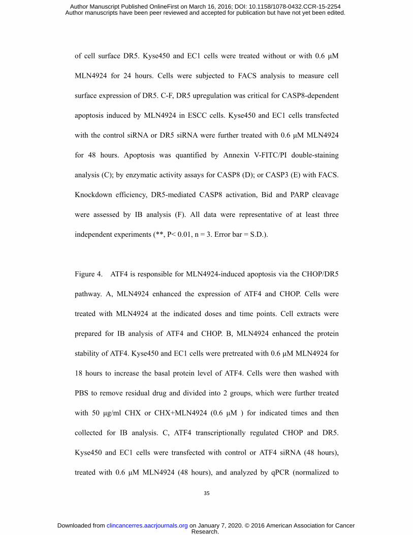
35
of cell surface DR5. Kyse450 and EC1 cells were treated without or with 0.6 μM
MLN4924 for 24 hours. Cells were subjected to FACS analysis to measure cell
surface expression of DR5. C-F, DR5 upregulation was critical for CASP8-dependent
apoptosis induced by MLN4924 in ESCC cells. Kyse450 and EC1 cells transfected
with the control siRNA or DR5 siRNA were further treated with 0.6 μM MLN4924
for 48 hours. Apoptosis was quantified by Annexin V-FITC/PI double-staining
analysis (C); by enzymatic activity assays for CASP8 (D); or CASP3 (E) with FACS.
Knockdown efficiency, DR5-mediated CASP8 activation, Bid and PARP cleavage
were assessed by IB analysis (F). All data were representative of at least three
independent experiments (**, P< 0.01, n = 3. Error bar = S.D.).
Figure 4. ATF4 is responsible for MLN4924-induced apoptosis via the CHOP/DR5
pathway. A, MLN4924 enhanced the expression of ATF4 and CHOP. Cells were
treated with MLN4924 at the indicated doses and time points. Cell extracts were
prepared for IB analysis of ATF4 and CHOP. B, MLN4924 enhanced the protein
stability of ATF4. Kyse450 and EC1 cells were pretreated with 0.6 μM MLN4924 for
18 hours to increase the basal protein level of ATF4. Cells were then washed with
PBS to remove residual drug and divided into 2 groups, which were further treated
with 50 μg/ml CHX or CHX+MLN4924 (0.6 μM ) for indicated times and then
collected for IB analysis. C, ATF4 transcriptionally regulated CHOP and DR5.
Kyse450 and EC1 cells were transfected with control or ATF4 siRNA (48 hours),
treated with 0.6 μM MLN4924 (48 hours), and analyzed by qPCR (normalized to
Research. on January 7, 2020. © 2016 American Association for Cancerclincancerres.aacrjournals.org Downloaded from
Author manuscripts have been peer reviewed and accepted for publication but have not yet been edited. Author Manuscript Published OnlineFirst on March 16, 2016; DOI: 10.1158/1078-0432.CCR-15-2254

36
GAPDH). D, DR5 was transcriptionally regulated by CHOP. Kyse450 and EC1 cells
were transfected with control or CHOP siRNA (48 hours), treated with 0.6μM
MLN4924 (48 hours), and analyzed by qPCR (normalized to GAPDH). E, The
expression of ATF4 and CHOP is responsible for MLN4924-induced apoptosis in
ESCC cells by the ATF4/CHOP/DR5 pathway. Knockdown efficiency, CASP8
activation, Bid and PARP cleavage were assessed by IB analysis. F, The expression of
ATF4 and CHOP was critical for MLN4924-induced apoptosis in ESCC cells.
Kyse450 and EC1 cells, transfected with control siRNA, ATF4 or CHOP siRNA, were
further treated with 0.6 μM MLN4924 for 48 hours. Apoptosis induction was
quantified by Annexin V-FITC/PI double-staining analysis (upper panel) or by
enzymatic activity assay for CASP8 (middle panel) or CASP3 by FACS (lower panel).
All data were representative of at least three independent experiments (**, P< 0.01, n
= 3. Error bar = S.D.).
Figure 5. Noxa is transactivated by ATF4 and participates in MLN4924-induced
apoptosis in ESCC cells. A, CASP9 knockdown reduced MLN4924-induced
apoptosis. Kyse450 and EC1 cells, transfected with siControl or siCASP9, were
treated with MLN4924 (0.6 μM) for 48 hours. Apoptosis was determined by the
Annexin V-FITC/PI double-staining analysis (left panel). Cell lysates were collected
and subjected to IB analysis for cleaved CASP3 and PARP. GAPDH served as a
loading control (right panel). B, MLN4924 increased the expression of Noxa at both
mRNA and protein levels. Kyse450 and EC1 cells were treated with MLN4924 (0.6
Research. on January 7, 2020. © 2016 American Association for Cancerclincancerres.aacrjournals.org Downloaded from
Author manuscripts have been peer reviewed and accepted for publication but have not yet been edited. Author Manuscript Published OnlineFirst on March 16, 2016; DOI: 10.1158/1078-0432.CCR-15-2254

37
μM) at the indicated concentrations and time. Cell extracts were prepared, and equal
amounts of protein were loaded and separated by SDS-PAGE and subjected to IB
analysis with anti-Noxa antibody. GAPDH served as a loading control (left panel).
The mRNA level of Noxa was determined by the qPCR assay (right panel). C and D,
Down-regulation of Noxa reduced MLN4924-induced apoptosis. Kyse450 and EC1
cells were transfected with control siRNA or Noxa siRNA and then treated with 0.6
μM MLN4924 for 48 hours. Apoptosis induction was quantified by Annexin
V-FITC/PI double-staining analysis (C, left panel) or CASP 3 activity analysis by
FACS (C, right panel). Knockdown efficiency and cleaved PARP/CASP3 were
assessed by IB analysis (D). E and F, ATF4 transcriptionally regulated Noxa. Kyse450
and EC1 cells were transfected (48 hours) with control or ATF4 siRNA, treated with
0.6 μM MLN4924 (48 hours). Transcriptional regulation of ATF4 on Noxa was
analyzed by qPCR (normalized to GAPDH) (E). Knockdown efficiency and
expression of Noxa were assessed by IB analysis (F). All data were representative of
at least three independent experiments (**, P< 0.01, n = 3. Error bar = S.D.).
Figure 6. MLN4924 suppressed esophageal tumor growth in vivo. Nude mice with
subcutaneously transplanted human esophageal cancer cells EC1-GFP or Kyse450
were administrated with MLN4924 as indicated in the Materials and Methods. A,
Whole-body images of EC1-GFP tumor model were captured twice a week. B, Tumor
size of both models was determined by caliper measurement and the data were
converted to tumor growth curves. C, Mice were sacrificed and tumor tissues were
Research. on January 7, 2020. © 2016 American Association for Cancerclincancerres.aacrjournals.org Downloaded from
Author manuscripts have been peer reviewed and accepted for publication but have not yet been edited. Author Manuscript Published OnlineFirst on March 16, 2016; DOI: 10.1158/1078-0432.CCR-15-2254

38
harvested, photographed, and weighed at the end of study (**, P < 0.01. Error bar =
S.D.). D, Proteins extracted from tumor tissues were analyzed by IB using anti-cullin1,
ATF4, CHOP, DR5, cleaved CASP8/3/PARP and Noxa antibodies. GAPDH was used
as a loading control. E, Schema of the mechanism for MLN4924-induced apoptosis in
ESCC.
Research. on January 7, 2020. © 2016 American Association for Cancerclincancerres.aacrjournals.org Downloaded from
Author manuscripts have been peer reviewed and accepted for publication but have not yet been edited. Author Manuscript Published OnlineFirst on March 16, 2016; DOI: 10.1158/1078-0432.CCR-15-2254

Figure 1
D E
G
F
A
B
Adjacent tissues ± + ++ +++ ++++
Tumor tissues
100 μm
0 20 40 60 80 100
0
20
40
60
80
100
Low expression
High expression
Su
rviv
al (%
)
P = 0.033
Months
Su
rviv
al (%
)
n = 45
n = 50
C
MLN
EC1
0 0.3 0.6 1.0
Kyse450
0 0.3 0.6 1.0
c-CASP3
c-PARP
GAPDH
CUL1
CUL1-N8
c-CASP8
70
40
25
70
35
(µM)
EC109
0 0.3 0.6 1.0
Kyse30
0 0.3 0.6 1.0
Kyse510
0 0.3 0.6 1.0
EC1
Kyse450
EC109
Kyse30
Kyse5100
20
40
60
80
100
120
0 0.1 0.3 0.6 (M)1.0
Ce
ll v
iab
ilit
y (%
) ** ** ** ** **
EC1
Kyse450
EC109
Kyse30
Kyse510
0
100
200
300
400
500
0 0.05 0.2 0.4 (M)
Co
lon
y n
um
be
r
**
** ****
**
0
10
20
30
40
50
Perc
en
tag
e (
%)
± + ++ +++ ++++
31/85
23/85
16/85 11/85
4/85 3/95
9/95
33/95 30/95
20/95
Adjacent tissues
Tumor tissues P < 0.01
EC1
Kyse450
EC109
Kyse30
Kyse510
0
10
20
30
40
50
0 0.3 0.6 1.0 (M)
*******
**
*
Casp
ase
-8 a
cti
vit
y (%
)
EC1
Kyse450
EC109
Kyse30
Kyse5100
10
20
30
40
50
0 0.3 0.6 1.0 (M)MLN
********
**
Ap
op
tosis
(%
)
EC1
Kyse450
EC109
Kyse30
Kyse510
0
10
20
30
40
50
0 0.3 0.6 1.0 (M)
*******
**
*
Casp
ase
-3 a
cti
vit
y (%
)
EC1
Kyse450
EC109
Kyse30
Kyse5100
10
20
30
40
50
0 0.3 0.6 1.0 (M)MLN
********
**
Ap
op
tosis
(%
)
Research. on January 7, 2020. © 2016 American Association for Cancerclincancerres.aacrjournals.org Downloaded from
Author manuscripts have been peer reviewed and accepted for publication but have not yet been edited. Author Manuscript Published OnlineFirst on March 16, 2016; DOI: 10.1158/1078-0432.CCR-15-2254

C
E
A B
D
DMSO MLN
0
5
10
15
20 **
Ap
op
tosis
(%
)
DMSO MLN
0
10
20
30 **
siControl siCASP8
Ap
op
tosis
(%
)
Kyse450 EC1
DMSO MLN
0
10
20
30 **
siControl siCASP8
Ap
op
tosis
(%
)
DMSO MLN
0
10
20
30
40
siControl siBid
**A
po
pto
sis
(%
)Kyse450 EC1
DMSO MLN
0
10
20
30
siControl siBid
**
Ap
op
tosis
(%
)
DMSO MLN
0
10
20
30
siControl siBid
**
Ap
op
tosis
(%
)
DMSO MLN
0
5
10
15
20
25
**
siControl siBid
Casp
ase
-3 a
cti
vit
y (%
) EC1
DMSO MLN
0
10
20
30 **
siControl siBid
Casp
ase
-3 a
cti
vit
y (%
) Kyse450
DMSO MLN
0
10
20
30
siControl siBid
**
Ap
op
tosis
(%
)
98.4%
1.5%
68.3%
31.2%
51.1%
48.3%
82.0%
18.0%
MLN (µM) 0 0.3 0.6 1.0
JC-1 monomers
JC
-1 a
gg
reg
ate
s
6.3%
93.4%
56.4%
42.9%
84.8%
15.1%
34.2%
65.2%
Kyse450 E
C1
GAPDH
Bid
tBid
MLN
EC1
0 0.3 0.6 1.0
Kyse450
0 0.3 0.6 1.0
c-CASP9
15
25
35
15 40
GAPDH
c-PARP
Bid
tBid
Kyse450 EC1
MLN
siBid
siControl
- - + +
- + - +
+ - + - - - + +
- + - +
+ - + -
70
15
25
35
(µM)
Figure 2
GAPDH
c-PARP
Bid tBid
Kyse450 EC1
MLN
siCASP8
siControl - - + +
- + - + + - + -
- - + +
- + - + + - + -
c-CASP8
15
25 40
70
35
G F
EC1 Kyse4500
10
20
30
40
50
60
0 0.3 0.6 1.0 (M)
**
**
Ce
lls lo
st
MM
P (
%)
Research. on January 7, 2020. © 2016 American Association for Cancerclincancerres.aacrjournals.org Downloaded from
Author manuscripts have been peer reviewed and accepted for publication but have not yet been edited. Author Manuscript Published OnlineFirst on March 16, 2016; DOI: 10.1158/1078-0432.CCR-15-2254

A B
0.7%
4.2%
70.5%
1.7%
7.6%
51.9%
Kyse450 EC1
DM
SO
Ig
G
DM
SO
D
R5 A
b
ML
N
DR
5 A
b
DMSO MLN
0
10
20
30
siControl siDR5
**
Ap
op
tosis
(%
)
Kyse450
DMSO MLN
0
10
20
30
siControl siDR5
**
Ap
op
tosis
(%
)
DMSO MLN
0
10
20
30
siControl siDR5
**
Ap
op
tosis
(%
)
EC1
DMSO MLN
0
10
20
30 **
siControl siDR5
Casp
ase
-8 a
cti
vit
y (%
) Kyse450
DMSO MLN
0
5
10
15
20
25 **
siControl siDR5
Casp
ase
-8 a
cti
vit
y (%
) EC1
DMSO MLN
0
10
20
30
siControl siDR5
**
Ap
op
tosis
(%
)
DMSO MLN
0
10
20
30 **
siControl siDR5
Casp
ase
-3 a
cti
vit
y (%
) Kyse450
DMSO MLN
0
5
10
15
20
25 **
siControl siDR5
Casp
ase
-3 a
cti
vit
y (%
) EC1
DMSO MLN
0
10
20
30
siControl siDR5
**
Ap
op
tosis
(%
)
C D
E F
DR5
DR5
GAPDH
MLN (µM) Time (h)
EC
1
Kyse450
0 0.3 0.6 1.0 0 12 24 48
GAPDH
55
35
35
40
55 40
c-CASP8
Kyse450 EC1
GAPDH
c-PARP
Bid tBid
DR5
MLN
siDR5
siControl - - + +
- + - +
+ - + -
- - + +
- + - +
+ - + -
40
55 40
25
15
70
35
Figure 3
Kyse450 EC1
0 12 24 48
0
1
2
3
4
EC1
Time (h)
DR
5 m
RN
A le
ve
l
( fo
ld in
cre
ase
)
**
0 12 24 48
0
1
2
3
4
Kyse450
**
Time (h)
DR
5 m
RN
A le
ve
l
( fo
ld in
cre
ase
)
Research. on January 7, 2020. © 2016 American Association for Cancerclincancerres.aacrjournals.org Downloaded from
Author manuscripts have been peer reviewed and accepted for publication but have not yet been edited. Author Manuscript Published OnlineFirst on March 16, 2016; DOI: 10.1158/1078-0432.CCR-15-2254

A
C
B
D
E
F Kyse450 EC1
DMSO MLN
0
1
2
3
4
5**
siControl siCHOP
DR
5 m
RN
A l
ev
el
( fo
ld i
nc
rea
se
)
DMSO MLN
0
1
2
3
4
5**
siControl siCHOP
DR
5 m
RN
A l
ev
el
( fo
ld i
nc
rea
se
)
DMSO MLN
0
1
2
3 **
siControl siCHOP
DR
5 m
RN
A l
ev
el
( fo
ld i
nc
rea
se
)
DMSO MLN
0
1
2
3 **
siControl siATF4
No
xa
mR
NA
le
ve
l
( fo
ld i
nc
rea
se
)
DMSO MLN
0
1
2
3
4
5 **
siControl siATF4
DR
5 m
RN
A l
ev
el
( fo
ld i
nc
rea
se
)
DMSO MLN
0
1
2
3
4
5 **
siControl siATF4
DR
5 m
RN
A l
ev
el
( fo
ld i
nc
rea
se
)
DMSO MLN
0
2
4
6
8
10
12 **
siControl siATF4
CH
OP
mR
NA
le
ve
l
( fo
ld i
nc
rea
se
)
DMSO MLN
0
2
4
6
8
10
12 **
siControl siATF4
CH
OP
mR
NA
le
ve
l
( fo
ld i
nc
rea
se
)
Kyse450 EC1
DMSO MLN
0
10
20
30 **
Ap
op
tosis
(%
)
DMSO MLN
0
10
20
30 **A
po
pto
sis
(%
)
Kyse450 EC1
DMSO MLN
0
5
10
15
20
25
siControl siATF4 siCHOP
**
Casp
ase
-8 a
cti
vit
y (%
)
DMSO MLN
0
10
20
30
siControl siATF4 siCHOP
**
Casp
ase
-8 a
cti
vit
y (%
)
DMSO MLN
0
10
20
30
siControl siATF4 siCHOP
**
Casp
ase
-3 a
cti
vit
y (%
)
DMSO MLN
0
5
10
15
20
25
siControl siATF4 siCHOP
**
Casp
ase
-3 a
cti
vit
y (%
)
DMSO MLN
0
5
10
15
20
25
siControl siATF4 siCHOP
**
Casp
ase
-3 a
cti
vit
y (%
)DMSO MLN
0
1
2
3 **
siControl siATF4
No
xa
mR
NA
le
ve
l
( fo
ld i
nc
rea
se
)
siCHOP
MLN
siATF4
siControl
- - + - - +
- - - + + +
- + - - + -
+ - - + - -
- - + - - +
- - - + + +
- + - - + -
+ - - + - -
Kyse450 EC1
GAPDH
c-PARP
ATF4
DR5
c-CASP8
Bid tBid
CHOP
40
35
55
25
55 40
25
15
70
Kyse450 EC1
Kyse450
EC
1
GAPDH
ATF4
CHOP
GAPDH
ATF4
CHOP
MLN (µM) Time (h)
0 0.3 0.6 1.0 0 12 24 48
35
35
55
55
25
25
Kyse450
GAPDH
ATF4
GAPDH
ATF4
EC
1
CHX+DMSO CHX+MLN
Time 0 15 30 45 60 90 0 15 30 45 60 90
35
35
55
55
(min)
Figure 4
Research. on January 7, 2020. © 2016 American Association for Cancerclincancerres.aacrjournals.org Downloaded from
Author manuscripts have been peer reviewed and accepted for publication but have not yet been edited. Author Manuscript Published OnlineFirst on March 16, 2016; DOI: 10.1158/1078-0432.CCR-15-2254

B
D
C
E
DMSO MLN
0
10
20
30
40 **
Ap
op
tosis
(%
)
DMSO MLN
0
10
20
30 **
Ap
op
tosis
(%
)
EC1 Kyse450
DMSO MLN
0
10
20
30
siControl siNoxa
**
Casp
ase
-3 a
cti
vit
y (%
)
DMSO MLN
0
10
20
30
40
siControl siNoxa
**
Casp
ase
-3 a
cti
vit
y (%
)EC1 Kyse450
DMSO MLN
0
10
20
30
siControl siNoxa
**
Casp
ase
-3 a
cti
vit
y (%
)
DMSO MLN
0
10
20
30
siControl siNoxa
**
Casp
ase
-3 a
cti
vit
y (%
)
A EC1 Kyse450
DMSO MLN
0
5
10
15
20
siControl siCASP9
**A
po
pto
sis
(%
)
DMSO MLN
0
5
10
15
20
siControl siCASP9
**
Ap
op
tosis
(%
)
DMSO MLN
0
10
20
30
40
50**
Ap
op
tosis
(%
)
F
DMSO MLN
0
1
2
3
4
5 **
siControl siATF4
No
xa
mR
NA
le
ve
l
( fo
ld i
nc
rea
se
)
DMSO MLN
0
1
2
3 **
siControl siATF4
No
xa
mR
NA
le
ve
l
( fo
ld i
nc
rea
se
)
DMSO MLN
0
1
2
3 **
siControl siATF4
No
xa
mR
NA
le
ve
l
( fo
ld i
nc
rea
se
)
Kyse450 EC1
GAPDH
c-PARP
Noxa
c-CASP3
Kyse450 EC1
MLN
siNoxa
siControl - - + +
- + - +
+ - + - - - + +
- + - +
+ - + -
10
25
70
35
Kyse450 EC1
MLN
siATF4
siControl - - + +
- + - +
+ - + - - - + +
- + - +
+ - + -
GAPDH
ATF4
Noxa
55
10
35
EC
1
Kyse450
0 0.3 0.6 1.0
MLN (µM) Time (h)
0 12 24 48
GAPDH
Noxa
GAPDH
Noxa
35
35
10
10
siCASP9
MLN
Kyse450
- + - + - - + +
EC1
GAPDH
c-PARP
c-CASP9
c-CASP3
- + - + - - + +
35 25
70
35
Figure 5
EC1 Kyse450
Time (h) Time (h) 0 12 24 48
0
2
4
6
**
mR
NA
le
ve
l
( fo
ld in
cre
ase
)
0 12 24 48
0
1
2
3
4 **
No
xa m
RN
A le
ve
l
( fo
ld in
cre
ase
)
Research. on January 7, 2020. © 2016 American Association for Cancerclincancerres.aacrjournals.org Downloaded from
Author manuscripts have been peer reviewed and accepted for publication but have not yet been edited. Author Manuscript Published OnlineFirst on March 16, 2016; DOI: 10.1158/1078-0432.CCR-15-2254

ML
N
Co
ntr
ol
1 4 8 11 13 15 17 Days
1 c
m
C
A B
D
EC1
1 cm MLN
Control
Control MLN
0.0
0.2
0.4
0.6
0.8
1.0 **
Tu
mo
r w
eig
ht
(g)
Kyse450
1 cm MLN
Control
Control MLN
0.0
0.2
0.4
0.6 **
Tu
mo
r w
eig
ht
(g)
Figure 6
Noxa
ATF4
CHOP
DR5
c-CASP3
c-PARP
GAPDH
Vehicle
1 2
MLN
1 2
c-CASP8
CUL1 CUL1-N8
70
40
55
25 55 40
10
25
70
35
EC
1
0.0
0.5
1.0
1.5 Control
MLN
1 4 8 11 13 15 17
Days post treatment
Tu
mo
r v
olu
me
(cm
3)
Kyse450 EC1
0.0
0.1
0.2
0.3
0.4
0.5
0.6 Control
MLN
0 1 8 164 12 20
Days post treatment
Tu
mo
r v
olu
me
(cm
3)
* * * *
Noxa
ATF4
CHOP
DR5
c-CASP3
c-PARP
GAPDH
c-CASP8
CUL1 CUL1-N8
70
40
55
25 55 40
10
25
70
35
Kyse450
Vehicle
1 2
MLN
1 2
E
MLN4924
ATF4
CASP8
Noxa
Mito
CHOP
DR5
tBid
Apoptosis
CASP9
CASP3
Neddylation
CRL/SCF
Research. on January 7, 2020. © 2016 American Association for Cancerclincancerres.aacrjournals.org Downloaded from
Author manuscripts have been peer reviewed and accepted for publication but have not yet been edited. Author Manuscript Published OnlineFirst on March 16, 2016; DOI: 10.1158/1078-0432.CCR-15-2254

Published OnlineFirst March 16, 2016.Clin Cancer Res Ping Chen, Tao Hu, Yupei Liang, et al. Cancer CellsPathway through ATF4-CHOP-DR5 Axis in Human Esophageal Neddylation Inhibition Activates the Extrinsic Apoptosis
Updated version
10.1158/1078-0432.CCR-15-2254doi:
Access the most recent version of this article at:
Material
Supplementary
http://clincancerres.aacrjournals.org/content/suppl/2016/03/16/1078-0432.CCR-15-2254.DC1
Access the most recent supplemental material at:
Manuscript
Authoredited. Author manuscripts have been peer reviewed and accepted for publication but have not yet been
E-mail alerts related to this article or journal.Sign up to receive free email-alerts
Subscriptions
Reprints and
To order reprints of this article or to subscribe to the journal, contact the AACR Publications
Permissions
Rightslink site. Click on "Request Permissions" which will take you to the Copyright Clearance Center's (CCC)
.http://clincancerres.aacrjournals.org/content/early/2016/03/16/1078-0432.CCR-15-2254To request permission to re-use all or part of this article, use this link
Research. on January 7, 2020. © 2016 American Association for Cancerclincancerres.aacrjournals.org Downloaded from
Author manuscripts have been peer reviewed and accepted for publication but have not yet been edited. Author Manuscript Published OnlineFirst on March 16, 2016; DOI: 10.1158/1078-0432.CCR-15-2254