Deep Massage for Back (plus some post. shoulder girdle and neck work)

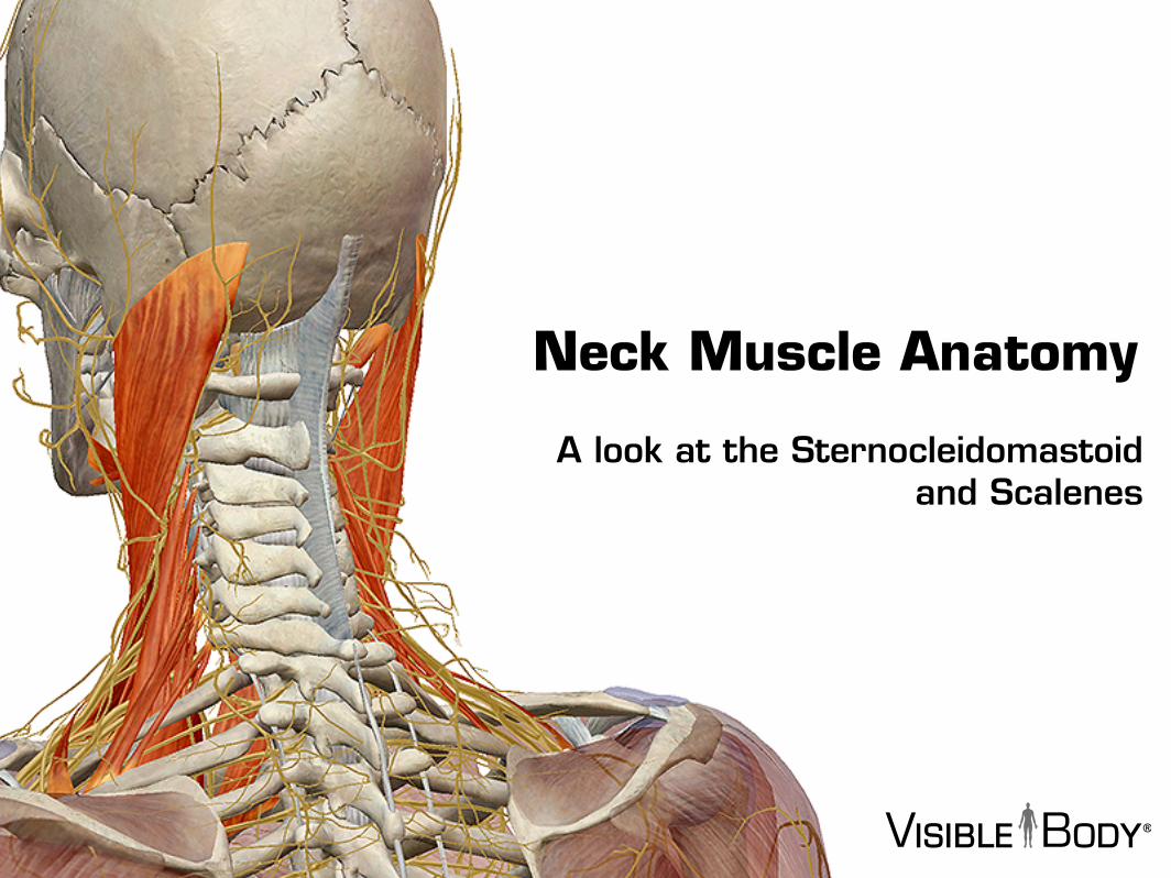
Your neck muscles are amazing. They are responsible for head movement, stabilizing the upper region of the body, assisting in swallowing, helping to elevate the rib cage during inhalation, and more.
Let’s take a look!

There are 26 muscles in the neck--10 pairs of 2 and 2 sets of 3, to be precise.
- Sterncleidomastoid- Scalenes- Platysma- Digastric- Omohyoid- Sternohyoid- Sternothyroid- Mylohyoid- Stylohyoid- Geniohyoid- Thyrohyoid
(And a partridge in a pear tree.)
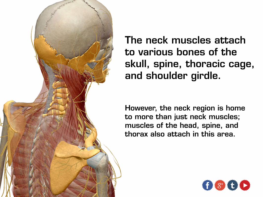
The neck muscles attach to various bones of the skull, spine, thoracic cage, and shoulder girdle.
However, the neck region is home to more than just neck muscles; muscles of the head, spine, and thorax also attach in this area.
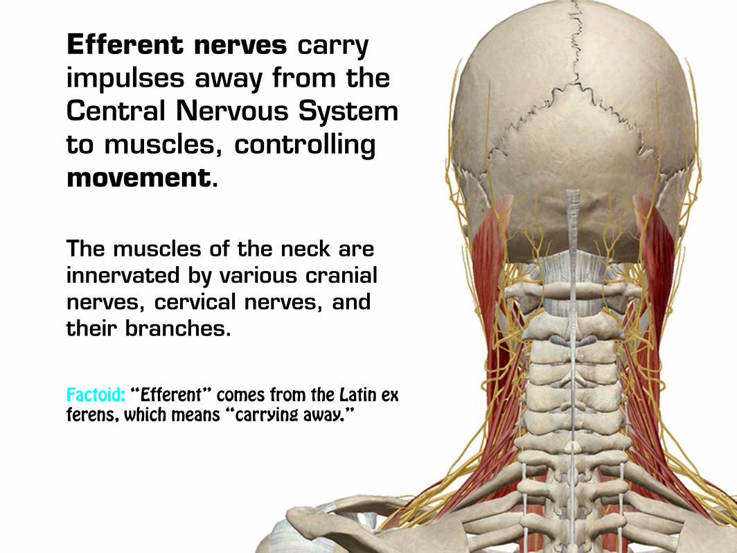
Efferent nerves carry impulses away from the Central Nervous System to muscles, controlling movement.
The muscles of the neck are innervated by various cranial nerves, cervical nerves, and their branches.
Factoid: “Efferent” comes from the Latin ex ferens, which means “carrying away.”
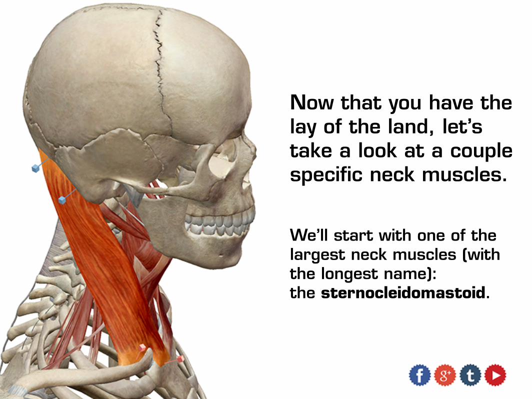
Now that you have the lay of the land, let’s take a look at a couple specific neck muscles.
We’ll start with one of the largest neck muscles (with the longest name): the sternocleidomastoid.
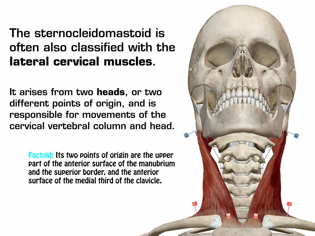
The sternocleidomastoid is often also classified with the lateral cervical muscles.
It arises from two heads, or two different points of origin, and is responsible for movements of the cervical vertebral column and head.
Factoid: Its two points of origin are the upper part of the anterior surface of the manubrium and the superior border, and the anterior surface of the medial third of the clavicle.

The sternocleidomastoid is innervated by the Accessory Nerve (XI) and branches from the anterior divisions of the cervical nerves (C02, C03).

The scalenus muscles, or scalenes, are sometimes classified in the lateral vertebral region.
It is a group of three, made up of the anterior, medius, and posterior scalenes.
Factoid: Scalene comes from the Latin word scalenus, meaning “uneven” or “unequal.”

The scalenes are innervated by the anterior divisions of the lower cervical nerves (C03--C08).
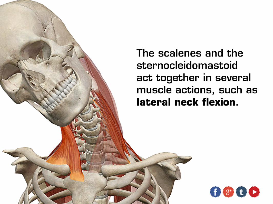
The scalenes and the sternocleidomastoid act together in several muscle actions, such as lateral neck flexion.
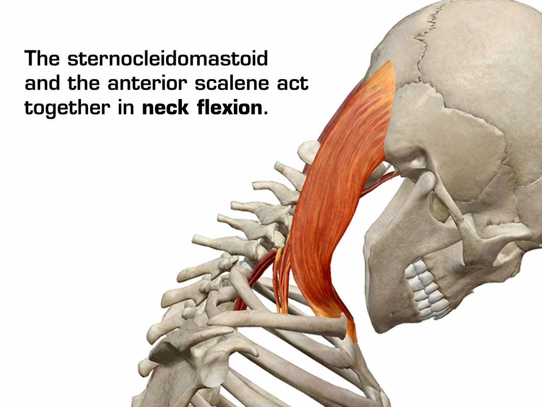
The sternocleidomastoid and the anterior scalene act together in neck flexion.
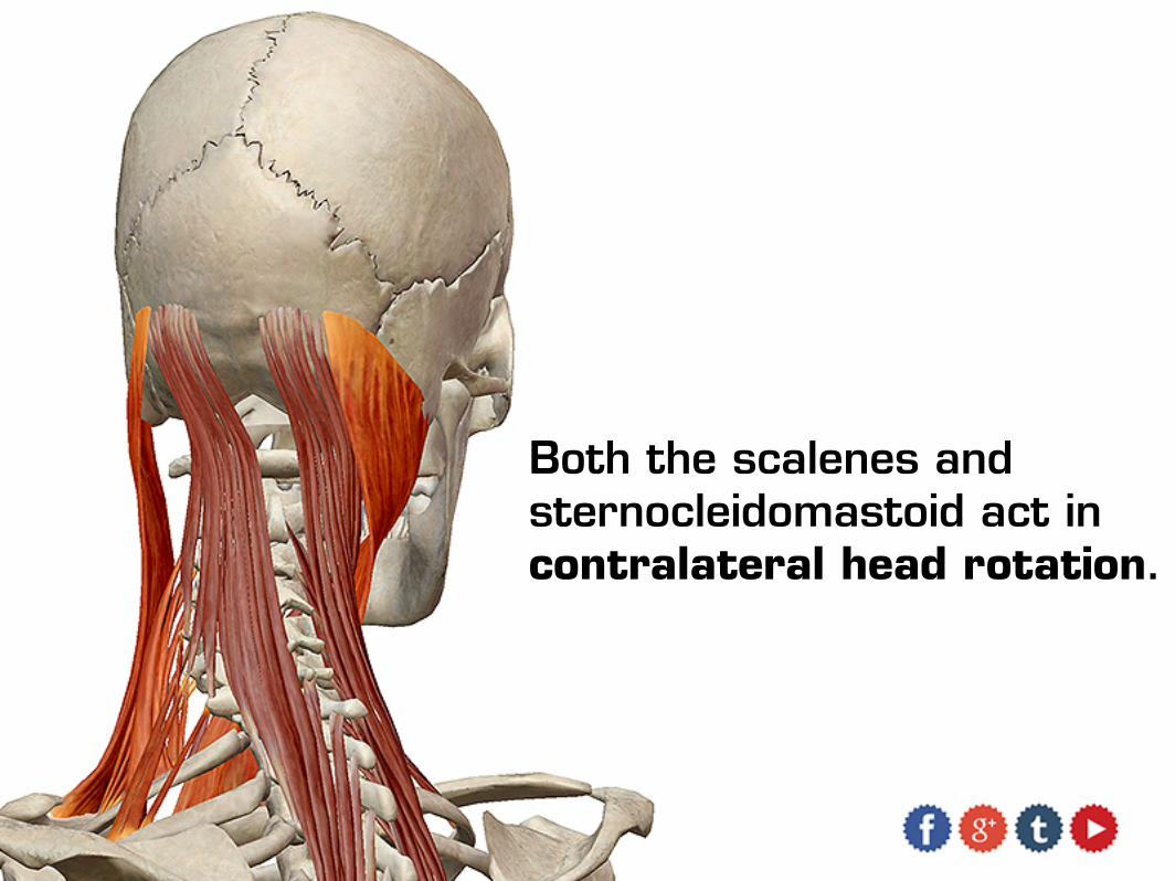
Both the scalenes and sternocleidomastoid act in contralateral head rotation.

Pretty amazing, huh?

All the images and text in this eBook came from Visible Body’s Muscle Premium app—an encyclopedic anatomical reference for human musculature.
MUSCLE PREMIUM
Content in the app includes:
600+ muscles, 200+ bones, peripheral nerves, and ligaments all rendered in interactive 3D, as well as dozens of interactive muscle action animations.
Functionality that allows you to rotate, zoom in/out, pan, hide, and add structures to see the anatomy from any angle. Customize and save views.
Dozens of quizzes to test your knowledge.
Muscle Premium is available for iOS, PC, Mac, and Android.
Site Licenses are available for Muscle Premium. Learn more here.