NEB Step-1 Day-8 (Review of Pelvis & Perineum)
113
-
Upload
drsaeed-shafi -
Category
Health & Medicine
-
view
86 -
download
4
Transcript of NEB Step-1 Day-8 (Review of Pelvis & Perineum)


Prof. Saeed Shafi

contd.
Learning Objectives
What is
• Urogenital ridge
– Nephrogenic cord
– Gonadal ridge
• Pronephros / mesonephros & metanephros?
• Ureteric bud & metanephric blastema are both mesodermal (intermediate mesoderm)


What is reciprocal induction?
• Branching morphogenesis of ureteric bud is bytranscription factor WT1 and glial derived neurotropicfactor(GDNF) produced by metanephric blastema
• Fibroblast growth factor (FGF2) and bone morphogeneticprotein (BMP-7) express by ureteric bud / metanephricdiverticulum induces mesenchymal condensation.
• Mesenchymal–epithelial transformation of metanephricblastema is by cytokine LIF (leukemia inhibitory factor).
• Transcription factors PAX-2, BF-2 and Wnt-4 produced byureteric bud help LIF.


Pronephros
• Early 4th week
• Cervical region
• Nonfunctioning
• Kidneys in fish

Mesonephros
• Late 4th week
• Caudal to pronephroi
• Function as interim kidney (4 – 9th week)
• Consist of tubules and mesonephric duct
• Degenerate towards end of 1st trimester

Metanephros • Ureteric bud
– Ureter– Pelvis– Calices – CD
• Metanephric blastema• Nephron • Glomeruli (increase 8 – 10 week, maximum on
32nd week)• All 1-M nephrons formed before birth• PCT increase after birth • GF in 9th week when new position is achieved


5th – 8th Week

Positional changes (6-9thW)
Rotation of hilum
Anomalies of kidneys
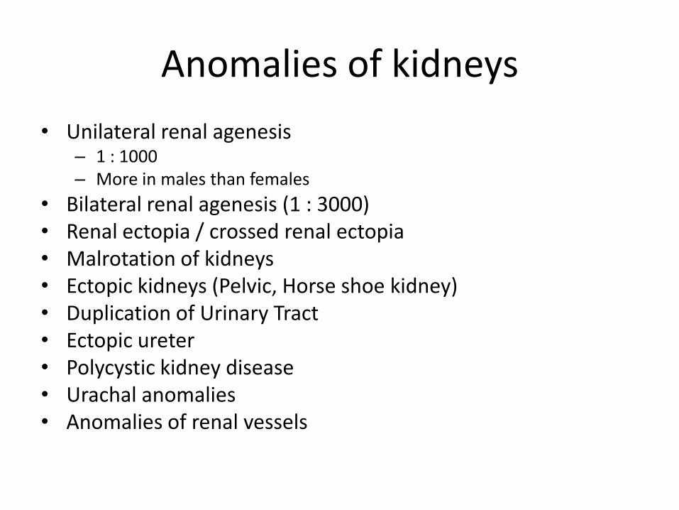
• Unilateral renal agenesis– 1 : 1000– More in males than females
• Bilateral renal agenesis (1 : 3000)• Renal ectopia / crossed renal ectopia• Malrotation of kidneys• Ectopic kidneys (Pelvic, Horse shoe kidney)• Duplication of Urinary Tract • Ectopic ureter • Polycystic kidney disease • Urachal anomalies • Anomalies of renal vessels

Anomalous vessels

Renal agenesis


Double ureter

Week 5 – Week 12


Nadia Gull of Peshawar changed to Toofan Khan

A newborn female with ambiguous genitalia, appearing like a male. Clitoris is enlarged and looks like a penis, labial
folds joined &look like scrotum.At puberty, the girl developed
excessive body hair growth, acne, menstrual irregularity, and infertility.

Development of Adrenals
• Cortex (W6 – Mesoderm)
• Medulla: Neural Crest Cells
• Location between root of dorsal mesentery and developing gonads
• Fetal cortex – mesothelial origin
• Permanent cortex
– Zona glomerulosa
– Zona fasciculata
– Zona reticularis (3 years)
At birth
Development of Adrenals
• Size of adrenals at birth 20 times larger than adults
• Larger than kidneys
• Fetal cortex regress in 1st year

Development of Adrenal glands
W-6 W-7

Development of Adrenal glands
New Born4 year

Cortical Differentiation
Differentiation of Suprarenal cortical zones begins during late fetal period.
ZONA GLOMERULOSA &ZONA FASCICULATA
are present at birth.
ZONA RETICULARIS is not recognizable until 3rd year of life.


• The suprarenal glands of the human fetus are 10-20 times larger than the adult gland relative to the body weight & are large as compared to the kidneys.
• This large gland results from the extensive size of the fetal cortex.
• The medulla remains relatively small until after birth.

• The suprarenal gland becomes smaller as the cortex regresses during the first year of life.
• The gland loses about 1/3rd of its weight during the first 2-3 weeks after birth & does not regain its original weight until the end of the second year.

CONGENITAL ADRENAL HYPERPLASIA
• -Abnormal increase in cells of the suprarenal cortex results in excessive androgen production during fetal period.
• In female causes masculinization of the external genitalia.
• In males this is undetected in early infancy. They have normal external genitalia.

• During childhood in both sexes androgen excess leads to rapid growth &accelerated skeletal maturation.
• The adrenogenital syndrome is associated with congenital adrenal hyperplasia(CAH).

Adrenogenital Syndrome
• This results in deficiency of supra-renal cortical enzymes necessary for the biosynthesis of many steroid hormones.
• Reduced hormone output results in increase release of ACTH that causes adrenal hyperplasia and excessive production of androgens.

Development of Genital ducts

Chromosome Complement
Type of Gonads
Male Phenotype Female phenotype
No hormone
Female external
Genetalia
Testosterone
Male external
genetalia

W-3
Intermediate
mesoderm
Urogenital Ridge
W-4
Genital Ridge
W-5
Nephrogenic
Cord
MesenchymePromordial germ
cell from Yolk Sac - W-4Mesothelium

Indifferent Gonads
• Week 5 – Thickening of mesothelium
– Proliferation of mesenchyme
– Migration of Primordial germ cells along dorsal mesentery of hindgut to gonadal ridge
Gonadal
ridge
formation

Indifferent Gonads
• Week 6 – Indifferent gonad with
– Cortex – gonadal cords
– Medulla –PG cells migrate into mesenchyme of gonads and incorporated into gonadal cords•If XX – cortex develops / medulla regress
•If XY – medulla develop / cortex regress
•Before 7-W, Gonads and genital ducts are similar-indifferent
gonads

Mesonephric
duct
Gonad

Contd
Development of Testes• Week 7• Indifferent gonad, under TDF coded by SRY gene
induces the differentiation of gonadal cords inmedulla into seminiferous cords
• Formation of tunica albuginea and loss of contact ofseminiferous cords with surface.
• Formation of mesorchium by degeneratingmesonephroi
• Week-8: Interstitial cells produce testosterone, whichdetermines the sexual differenciation of male genitalducts.
• Testosterone production is stimulated by hCG & ismaximum at 8-12 weeks

Contd
Development of Testes
• In absence of Y-chromosome, indifferent gonads cortex develop and medulla degenerate(Ovary)
• Development of female genital ducts is not dependant on any hormone.
• Sertoli cell (mesothelial origin) produce AMH which supresses development of paramesonephric ducts in male

• Gonadal cords – seminiferous cords –retetestis
• Rete testis become continuous with 15 – 20 mesonephric tubules to form efferent ductules.
• Mesonephric duct become duct of epididymis
• Seminiferous tubules remain solid till puberty

PGC
Leydig’s cell Mesenchyme
Yolk Sac
Sertoli Cells
MIS/AMHMesothelium
Testosteron Male genitalia

• PGC(Primordial germ cells)- yolk sac
• Sertoli cells - mesothelium
• Interstitial cells -mesenchyme


Ovarian development
• Slower than testis. Ovary can’t befound histologically till week 10th.
• Gonadal cords & medulla regress
• Cortex cords develop, extend fromsurface epithelium into underlyingmesenchyme

Ovarian development
• Primordial cells migrate into cortical cords
• Week 16- cortical cords breakup into clusters / primordial follicles
• No orgonia form postnatally
• Ovary separates from mesonephroi and develop mesentry - mesovarium

Genital ducts in male
• Week 5 – 6: Indifferent state
• Both ducts present in male and female
• Week 8: SRY – TDF – Leydig’s cells – testesterone –
mesonephric duct differenciation
• Proximal part – duct of epididymis
• Distal part – ductus deferencs and ejaculatory duct
• AMH/MIF leads to regression of paramesonephric duct in male


Female genital ducts
• Paramesonephric duct
• Lateral to mesonephric duct
• Derived from invagination of mesothelium
• Cranial part →Fallopian tubes
• Caudal part → Utero-vaginal primordium
• UVP project into dorsal wall of UGS
• Sinovaginal bulbs-vaginal plate-canalization
• Hymen

Mesonephros


Review of Pelvis & Perineum
Prof. Saeed Shafi


Learning Outcomes
???


R Ureter


Anorectal canal

DERIVATIVES OF CLOACA
•Cloaca is the distal dilated end of hindgut
• ventrally it is connected to a finger like diverticulumallantoise
• Partitioning of cloaca (W 5 – 7) by urorectal septum into
• urogenital sinus (anterior)
• anorectal canal (posterior)
•Partitioning of cloacal membrane into • anal and urogenital membrane
•Partitioning of cloacal sphincter into • external anal sphincter and urogenital sphincter
•Parineal body is at the site of intersection of anal membrane and urorectal septum

DEVELOPMENT OF ANAL CANAL
•Upper 2/3rd (endodermal) from hindgut
•Lower 1/3rd (ectodermal) from proctodeum
•Junction between these is at the level of pectinate line / anal valves / anal membrane
• Innervation & blood supply of anal canal

DEVELOPMENT OF ANAL CANAL

DEVELOPMENTAL ANOMALIES
Low annorectal anomalies (imperforate anus / ectopic
anus, anal agenesis, Persistent cloaca etc)
High anorectal anomalies (with or without fistulae)
Anal agenesis with perineal fistula
Ano-rectal agenesis with recto-vaginal fistula
Anorectal agenesis with recto-uretheral fistula


Anal
Musosa


Male vs. Female Pelvis
MaleFemale
Pelvic Inlet
Pelvic Outlet
Pelvic Cavity
Pelvic Arch

Perineum
• Definition
• Boundaries
• Subdivisions
– Anal triangle
– Urogenital triangle


Anal triangle
• Boundaries
• Contents
– Anal canal
– Ischioanal fossae


External anal sphincter

Relations


Urogenital Triangle
• Boundaries
• Layers– Skin
– Superficial fascia – 2 layers
– Deep fascia and muscles
– Fascia and muscles of urogenital diaphragm
• Contents – of male urogenital triangle
– of female urogenital triangle
• Pouches / spaces– Superficial
– Deep

Male urogenital triangle

Clinical aspects of perineum
• Female– Pudendal nerve block
– Episiotomy
– Perineal injuries during childbirth
– Bartholinitis
• Male– Urethral cathetrization
– Circumcision
– Rupture of urethra

Summary / Take Home Message