NCI Funding for Radiomics - AMOS...
14
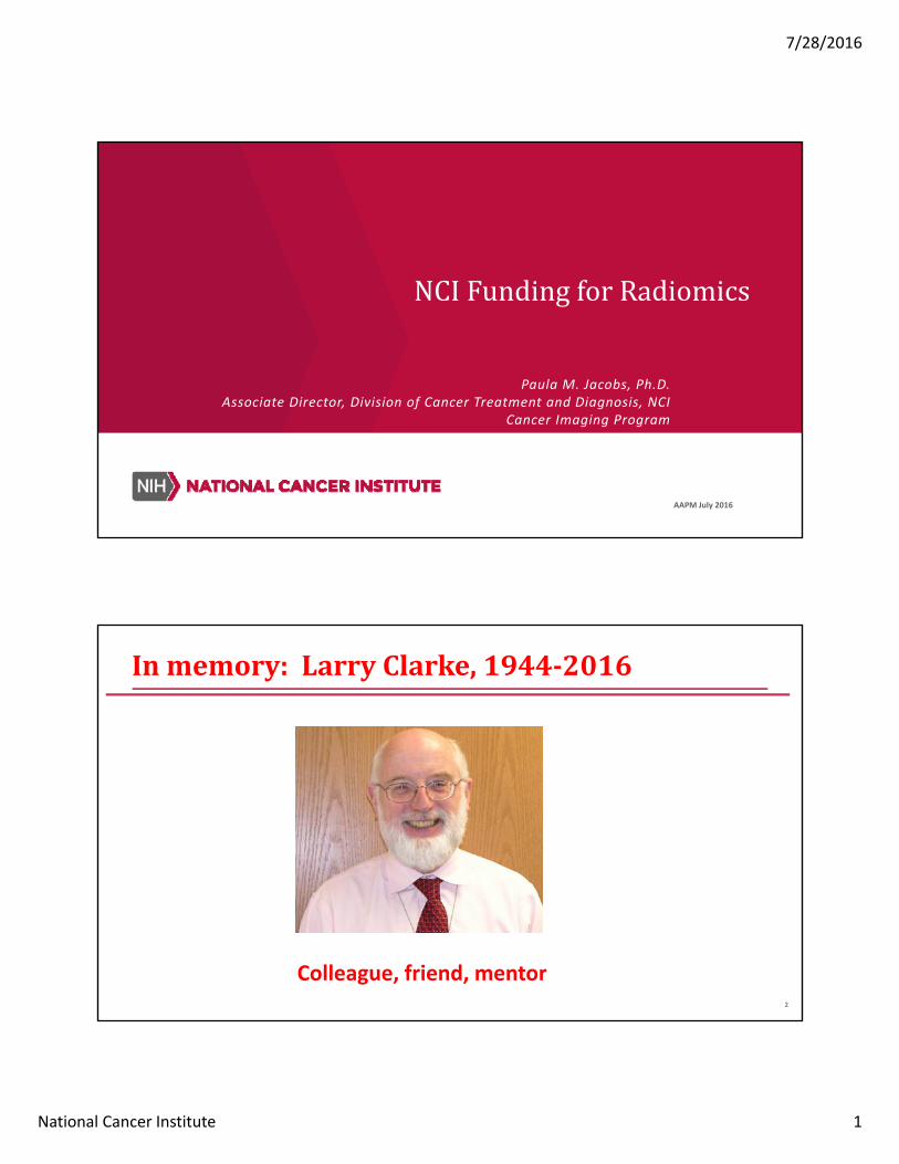
7/28/2016 National Cancer Institute 1 NCI Funding for Radiomics Paula M. Jacobs, Ph.D. Associate Director, Division of Cancer Treatment and Diagnosis, NCI Cancer Imaging Program June 2015 AAPM July 2016 2 In memory: Larry Clarke, 1944‐2016 Colleague, friend, mentor
Transcript of NCI Funding for Radiomics - AMOS...
7/28/2016
National Cancer Institute 1
NCIFundingforRadiomics
Paula M. Jacobs, Ph.D.Associate Director, Division of Cancer Treatment and Diagnosis, NCI
Cancer Imaging Program
June 2015 AAPM July 2016
2
Inmemory:LarryClarke,1944‐2016
Colleague, friend, mentor

7/28/2016
National Cancer Institute 2
3
Technology Development: Basic Research to Clinical Trials
Multi‐Site Preparation and Validation Studies
Development
Early Validation StudiesBasic Research Clinical Trials
IMAT, EBRG, BRP U01, R01 & R21, R33
SBIR & STTR
Early Phase Clinical Trials
IGDD, Co‐clinical
AIP, QIN, ITCR, Global Health
Parent R01, R03, R15
4
GeneralFunding
Funding Opportunities and Notices‐ NIH & NCI
• http://grants.nih.gov/grants/guide/• http://www.cancer.gov/researchandfunding/funding/announcements
Common types of grant
• Parent Announcements– R01, R21, R03, etc.
• Request for applications (RFA) Set aside funds• Program announcement (PA/PAR) R01, R21, U01, etc.
• SBIR/STTR

7/28/2016
National Cancer Institute 3
5
FundingInitiativesthatsupportTechnologyDevelopment
Innovative Molecular Analysis Technologies in Cancer Research (IMAT)
Bioengineering Research Partnership and Grants
Nanotechnology
Quantitative Imaging
Academic Industrial Partnerships
Information Technology for Cancer Research
Technologies for Global Health
SBIR/STTR
6
FundingInitiativesthatsupportTechnologyDevelopment
Innovative Molecular Analysis Technologies in Cancer Research (IMAT)
Bioengineering Research Partnership and GrantsNanotechnology
Quantitative Imaging
Academic Industrial Partnerships
Information Technology for Cancer Research
Technologies for Global Health
SBIR/STTR

7/28/2016
National Cancer Institute 4
7
Quantitative Imaging Network
Quantitative Imaging for Evaluation of Responses to Cancer Therapies: U01
PAR 14‐116
8
Rationale
Multi‐center clinical trials need validated imaging tools to measure therapy response
• Improve evaluation of therapies with quantitative imaging
• Reduce response variability to increase power
Validate quantitative imaging techniques on commercial platforms to support multi‐center multi‐platform trials
Collaborative efforts include multi‐site algorithm challenges with shared data

7/28/2016
National Cancer Institute 5
9
QuantitativeImagingNetwork
A cooperative agreement (U01) grant
Develop, test, and validate quantitative imaging methodsfor evaluating response to therapies
Managed as a network
Trans‐institution working groups
Some data sharing between groups required
All data and algorithms will be public eventually
QINtoolsReachingClinicalWorkflow
Institution Concept Development Testing & Optimization
Clinical Testing Commercialization Clinical Workflow
Brigham & Women's Hospital
3-D Slicer for Medical Image Visualization Open Source
Brigham & Women's Hospital
mpReview: Annotation for multiparamagnetic MRI Open Source
Brigham & Women's Hospital *
OncoQuant: DCE-MRI Analysis Not yet publically
available
Stanford University **
ePAD Clinical Viewer Open Source
* with GE Global Research Basic Research Clinical Research Community
** Active User's Group

7/28/2016
National Cancer Institute 6
11
Examples of Radiomics in QINGillies, Moffitt
Aerts, Dana Farber
12
ThePipelineforRadiomic ImageProcessing
Assemble cohorts of high‐quality images with matching outcome data
Segment lesions
Extract regions of interest and process data from them:
• Semantic features: e.g. size, shape, location
• Agnostic features: e.g. wavelet parameters, histogram skewness
Data mining, combining imaging information with clinical information

7/28/2016
National Cancer Institute 7
13
AFeatureExample:Convexity
Convexity is the ratio of thetumor border (from segmentation)to the perimeter of a convex hullsurrounding the tumor.
Here, the blue is thetumor border and redis the convex hullperimeter
Convexity tracks tumor morphology
Convexity is predictive of patient overall survival when dichotomized at the median value.
14
FeatureSizeReduction
Genomic Imformation
Image Parameters
Clinical Information
As many as 400 to 500features are firstconsidered
Through data mining and analysis in specific cancer studies, 4 to 7 parameters may prove to be sufficient for therapy response
The reduced featureset will be differentfor different tumortypes and organ sites.
7/28/2016
National Cancer Institute 8
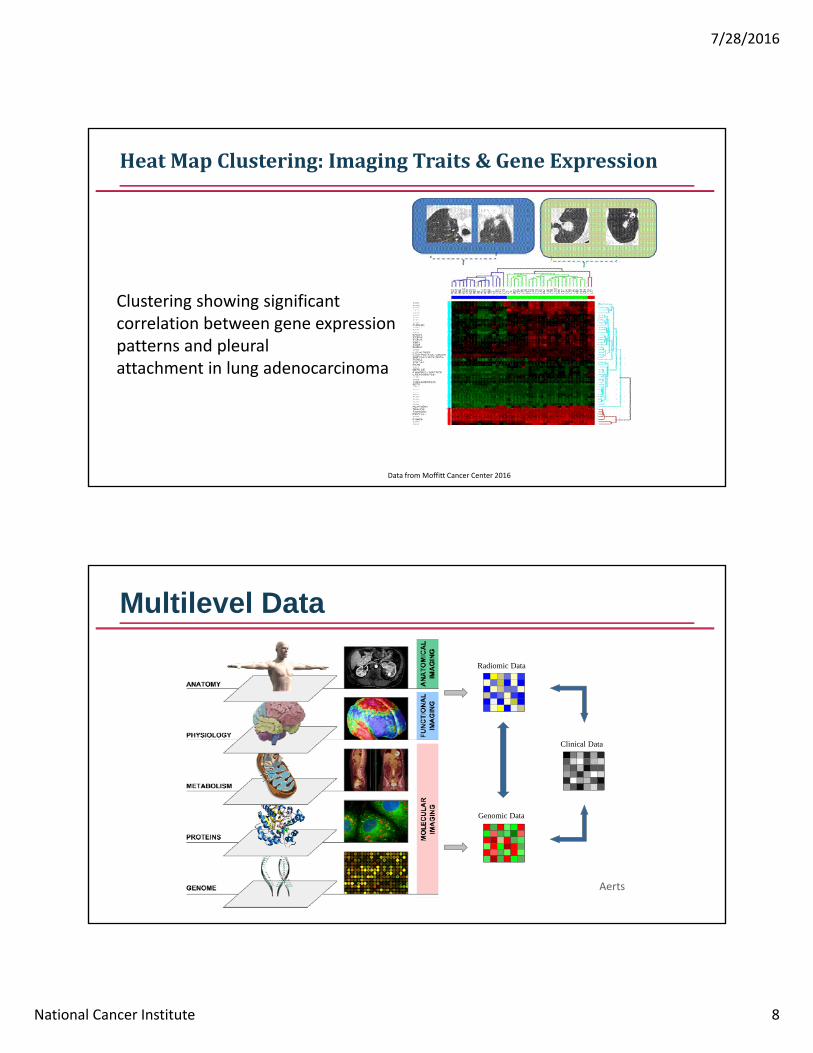
HeatMapClustering:ImagingTraits&GeneExpression
Clustering showing significant correlation between gene expression patterns and pleuralattachment in lung adenocarcinoma
Data from Moffitt Cancer Center 2016
Multilevel Data
Radiomic Data
Clinical Data
Genomic Data
Aerts

7/28/2016
National Cancer Institute 9
• Radiomics analysis on CT imaging of >1000 patients with Lung or H&N cancer
• Developed and Validate a prognostic radiomics signature that can be applied across cancer types
• Imaging‐Genomics analysis showed strong correlations between radiomics and genomics data
Imaging-Genomics across cancer types
*Aerts et al. Nature Comm. 2014
18
Academic‐IndustrialPartnershipsforTranslationofTechnologiesforCancerDiagnosisandTreatment
R01: PAR‐15‐075

7/28/2016
National Cancer Institute 10
19
Rationale
There is a constant need for adaptation, optimization and validation and eventual commercial dissemination of novel imaging technologies.
These imaging methods need to be fully integrated into commercially supported imaging platforms.
Research partnerships between academic and industry are therefore a critical on‐going requirement for imaging research.
Academic‐Industrial Partnerships for Translation of Technologies for Cancer Diagnosis and Treatment (R01): PAR‐15‐075
20
AIPProgramStructure
Research and Innovation: Clear translational research strategy for proposed technology.• Focuses on cancer detection and diagnosis, prediction and measurement of response to therapy, including image guided
interventions.
Project oriented toward clinical use: Should include physicians as key participants to provide essential expertise in oncology, pathology and/or other clinical science and practice appropriate to the planned outcome.
Project oriented toward pre‐clinical use: Translations of technologies to enhance the research performance of existing systems or provide new methods for a targeted cancer research problem.
• Focus on the optimization of advanced prototype imaging technologies and methods across pre‐clinical and clinical applications.
Partnership Structure: Partnerships for academic and industrial collaboration with translational
research goals. Must include at least one lead academic and one lead industrial organization
Foreign Institutions: Eligible to apply.

7/28/2016
National Cancer Institute 11
21
Information Technologies for Cancer Research: ICTR
R21, U01, U24
22
InformationTechnologiesforCancerResearch
Mission: Promote research‐driven informatics technology to address priority needs in cancer research.
Scope: Serve informatics needs that span the cancer research continuum and provide support for informatics resources:
• development of innovative methods and algorithms,
• early and advanced stage software development,
• sustainment of high‐value resources on which the research community has come to depend.
ITCR supports a wide range of informatics tools to serve current and emerging needs across the cancer research continuum.

7/28/2016
National Cancer Institute 12
23
ITCRPARs:FourFundingOpportunities
Supporting successive stages of informatics technology development.
Algorithm development (R21): PAR‐15‐334: Development of Innovative Informatics
Methods and Algorithms for Cancer Research and Management
Prototyping and Hardening (U01): PAR‐15‐332: Early‐Stage Development of Informatics
Technologies for Cancer Research and Management
Enhancement and Dissemination (U24): PAR‐15‐331: Development and enhancement of
emerging Informatics Technologies to improve acquisition, management, analysis and dissemination of data and knowledge to support Cancer Research (includes foreign investigators)
Sustainment (U24): PAR‐15‐333: Continued development and sustainment of high‐value
informatics research resources to serve current and emerging needs across the cancer research continuum (includes foreign investigators)
24
ProgramStructure
Algorithm Development
Prototype & Hardening
Enhancement and Dissemination
Sustainment
PAR-15-334R21: Innovative computational researchUp to $275K DC for 2 years
PAR-15-332 U01: Early stage developmentUp to $300K DC/year for 3 years
PAR-15-331 U24: Advanced stage developmentUp to $600K DC/year for 5 years
PAR-15-333 U24: Sustain highly-accessed resourcesNo budget ceiling; up to 5 years
7/28/2016
National Cancer Institute 13
25
SomeimagerelatedITCRgrants:http://itcr.nci.nih.gov/fp
John Quackenbush, Hugo Aerts (co-PIs)
Dana-Farber Cancer Institute
Quantitative Radiomics System Decoding the Tumor Phenotype
Bruce Rosen and Jayashree Kalpathy-Cramer (co-PIs)
Massachusetts General Hospital
Informatics Tools for Optimized Imaging Biomarkers for Cancer Research & Discovery
Christos Davatzikos University of Pennsylvania
Cancer Imaging Phenomics Software Suite: Application to Brain and Breast Cancer
Gordon Harris Harvard Medical School
Extensible Open-Source Zero-Footprint Web Viewer for Oncologic Imaging Research
Ron Kikinis and Andrey Fedorov (co-PIs)
Brigham and Women's Hospital and Harvard Medical School
Quantitative image informatics for cancer research (QIICR)
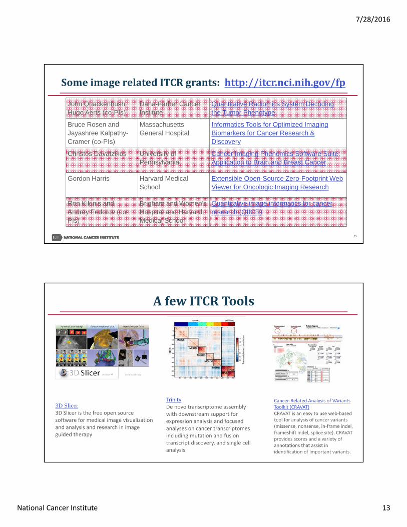
AfewITCRTools
Cancer‐Related Analysis of VAriantsToolkit (CRAVAT)CRAVAT is an easy to use web‐based tool for analysis of cancer variants (missense, nonsense, in‐frame indel, frameshift indel, splice site). CRAVAT provides scores and a variety of annotations that assist in identification of important variants.
TrinityDe novo transcriptome assembly with downstream support for expression analysis and focused analyses on cancer transcriptomes including mutation and fusion transcript discovery, and single cell analysis.
3D Slicer3D Slicer is the free open source software for medical image visualization and analysis and research in image guided therapy

7/28/2016
National Cancer Institute 14
www.cancer.gov www.cancer.gov/espanol
Imaging.cancer.gov [email protected]