NBA-7302; No.of Pages16 ARTICLE IN PRESS _Neurobiol... · Please cite this article in press as:...
16
Please cite this article in press as: Paban, V., et al., Neurotrophic signaling molecules associated with cholinergic damage in young and aged rats. Environmental enrichment as potential therapeutic agent. Neurobiol. Aging (2009), doi:10.1016/j.neurobiolaging.2009.03.010 ARTICLE IN PRESS NBA-7302; No. of Pages 16 Neurobiology of Aging xxx (2009) xxx–xxx Neurotrophic signaling molecules associated with cholinergic damage in young and aged ratsEnvironmental enrichment as potential therapeutic agent Véronique Paban ∗ , Caroline Chambon, Christine Manrique, Claude Touzet, Béatrice Alescio-Lautier Université d’Aix-Marseille I - Laboratoire de Neurobiologie Intégrative et Adaptative - UMR/CNRS 6149 - Pôle 3C - 3 Place Victor Hugo, 13331 Marseille Cedex 03, France Received 6 November 2008; received in revised form 16 March 2009; accepted 18 March 2009 Abstract The aim of this study was to determine the neurobiological bases of behavioral deficits associated with cholinergic damage and the potential of long-term environmental enrichment as a therapeutic agent. Rats were submitted to intra-structures injection of 192 IgG-saporin and then behaviorally tested 1 month and 1 year post-lesion in a nonmatching-to-position task. The gene expression changes were assessed by cDNA macroarray technology using the GE array Q series designed to profile the expression of neurotrophic signaling molecules. Results showed that (1) cholinergic injury modulated the expression of genes such as brain-derived neurotrophin factor but also genes associated with inflammatory response, neuron apoptosis, regulation of angiogenesis, and synaptic plasticity, (2) aging is associated with regulation of glial proliferation and apoptosis, and (3) long-term enriched environment housing enhanced behavioral performance in lesioned and non-lesioned rats and upregulated gene expression. This therapeutic role of the enriched environment seemed to be associated with a suppression of expression of genes involved in apoptosis, glial cell differentiation, and cell cycle, but also with an enhanced expression of a subset of genes involved in signal transduction. © 2009 Elsevier Inc. All rights reserved. Keywords: 192 IgG-saporin; bdnf; GE array; Immunohistochemistry; T-maze 1. Introduction The cholinergic basal forebrain is a neuronal system known to be implicated in cognitive processes like memory and attention (Baxter and Chiba, 1999; Sarter et al., 2003; Wenk, 1997). Many studies have dealt with the cholinergic system in aging (Gallagher and Colombo, 1995; Sarter and Bruno, 1998) and in related pathologies like Alzheimer’s disease (AD) (Casu et al., 2002; Dickinson-Anson et al., 2003; Zhang, 2004). Many years ago the cholinergic hypoth- esis of aging and AD emerged from human and animal data (Gallagher and Colombo, 1995; Muir, 1997), establishing a link between the cognitive decline observed in aged healthy ∗ Corresponding author. Tel.: +33 488576836; fax: +33 488576818. E-mail address: [email protected] (V. Paban). persons and in AD patients and alterations of the choliner- gic basal forebrain (Bartus, 2000; Shinotoh et al., 2000; Yan and Feng, 2004). Anatomically, the cholinergic basal fore- brain includes cells in the nucleus basalis of Meynert (called basalis magnocellularis nucleus (NBM) in animals) and cells in the complex medial septum (MS)/diagonal band of Broca (DBB). These cells project to the entire cortical mantle and the amygdala and to the hippocampus, respectively (Fibiger, 1982). In animals, the cholinergic system has been largely studied through experiments involving brain lesions, which showed a wide range of cognitive deficits. The immunotoxin 192 IgG- saporin is considered a valid tool to study the consequences of specific cholinergic loss in the basal forebrain (Paban et al., 2005a; Perry et al., 2001). The functional consequences of administration of the immunotoxin 192 IgG-saporin have 0197-4580/$ – see front matter © 2009 Elsevier Inc. All rights reserved. doi:10.1016/j.neurobiolaging.2009.03.010
Transcript of NBA-7302; No.of Pages16 ARTICLE IN PRESS _Neurobiol... · Please cite this article in press as:...
N
A
obm(ragit©
K
1
kaWsBd2e(l
0d
ARTICLE IN PRESSBA-7302; No. of Pages 16
Neurobiology of Aging xxx (2009) xxx–xxx
Neurotrophic signaling molecules associated with cholinergicdamage in young and aged ratsEnvironmental enrichment
as potential therapeutic agentVéronique Paban ∗, Caroline Chambon, Christine Manrique,
Claude Touzet, Béatrice Alescio-LautierUniversité d’Aix-Marseille I - Laboratoire de Neurobiologie Intégrative et Adaptative - UMR/CNRS 6149 - Pôle 3C - 3 Place Victor Hugo,
13331 Marseille Cedex 03, France
Received 6 November 2008; received in revised form 16 March 2009; accepted 18 March 2009
bstract
The aim of this study was to determine the neurobiological bases of behavioral deficits associated with cholinergic damage and the potentialf long-term environmental enrichment as a therapeutic agent. Rats were submitted to intra-structures injection of 192 IgG-saporin and thenehaviorally tested 1 month and 1 year post-lesion in a nonmatching-to-position task. The gene expression changes were assessed by cDNAacroarray technology using the GE array Q series designed to profile the expression of neurotrophic signaling molecules. Results showed that
1) cholinergic injury modulated the expression of genes such as brain-derived neurotrophin factor but also genes associated with inflammatoryesponse, neuron apoptosis, regulation of angiogenesis, and synaptic plasticity, (2) aging is associated with regulation of glial proliferation andpoptosis, and (3) long-term enriched environment housing enhanced behavioral performance in lesioned and non-lesioned rats and upregulated
ene expression. This therapeutic role of the enriched environment seemed to be associated with a suppression of expression of genesnvolved in apoptosis, glial cell differentiation, and cell cycle, but also with an enhanced expression of a subset of genes involved in signalransduction.2009 Elsevier Inc. All rights reserved.
ze
pgabbi(t1
eywords: 192 IgG-saporin; bdnf; GE array; Immunohistochemistry; T-ma
. Introduction
The cholinergic basal forebrain is a neuronal systemnown to be implicated in cognitive processes like memorynd attention (Baxter and Chiba, 1999; Sarter et al., 2003;enk, 1997). Many studies have dealt with the cholinergic
ystem in aging (Gallagher and Colombo, 1995; Sarter andruno, 1998) and in related pathologies like Alzheimer’sisease (AD) (Casu et al., 2002; Dickinson-Anson et al.,003; Zhang, 2004). Many years ago the cholinergic hypoth-
Please cite this article in press as: Paban, V., et al., Neurotrophic signalaged rats. Environmental enrichment as potential therapeutic agent. Neu
sis of aging and AD emerged from human and animal dataGallagher and Colombo, 1995; Muir, 1997), establishing aink between the cognitive decline observed in aged healthy
∗ Corresponding author. Tel.: +33 488576836; fax: +33 488576818.E-mail address: [email protected] (V. Paban).
twsoao
197-4580/$ – see front matter © 2009 Elsevier Inc. All rights reserved.oi:10.1016/j.neurobiolaging.2009.03.010
ersons and in AD patients and alterations of the choliner-ic basal forebrain (Bartus, 2000; Shinotoh et al., 2000; Yannd Feng, 2004). Anatomically, the cholinergic basal fore-rain includes cells in the nucleus basalis of Meynert (calledasalis magnocellularis nucleus (NBM) in animals) and cellsn the complex medial septum (MS)/diagonal band of BrocaDBB). These cells project to the entire cortical mantle andhe amygdala and to the hippocampus, respectively (Fibiger,982).
In animals, the cholinergic system has been largely studiedhrough experiments involving brain lesions, which showed aide range of cognitive deficits. The immunotoxin 192 IgG-
ing molecules associated with cholinergic damage in young androbiol. Aging (2009), doi:10.1016/j.neurobiolaging.2009.03.010
aporin is considered a valid tool to study the consequencesf specific cholinergic loss in the basal forebrain (Paban etl., 2005a; Perry et al., 2001). The functional consequencesf administration of the immunotoxin 192 IgG-saporin have

INNBA-7302; No. of Pages 16
2 ogy of A
bWttet
1talwutelms1c
nwA(taa2ottua2esratappmfiIhioesiosno
2
2
eim2tatfmbg
IstaohoAsttotdRtqf
2
tRmTiinc±±o(
ARTICLEV. Paban et al. / Neurobiol
een reviewed (McGaughy et al., 2000; Rossner et al., 1995;renn and Wiley, 1998). The cognitive deficits consecutive
o cholinergic lesion depend on many parameters like injec-ion type (intracerebroventricular or intra-strutures), lesionxtent (MS/DBB lesion, NBM lesion, or global lesion), andask demand (Parent and Baxter, 2004).
We have previously shown that the behavioral effects of92 IgG-saporin-induced cholinergic lesion depended on theime at which testing is conducted after surgery (Paban etl., 2005a). In particular, we demonstrated that at short post-esion time – less than 1 month – no deficit was observedhereas at longer post-lesion times – from 1 month andp to 1 year – rats showed memory impairments. Althoughhe behavioral effects of cholinergic immunolesion are wellstablished, there are only few data on underlying molecu-ar events. We have previously shown that the expression of
olecules such as the highly polysialylated neural cell adhe-ion molecule (PSA-NCAM) was upregulated 1 month andyear after surgery, suggesting that remodeling occurred in
holinergic-lesioned rats (Chambon et al., 2007).The aim of the present study was to determine the
eurobiological bases of behavioral deficits associatedith cholinergic damage in young and middle-aged rats.ttention was focused on neuroplasticity-related molecules
Johansson, 2004). Indeed, several studies have indicatedhat brain injuries are associated with altered expression ofnumber of genes encoding neurotrophins, growth factors,
nd signaling molecules (Lessmann et al., 2003; McAllister,000; Patz and Wahle, 2004). Thus, we used a technol-gy allowing expression-profiling analyses of several genes,hat is, cDNA arrays. This technique presents the advan-age of evaluating many genes at the same time and is nowsed in many fields of research including aging (Fraser etl., 2005; Lee et al., 2000; McCarroll et al., 2004; Prolla,002) and models of pathologies (Cheng et al., 2007; Weit al., 1999). We used a cDNA macroarray GE Array Qeries (MM18) including 96 gene fragments for studying neu-otrophic signaling gene expression after cholinergic injuryt 1 month and 1 year post-lesion times. Because of the cen-ral importance of brain-derived neurotrophic factor (bdnf)s a neurotrophic molecule, its expression was analyzed at arotein level by immunohistochemistry. Interestingly, we hadreviously shown that, during entry to senescence (around 15onths old), housing in an enriched environment had bene-cial effects on cognitive performance (Paban et al., 2005a).ndeed, 192 IgG-saporin-lesioned rats performed better whenoused in an enriched environment relative to animals housedn standard cage conditions, raising the question of the usef enriched environment as therapeutic agent. The molecularvents underlying these beneficial effects are not fully under-tood (Cotman and Berchtold, 2002). Thus, to gain insightnto this question, we compared the gene expression profile
Please cite this article in press as: Paban, V., et al., Neurotrophic signalaged rats. Environmental enrichment as potential therapeutic agent. Neu
f lesioned rats housed in enriched environment to the one oftandard-lesioned rats. In particular, the goal was to identifyew molecular and cellular pathways that can be targeted,pening new directions for treatment strategies.
orb(
PRESSging xxx (2009) xxx–xxx
. Materials and methods
.1. Animals and experimental design
Male Wistar rats (Charles River, France) were used in thexperiments. Animals were housed in standard conditions,.e., in groups of 2–3 rats per cage or in enriched environ-
ent, i.e., in groups of 10 rats in a cage of approximatelysquare meters equipped with a rearrangeable set of plastic
ubes, a running wheel, nesting material, and toys (Paban etl., 2005a). Rats were reared under 12 h light/12 h dark condi-ions with ad libitum access to food and water, except whenood-deprived during behavioral training. Every effort wasade to minimize animal suffering and to reduce the num-
er of rats used. All experiments conformed to internationaluidelines on the ethical use of animals.
Sixty-one rats were used. Thirty were injected with 192gG-saporin (SAP) and thirty-one with phosphate bufferaline solution only (PBS). Rats were 3 months old at theime of the surgery and were tested behaviorally 1 monthfter surgery – groups 1-month (PBS, N = 10; SAP, N = 10)r 1 year – groups 1-year (PBS, N = 11; SAP, N = 9). Rats wereoused in a standard environment during all this period. Twother groups of rats were used (PBS, N = 10; SAP, N = 11).s for group 1-year, these animals were tested 1 year after
urgery but were housed in an enriched environment duringhe entire experimental period (from surgery to behavioralesting); the rats were then 15 months old at the beginningf the behavioral session. Rats were killed immediately afterhe behavioral testing by anesthesia with isoflurane gas andecapitated. The brains were immediately cut on ice withNase-free instruments through the interhemispheric sulcus:
he right hemisphere was used for RNA isolation and subse-uent macroarray/PCR processings and the left hemisphereor immunohistochemistry.
.2. Cholinergic lesion surgery
Animals were anesthetized with an intramuscular injec-ion of a solution of Imalgen 500 (ketamine, 62.5 mg/kg),ompun (xylazine, 3.17 mg/kg) and Calmivet (acepromazinealeate, 0.62 mg/kg) and placed in a stereotaxic instrument.he immunotoxin 192 IgG-saporin (Chemicon, France) or
ts vehicle solution (phosphate-buffered solution) was slowlynjected bilaterally into the medial septum (MS) and theucleus basalis magnocellularis (NBM) at the followingoordinates: from the interaural point, AP −8.5 mm, ML0.4 mm, DV +3.4 mm for the MS, and AP −7.5 mm, ML2.5 mm, DV +2.5 mm for the NBM. The following doses
f 192 IgG-saporin were used: MS (37.5 ng/side), and NBM75 ng/side) to allow for maximal ChAT depletion. The toxin
ing molecules associated with cholinergic damage in young androbiol. Aging (2009), doi:10.1016/j.neurobiolaging.2009.03.010
r the vehicle was injected with a 10 �l Hamilton microsy-inge in a volume of 0.5 �l, allowing 5 min for diffusionefore the cannula was retracted as previously describedChambon et al., 2007; Paban et al., 2005a,b).

INNBA-7302; No. of Pages 16
ogy of A
2
tatdtOdctrAasoe(aiaoia
2
phsotnfiettwgaksvgtgctnTfiar
ba(L
2(
fgIfmyrscl(aatAsuisnfATGCGAGGTGTGtb1dpD
2
ARTICLEV. Paban et al. / Neurobiol
.3. Nonmatching-to-position task
The animals were trained in a nonmatching-to-positionask in a T-maze apparatus, as described in detail (Paban etl., 2005a,b). Briefly, the rats were gradually food-deprivedo 85% of the free-feeding level. The learning consisted of 8aily trials until the criterion for acquisition was met. Eachrial consisted of paired runs: one forced and one choice run.n the forced run, the side arm was closed by the guillotineoor and the other door was open with food pellets in theup. This forced the animal to enter a pre-selected arm andhen allowed it to eat the food. Immediately after the forcedun, the rat was placed again at the start box for a choice run.t this time, no guillotine door was lowered, and the rat was
llowed to choose between the two arms. The criterion forcoring an arm visit consisted of the rat placing a hind leg inne of the arms. No backtracking was permitted. When the ratntered the arm opposite to the one rewarded in the forced runnonmatching), a correct response was recorded, and it wasllowed to eat the pellets before returning to its cage. If not,t was confined to the incorrect arm for approximately 20 snd then returned to its cage. The rats were tested in groupsf 3 or 4 with each one given one trial per turn, so that thentertrial interval was about 4–6 min. Acquisition was defineds 7 correct trials on 2 consecutive days.
.4. RNA isolation and macroarray processing
Four rats per group were used. Medial septum, hip-ocampus, and frontal and entorhinal cortices from the rightemisphere were removed. Ten to fifteen micrograms of tis-ue were taken from each region, pooled, and mixed in 1 mlf Trizol reagent. A phase separation was carried out onhe homogenate to separate the different molecular compo-ents: RNA, DNA, and proteins. Total RNA was isolatedrom the aqueous phase by the Trizol reagent protocol. Itsntegrity and quality were checked using 1% agarose gellectrophoresis, and its concentration was measured by spec-rophotometry (A = 260 nm). For each rat (one chip per rat),otal RNA was reversed transcribed, radioactively markedith 32P, and hybridized to the GE Array Q series MM18ene array (Super Array Bioscience Corporation, France)ccording to the protocol provided by the manufacturer. Thisit is designed to profile the expression of 96 neurotrophicignaling molecules involved in growth, differentiation, sur-ival, and apoptosis. They are classified in 7 subsets ofenes: neurotrophins and receptors (Subset 1, S1), neuropep-ides and receptors (S2), growth factors involved in neuronalrowth and differentiation (S3), chemokine receptors (S4),ytokines and receptors (S5), signaling molecules indica-ive of downstream pathways (S6), and genes involved ineuronal apoptosis in response to neurotrophic factors (S7).
Please cite this article in press as: Paban, V., et al., Neurotrophic signalaged rats. Environmental enrichment as potential therapeutic agent. Neu
he radioactive signal was detected after exposure to X-raylm (Kodak, France). X-ray films were then scanned andnalyzed with the quantification software Quantity One (Bio-ad, France). All raw signal intensities were corrected for
gwsR
PRESSging xxx (2009) xxx–xxx 3
ackground by subtracting the signal intensity of a blanknd were normalized to the mean of housekeeping genesbeta-actin, GAPDH, cyclophilin A, and ribosomal protein13a).
.5. Reverse transcription-polymerase chain reactionRT-PCR)
RT-PCR was performed on selected genes using RNArom the same animals as for macroarray analysis. Fourenes chosen from the array data analysis were examined.t seems of interest to select genes with a highly dif-erential expression and genes that showed no expressionodification in order to show that the macroarray method
ields reliable results. Based on our cDNA macroarrayesults, bdnf and orexine receptor 2 (hcrt2) were cho-en as genes showing a significant and high expressionhange in 192 IgG-saporin-treated rats whatever the post-esion time. Neurotrophic tyrosine kinase receptor type 3ntrk3) and fibroblast growth factor 9 (fgf9) were chosens genes showing no significant change between lesionednd non-lesioned rats. For every RNA preparation, a nega-ive control was run in parallel, omitting the RNA sample.
1-�g RNA sample was used for one-step RT-PCR analy-is kit (Qiagen) according to the manufacturer’s instructions,sing a Gene Amp PCR system 9700 (Applied Biosystems),n 50 �l reactions containing 10 �M of each of the genepecific primers for bdnf, hcrtr2, ntrk3, fgf9, puc18 (foregative control), and beta-actin (for positive control). Theollowing oligonucleotide primers were used: for bdnf, 5′-TGAAGGCTGCGCCCATGAAAGAAA-3′ and 5′-TCCT-ATGAACCGCCAGCCAATTCTC-3′; for hcrtr2, 5′-TGC-GTACCTGTGGAGGGAATACCTA-3′ and 5′-GCGCCA-GTGCTCTGAGAGTTTTGATA-3′; for ntrk3, 5′-TCGG-AATTGAGACTGGAGCAGAACT-3′ and 5′-AGCATCC-GCGATGAAGGTGTAGTGA-3′; for fgf9, 5′-CCACCTG-GTCAGTCCGAAGCA-3′ and 5′-GGTACTTTGTCAGG-TCCACTGGTCTA-3′; or puc18, 5′-TTCACACCGCATA-GGTGCACTCTCA-3′ and 5′-TGTTGCCATTGCTACA-GCATCGT-3′; for actin, 5′-TGACCCTGAAGTACCCCA-TGAACACG-3′ and 5′-ATACCCAGGAAGGAAGGCT-GAAGAGA-3′. Amplified PCR products were subjected
o 1% agarose gel electrophoresis and stained with ethidiumromide. Gel images were captured with an Imager (Bio-D, Fisher Bioblock Scientifics SAA) and the fluorescentensities of the PCR products were evaluated through com-arison with different concentrations of the correspondingNA ladder.
.6. Immunohistochemistry
Immunohistochemistry was used to determine whether
ing molecules associated with cholinergic damage in young androbiol. Aging (2009), doi:10.1016/j.neurobiolaging.2009.03.010
ene expression identified by macroarray was associatedith the synthesis of its protein. Because the macroarray
tudy showed a characteristic change in bdnf messengerNA no matter what the experimental conditions were (see
INNBA-7302; No. of Pages 16
4 ogy of A
Sig4piaads0aCt(Hilauaifecwacaoc
(1wapTg0wd(
2
wLemtNi
s
Mc
MiteisgeaEtsdEa(2fg
3
3
ma((T1tataa1
ataGswaiic
ARTICLEV. Paban et al. / Neurobiol
ection 3), we investigated bdnf at the protein level bymmunohistochemical analysis. The left hemispheres (4 perroup of rats) were frozen in isopentane solution and cut into0-�m serial coronal sections. The sections were fixed in 4%araformaldehyde for 3 h and then washed 3 times for 10 minn 0.3% H2O2 and 0.05 Tris-buffered saline (TBS) solution,nd then incubated for 48 h at 4 ◦C with a rabbit polyclonalntibody to bdnf (Santa Cruz Biotechnology, CA, 1:1000ilution in TBS 0.3% Triton X-100). After this procedure,ections were washed 3 times for 10 min in 0.3% H2O2 and.05 Tris-buffered saline (TBS) solution, incubated for 2 h inbiotinylated rabbit anti-goat IgG (Chemicon International,A; 1:500 dilution in TBS 0.3% Triton X-100), washed 3
imes for 10 min in TBS, incubated in avidin–biotin complex1 h), and then immersed in 0.25% diaminobenzidine, 0.01%
2O2 solution, mounted on gelatinized slides, dehydratedn alcohol, and coverslipped with DePex for examination byight microscopy. A quantitative analysis of bdnf immunore-ctivity was performed via computer-assisted image analysissing a Leitz Aristoplan light microscope equipped withNikon high-resolution digital camera (756 × 581 pixels)
nterfaced to a PC computer, Image software (Lucia, Nikon)or capturing and processing the images. The zones of inter-st were acquired with a 10× objective lens. Measurementonsisted of computing the area of the labelled cells. Thisas performed using a grey-level method which consisted of
djusting a threshold brightness value. Thus, only labelledells with a grey value above this cutoff were taken intoccount. Bdnf immunoreactivity was quantified at the levelf the medial septum, hippocampus, frontal and entorhinalortices.
Immunohistochemistry of choline acetyltransferaseChAT) was done to investigate the immunotoxicity of92 IgG-saporin. The immunochemical staining for ChATas carried out as described above, in which the primary
ntibody was a goat polyclonal antibody against ChATrotein (Chemicon, France, 1:500 dilution in TBS 0.3%riton X-100) and the secondary antibody was a biotinylatedoat anti-rabbit IgG (Vector, France, 1:200 dilution in TBS.3% Triton X-100). Also, parallel sections were stainedith cresyl violet to determine the extent of nonspecificamage due to the needle tracks, as described previouslyPaban et al., 2005a,b).
.7. Statistical analysis
For the behavioral data, analysis of variance (ANOVA)as used to study the main effects of Group and Post-esion Time and their interaction. The effects of the enrichednvironment were analyzed using ANOVA to determine theain effects of Group and Environment and their interac-
ion. Further post hoc comparisons were made using the
Please cite this article in press as: Paban, V., et al., Neurotrophic signalaged rats. Environmental enrichment as potential therapeutic agent. Neu
ewman–Keuls t-test. Differences were considered signif-cant when P < 0.05.
For immunohistochemistery data, bdnf protein expres-ion between PBS and SAP groups was assessed using
3
a
PRESSging xxx (2009) xxx–xxx
ann–Whitney U test. Differences were considered signifi-ant when P < 0.05.
For the gene expression macroarray analysis, theann–Whitney U test was used for paired comparison
n each experimental condition. Genes of interest werehose satisfying the following two criteria: (1) the differ-nce in P value is less than 0.05, (2) the fold changes >1.5. These genes were selected for further analy-es. In particular, the expression data of these selectedenes were used to calculate Pearson correlations betweenvery pair-wise combination of genes of interest. A hier-rchical clustering analysis was then performed usinguclidian distance and Ward linkage to visualize clus-
ers of highly correlated genes that might be involved inome common regulation pathway. Functional profiling ofifferentially expressed genes was performed with Onto-xpress (http://vortex.cs.wayne.edu/projects.htm) (Khatri etl., 2002; Draghici et al., 2003) and DAVID websiteshttp://david.abcc.ncifcrf.gov/ease/ease.jsp) (Hosack et al.,003), which allowed us to thoroughly characterize sets ofunctionally related genes based on Gene-Ontology cate-ories.
. Results
.1. Behavioral analysis
The results are shown in Fig. 1. ANOVA on the perfor-ance of rats tested at 1 month and 1 year post-lesion time
nd housed in standard environment revealed a Group effectF (1, 36) = 17.85, P < 0.0002), a Post-Lesion Time effectF (1, 36) = 43.39, P < 0.0001), but no Group X Post-Lesionime interaction (F (1, 36) = 0.22, P = 0.64), indicating that92 IgG-saporin (SAP)-lesioned rats had worse performancehan control rats (PBS), irrespective of the post-lesion timest which animals were tested, 1 month vs. 1 year. Note thathe number of days to meet the criterion increased with thege of rats. Thus, groups 1 year (15 months old), PBS as wells SAP, needed more time to reach the criterion than groupsmonth (4 months old).ANOVA on the performance of rats housed in standard
nd enriched environment and tested at 1 year post-lesionime yielded a Group effect (F (1, 37) = 17.49, P < 0.0002),n Environment effect (F (1, 37) = 118.05, P < 0.0001), but noroup × Environment interaction (F (1, 37) = 3.22, P = 0.08),
howing that PBS and SAP rats displayed better performancehen housed in an enriched environment. Student’s t-test
nalysis on the performance of PBS and SAP rats housedn enriched environment yielded a Group effect (P < 0.05),ndicating that lesioned rats needed more time to reach theriterion than control rats.
ing molecules associated with cholinergic damage in young androbiol. Aging (2009), doi:10.1016/j.neurobiolaging.2009.03.010
.2. Macroarray analysis
The results are shown in Fig. 2 and Tables 1–3. One monthfter 192 IgG-saporin cholinergic immunolesion, among the
ARTICLE IN PRESSNBA-7302; No. of Pages 16
V. Paban et al. / Neurobiology of Aging xxx (2009) xxx–xxx 5
Table 1Gene expression changes in 192 IgG-saporin-lesioned rats at 1 month (A) and 1 year (B) post-lesion. Only genes showing significantly (P < 0.05) differentchanges greater than 1.5-fold are reported. Gene symbols, bank accession numbers, and names are provided. S1, neurotrophins and receptors; S2, neuropeptidesand receptors; S3, growth factors involved in neuronal growth and differentiation; S4, chemokine receptors; S5, cytokines and receptors; S6, signaling moleculesindicative of downstream pathways; S7 genes involved in neuronal apoptosis in response to neurotrophic factors. Positive fold change values indicate an increasein gene expression in SAP-treated rats. (C) 2-way Venn diagram indicating the gene expression profiles following cholinergic injury at 1 month vs. 1 yearpost-lesion. The common genes, i.e., genes affected by the lesioning no matter what the post-lesion time was, are noted in the center.
Gene symbol Gene Bank No. Gene name Subset P-value Fold change
Abdnf NM 007540 Brain-derived neurotrophic factor S1 <0.02 4.89cntfr NM 016673 Ciliary neurotrophic factor receptor S1 <0.02 2.80crh BE993430 Corticotropin releasing hormone S1 <0.02 1.98hcrtr2 AF394597 Orexine receptor 2 S1 <0.02 3.07snt-1 BB661937 Suc-1 associated neurotrophic factor target S1 <0.04 1.56tac1 NM 00931 Tachykinin 1 S1 <0.02 3.27gpr74 AF236084 G-protein-coupled recptor 74 S2 <0.02 1.54ptn D90225 Pleiotrophin S3 <0.02 3.55tgfa U65016 Transforming growth factor alpha S3 <0.02 2.29cx3cr1 AF102269 CX3 CR1 S4 <0.02 1.59lifr NM 013584 Leukaemia inhibitory factor receptor S5 <0.02 1.62stat5a NM 011488 Signal transducer and activator of transcription S6 <0.02 1.57
Bbdnf NM 007540 Brain-derived neurotrophic factor S1 <0.02 5.84cntfr NM 016673 Ciliary neurotrophic factor receptor S1 <0.04 1.55crhbp BE656478 Corticotropin-releasing factor binding protein S1 <0.02 3.52gfra3 NM 010280 Glial-derived neurotrophic factor receptor alpha 3 S1 <0.02 3.03hcrtr2 AF394597 Orexine receptor 2 S1 <0.02 1.59mt3 NM 013603 Metallothionein 3 S1 <0.02 3.81nr1i2 AF031814 Pregnane X receptor S1 <0.02 2.52ntf3 NM 008742 Neurotrophin 3 S1 <0.04 2.26tac1 NM 00931 Tachykinin 1 S1 <0.02 2.89gpr74 AF236084 G-protein-coupled recptor 74 S2 <0.02 1.96fgf2 M30644 Fibroblast growth factor 2 S3 <0.04 4.56ptn D90225 Pleiotrophin S3 <0.02 2.71cmkar4 AB000803 CXCR4 S4 <0.02 3.17lif NM 008501 Leukaemia inhibitory factor S5 <0.02 1.96jun J04115 Jun oncogene S6 <0.02 3.22apaf1 NM 009684 Apoptotic protease activating factor 1 S7 <0.02 3.76trp53 K01700 Transformation related protein 53 S7 <0.02 1.99
C
9Mems(nw
tcbw
6 genes that were represented in the GE Array Q seriesM18, 12 genes showed significantly altered expression lev-
ls and all were upregulated. Twenty-six transcripts changedore than 1.5-fold between control (PBS) and 192 IgG-
Please cite this article in press as: Paban, V., et al., Neurotrophic signalaged rats. Environmental enrichment as potential therapeutic agent. Neu
aporin (SAP)-treated rats housed in standard environmentFig. 2A and B and Table 1A). Only genes showing sig-ificantly (P < 0.05) different changes, greater than 1.5-fold,ere reported in tables and figures and considered for fur-
ec3o
her analyses. When we took into account the functionallassification in 7 subsets as described above, these geneselong mainly to S1. Note that the highest upregulationas seen in bdnf with a 4.89-fold increase in mRNA lev-
ing molecules associated with cholinergic damage in young androbiol. Aging (2009), doi:10.1016/j.neurobiolaging.2009.03.010
ls in SAP vs. PBS rats. Further analysis using hierarchicallustering showed that the 12 genes could be split intowell-identified clusters. The functional profiles of each
f these clusters were obtained using extensive literature
ARTICLE IN PRESSNBA-7302; No. of Pages 16
6 V. Paban et al. / Neurobiology of Aging xxx (2009) xxx–xxx
Table 2Gene expression changes following long-term enriched environment housing in 192 IgG-saporin-immunolesioned rats (A, PBS vs. SAP-lesioned rats housedin enriched environment and tested 1 year post-lesion). Only genes showing significantly (P < 0.05) different changes greater than 1.5-fold are reported. Genesymbols, bank accession numbers, and names are provided. S1, neurotrophins and receptors; S2, neuropeptides and receptors; S3, growth factors involved inneuronal growth and differentiation; S4, chemokine receptors; S5, cytokines and receptors; S6, signaling molecules indicative of downstream pathways; S7genes involved in neuronal apoptosis in response to neurotrophic factors. Positive fold change values indicate an increase in gene expression in SAP-treatedrats. (B) 2-way Venn diagram indicating the gene expression profiles following cholinergic injury when rats were housed in standard vs. enriched environment.The common genes, i.e., genes affected by the cholinergic injury no matter what the housing condition was are noted in the center. (C) 3-way Venn diagramindicating the gene expression profiles after cholinergic injury at 1 month and 1 year post-lesion of rats housed in standard or enriched environment.
Gene symbol Gene Bank No. Gene name Subset P-value Fold change
Abdnf NM 007540 Brain-derived neurotrophic factor S1 <0.02 4.97crhbp BE656478 Corticotropin-releasing factor binding protein S1 <0.02 2.75gfra3 NM 010280 Glial-derived neurotrophic factor receptor alpha 3 S1 <0.02 1.58hcrtr2 AF394597 Orexine receptor 2 S1 <0.02 3.27ntf3 NM 008742 Neurotrophin 3 S1 <0.02 2.01npy6r NM 010935 Neuropeptide Y receptor Y6 S2 <0.02 1.84il6ra X53802 Interleukin 6 receptor alpha S5 <0.02 1.59fos V00727 c-fos oncogene S6 <0.04 1.51
B
C
ss
•
•
•
earches in PubMed and Onto-Express and DAVID web-ites.Cluster 1 included crh, gpr74, snt1, and lifr genes andwould be involved in biological processes such as inflam-
Please cite this article in press as: Paban, V., et al., Neurotrophic signalaged rats. Environmental enrichment as potential therapeutic agent. Neu
matory processes and positive regulation of cell adhesion.Cluster 2 included cx3cr1 and stat5a genes and would beinvolved in biological processes such as positive regula-tion of inflammatory response, regulation of cell adhesion,growth, and proliferation.
a
dt
Cluster 3 included bdnf, cntfr, hcrt2, tac1, ptn, and tgfagenes and would be involved in biological processes suchas regulation of cellular plasticity, dendrite development,cell proliferation, and neuron recognition.
The full names of all transcripts are given in tables. To
ing molecules associated with cholinergic damage in young androbiol. Aging (2009), doi:10.1016/j.neurobiolaging.2009.03.010
void redundancy, only abbreviations are used.One year after cholinergic immunolesion, 17 transcripts
emonstrated a significant expression change between con-rol and 192 IgG-saporin-treated rats housed in standard
ARTICLE IN PRESSNBA-7302; No. of Pages 16
V. Paban et al. / Neurobiology of Aging xxx (2009) xxx–xxx 7
Table 3Gene expression changes during aging. (A) Gene expression changes in 4- vs. 15-month-old PBS control rats housed in standard condition. (B) Gene expressionchanges in 15-month-old control rats housed in standard vs. enriched environment. Only genes showing significantly (P < 0.05) different changes greater than1.5-fold are reported. Gene symbols, bank accession numbers, and names are provided. S1, neurotrophins and receptors; S2, neuropeptides and receptors; S3,growth factors involved in neuronal growth and differentiation; S4, chemokine receptors; S5, cytokines and receptors; S6, signaling molecules indicative ofdownstream pathways; S7 genes involved in neuronal apoptosis in response to neurotrophic factors. Positive fold change values indicate an increase in geneexpression and negative values indicate a decrease in 15-month-old control rats housed in standard condition (A) and in 15-month-old control rats housed inenriched environment (B).
Gene symbol Gene Bank No. Gene name Subset P-value Fold change
Agmfb NM 022023 Glial maturation factor beta S1 <0.02 2.16hcrtr2 AF394597 Orexine receptor 2 S1 <0.04 −2.58nrg4 NM 032002 Neuregulin 4 S1 <0.02 −3.1nmbr NM 008703 Neuromedin B receptor S2 <0.04 −2.75fgfr1 M33760 Fibroblast growth factor 1 S3 <0.02 1.79tgfa U65016 Transforming growth factor alpha S3 <0.02 1.73tgfb1 M13177 Transforming growth factor beta 1 S3 <0.02 −2.99il1b M15131 Interleukin 1-beta S5 <0.04 −2.79jun J04115 Jun oncogene S6 <0.02 2.11myc X01023 c-myc gene S6 <0.02 −3.48stat6 NM 009284 Signal transducer and activator of transcription 6 S6 <0.04 1.53apaf1 NM 009684 Apoptotic protease activating factor 1 S7 <0.04 3.06
Bbdnf NM 007540 Brain-derived neurotrophic factor S1 <0.02 5.14gmfb NM 022023 Glial maturation factor beta S1 <0.02 3.16gdnf NM 010275 Glial-derived neurotrophic factor S1 <0.04 1.57gfra3 NM 010280 Glial-derived neurotrophic factor receptor alpha 3 S1 <0.02 1.60mt3 NM 013603 Metallothionein 3 S1 <0.02 2.88ngfb NM 013609 Nerve growth factor beta S1 <0.02 1.94pspn NM 008954 Persephin S1 <0.04 1.59tac1 NM 00931 Tachykinin 1 S1 <0.02 2.33gpr74 AF236084 G-protein-coupled recptor 74 S2 <0.04 1.56fgf2 M30644 Basic fibroblast growth factor S3 <0.02 2.10ptn D90225 Pleiotrophin S3 <0.02 1.67cmkar4 AB000803 CXCR4 S4 <0.02 1.83
vator ofotein 53
emtecb
•
•
•
ew
Ta
chrshSabS8
•
•
myc X01023 c-myc genestat5a NM 011488 Signal transducer and actitrp53 K01700 Transformation related pr
nvironment; all were upregulated. Thirty-one genes changedore than 1.5-fold (Fig. 2C and D, and Table 1B). Most of
hem belonged to S1. The bdnf gene (S1) showed the high-st fold change, followed by the fgf2 gene (S3). Hierarchicallustering analysis showed that the 17 genes selected coulde grouped into 3 clusters.
Cluster 1 included bdnf, crhbp, ntf3, tac1, gpr74, ptn,cmkar4, lif, apaf, and trp53 genes and would beinvolved in biological processes such as regulation ofapoptosis.Cluster 2 included gfra3, hcrt2, mt3, nr1i2, and fgf2 genesand would be involved in biological processes such aspositive regulation of cell differentiation, regulation ofneurogenesis, and angiogenesis.Cluster 3 included cntfr and jun genes and would beinvolved in biological processes such as regulation of cellcycle and positive regulation of cell proliferation.
Please cite this article in press as: Paban, V., et al., Neurotrophic signalaged rats. Environmental enrichment as potential therapeutic agent. Neu
Interestingly, 6 genes were found in common, i.e., theirxpressions were altered by cholinergic damage no matterhat the post-lesion time was 1 month or 1 year (Table 1C).
•
S6 <0.04 1.58transcription S6 <0.04 1.62
S7 <0.04 1.57
he 6 genes were bdnf, cntfr, hcrtr2, tac1, gpr74, and ptn andll were upregulated. Most of them belong to subset 1.
Similar analyses were done to evaluate gene expressionhanges following cholinergic damage when rats wereoused in enriched environment. Among the 96 genesepresented on the GE Array, 8 genes demonstrated aignificant expression change between PBS and SAP ratsoused in enriched environment; all were upregulated.eventeen genes showed a fold change > 1.5 (Fig. 2E and F,nd Table 2A). These genes belong mainly to S1, includingdnf gene, which showed the highest fold change, then S2,5, and S6. Hierarchical clustering results indicated that thegenes could be grouped into 3 clusters.
Cluster 1 included npy6r and fos genes and would beinvolved in biological processes such as signal transduc-tion, axon guidance, and synaptic plasticity.Cluster 2 included gfra3 and il6ra genes and would be
ing molecules associated with cholinergic damage in young androbiol. Aging (2009), doi:10.1016/j.neurobiolaging.2009.03.010
involved in biological processes such as axon guidanceand synaptic plasticity.Cluster 3 included bdnf, crhbp, hcrt2, and ntf3 genesand would be involved in biological processes such as

ARTICLE INNBA-7302; No. of Pages 16
8 V. Paban et al. / Neurobiology of A
Fig. 1. Performance of control (PBS) and 192 IgG-saporin-lesioned (SAP)rats in the nonmatching-to-position task at 1 month and 1 year post-lesionand enriched environmental housing. The histograms represent the average(mean ± S.E.M.) numbers of days required to reach the criterion. Lesionedrats had worse performance than control rats no matter what the time afterspt
stmueshse
eyebocdts1n
uap
(a9ncu(c
•and would be involved in biological processes such asregulation of astrocytes/glial proliferation and growth.
• Cluster 2 included gmfb, hcrt2, nrg4, tgfa, il1b, myc, andsta6 genes and would be involved in biological processes
Fig. 2. Gene expression patterns were determined by GE Array Q seriesMM18 designed to profile the expression of 96 neurotrophic signaling
urgery was. In enriched environment, lesioned and non-lesioned rats dis-layed better performance. However, SAP-enriched rats required more dayshan PBS rats to reach the criterion. *P < 0.05; **P < 0.01 (vs. PBS group).
axon guidance, dendrite development, positive regulationof neuron differentiation, regulation of synaptic plasticity,and neuron apoptosis.
Note that 5 genes were found in common, i.e., their expres-ions were affected by the cholinergic injury no matter whathe housing condition was—standard or enriched environ-
ent (Table 2B). There were 3 genes whose expressions werepregulated in lesioned rats but only if they were housed innriched environment. There were 12 genes whose expres-ions were upregulated in lesioned rats but only if they wereoused in standard environment. In other words, the expres-ion of these 12 genes was repressed by housing in enrichednvironment.
Interestingly, a 3-way Venn diagram including the genexpression profiles after cholinergic injury at 1 month and 1ear post-lesion times of rats housed in standard or enrichednvironment revealed 6 groups of genes (Table 2C): (1)dnf and hcrt2 genes were common to all groups, regardlessf lesioning or age; (2) crhbp, gfra3, and ntf3 genes wereommon to middle-aged rats, regardless of housing con-ition; (3) cntfr, tac1, gpr74, and ptn genes were common
Please cite this article in press as: Paban, V., et al., Neurotrophic signalaged rats. Environmental enrichment as potential therapeutic agent. Neu
o lesioned rats, regardless of the post-lesion time; (4) crh,nt-1, tgfa, cx3cr1, lifr, and stat5a genes were unique to
month post-lesion-standard condition group; (5) mt3,r1i2, tgf2, cmkar4, lif, jun, apaf1, and trp53 genes were
mSia(
PRESSging xxx (2009) xxx–xxx
nique to 1 year post-lesion-standard condition group;nd (6) npyr6, il6ra, and fos genes were unique to 1 yearost-lesion-enriched condition group.
Comparing groups 1 month (4 months old) and 1 year15 months old) of PBS control rats allowed us to evalu-te the gene expression changes during aging. Among the6 genes represented on the GE Array, 12 showed a sig-ificant differential expression and 29 genes showed a foldhange > 1.5. Five genes were downregulated and 6 werepregulated (Fig. 2A and C and Table 3A). The myc genebelonging to S6) showed the highest fold change. Hierar-hical clustering identified two clusters.
Cluster 1 included nmbr, fgfr1, tgfb1, jun, and apaf1 genes
ing molecules associated with cholinergic damage in young androbiol. Aging (2009), doi:10.1016/j.neurobiolaging.2009.03.010
olecules involved in growth, differentiation, survival, and apoptosis.quared spots indicate the position of the differentially expressed genes
n control (PBS) and 192 IgG-saporin-lesioned (SAP) rats at 1 month (And B) and 1 year (C and D) after surgery. Rats were housed in standardA–D) or enriched environment (E and F).
IN PRESSNBA-7302; No. of Pages 16
ogy of Aging xxx (2009) xxx–xxx 9
rdbrTEwci
•
•
•
3
qmuyitiiSim
3
wrairr(bsaikc
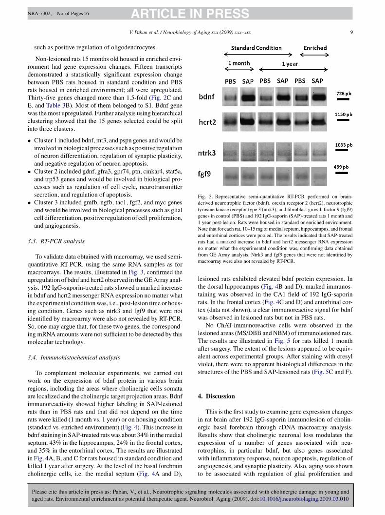
Fig. 3. Representative semi-quantitative RT-PCR performed on brain-derived neurotrophic factor (bdnf), orexin receptor 2 (hcrt2), neurotrophictyrosine kinase receptor type 3 (ntrk3), and fibroblast growth factor 9 (fgf9)genes in control (PBS) and 192 IgG-saporin (SAP)-treated rats 1 month and1 year post-lesion. Rats were housed in standard or enriched environment.Note that for each rat, 10–15 mg of medial septum, hippocampus, and frontaland entorhinal cortices were pooled. The results indicated that SAP-treatedrats had a marked increase in bdnf and hcrt2 messenger RNA expressionnfm
lttrtw
lTaavs
4
ieRe
ARTICLEV. Paban et al. / Neurobiol
such as positive regulation of oligodendrocytes.
Non-lesioned rats 15 months old housed in enriched envi-onment had gene expression changes. Fifteen transcriptsemonstrated a statistically significant expression changeetween PBS rats housed in standard condition and PBSats housed in enriched environment; all were upregulated.hirty-five genes changed more than 1.5-fold (Fig. 2C and, and Table 3B). Most of them belonged to S1. Bdnf geneas the most upregulated. Further analysis using hierarchical
lustering showed that the 15 genes selected could be splitnto three clusters.
Cluster 1 included bdnf, mt3, and pspn genes and would beinvolved in biological processes such as positive regulationof neuron differentiation, regulation of synaptic plasticity,and negative regulation of neuron apoptosis.Cluster 2 included gdnf, gfra3, gpr74, ptn, cmkar4, stat5a,and trp53 genes and would be involved in biological pro-cesses such as regulation of cell cycle, neurotransmittersecretion, and regulation of apoptosis.Cluster 3 included gmfb, ngfb, tac1, fgf2, and myc genesand would be involved in biological processes such as glialcell differentiation, positive regulation of cell proliferation,and angiogenesis.
.3. RT-PCR analysis
To validate data obtained with macroarray, we used semi-uantitative RT-PCR, using the same RNA samples as foracroarrays. The results, illustrated in Fig. 3, confirmed the
pregulation of bdnf and hcrt2 observed in the GE Array anal-sis. 192 IgG-saporin-treated rats showed a marked increasen bdnf and hcrt2 messenger RNA expression no matter whathe experimental condition was, i.e., post-lesion time or hous-ng condition. Genes such as ntrk3 and fgf9 that were notdentified by macroarray were also not revealed by RT-PCR.o, one may argue that, for these two genes, the correspond-
ng mRNA amounts were not sufficient to be detected by thisolecular technology.
.4. Immunohistochemical analysis
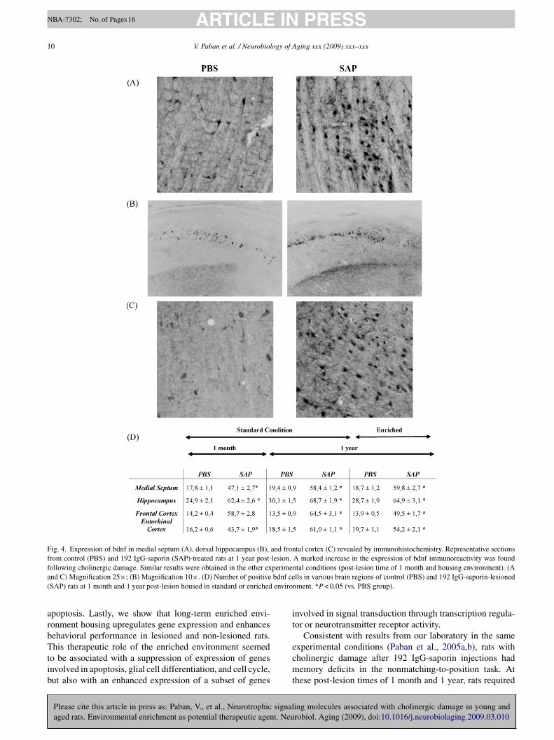
To complement molecular experiments, we carried outork on the expression of bdnf protein in various brain
egions, including the areas where cholinergic cells somatare localized and the cholinergic target projection areas. Bdnfmmunoreactivity showed higher labeling in SAP-lesionedats than in PBS rats and that did not depend on the timeats were killed (1 month vs. 1 year) or on housing conditionstandard vs. enriched environment) (Fig. 4). This increase indnf staining in SAP-treated rats was about 34% in the medialeptum, 43% in the hippocampus, 24% in the frontal cortex,
Please cite this article in press as: Paban, V., et al., Neurotrophic signalaged rats. Environmental enrichment as potential therapeutic agent. Neu
nd 35% in the entorhinal cortex. The results are illustratedn Fig. 4A, B, and C for rats housed in standard condition andilled 1 year after surgery. At the level of the basal forebrainholinergic cells, i.e. the medial septum (Fig. 4A and D),
rwat
o matter what the experimental condition was, confirming data obtainedrom GE Array analysis. Ntrk3 and fgf9 genes that were not identified byacroarray were also not revealed by RT-PCR.
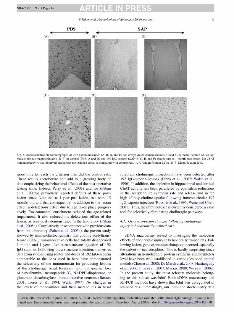
esioned rats exhibited elevated bdnf protein expression. Inhe dorsal hippocampus (Fig. 4B and D), marked immunos-aining was observed in the CA1 field of 192 IgG-saporinats. In the frontal cortex (Fig. 4C and D) and entorhinal cor-ex (data not shown), a clear immunoreactive signal for bdnfas observed in lesioned rats but not in PBS rats.No ChAT-immunoreactive cells were observed in the
esioned areas (MS/DBB and NBM) of immunolesioned rats.he results are illustrated in Fig. 5 for rats killed 1 monthfter surgery. The extent of the lesions appeared to be equiv-lent across experimental groups. After staining with cresyliolet, there were no apparent histological differences in thetructures of the PBS and SAP-lesioned rats (Fig. 5C and F).
. Discussion
This is the first study to examine gene expression changesn rat brain after 192 IgG-saporin immunolesion of cholin-rgic basal forebrain through cDNA macroarray analysis.esults show that cholinergic neuronal loss modulates thexpression of a number of genes associated with neu-
ing molecules associated with cholinergic damage in young androbiol. Aging (2009), doi:10.1016/j.neurobiolaging.2009.03.010
otrophins, in particular bdnf, but also genes associatedith inflammatory response, neuron apoptosis, regulation of
ngiogenesis, and synaptic plasticity. Also, aging was showno be associated with regulation of glial proliferation and
ARTICLE IN PRESSNBA-7302; No. of Pages 16
10 V. Paban et al. / Neurobiology of Aging xxx (2009) xxx–xxx
Fig. 4. Expression of bdnf in medial septum (A), dorsal hippocampus (B), and frontal cortex (C) revealed by immunohistochemistry. Representative sectionsfrom control (PBS) and 192 IgG-saporin (SAP)-treated rats at 1 year post-lesion. A marked increase in the expression of bdnf immunoreactivity was foundf perimea bdnf ce( enviro
arbTtib
it
ollowing cholinergic damage. Similar results were obtained in the other exnd C) Magnification 25×; (B) Magnification 10×. (D) Number of positiveSAP) rats at 1 month and 1 year post-lesion housed in standard or enriched
poptosis. Lastly, we show that long-term enriched envi-onment housing upregulates gene expression and enhancesehavioral performance in lesioned and non-lesioned rats.
Please cite this article in press as: Paban, V., et al., Neurotrophic signalaged rats. Environmental enrichment as potential therapeutic agent. Neu
his therapeutic role of the enriched environment seemedo be associated with a suppression of expression of genesnvolved in apoptosis, glial cell differentiation, and cell cycle,ut also with an enhanced expression of a subset of genes
ecmt
ntal conditions (post-lesion time of 1 month and housing environment). (Alls in various brain regions of control (PBS) and 192 IgG-saporin-lesioned
nment. *P < 0.05 (vs. PBS group).
nvolved in signal transduction through transcription regula-or or neurotransmitter receptor activity.
Consistent with results from our laboratory in the same
ing molecules associated with cholinergic damage in young androbiol. Aging (2009), doi:10.1016/j.neurobiolaging.2009.03.010
xperimental conditions (Paban et al., 2005a,b), rats withholinergic damage after 192 IgG-saporin injections hademory deficits in the nonmatching-to-position task. At
hese post-lesion times of 1 month and 1 year, rats required
ARTICLE IN PRESSNBA-7302; No. of Pages 16
V. Paban et al. / Neurobiology of Aging xxx (2009) xxx–xxx 11
F , and En IgG-sai with co
mTdtelmesilefsf1Idctoog2t
f11CihI2t
4i
eltalme
ig. 5. Representative photomicrographs of ChAT-immunostained (A, B, Ducleus basalis magnocellularis (D–F) of control (PBS, A and D) and 192mmunoreactivity was observed throughout the lesioned areas, as compared
ore time to reach the criterion than did the control rats.hese results corroborate and add to a growing body ofata emphasizing the behavioral effects of the post-operativeesting time. Indeed, Perry et al. (2001) and we (Pabant al., 2005a) previously reported deficits at these post-esion times. Note that at 1 year post-lesion, rats were 15
onths old and that consequently, in addition to the lesionffect, a deleterious effect due to age takes place progres-ively. Environmental enrichment reduced the age-relatedmpairment. It also reduced the deleterious effect of theesion, as previously demonstrated in the laboratory (Pabant al., 2005a). Correlatively, in accordance with previous datarom the laboratory (Paban et al., 2005a), the present studyhowed by immunohistochemistry that choline acetyltrans-erase (ChAT) immunoreative cells had totally disappeared
month and 1 year after intra-structure injection of 192gG-saporin. Following intra-structure injection, numerousata from studies using routes and doses of 192 IgG-saporinomparable to the ones used in here have demonstratedhe selectivity of the immunotoxin for producing lesionsf the cholinergic basal forebrain with no specific loss
Please cite this article in press as: Paban, V., et al., Neurotrophic signalaged rats. Environmental enrichment as potential therapeutic agent. Neu
f parvalbumin-, neuropeptide Y-, NADPH-diaphorase, orlutamate decarboxylase-immunoreactive neurons (Baxter,001; Torres et al., 1994; Wenk, 1997). No changes inhe levels of monoamines and their metabolites in basal
IiRl
) and cresyl violet-stained sections (C and F) in medial septum (A–C) andporin (SAP, B, C, E, and F)-treated rats at 1 month post-lesion. No ChATntrol rats. (A–C) Magnification 2.5×; (D–F) Magnification 25×.
orebrain cholinergic projections have been detected after92 IgG-saporin lesions (Pizzo et al., 2002; Walsh et al.,996). In addition, the depletion in hippocampal and corticalhAT activity has been paralleled by equivalent reductions
n the acetylcholine synthesis rate and release and in theigh-affinity choline uptake following intraventricular 192gG-saporin injection (Rossner et al., 1995; Waite and Chen,001). Thus, the immunotoxin is currently considered a validool for selectively eliminating cholinergic pathways.
.1. Gene expression changes following cholinergicnjury in behaviorally trained rats
cDNA macroarray served to investigate the molecularffects of cholinergic injury in behaviorally trained rats. Fol-owing lesion, gene expression changes concerned especiallyhe subset of neurotrophins. This is hardly surprising sincelterations in neurotrophin protein synthesis and/or mRNAevel have been well established in various lesioned-animal
odels (Chen et al., 2008; De March et al., 2008; Hattiangadyt al., 2006; Imai et al., 2007; Macias, 2008; Wu et al., 2006).
ing molecules associated with cholinergic damage in young androbiol. Aging (2009), doi:10.1016/j.neurobiolaging.2009.03.010
n the present study, the most relevant molecule belong-ng to this subset was bdnf. Both cDNA macroarray andT-PCR methods have shown that bdnf was upregulated in
esioned rats. Interestingly, our immunohistochemistry data
INNBA-7302; No. of Pages 16
1 ogy of A
soifosuireXii2tgacgtimrc(ap
cogtrPlmga(2bte2riepaagaIaip
e2Keefnigwa(gtwwibbupdC2aeotanaitrada
itlaIiisgrrftw
ARTICLE2 V. Paban et al. / Neurobiol
howed that cholinergic-lesioned rats exhibited an elevationf bdnf protein expression in various brain regions, includ-ng the medial septum, the dorsal hippocampus, and therontal and entorhinal cortices. Bdnf is known to play vari-us roles in the central nervous system, including promotingurvival, maintenance, and growth of distinct neuronal pop-lations (Labelle and Leclerc, 2000). In particular, bdnf isnvolved in the maintenance of forebrain cholinergic neu-ons (Gómez-Palacio-Schjetnan and Escobar, 2008; Morset al., 1993; Pencea et al., 2001; Taupin and Gage, 2002;uan et al., 2008). Furthermore, bdnf plays a crucial role
n cognition, learning, and memory formation by modulat-ng synaptic plasticity (Schindowski et al., 2008; Todd et al.,007). Hsieh et al. (2003), to our knowledge, are the only teamo have applied cDNA microarray technology to investigateene expression changes after cholinergic damage. They usedcDNA collection containing 9600 sequence-verified humanDNA clones spotted on a nylon membrane to explore theene expression profile in the hippocampus of scopolamine-reated rats. Animals were killed 30 min after intracisternalnjection. In spite of some experimental differences (ani-
al models, structures, and cDNA array technology), theiresults add to ours, indicating an upregulation of genes asso-iated with the signaling pathways of muscarinic receptorse.g., overexpression of inositol 1,4,5-triphosphate receptor)nd with Alzheimer’s disease (e.g., upregulation of amyloid,rotein tau) in treated rats.
Interestingly, we have shown that gene expressionhanges following cholinergic immunolesion dependedn the post-lesion time. At 1 month post-lesion, 12enes showed differential expression. To extract informa-ion from gene expression and discover more complexelationships, we used extensive literature searches inubMed and then relied on the GO database. In particu-
ar, we used Onto-Express which is able to identify theost relevant biological processes from lists of selected
enes (http://vortex.cs.wayne.edu/projects.htm) (Draghici etl., 2003; Khatri et al., 2002) and DAVID websiteshttp://david.abcc.ncifcrf.gov/ease/ease.jsp) (Dennis et al.,003). Thus, the biological processes the most representedy the cluster including cx3cr1 and stat5a genes, but also byhe cluster including lifr (Covey and Levison, 2007; Märzt al., 2002), crh (Bayatti and Behl, 2005; Hanstein et al.,008), or gpr74, would be positive regulation of inflammatoryesponse, cell adhesion, and growth. In contrast, the clusterncluding genes such as bdnf (Kohara et al., 2003; Tolwanit al., 2002), cntfr (Lin et al., 1998; Sleeman et al., 2000),tn (Iseki et al., 2002), or tac1 genes would be involved incommon way in cellular plasticity, dendrite development,
nd, notably, neuron recognition. At 1 year post-lesion, 17enes showed differential expression. The bdnf gene as wells its protein product still showed a high level of expression.
Please cite this article in press as: Paban, V., et al., Neurotrophic signalaged rats. Environmental enrichment as potential therapeutic agent. Neu
t belonged to a cluster that also includes trp53, ntf3, ptn,nd notably, apaf1. The biological profiling of this clusterndicated it is involved in regulation of neuron apoptosis, inarticular (Ferrer and Planas, 2003; Heaton et al., 2002; Liot
tume
PRESSging xxx (2009) xxx–xxx
t al., 2004; Ma et al., 2002; Troy et al., 2002; Peria et al.,007). The second cluster including fgf2 (Jin et al., 2005;iprianova et al., 2004), gfra3 (Wang et al., 2002; Wang
t al., 2004), nr1i2 (Langmade et al., 2006), mt3 (Carrascot al., 2003), and lif (Holmberg and Patterson, 2006) genesor instance would be implicated in glial cell differentiation,egative regulation of neurogenesis, and interestingly in pos-tive regulation of angiogenesis. The two highly correlatedenes – cntfr and jun genes – identified in the third clusterould be involved in a common cellular pathway associ-
ted with regenerative program/cell body response to injuryDhandapani et al., 2003). When one considers all of the sixenes (bdnf, cntfr, hcrtr2, tac1, gpr74, and ptn) affected byhe cholinergic injury no matter what the post-lesion timeas (1 month or 1 year), it is interesting to note that thereere all up-regulated, suggesting that cholinergic depletion
n rats behaviorally tested elicited stimulation of cerebraliological processes. More precisely, these genes seem toe involved in common biological processes such as reg-lation of neuron differentiation and synaptic plasticity. Inarticular, they stimulate axon target recognition and den-rite development (Bayer et al., 2004; Bevan et al., 2008;arter et al., 2002; Fukumitsu et al., 2006; Hienola et al.,004; Ozog et al., 2007; Sheridan and Adler, 2006; Smithnd Pang, 2005). Altogether, cholinergic injury induced genexpression changes that seem to be associated with synthesisf potent survival factors for neurons, which may be impor-ant for reducing tissue destruction following inflammatoryttacks, and also associated with structural remodeling of theeuronal network, which appeared to start as soon as 1 monthfter surgery and be maintained up to 1 year. Interestingly,t appears that neurotrophins were not able to compensatehe behavioral deficits since lesioned rats performed badlyelative to controls; genes associated with neuronal deathnd inflammatory response might account for the behavioraleficits-induced cholinergic injury. Further pharmacologicalpproaches should give more insight into the question.
Lesioned rats housed in enriched environment and behav-orally tested 1 year post-lesion had gene expression changeshat affected notably bdnf, crhbp, hcrt2, and ntf3 genes. Bio-ogical profiling of this cluster indicated that these genes weressociated with axon guidance and dendrite development.nterestingly, for rats submitted to 192 IgG-saporin injectionsnto the NBM, Mandolesi et al. (2008) showed that housingn enriched environment enhanced the number of dendriticpines in the parietal neurons. Also, biological profiling sug-ested the involvement of cellular pathways associated withegulation of apoptosis through ntf3 gene. Bates et al. (2002)eported that ntf3, classically known as a neuronal survivalactor, could also promote cell death during acute stroke. Thewo other clusters, npy6r - fos genes and gfra3 - il6ra genes,ere associated with processes of regulation of synaptic plas-
ing molecules associated with cholinergic damage in young androbiol. Aging (2009), doi:10.1016/j.neurobiolaging.2009.03.010
icity. Altogether, we have shown that enriched environmentpregulated gene expression and enhanced behavioral perfor-ance in cholinergic lesioned rats. This therapeutic role of the
nriched environment seemed to have a two-way effect: (1)
INNBA-7302; No. of Pages 16
ogy of A
iatapggHHimimowTcsthnao2rma
4b
ruaTrideOw(2eieigaWntsis
iii
eatat2tatTerlR
A
ca
R
B
B
B
B
B
B
B
B
B
ARTICLEV. Paban et al. / Neurobiol
nducing the expression of certain genes, such as npy6r, il6ra,nd fos genes, i.e., genes associated with signal transductionhrough transcription regulator or neurotransmitter receptorctivity (Bensadoun et al., 2001; Zhang et al., 2002) and (2)reventing the expression of other genes, e.g., cntfr, tac1,pr74, apaf1, i.e., genes involved in regulation of apoptosis,lial cell differentiation, and cell cycle (Carrasco et al., 2003;eaton et al., 2002; Langmade et al., 2006; Peria et al., 2007).owever, any interpretation of such data calls for further stud-
es. In particular, pharmacological approaches should provideore insight. Interestingly, the 3-way Venn diagram indicat-
ng the gene expression profiles after cholinergic injury at 1onth and 1 year post-lesion times of rats housed in standard
r enriched environment revealed that bdnf and hcrt2 genesere common to all groups, regardless of lesioning or age.he crucial role of bdnf in synaptic plasticity has been dis-ussed above. Hcrt receptor 2 is highly expressed in medialeptum- diagonal band of Broca. It mediates the action ofhe hcrt peptides (orexins also known as hypocretins). Hcrtas a strong and direct excitatory effect on the release ofeurotransmitters such as glutamate and acetylcholine. It haslso been involved in motivated behaviors through an actionn attentional and sensory systems (Siegel, 2004; Wu et al.,004). Sutcliffe and De Lecea (2000) discussed the complexole of the hcrt peptides, notably in some aspects of energyetabolism, cardiovascular function, hormone homeostasis,
nd sleep-wake behaviors.
.2. Gene expression changes during aging inehaviorally trained rats
Gene expression changes in non-lesioned middle-agedats (15 months old) and behaviorally trained consisted ofpregulation of gmfb, fgfr1, stat6, and apaf1 for instancend downregulation of hcrtr2, nrg4, il1b, and myc genes.hese genes could be split into two clusters of highly cor-
elated genes. Biological profiling suggested an involvementn regulation of astrocytes/glial proliferation and also oligo-endrocytes (Iseki et al., 2002; Isono et al., 2003; Lesnét al., 2002; Matsumura et al., 2003; Zeis et al., 2008).ur data also suggested that aging would be associatedith apoptosis through activation of genes such as apaf1
Cregan et al., 2002) and myc oncogene (Morrish et al.,003). The present study extends our knowledge on genexpression-profiling during aging. Indeed, literature datandicate that rodent aging was associated mainly with alteredxpression of genes related to inflammation, protein process-ng, oxidative stress, lipid/protein metabolism, neurite/axonrowth, and cytoskeletal/extracellular assembly (Blalock etl., 2003; Cheng et al., 2007; Prolla, 2002; Rowe et al., 2007;eindruch et al., 2002). The use of different statistical tech-
iques and gene microarray technologies makes it difficult
Please cite this article in press as: Paban, V., et al., Neurotrophic signalaged rats. Environmental enrichment as potential therapeutic agent. Neu
o directly compare studies. Moreover, the brain regions,pecies, strain, and ages all differed in these studies. Interest-ngly, in spite of these experimental differences some geneshowed a similar expression pattern: myc, which plays roles
B
PRESSging xxx (2009) xxx–xxx 13
n cell proliferation, differentiation, and death and tgfa, whichs an endogenous, mitogenic ligand that can promote changesn astrocytes/glial proliferation and survival.
These rats (non-lesioned, 15 months old) housed innriched environment had gene expression changes thatffected pspn, bdnf, and mt3 genes. These genes belongo the same cluster, suggesting that they could act togethers modulators of excitotoxicity in the central nervous sys-em, with pronounced neuroprotective activity (Golden et al.,003). Biological profiling of the two other clusters suggestedhe involvement of processes associated with regulation ofngiogenesis (Presta et al., 2005; Zhang et al., 2008), neuro-ransmitter secretion (Huh et al., 2008), and cellular plasticity.hese results add to a growing body of data showing the ben-ficial effect of environmental enrichment on the brain of oldodents when enrichment is applied during a long period orate in life (Branchi et al., 2004; Cotman and Berchtold, 2002;onnback et al., 2005; Van Praag et al., 2000).
cknowledgements
The authors thank the Génopole of Luminy for techni-al assistance, and Magali Jaffard and Céline Charrier forssistance in experiments.
eferences
artus, R.T., 2000. On neurodegenerative diseases, models, and treatmentstrategies: lessons learned and lessons forgotten a generation followingthe cholinergic hypothesis. Exp. Neurol. 163, 495–529.
ates, B., Hirt, L., Thomas, S.S., Akbarian, S., Le, D., Amin-Hanjani,S., Whalen, M., Jaenisch, R., Moskowitz, M.A., 2002. Neurotrophin-3 promotes cell death induced in cerebral ischemia, oxygen-glucosedeprivation, and oxidative stress: possible involvement of oxygen freeradicals. Neurobiol. Dis. 9, 24–37.
axter, M.G., Chiba, A.A., 1999. Cognitive functions of the basal forebrain.Curr. Opin. Neurobiol. 9, 178–183.
axter, M.G., 2001. Effects of selective immunotoxic lesions on learningand memory. Methods Mol. Biol. 166, 249–265.
ayatti, N., Behl, C., 2005. The neuroprotective actions of corticotropinreleasing hormone. Ageing Res. Rev. 4, 258–270.
ayer, L., Serafin, M., Eggermann, E., Saint-Mleux, B., Machard, D.,Jones, B.E., Mühlethaler, M., 2004. Exclusive postsynaptic action ofhypocretin-orexin on sublayer 6b cortical neurons. J. Neurosci. 24,6760–6764.
ensadoun, J.C., de Almeida, L.P., Dréano, M., Aebischer, P., Déglon, N.,2001. Neuroprotective effect of interleukin-6 and IL6/IL6R chimera inthe quinolinic acid rat model of Huntington’s syndrome. Eur. J. Neurosci.11, 1753–1761.
evan, S., Vakharia, V., Parker, D., 2008. Changes in gene expression andintegrin-mediated structural changes are associated with long-term plas-ticity of a spinal cord locomotor network. Neuroscience 152, 160–168.
lalock, E.M., Chen, K.C., Sharrow, K., Herman, J.P., Porter, N.M., Foster,T.C., Landfield, P.W., 2003. Gene microarrays in hippocampal aging:
ing molecules associated with cholinergic damage in young androbiol. Aging (2009), doi:10.1016/j.neurobiolaging.2009.03.010
statistical profiling identifies novel processes correlated with cognitiveimpairment. J. Neurosci. 23, 3807–3819.
ranchi, I., Francia, N., Alleva, E., 2004. Epigenetic control of neurobe-havioural plasticity: the role of neurotrophins. Behav. Pharmacol. 15,353–362.

INNBA-7302; No. of Pages 16
1 ogy of A
C
C
C
C
C
C
C
C
C
D
D
D
D
D
F
F
F
F
G
G
G
H
H
H
H
H
H
H
H
I
I
I
J
J
K
K
K
L
ARTICLE4 V. Paban et al. / Neurobiol
arrasco, J., Penkowa, M., Giralt, M., Camats, J., Molinero, A., Camp-bell, I.L., Palmiter, R.D., Hidalgo, J., 2003. Role of metallothionein-IIIfollowing central nervous system damage. Neurobiol. Dis. 13, 22–36.
arter, A.R., Chen, C., Schwartz, P.M., Segal, R.A., 2002. Brain-derivedneurotrophic factor modulates cerebellar plasticity and synaptic ultra-structure. J. Neurosci. 22, 1316–1327.
asu, M.A., Wong, T.P., De Koninck, Y., Ribeiro-da-Silva, A., Cuello, A.C.,2002. Aging causes a preferential loss of cholinergic innervation of char-acterized neocortical pyramidal neurons. Cereb. Cortex. 12, 329–337.
hambon, C., Paban, V., Manrique, C., Alescio-Lautier, B., 2007. Behavioraland immunohistological effects of cholinergic damage in immunole-sioned rats: alteration of c-Fos and polysialylated neural cell adhesionmolecule expression. Neuroscience 147, 893–905.
hen, Q., Smith, G.M., Shine, H.D., 2008. Immune activation is requiredfor NT-3-induced axonal plasticity in chronic spinal cord injury. Exp.Neurol. 209, 497–509.
heng, X.R., Zhou, W.X., Zhang, Y.X., Zhou, D.S., Yang, R.F., Chen,L.F., 2007. Differential gene expression profiles in the hippocam-pus of senescence-accelerated mouse. Neurobiol. Aging 28, 497–506.
otman, C.W., Berchtold, N.C., 2002. Exercise: a behavioral interventionto enhance brain health and plasticity. Trends Neurosci. 25, 295–301.
ovey, M.V., Levison, S.W., 2007. Leukemia inhibitory factor partic-ipates in the expansion of neural stem/progenitors after perinatalhypoxia/ischemia. Neuroscience 148, 501–509.
regan, S.P., Fortin, A., MacLaurin, J.G., Callaghan, S.M., Cecconi, F., Yu,S.W., Dawson, T.M., Dawson, V.L., Park, D.S., Kroemer, G., Slack,R.S., 2002. Apoptosis-inducing factor is involved in the regulation ofcaspase-independent neuronal cell death. J. Cell. Biol. 158, 507–517.
e March, Z., Zuccato, C., Giampà, C., Patassini, S., Bari, M., Gasperi,V., De Ceballos, M.L., Bernardi, G., Maccarrone, M., Cattaneo, E.,Fusco, F.R., 2008. Cortical expression of brain derived neurotrophicfactor and type-1 cannabinoid receptor after striatal excitotoxic lesions.Neuroscience 152, 734–740.
ennis Jr., G., Sherman, B.T., Hosack, D.A., Yang, J., Gao, W., Lane, H.C.,Lempicki, R.A., 2003. DAVID: database for annotation, visualization,and integrated discovery. Genome Biol. 4, 3–7.
handapani, K.M., Hadman, M., De Sevilla, L., Wade, M.F., Mahesh, V.B.,Brann, D.W., 2003. Astrocyte protection of neurons: role of transforminggrowth factor-beta signaling via a c-Jun-AP-1 protective pathway. J. Biol.Chem. 278, 43329–43339.
ickinson-Anson, H., Winkler, J., Fisher, L.J., Song, H.J., Poo, M., Gage,F.H., 2003. Acetylcholine-secreting cells improve age-induced memorydeficits. Mol. Ther. 8, 51–61.
raghici, S., Khatri, P., Bhavsar, P., Shah, A., Krawetz, S.A., Tainsky, M.A.,2003. Onto-Tools, the toolkit of the modern biologist: Onto-Express,Onto-Compare Onto-Design and Onto-Translate. Nucleic Acids Res.31, 3775–3781.
errer, I., Planas, A.M., 2003. Signaling of cell death and cell survival fol-lowing focal cerebral ischemia: life and death struggle in the penumbra.J. Neuropathol. Exp. Neurol. 62, 329–339.
ibiger, H.C., 1982. The organization and some projections of cholinergicneurons of the mammalian forebrain. Brain Res. 257, 327–388.
raser, H.B., Khaitovich, P., Plotkin, J.B., Paabo, S., Eisen, M.B., 2005.Aging and gene expression in the primate brain. PLoS Biol. 3, 274–287.
ukumitsu, H., Ohtsuka, M., Murai, R., Nakamura, H., Itoh, K., Furukawa,S., 2006. Brain-derived neurotrophic factor participates in determinationof neuronal laminar fate in the developing mouse cerebral cortex. J.Neurosci. 26, 13218–13230.
allagher, M., Colombo, P.J., 1995. Ageing: the cholinergic hypothesis ofcognitive decline. Curr. Opin. Neurobiol. 5, 161–168.
olden, J.P., Milbrandt, J., Johnson Jr., E.M., 2003. Neurturin and persephin
Please cite this article in press as: Paban, V., et al., Neurotrophic signalaged rats. Environmental enrichment as potential therapeutic agent. Neu
promote the survival of embryonic basal forebrain cholinergic neuronsin vitro. Exp. Neurol. 184, 447–455.
ómez-Palacio-Schjetnan, A., Escobar, M.L., 2008. In vivo BDNF mod-ulation of adult functional and morphological synaptic plasticity athippocampal mossy fibers. Neurosci. Lett. 445, 62–67.
L
PRESSging xxx (2009) xxx–xxx
anstein, R., Lu, A., Wurst, W., Holsboer, F., Deussing, J.M., Clement,A.B., Behl, C., 2008. Transgenic overexpression of corticotropin releas-ing hormone provides partial protection against neurodegeneration inan in vivo model of acute excitotoxic stress. Neuroscience 156, 712–721.
attiangady, B., Rao, M.S., Zaman, V., Shetty, A.K., 2006. Incorporation ofembryonic CA3 cell grafts into the adult hippocampus at 4-months afterinjury: effects of combined neurotrophic supplementation and caspaseinhibition. Neuroscience 139, 1369–1383.
eaton, M.B., Madorsky, I., Paiva, M., Mayer, J., 2002. Influence of ethanolon neonatal cerebellum of BDNF gene-deleted animals: analyses ofeffects on Purkinje cells, apoptosis-related proteins, and endogenousantioxidants. J. Neurobiol. 51, 160–176.
ienola, A., Pekkanen, M., Raulo, E., Vanttola, P., Rauvala, H., 2004. HB-GAM inhibits proliferation and enhances differentiation of neural stemcells. Mol. Cell. Neurosci. 26, 75–88.
olmberg, K.H., Patterson, P.H., 2006. Leukemia inhibitory factor is a keyregulator of astrocytic, microglial and neuronal responses in a low-dosepilocarpine injury model. Brain Res. 1075, 26–35.
osack, D.A., Dennis Jr., G., Sherman, B.T.H., Lane, C.H., Lempicki, R.A.,2003. Identifying biological themes within lists of genes with EASE.Genome Biol. 4, R70.
sieh, M.T., Hseih, C.L., Lin, L.W., Wu, C.R., Huang, G.S., 2003.Differential gene expression of scopolamine-treated rat hippocampus-application of cDNA microarray technology. Life Sci. 73, 1007–1016.
uh, C.Y., Danik, M., Manseau, F., Trudeau, L.E., Williams, S., 2008.Chronic exposure to nerve growth factor increases acetylcholine andglutamate release from cholinergic neurons of the rat medial septumand diagonal band of Broca via mechanisms mediated by p75NTR. J.Neurosci. 28, 1404–1409.
mai, F., Suzuki, H., Oda, J., Ninomiya, T., Ono, K., Sano, H., Sawada,M., 2007. Neuroprotective effect of exogenous microglia in global brainischemia. J. Cereb. Blood Flow Metab. 27, 488–500.
seki, K., Hagino, S., Mori, T., Zhang, Y., Yokoya, S., Takaki, H., Tase,C., Murakawa, M., Wanaka, A., 2002. Increased syndecan expressionby pleiotrophin and FGF receptor-expressing astrocytes in injured braintissue. Glia 39, 1–9.
sono, M., Goda, M., Kobayashi, H., Wu, J.L., 2003. TGF-alpha overex-pression induces astrocytic hypertrophy after cortical stab wound injury.Neurol. Res. 25, 546–550.
in, K., LaFevre-Bernt, M., Sun, Y., Chen, S., Gafni, J., Crippen, D., Logvi-nova, A., Ross, C.A., Greenberg, D.A., Ellerby, L.M., 2005. FGF-2promotes neurogenesis and neuroprotection and prolongs survival in atransgenic mouse model of Huntington’s disease. Proc. Natl. Acad. Sci.U.S.A. 102, 18189–18194.
ohansson, B.B., 2004. Brain plasticity in health and disease. Keio. J. Med.53, 231–246.
hatri, P., Draghici, S., Ostermeier, G.C., Krawetz, S.A., 2002. Profilinggene expression using onto-express. Genomics 79, 266–270.
iprianova, I., Schindowski, K., von Bohlen und Halbach, O., Krause, S.,Dono, R., Schwaninger, M., Unsicker, K., 2004. Enlarged infarct volumeand loss of BDNF mRNA induction following brain ischemia in micelacking FGF-2. Exp. Neurol. 189, 252–260.
ohara, K., Kitamura, A., Adachi, N., Nishida, M., Itami, C., Nakamura,S., Tsumoto, T., 2003. Inhibitory but not excitatory cortical neuronsrequire presynaptic brain-derived neurotrophic factor for dendritic devel-opment, as revealed by chimera cell culture. J. Neurosci. 23, 6123–6131.
abelle, C., Leclerc, N., 2000. Exogenous BDNF, NT-3 and NT-4 differen-tially regulate neurite outgrowth in cultured hippocampal neurons. BrainRes. Dev. 123, 1–11.
ing molecules associated with cholinergic damage in young androbiol. Aging (2009), doi:10.1016/j.neurobiolaging.2009.03.010
angmade, S.J., Gale, S.E., Frolov, A., Mohri, I., Suzuki, K., Mellon, S.H.,Walkley, S.U., Covey, D.F., Schaffer, J.E., Ory, D.S., 2006. Pregnane Xreceptor (PXR) activation: a mechanism for neuroprotection in a mousemodel of Niemann–Pick C disease. Proc. Natl. Acad. Sci. U.S.A. 103,13807–13812.
INNBA-7302; No. of Pages 16
ogy of A
L
L
L
L
L
M
M
M
M
M
M
M
M
M
M
M
O
P
P
P
P
P
P
P
P
P
P
R
R
R
S
S
S
S
S
S
S
S
S
T
ARTICLEV. Paban et al. / Neurobiol
ee, C.K., Weindruch, R., Prolla, T.A., 2000. Gene-expression profile of theageing brain in mice. Nat. Genet. 25, 294–297.
esné, S., Blanchet, S., Docagne, F., Liot, G., Plawinski, L., MacKenzie,E.T., Auffray, C., Buisson, A., Piétu, G., Vivien, D., 2002. Transforminggrowth factor-beta1-modulated cerebral gene expression. J. Cereb. BloodFlow Metab. 22, 1114–1123.
essmann, V., Gottmann, K., Malcangio, M., 2003. Neurotrophin secre-tion: current facts and future prospects. Prog. Neurobiol. 69, 341–374.
in, T.N., Wang, P.Y., Chi, S.I., Kuo, J.S., 1998. Differential regulation ofciliary neurotrophic factor (CNTF) and CNTF receptor alpha (CNTFRalpha) expression following focal cerebral ischemia. Mol Brain Res. 55,71–80.
iot, G., Gabriel, C., Cacquevel, M., Ali, C., MacKenzie, E.T., Buisson,A., Vivien, D., 2004. Neurotrophin-3-induced PI-3 kinase/Akt signal-ing rescues cortical neurons from apoptosis. Exp. Neurol. 187, 38–46.
a, P., Magut, M., Chen, X., Chen, C.Y., 2002. P53 is necessary for theapoptotic response mediated by a transient increase of Ras activity. Mol.Cell Biol. 22, 2928–2938.
acias, M., 2008. Injury induced dendritic plasticity in the mature centralnervous system. Acta Neurobiol. Exp. (Wars) 68, 334–346.
andolesi, L., De Bartolo, P., Foti, F., Gelfo, F., Federico, F., Leggio,M.G., Petrosini, L., 2008. Environmental enrichment provides a cog-nitive reserve to be spent in the case of brain lesion. J. Alzheimers Dis.15, 11–28.
ärz, P., Ozbek, S., Fischer, M., Voltz, N., Otten, U., Rose-John, S., 2002.Differential response of neuronal cells to a fusion protein of ciliary neu-rotrophic factor/soluble CNTF-receptor and leukemia inhibitory factor.Eur. J. Biochem. 269, 3023–3031.
atsumura, I., Tanaka, H., Kanakura, Y., 2003. E2F1 and c-Myc in cellgrowth and death. Cell Cycle 2, 333–338.
cAllister, A.K., 2000. Cellular and molecular mechanisms of dendritegrowth. Cereb. Cortex. 10, 963–973.
cCarroll, S.A., Murphy, C.T., Zou, S., Pletcher, S.D., Chin, C.S., Jan,Y.N., Kenyon, C., Bargmann, C.I., Li, H., 2004. Comparing genomicexpression patterns across species identifies shared transcriptional profilein aging. Nat. Genet. 36, 197–204.
cGaughy, J., Everitt, B.J., Robbins, T.W., Sarter, M., 2000. The role of cor-tical cholinergic afferent projections in cognition: impact of new selectiveimmunotoxins. Behav. Brain Res. 115, 251–263.
orrish, F., Giedt, C., Hockenbery, D., 2003. c-MYC apoptotic function ismediated by NRF-1 target genes. Genes Dev. 17, 240–255.
orse, J.K., Wiegand, S.J., Anderson, K., You, Y., Cai, N., Carnahan, J.,Miller, J., DiStefano, P.S., Altar, C.A., Lindsay, R.M., et al., 1993. Brain-derived neurotrophic factor (BDNF) prevents the degeneration of medialseptal cholinergic neurons following fimbria transection. J. Neurosci. 13,4146–4156.
uir, J.L., 1997. Acetylcholine, aging, and Alzheimer’s disease. Pharmacol.Biochem. Behav. 56, 687–696.
zog, M.A., Modha, G., Church, J., Reilly, R., Naus, C.C., 2007. Co-administration of ciliary neurotrophic factor with its soluble receptorprotects against neuronal death and enhances neurite outgrowth. J. Biol.Chem. 283, 6546–6560.
aban, V., Jaffard, M., Chambon, C., Malafosse, M., Alescio-Lautier, B.,2005a. Time course of behavioral changes following basal forebraincholinergic damage in rats: environmental enrichment as a therapeuticintervention. Neuroscience 132, 13–32.
aban, V., Chambon, C., Jaffard, M., Alescio-Lautier, B., 2005b. Behavioraleffects of basal forebrain cholinergic lesions in young adult and agingrats. Behav. Neurosci. 119, 933–945.
arent, M.B., Baxter, M.G., 2004. Septohippocampal acetylcholine:
Please cite this article in press as: Paban, V., et al., Neurotrophic signalaged rats. Environmental enrichment as potential therapeutic agent. Neu
involved in but not necessary for learning and memory? Learn Mem.11, 9–20.
atz, S., Wahle, P., 2004. Neurotrophins induce short-term and long-term changes of cortical neurotrophin expression. Eur. J. Neurosci. 20,701–708.
T
PRESSging xxx (2009) xxx–xxx 15
encea, V., Bingaman, K.D., Freedman, L.J., Luskin, M.B., 2001. Neuro-genesis in the subventricular zone and rostral migratory stream of theneonatal and adult primate forebrain. Exp. Neurol. 172, 1–16.
eria, F.M., Neder, L., Marie, S.K., Rosemberg, S., Oba-Shinjo, S.M.,Colli, B.O., Gabbai, A.A., Malheiros, S.M., Zago, M.A., Panepucci,R.A., Moreira-Filho, C.A., Okamoto, O.K., Carlotti Jr., C.G., 2007.Pleiotrophin expression in astrocytic and oligodendroglial tumors and it’scorrelation with histological diagnosis, microvascular density, cellularproliferation and overall survival. J. Neurooncol. 84, 255–261.
erry, T., Hodges, H., Gray, J.A., 2001. Behavioural, histologicaland immunocytochemical consequences following 192 IgG-saporinimmunolesions of the basal forebrain cholinergic system. Brain Res.Bull. 54, 29–48.
izzo, D.P., Thal, L.J., Winkler, J., 2002. Mnemonic deficits in animalsdepend upon the degree of cholinergic deficit and task complexity. Exp.Neurol. 177, 292–305.
resta, M., Dell’Era, P., Mitola, S., Moroni, E., Ronca, R., Rusnati, M.,2005. Fibroblast growth factor/fibroblast growth factor receptor systemin angiogenesis. Cytokine Growth Factor Rev. 16, 159–178.
rolla, T.A., 2002. DNA microarray analysis of the aging brain. Chem.Senses 27, 299–306.
onnback, A., Dahlqvist, P., Svensson, P.A., Jernas, M., Carlsson, B.,Carlsson, L.M., Olsson, T., 2005. Gene expression profiling of the rat hip-pocampus one month after focal cerebral ischemia followed by enrichedenvironment. Neurosci. Lett. 385, 173–178.
ossner, S., Schliebs, R., Hartig, W., Bigl., V., 1995. 192IGG-saporin-induced selective lesion of cholinergic basal forebrain system:neurochemical effects on cholinergic neurotransmission in rat cerebralcortex and hippocampus. Brain Res. Bull. 38, 371–381.
owe, W.B., Blalock, E.M., Chen, K.C., Kadish, I., Wang, D., Bar-rett, J.E., Thibault, O., Porter, N.M., Rose, G.M., Landfield, P.W.,2007. Hippocampal expression analyses reveal selective association ofimmediate-early, neuroenergetic, and myelinogenic pathways with cog-nitive impairment in aged rats. J. Neurosci. 27, 3098–3110.
arter, M., Bruno, J.P., 1998. Age-related changes in rodent cortical acetyl-choline and cognition: main effects of age versus age as an interveningvariable. Brain Res. Rev. 27, 143–156.
arter, M., Bruno, J.P., Givens, B., 2003. Attentional functions of corti-cal cholinergic inputs: what does it mean for learning and memory?Neurobiol. Learn Mem. 80, 245–256.
chindowski, K., Belarbi, K., Bretteville, A., Ando, K., Buée, L., 2008. Neu-rogenesis and cell cycle-reactivated neuronal death during pathogenic tauaggregation. Genes Brain Behav. 7, 92–100.
heridan, R.E., Adler, M., 2006. Growth factor dependent cholinergic func-tion and survival in primary mouse spinal cord cultures. Life Sci. 79,591–595.
hinotoh, H., Namba, H., Fukushi, K., Nagatsuka, S., Tanaka, N., Aot-suka, A., Ota, T., Tanada, S., Irie, T., 2000. Progressive loss of corticalacetylcholinesterase activity in association with cognitive decline inAlzheimer’s disease: a positron emission tomography study. Ann. Neu-rol. 48, 194–200.
iegel, J.M., 2004. Hypocretin (orexin): role in normal behavior and neu-ropathology. Annu. Rev. Psychol. 55, 125–148.
leeman, M.W., Anderson, K.D., Lambert, P.D., Yancopoulos, G.D., Wie-gand, S.J., 2000. The ciliary neurotrophic factor and its receptor CNTFRalpha. Pharm. Acta Helv. 74, 265–272.
mith, H.R., Pang, K.C., 2005. Orexin-saporin lesions of the medial septumimpair spatial memory. Neuroscience 132, 261–271.
utcliffe, J.G., De Lecea, L., 2000. The hypocretins: excitatory neuromod-ulatory peptides for multiple homeostatic systems, including sleep andfeeding. J. Neurosci. Res. 62, 161–168.
aupin, P., Gage, F.H., 2002. Adult neurogenesis and neural stem cellsof the central nervous system in mammals. J. Neurosci. Res. 69,
ing molecules associated with cholinergic damage in young androbiol. Aging (2009), doi:10.1016/j.neurobiolaging.2009.03.010
745–749.odd, K.J., Auld, D.S., Robitaille, R., 2007. Neurotrophins modulate
neuron–glia interactions at a vertebrate synapse. Eur. J. Neurosci. 25,1287–1296.
INNBA-7302; No. of Pages 16
1 ogy of A
T
T
T
V
W
W
W
W
W
W
W
W
W
W
X
Y
Z
Z
Z
ARTICLE6 V. Paban et al. / Neurobiol
olwani, R.J., Buckmaster, P.S., Varma, S., Cosgaya, J.M., Wu, Y., Suri, C.,Shooter, E.M., 2002. BDNF overexpression increases dendrite complex-ity in hippocampal dentate gyrus. Neuroscience 114, 795–805.
orres, E.M., Perry, T.A., Blockland, A., Wilkinson, L.S., Wiley, R.G., Lappi,D.A., Dunnet, S.B., 1994. Behavioural, histochemical and biochemicalconsequences of selective immunolesions in discrete regions of the basalforebrain cholinergic system. Neuroscience 63, 95–122.
roy, C.M., Friedman, J.E., Friedman, W.J., 2002. Mechanisms of p75-mediated death of hippocampal neurons. Role of caspases. J. Biol. Chem.277, 34295–34302.
an Praag, H., Kempermann, G., Gage, F.H., 2000. Neural consequences ofenvironmental enrichment. Nat. Rev. Neurosci. 1, 191–198.
aite, J.J., Chen, A.D., 2001. Differential changes in rat cholinergic param-eters subsequent to immunotoxic lesion of the basal forebrain nuclei.Brain Res. 918, 113–120.
alsh, T.J., Herzog, C.D., Gandhi, C., Stackman, R.W., Wiley, R.G., 1996.Injection of IgG 192-saporin into the medial septum produces cholinergichypofunction and dose-dependent working memory deficits. Brain Res.726, 69–79.
ang, W., Redecker, C., Bidmon, H.J., Witte, O.W., 2004. Delayed neuronaldeath and damage of GDNF family receptors in CA1 following focalcerebral ischemia. Brain Res. 1023, 92–101.
ang, Y., Chang, C.F., Morales, M., Chiang, Y.H., Hoffer, J., 2002. Pro-tective effects of glial cell line-derived neurotrophic factor in ischemicbrain injury. Ann. N. Y. Acad. Sci. 962, 423–437.
Please cite this article in press as: Paban, V., et al., Neurotrophic signalaged rats. Environmental enrichment as potential therapeutic agent. Neu
ei, X., Zhang, Y., Zhou, J., 1999. Alzheimer’s disease-related gene expres-sion in the brain of senescence accelerated mouse. Neurosci. Lett. 268,139–142.
eindruch, R., Kayo, T., Lee, C.K., Prolla, T.A., 2002. Gene expressionprofiling of aging using DNA microarrays. Mech. Ageing 123, 177–193.
Z
PRESSging xxx (2009) xxx–xxx
enk, G.L., 1997. The nucleus basalis magnocellularis cholinergic system:one hundred years of progress. Neurobiol. Learn Mem. 67, 85–95.
renn, C.C., Wiley, R.G., 1998. The behavioral functions of the cholinergicbasal forebrain: lessons from 192 IgG-saporin. Int. J. Dev. Neurosci. 16,595–602.
u, Z.L., Ciallella, J.R., Flood, D.G., O’Kane, T.M., Bozyczko-Coyne, D.,Savage, M.J., 2006. Comparative analysis of cortical gene expressionin mouse models of Alzheimer’s disease. Neurobiol. Aging 27, 377–386.
u, M., Zaborszky, L., Hajszan, T., Van Den Pol, A.N., Alreja, M., 2004.Hypocretin/orexin innervation and excitation of identified septohip-pocampal cholinergic neurons. J. Neurosci. 24, 3527–3536.
uan, A.G., Long, D.H., Gu, H.G., Yang, D.D., Hong, L.P., Leng, S.L.,2008. BDNF improves the effects of neural stem cells on the rat modelof Alzheimer’s disease with unilateral lesion of fimbria-fornix. Neurosci.Lett. 440, 331–335.
an, Z., Feng, J., 2004. Alzheimer’s disease: interactions between choliner-gic functions and beta-amyloid. Curr. Alzheimer Res. 1, 241–248.
eis, T., Graumann, U., Reynolds, R., Schaeren-Wiemers, N., 2008. Normal-appearing white matter in multiple sclerosis is in a subtle balance betweeninflammation and neuroprotection. Brain 131, 288–303.
hang, J., Zhang, D., McQuade, J.S., Behbehani, M., Tsien, J.Z., Xu, M.,2002. c-fos regulates neuronal excitability and survival. Nat. Genet. 4,416–420.
hang, W., Chen, Y., Swift, M.R., Tassi, E., Stylianou, D.C., Gibby, K.A.,
ing molecules associated with cholinergic damage in young androbiol. Aging (2009), doi:10.1016/j.neurobiolaging.2009.03.010
Riegel, A.T., Wellstein, A., 2008. Effect of FGF binding protein-3 onvascular permeability. J. Biol. Chem. 283, 28329–28337.
hang, X., 2004. Cholinergic activity and amyloid precursor protein process-ing in aging and Alzheimer’s disease. Curr. Drug Targets CNS Neurol.Disord. 3, 137–152.


![[XLS] · Web viewMALLIKARJUN KHERADKAR KHERADKARAGRICUL 1609 E 6TH LANE NEAR MILK CENTRE RAJARAMPURI KOLHAPUR MAHARASHTRA PM000727 MADHURI DEBI SARAF PABAN SARAFHOUSEWIFE P 31 PM000746](https://static.fdocuments.in/doc/165x107/5aadecbc7f8b9a6b308b69e7/xls-viewmallikarjun-kheradkar-kheradkaragricul-1609-e-6th-lane-near-milk-centre.jpg)