Updates to the Armed Forces Reportable Medical Events Guidelines and Navy DRSi
Upload
armandokinCategory
view
217download
0
8/12/2019 Navy Medical
http://slidepdf.com/reader/full/navy-medical 1/16
ANATOMICAL DIAGRAMS
FOR MEDICAL STUDENTS
G E T K N O W L E D G E T H A T E V E R Y A S P I R I N G P H Y S I CI A N N E E D S
8/12/2019 Navy Medical
http://slidepdf.com/reader/full/navy-medical 2/16
A GLOBAL FORCE FOR GOOD.™
MEDICINE CAN BE YOUR PASSION – AND YOUR MISSION
Navy Medicine does more than provide world-class health careto servicemembers and their families. It also routinely bringshope and healing to patients in need around the globe.
Think about why you want to be a physician. Besides the respect and pay you know you’ll earn.
Besides the independence and impact you know you’ll have. At the heart of it all is the desire
to improve the lives of those around you. To make the world a better place. To give back.
Nowhere is such a promising future more possible than in the world of Navy Medicine.
Consider starting – and distinguishing – your medical career in America’s Navy
As a Navy Physician and Medical Officer in the Navy Medical Corps, you can pursue your true
passion for helping others. Here you can:
• Practice patient-focused medicine in any of more than 30 specialty/subspecialty areas –
without the business-related hassles found in civilian practice
• Gain unrivaled experience – which includes the chance to take part in humanitarian efforts
• Be affiliated with a world-class health-care network – after receiving financial assistance
that can help pay for medical school
First things first: study hard and finish your medical degreeFor now, focus on your studies. Use the diagrams. Share them. Or feel free to pass them on.
It’s the least we can do to help you gain the knowledge that you will need as a physician.
And remember: America’s Navy can not only help you to be the best physician you can be,
but it can also help fund your medical education through available scholarship programs
right now. At any accredited medical school that you are accepted to or currently attending.
And there’s no service commitment until after you graduate.
8/12/2019 Navy Medical
http://slidepdf.com/reader/full/navy-medical 3/16
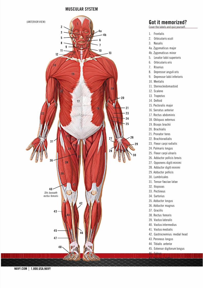
MUSCULAR SYSTEM
(ANTERIOR VIEW) Got it memorized?Cover the labels and quiz yourself.
1. Frontalis
2. Orbicularis oculi
3. Nasalis
4a. Zygomaticus major
4b. Zygomaticus minor
5. Levator labii superioris
6. Orbicularis oris
7. Risorius
8. Depressor anguli oris
9. Depressor labii inferioris
10. Mentalis
11. Sternocleidomastoid
12. Scalene
13. Trapezius
14. Deltoid15. Pectoralis major
16. Serratus anterior
17. Rectus abdominis
18. Obliquus externus
19. Biceps brachii
20. Brachialis
21. Pronator teres
22. Brachioradialis
23. Flexor carpi radialis
24. Palmaris longus
25. Flexor carpi ulnaris
26. Adductor pollicis brevis
27. Opponens digiti minimi
28. Adductor digiti minimi
29. Adductor pollicis
30. Lumbricales
31. Tensor fasciae latae
32. Iliopsoas
33. Pectineus
34. Sartorius
35. Adductor longus36. Adductor magnus
37. Gracilis
38. Rectus femoris
39. Vastus lateralis
40. Vastus intermedius
41. Vastus medialis
42. Gastrocnemius: medial head
43. Peroneus longus
44. Tibialis anterior
45. Extensor digitorum longus
46. Soleus
47. Peroneus tertius
48. Digitorum brevis
8/12/2019 Navy Medical
http://slidepdf.com/reader/full/navy-medical 4/16
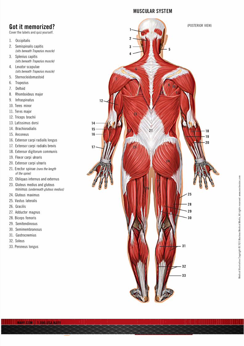
MUSCULAR SYSTEM
(POSTERIOR VIEW)Got it memorized?Cover the labels and quiz yourself.
1. Occipitalis
2. Semispinalis capitis(sits beneath Trapezius muscle)
3. Splenius capitis
(sits beneath Trapezius muscle)
4. Levator scapulae(sits beneath Trapezius muscle)
5. Sternocleidomastoid
6. Trapezius
7. Deltoid
8. Rhomboideus major
9. Infraspinatus
10. Teres minor
11. Teres major
12. Triceps brachii
13. Latissimus dorsi
14. Brachioradialis
15. Anconeus
16. Extensor carpi radialis longus
17. Extensor carpi radialis brevis
18. Extensor digitorum communis
19. Flexor carpi ulnaris
20. Extensor carpi ulnaris
21. Erector spinae (runs the length
of the spine)
22. Obliquus internus and externus
23. Gluteus medius and gluteusminimus (underneath gluteus medius)
24. Gluteus maximus
25. Vastus lateralis
26. Gracilis
27. Adductor magnus
28. Biceps femoris
29. Semitendinosus
30. Semimembranosus
31. Gastrocnemius
32. Soleus33. Peroneus longus
8/12/2019 Navy Medical
http://slidepdf.com/reader/full/navy-medical 5/16
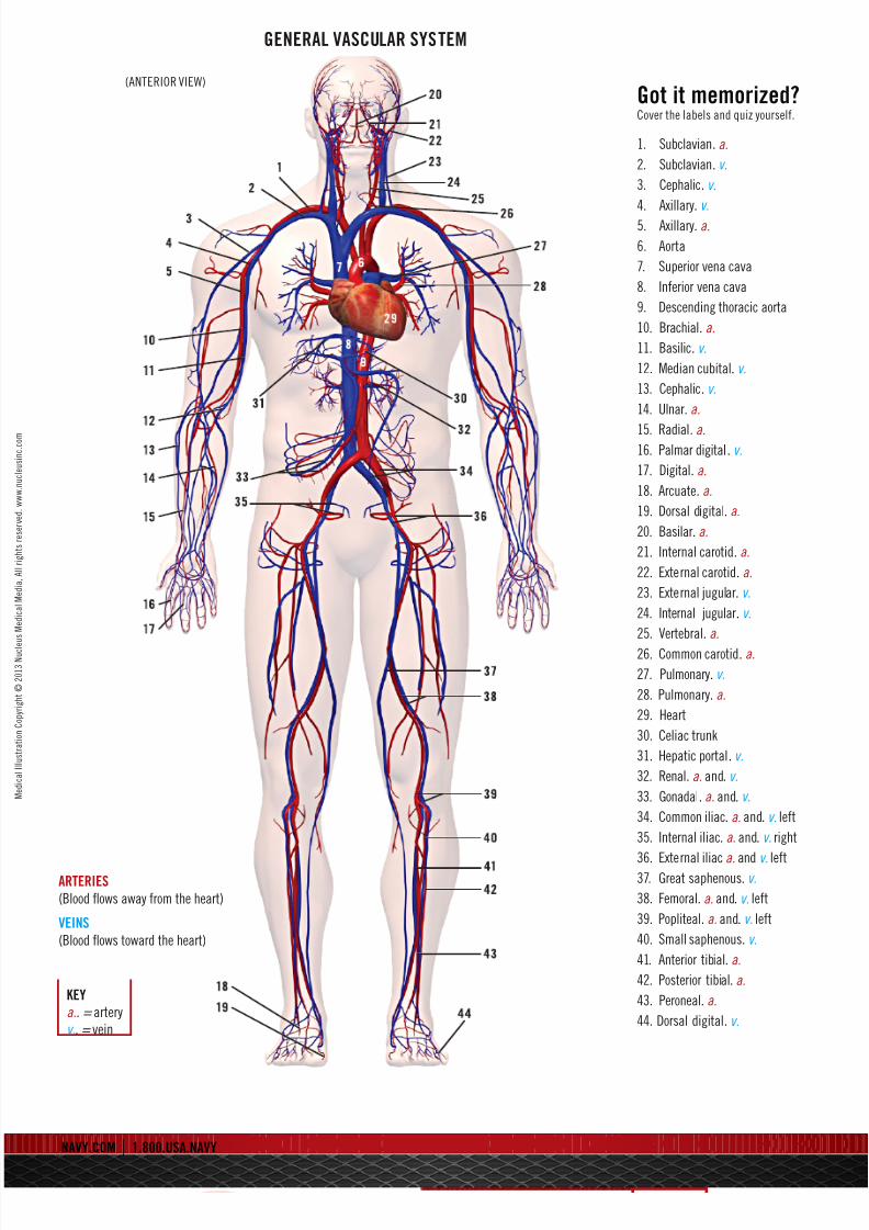
GENERAL VASCULAR SYSTEM
(ANTERIOR VIEW)
KEY
a.. = artery
v.. = vein
ARTERIES
(Blood flows away from the heart)
VEINS
(Blood flows toward the heart)
Got it memorized?Cover the labels and quiz yourself.
1. Subclavian. a.
2. Subclavian. v.
3. Cephalic. v.
4. Axillary. v.
5. Axillary. a.
6. Aorta
7. Superior vena cava
8. Inferior vena cava
9. Descending thoracic aorta
10. Brachial. a.
11. Basilic. v.
12. Median cubital. v.
13. Cephalic. v.
14. Ulnar. a.
15. Radial. a.16. Palmar digital. v.
17. Digital. a.
18. Arcuate. a.
19. Dorsal digital. a.
20. Basilar. a.
21. Internal carotid. a.
22. External carotid. a.
23. External jugular. v.
24. Internal jugular. v.
25. Vertebral. a.
26. Common carotid. a.
27. Pulmonary. v.
28. Pulmonary. a.
29. Heart
30. Celiac trunk
31. Hepatic portal. v.
32. Renal. a. and. v.
33. Gonadal. a. and. v.
34. Common iliac. a. and. v. left
35. Internal iliac. a. and. v. right
36. External iliac a. and v. left37. Great saphenous. v.
38. Femoral. a. and. v. left
39. Popliteal. a. and. v. left
40. Small saphenous. v.
41. Anterior tibial. a.
42. Posterior tibial. a.
43. Peroneal. a.
44. Dorsal digital. v.

8/12/2019 Navy Medical
http://slidepdf.com/reader/full/navy-medical 6/16
HEART
Got it memorized?Cover the labels and quiz yourself.
1. Superior vena cava
2. Ascending aorta. a.
3. Coronary right. a.
4. Subclavian right. a.
5. Common carotid left. a.
6. Subclavian left. a.
7. Aortic arch
8. Pulmonary right. v.
9. Pulmonary left. v.
10. Left circumflex. a.
11. Left anterior descending
12. Apex of the heart
13. Descending aorta. a.
14. Inferior vena cava
15. Cardiac muscle
16. Marginal branches. a.
17. Pulmonary superior/inferior. a.
A. Right atrium
B. Right ventricle
C. Left atrium
D. Left ventricle
E. Papillary muscles
F. Chordae tendineae
G. Tricuspid valveH. Mitral valve
. Pulmonary valve
. Aortic valve
KEY:..
a.. = artery
v.. = vein
8/12/2019 Navy Medical
http://slidepdf.com/reader/full/navy-medical 7/16
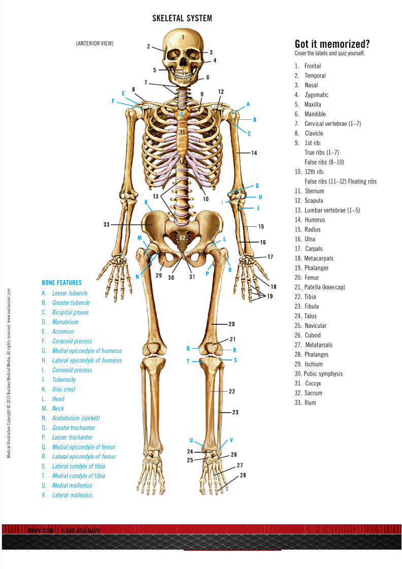
SKELETAL SYSTEM
(ANTERIOR VIEW) Got it memorized?Cover the labels and quiz yourself.
1. Frontal
2. Temporal
3. Nasal
4. Zygomatic
5. Maxilla
6. Mandible
7. Cervical vertebrae (1–7)
8. Clavicle
9. 1st rib:
True ribs (1–7)
False ribs (8–10)
10. 12th rib:
False ribs (11–12) Floating ribs
11. Sternum
12. Scapula
13. Lumbar vertebrae (1–5)
14. Humerus
15. Radius
16. Ulna
17. Carpals
18. Metacarpals
19. Phalanges
20. Femur
21. Patella (kneecap)
22. Tibia23. Fibula
24. Talus
25. Navicular
26. Cuboid
27. Metatarsals
28. Phalanges
29. Ischium
30. Pubic symphysis
31. Coccyx
32. Sacrum
33. Ilium
BONE FEATURES
A. Lesser tubercle
B. Greater tubercle
C. Bicipital groove
D. Manubrium
E. Acromion
F. Coracoid process
G. Medial epicondyl e of humerus
H. Lateral epicondyle of humerus
I. Coronoid process
J. Tuberosity
K. Iliac crest
L. Head M. Neck
N. Acetabulum (socket)
O. Greater trochanter
P. Lesser trochanter
Q. Medial epicondyle of femur
R. Lateral epicondyle of femur
S. Lateral condyle of tibia
T. Medial condyle of tibia
U. Medial malleolus
V. Lateral malleolus
8/12/2019 Navy Medical
http://slidepdf.com/reader/full/navy-medical 8/16
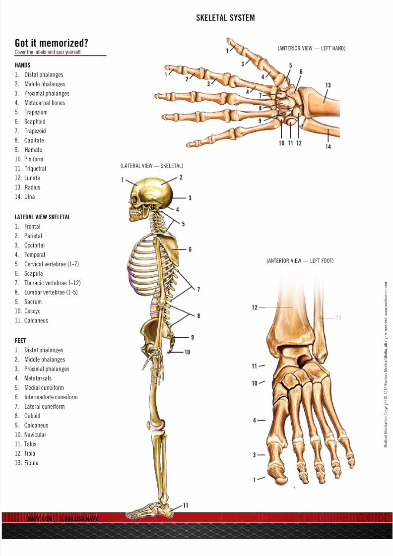
SKELETAL SYSTEM
(ANTERIOR VIEW — LEFT HAND)Got it memorized?Cover the labels and quiz yourself.
HANDS
1. Distal phalanges
2. Middle phalanges
3. Proximal phalanges
4. Metacarpal bones
5. Trapezium
6. Scaphoid
7. Trapezoid
8. Capitate
9. Hamate
10. Pisiform
11. Triquetral
12. Lunate
13. Radius
14. Ulna
LATERAL VIEW SKELETAL
1. Frontal
2. Parietal
3. Occipital
4. Temporal
5. Cervical vertebrae (1-7)
6. Scapula
7. Thoracic vertebrae 1-12)
8. Lumbar vertebrae (1-5)9. Sacrum
10. Coccyx
11. Calcaneus
FEET
1. Distal phalanges
2. Middle phalanges
3. Proximal phalanges
4. Metatarsals
5. Medial cuneiform6. Intermediate cuneiform
7. Lateral cuneiform
8. Cuboid
9. Calcaneus
10. Navicular
11. Talus
12. Tibia
13. Fibula
(LATERAL VIEW — SKELETAL)
(ANTERIOR VIEW — LEFT FOOT)

8/12/2019 Navy Medical
http://slidepdf.com/reader/full/navy-medical 9/16
SKULL AND BRAIN
(LATERAL VIEW) (ANTERIOR VIEW)
Got it memorized?Cover the labels and quiz yourself.
SKULL BONES
1. Frontal
2. Temporal
3. Parietal
4. Occipital
5. Lacrimal
6. Nasal
7. Zygomatic
8. Maxilla
9. Mandible
10. Sphenoid
11. Vomer
(LATERAL VIEW) (TOP SLICE VIEW)
BRAIN
1. Frontal lobe
2. Temporal lobe
3. Parietal lobe
4. Occipital lobe
5. Cerebellum
6. Medulla oblo
7. Corpus Callos
Frontal Lobe of the Cerebrum —
the top, front regions of each of thecerebral hemispheres. They are used
for reasoning, emotions, judgment and
voluntary movement.
Temporal Lobe of the Cerebrum —
the region at the lower side of each
cerebral hemisphere. It contains cente
of hearing and memory (located at the
sides of the head).
Parietal Lobe of the Cerebrum —
the middle lobe of each cerebral
hemisphere between the frontal andoccipital lobes. It contains important
sensory centers (located at the upper
rear of the head).
Pituitary Gland — a gland attached
to the base of the brain (located betwe
the pons and the corpus callosum) tha
secretes hormones.
Occipital Lobe of the Cerebrum —
the region at the back of each cerebral
hemisphere that contains the centers
of vision and reading ability (located
at the back of the head).
Cerebellum — the part of the brain
below the back of the cerebrum. It
regulates balance, posture, movement
and muscle coordination.
Corpus Callosum — a large bundle
of nerve fibers that connect the left an
right cerebral hemispheres. In the late
section, it looks a bit like a “C” on its s
Medulla Oblongata — the lowest sec
of the brainstem (top end of the spinal
cord). It controls automatic functionsincluding heartbeat, breathing, etc.

8/12/2019 Navy Medical
http://slidepdf.com/reader/full/navy-medical 10/16
FUNCTIONAL AREAS OF THE BRAIN
(LATERAL VIEW)
(SAGITTAL VIEW)
(SUPERIOR VIEW) (INFERIOR VIEW)
Got it memorized?Cover the labels and quiz yourself.
. Visual Area:
Sight
Image recognition
Image perception
2. Association Area:
Short-term memory
Equilibrium
Emotion
3. Motor Function Area:
Initiation of voluntary muscles
4. Broca’s Area:
Muscles of speech
5. Auditory Area:
Hearing
6. Emotional Area:Pain
Hunger
“Fight or flight” response
7. Sensory Association Area
8. Olfactory Area:
Smelling
9. Sensory Area:
Sensation from muscles and skin
0. Somatosensory Association Area:
Evaluation of weight, texture,
temperature, etc., for objectrecognition
1. Wernicke’s Area:
Written and spoken language
comprehension
2. Motor Function Area:
Eye movement and orientation
3. Higher Mental Functions:
Concentration
Planning
Judgment
Emotional expression
Creativity
Inhibitions
Functional Area of
he Cerebellum
4. Motor Functions:
Coordination of movement
Balance and equilibrium
Posture
8/12/2019 Navy Medical
http://slidepdf.com/reader/full/navy-medical 11/16
8/12/2019 Navy Medical
http://slidepdf.com/reader/full/navy-medical 12/16
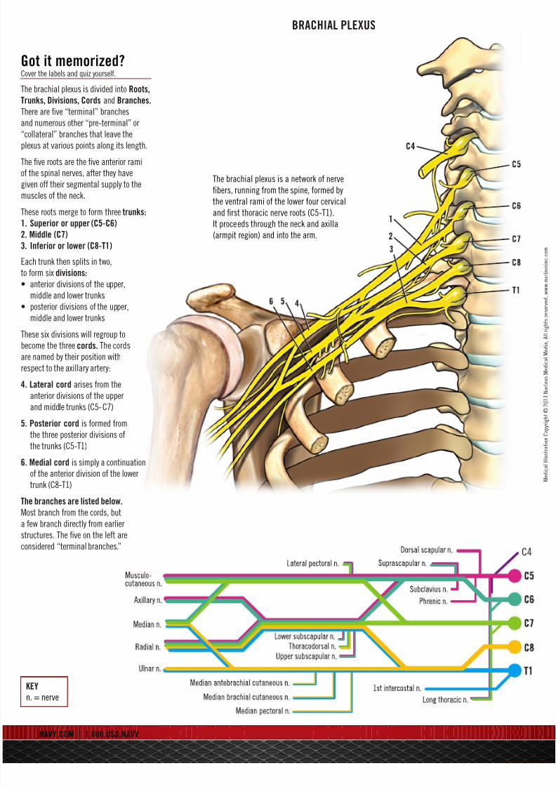
BRACHIAL PLEXUS
The brachial plexus is a network of nerve
fibers, running from the spine, formed by
the ventral rami of the lower four cervical
and first thoracic nerve roots (C5-T1).
It proceeds through the neck and axilla
(armpit region) and into the arm.
Got it memorized?Cover the labels and quiz yourself.
The brachial plexus is divided into Roots,
Trunks, Divisions, Cords and Branches.
There are five “terminal” branches
and numerous other “pre-terminal” or
“collateral” branches that leave theplexus at various points along its length.
The five roots are the five anterior rami
of the spinal nerves, after they have
given off their segmental supply to the
muscles of the neck.
These roots merge to form three trunks:
. Superior or upper (C5-C6)
2. Middle (C7)
3. Inferior or lower (C8-T1)
Each trunk then splits in two,
o form six divisions:• anterior divisions of the upper,
middle and lower trunks
• posterior divisions of the upper,
middle and lower trunks
These six divisions will regroup to
become the three cords. The cords
are named by their position with
espect to the axillary artery:
4. Lateral cord arises from the
anterior divisions of the upper
and middle trunks (C5-C7)
5. Posterior cord is formed from
the three posterior divisions of
the trunks (C5-T1)
6. Medial cord is simply a continuation
of the anterior division of the lower
trunk (C8-T1)
The branches are listed below.
Most branch from the cords, but
a few branch directly from earlier
structures. The five on the left are
considered “terminal branches.”
KEYn. = nerve
8/12/2019 Navy Medical
http://slidepdf.com/reader/full/navy-medical 13/16
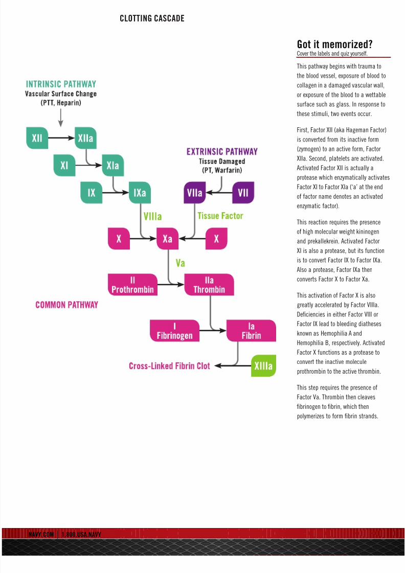
CLOTTING CASCADE
Got it memorized?Cover the labels and quiz yourself.
This pathway begins with trauma to
the blood vessel, exposure of blood to
collagen in a damaged vascular wall
or exposure of the blood to a wettable
surface such as glass. In response to
these stimuli, two events occur.
First, Factor XII (aka Hageman Factor
is converted from its inactive form
(zymogen) to an active form, Factor
XIIa. Second, platelets are activated.
Activated Factor XII is actually a
protease which enzymatically activat
Factor XI to Factor XIa (‘a’ at the end
of factor name denotes an activated
enzymatic factor).
This reaction requires the presence
of high molecular weight kininogen
and prekallekrein. Activated Factor
XI is also a protease, but its function
is to convert Factor IX to Factor IXa.
Also a protease, Factor IXa then
converts Factor X to Factor Xa.
This activation of Factor X is also
greatly accelerated by Factor VIIIa.
Deficiencies in either Factor VIII or
Factor IX lead to bleeding diatheses
known as Hemophilia A and
Hemophilia B, respectively. Activated
Factor X functions as a protease to
convert the inactive molecule
prothrombin to the active thrombin.
This step requires the presence of
Factor Va. Thrombin then cleaves
fibrinogen to fibrin, which then
polymerizes to form fibrin strands.
8/12/2019 Navy Medical
http://slidepdf.com/reader/full/navy-medical 14/16
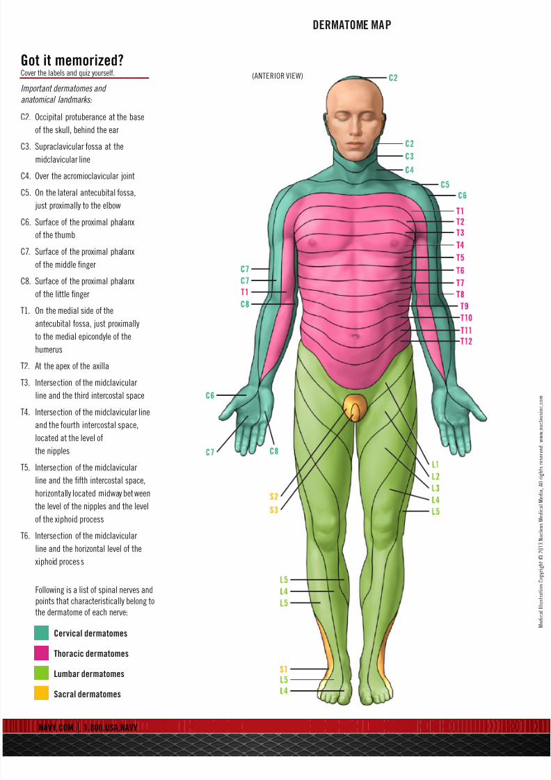
DERMATOME MAP
(ANTERIOR VIEW)
Got it memorized?Cover the labels and quiz yourself.
mportant dermatomes and
anatomical landmarks:
C2. Occipital protuberance at the base
of the skull, behind the ear
C3. Supraclavicular fossa at the
midclavicular line
C4. Over the acromioclavicular joint
C5. On the lateral antecubital fossa,
just proximally to the elbow
C6. Surface of the proximal phalanx
of the thumb
C7. Surface of the proximal phalanx
of the middle finger
C8. Surface of the proximal phalanx
of the little finger
T1. On the medial side of the
antecubital fossa, just proximally
to the medial epicondyle of the
humerus
T2. At the apex of the axilla
T3. Intersection of the midclavicular
line and the third intercostal space
T4. Intersection of the midclavicular line
and the fourth intercostal space,
located at the level of
the nipples
T5. Intersection of the midclavicular
line and the fifth intercostal space,
horizontally located midway between
the level of the nipples and the level
of the xiphoid process
T6. Intersection of the midclavicular
line and the horizontal level of the
xiphoid process
Following is a list of spinal nerves and
points that characteristically belong to
the dermatome of each nerve:
Cervical dermatomes
Thoracic dermatomes
Lumbar dermatomes
Sacral dermatomes
8/12/2019 Navy Medical
http://slidepdf.com/reader/full/navy-medical 15/16
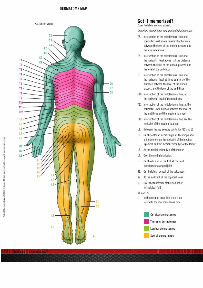
DERMATOME MAP
(POSTERIOR VIEW)
NAVY.COM | 1.800.USA.NAVY
Got it memorized?Cover the labels and quiz yourself.
Important dermatomes and anatomical landmarks:
T7. Intersection of the midclavicular line and
horizontal level at one quarter the distance
between the level of the xiphoid process and
the level umbilicus
T8. Intersection of the midclavicular line and
the horizontal level at one half the distance
between the level of the xiphoid process and
the level of the umbilicus
T9. Intersection of the midclavicular line and
the horizontal level at three quarters of the
distance between the level of the xiphoid
process and the level of the umbilicus
T10. Intersection of the midclavicular line, at
the horizontal level of the umbilicus
T11. Intersection of the midclavicular line, at the
horizontal level midway between the level of
the umbilicus and the inguinal ligament
T12. Intersection of the midclavicular line and the
midpoint of the inguinal ligament
L1. Between the key sensory points for T12 and L
L2. On the anterior medial thigh, at the midpoint
a line connecting the midpoint of the inguinal
ligament and the medial epicondyle of the fem
L3. At the medial epicondyle of the femurL4. Over the medial malleolus
L5. On the dorsum of the foot at the third
metatarsophalangeal joint
S1. On the lateral aspect of the calcaneus
S2. At the midpoint of the popliteal fossa
S3. Over the tuberosity of the ischium or
infragluteal fold
S4 and S5.
In the perianal area, less than 1 cm
lateral to the mucocutaneous zone
Cervical dermatomes
Thoracic dermatomes
Lumbar dermatomes
Sacral dermatomes

8/12/2019 Navy Medical
http://slidepdf.com/reader/full/navy-medical 16/16
The physician you want to be – and the specialty you want to practice.
From preventive care to emergency treatment to surgery, you’ll find Navy Medicine at the forefront.
Pioneering advances in the field. Providing the opportunity to practice in any of the following areas:
Aerospace Medicine
Anesthesiology
Dermatology
Emergency Medicine
Family Medicine
Fleet Marine Corps Medicine
Geriatrics
Internal Medicine*
Neonatology
Neurology
Nuclear Medicine
Obstetrics/Gynecology
Occupational Medicine
Ophthalmology*
Osteopathic Medicine
Otolaryngology
Pain Management
Pathology*
Pediatrics*
Physical Medicine
Plastic and Reconstructive Surgery
Preventive Medicine
Psychiatry*
Radiology*
Sports Medicine
Surface Medicine
Surgery*
Transfusion Medicine
Tropical Medicine
Undersea/Diving Medicine
Urology
*Additional subspecialtiesmay be considered.
Find out more about Navy Medicine and hear real stories
from other Navy Health Care professionals, like Dr. Nassiri,
at navy.com/careers/healthcare