NATURE REVIEWS | | ADVANCE ONLINE PUBLICATION · PDF filestudying the mechanism of action ......
19
Although there have been many notable drug development achievements in recent years, several disease areas such as neuro- degeneration and aggressive cancers remain largely intractable. For example, substantial investment in the development of novel therapeutics for Alzheimer disease, Parkinson disease and motor neuron diseases has largely failed 1–3 . This failure can be attributed, in part, to our limited understanding of the targets that may prevent or repair neuronal damage, and to a lack of robust disease-relevant preclinical models 1,4 . In the field of cancer, important progress has been made in the discovery of new drugs, including those based on target-directed precision medicine strategies 5 . However, for aggressive cancers, such as glioma, pancreatic, oesophageal and several lung cancers, many promising drug candidates developed from standard cell-line screens Finally, a substantial proportion of clinical trial failures for novel medicines overall are due to safety issues such as cardiotoxicity and hepatotoxicity 8 , and serious toxicity issues are often discovered only after clinical development has been completed 9,10 . Thus, more predictive toxicology models would contribute substantially towards more successful clinical translation and improved patient care. Despite advances in target- and cell-based screening technologies, the majority of drug discovery projects remain dependent on cell culture systems that were developed several decades ago, incorporating immortalized cell lines; many consider the use of such assay systems to be questionable owing to their poor disease relevance. Although many traditional models provide valuable tools for studying the mechanism of action (MOA) of drugs and have helped identify successful drug candidates in the past, it is our opinion that the widespread use of contemporary cell culture assay systems must be revisited and that current efforts should be directed towards the development of new models, new assay formats and innovative screening technologies that better recapitulate in vivo physiology. In this article, after briefly summarizing the limitations of traditional cell-based models of disease, we discuss how emerging developments in (patient-derived) ex‑vivo cultures, induced pluripotent stem cell (iPSC) technology, three-dimensional (3D) co-culture and organotypic systems, complemented by advances in single-cell imaging, microfluidics and gene editing technologies, are well positioned to advance preclinical disease modelling and drug screening across challenging disease areas. We outline a set of principles for defining ‘disease-relevant assays’ (TABLE 1) and highlight methodological and analytical gaps, fundamental challenges and new opportunities to exploit and combine more advanced in vitro models with emerging technologies. We conclude by discussing the need to further evolve translational funding schemes and precompetitive research consortia to support the future development of new preclinical models and assay-screening technologies that provide more robust target validation and greater clinical predictivity. and in vivo xenograft models did not show clinical efficacy. The poor clinical translation can largely be attributed to the failure of these models to recapitulate key patho- physiological features of the human disease, including complex inter- and intratumour heterogeneity, poor drug penetration through tissue, host-stroma–tumour cell interactions, and the cancer stem cell niche, all of which may have profound effects on the therapeutic response in vivo. In addition, given the periodic emergence of new and old infections 6 , and the pressing challenges from antimicrobial resistance and pandemic threats such as Ebola and Zika, modern, disease-relevant, cell-based phenotypic assay methodologies may represent valuable assets for improving our knowledge of the dynamics of host– pathogen interactions in their natural environment 7 and the development of new therapies (BOX 1). OPINION Screening out irrelevant cell-based models of disease Peter Horvath, Nathalie Aulner, Marc Bickle, Anthony M. Davies, Elaine Del Nery, Daniel Ebner, Maria C. Montoya, Päivi Östling, Vilja Pietiäinen, Leo S. Price, Spencer L. Shorte, Gerardo Turcatti, Carina von Schantz and Neil O. Carragher Abstract | The common and persistent failures to translate promising preclinical drug candidates into clinical success highlight the limited effectiveness of disease models currently used in drug discovery. An apparent reluctance to explore and adopt alternative cell- and tissue-based model systems, coupled with a detachment from clinical practice during assay validation, contributes to ineffective translational research. To help address these issues and stimulate debate, here we propose a set of principles to facilitate the definition and development of disease-relevant assays, and we discuss new opportunities for exploiting the latest advances in cell-based assay technologies in drug discovery, including induced pluripotent stem cells, three-dimensional (3D) co-culture and organ-on-a-chip systems, complemented by advances in single-cell imaging and gene editing technologies. Funding to support precompetitive, multidisciplinary collaborations to develop novel preclinical models and cell-based screening technologies could have a key role in improving their clinical relevance, and ultimately increase clinical success rates. A GUIDE TO DRUG DISCOVERY NATURE REVIEWS | DRUG DISCOVERY ADVANCE ONLINE PUBLICATION | 1 PERSPECTIVES ©2016MacmillanPublishersLimited,partofSpringerNature.Allrightsreserved.
Transcript of NATURE REVIEWS | | ADVANCE ONLINE PUBLICATION · PDF filestudying the mechanism of action ......
Although there have been many notable drug development achievements in recent years, several disease areas such as neurodegeneration and aggressive cancers remain largely intractable. For example, substantial investment in the development of novel therapeutics for Alzheimer disease, Parkinson disease and motor neuron diseases has largely failed1–3. This failure can be attributed, in part, to our limited understanding of the targets that may prevent or repair neuronal damage, and to a lack of robust diseaserelevant preclinical models1,4.
In the field of cancer, important progress has been made in the discovery of new drugs, including those based on targetdirected precision medicine strategies5. However, for aggressive cancers, such as glioma, pancreatic, oesophageal and several lung cancers, many promising drug candidates developed from standard cellline screens
Finally, a substantial proportion of clinical trial failures for novel medicines overall are due to safety issues such as cardiotoxicity and hepatotoxicity8, and serious toxicity issues are often discovered only after clinical development has been completed9,10. Thus, more predictive toxicology models would contribute substantially towards more successful clinical translation and improved patient care.
Despite advances in target and cellbased screening technologies, the majority of drug discovery projects remain dependent on cell culture systems that were developed several decades ago, incorporating immortalized cell lines; many consider the use of such assay systems to be questionable owing to their poor disease relevance. Although many traditional models provide valuable tools for studying the mechanism of action (MOA) of drugs and have helped identify successful drug candidates in the past, it is our opinion that the widespread use of contemporary cell culture assay systems must be revisited and that current efforts should be directed towards the development of new models, new assay formats and innovative screening technologies that better recapitulate in vivo physiology.
In this article, after briefly summarizing the limitations of traditional cellbased models of disease, we discuss how emerging developments in (patientderived) ex‑vivo cultures, induced pluripotent stem cell (iPSC) technology, threedimensional (3D) coculture and organotypic systems, complemented by advances in singlecell imaging, microfluidics and gene editing technologies, are well positioned to advance preclinical disease modelling and drug screening across challenging disease areas. We outline a set of principles for defining ‘diseaserelevant assays’ (TABLE 1) and highlight methodological and analytical gaps, fundamental challenges and new opportunities to exploit and combine more advanced in vitro models with emerging technologies. We conclude by discussing the need to further evolve translational funding schemes and precompetitive research consortia to support the future development of new preclinical models and assayscreening technologies that provide more robust target validation and greater clinical predictivity.
and in vivo xenograft models did not show clinical efficacy. The poor clinical translation can largely be attributed to the failure of these models to recapitulate key pathophysiological features of the human disease, including complex inter and intratumour heterogeneity, poor drug penetration through tissue, hoststroma–tumour cell interactions, and the cancer stem cell niche, all of which may have profound effects on the therapeutic response in vivo.
In addition, given the periodic emergence of new and old infections6, and the pressing challenges from antimicrobial resistance and pandemic threats such as Ebola and Zika, modern, diseaserelevant, cellbased phenotypic assay methodologies may represent valuable assets for improving our knowledge of the dynamics of host–pathogen interactions in their natural environment7 and the development of new therapies (BOX 1).
O P I N I O N
Screening out irrelevant cell-based models of diseasePeter Horvath, Nathalie Aulner, Marc Bickle, Anthony M. Davies, Elaine Del Nery, Daniel Ebner, Maria C. Montoya, Päivi Östling, Vilja Pietiäinen, Leo S. Price, Spencer L. Shorte, Gerardo Turcatti, Carina von Schantz and Neil O. Carragher
Abstract | The common and persistent failures to translate promising preclinical drug candidates into clinical success highlight the limited effectiveness of disease models currently used in drug discovery. An apparent reluctance to explore and adopt alternative cell- and tissue-based model systems, coupled with a detachment from clinical practice during assay validation, contributes to ineffective translational research. To help address these issues and stimulate debate, here we propose a set of principles to facilitate the definition and development of disease-relevant assays, and we discuss new opportunities for exploiting the latest advances in cell-based assay technologies in drug discovery, including induced pluripotent stem cells, three-dimensional (3D) co-culture and organ-on-a-chip systems, complemented by advances in single-cell imaging and gene editing technologies. Funding to support precompetitive, multidisciplinary collaborations to develop novel preclinical models and cell-based screening technologies could have a key role in improving their clinical relevance, and ultimately increase clinical success rates.
A G U I D E TO D R U G D I S C OV E RY
NATURE REVIEWS | DRUG DISCOVERY ADVANCE ONLINE PUBLICATION | 1
PERSPECTIVES
© 2016
Macmillan
Publishers
Limited,
part
of
Springer
Nature.
All
rights
reserved.

Limitations of traditional disease modelsTraditional cell culture methods typically rely on cancer cells or immortalized cells grown within artificial environments, on nonphysiological substrates such as functionalized plastic and glass. Although these methods have facilitated the discovery of many basic biological processes, they often fail to provide an adequate platform for drug discovery owing to their inadequate representation of key physiological characteristics. These problems can be broadly categorized into the following limitations.
Limitations due to cells. Most cellbased assay screens have traditionally been performed using transformed or immortalized cell lines. These have been cultured for many generations, resulting in a substantial drift in their genetic, epigenetic and physiological characteristics, which means they are not a good model of primary tissue cells11,12. The gross genetic and epigenetic abnormalities (characterized by multiple genetic rearrangements and amplified gene copy numbers) associated with longterm culture confound pharmacogenomic and functional genomic studies. Genetic adaptation resulting from longterm in vitro cultures also contributes to heterogeneity in cultures of the same cell line between passages, batches and laboratories.
Limitations due to culture conditions. The media most commonly used for cell culture are designed for fast cell growth, incorporating large concentrations of fetal serum and nutrients, which may promote dedifferentiation of primary cell types into more embryonic or fetallike phenotypes13. With the development of primary cell and differentiated stem cell cultures, the use of high glucose and growth factor media has been eschewed for defined culture media to promote cellular identity rather than rapid growth. Cells are often grown in standard incubators under high oxygen partial pressure (approximately 20%), which does not represent the steadystate conditions of human organs and tissues (fluctuating between 1% in the dermis, and 14% in arterial blood)14–17. Such conditions poorly recapitulate the distinct micro environments that define normal and diseased tissue phenotypes. This has a profound impact on cell metabolism, reactive oxygen species (ROS) production, mitochondrial functions and, ultimately, the differentiation and function of cells18,19. Additionally,
Box 1 | Development of improved models of infectious diseases
Pathogen biology and infectious disease operate on multiple cellular and tissue scales, wherein the biology of the infectious microorganism and its target cell depend on the contextual interplay of pathogen development stages with distinct host tissues, organs and the immune response. One important example of this complexity is the lifecycle of Plasmodium falciparum, the parasite that causes malaria in humans. There are several possible strategies to screen for lead compounds using phenotypic approaches, including screening at the human liver-stage, blood cell-stage or the insect-stage of the parasite life cycle. The use of cell-based phenotypic assays is offering promise in the identification of novel therapeutic classes that target multiple stages of parasite development178.
However, the nature of the targeted cells and the virulence of an infectious agent are intimately linked, and can sometimes only be appropriately recapitulated by very specific host–pathogen combinations. Along these lines, a series of studies to identify new chemical entities that are active against Leishmania subspecies, the causal agents of human leishmaniasis, clearly demonstrated that developing an assay targeting the insect promastigote stage of the parasite does not yield viable lead compounds. Instead, a much longer and more sophisticated methodology is required for a primary macrophage assay supporting infection-competent (replicative) parasites, which reduces screening throughput but greatly enhances the quality of results and identification of viable lead compounds179–181.
A major concern in the field of infectious disease modelling is the use of perpetually cultured laboratory strains of infectious disease organisms despite their poor resemblance to ‘real-world’ pathogens. Bacterial growth conditions in vitro differ fundamentally from the conditions found in natural ecosystems, including infection sites. Indeed, as a result of the high degree of plasticity of their genome, bacterial strains adapt quickly to the optimized laboratory mono-culture conditions and, therefore, rapidly lose key pathophysiological characteristics182. In the late nineteenth century, Louis Pasteur had already recognized that laboratory adaptation of bacteria is associated with attenuation of virulence towards the host species, and this idea was exploited by his colleagues Calmette and Guérin, leading to the development of the BCG (bacille Calmette–Guérin) vaccine in 1921 (REF. 183).
It is now well established that bacteria do not exist in isolation but rather live as communities, behaving collectively to adapt to new host environments and modes of growth184. Quorum-sensing (QS) — that is, cell-to-cell communication by the production and release of autoinducers in the environment — has a key role in Pseudomonas aeruginosa pathogenicity. The large variation of expression of QS genes observed upon culture in different environments emphasizes the importance of clearly identifying and mimicking the actual habitat that bacteria encounter in vivo when developing an assay to design new therapies185. Along these lines, most bacterial chronic infectious diseases are associated with the formation of polymicrobial biofilms. Biofilms protect the bacteria from the host innate- and adaptive immune response and represent an ideal setting for horizontal gene transfer (HGT), thus creating new virulent strains and resistance by creating a communal distributed ‘supra-genome’186.
A way to develop more physiological infectious disease models is to mimic as closely as possible the in vivo ecosystems that the pathogens might encounter in an infected host, to recreate the microenvironments required for the virulence and eventually to identify drugs that might boost the host defence mechanisms. Many pathogens that infect humans have been studied in model animals such as Drosophila melanogaster, Caenorhabditis elegans, zebrafish or mice; unfortunately, not all of them are easily amenable to higher-throughput drug screening, nor do they properly model the human response to pathogen infection. Nonetheless, in an elegant study, Kim and co-workers describe how host-cell autophagy activated by antibiotics is required for an effective antimycobacterial drug response through conserved mechanisms between fruitflies and mammals187. Although small model organisms hold much promise for infectious disease drug discovery, they still have many drawbacks such as their temperature, discrepancies between the anaerobic nature of the human intestine and non-conserved host targets across species. These shortcomings might be overcome using synthetic microtissues such as the human-gut-on-a-chip188 or small-airway-on-a-chip microfluidic devices189.
Many pathogens require high biosafety level laboratory confinement (BSL3 and above), which presents several challenges to developing assays under these conditions. The continued development of automated liquid handling and automated microscopy platforms, which enable remote control, including image acquisition and analysis, help support assay development and screening under high-level biosafety restrictions. The lack of an effective cure or preventive measures against emerging antibiotic-resistant bacteria and re-emerging viral pandemics (for example, Ebola, Marburg and Zika) calls for renewed efforts to develop innovative technologies and methodologies for drug screening in the infectious disease area. Comparative phenotypic screening across microbial resistant subspecies combined with relevant host systems may guide drug discovery towards novel therapeutic classes targeting infectious disease resistance mechanisms and new host-oriented therapies190,191.
P E R S P E C T I V E S
2 | ADVANCE ONLINE PUBLICATION www.nature.com/nrd
© 2016
Macmillan
Publishers
Limited,
part
of
Springer
Nature.
All
rights
reserved. ©
2016
Macmillan
Publishers
Limited,
part
of
Springer
Nature.
All
rights
reserved.

conventional tissue culture systems do not readily permit the formation of shortrange gradients of nutrients, hormones and oxygen that are often experienced by cells, depending on the distance to the nearest blood vessel. The liver is a wellknown example of this, with gradients in the lobules between the central vein and the portal artery leading to zonation20. This can be mimicked with the adoption of microfluidic systems that deliver nutrients, dissolve gases and remove waste products21.
Limitations due to lack of appropriate cell culture substrates and bioengineering tools. The twodimensional (2D) planar substrates on which cells are typically grown are stiff, demonstrating high (gigapascal) tensile strength and mechanical resistance to deformation, unlike most substrates found in the human body (which are on the millipascal to kilopascal scales), with the exception of bone and cartilage22. Hence, the plastic or glass used in cell culture may not accurately represent the normal in vivo mechanical environment23,24. For example, in the case of liver culture, most differentiation protocols require the use of sandwich cultures, where the cells are grown between layers of either collagen or other extracellular matrix (ECM) proteins. The mechanical properties of these supporting matrices are generally not well characterized, despite the fact that such properties are known to have an impact on cellular function and differentiation in tissues25,26. In these types of experiments, where a minimal quantity of deposed hydrogel is used, it is likely that the cells would encounter a stiff environment, which the liver would normally only encounter during fibrosis or cirrhosis. Thus, toxicology assays are typically carried out under pathological rather than healthy liver conditions. Microfluidic devices, which utilize mechanical actuation systems to recapitulate mechanical forces or generate the shear forces that tissues experience in living bodies, are beginning to be used; however, they will require further development and refinement if they are to be used for more general screening applications27.
A further challenge evident in tissue modelling within current in vitro assays is the absence of more physiologically relevant ECM. For example, the popular use of Matrigel and collagen type I as an ECM substrate in hepatocyte cultures does not represent the ECM proteins predominantly found in the liver28. Many pathologies are associated with changes in ECM production
recapitulate the full complexity of human disease, below and in FIG. 1 we outline the latest developments in cellbased models and assay technologies that are beginning to address the limitations of traditional and contemporary in vitro assays.
Enhancements in disease modellingPrimary and patient-derived cell models. The adaptation of patientderived primary cell samples, as well as fresh human tissue samples, for ex vivo and in vitro translational research applications aims to overcome many of the disadvantages of using transformed cell lines for drug discovery33. These samples also offer a more clinically relevant model for testing novel gene and cellbased therapies. However, the lack of culture systems possessing the robustness, scalability and flexibility needed by companies has hampered the adoption of in vitro primary cellbased research tools, including patientderived cell models, at the earliest stages of drug development.
In cancer, highly selective drugs targeted at genetically defined clinical subtypes are needed to support a more patientcentric approach to drug development34,35. Potential drugs have been tested in vitro and ex vivo against wellcharacterized patientderived primary cancer subtypes for various cancers, but often without a direct impact on treatment36–38. In leukemias, however, where the ex vivo material (for example, suspension cells) is more readily available for drug testing than in solid tumours, patientderived samples have recently been utilized for potential drug repositioning39,40 and combined with molecular profiling to identify clinically actionable drugs for personalized acute myeloid leukaemia (AML) therapy40. Although primary leukaemic cells can be used without further expansion for ex vivo drug testing33,41, the drug responses may vary depending on the cell culture assay conditions. Importantly, several studies highlight the importance of the interaction of leukaemic cells with the bone marrow stromal microenvironment, which can be partly mimicked using cocultures of leukaemic cells with human bone marrowderived mesenchymal stem cells42,43.
The extension of patientcentric primary ex vivo drug profiling to higher throughput applications and primary cells derived from solid tumours or normal tissue presents several challenges44. The development of coculture protocols45 has enabled a relatively rapid production and scaleup of high amounts of conditionally
that have a considerable impact on cell and tissue function. Thus, recapitulating both physiological and pathophysiological ECM composition and structure is an important consideration for in vitro cellular models.
Limitations due to lack of appropriate co-culture methods. Cellculture screening assays traditionally use a single cell type, whereas cells in vivo are either in direct contact or communicate over a long range with many different cell types. As most biological processes and pathologies involve the interaction of multiple cell types, these should ideally be incorporated into in vitro cellular assays whenever possible. For example, most toxicology assays use only hepatocytes, but although 80% of the liver volume consists of hepatocytes (60% of the cells), other important cell types within the liver include stellate cells, resident macrophages (Kupffer cells), sinusoidal endothelial cells and some nonparenchymal cells. Both stellate cells and Kupffer cells are known to be important for some compound toxicities and should therefore be incorporated into in vitro toxicology assays29,30. Neurodegeneration, wherein both astrocytes and glial cells are responsible for protecting neurons but are also known to cause neural death, provides a compelling case for the use of mixed cell cultures of distinct cell types31. Further development of coculture methods, which incorporate disease cells with relevant immune subcompartments, is also urgently needed to help better understand and address the role of the host immune system in the patho genesis and therapeutic outcomes of many diseases32. These considerations are of particular importance for pathogen biology and infectious diseases, which operate at multiple cellular and tissue levels (BOX 1).
Tackling the limitations. Efforts to address the limitations of traditional preclinical assays by employing new models face various imposing challenges, including: how to mimic the microenvironment and heterogeneity of normal and disease tissue; how to take into account the environmental and genetic or epigenetic factors governing disease aetiology and therapeutic outcomes; how to understand the effect of drugs upon the whole physiological entity, for example, across multiple cell types and organs of the human body; and how to interpret the role of the host immune system in the pathogenesis of a particular disease. Although it is clear that no single preclinical model or screening assay will faithfully
P E R S P E C T I V E S
NATURE REVIEWS | DRUG DISCOVERY ADVANCE ONLINE PUBLICATION | 3
© 2016
Macmillan
Publishers
Limited,
part
of
Springer
Nature.
All
rights
reserved. ©
2016
Macmillan
Publishers
Limited,
part
of
Springer
Nature.
All
rights
reserved.

Table 1 | Defining principles of disease-relevant assays
No. Principle Activity Justification Disease-specific considerations
1 Define the translational research objective of the assay
Ensure that assay selection is linked to a definitive clinical question or actual clinical scenario
Accurately inform disease positioning and enhance successful translation of cell-based screening programmes
Generic
2 Adopt the human physiology assay checklist
Acknowledge assay limitations by adopting a human physiology assay checklist (see main text), which would enable unbiased evaluation of which assay conditions accurately recapitulate human tissue physiology and pathophysiology and which do not
Supporting appropriate assay selection and interpretation of assay data to guide subsequent preclinical studies and further assay development to enhance translational success
Generic
3 Retrospectively analyse and measure the predictive value of each assay
Recapitulate poor drug response or known clinical resistance mechanisms through retrospective analysis of approved and failed drugs to determine the positive and negative predictive value (PPV and NPV, respectively) of each assay. Retrospective studies support reverse engineering of preclinical assays to improve PPV and NPV
Validation of assay predictivity to enhance clinical translation
Generic
4 Ensure the tissue context is appropriate
Accurately represent the appropriate tissue context of normal or diseased tissue through the development and detailed characterization of patient-derived, primary human cell and induced pluripotent stem cell (iPSC) differentiation protocols
Generate cell-based screening assays with appropriate tissue context and endogenous pathway network biology across disease areas to support target discovery, target validation and drug mechanism-of-action studies under the most relevant conditions
Generic
5 Ensure the genomic context is appropriate
Ensure the genomic characteristics accurately represent patient populations and disease subtypes
Design bespoke suites of cell-based screens representing disease subtypes or patient populations to inform disease positioning of assay screening hits and new drug targets
• Generic• Cancer subtypes• Hereditary and
spontaneous neurological disorders
• Personalized cardiovascular disease
6 Capture heterogeneity within cell populations
Define the heterogeneity of cell-based assays, ensuring that a heterogeneous phenotypic response within individual cell subpopulations is measured
Embrace disease heterogeneity by analysing the drug’s mechanism of action and target activity at a single-cell level and across subpopulations in complex assay formats to inform future preclinical and clinical development
• Generic• Cancer• Inflammation and
immune disorders• Regenerative
medicine
7 Use mixed cell culture models and relevant host biology
Use mixed cell culture models that represent the multicellular composition of normal and diseased tissue. Incorporate appropriate host cell biology relevant to the disease
Develop more relevant multicellular screening assays to support novel target discovery and drug combinations exploiting both disease and host cell biology
• Generic• Cancer• Inflammation and
immune disorders• Infectious disease• Liver toxicity and
disease• Cardiovascular disease
8 Use defined cell growth media
Use defined cell growth media that represent steady-state levels of human tissue nutrients under normal and pathophysiological conditions
Accurately model the environmental conditions of normal and diseased tissue to enhance the translation and prediction of in vivo efficacy or toxicity
Generic
9 Ensure substrate tension and mechanical forces are appropriate
Ensure that substrate tensions and mechanical forces are representative of human tissues under normal and pathophysiological conditions
Accurately model the environmental conditions of normal and diseased tissue architecture and mechanotrans-duction to enhance the translation and prediction of in vivo efficacy or toxicity
• Generic• Cardiovascular disease• Musculoskeletal
disorders• Fibrosis• Cancer
10 Ensure the atmospheric conditions are appropriate
Ensure that the atmospheric conditions of physiological and pathophysiological gaseous tensions, including oxygen pressure and pH conditions, are tailored to specific tissue types
Accurately model environmental conditions of normal and diseased tissue, including different physiological levels of normoxia and pathophysiological levels of hypoxia within in vivo tissues to enhance the translation and prediction of in vivo efficacy or toxicity
• Skin diseases• Cardiovascular disease• Lung and respiratory
diseases• Liver zonation• Ischaemia• Cancer hypoxia
P E R S P E C T I V E S
4 | ADVANCE ONLINE PUBLICATION www.nature.com/nrd
© 2016
Macmillan
Publishers
Limited,
part
of
Springer
Nature.
All
rights
reserved. ©
2016
Macmillan
Publishers
Limited,
part
of
Springer
Nature.
All
rights
reserved.

reprogrammed cells from surgical and accessible biopsy specimens, from both healthy and tumorigenic tissues such as the lung, breast, prostate, pancreas, colon and kidney44,46. In vitro cell culture conditions modify cells over time, and may even lead to loss of expression markers of the original sample and enrichment of specific cell populations. It is therefore essential to ensure that these cells represent the original tissue and genomic background of the individuals from whom they were derived by extensive genotypic and singlecell phenotypic characterization. Living organoid biobanks for solid tumours can complement cell line and xenograftbased drug studies by providing an improved model for complex tissue architecture. This was demonstrated in a recent
patientderived primary samples requires close collaboration between researchers, clinics and biobanks to find representative samples combined with relevant clinical data, and to establish standardized sample handling procedures for sensitive live tissues and cells. The Finnish Haematology Registry Biobank (FHRB) provides an exemplar of an operative biobank, functioning as a valuable source of patient material for precision medicine approaches in leukaemia40,48. The exploitation of biobanks to support drug testing of ex vivo patient cells collected from across both large and smaller patient cohorts can help to prioritize and derisk drug candidates for largerscale clinical testing, to support patientstratified medicine strategies and to systematically identify novel drugrepositioning opportunities48.
proofofconcept study, in which the living organoids of 20 patients with colorectal cancer, sharing identical gene expression profiles and genetics with the corresponding original tumours, were screened against 83 compounds; the findings revealed good reproducibility and correlation with individual oncogenic mutations47.
Overcoming the challenges related to the expansion of primary patientderived ex vivo cultures for higherthroughput screening across distinct patient cohorts will require access to numerous representative patient samples for simultaneous testing, scaleup of limited primary cell material and effective integration of drug sensitivity phenotypic data with the molecular characterization and clinical data associated with each patient sample. Access to highquality
Table 1 (cont.) | Defining principles of disease-relevant assays
No. Principle Activity Justification Disease-specific considerations
11 Ensure the extracellular matrix composition is relevant
Extracellular matrix compositions should be relevant and represent the physiological and pathophysiological composition and architecture
Accurately model extracellular environments of normal and diseased tissue to ensure appropriate cell and tissue architecture, cell differentiation and cell function in order to enhance clinical translation of in vitro assay screens
Generic
12 Ensure clinically equivalent dosing is used
Mimic drug uptake and retention within normal and diseased tissue by simulating tissue perfusion. Incorporate short-term dosing and drug wash-out protocols that predict in vivo pharmacokinetic properties of specific drug candidates or predictions of common drug-like properties
Improved prediction of in vivo and clinical activity, supporting more accurate scheduling and dosing of drugs and drug combinations
Generic
13 Simulate systemic multi-tissue effects
Design organ-on-chip or multi-tissue models that mimic in vivo compound metabolism, paracrine signalling between tissue and systemic tissue effects, and associated co-morbidities
Enhanced modelling of complex human physiology and whole-organism systems to predict clinical efficacy and toxicity across multiple organs and tissues
• Generic• Metabolic
14 Incorporate appropriately aged cells to model human disease
Utilize primary cells from appropriately aged donors or adapt cell differentiation protocols to generate appropriately aged cell models
Ensure the assay represents the expected age of the patient (for example, embryonic, paediatric, adult and late-onset degenerative disease)
• Generic• Developmental
malformations• Neurodegeneration• Cancer
15 Model distinct stages of the disease life cycle
Design models of precursory, early-stage, mid-stage, late-stage and dormant disease
Ensure design of assays that appropriately guide targets, hits and candidate drug therapies for prevention, cure, stabilization and palliative use
• Cancer: premalignant tumours
• Arthritis• Neurodegeneration:
early-onset and late-stage disease
16 Incorporate appropriate disease-causing ‘perturbagens’
Accurately model the environmental and physiological toxins that are responsible for disease initiation and progression. Ensure relevant levels and types of toxin, as well as exposure duration, are incorporated into cell models
Support the development of assays representing disease progression models that accurately reflect disease aetiology to inform clinical positioning and enhance clinical translation
• Drug-induced liver injury
• Cancer• Cardiovascular disease• Lung and respiratory
disease
17 Define relevant assay end points
Ensure assay end points translate to resolution of disease pathophysiology or guide therapeutic interventions towards desired clinical outcomes
Enhance clinical translation of new targets and candidate drugs derived from preclinical assay systems
Generic
P E R S P E C T I V E S
NATURE REVIEWS | DRUG DISCOVERY ADVANCE ONLINE PUBLICATION | 5
© 2016
Macmillan
Publishers
Limited,
part
of
Springer
Nature.
All
rights
reserved. ©
2016
Macmillan
Publishers
Limited,
part
of
Springer
Nature.
All
rights
reserved.

Nature Reviews | Drug Discovery
Patient-deriveddiagnostic cell assays Gene-editing/CRISPR–Cas9
3D assay formats
iPSCs
Microfluidics
Automated image-basedscreening platforms
Advancing cell-based assay technologies
A new era of robust hypothesis generation and exploration of target biology and drug mechanism-of-action under appropriate physiological and pathophysiological contexts
Cellular molecular analysis (for example, next-generation sequencing) and bioinformatics
Image-analysis andimage-informatics pipelines
Further expansion of primary cells for highthroughput screening (HTS) of smallmolecule or antibody libraries remains challenging, however, and will require the development of new technology platforms, including miniaturized assay screening formats and defined culture conditions, that provide sufficient sample throughput and stability.
Until these challenges are met, HTS of compound libraries across transformed and immortalized cell line models, when integrated with molecular profiling, may still provide useful opportunities to advance drug MOA studies, target identification and patient stratification hypotheses. A recent study used correlationbased analyses to associate the sensitivity of 481 compounds tested across 860 human cancer cell lines with the basal gene expression profile of each cell line to reveal new target mechanisms for several compounds49. Furthermore, the application of multiparametric genetic or imagebased phenotypic profiling assays in established cell lines, combined with multivariate statistics and machine learning methods, has been used to patternmatch
A major breakthrough in the ability to develop tissuespecific cellbased disease models, including patientderived cell assays at a sufficient scale, has been achieved through the development of iPSC technology53. New opportunities presented by iPSC technology in disease modelling and translational research have recently been reviewed in depth54,55. We therefore focus our discussion below on advantages and some limitations specifically related to cellbased assay development and screening, and selected exemplar applications for neurodegenerative diseases, cardiotoxicity testing and metabolic diseases.
iPSCs have several advantages as a platform for drug screening. They represent normal primary cells with a mostly stable genotype compared with transformed cell lines, and they possess an intrinsic capacity for selfrenewal, facilitating their propagation and expansion for drug screening. iPSCs can also be reprogrammed into many different tissuespecific cell types and they can be derived from any patient in unlimited quantities. Importantly, iPSCs are amenable to detailed genetic
compoundinduced transcriptomic or phenotypic fingerprints with reference data sets to predict the MOA of a compound and postulate new disease indications50–52. Thus, although transformed cell line assays may poorly represent disease, integration with indepth genomic and phenotypic profiling to understand MOAs and elucidate new targets may represent the best use of these wellcharacterized transformed or immortalized cell line culture resources.
Induced pluripotent stem cell technology. Although primary human and patientderived ex vivo models are considered to be of high value, the availability of the relevant tissue is a limiting factor for modelling many disease phenotypes. The ability to scale up and expand primary cellderived cultures for HTS applications, while still maintaining the relevant genomic epigenetic and tissue architecture of the original tissue, also remains a challenge. These limitations have hampered drug discovery in several disease areas, most notably in neurodegeneration and psychiatric disorders.
Figure 1 | Novel assay technologies and their integration. Advances in patient-derived primary cell models; induced pluripo-tent stem cell (iPSC) technology; three-dimensional (3D) ex vivo and multicellular models, and microfluidic devices; CRISPR–Cas9 gene-editing; automated imaging and image analysis platforms; and
molecular cell profiling technologies, including advanced proteomic and genomic methodology (such as next-generation sequencing and bioinformatics) individually and together present new opportunities for incorporating more relevant physiological models into drug discovery.
P E R S P E C T I V E S
6 | ADVANCE ONLINE PUBLICATION www.nature.com/nrd
© 2016
Macmillan
Publishers
Limited,
part
of
Springer
Nature.
All
rights
reserved. ©
2016
Macmillan
Publishers
Limited,
part
of
Springer
Nature.
All
rights
reserved.

characterization and to new gene editing technologies, thus presenting an excellent opportunity for directly linking phenotype to genotypes. These properties facilitate pharmacogenomics studies and the development of matched pairs of genetically defined disease phenotypes and isogenic controls for screening.
However, iPSCs also have several limitations. First, the persistence of residual epigenetic memory, from the somatic cells from which the iPSC cells were derived, may adversely influence or confound the phenotypic response to candidate therapies and in drug screening56,57. Second, iPSC disease models have tended to focus on rare, monogenic, hereditary forms of disease rather than more common, spontaneous forms that are characterized by complex genetic traits involving multiple unknown genetic and epigenetic factors. New advances in multigene editing and synthetic biology approaches in patientderived iPSC models may begin to address these challenges58–60. Third, the rapid differentiation protocols and embryonic nature of iPSCs and their derivatives may not be optimal for modelling lateonset disorders associated with ageing. Long differentiation protocols have been applied to help develop mature differentiated cell types, and exogenous stressors or ectopic expression of agerelated genes have been used to induce ageinglike features of iPSCderived models of Parkinson disease61. However, these approaches only partly address this issue of cellular ageing. Finally, a practical limitation of iPSC models is the long differentiation protocols required. Despite these limitations, iPSCderived cultures of cardiomyocytes62–66 and neurons66–68, as well as intestinal69 and lung70 tissues, have been developed as heart, cerebral, intestinal and pulmonary disease models, respectively, and have been used in drug screening62,63,65–67,71.
For example, the development of automated phenotypic screening assays incorporating differentiated iPSCs that address specific neurodegenerative diseases has recently begun to yield new potential therapeutic targets and lead compounds. Neurons made from iPSCs, derived from a patient with Rett syndrome, exhibiting reduced spine density and smaller cell bodies, were used in a drug screen that identified two molecules, insulinlike growth factor 1 (IGF1) and gentamycin, that were able to rescue synaptic defects72,73. In a recent study of a large cohort of healthy controls and patients with amyotrophic lateral sclerosis (ALS), fibroblasts were reprogrammed into pluripotency. The
assays77 to automated patch clamp85,86 and microelectrode arrays (MEA)78,83,86. Opticsbased highthroughput assays have been developed for monitoring voltage or ionsensitive dyes using kinetic plate readers and highcontent imaging platforms78,87–89.
Stemcellderived cardiomyocytes, in combination with imagebased highcontent screening technologies, have proven to be very effective for analysing the structural cardiotoxicity associated with anticancer therapies90. Druginduced heart failure can also arise from impaired cardiac function characterized by changes in cardiomyocyte contractility, which can be studied using realtime cell analysis (RTCA), an impedancebased highthroughput technology91. However, cardiomyocyte contractility is rarely monitored in preclinical toxicology studies and remains a particularly challenging task owing to the high speed of cell beating and the complexity of the process, which requires advanced phenotypic approaches. Several new methods have been developed to quantify cardiomyocyte contractility based on digital holographic microscopy (DHM)92, muscular tissue films (MTFs)93 or dynamic monolayer force microscopy (DMFM)94. With the exception of labelfree DHM, which is relatively inexpensive, the complexity and cost of many of the other techniques currently limit their application to HTS.
Cultures of patientderived fibroblasts have been used extensively to characterize and/or to find new treatments for inherited metabolic diseases, such as genetic enzyme deficiencies and lipid storage diseases (for example, NiemannPick disease type C95). The advantage of fibroblasts is that they are easy to collect, store and expand for several passages and, in general, they do not require complex culture conditions. Importantly, the fibroblasts can be reprogrammed into iPSCs and further differentiated into the most appropriate cell types relevant to specific metabolic disorders. In diabetes, this approach has been utilized for in vitro production of functional stem cellderived βcells from fibroblasts of patients with diabetes96, which can be used to search for new diabetes targets and molecules that promote pancreatic βcell proliferation and function or suppress βcell apoptosis97. Similar approaches exploiting iPSC technology are being applied to other diseases such as muscular dystrophies98, cardiovascular disease99 and aldehyde dehydrogenase 2 deficiency100. Importantly, many of these iPSC models can be readily expanded for HTS.
cells were subsequently configured into a highcontent chemical screen, resulting in the identification of several US Food and Drug Administration (FDA)approved smallmolecule modulators, demonstrating the feasibility of patientderived iPSCbased disease modelling for drug repurposing and screening74. In another study, αsynuclein defective cortical neurons were generated using iPSCs from patients at risk of developing Parkinson disease. By identifying pathogenic phenotypic end points suitable for cellbased screening assays, these studies identified new potential therapeutic targets, such as the ubiquitin ligase NEDD (neural precursor cellexpressed, developmentally downregulated), which rescues the αsynuclein toxicity associated with neurons derived from patients with Parkinson disease75. Another recent smallmolecule chemical screen using human iPSCderived dopaminergic neurons in a rapid 96well screening format identified several potential neuroprotective candidates for Parkinson disease76.
Cardiotoxicity testing has traditionally focused on in vitro electrophysiology assays to assess the risk of arrhythmia, yet a major limitation of these assays has been the dependence on the use of cell lines engineered to express single ion channels77,78. These reductionist approaches are poor predictors of the risk of arrhythmias as a cardiac action potential in vivo involves the cooperation of multiple ion channels. Stem cell technology can be used to provide an unlimited supply of cardiomyocytes that more faithfully reproduce human cardiac electrophysiology and that can be used for in vitro HTS approaches, which are beginning to revolutionize the field79.
However, current HTS approaches are still limited by the maturation state of stemcellderived cells, which do not recapitulate completely the contractile function of adult cardiomyocytes80. In vitro engineered 2D81 and 3D82 cardiac tissue models have now been developed as low or mediumthroughput screening platforms using stemcellderived cardiomyocytes with an improved maturation status, and these models hold great promise for the future study of cardiotoxicity and myocardial dysfunction. Screening platforms have also been developed from neonatal rat primary cardiac myocytes83,84. Although highly efficient, some functional differences to human models exist. Medium and highthroughput screening technologies for recording cardiomyocyte function currently span from the classic radioligand binding
P E R S P E C T I V E S
NATURE REVIEWS | DRUG DISCOVERY ADVANCE ONLINE PUBLICATION | 7
© 2016
Macmillan
Publishers
Limited,
part
of
Springer
Nature.
All
rights
reserved. ©
2016
Macmillan
Publishers
Limited,
part
of
Springer
Nature.
All
rights
reserved.
Overall, it is too early to accurately measure the impact of iPSC technology. However, differentiated iPSC assays, combined with more informative functional screening technologies, provide improved models of normal and diseased tissue compared with traditional assays, leading many investigators to anticipate that these assays will ultimately improve clinical success rates.
Precise genome editing. Advances in genomeediting tools have opened new avenues for developing cheaper, faster and more translatable in vitro and in vivo models of human diseases. The most prominent is the recent discovery of CRISPR–Cas9 technology, and its exploitation as a gene editing tool in mammalian cell systems101. Briefly, the CRISPR–Cas9 gene editing technology consists of two components: a DNA sequencespecific ‘guide’ RNA (gRNA) and a nonspecific CRISPRassociated endonuclease (Cas9). These components are introduced into model cells and, once expressed in cells, the Cas9 protein and the gRNA form a riboprotein complex, which binds and cleaves the DNA if sufficient homology exists between the gRNA and target sequences. Subsequent insertion of new ‘designer’ DNA sequences into the targeted site is then possible through endogenous DNA repair mechanisms. More detailed information on the CRISPR–Cas9 technology can be found in recent reviews102,103.
The great advantage of CRISPR–Cas9 gene editing is the precise nature of the editing; initially, CRISPR was applied to ‘knock out’ target genes in various cell types and organisms, but modified versions of the CRISPR–Cas9 system have recently been developed to recruit heterologous domains, including transcriptional coregulators that selectively activate or repress target genes102. Additionally, to assist in genome edit detection, purification and visualization, epitope tags and reporter molecules have been incorporated into the genome editing constructs to further expand the utility of this technique to image DNA editions and their protein products in live cells104. In contrast to traditional genetic engineering approaches such as sitedirected mutagenesis and gene targeting in embryonic stem cells, the CRISPR–Cas9 system is more efficient, faster and cheaper, and has been used to efficiently modify endogenous genes in a variety of cell types and organisms that have traditionally been challenging to genetically manipulate.
architecture in 96 and 384well formats. Examples of advanced multicellular 3D spheroid screening assays, designed to address specific clinical scenarios, include the application of a coculture model composed of normal human dermal fibroblasts (NHDFs) growing together with red fluorescent protein (RFP)labelled breast cancer cells for highthroughput phenotypic screening of radiationresistant tumour cells113. This assay was quantified by realtime highcontent imaging in a format that is suitable for scaleup to HTS of drug combinations that sensitize cells to radiotherapy, chemotherapy or both113. Further highcontent imagebased 3D spheroid assays have been applied to smallmolecule screens investigating compounds that specifically target dormant tumour cells within the inner core of tumour spheroids or compounds that prevent either fibroblast or immune invasion into tumour spheroids114–116. Such 3D spheroid assay formats have also been used as tissue surrogates to study immune infiltration into specific tissue types and represent a rapid and costeffective alternative to animal models for studying host immune responses114,115.
Although various successful studies have demonstrated the practical implementation of 3D formats to small, medium and highthroughput screening assays113,117, the adoption of 3D tissue culture into routine screening has been sluggish, in part owing to a number of remaining technical issues. First, although animalderived basement membrane extract (BME) hydrogels often support the growth of difficulttoculture cells such as primary cells, their physical and chemical properties are fixed, their composition is undefined and there is inevitable batchtobatch variation associated with these natural products, which is considered a major hindrance to obtaining reproducible results118. To promote more costeffective and reproducible 3D cell culture screening platforms, synthetic biomaterials have been developed (BOX 2; FIG. 2). However, the lack of organic ECM proteins and appropriate extracellular environmental signalling cues mediated by the binding of ECM proteins to cellsurface receptors limits the physiological relevance of synthetic 3D biomaterial substrates. Even peptidederived gels have yet to recapitulate sufficient functionality for the development of 3D tissues from most cells. Those cells that do grow in inert hydrogels, scaffolds or in hanging drop or lowattachment plates may do so through the secretion of
The ease of generating gRNAs makes CRISPR one of the most scalable genome editing technologies, and it has been utilized for genomewide screens to identify new target hypotheses and elucidate the MOA of existing drugs or mechanisms of drug resistance105–107. Further development of arrayed libraries will facilitate the application of geneediting technology to a broader range of cellbased phenotypic assays108. Combining technologies — such as CRISPR–Cas9 with iPSC technology and advancing phenotypic assay formats — presents a further opportunity to generate genetically defined cellbased assay models at a sufficient scale, recapitulating precise genetic drivers of disease aetiology. For a more detailed discussion of how CRISPR–Cas9 tools are being combined with iPSC technology to generate new disease models, highfidelity isogenic pairs for counterscreening and lineage reporters, as well as to progress target identification and validation studies, we refer readers to REFS 109–111. CRISPR–Cas9 has been used in vivo to directly mutate tumour suppressor genes and oncogenes in the mouse liver112, demonstrating the potential to develop new in vivo models of disease and customdesigned preclinical drug discovery cascades that bridge the gap between in vitro screening assays and in vivo proofofconcept studies.
Three-dimensional cell culture models. Culturing cells in 3D environments can favour the formation of multicellular tissues with the appropriate cell–cell and cell–ECM interactions and architecture that are important drivers of tissue differentiation and function. The use of 3D cellular models for in vitro disease modelling and screening is especially useful in instances where aberrant tissue organization is associated with disease pathology and progression: for example, in neurodegenerative disorders, fibrosis, solid cancers and cystopathies.
Many options for 3D in vitro and ex vivo models are emerging that utilize both natural and synthetic biomaterials, each with advantages and limitations (BOX 2; FIG. 2). New 3D assay formats have been developed specifically for medium to highthroughput screening, including commercially available options such as microtissue products (InSphero 3D InSight), nanoculture spheroid plates (SCIVAX), micropattern plates (Cytoo), aligned (NanoAligned) or randomly oriented (NanoECM) polymer nanofibre plates and lowdensity 3D cell suspension media (Happy Cell). These assays provide robust 3D cell culture
P E R S P E C T I V E S
8 | ADVANCE ONLINE PUBLICATION www.nature.com/nrd
© 2016
Macmillan
Publishers
Limited,
part
of
Springer
Nature.
All
rights
reserved. ©
2016
Macmillan
Publishers
Limited,
part
of
Springer
Nature.
All
rights
reserved.

endogenous ECM proteins or as a result of oncogenic mutations that confer anchorage independence. The adoption of hybrid matrices combining synthetic and organic biomaterial has recently gained popularity for drug testing in cancer cell models119, tissueengineering matrices120–123 and the development of more complex innovative immunocompetent 3D culture models comprising dendritic cells cocultured with fibroblasts and keratinocytes114. Indeed, the addition of cells that are responsible for producing the ECM in vivo (for example, stromal fibroblasts and stellate cells in the case of the liver) into 3D coculture systems represents an alternative approach for incorporating more physiological ECM constituents into synthetic 3D scaffolds.
Further practical limitations of 3D cellculture models include: the high cost of biomaterials; higher viscosity and temperaturesensitive gelation hindering automated handling of gels in the liquid state; sample processing (for example, antibody staining and sample washing) for highcontent analysis; and the challenge of defining optimal cell ratios, culture conditions and ECM constituents for 3D coculture models. However, the integration of factorial design strategies and evolutionarily inspired genetic algorithms, together with advances in cell culture automation and phenotypic analysis, are well placed to advance complex assay optimization124–126. Perhaps the most challenging aspect of highcontent screening of 3D cultures, however, is image capture and analysis, which requires new advanced microscopy and imageinformatics solutions. Nevertheless, emerging microfluidic and highresolution 3D imaging technologies such as light sheet fluorescence microscopy (LSFM) and selective plane illumination microscopy (SPIM) hold great promise for advancing 3D culturebased assays, although they are not yet adapted to a screening setting127. Such technologies are discussed in more depth below.
The poor penetration and perfusion of drugs into 3D in vitro models can present limitations for drug testing and screening but it also presents new opportunities to mimic fibrotic and poorly vascularized tissues associated with several diseases in which poor drug perfusion contributes to poor clinical efficacy128,129. This aspect of pathophysiological drug resistance is not recapitulated in 2D cell culture models and may only be partly addressed in 3D multicellular spheroid models. This more complex aspect of disease pathophysiology can be recapitulated in some in vivo
and clinical relevance while taking into account the issues of throughput, scale and cost are in great demand. One such approach is the organonachip technology. These devices are essentially miniaturized microfluidic perfusion systems that permit longterm in vitro growth and the propagation of primary stem cells and tissues in a format that is both economically and ethically viable with the potential to scale up for highthroughput discovery campaigns. Although still early in their development, several organonachip assay formats have been evaluated; two such examples include a multiorgan chip comprising liver, tumour and bone marrow cell lines133, and a fourorganchip system that mimics human liver, skin, intestine and kidney134. The advantage of these systems is that they offer a means of modelling the complex tissue microenvironment and the communication between distinct tissues in vivo. These systems are reported to produce levels of tissue and organ functionality that are not possible with conventional 2D or 3D
models, such as genetically engineered mouse models of pancreatic cancer128, but in vivo models are not practical or costeffective for screening larger numbers of candidate drugs in a sufficiently rapid manner. However, 3D organotypic in vitro coculture assays (for example, composed of stromal fibroblasts and cancer cells) are faster and recapitulate the fibrosis and poor drug penetration observed in genetically engineered mouse models of pancreatic cancer and in the human disease130. Such assays can predict the poor clinical response of solid tumours to smallmolecule kinase inhibitors such as dasatinib, and they are suitable for identifying new drugs and drug combination strategies that combat poor tissue perfusion131,132. The development of such predictive preclinical assays into higherthroughput and reproducible screening formats is imperative.
Organ-on-a-chip and microfluidic technologies. New approaches that can offer a satisfactory level of biological complexity
Box 2 | Two-dimensional versus three-dimensional cell biology
Cellular growth in two-dimensional (2D) versus three-dimensional (3D) in vitro culture models differs with regard to critical environmental factors. First, the mechanical factors differ; cells grown in 2D cultures are subject to stiffer conditions (that is, less compliant mechanical conditions) than those grown in 3D cultures, which better resemble the mechanical forces exerted on cells in vivo. Second, the biochemical environment differs; access to nutrients, oxygen, ions, gradients and drugs is critical within tissues in vivo and is clearly distinct between 2D and 3D culture models. Third, the environmental context differs, as physiological cell–cell and cell–extracellular matrix (ECM) interactions are severely compromised in most 2D cultures. These factors can influence intracellular signal transduction pathways, leading to differential gene expression patterns, with important implications for the polarization and differentiation status of cells192–194. Accordingly, screening run in parallel in 2D and 3D assays has led to different results193.
As reviewed in REFS 195–204, there are multiple static and microfluidic systems that facilitate the development of new 3D in vitro models of disease (FIG. 2). A range of biomaterial scaffolds for improving the physiological relevance of in vitro assays are increasingly being adopted using different natural and synthetic materials in 3D or 2D cell culture models, and for bioprinted organotypic tissue and organs197,202,205. Scaffold-based 3D cultures can be generated by seeding cells on an acellular 3D matrix or by dispersing cells in a liquid matrix followed by solidification or polymerization.
The most common scaffolds used fall into two broad categories. The first is biologically derived materials or natural hydrogels that commonly include, but are not limited to, collagen, fibrin, hyaluronic acid, Matrigel and derivatives of natural materials such as chitosan, alginate and silk fibres206. The second category is synthetically derived matrices, including polyvinyl alcohol (PVA), polylactide-co-glycolide (PLG), polycaprolactone (PLA) and polyethylene glycol (PEG) hydrogels, which offer more flexibility for tuning chemical composition and mechanical properties as they can be selected or tuned to be hydrolysable or biodegradable206,207.
To enrich their potential as ‘bioactive’ materials, those scaffolds are generally supplemented with ECM proteins122, active peptide sequences208,209 or nucleic acid aptamers210. Magnetic nanoparticles such as magnetite (Fe3O4)
211 are used to create magnetic hydrogels, which allow for greater control of the swelling and collapsing properties of the hydrogels using an external magnetic field212. These 3D systems better reflect the in vivo scenario, allowing, for example, epithelial morphogenetic processes, including the formation of tubules and cysts, and modelling epithelial acini (reminiscent of those found in lung alveolae, mammary and salivary glands, and in pancreatic and kidney cysts), which in many instances have been reported to be functional27,213,214. Thus, different 3D assays can be considered to bridge the gap between 2D cultured cells and in vivo models.
P E R S P E C T I V E S
NATURE REVIEWS | DRUG DISCOVERY ADVANCE ONLINE PUBLICATION | 9
© 2016
Macmillan
Publishers
Limited,
part
of
Springer
Nature.
All
rights
reserved. ©
2016
Macmillan
Publishers
Limited,
part
of
Springer
Nature.
All
rights
reserved.

Nature Reviews | Drug Discovery
Inorganic synthetic matrix cell culture scaffolds (nanofibres, hydrogels and polymers) • Improved physiological relevance over plastic or glass substrates but
limited functionality and relevant ECM–cell communication relative to organic ECM
• Cost-effective and commercial supplies amenable to standard 96- and 384-well formats support high-throughput application and adoption across laboratories
3D microtissues and multicellular spheroid models • Improved physiological relevance over 2D mono-culture assays• Many 96- and 384-well assay formats amenable for high-throughput/
high-content screening• Commercial supply of standard plate-based consumables and reagents
supports adoption across laboratories
3D organic matrix (for example, Collagen, Matrigel) cell assays • Improved physiological relevance over standard 2D and synthetic
substrates especially if appropriate cocktails and native structure of ECM constituents considered
• Amenable to standard 96- and 384-well assay formats and commercial supply supports adoption across many laboratories. However, high costs and batch-to-batch variability limit high-throughput application
Multicellular organotypic and air–liquid interface assays• High physiological relevance when using appropriate cell types and
organic ECM preparations• Current formats are not suitable for high-throughput screening• Established protocols widely published; however, complex assay set-up
and lack of commercial supplies limit widespread adoption
Ex-vivo tissue culture assays• High physiological relevance when using freshly isolated human tissue
samples and appropriate assay end points• Primary-derived tissue formats are not readily amenable to scale up for
high-throughput screening• Limited supplies of fresh human tissue, short lifespan of tissue viability
and complex analysis of assay endpoints limit widespread adoption
Organ-on-a-chip and microfluidic assay systems• High physiological relevance when using appropriate combinations of
multiple cell types, tissues, matrix substrates, mechanical stimuli and perfusion or excretion of nutrients
• The most relevant formats are currently restricted to low-throughput applications and not compatible with standard automation and assay screening platforms, limiting adoption across laboratories
Throughput/adoption
Physiological relevance
culture systems, such as kidney tubular epithelial cells and, as previously discussed, hepatocytes21.
Another advantage of these microfluidic systems is their ability to recapitulate the haemodynamic forces generated by blood flow, which are important in governing the normal homeostatic function of the endothelial cell layer lining the inner lumen of the vascular wall and the subendothelial
advances in microfluidic designs, such as the multiorgan tissue flow (MOTiF) chip to enable more precise supply of nutrients and discharge of catabolic metabolites under controlled shear stress, contribute to the increased utility and physiological relevance of microfluidic cardiovascular models137.
Other examples of recent advances in microfluidic devices for advanced cell culture include the lungonachip, a microfluidic system that mimics the critical physical and biological features typically found at the alveolar–capillary interface of the human lung. This system can be used to mimic complex pathophysiological responses to stimuli such as those elicited when bacteria and inflammatory cytokines are introduced into the alveolar space27. Increasing efforts are being made to produce cardiac tissue on microfluidic devices using cardiomyocyte models as beating heartonachip platforms that can be used to measure contractility and electrophysiology to test cardiac pharmaceuticals as well as to assess potential cardiotoxic effects during drug discovery81,82.
The development of microfluidic devices that include temporal and spatial measurements on single cells is further enhanced by methods that manipulate cell movement and collection. Hydrodynamic cell trapping systems have recently overcome the throughput limitations of previous methods for manipulating cells, such as acoustic tweezers or fluorescenceactivated cell sorting, by enabling rapid, robust and highthroughput handling of single cells138. Microfluidic manipulation of single cells has many applications, including elucidating mechanisms of stem cell renewal and differentiation139. Miniaturized methods for manipulating and analysing small populations or single cell phenotypes are complemented by recent advances in ultrasensitive methods for proteomic and genomic analysis140,141. As an example, SalehiReyhani et al.142 reported the development of a microfluidic antibody capture chip, integrated with total internal reflection fluorescence (TIRF) detection and robust cell lysis to monitor p53 levels within single cells. Progress in wholegenome and wholetranscriptome amplification combined with nextgeneration sequencing platforms, has facilitated the advancement of singlecell genomics. An integrated microfluidic device, which couples singlecell capture, an enzymatic reaction and quantitative mRNA detection within a single platform, has recently been developed143. This platform distinguished stochastic
vascular smooth muscle cells. The development of microfluidic perfusion chambers that recapitulate the pulsatile nature of blood flow and regions of high and low shear stress known to regulate endothelial and smooth cell function have helped to more effectively model normal homeostatic vascular function and the pathophysiology associated with cardiovascular disease135,136. The latest
Figure 2 | Evolution of more physiologically relevant cell-culture assay systems. In contrast to traditional two-dimensional (2D) cultures of cells as monolayers on flat surfaces, three-dimensional (3D) assays allow cells to grow by forming more complex 3D structures, which better reflect the phys-iological architecture of tissues and organs in vivo. Several new technology developments and culture methods have enabled the design of more consistent and informative 2D and 3D cell culture assays, which can be tailored to address specific biological and clinical questions (BOX 2). Specific develop-ments that are relevant here include: synthetic nanofibre, hydrogel and polymer scaffolds for 3D cul-ture209,218–221; spheroid and microtissue assays222; 3D organic matrix assays223; multicellular organotypic assays130,172; ex vivo tissue assays224; and microfluidic and organ-on-a-chip devices incorporating 3D cell culture substrates, defined mechanical stimuli and controlled perfusion with nutrient media225–228. Comparative models of increasing complexity and physiological relevance, although not always suit-able for primary screening, may be better positioned further down the cascade as secondary assays or target validation tools to provide increased confidence in the translational potential of novel lead compounds and new target hypotheses. ECM, extracellular matrix.
P E R S P E C T I V E S
10 | ADVANCE ONLINE PUBLICATION www.nature.com/nrd
© 2016
Macmillan
Publishers
Limited,
part
of
Springer
Nature.
All
rights
reserved. ©
2016
Macmillan
Publishers
Limited,
part
of
Springer
Nature.
All
rights
reserved.

variation in gene expression between two distinct cell populations at the singlecell level, which would otherwise be masked when analysed at the population level143.
Combining microfluidics with imagebased or labelfree methods for quantifying cell phenotypes at the singlecell level enables miniaturized phenotypic analysis of rare subpopulations and primary cells without the need for bulk expansion in vitro. Developments have included the use of optically encoded dropletmicrofluidics to enable HTS of compound libraries across single cells144. Collaboration between industry and tissue engineering academic groups should encourage further development and adoption of these technologies by a wider community, bringing microfluidic devices, artificial extracellular matrices of tuneable stiffness and mixed cell culture models to a greater number of laboratories.
Advanced microscopy and image analysis tools. One advantage of automated microscopic imaging over other HTS platforms is its provision of information on functional data points together with associated spatial information in x, y and z dimensions. This allows cellbased screening assay formats to progress towards more complex, heterogeneous coculture and 3D models. Novel ex vivo cell models such as those previously described from patients with solid tumours, as well as wholeorganism models used in drug testing, are often a source of heterogeneous cell types and present challenges for assay quantification. The heterogeneity that characterizes these models and the original tissues that they represent may drift as a consequence of cell culture conditions, and should be taken into account during assay development and analysis. The widely used wholewell measurements, based on standard luminescence, fluorescence or other similar assays, do not reveal the heterogeneity in response to culture conditions or drug exposure. Such assays also do not recognize or quantify subpopulations of cells carrying specific markers (for example, amplification of a cancer biomarker to inform patient stratification), which can be scored by highcontent imaging, and other singlecell technologies.
Microscopy technologies have progressed remarkably over the past few years. Advances in optics, robotics and computational techniques, as well as an expanding repertoire of contrast markers, including functional livecell reporters, are
and potential personalized healthcare strategies157. The CANScript technology was recently developed to combine ex vivo phenotypic responses of heterogeneous patientderived tumour tissues with nextgeneration proteomics and genomic data in order to predict clinical outcomes158. Following integration of experimental ex vivo data with genomic, proteomic and clinical data, machine learning was utilized to predict the clinical outcomes of chemotherapy in patients with head and neck squamous cell carcinoma and colorectal cancer158. Further development of such ‘integrative’ bioinformatics tools, combining clinical expression or the mutation status of specific targets with cellular networks, chemical tools and preclinical activity, is exemplified by the CanSAR knowledge base, which enables evaluation of the target biology, drug MOA and patient stratification hypothesis within the context of pathway networks and integrated biological systems159,160.
Although such bespoke bioinformatics solutions demonstrate promise, a major challenge to exploiting the latest functional genomics, proteomics and phenomics technologies is how to integrate large orthogonal datasets in a robust manner to accurately inform the drug discovery process and predict viable clinical development strategies161. New advances in network biology and graph theory offer approaches for such integration but require further development and validation162. A common weakness of current bioinformatics databases incorporating functional preclinical data is the lack of any standardization of phenotypic assay operation, data reporting and evaluation of the relevance of the phenotypic assay. Indeed, two recently reported largescale pharmacogenomic studies in cancer cell lines have resulted in followup discussion and debate of the inconsistency between study results163–165. This could be due to the lack of standardization in experimental assay design, execution and data analysis. However, high correlation for both drug testing and the genomic data could be achieved if common standards for experimental assays and bioinformatics methods are used166. These examples further highlight the need for new, betterdefined and more robust screening assays through the establishment of strict standard operating procedures and bioinformatics data analytics procedures, allowing statistical comparisons and validations across laboratories167. Further
contributing to the widespread adoption of imagebased screening platforms that provide highly dynamic and quantitative fluorescence readouts in cellbased assay systems145–147. Noninvasive, labelfree imaging techniques have recently emerged, fulfilling the requirements of minimal cell manipulation for cellbased assays in a highcontent screening context. Among these labelfree techniques, DHM provides quantitative information that is automated for endpoint and timelapse imaging using 96 and 384well plates148–150. Similarly, labelfree optical techniques such as phase contrast or differential interference contrast (DIC) can be digitally reconstructed and quantified151. Light sheet fluorescence microscopy (LSFM) holds great promise for the analysis of large numbers of samples, in 3D high resolution and with fast recording speed and minimal photoinduced cell damage. LSFM has gained increasing popularity in various research areas, including neuroscience, plant and developmental biology, toxicology and drug discovery, although it is not yet adapted to an automated HTS setting127,152,153. Currently, the majority of 3D image analysis software is applied to single images or in a semiautomated lowthroughput manner using predominantly customized solutions because no communitywide accepted tools exist154,155. Imagebased multiparametric phenotypic profiling, including morphology, topology and texture parameters such as wavelet and image moments, have begun to address the challenges of automated segmentation and mathematical descriptor extraction for 3D cell profiling155,156.
Optimizing bioinformatics solutions and phenotypic analysis towards systems pharmacology. Developments in microscopic imaging provide a strong example of the impact of new technologies on functional biology and cellular pharmacology studies. Rapid and transformative advances in other areas of technology, including proteomic, lipidomic, transcriptomic, epigenetic and mass spectrometry imaging technologies, all support a move away from reductionist approaches in drug discovery to a more holistic ‘systems pharmacology’ approach. Systems pharmacology describes a broader view of drug activity whereby targets are considered part of integrated biological networks and wherein phenotypic response is linked to genotype and epigenetic considerations, supporting a more indepth understanding of a drug’s MOA
P E R S P E C T I V E S
NATURE REVIEWS | DRUG DISCOVERY ADVANCE ONLINE PUBLICATION | 11
© 2016
Macmillan
Publishers
Limited,
part
of
Springer
Nature.
All
rights
reserved. ©
2016
Macmillan
Publishers
Limited,
part
of
Springer
Nature.
All
rights
reserved.
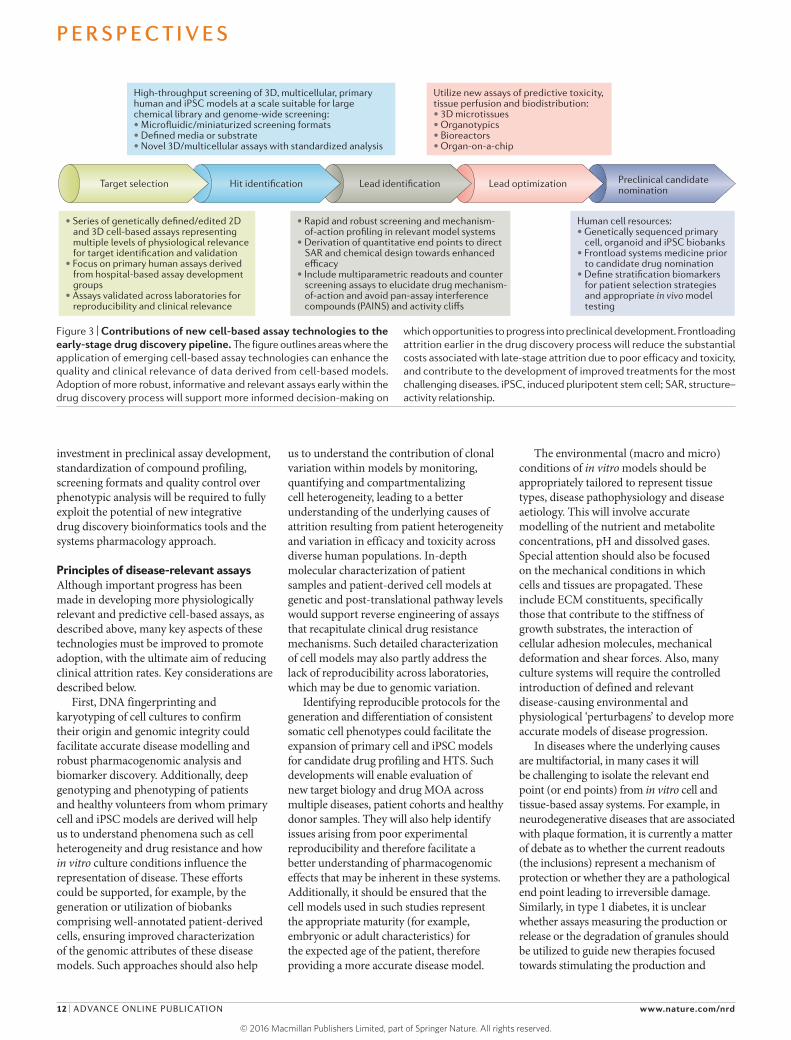
Nature Reviews | Drug Discovery
• Series of genetically defined/edited 2D and 3D cell-based assays representing multiple levels of physiological relevance for target identification and validation
• Focus on primary human assays derived from hospital-based assay development groups
• Assays validated across laboratories for reproducibility and clinical relevance
• Rapid and robust screening and mechanism-of-action profiling in relevant model systems
• Derivation of quantitative end points to direct SAR and chemical design towards enhanced efficacy
• Include multiparametric readouts and counter screening assays to elucidate drug mechanism-of-action and avoid pan-assay interference compounds (PAINS) and activity cliffs
Human cell resources:• Genetically sequenced primary
cell, organoid and iPSC biobanks• Frontload systems medicine prior
to candidate drug nomination• Define stratification biomarkers
for patient selection strategies and appropriate in vivo model testing
High-throughput screening of 3D, multicellular, primary human and iPSC models at a scale suitable for large chemical library and genome-wide screening:• Microfluidic/miniaturized screening formats• Defined media or substrate• Novel 3D/multicellular assays with standardized analysis
Utilize new assays of predictive toxicity, tissue perfusion and biodistribution:• 3D microtissues• Organotypics• Bioreactors• Organ-on-a-chip
Target selection Hit identification Lead identification Lead optimization Preclinical candidatenomination
investment in preclinical assay development, standardization of compound profiling, screening formats and quality control over phenotypic analysis will be required to fully exploit the potential of new integrative drug discovery bioinformatics tools and the systems pharmacology approach.
Principles of disease-relevant assaysAlthough important progress has been made in developing more physiologically relevant and predictive cellbased assays, as described above, many key aspects of these technologies must be improved to promote adoption, with the ultimate aim of reducing clinical attrition rates. Key considerations are described below.
First, DNA fingerprinting and karyotyping of cell cultures to confirm their origin and genomic integrity could facilitate accurate disease modelling and robust pharmacogenomic analysis and biomarker discovery. Additionally, deep genotyping and phenotyping of patients and healthy volunteers from whom primary cell and iPSC models are derived will help us to understand phenomena such as cell heterogeneity and drug resistance and how in vitro culture conditions influence the representation of disease. These efforts could be supported, for example, by the generation or utilization of biobanks comprising wellannotated patientderived cells, ensuring improved characterization of the genomic attributes of these disease models. Such approaches should also help
us to understand the contribution of clonal variation within models by monitoring, quantifying and compartmentalizing cell heterogeneity, leading to a better understanding of the underlying causes of attrition resulting from patient heterogeneity and variation in efficacy and toxicity across diverse human populations. Indepth molecular characterization of patient samples and patientderived cell models at genetic and posttranslational pathway levels would support reverse engineering of assays that recapitulate clinical drug resistance mechanisms. Such detailed characterization of cell models may also partly address the lack of reproducibility across laboratories, which may be due to genomic variation.
Identifying reproducible protocols for the generation and differentiation of consistent somatic cell phenotypes could facilitate the expansion of primary cell and iPSC models for candidate drug profiling and HTS. Such developments will enable evaluation of new target biology and drug MOA across multiple diseases, patient cohorts and healthy donor samples. They will also help identify issues arising from poor experimental reproducibility and therefore facilitate a better understanding of pharmacogenomic effects that may be inherent in these systems. Additionally, it should be ensured that the cell models used in such studies represent the appropriate maturity (for example, embryonic or adult characteristics) for the expected age of the patient, therefore providing a more accurate disease model.
The environmental (macro and micro) conditions of in vitro models should be appropriately tailored to represent tissue types, disease pathophysiology and disease aetiology. This will involve accurate modelling of the nutrient and metabolite concentrations, pH and dissolved gases. Special attention should also be focused on the mechanical conditions in which cells and tissues are propagated. These include ECM constituents, specifically those that contribute to the stiffness of growth substrates, the interaction of cellular adhesion molecules, mechanical deformation and shear forces. Also, many culture systems will require the controlled introduction of defined and relevant diseasecausing environmental and physiological ‘perturbagens’ to develop more accurate models of disease progression.
In diseases where the underlying causes are multifactorial, in many cases it will be challenging to isolate the relevant end point (or end points) from in vitro cell and tissuebased assay systems. For example, in neurodegenerative diseases that are associated with plaque formation, it is currently a matter of debate as to whether the current readouts (the inclusions) represent a mechanism of protection or whether they are a pathological end point leading to irreversible damage. Similarly, in type 1 diabetes, it is unclear whether assays measuring the production or release or the degradation of granules should be utilized to guide new therapies focused towards stimulating the production and
Figure 3 | Contributions of new cell-based assay technologies to the early-stage drug discovery pipeline. The figure outlines areas where the application of emerging cell-based assay technologies can enhance the quality and clinical relevance of data derived from cell-based models. Adoption of more robust, informative and relevant assays early within the drug discovery process will support more informed decision-making on
which opportunities to progress into preclinical development. Frontloading attrition earlier in the drug discovery process will reduce the substantial costs associated with late-stage attrition due to poor efficacy and toxicity, and contribute to the development of improved treatments for the most challenging diseases. iPSC, induced pluripotent stem cell; SAR, structure–activity relationship.
P E R S P E C T I V E S
12 | ADVANCE ONLINE PUBLICATION www.nature.com/nrd
© 2016
Macmillan
Publishers
Limited,
part
of
Springer
Nature.
All
rights
reserved. ©
2016
Macmillan
Publishers
Limited,
part
of
Springer
Nature.
All
rights
reserved.

Nature Reviews | Drug Discovery
Standardized collection of fresh tissuebiopsy samples for assay development
Fresh human tissue biobank dedicated to development of a comprehensive quality controlled primary human cell assay resource across healthy volunteer and patient cohorts
Open-source qualified/validatedassays and results (reproduced between assay development labs)
Large cohort studies: biomarker and personalized medicine strategies
• Academic community
• Clinicians/health- care industry
Precompetitiveaccess to assays
and data
Assay development lab 1
Precompetitive assay developmentand validation network
Assay development lab 2
Assay development lab 3
Assay development lab 4
Anonymized clinical-pathologyreports linked to sequencing data
release of insulin, or whether the degradation of old insulinsecreting granules should be prioritized to allow newly synthesized granules to fuse with the plasma membrane. Thus, as previously discussed by others168, the selection of relevant end points, which translate into clinical phenotypes or biomarkers of disease outcome, may be most beneficial.
Additional considerations include:• development of multiscale tissue assays
to mimic organismlevel physiology to accurately model drug metabolism, comorbidities and systemic paracrine effects;
• incorporation of clinically equivalent dosing strategies that mimic known or predict expected in vivo pharmacokinetic properties of various therapeutic modalities;
• derivation of iPSCs from multiple patients and tissue types, containing multiple diverse genomic alterations, in order to reflect broader patient populations and more common disorders;
• comparison of the drug–response data between patients and patientderived cell models to evaluate the predictive value of the models;
• integration of molecular genetic, epigenetic and proteomic data sets with robust phenotypic measurements in both clinical and preclinical settings with computational bioinformatics, supporting multiparametric validation of drug targets and assay relevance through a lessreductionist ‘systems pharmacology’ approach; and
• adoption of a human physiology assay checklist, based on unbiased evaluation of which assay conditions accurately recapitulate the human tissue physiology or pathophysiology under study, and the limitations that each assay will inevitably have with regard to recapitulating the full complexity of human disease.With the above considerations in mind,
in TABLE 1 we provide our proposal for a set of defining principles of diseaserelevant assays and justify their potential impact on drug discovery. Although many principles are generic and can be applied across disease models, certain principles have disease specific considerations, which require more indepth investigation and modelling, as indicated in TABLE 1. We anticipate that no single model or assay will be perfect and many of them will fail to recapitulate the full complexity of the human disease conditions that they intend to represent. However, we
believe that careful consideration of the limitations of each assay and the defining principles outlined in TABLE 1 will lead to more critical evaluation of the predictive value of every cellbased assay, and also lead to more cautious and appropriate interpretations of the results derived from these assays.
These defining principles are also intended to encourage further discussion and debate on preclinical assay screening standards and their clinical relevance in order to help drive forward the field of preclinical assay development towards improved clinical success rates. We anticipate that broad communication of such principles across diverse scientific disciplines will both stimulate and
guide further investment in innovative solutions and the development of new technologies that address these challenges. In FIG. 3, we highlight how integration of the technologies discussed above could contribute to earlystage drug discovery.
Assay validation and translationValidating new preclinical models for physiological and disease relevance is not trivial, and arguably the best validation is evidence of prediction of clinical efficacy or toxicity. The majority of new cellular assay technologies described in this article have not yet reached a level of maturity that would permit any clear conclusions regarding their predictive value. Currently,
Figure 4 | Precompetitive consortia facilitating predictive assay development, experimental and personalized medicine strategies. The development of screening assays and preclinical models that better predict clinical outcomes is a major challenge, and has not received the neces-sary investment from translational funding bod-ies and industry. Here, we present a model for the precompetitive development of more predictive and reproducible cell-based assays though aca-demic and industrial consortia. Facilitated by the access to human cell and tissue biobanks, we pro-pose a network of core cell-based screening lab-oratories that are proficient in assay quality control and automated cell-based screening technology, to develop assays to common stand-ards. New assays will be qualified for disease rel-evance in partnership with disease area experts and clinicians performing retrospective studies with approved drugs and failed clinical candi-dates, where possible, to determine the positive and negative predictive value of each assay. Assay protocols and assay results relating to the identification of new targets, lead compounds and repositioning opportunities will be validated for reproducibility by benchmarking assay per-formance and confirming results between dis-tinct core laboratories within assay development networks. We believe that academic, industrial and government funding of such precompetitive activity will provide open-source access to high-quality assays and results to be exploited by the academic and health-care communities and the biopharmaceutical industry. These activities will drive substantial improvements in the qual-ity of the academic literature, publishing new target hypotheses and new drug combination and repositioning opportunities. Such precom-petitive activity will also improve the quality of assays used by academic and industrial groups for screening internal chemical and biological libraries. Follow-up studies on promising leads or drug candidates could be performed on larger cohorts of patient-derived cell models through the consortia and/or access to the academic network of core laboratories.
P E R S P E C T I V E S
NATURE REVIEWS | DRUG DISCOVERY ADVANCE ONLINE PUBLICATION | 13
© 2016
Macmillan
Publishers
Limited,
part
of
Springer
Nature.
All
rights
reserved. ©
2016
Macmillan
Publishers
Limited,
part
of
Springer
Nature.
All
rights
reserved.

the adoption of new technologies often depends on an organic rise in utilization and acceptance. However, rather than simply waiting for a sufficiently large body of evidence to be generated by pioneering drug discovery efforts, a concerted effort is needed to compare the predictive power of new cellbased models for outcomes in patients. Such efforts would ideally involve testing collections of known molecules that have demonstrable clinical efficacies and toxicities to support backward translation to determine the predictive value of in vitro models. Backward translation studies may also encompass reverse engineering of in vitro model systems to predict past clinical trial failures by recapitulating known mechanisms of drug resistance or relapse observed in the clinic.
Recent examples of such work have focused on evolving hepatotoxicity assays utilizing primary hepatocytes, stemcell derived hepatocytes and hepatocyte cell line cultures grown on a range of defined organic or synthetic matrix substrates. These systems were then tested against a panel of drugs to predict agents that result in druginduced liver injury in humans169–171. Similar approaches using stemcellderived cardiomyocytes to predict cardiotoxicity are also under development, as previously discussed.
Examples of backward translation studies to predict the clinical efficacy of therapeutic agents are rare, which is probably a reflection of the lack of research funding available to support backward translation studies and assay model development. However, to demonstrate the predictive and translational potential of new preclinical models described in this article, we highlight two case studies: the Hedgehog signalling inhibitor vismodegib and an αvβ6 blocking antibody (264RAD). Both of these examples exploit the unique aspects of the 3D organotypic pancreatic cancer model previously described, which is composed of a coculture of established pancreatic cancer cells cultured on dense collagen matrix contracted by fibroblasts130,172. This model recapitulates in vivo pharmacology issues relating to poor drug penetration and inadequate clinical efficacy131,132.
In the first example, studies that used both the in vitro organotypic cell culture model and comparative in vivo transgenic models showed that cyclopamine, which targets Hedgehog signalling in the stromal cell compartment, leads to increased penetration and hence activity of the anticancer drugs dasatinib and gemcitabine in pancreatic cancer128,132. Clinical trials of the approved Hedgehog signalling inhibitor vismodegib have been instigated in pancreatic cancer, and
the recently published preliminary results indicate an improved efficacy response173. In the second example, pioneering studies demonstrating that targeting αvβ6 suppressed cancer cell invasion in 3D organotypic assay formats174,175 have contributed to the progression of the novel αvβ6blocking antibody therapy (264RAD) into a phase I trial in pancreatic cancer; see the Barts Cancer Institute website for further information.
Finally, a recent article published by Scannell and Bosley176 provides further justification for increased investment in backward translation studies. This work, which describes a quantitative decisiontheoretic model of declining R&D productivity, illustrates how small improvements in the positive predictive value (PPV) of preclinical assays can outperform the documented advances in assay automation and throughput with regard to improving R&D productivity as defined by the successful translation of drug discovery to positive clinical outcomes176. Thus, a clearly defined pathway to assay validation and a subsequent development of a repository of assays with known positive and negative predictive values (PPVs and NPVs) would be of considerable value. As far as we are aware, there is no unifying systematic approach to measuring and recording the clinical impact of established assays or new assay technologies and approaches. We suggest that a routine evaluation of the PPV or NPV of all assays used in drug discovery would provide a valuable resource to the translational research community.
The need for funding and consortiaAcademic–industrial partnerships have existed in some form since the birth of the pharmaceutical industry over 100 years ago, but have been increasingly appreciated and pursued more recently in a wide range of contexts, from identifying and validating novel therapeutic targets through to addressing drug development challenges that are too broad for any one organization to tackle effectively alone, which are often addressed through consortia. There remains, however, a gap in the development of such consortia in the area of preclinical model development, and here we put forward three proposals to address this.
First, we propose that direct funding for the development of preclinical models and assayscreening technologies with improved clinical relevance is an aspect of the drug development process that can be most effectively addressed by
Box 3 | Phenotypic drug discovery versus target-directed drug discovery
Target-directed drug discovery (TDD) has been the predominant strategy of the pharmaceutical and biotechnology industry for the past 25 years. It is characterized by the identification and optimization of compounds that modulate a pre-nominated target that is implicated in disease progression, often using high-throughput screening to identify initial hits. Phenotypic drug discovery (PDD) has been defined as the generation of hit or lead molecules without any prior knowledge of the target. Such compounds are typically identified and developed through empirical testing in physiological in vivo models or from cell-based phenotypic screening assays.
An initial retrospective analysis of all drugs approved by the US Food and Drug Administration (FDA) between 1999 and 2008 indicated that of the first-in-class small-molecule medicines approved, 37% (28 drugs) were initially identified by a PDD approach, whereas 23% (17 drugs) were identified by TDD215. Follow-up retrospective studies in cancer216 and across disease areas217 produced different results, in part owing to differences in the definition of phenotypic screening and in part owing to differences in the time period studied, with TDD showing substantial success in cancer in more recent years. In cancer, 31 novel small-molecule drugs approved by the FDA between 1999 and 2013 were discovered by TDD (including 21 kinase inhibitors), whereas 17 novel small-molecule drugs approved during the same period were discovered by PDD (including a single kinase inhibitor, trametinib, targeting MAPK/ERK kinase (MEK))216. A retrospective analysis by Eder and colleagues217 across all disease areas, using a refined selection criteria, identified 45 novel small molecules approved during 1999 and 2013 that were discovered through TDD. At the same time, 33 approved small molecules were discovered by non-target-directed methods, including 8 through unbiased phenotypic screening of large chemical libraries and 25 through chemocentric approaches, representing a combination of rational drug design and phenotypic screening/validation. These studies indicate that PDD and TDD can be considered complementary approaches. Further investment in more predictive cell-based assay systems could advance both PDD and TDD strategies, ultimately resulting in improved clinical success rates.
P E R S P E C T I V E S
14 | ADVANCE ONLINE PUBLICATION www.nature.com/nrd
© 2016
Macmillan
Publishers
Limited,
part
of
Springer
Nature.
All
rights
reserved. ©
2016
Macmillan
Publishers
Limited,
part
of
Springer
Nature.
All
rights
reserved.
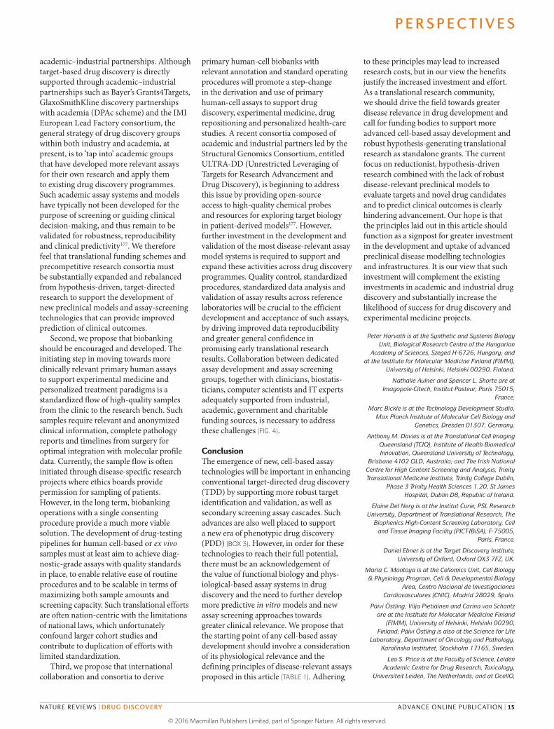
academic–industrial partnerships. Although targetbased drug discovery is directly supported through academic–industrial partnerships such as Bayer’s Grants4Targets, GlaxoSmithKline discovery partnerships with academia (DPAc scheme) and the IMI European Lead Factory consortium, the general strategy of drug discovery groups within both industry and academia, at present, is to ‘tap into’ academic groups that have developed more relevant assays for their own research and apply them to existing drug discovery programmes. Such academic assay systems and models have typically not been developed for the purpose of screening or guiding clinical decisionmaking, and thus remain to be validated for robustness, reproducibility and clinical predictivity177. We therefore feel that translational funding schemes and precompetitive research consortia must be substantially expanded and rebalanced from hypothesisdriven, targetdirected research to support the development of new preclinical models and assayscreening technologies that can provide improved prediction of clinical outcomes.
Second, we propose that biobanking should be encouraged and developed. The initiating step in moving towards more clinically relevant primary human assays to support experimental medicine and personalized treatment paradigms is a standardized flow of highquality samples from the clinic to the research bench. Such samples require relevant and anonymized clinical information, complete pathology reports and timelines from surgery for optimal integration with molecular profile data. Currently, the sample flow is often initiated through diseasespecific research projects where ethics boards provide permission for sampling of patients. However, in the long term, biobanking operations with a single consenting procedure provide a much more viable solution. The development of drugtesting pipelines for human cellbased or ex vivo samples must at least aim to achieve diagnosticgrade assays with quality standards in place, to enable relative ease of routine procedures and to be scalable in terms of maximizing both sample amounts and screening capacity. Such translational efforts are often nationcentric with the limitations of national laws, which unfortunately confound larger cohort studies and contribute to duplication of efforts with limited standardization.
Third, we propose that international collaboration and consortia to derive
primary humancell biobanks with relevant annotation and standard operating procedures will promote a stepchange in the derivation and use of primary humancell assays to support drug discovery, experimental medicine, drug repositioning and personalized healthcare studies. A recent consortia composed of academic and industrial partners led by the Structural Genomics Consortium, entitled ULTRADD (Unrestricted Leveraging of Targets for Research Advancement and Drug Discovery), is beginning to address this issue by providing opensource access to highquality chemical probes and resources for exploring target biology in patientderived models177. However, further investment in the development and validation of the most diseaserelevant assay model systems is required to support and expand these activities across drug discovery programmes. Quality control, standardized procedures, standardized data analysis and validation of assay results across reference laboratories will be crucial to the efficient development and acceptance of such assays, by driving improved data reproducibility and greater general confidence in promising early translational research results. Collaboration between dedicated assay development and assay screening groups, together with clinicians, biostatisticians, computer scientists and IT experts adequately supported from industrial, academic, government and charitable funding sources, is necessary to address these challenges (FIG. 4).
ConclusionThe emergence of new, cellbased assay technologies will be important in enhancing conventional targetdirected drug discovery (TDD) by supporting more robust target identification and validation, as well as secondary screening assay cascades. Such advances are also well placed to support a new era of phenotypic drug discovery (PDD) (BOX 3). However, in order for these technologies to reach their full potential, there must be an acknowledgement of the value of functional biology and physiologicalbased assay systems in drug discovery and the need to further develop more predictive in vitro models and new assay screening approaches towards greater clinical relevance. We propose that the starting point of any cellbased assay development should involve a consideration of its physiological relevance and the defining principles of diseaserelevant assays proposed in this article (TABLE 1). Adhering
to these principles may lead to increased research costs, but in our view the benefits justify the increased investment and effort. As a translational research community, we should drive the field towards greater disease relevance in drug development and call for funding bodies to support more advanced cellbased assay development and robust hypothesisgenerating translational research as standalone grants. The current focus on reductionist, hypothesisdriven research combined with the lack of robust diseaserelevant preclinical models to evaluate targets and novel drug candidates and to predict clinical outcomes is clearly hindering advancement. Our hope is that the principles laid out in this article should function as a signpost for greater investment in the development and uptake of advanced preclinical disease modelling technologies and infrastructures. It is our view that such investment will complement the existing investments in academic and industrial drug discovery and substantially increase the likelihood of success for drug discovery and experimental medicine projects.
Peter Horvath is at the Synthetic and Systems Biology Unit, Biological Research Centre of the Hungarian
Academy of Sciences, Szeged H-6726, Hungary; and at the Institute for Molecular Medicine Finland (FIMM),
University of Helsinki, Helsinki 00290, Finland.
Nathalie Aulner and Spencer L. Shorte are at Imagopole-Citech, Institut Pasteur, Paris 75015,
France.
Marc Bickle is at the Technology Development Studio, Max Planck Institute of Molecular Cell Biology and
Genetics, Dresden 01307, Germany.
Anthony M. Davies is at the Translational Cell Imaging Queensland (TCIQ), Institute of Health Biomedical Innovation, Queensland University of Technology,
Brisbane 4102 QLD, Australia; and The Irish National Centre for High Content Screening and Analysis, Trinity Translational Medicine Institute, Trinity College Dublin,
Phase 3 Trinity Health Sciences 1.20, St James Hospital, Dublin D8, Republic of Ireland.
Elaine Del Nery is at the Institut Curie, PSL Research University, Department of Translational Research, The
Biophenics High-Content Screening Laboratory, Cell and Tissue Imaging Facility (PICT-IBiSA), F-75005,
Paris, France.
Daniel Ebner is at the Target Discovery Institute, University of Oxford, Oxford OX3 7FZ, UK.
Maria C. Montoya is at the Cellomics Unit, Cell Biology & Physiology Program, Cell & Developmental Biology
Area, Centro Nacional de Investigaciones Cardiovasculares (CNIC), Madrid 28029, Spain.
Päivi Östling, Vilja Pietiäinen and Carina von Schantz are at the Institute for Molecular Medicine Finland
(FIMM), University of Helsinki, Helsinki 00290, Finland; Päivi Östling is also at the Science for Life
Laboratory, Department of Oncology and Pathology, Karolinska Institutet, Stockholm 17165, Sweden.
Leo S. Price is at the Faculty of Science, Leiden Academic Centre for Drug Research, Toxicology,
Universiteit Leiden, The Netherlands; and at OcellO,
P E R S P E C T I V E S
NATURE REVIEWS | DRUG DISCOVERY ADVANCE ONLINE PUBLICATION | 15
© 2016
Macmillan
Publishers
Limited,
part
of
Springer
Nature.
All
rights
reserved. ©
2016
Macmillan
Publishers
Limited,
part
of
Springer
Nature.
All
rights
reserved.

J.H Oortweg 21, 2333 CH, Leiden, The Netherlands.
Gerardo Turcatti is at the Biomolecular Screening Facility, Swiss Federal Institute of Technology (EPFL),
Lausanne CH-1015, Switzerland.
Neil O. Carragher is at the Cancer Research UK Edinburgh Centre, Institute of Genetics and Molecular
Medicine, University of Edinburgh, Edinburgh EH4 2XR, UK.
All authors are part of the European Cell-Based Assays Interest Group.
Correspondence to N.C. [email protected]
doi:10.1038/nrd.2016.175 Published online 12 Sep 2016
1. Waldmeier, P., Bozyczko-Coyne, D., Williams, M. & Vaught, J. L. Recent clinical failures in Parkinson’s disease with apoptosis inhibitors underline the need for a paradigm shift in drug discovery for neurodegenerative diseases. Biochem. Pharmacol. 72, 1197–1206 (2006).
2. Bolognesi, M. L., Matera, R., Minarini, A., Rosini, M. & Melchiorre, C. Alzheimer’s disease: new approaches to drug discovery. Curr. Opin. Chem. Biol. 13, 303–308 (2009).
3. Paul, S. M. et al. How to improve R&D productivity: the pharmaceutical industry’s grand challenge. Nat. Rev. Drug Discov. 9, 203–214 (2010).
4. Mangialasche, F., Solomon, A., Winblad, B., Mecocci, P. & Kivipelto, M. Alzheimer’s disease: clinical trials and drug development. Lancet Neurol. 9, 702–716 (2010).
5. Hoelder, S., Clarke, P. A. & Workman, P. Discovery of small molecule cancer drugs: successes, challenges and opportunities. Mol. Oncol. 6, 155–176 (2012).
6. Morens, D. M., Folkers, G. K. & Fauci, A. S. The challenge of emerging and re-emerging infectious diseases. Nature 430, 242–249 (2004).
7. Marston, H. D., Folkers, G. K., Morens, D. M. & Fauci, A. S. Emerging viral diseases: confronting threats with new technologies. Sci. Transl Med. 6, 253ps210 (2014).
8. Arrowsmith, J. Trial watch: Phase II failures: 2008–2010. Nat. Rev. Drug Discov. 10, 328–329 (2011).
9. Laverty, H. et al. How can we improve our understanding of cardiovascular safety liabilities to develop safer medicines? Br. J. Pharmacol. 163, 675–693 (2011).
10. Bass, N. M. in Current Diagnosis & Treatment in Gastroenterology (eds Friedman, S. E., Grendell, J. H. & McQuaid, K. R.) 664–679 (Lang Medical Books/McGraw-Hill, 2003).
11. Masters, J. R. & Stacey, G. N. Changing medium and passaging cell lines. Nat. Protoc. 2, 2276–2284 (2007).
12. Nestor, C. E. et al. Rapid reprogramming of epigenetic and transcriptional profiles in mammalian culture systems. Genome Biol. 16, 11 (2015).
13. Morris, C. C. Maintenace and loss in tissue culture of specific cell characteristics. Adv. Appl. Microbiol. 4, 117–212 (1962).
14. Carreau, A., Hafny-Rahbi, B. E., Matejuk, A., Grillon, C. & Kieda, C. Why is the partial oxygen pressure of human tissues a crucial parameter? Small molecules and hypoxia. J. Cell. Mol. Med. 15, 1239–1253 (2011).
15. Newby, D., Marks, L. & Lyall, F. Dissolved oxygen concentration in culture medium: assumptions and pitfalls. Placenta 26, 353–357 (2005).
16. Sullivan, M., Galea, P. & Latif, S. What is the appropriate oxygen tension for in vitro culture? Mol. Hum. Reprod. 12, 653 (2006).
17. Halliwell, B. Cell culture, oxidative stress, and antioxidants: avoiding pitfalls. Biomed. J. 37, 99–105 (2014).
18. Tiede, L. M., Cook, E. A., Morsey, B. & Fox, H. S. Oxygen matters: tissue culture oxygen levels affect mitochondrial function and structure as well as responses to HIV viroproteins. Cell Death Dis. 2, e246 (2011).
19. Redshaw, Z. & Loughna, P. T. Oxygen concentration modulates the differentiation of muscle stem cells toward myogenic and adipogenic fates. Differentiation 84, 193–202 (2012).
20. Gebhardt, R. & Matz-Soja, M. Liver zonation: novel aspects of its regulation and its impact on homeostasis. World J. Gastroenterol. 20, 8491–8504 (2014).
21. Bhatia, S. N. & Ingber, D. E. Microfluidic organs-on-chips. Nat. Biotechnol. 32, 760–772 (2014).
22. Wells, R. G. The role of matrix stiffness in regulating cell behavior. Hepatology 47, 1394–1400 (2008).
23. Discher, D. E., Janmey, P. & Wang, Y.-l. Tissue cells feel and respond to the stiffness of their substrate. Science 310, 1139–1143 (2005).
24. Levental, I., Georges, P. C. & Janmey, P. A. Soft biological materials and their impact on cell function. Soft Matter 3, 299–306 (2007).
25. Engler, A. J. et al. Myotubes differentiate optimally on substrates with tissue-like stiffness: pathological implications for soft or stiff microenvironments. J. Cell Biol. 166, 877–887 (2004).
26. Semler, E. J., Ranucci, C. S. & Moghe, P. V. Mechanochemical manipulation of hepatocyte aggregation can selectively induce or repress liver-specific function. Biotechnol. Bioeng. 69, 359–369 (2000).
27. Huh, D. et al. Reconstituting organ-level lung functions on a chip. Science 328, 1662–1668 (2010).
28. Wells, R. G. Cellular sources of extracellular matrix in hepatic fibrosis. Clin. Liver Dis. 12, 759–768 (2008).
29. Teranishi, Y. et al. Involvement of hepatic stellate cell cytoglobin in acute hepatocyte damage through the regulation of CYP2E1-mediated xenobiotic metabolism. Lab Invest. 95, 515–524 (2015).
30. Roberts, R. A. et al. Role of the Kupffer cell in mediating hepatic toxicity and carcinogenesis. Toxicol. Sci. 96, 2–15 (2007).
31. Lobsiger, C. S. & Cleveland, D. W. Glial cells as intrinsic components of non-cell autonomous neurodegenerative disease. Nat. Neurosci. 10, 1355–1360 (2007).
32. McCormack, E. et al. Bi-specific TCR-anti CD3 redirected T-cell targeting of NY-ESO-1- and LAGE-1-positive tumors. Cancer Immunol. Immunother. 62, 773–785 (2013).
33. Pieters, R. et al. In vitro drug sensitivity of cells from children with leukemia using the MTT assay with improved culture conditions. Blood 76, 2327–2336 (1990).
34. Hollingsworth, S. J. & Biankin, A. V. The challenges of precision oncology drug development and implementation. Publ. Health Genom. 18, 338–348 (2015).
35. Biankin, A. V., Piantadosi, S. & Hollingsworth, S. J. Patient-centric trials for therapeutic development in precision oncology. Nature 526, 361–370 (2015).
36. Iwadate, Y., Fujimoto, S., Namba, H. & Yamaura, A. Promising survival for patients with glioblastoma multiforme treated with individualised chemotherapy based on in vitro drug sensitivity testing. Br. J. Cancer 89, 1896–1900 (2003).
37. Bosanquet, A. G. & Bell, P. B. Ex vivo therapeutic index by drug sensitivity assay using fresh human normal and tumor cells. J. Exp. Ther. Oncol. 4, 145–154 (2004).
38. Villman, K., Blomqvist, C., Larsson, R. & Nygren, P. Predictive value of in vitro assessment of cytotoxic drug activity in advanced breast cancer. Anticancer Drugs 16, 609–615 (2005).
39. Eriksson, A. et al. Drug screen in patient cells suggests quinacrine to be repositioned for treatment of acute myeloid leukemia. Blood Cancer J. 5, e307 (2015).
40. Pemovska, T. et al. Individualized systems medicine strategy to tailor treatments for patients with chemorefractory acute myeloid leukemia. Cancer Discov. 3, 1416–1429 (2013).
41. Yamada, S. et al. Distinctive multidrug sensitivity and outcome of acute erythroblastic and megakaryoblastic leukemia in children with Down syndrome. Int. J. Hematol. 74, 428–436 (2001).
42. Aljitawi, O. S. et al. A novel three-dimensional stromal-based model for in vitro chemotherapy sensitivity testing of leukemia cells. Leuk. Lymphoma 55, 378–391 (2014).
43. Bakker, E., Qattan, M., Mutti, L., Demonacos, C. & Krstic-Demonacos, M. The role of microenvironment and immunity in drug response in leukemia. Biochim. Biophys. Acta 1863, 414–426 (2016).
44. Saeed, K. et al. Comprehensive drug testing of patient-derived conditionally reprogrammed cells from castration-resistant prostate cancer. Eur. Urol. http://dx.doi.org/10.1016/j.eururo.2016.04.019 (2016).
45. Suprynowicz, F. A. et al. Conditionally reprogrammed cells represent a stem-like state of adult epithelial cells. Proc. Natl Acad. Sci. USA 109, 20035–20040 (2012).
46. Yuan, H. et al. Use of reprogrammed cells to identify therapy for respiratory papillomatosis. N. Engl. J. Med. 367, 1220–1227 (2012).
47. van de Wetering, M. et al. Prospective derivation of a living organoid biobank of colorectal cancer patients. Cell 161, 933–945 (2015).
48. Pemovska, T. et al. Axitinib effectively inhibits BCR-ABL1(T315I) with a distinct binding conformation. Nature 519, 102–105 (2015).
49. Rees, M. G. et al. Correlating chemical sensitivity and basal gene expression reveals mechanism of action. Nat. Chem. Biol. 12, 109–116 (2016).
50. Lamb, J. et al. The Connectivity Map: using gene-expression signatures to connect small molecules, genes, and disease. Science 313, 1929–1935 (2006).
51. Perlman, Z. E. et al. Multidimensional drug profiling by automated microscopy. Science 306, 1194–1198 (2004).
52. Caie, P. D. et al. High-content phenotypic profiling of drug response signatures across distinct cancer cells. Mol. Cancer Ther. 9, 1913–1926 (2010).
53. Takahashi, K. & Yamanaka, S. Induction of pluripotent stem cells from mouse embryonic and adult fibroblast cultures by defined factors. Cell 126, 663–676 (2006).
54. Avior, Y., Sagi, I. & Benvenisty, N. Pluripotent stem cells in disease modelling and drug discovery. Nat. Rev. Mol. Cell Biol. 17, 170–182 (2016).
55. Singh, V. K., Kumar, N., Kalsan, M., Saini, A. & Chandra, R. Mechanism of induction: induced pluripotent stem cells (iPSCs). J. Stem Cells 10, 43–62 (2015).
56. Bar-Nur, O., Russ, H. A., Efrat, S. & Benvenisty, N. Epigenetic memory and preferential lineage-specific differentiation in induced pluripotent stem cells derived from human pancreatic islet beta cells. Cell Stem Cell 9, 17–23 (2011).
57. Kim, K. et al. Donor cell type can influence the epigenome and differentiation potential of human induced pluripotent stem cells. Nat. Biotechnol. 29, 1117–1119 (2011).
58. Sakurai, T. et al. A non-inheritable maternal Cas9-based multiple-gene editing system in mice. Sci. Rep. 6, 20011 (2016).
59. Martella, A., Pollard, S. M., Dai, J. & Cai, Y. Mammalian synthetic biology: time for big MACs. ACS Synth. Biol. http://dx.doi.org/10.1021/acssynbio.6b00074 (2016).
60. Annaluru, N., Ramalingam, S. & Chandrasegaran, S. Rewriting the blueprint of life by synthetic genomics and genome engineering. Genome Biol. 16, 125 (2015).
61. Miller, J. D. et al. Human iPSC-based modeling of late-onset disease via progerin-induced aging. Cell Stem Cell 13, 691–705 (2013).
62. Liang, P. et al. Drug screening using a library of human induced pluripotent stem cell-derived cardiomyocytes reveals disease-specific patterns of cardiotoxicity. Circulation 127, 1677–1691 (2013).
63. Matsa, E. et al. Drug evaluation in cardiomyocytes derived from human induced pluripotent stem cells carrying a long QT syndrome type 2 mutation. Eur. Heart J. 32, 952–962 (2011).
64. Navarrete, E. G. et al. Screening drug-induced arrhythmia [corrected] using human induced pluripotent stem cell-derived cardiomyocytes and low-impedance microelectrode arrays. Circulation 128, S3–S13 (2013).
65. Mioulane, M., Foldes, G., Ali, N. N., Schneider, M. D. & Harding, S. E. Development of high content imaging methods for cell death detection in human pluripotent stem cell-derived cardiomyocytes. J. Cardiovasc. Transl. Res. 5, 593–604 (2012).
66. Xu, X. et al. Prevention of β-amyloid induced toxicity in human iPS cell-derived neurons by inhibition of cyclin-dependent kinases and associated cell cycle events. Stem Cell Res. 10, 213–227 (2013).
67. Usher, L. C. et al. A chemical screen identifies novel compounds that overcome glial-mediated inhibition of neuronal regeneration. J. Neurosci. 30, 4693–4706 (2010).
68. Dimos, J. T. et al. Induced pluripotent stem cells generated from patients with ALS can be differentiated into motor neurons. Science 321, 1218–1221 (2008).
69. Spence, J. R. et al. Directed differentiation of human pluripotent stem cells into intestinal tissue in vitro. Nature 470, 105–109 (2011).
70. Nalayanda, D. D. et al. An open-access microfluidic model for lung-specific functional studies at an air-liquid interface. Biomed. Microdevices 11, 1081–1089 (2009).
P E R S P E C T I V E S
16 | ADVANCE ONLINE PUBLICATION www.nature.com/nrd
© 2016
Macmillan
Publishers
Limited,
part
of
Springer
Nature.
All
rights
reserved. ©
2016
Macmillan
Publishers
Limited,
part
of
Springer
Nature.
All
rights
reserved.

71. Ebert, A. D. et al. Induced pluripotent stem cells from a spinal muscular atrophy patient. Nature 457, 277–280 (2009).
72. Marchetto, M. C. et al. A model for neural development and treatment of Rett syndrome using human induced pluripotent stem cells. Cell 143, 527–539 (2010).
73. Koch, P. et al. Presenilin-1 L166P mutant human pluripotent stem cell-derived neurons exhibit partial loss of gamma-secretase activity in endogenous amyloid-beta generation. Am. J. Pathol. 180, 2404–2416 (2012).
74. Burkhardt, M. F. et al. A cellular model for sporadic ALS using patient-derived induced pluripotent stem cells. Mol. Cell Neurosci. 56, 355–364 (2013).
75. Chung, C. Y. et al. Identification and rescue of alpha-synuclein toxicity in Parkinson patient-derived neurons. Science 342, 983–987 (2013).
76. Peng, J., Liu, Q., Rao, M. S. & Zeng, X. Using human pluripotent stem cell-derived dopaminergic neurons to evaluate candidate Parkinson’s disease therapeutic agents in MPP+ and rotenone models. J. Biomol. Screen 18, 522–533 (2013).
77. Chiu, P. J. et al. Validation of a [3H]astemizole binding assay in HEK293 cells expressing HERG K+ channels. J. Pharmacol. Sci. 95, 311–319 (2004).
78. Huang, X. P., Mangano, T., Hufeisen, S., Setola, V. & Roth, B. L. Identification of human Ether-a-go-go related gene modulators by three screening platforms in an academic drug-discovery setting. Assay Drug Dev. Technol. 8, 727–742 (2010).
79. Gintant, G., Sager, P. T. & Stockbridge, N. Evolution of strategies to improve preclinical cardiac safety testing. Nat. Rev. Drug Discov. 15, 457–471 (2016).
80. Ivashchenko, C. Y. et al. Human-induced pluripotent stem cell-derived cardiomyocytes exhibit temporal changes in phenotype. Am. J. Physiol. Heart Circ. Physiol. 305, H913–H922 (2013).
81. Ma, Z. et al. Self-organizing human cardiac microchambers mediated by geometric confinement. Nat. Commun. 6, 7413 (2015).
82. Schaaf, S. et al. Human engineered heart tissue as a versatile tool in basic research and preclinical toxicology. PLoS ONE 6, e26397 (2011).
83. Meyer, T., Leisgen, C., Gonser, B. & Gunther, E. QT-screen: high-throughput cardiac safety pharmacology by extracellular electrophysiology on primary cardiac myocytes. Assay Drug Dev. Technol. 2, 507–514 (2004).
84. Hansen, A. et al. Development of a drug screening platform based on engineered heart tissue. Circ. Res. 107, 35–44 (2010).
85. Bridgland-Taylor, M. H. et al. Optimisation and validation of a medium-throughput electrophysiology-based hERG assay using IonWorks HT. J. Pharmacol. Toxicol. Methods 54, 189–199 (2006).
86. Farre, C. et al. Port-a-patch and patchliner: high fidelity electrophysiology for secondary screening and safety pharmacology. Comb. Chem. High Throughput Screen 12, 24–37 (2009).
87. Sirenko, O. et al. Multiparameter in vitro assessment of compound effects on cardiomyocyte physiology using iPSC cells. J. Biomol. Screen 18, 39–53 (2013).
88. Lu, H. R. et al. High throughput measurement of Ca++ dynamics in human stem cell-derived cardiomyocytes by kinetic image cytometery: a cardiac risk assessment characterization using a large panel of cardioactive and inactive compounds. Toxicol. Sci. 148, 503–516 (2015).
89. Cerignoli, F. et al. High throughput measurement of Ca2+ dynamics for drug risk assessment in human stem cell-derived cardiomyocytes by kinetic image cytometry. J. Pharmacol. Toxicol. Methods 66, 246–256 (2012).
90. Pointon, A., Abi-Gerges, N., Cross, M. J. & Sidaway, J. E. Phenotypic profiling of structural cardiotoxins in vitro reveals dependency on multiple mechanisms of toxicity. Toxicol. Sci. 132, 317–326 (2013).
91. Peters, M. F., Lamore, S. D., Guo, L., Scott, C. W. & Kolaja, K. L. Human stem cell-derived cardiomyocytes in cellular impedance assays: bringing cardiotoxicity screening to the front line. Cardiovasc. Toxicol. 15, 127–139 (2015).
92. Rappaz, B. et al. Automated multi-parameter measurement of cardiomyocytes dynamics with digital holographic microscopy. Opt. Express 23, 13333–13347 (2015).
93. Grosberg, A. et al. Muscle on a chip: in vitro contractility assays for smooth and striated muscle. J. Pharmacol. Toxicol. Methods 65, 126–135 (2012).
94. Banerjee, I. et al. Cyclic stretch of embryonic cardiomyocytes increases proliferation, growth, and expression while repressing Tgf-β signaling. J. Mol. Cell Cardiol. 79, 133–144 (2015).
95. Macias-Vidal, J. et al. The proteasome inhibitor bortezomib reduced cholesterol accumulation in fibroblasts from Niemann-Pick type C patients carrying missense mutations. FEBS J. 281, 4450–4466 (2014).
96. Millman, J. R. et al. Generation of stem cell-derived β-cells from patients with type 1 diabetes. Nat. Commun. 7, 11463 (2016).
97. Hua, H. et al. iPSC-derived β cells model diabetes due to glucokinase deficiency. J. Clin. Invest. 123, 3146–3153 (2013).
98. Smith, A. S., Davis, J., Lee, G., Mack, D. L. & Kim, D. H. Muscular dystrophy in a dish: engineered human skeletal muscle mimetics for disease modeling and drug discovery. Drug Discov. Today http://dx.doi.org/10.1016/j.drudis.2016.04.013 (2016).
99. Chen, I. Y., Matsa, E. & Wu, J. C. Induced pluripotent stem cells: at the heart of cardiovascular precision medicine. Nat. Rev. Cardiol. 13, 333–349 (2016).
100. Ebert, A. D. et al. Characterization of the molecular mechanisms underlying increased ischemic damage in the aldehyde dehydrogenase 2 genetic polymorphism using a human induced pluripotent stem cell model system. Sci. Transl Med. 6, 255ra130 (2014).
101. Cong, L. et al. Multiplex genome engineering using CRISPR/Cas systems. Science 339, 819–823 (2013).
102. Sander, J. D. & Joung, J. K. CRISPR-Cas systems for editing, regulating and targeting genomes. Nat. Biotechnol. 32, 347–355 (2014).
103. Doudna, J. A. & Charpentier, E. Genome editing. The new frontier of genome engineering with CRISPR-Cas9. Science 346, 1258096 (2014).
104. Chen, B. & Huang, B. Imaging genomic elements in living cells using CRISPR/Cas9. Methods Enzymol. 546, 337–354 (2014).
105. Shalem, O. et al. Genome-scale CRISPR-Cas9 knockout screening in human cells. Science 343, 84–87 (2014).
106. Shalem, O., Sanjana, N. E. & Zhang, F. High-throughput functional genomics using CRISPR-Cas9. Nat. Rev. Genet. 16, 299–311 (2015).
107. Wang, T., Wei, J. J., Sabatini, D. M. & Lander, E. S. Genetic screens in human cells using the CRISPR-Cas9 system. Science 343, 80–84 (2014).
108. Agrotis, A. & Ketteler, R. A new age in functional genomics using CRISPR/Cas9 in arrayed library screening. Front. Genet. 6, 300 (2015).
109. Musunuru, K. Genome editing of human pluripotent stem cells to generate human cellular disease models. Dis. Model. Mech. 6, 896–904 (2013).
110. Xue, H., Wu, J., Li, S., Rao, M. S. & Liu, Y. Genetic modification in human pluripotent stem cells by homologous recombination and CRISPR/Cas9 system. Methods Mol. Biol. 1307, 173–190 (2016).
111. Moore, J. D. The impact of CRISPR-Cas9 on target identification and validation. Drug Discov. Today 20, 450–457 (2015).
112. Xue, W. et al. CRISPR-mediated direct mutation of cancer genes in the mouse liver. Nature 514, 380–384 (2014).
113. Anastasov, N. et al. A 3D-microtissue-based phenotypic screening of radiation resistant tumor cells with synchronized chemotherapeutic treatment. BMC Cancer 15, 466 (2015).
114. Chau, D. Y., Johnson, C., MacNeil, S., Haycock, J. W. & Ghaemmaghami, A. M. The development of a 3D immunocompetent model of human skin. Biofabrication 5, 035011 (2013).
115. Wenzel, C., Otto, S., Prechtl, S., Parczyk, K. & Steigemann, P. A novel 3D high-content assay identifies compounds that prevent fibroblast invasion into tissue surrogates. Exp. Cell Res. 339, 35–43 (2015).
116. Wenzel, C. et al. 3D high-content screening for the identification of compounds that target cells in dormant tumor spheroid regions. Exp. Cell Res. 323, 131–143 (2014).
117. Krausz, E. et al. Translation of a tumor microenvironment mimicking 3D tumor growth co-culture assay platform to high-content screening. J. Biomol. Screen 18, 54–66 (2013).
118. Vukicevic, S. et al. Identification of multiple active growth factors in basement membrane Matrigel suggests caution in interpretation of cellular activity related to extracellular matrix components. Exp. Cell Res. 202, 1–8 (1992).
119. Sieh, S. et al. Phenotypic characterization of prostate cancer LNCaP cells cultured within a bioengineered microenvironment. PLoS ONE 7, e40217 (2012).
120. Phelps, E. A., Landazuri, N., Thule, P. M., Taylor, W. R. & Garcia, A. J. Bioartificial matrices for therapeutic vascularization. Proc. Natl Acad. Sci. USA 107, 3323–3328 (2010).
121. Xu, T. et al. Hybrid printing of mechanically and biologically improved constructs for cartilage tissue engineering applications. Biofabrication 5, 015001 (2013).
122. Mironi-Harpaz, I., Berdichevski, A. & Seliktar, D. Fabrication of PEGylated fibrinogen: a versatile injectable hydrogel biomaterial. Methods Mol. Biol. 1181, 61–68 (2014).
123. Harrington, H. et al. Immunocompetent 3D model of human upper airway for disease modeling and in vitro drug evaluation. Mol. Pharm. 11, 2082–2091 (2014).
124. Altekar, M. et al. Assay optimization: a statistical design of experiments approach. Clin. Lab Med. 27, 139–154 (2007).
125. Havel, J., Link, H., Hofinger, M., Franco-Lara, E. & Weuster-Botz, D. Comparison of genetic algorithms for experimental multi-objective optimization on the example of medium design for cyanobacteria. Biotechnol. J. 1, 549–555 (2006).
126. Shaw, R., Fitzek, M., Mouchet, E., Walker, G. & Jarvis, P. Overcoming obstacles in the implementation of factorial design for assay optimization. Assay Drug Dev. Technol. 13, 88–93 (2015).
127. Pampaloni, F. et al. Tissue-culture light sheet fluorescence microscopy (TC-LSFM) allows long-term imaging of three-dimensional cell cultures under controlled conditions. Integr. Biol. (Camb.) 6, 988–998 (2014).
128. Olive, K. P. et al. Inhibition of Hedgehog signaling enhances delivery of chemotherapy in a mouse model of pancreatic cancer. Science 324, 1457–1461 (2009).
129. Smyth, M. J., Pietersz, G. A. & McKenzie, I. F. Use of vasoactive agents to increase tumor perfusion and the antitumor efficacy of drug-monoclonal antibody conjugates. J. Natl Cancer Inst. 79, 1367–1373 (1987).
130. Froeling, F. E., Marshall, J. F. & Kocher, H. M. Pancreatic cancer organotypic cultures. J. Biotechnol. 148, 16–23 (2010).
131. Chee, C. E. et al. Phase II study of dasatinib (BMS-354825) in patients with metastatic adenocarcinoma of the pancreas. Oncologist 18, 1091–1092 (2013).
132. Nobis, M. et al. Intravital FLIM-FRET imaging reveals dasatinib-induced spatial control of Src in pancreatic cancer. Cancer Res. 73, 4674–4686 (2013).
133. Sung, J. H., Kam, C. & Shuler, M. L. A microfluidic device for a pharmacokinetic-pharmacodynamic (PK-PD) model on a chip. Lab. Chip 10, 446–455 (2010).
134. Maschmeyer, I. et al. A four-organ-chip for interconnected long-term co-culture of human intestine, liver, skin and kidney equivalents. Lab. Chip 15, 2688–2699 (2015).
135. Mantella, L. E., Quan, A. & Verma, S. Variability in vascular smooth muscle cell stretch-induced responses in 2D culture. Vasc. Cell 7, 7 (2015).
136. Zhang, X., Huk, D. J., Wang, Q., Lincoln, J. & Zhao, Y. A microfluidic shear device that accommodates parallel high and low stress zones within the same culturing chamber. Biomicrofluidics 8, 054106 (2014).
137. Raasch, M. et al. Microfluidically supported biochip design for culture of endothelial cell layers with improved perfusion conditions. Biofabrication 7, 015013 (2015).
138. Kobel, S., Valero, A., Latt, J., Renaud, P. & Lutolf, M. Optimization of microfluidic single cell trapping for long-term on-chip culture. Lab. Chip 10, 857–863 (2010).
139. Lecault, V. et al. High-throughput analysis of single hematopoietic stem cell proliferation in microfluidic cell culture arrays. Nat. Methods 8, 581–586 (2011).
140. Bendall, S. C. et al. Single-cell mass cytometry of differential immune and drug responses across a human hematopoietic continuum. Science 332, 687–696 (2011).
141. Johansson, H. J. et al. Retinoic acid receptor alpha is associated with tamoxifen resistance in breast cancer. Nat. Commun. 4, 2175 (2013).
142. Salehi-Reyhani, A. et al. A first step towards practical single cell proteomics: a microfluidic antibody capture chip with TIRF detection. Lab. Chip 11, 1256–1261 (2011).
P E R S P E C T I V E S
NATURE REVIEWS | DRUG DISCOVERY ADVANCE ONLINE PUBLICATION | 17
© 2016
Macmillan
Publishers
Limited,
part
of
Springer
Nature.
All
rights
reserved. ©
2016
Macmillan
Publishers
Limited,
part
of
Springer
Nature.
All
rights
reserved.

143. Toriello, N. M. et al. Integrated microfluidic bioprocessor for single-cell gene expression analysis. Proc. Natl Acad. Sci. USA 105, 20173–20178 (2008).
144. Brouzes, E. et al. Droplet microfluidic technology for single-cell high-throughput screening. Proc. Natl Acad. Sci. USA 106, 14195–14200 (2009).
145. Bickle, M. The beautiful cell: high-content screening in drug discovery. Anal. Bioanal Chem. 398, 219–226 (2010).
146. Isherwood, B. et al. Live cell in vitro and in vivo imaging applications: accelerating drug discovery. Pharmaceutics 3, 141–170 (2011).
147. Kummel, A. et al. Integration of multiple readouts into the z’ factor for assay quality assessment. J. Biomol. Screen 15, 95–101 (2010).
148. Kuhn, J. et al. Label-free cytotoxicity screening assay by digital holographic microscopy. Assay Drug Dev. Technol. 11, 101–107 (2013).
149. Rappaz, B., Breton, B., Shaffer, E. & Turcatti, G. Digital holographic microscopy: a quantitative label-free microscopy technique for phenotypic screening. Comb. Chem. High Throughput Screen 17, 80–88 (2014).
150. Rappaz, B., Kuttler, F., Breton, B. & Turcatti, G. in Label-Free Bisensor Methods in Drug Discovery (ed. Fang, Y.) 307–325 (Springer Science+Business Media, 2015).
151. Koos, K., Molnár, J., Kelemen, L., Tamás, G. & Horvath, P. DIC image reconstruction using an energy minimization framework to visualize optical path length distribution. Sci. Rep.6, 30420 (2015).
152. Swoger, J., Pampaloni, F. & Stelzer, E. H. Imaging cellular spheroids with a single (selective) plane illumination microscope. Cold Spring Harb. Protoc. 2014, 106–113 (2014).
153. Pampaloni, F., Ansari, N. & Stelzer, E. H. High-resolution deep imaging of live cellular spheroids with light-sheet-based fluorescence microscopy. Cell Tissue Res. 352, 161–177 (2013).
154. Kankaanpaa, P. et al. BioImageXD: an open, general-purpose and high-throughput image-processing platform. Nat. Methods 9, 683–689 (2012).
155. Di, Z. et al. Ultra high content image analysis and phenotype profiling of 3D cultured micro-tissues. PLoS ONE 9, e109688 (2014).
156. Sandercock, A. M. et al. Identification of anti-tumour biologics using primary tumour models, 3D phenotypic screening and image-based multi-parametric profiling. Mol. Cancer 14, 147 (2015).
157. Zhao, S. & Iyengar, R. Systems pharmacology: network analysis to identify multiscale mechanisms of drug action. Annu. Rev. Pharmacol. Toxicol. 52, 505–521 (2012).
158. Majumder, B. et al. Predicting clinical response to anticancer drugs using an ex vivo platform that captures tumour heterogeneity. Nat. Commun. 6, 6169 (2015).
159. Mitsopoulos, C., Schierz, A. C., Workman, P. & Al-Lazikani, B. Distinctive behaviors of druggable proteins in cellular networks. PLoS Comput. Biol. 11, e1004597 (2015).
160. Bulusu, K. C., Tym, J. E., Coker, E. A., Schierz, A. C. & Al-Lazikani, B. canSAR: updated cancer research and drug discovery knowledgebase. Nucleic Acids Res. 42, D1040–D1047 (2014).
161. Hansen, J. & Iyengar, R. Computation as the mechanistic bridge between precision medicine and systems therapeutics. Clin. Pharmacol. Ther. 93, 117–128 (2013).
162. Pavlopoulos, G. A., Hooper, S. D., Sifrim, A., Schneider, R. & Aerts, J. Medusa: a tool for exploring and clustering biological networks. BMC Res. Notes 4, 384 (2011).
163. Garnett, M. J. et al. Systematic identification of genomic markers of drug sensitivity in cancer cells. Nature 483, 570–575 (2012).
164. Haibe-Kains, B. et al. Inconsistency in large pharmacogenomic studies. Nature 504, 389–393 (2013).
165. Barretina, J. et al. The Cancer Cell Line Encyclopedia enables predictive modelling of anticancer drug sensitivity. Nature 483, 603–607 (2012).
166. The Cancer Cell Line Encyclopedia Consortium & The Genomics of Drug Sensitivity in Cancer Consortium. Pharmacogenomic agreement between two cancer cell line data sets. Nature 528, 84–87 (2015).
167. Hatzis, C. et al. Enhancing reproducibility in cancer drug screening: how do we move forward? Cancer Res. 74, 4016–4023 (2014).
168. Vincent, F. et al. Developing predictive assays: the phenotypic screening “rule of 3”. Sci. Transl Med. 7, 293ps215 (2015).
169. O’Brien, P. J. et al. High concordance of drug-induced human hepatotoxicity with in vitro cytotoxicity measured in a novel cell-based model using high content screening. Arch. Toxicol. 80, 580–604 (2006).
170. Pilling, J., Garside, H. & Ainscow, E. Development of a quantitative 96-well method to image glycogen storage in primary rat hepatocytes. Mol. Cell Biochem. 341, 73–78 (2010).
171. Szkolnicka, D. et al. Accurate prediction of drug-induced liver injury using stem cell-derived populations. Stem Cells Transl. Med. 3, 141–148 (2014).
172. Timpson, P. et al. Organotypic collagen I assay: a malleable platform to assess cell behaviour in a 3-dimensional context. J. Vis. Exp. 56, e3089 (2011).
173. Kim, E. J. et al. Pilot clinical trial of hedgehog pathway inhibitor GDC-0449 (vismodegib) in combination with gemcitabine in patients with metastatic pancreatic adenocarcinoma. Clin. Cancer Res. 20, 5937–5945 (2014).
174. Morgan, M. R. et al. Psoriasin (S100A7) associates with integrin β6 subunit and is required for αvβ6-dependent carcinoma cell invasion. Oncogene 30, 1422–1435 (2011).
175. Moore, K. M. et al. Therapeutic targeting of integrin αvβ6 in breast cancer. J. Natl Cancer Inst. 106, dju169 (2014).
176. Scannell, J. W. & Bosley, J. When quality beats quantity: decision theory, drug discovery, and the reproducibility crisis. PLoS ONE 11, e0147215 (2016).
177. Edwards, A. M. et al. Preclinical target validation using patient-derived cells. Nat. Rev. Drug Discov. 14, 149–150 (2015).
178. Baragana, B. et al. A novel multiple-stage antimalarial agent that inhibits protein synthesis. Nature 522, 315–320 (2015).
179. Aulner, N. et al. High content analysis of primary macrophages hosting proliferating Leishmania amastigotes: application to anti-leishmanial drug discovery. PLoS Negl Trop. Dis. 7, e2154 (2013).
180. De Muylder, G. et al. A screen against Leishmania intracellular amastigotes: comparison to a promastigote screen and identification of a host cell-specific hit. PLoS Negl Trop. Dis. 5, e1253 (2011).
181. Siqueira-Neto, J. L. et al. An image-based high-content screening assay for compounds targeting intracellular Leishmania donovani amastigotes in human macrophages. PLoS Negl Trop. Dis. 6, e1671 (2012).
182. Fux, C. A., Shirtliff, M., Stoodley, P. & Costerton, J. W. Can laboratory reference strains mirror “real-world” pathogenesis? Trends Microbiol. 13, 58–63 (2005).
183. Calmette, A., Boquet, A. & Negre, L. Contribution à l’étude du bacille tuberculeux bilié. Ann. l’Institut Pasteur 9, 561–570 (in French) (1921).
184. de Kievit, T. R. & Iglewski, B. H. Bacterial quorum sensing in pathogenic relationships. Infect. Immun. 68, 4839–4849 (2000).
185. Wagner, V. E., Bushnell, D., Passador, L., Brooks, A. I. & Iglewski, B. H. Microarray analysis of Pseudomonas aeruginosa quorum-sensing regulons: effects of growth phase and environment. J. Bacteriol. 185, 2080–2095 (2003).
186. Ehrlich, G. D. et al. The distributed genome hypothesis as a rubric for understanding evolution in situ during chronic bacterial biofilm infectious processes. FEMS Immunol. Med. Microbiol. 59, 269–279 (2010).
187. Kim, J. J. et al. Host cell autophagy activated by antibiotics is required for their effective antimycobacterial drug action. Cell Host Microbe 11, 457–468 (2012).
188. Kim, H. J., Li, H., Collins, J. J. & Ingber, D. E. Contributions of microbiome and mechanical deformation to intestinal bacterial overgrowth and inflammation in a human gut-on-a-chip. Proc. Natl Acad. Sci. USA 113, E7–E15 (2016).
189. Benam, K. H. et al. Small airway-on-a-chip enables analysis of human lung inflammation and drug responses in vitro. Nat. Methods 13, 151–157 (2016).
190. Guiguemde, W. A. et al. Chemical genetics of Plasmodium falciparum. Nature 465, 311–315 (2010).
191. Sundaramurthy, V. et al. Integration of chemical and RNAi multiparametric profiles identifies triggers of intracellular mycobacterial killing. Cell Host Microbe 13, 129–142 (2013).
192. Sun, T., Jackson, S., Haycock, J. W. & MacNeil, S. Culture of skin cells in 3D rather than 2D improves their ability to survive exposure to cytotoxic agents. J. Biotechnol. 122, 372–381 (2006).
193. Pickl, M. & Ries, C. H. Comparison of 3D and 2D tumor models reveals enhanced HER2 activation in 3D associated with an increased response to trastuzumab. Oncogene 28, 461–468 (2009).
194. McBeath, R., Pirone, D. M., Nelson, C. M., Bhadriraju, K. & Chen, C. S. Cell shape, cytoskeletal tension, and RhoA regulate stem cell lineage commitment. Dev. Cell 6, 483–495 (2004).
195. Choi, J., Lee, E. K., Choo, J., Yuh, J. & Hong, J. W. Micro 3D cell culture systems for cellular behavior studies: culture matrices, devices, substrates, and in-situ sensing methods. Biotechnol. J. 10, 1682–1688 (2015).
196. van Duinen, V., Trietsch, S. J., Joore, J., Vulto, P. & Hankemeier, T. Microfluidic 3D cell culture: from tools to tissue models. Curr. Opin. Biotechnol. 35, 118–126 (2015).
197. Huh, D., Hamilton, G. A. & Ingber, D. E. From 3D cell culture to organs-on-chips. Trends Cell Biol. 21, 745–754 (2011).
198. Pampaloni, F., Reynaud, E. G. & Stelzer, E. H. The third dimension bridges the gap between cell culture and live tissue. Nat. Rev. Mol. Cell Biol. 8, 839–845 (2007).
199. Eglen, R. M. & Randle, D. H. Drug discovery goes three-dimensional: goodbye to flat high-throughput screening? Assay Drug Dev. Technol. 13, 262–265 (2015).
200. Sittampalam, S. et al. Three-dimensional cell culture assays: are they more predictive of in vivo efficacy than 2D monolayer cell-based assays? Assay Drug Dev. Technol. 13, 254–261 (2015).
201. Knowlton, S., Onal, S., Yu, C. H., Zhao, J. J. & Tasoglu, S. Bioprinting for cancer research. Trends Biotechnol. 33, 504–513 (2015).
202. Murphy, S. V. & Atala, A. 3D bioprinting of tissues and organs. Nat. Biotechnol. 32, 773–785 (2014).
203. Shamir, E. R. & Ewald, A. J. Three-dimensional organotypic culture: experimental models of mammalian biology and disease. Nat. Rev. Mol. Cell Biol. 15, 647–664 (2014).
204. Fitzgerald, K. A. et al. Life in 3D is never flat: 3D models to optimise drug delivery. J. Control Release 215, 39–54 (2015).
205. Han, S. et al. Constructive remodeling of a synthetic endothelial extracellular matrix. Sci. Rep. 5, 18290 (2015).
206. Verhulsel, M. et al. A review of microfabrication and hydrogel engineering for micro-organs on chips. Biomaterials 35, 1816–1832 (2014).
207. Rimann, M. & Graf-Hausner, U. Synthetic 3D multicellular systems for drug development. Curr. Opin. Biotechnol. 23, 803–809 (2012).
208. Cushing, M. C. & Anseth, K. S. Materials science. Hydrogel cell cultures. Science 316, 1133–1134 (2007).
209. Worthington, P., Pochan, D. J. & Langhans, S. A. Peptide hydrogels — versatile matrices for 3D cell culture in cancer medicine. Front. Oncol. 5, 92 (2015).
210. Chen, N., Zhang, Z., Soontornworajit, B., Zhou, J. & Wang, Y. Cell adhesion on an artificial extracellular matrix using aptamer-functionalized PEG hydrogels. Biomaterials 33, 1353–1362 (2012).
211. Souza, G. R. et al. Three-dimensional tissue culture based on magnetic cell levitation. Nat. Nanotechnol. 5, 291–296 (2010).
212. Bumpers, H. L., Janagama, D. G., Manne, U., Basson, M. D. & Katkoori, V. Nanomagnetic levitation three-dimensional cultures of breast and colorectal cancers. J. Surg. Res. 194, 319–326 (2015).
213. Park, J., Koito, H., Li, J. & Han, A. Microfluidic compartmentalized co-culture platform for CNS axon myelination research. Biomed. Microdevices 11, 1145–1153 (2009).
214. Kane, B. J., Zinner, M. J., Yarmush, M. L. & Toner, M. Liver-specific functional studies in a microfluidic array of primary mammalian hepatocytes. Anal. Chem. 78, 4291–4298 (2006).
215. Swinney, D. C. & Anthony, J. How were new medicines discovered? Nat. Rev. Drug Discov. 10, 507–519 (2011).
216. Moffat, J. G., Rudolph, J. & Bailey, D. Phenotypic screening in cancer drug discovery — past, present and future. Nat. Rev. Drug Discov. 13, 588–602 (2014).
217. Eder, J., Sedrani, R. & Wiesmann, C. The discovery of first-in-class drugs: origins and evolution. Nat. Rev. Drug Discov. 13, 577–587 (2014).
P E R S P E C T I V E S
18 | ADVANCE ONLINE PUBLICATION www.nature.com/nrd
© 2016
Macmillan
Publishers
Limited,
part
of
Springer
Nature.
All
rights
reserved. ©
2016
Macmillan
Publishers
Limited,
part
of
Springer
Nature.
All
rights
reserved.

218. Hosseinkhani, H., Hosseinkhani, M., Hattori, S., Matsuoka, R. & Kawaguchi, N. Micro and nano-scale in vitro 3D culture system for cardiac stem cells. J. Biomed. Mater. Res. A 94, 1–8 (2010).
219. Puschmann, T. B. et al. HB-EGF affects astrocyte morphology, proliferation, differentiation, and the expression of intermediate filament proteins. J. Neurochem. 128, 878–889 (2014).
220. Seliktar, D. Designing cell-compatible hydrogels for biomedical applications. Science 336, 1124–1128 (2012).
221. Miyagawa, Y. et al. A microfabricated scaffold induces the spheroid formation of human bone marrow-derived mesenchymal progenitor cells and promotes efficient adipogenic differentiation. Tissue Eng. Part A 17, 513–521 (2011).
222. Kelm, J. M., Timmins, N. E., Brown, C. J., Fussenegger, M. & Nielsen, L. K. Method for generation of homogeneous multicellular tumor spheroids applicable to a wide variety of cell types. Biotechnol. Bioeng. 83, 173–180 (2003).
223. Carragher, N. O. Profiling distinct mechanisms of tumour invasion for drug discovery: imaging adhesion, signalling and matrix turnover. Clin. Exp. Metastasis 26, 381–397 (2009).
224. Kenny, H. A., Krausz, T., Yamada, S. D. & Lengyel, E. Use of a novel 3D culture model to elucidate the role of mesothelial cells, fibroblasts and extra-cellular matrices on adhesion and invasion of ovarian cancer cells to the omentum. Int. J. Cancer 121, 1463–1472 (2007).
225. Du, G., Fang, Q. & den Toonder, J. M. Microfluidics for cell-based high throughput screening platforms-A review. Anal. Chim. Acta 903, 36–50 (2016).
226. Esch, E. W., Bahinski, A. & Huh, D. Organs-on-chips at the frontiers of drug discovery. Nat. Rev. Drug Discov. 14, 248–260 (2015).
227. Baker, M. Tissue models: a living system on a chip. Nature 471, 661–665 (2011).
228. Wood, L., Kamm, R. & Asada, H. Stochastic modeling and identification of emergent behaviours of an endothelial cell population in angiogenic pattern formation. Int. J. Robot. Res. 30, 659–677 (2011).
AcknowledgementsThe authors thank H. Ebner for assistance in the writing of this manuscript. E.D.N is supported by the program Paris Al l iance of Cancer Research Inst i tutes (PACRI) , Investissements d’Avenir, launched by the French government with the reference ANR-11-PHUC-002. N.A. and S.L.S. are grateful for support from the 7th Framework Programme of the European Commission (LEISHDRUG project, 223414) and the French Government (L’Agence nationale de la recherche (ANR)) programmes: Investissements d’Avenir pro-gramme (‘Laboratoire d’Excellence Integrative Biology of Emerging Infectious Diseases’; grant ANR-10-LABX-62-IBEID); France BioImaging (FBI; grant ANR-10-INSB-04-01) and the Fondation Française pour la Recherche Médicale (FRM; Grands Équipements Program). N.O.C. acknowledges a fellowship award from Research Councils UK (RCUK). P.H. acknowledges support from the Hungarian National Brain Research Program (grant MTA-SE-NAP B-BIOMAG). V.P. and P.H. acknowledge support from the TEKES Finland Distinguished Professor Programme (FiDiPro) Fellow Grant (40294/13). M.C.M is supported by the Spanish Ministry of Economy and Competitiveness (MINECO) and the The Centro Nacional de Investigaciones Cardiovasculares Carlos III (CNIC) foundation, and received funding from the Severo Ochoa Center of Excellence (MINECO award SEV-2015-0505), MINECO (grant BIO2014-62200-EXP) and the Innovative Training Networks (ITN) EU Horizon 2020 (EU-H2020) pro-gramme (grant 641639 BIOPOL). V.P. and P.Ö. received funding from the European Union’s 7th Framework Programme (FP7/2007–2013; grant 258068); EU-FP7
Systems Microscopy Network of Excellence (NoE) project, the Sigrid Juselius Foundation, the Cancer Society of Finland, the Academy of Finland (Centre of Excellence in Translational Cancer Biology), the Magnus Ehrnrooth foundation and the TEKES FiDiPro Fellow Grant (40294/13), and TEKES New Generation Biobanking Grant (40294/11). Research in the Kallioniemi group at the Science for Life Laboratory received funding from K. Wallenberg and A. Wallenberg (grant 2015.0291), and the Karolinska Institutet. G.T. is supported by École Polytechnique Fédérale de Lausanne (EPFL) and the Swiss National Science Foundation/ National Centres of Competence in Research (SNF/NCCR) in Chemical Biology. D.E. acknowledges research support from Cancer Research UK (CRUK) and the Higher Education Funding Council for England (HEFCE).
Competing interestsThe authors declare competing interests: see Web version for details.
FURTHER INFORMATIONBart’s Cancer Institute (“Promising antibody against cancer cells characterised at BCI to be developed for clinical trials”): http://bci.qmul.ac.uk/news/general-news/item/264rad-antibody-bci-medimmune-cruk-trialsBayer’s Grants4Targets: https://grants4targets.bayer.com/CanSAR knowledge base: https://cansar.icr.ac.uk/European Cell-Based Assays Interest Group: https://www.eucai.orgGlaxoSmithKline discovery partnerships with academia (DPAc scheme): http://www.dpac.gsk.com/IMI European Lead Factory consortium: https://www.europeanleadfactory.eu/ULTRA-DD (Unrestricted Leveraging of Targets for Research Advancement and Drug Discovery): http://www.ultra-dd.org/
ALL LINKS ARE ACTIVE IN THE ONLINE PDF
P E R S P E C T I V E S
NATURE REVIEWS | DRUG DISCOVERY ADVANCE ONLINE PUBLICATION | 19
© 2016
Macmillan
Publishers
Limited,
part
of
Springer
Nature.
All
rights
reserved. ©
2016
Macmillan
Publishers
Limited,
part
of
Springer
Nature.
All
rights
reserved.


![Negative Regulation of Autophagy by UBA6-BIRC6 Mediated … · 555 and Ser-777 catalyzed by AMP-activated protein kinase (AMPK) [18] [19]. On the other hand, the mechanistic target](https://static.fdocuments.in/doc/165x107/6142e5797bbb8b3311171c5f/negative-regulation-of-autophagy-by-uba6-birc6-mediated-555-and-ser-777-catalyzed.jpg)