NATURAL CALCIUM CARBONATE FOR BIOMEDICAL APPLICATIONS
72
NATURAL CALCIUM CARBONATE FOR BIOMEDICAL APPLICATIONS A thesis submitted in partial fulfilment for the degree of MASTER OF TECHNOLOGY (INTEGRATED) IN BIOTECHNOLOGY Submitted by SONALI SUDHIR SALI Under the Guidance of Dr. S. VIJAYALAKSHMI CENTRE FOR RESEARCH IN NANOTECHNOLOGY AND SCIENCE INDIAN INSTITUTE OF TECHNOLOGY – BOMBAY (IITB) Powai, Mumbai – 400076. SCHOOL OF BIOTECHNOLOGY AND BIOINFORMATICS D. Y. PATIL UNIVERSITY MAY – 2015
Transcript of NATURAL CALCIUM CARBONATE FOR BIOMEDICAL APPLICATIONS

NATURAL CALCIUM CARBONATE FOR BIOMEDICAL
APPLICATIONS
A thesis submitted in partial fulfilment for the degree of
MASTER OF TECHNOLOGY (INTEGRATED)
IN
BIOTECHNOLOGY
Submitted by
SONALI SUDHIR SALI
Under the Guidance of
Dr. S. VIJAYALAKSHMI
CENTRE FOR RESEARCH IN NANOTECHNOLOGY AND SCIENCE
INDIAN INSTITUTE OF TECHNOLOGY – BOMBAY (IITB)
Powai, Mumbai – 400076.
SCHOOL OF BIOTECHNOLOGY AND BIOINFORMATICS
D. Y. PATIL UNIVERSITY
MAY – 2015

i

ii

iii

iv
Dedicated to my mother…

v
ACKNOWLEDGEMENTS
This thesis would simply not have been possible without the work, guidance and
endless belief and support of my guide Dr. S. Vijayalakshmi. She really did show me what it
takes to pursue research and kept me going. Her patient, modest and kind nature helped me
understand and do better. Her warm words of appreciation and encouragement made me want
to go ahead in the field, which will never be forgotten.
I would also like to thank Mohammad Furquan who helped and supported
throughout the project.
I extend gratitude to my fellow interns and friends for their genuine encouragement;
Hafis Rahman and Sakshi Anand and Varun S., discussions with whom proved very
useful.
And lastly I am grateful to my mother Vaishali N. Sarode for her constant love and
support throughout difficult times.

vi
ABSTRACT
Sea shells are found to be a very rich natural resource for calcium carbonate.
Nanoparticles found in sea shells are in the prime target of researchers in the biomedical
application field. Sea shells are made up of CaCO3 mainly in the aragonite form, which are
columnar or fibrous or microsphere structured crystals. The porous nature of CaCO3 is
advantageous for various drug delivery applications. The bioactivity of nanoparticles of sea
shell has been studied in this work. The sea shells collected from the beach were thoroughly
washed, dried and pulverized. The powder was sieved and particles in the range of 45-63
microns were collected. The powdered sea shells were characterized using X-Ray Diffraction
(XRD) and Field Emission Gun-Scanning Electron Microscopy (FE-SEM). The XRD data
showed that the particles were mainly microspheres. Traces of calcite and vaterite were also
present. Experiments were conducted to study the aspirin and strontium ranelate drug
loading into the sea shell powder using soak and dry method. Different concentration of (500
µg to 10 mg/ml) drug solution was made in ethanol and water. About 100 mg of the sea shell
powder was soaked in 5 ml of the drug solutions and was kept soaking for 48 hrs with
intermittent ultrasonication. The mixture was then gently dried in a vacuum oven at 40oC.
The FE-SEM images were taken for all the feed and product samples. The in vitro drug
release studies were done using Phosphate Buffered Saline (PBS). The FE-SEM images
displayed a distribution of differently sized and shaped particles. The sea shells due to its
natural porosity and crystallinity are expected to be useful for drug delivery. About 50% drug
entrapment efficiency for aspirin and 39% for strontium ranelate was seen. A burst release of
the drug (80%) was observed within two hours for both the drugs studied. Rest of the drug
was released slowly in 19 hrs. Further modification of the sea shell with non-toxic polymers
is also planned as a part of this work. Sea shell powder has become a potential candidate for
drug delivery due to all the aforementioned advantages.

vii
TABLE OF CONTENTS
D.Y. PATIL CERTIFICATE………………..…………………………………………...…..i
IIT-BOMBAY CERTIFICATE……………………………………………………….…….ii
DECLARATION BY THE CANDIDATE……………………………………….………..iii
ACKNOWLEDGEMENTS...………………………………………………..……………....v
ABSTRACT………………………………………………………………..………………...vi
Table of Contents………………………………………………………….………………..vii
List of Figures………………………………………………………………...………………x
List of Tables………………………………………………………………….……………xiii
Abbreviations………………………………………………………………..……………...xiv
CHAPTER 1
1.1 INTRODUCTION…………………………………………………….……………...…..1
1.1.1 Biomedical applications of Nanotechnology…………….………………….…........2
1.1.2 Nanotechnology based drug delivery systems……………………………..…….....4
1.1.3 Drugs handled……………………………………………………..………….………8
1.1.3.1 Aspirin…………………………………………………….………………….………..8
1.1.3.2 Strontium ranelate…………………………………….....……………….….……….10
1.1.4 Natural Calcium carbonate…………………………………….………….……….11
CHAPTER 2
2.1 EXPERIMENTAL: MATERIALS AND METHODS………………………………..16
2.1.1 Materials……………………………………………...............……..………………17
2.1.1.1 Chemicals and reagents……………………………………….……………………...17
2.1.1.2 Instruments…………………………………………….....…….…………………….17
viii
2.1.2 Preparation of CaCO3 nanospheres…...……..……..…………………...………….18
2.1.3 Preparation of PBS buffer…….…………………………….……………..………..18
2.1.4 Drug loading………………………………………………….…………….………..20
2.1.4.1 Aspirin……………………………………………………….....…….………………20
2.1.4.2 Strontium ranelate…………………………………………….……………..……….22
2.1.5 Characterization and quantification………………………………….……….…...24
2.1.5.1 XRD………………………………………………………….………….…………...24
2.1.5.2 FE-SEM……………...……………....………….……………….…….…...………...24
2.1.5.3 HR-LCMS………………………………...…….………………………..…………..24
2.1.5.4 UV-Vis Spectroscopy………………………...…………………..……………….....25
2.1.6 Drug release…………………………………....…………..………………………...26
2.1.6.1 Aspirin…………………………………...……………………..……………….……26
2.1.6.2 Strontium ranelate……………………………...…………………..….……………..28
CHAPTER 3
3.1 RESULTS AND DISCUSSION………………………………..………………….......30
3.1.1 Characterization and quantification…………………..…………………………….31
3.1.1.1 X-Ray Diffraction (XRD)……………………………………….…...…………........31
3.1.1.2 Field emission-Scanning Electron Microscopy (FE-SEM) and (EDX)………...........32
3.1.2 Drug loading studies………………………………………………..……….…….......34
3.1.2.1 Aspirin………………………………………………………………....……………..35
3.1.2.2 Strontium ranelate…………………………………………………...…….…………38
ix
3.1.3 Drug release studies…………………………………………………...….…………41
3.1.3.1 Aspirin………………………………………………………………...……………...41
3.1.3.2 Strontium ranelate…………………………………………………..……..…………51
CHAPTER 4
Conclusion and Future scope……………………………………………..……….……….54
REFERENCES……………………………………………………………..……….….…...55
APPENDIX………………………………………………………...…………………..……57

x
LIST OF FIGURES
CHAPTER 1
Fig 1.1: (a) Nanoparticles (b) nanocapsules (c) nanotubes (d) nanogels………..………...7
Fig 1.2: Structure of Aspirin…………………………………………..………………...…….9
Fig 1.3: Structure of strontium ranelate……………………………………………………...10
Fig. 1.4: Sea shells…………………………………………………………..…………........12
CHAPTER 2
Fig 2.1: Sea shells grinded into powder and collected…………………………..…………18
CHAPTER 3
Fig 3.1: Shows the XRD pattern of natural calcium carbonate obtained from the sea
shells……………………………………………………………………………………..…...31
Fig 3.2: FE-SEM image of Calcium carbonate…..…………...………...……………………32
Fig 3.3: FE-SEM images of Aspirin…….………………………….…………………..…....33
Fig 3.4: FE-SEM images of Strontium ranelate……………..…………………..…….…..…33
Fig 3.5: Shows the weight % of chemical composition of CaCO3……….……………….....34
Fig 3.6: Aspirin loading of ratio 1:16 as per Table 3.2……………………...………….........35
Fig 3.7: Aspirin loading of ratio 1:4 as per Table 3.2……………………………....…….….36
Fig 3.8: Aspirin loading of ratio 1:1 as per Table 3.2…………………………….....…….....36
Fig 3.9: Aspirin loading of ratio 5:2 as per Table 3.2……………….……………....…….…37

xi
Fig 3.10: Aspirin loading of ratio 5:1 as per Table 3.2……………………...………...…......37
Fig 3.11: SR loading for ratio 1:20 as per Table 3.3………………………...………...…......38
Fig 3.12: SR loading for ratio 1:4 as per Table 3.3………………………...………………...39
Fig 3.13: SR loading for ratio 1:2 as per Table 3.3…….…………...………………..............39
Fig 3.14: SR loading for ratio 1:1 as per Table 3.3…………….……...……………......…....40
Fig 3.15: SR loading for ratio 2:1 as per Table 3.3………….………………......…………...40
Fig 3.16: The results for the concentration 500 µg/ml show that aspirin released showed fast
release from the 2nd hour till the last hour. Although considering the peak areas, it can be seen
that the release shows exponential increase at the beginning of second hour and reduces as it
moves towards the last hour……………………………………………………………....42-43
Fig 3.17: The HR-LCMS results for the concentration 1000 µg/ml show that aspirin released
at a rapid release right from the first hour till the last hour. Although considering the peak
areas, it can be seen that the release shows exponential increase at the beginning of second
hour and reduces as it moves towards the last hour............................................................43-44
Fig 3.18: The HR-LCMS results for the concentration 2000 µg/ml show that aspirin released
at a rapid release right from the second hour till the last hour. Although considering the peak
areas, it can be seen that the release shows exponential increase at the beginning of second
hour and reduces as it moves towards the last hour………………………………………45-46
Fig 3.19: The HR-LCMS results for the concentration 5000 µg/ml show that aspirin released
at a rapid release right from the first hour till the last hour. Although considering the peak
areas, it can be seen that the release shows exponential increase at the beginning of second
hour and reduces as it moves towards the last hour……………………………...……….46-47
Fig 3.20: The HR-LCMS results for the concentration 10,000 µg/ml show that aspirin
released at a rapid release right from the first hour till the last hour. But compared to first 5
hours, the release was lesser. Although considering the peak areas, it can be seen that the

xii
release shows exponential increase at the beginning of second hour and reduces as it moves
towards the last hour…………………………………………………………….….…….48-49
Fig 3.21: The calibration curve of linearity for Aspirin…………...……….…………....…..49
Fig 3.22: The graphical representation of the release of all the concentrations………….….50
Fig 3.23: Overlay spectra of linearity (5-100 μg/ml) of Strontium ranelate…………...….…51
Fig 3.24: Calibration Curve of Strontium ranelate…………………………...….…….....….52

xiii
LIST OF TABLES
CHAPTER 2
Table 2.1: Diluted aspirin solutions for loading of CaCO3………………………….………20
Table 2.2: Drug loading of Aspirin……...………………………………………….……….21
Table 2.3: Diluted SR solutions for loading of CaCO3…………………………….………..22
Table 2.4: Drug loading of SR…………...………………………………………….………23
Table 2.5: Aspirin dialysis………...…………….………………………………….………..27
Table 2.6: Standards for Aspirin……….....…………...……...……………………….…….27
Table 2.7: SR dialysis………………….…...…………………………...………….……….28
Table 2.8: Standards for SR……………………...………………………………….………29
CHAPTER 3
Table 3.1: High-Resolution LCMS areas according to concentration and time….…………41
Table 3.2: The ratios of aspirin drug to calcium carbonate for loading………………….….50
Table 3.3: Loading efficiency of SR……………………………………...…………………52

xiv
ABBREVIATIONS
CaCO3 – Calcium Carbonate
XRD – X-Ray Diffraction
FE-SEM – Field Emission-Scanning Electron Microscopy
TEM – Transmission Electron Microscopy
HR-LCMS – High Resolution-Liquid Chromatography Mass Spectrometry
PBS – Phosphate Buffer Saline
SR – Strontium ranelate
EDS – Energy Dispersive X-Ray Spectroscopy
FTIR – Fourier Transform Infrared Spectroscopy
TGA – Thermogravimetric Analysis
ASA – Acetylsalysilic acid

1
CHAPTER 1
1.1 INTRODUCTION

2
1.1.1 BIOMEDICAL APPLICATIONS OF NANOTECHNOLOGY
There are three main categories of applications in biomedicine: drugs, diagnostic techniques,
and prostheses and implants. There is a lot of interest booming for applications outside the body in
diagnostic biosensors and ‘lab-on-a-chip’ techniques to analyse blood and other samples. The
biological sensors or biosensors are made up of a biological element that may be an antibody, an
enzyme or a nucleic acid, and a transducer. The biological element fixed in the chip interacts with
the analyte being tested which creates a biological response is efficiently converted to an electrical
signal by the transducer. Biological sensors have become extremely valuable devices to measure a
broad range of spectra of analytes comprising of gases, ions, organic compounds and bacteria.
Nanodrugs is a concept that is on an exponential rise today in nanotechnology research. Be it
carbon buckyball or nanotubes or any other nanopowder, they contribute generously to the drug
delivery system. Nanodrugs are designed to greatly enhance their capacity to conduct therapeutics.
At first the nanodrugs comprised of anti-cancer drugs or any other drugs loaded into/onto
synthesized nanocarriers without any targeting features. Non-targeted nanodrugs have given an
opportunity to carry large amounts of drugs, along with poorly water-soluble and/or permeable
drugs. Many products like toothpastes, sunscreens and various medicines are already commercialised
and sold in the market today. Quantum dot based cosmetics are sold in large quantities. [1]
Clinical diagnostics is the most promising application for very little amounts of fluids that can be
tested on lab-on-a-chip system. Conventional or traditional microfluidic devices are based on
continuous unbroken fluid film flow in microchannels, but give very little flexibility in
reconfigurability and scalability. Thus a fully integrated and reconfigurable “digital” microfluidic
lab-on-a-chip which is droplet based is made for clinical diagnosis of human physiological fluids has
been designed. The droplets act as solution-phase in the reaction chambers on the chip and are
manipulated by using the electrowetting effect. Thus we can obtain a repeatable and reliable high-
speed movement of microdroplets of human whole blood, plasma, saliva, serum, sweat, tear and
urine which is demonstrated to attain a good compatibility of these fluids with the chip’s
electrowetting platform. The lab-on-a-chip comprises of sample injection on chip reservoirs, droplet
forming structures, fluid paving its pathways, mixing areas wherever necessary and lastly optical
detection sites.
Another application is imaging for therapy in which there are sophisticated machines used to
obtain images of the inside of your body. Molecular imaging is not merely a traditional process of
forming an image and interpreting it, but it is intended for diagnostic accuracy and sensitivity by
3
giving an in vivo image of cell-drug chemistry or interaction and effects. It gives an efficient image
contrast and high definition resolution which are necessary to view images properly, but it focuses
more on depicting the enhanced consequences of microscopic pathologies by focusing on the
molecular components or processes that show the actual mechanisms of diseases. Medical imaging
technologies help for instant diagnosis and analysis of various pathologies. To increase the
sensitivity along with usage, technologies like CT and MRI depend on administering contrast agents
intravenously. The current types of contrast agents have definitely allowed for rapid diagnosis but
they still have many negative points including no tissue specificity and systemic toxicity. Through
the numerous advances made in nanotechnology and materials science, now creation a new
generation of contrast agents that eliminate these problems, and are capable of providing more
sensitive and specific information. [1] [2] [3] [4]
Nanotechnology is currently under intense development for its applications in cancer
imaging, molecular diagnosis and targeted drug delivery. Cancer was previously considered an
incurable disease. Today most of the patients diagnosed at an early stage survive their illness.
Advances in cancer diagnostics and therapeutics in the last few years are majorly responsible for this
dramatic improvement. Delivering drug-loaded nanoparticles to cancerous cells has been focused on
passive and active targeting methods. In comparison with passive targeting, that uses manipulation of
kinetics of the drugs and size reduction of nanoparticles, active targeting is done by delivering drug-
loaded nanoparticles to specifically identified sites while having minimum undesirable effect
anywhere else in the body. Active cancer cell targeting is done by administration of nanoparticles
with targeting molecules bound on the particle surface that can detect or recognize and then bind to
specific ligands that in turn recognize only the cancer cells. In local drug delivery, the cytotoxic drug
loaded in the nanoparticles can be directly delivered to cancerous cells so that there is minimum
harmful toxicity of the drug towards the non-cancerous cells surrounding the targeted tissue. This
approach is useful for initial primary tumours that have not yet become metastatic in nature. For
metastatic cancer cells, the location, size and amount of tumour in the body must be visualized or
accessed precisely, which is not exactly possible due to tumour nature thus making local delivery
impractical. In this case, the drug delivery vehicle would be administered systemically, in which the
drug travels through the entire body. An active compound might be bonded to a particle’s surface or
inserted in a nanotube. When linked with ligands that target biological compounds, such as peptides,
monoclonal antibodies or small molecules, the nanoparticles are efficiently used to target malignant
cancerous tumours with high binding capacity and specificity. In the diameter size range of 5-100
nm, nanoparticles have a large surface area and a functional group for binding to multiple diagnostic

4
and therapeutic agents. Recent advances have thus led to multifunctional nanoparticle for targeted
cancer therapy. [5] [6]
1.1.2 NANOTECHNOLOGY BASED DRUG DELIVERY SYSTEMS
Conventional drug administration forms are pills, ointments, eye drops, and intravenous
solutions. Today, various new drug delivery systems have been developed. These systems include
drug modification chemically, drug entrapment in polymeric materials, drug entrapment in small
pores that are injected later into the bloodstream. This system is the absorption of drug across
a biological membrane, but the targeted release system releases it in the form of a dosage. The plus
point of the targeted release system is the reduction in the periodic outflow of the dosages, thus
having a uniform effect of the drug, reduction of its side-effects, and also reduced irregularity in
circulating drug levels. The disadvantage is high cost, making productivity difficult and lesser ability
of adjusting the dosages. Thus only a certain amount of a therapeutic agent is released for a long time
period into a targeted diseased area. Thus prevents damage to the healthy tissue.
Nano drug delivery takes advantage that, nanoscaled materials (10−9 to 10−7 m) can possess
very different physical properties, mechanical and optical and electrical which are different from
those seen in the macroscopic counterparts. Nano-scale drug-delivery systems can be produced to
mix and match different biological and synthetic modules, for various applications, comprising of
injectable, oral, topical, implantable, inhalable and transdermal drug delivery. Nanoparticles can also
be functionalized with biomolecules by various methods including physical adsorption, binding
recognition and covalent coupling. Nanoparticles can be modified to get efficient targeting to
specific organelles. Many of the properties of these delivery systems can be tailored desirably for
specific applications such as solubility, biodistribution, biocompatibility, biodegradability, drug
release, drug encapsulation and shape. Spherical nanoparticles are the simplest to create but other
shapes and constructions also offer advantages for certain applications. [7]
The various materials are polymers, nanocapsules, nanotubes and nanogels. To look into a
few in depth, firstly polymers. These types of therapeutics include specially designed drugs that can
be macromolecules. The advantage is their comfort with chemical changes, giving a good chemical
composition, functional surface and the possibility of three-dimensional structures. Several polymers
are used for clinical therapies: synthetic polymers, natural polymers and pseudosynthetic polymers.
Polymers offer diversity in chemistry, varied possible morphologies, allowing them a good use of
materials that are suitable for applications in nanoscale drug-delivery systems. Study of the structural

5
and functional relationships of polymers is increasing their utility. The range of polymeric structures
is quite wide: linear, block, graft, multivalent, branched, cross-linked, dendronic and star-shaped
polymers. Polymer structure can be of great importance as the effectiveness of drug carriers depends
on the polymer’s chemical composition, backbone stability and water solubility. The polymer
structure not only affects the carrier’s physicochemical properties but also affects its drug-loading
efficacy, rate of drug-release and also rendering drastic effect on its bioavailability. Drugs can be
physically loaded in the polymers or covalently attached to the polymer backbone. Polymers are
heterogeneous mixtures of chains of different lengths. Recent research has shown that polymers are
suited for in vivo applications. Scientists are meanwhile continuing to explore new biodegradable
polymers that show better three-dimensional structures and are more efficiently suited for frequent
parenteral administration.
Lipid and polymeric nanocapsules can provide controlled release of the drug and efficient
site targeting. The composition of the outer coating with this material, determines their dispersion
stability. Nanocapsule fabrication can be done by precipitation, layer-by-layer deposition and self-
assembly procedures. Important parameters are capsule size and radius distribution, thickness,
membrane quality and type. Lipid-based nanocapsules can be easily modified artificially to bring
desired changes in the membrane permeability by inserting channel and targeting specific
cells by attaching antibodies. The use of lipids is although limited because of their instability inside
the biological media and also because of their sensitivity to a wide range of external parameters, like
temperature, pH and osmotic pressure. The stability these nanocapsules can be improved by
conjugating lipids with polymers.
Nanotubes offer advantages more than spherical nanoparticles for some applications. The
inner volumes can be filled with drugs and because the inner and outer surfaces of some types of
nanotubes are different, they are separately modified to take in the drugs. Finally, the open-mouthed
structure on both the sides makes drug loading easy. Nanotubes can be fabricated from many
materials and via distinct routes, ranging from self-assembly to deliberate deposition. Examples
include fullerene carbon nanotubes, cyclic peptide nanotubes and template-synthesized nanotubes. In
template approach that is the most versatile way to fabricate nanotubes in which they are made by
depositing the material (polymer, silica, metal, or carbon) within the cylindrical pores of a solid
surface. The outer diameter is decided by the template diameter and the inner diameter by its
deposition time. Future work may focus on manipulating the coating to control the drug release. This
approach has several advantages like increased control, greater efficiency than standard transfection
reagents and decreased cytotoxicity.

6
Hydrogel matrices are biocompatible and are used in drug delivery as they can prevent
payload aggregation. Hydrogels as drug carriers have the biggest advantage that they can be
synthesized in the absence of drugs. Generally, the drug is loaded along by self-assembly based on
non-covalent interactions between them. Nano-scale hydrogels, or “nanogels”, offer a very high
drug-loading capacity. Hydrogels are hydrophilic in nature, have three-dimensional cross-linked
networks that swell up when in contact with water. They respond to ionic strength, pH and
temperature. They combine the properties of gels with those of colloids, which are high surface-to-
volume ratio and small size. Polymeric nanogels have long stability, controlled release, low levels of
cytotoxicity and have better protection from enzymatic degradation. However hydrogel coating is
difficult although they have been coated with lipids to manage colloidal stability, but they have low
coating efficiency.
Stem cells have a huge potential in medicine: from repairing heart cells to replacing nerve
cells which are lost in the brain of a patient with Parkinson's disease. Using stem cells as a therapy
means making them grow into the desired type of tissue. Inside the body this happens due to exact
chemical and physical signals sent by brain, but not all of them are understood or characterised.
Usage of chemicals to make the stem cells to desired cells has worked in laboratories, but results are
not often safe or predictable. A team from North-western University in the US has said they have a
solution according to which they can direct the development of stem cells using physical properties,
by transforming a technique called scanning probe lithography that traces 3-D microscopic shapes
and constructs them on flat surfaces. Placing the stem cells on this surface, devoid of any chemicals,
the stem cells can be induced to develop into bone cells. Extending this same technique, it may be
possible to transform stem cells into any type of cells. The body needs repairs to be carried out for
which mesenchymal stem cells or MSCs can enter the blood circulation system and travel around the
body and get attached wherever they are needed. MSCs can develop into a wide range of tissue
types, so they are pluripotent. The developments that happen depend partly, on the molecular
structures in the matrix around the cells which make up the tissue. Scientists have mimicked this
real-life situation, by using the molecular structures in the matrix around the cell as a pattern. Then
with an array of pyramid-like points that are thousand times smaller than the tip of a pencil are used
to build a nanolandscape, molecule by molecule, with sculptures in sizes ranging from nano to
microscale, on a small glass piece. This technique is called as polymer pen lithography. The
researchers grew MSCs on a type of nanoscopic sculpture, and also were able to direct the stem cells
into osteocytes (bone cells). The purpose and potential of this tool is to take pluripotent stem cells
from a patient, grow them on a selected 3-D matrix to convert them progressively and rapidly into any

7
particular type of cell of our choice. Then return the cells to the patient for repair of damaged tissues.
One important aspect of this work is that, it provides proof that stem-cell developmental fate can be
changed by the solely using the 3-D molecular structure. [8]
Fig 1.1: (a) Nanoparticles (b) nanocapsules (c) nanotubes (d) nanogels [8]
8
1.1.3 DRUGS HANDLED
1.1.3.1 ASPIRIN
Aspirin is also known as acetylsalicylic acid. The IUPAC name is 2-(acetoxy) benzoic acid.
Its chemical formula is C9H8O4. [9] It is a salicylate drug, mostly used as an analgesic to relieve
minor levels of aches and pains. Also used as an antipyretic so as to reduce fever, and as an anti-
inflammatory medication. It reduces certain natural substances in the body that cause pain,
inflammation and fever. It is sometimes used to treat chest pain (angina) and strokes. It also relieves
mild to moderate pain from conditions such as muscle aches, toothaches, common cold, and
headaches. Aspirin is also known as a nonsteroidal anti-inflammatory drug (NSAID). People may be
directed to take a low dose of aspirin to prevent blood clots. This effect reduces the risk of stroke and
heart attack. If there has been a surgery on clogged arteries (such as bypass surgery, carotid
endarterectomy, coronary stent), patient may be directed to use aspirin in low doses as a "blood
thinner" to prevent blood clots. This is because it has an antiplatelet effect which is inhibiting the
production of thromboxane in the blood, which is used to bind platelets together to create a blockage
or obstruction over damaged walls of blood vessels for the blood to not ooze out. But this platelet
patch can become very large and may block the blood flow, which is why aspirin can be used for a
long-term, at low doses, to prevent heart attacks, strokes, and blood clot formation in people at high
risk of developing blood clots. [10] Aspirin has certain negative points too. It can cause Reye's
syndrome which is a serious and sometimes fatal condition in children. [11] The metabolization is
rapid hydrolysation to salicylic acid which is conjugated in the liver to the metabolites. For the
treatment of moderate to severe pain it is frequently used along with other opioid analgesic and other
non-steroidal-anti-inflammatory drugs. Severe pains can be treated with combination of other
different drugs with aspirin. Rheumatic fever and rheumatic arthritis can also be treated to some
extent. It can be used in the treatment of pericarditis, coronary artery disease, myocardial infarction
and colorectal cancer. Patients diagnosed with colorectal cancer who regularly use aspirin have a
lower risk of colorectal cancer compared to patients not using aspirin. Sometimes one medicine can
reduce the effect of another medicine - this is called drug interaction. Aspirin interacts with anti-
inflammatory painkillers - such as ibuprofen, diclofenac, indomethacin, and naproxen but may also
increase the risk of bleeding in stomach if taken along with aspirin. Aspirin also interacts with
methotrexate which is used for the treatment of cancer. Aspirin can make it difficult for the body to
remove methotrexate, causing highly dangerous levels of methotrexate in body. It reacts with

9
warfarin which is an anticoagulant drug and obstructs the blood clotting. Aspirin if taken with
warfarin can also reduce its anticoagulant effects, thus increasing the risk of bleeding.
Fig 1.2: Structure of Aspirin [12]
The side effects of aspirin include irritation of the stomach or gut, indigestion, nausea and
more severe but less common effects include vomiting, inflammation or bleeding of stomach,
bruising and the extremely rare effect due to low dose aspirin may be hemorrhagic stroke. Aspirin is
poorly soluble in water and thus can also cause gastrointestinal irritation. About 50–80% of
salicylate in the blood binds to albumin, and the rest remains in the active state. The protein binding
is depends on concentration. The chemical properties say that it is stable in dry air, but slowly
hydrolyses when in contact with moisture to acetic and also salicylic acids. In solution with alkalis,
the hydrolysis takes place very fast. Aspirin is used for drug delivery because it is widely used for
various conditions. Many researches have been done till date for delivery of drug using aspirin.
Aspirin has never been assigned to pregnant women by the FDA. Although, aspirin is
definitely considered in pregnancy category D, if full dose aspirin is administered in the third month.
Using nonsteroidal anti-inflammatory drugs in the duration of the third trimester of pregnancy should
be avoided thoroughly as it has bad effects on fetal cardiovascular system. Usage in pregnancy is
associated with adverse alterations in maternal and fetal hemostasis. High doses are associated with
increased perinatal mortality. During the first two months of pregnancy, aspirin should only be
administered when clearly needed.
Increased profuse bleeding can take place during delivery if aspirin is used 1 week before
and/or while there is labor and delivery. Prolonged labor has been seen in women as aspirin inhibits
prostaglandin. [13] [14]

10
1.1.3.2 STRONTIUM RANELATE
Strontium ranelate comprises of an organic part i.e. ranelic acid and of two atoms of stable
non-radioactive strontium. Strontium is a metal naturally found as a non-radioactive element. Almost
99% of the strontium in the human body is concentrated in the bones. Several different forms of
strontium are used by scientists for testing if it can be administered orally to treat osteoporosis.
Radioactive strontium-89 is administered intravenously for prostate cancer and bone cancer.
Strontium chloride hexahydrate is used in toothpaste to reduce pain in sensitive teeth. Strontium
chloride is a form found in dietary supplements. These supplements are used for building bones. But
there is not much available information about the safety of strontium chloride when taken orally.
Strontium ranelate increases bone formation and prevents bone loss in postmenopausal women with
osteoporosis. Strontium in dietary has these effects. A radioactive strontium form can kill some
cancer cells but this type is not available in dietary supplements. Using strontium for osteoarthritis
has developed suggesting it will boost collagen and cartilage formation in joints. There is some
interest in study of strontium to prevent tooth decay because there are fewer dental caries in some
population groups that drink public water containing high levels of strontium.
Fig 1.3: Structure of strontium ranelate [15]
It is effective for bone pain related to bone cancer. Strontium-89 chloride when administered
intravenously reduces pain in metastatic bone cancer. It is effective for sensitive teeth in a way that
using strontium chloride with strontium acetate in toothpaste minimizes pain in sensitive teeth. For
osteoporosis evidences show that taking strontium ranelate orally reduces the risk of vertebral and
nonvertebral fractures. It increases bone mass in postmenopausal women with osteoporosis. In
prostate cancer giving strontium-89 chloride intravenously slows the growth of prostate. In
osteoarthritis, taking strontium ranelate orally for 3 years improves backache and prevents spinal
osteoarthritis from becoming severe. For dental cavities also it is very useful. More evidence on the
basis of research is needed to know the effectiveness of strontium for the aforementioned uses.

11
Strontium is safe when taken orally in certain amounts found in food. Typical diet should
include 0.5 to 1.5 mg/day of strontium. The prescribed form is strontium-89 chloride which is
also safe when administered intravenously. Toothpastes that contain strontium have received safety
approval from the U.S. Food and Drug Administration (FDA). Taking strontium ranelate orally for
56 months is also safe. It can cause side effects such as diarrhea, stomach pain, and headache.
Taking high doses of strontium orally is unsafe. High doses of strontium may damage the bones.
There is lack of enough information to know if the form in dietary supplements is safe. Paget's
disease which is a bone disease, the bones of people with Paget's disease take up more strontium. In
kidney problems strontium is eliminated by the kidneys and so it can build up in people with poor
kidney functioning. Use strontium supplements with caution if you have kidney disease. Strontium
ranelate should not be used if kidney disease is advanced. In blood clotting disorders, strontium
ranelate causes a small increase in risk of blood clots. There is some concern that strontium might be
more likely to cause blot clots in people with blood clotting disorders. It’s best not to use strontium if
you have a clotting disorder. [16]
In vitro, strontium ranelate increases collagen and non-collagenic proteins synthesis by
mature osteoblast enriched cells. Strontium ranelate helps bone formation by enhancing pre-
osteoblastic cells replication. It stimulates replication of osteoprogenitor cells, collagen and non-
collagenic protein synthesis in osteoblasts providing enough evidence to consider SR as a bone
forming agent. In the mouse culture system, SR initiates a dose-dependent inhibition of calcium
release. These effects show that SR affects bone resorption due to inhibition of osteoclast activity. In
normal rats, administration of SR increases improvement in properties of the humerus and lumbar
vertebra with an increase in bone dimension, and volume. The combinational intake of SR and
calcium reduces the bio-availability of SR. It does not cause any adverse reaction. [17]
1.1.4 NATURAL CALCIUM CARBONATE
Humanity has valued sea shells for their beauty as ornaments and utility as tools for
thousands of years. But even alchemists never tried transmuting base materials into the very fine
interlayering necessary to create a shell's strength, hardness, and toughness.
A seashell or sea shell, also known simply as a shell, is a hard, protective outer layer created
by an animal that lives in the sea. The shell is part of the body of the animal. Empty seashells are
often found washed up on beaches by beachcombers. The shells are empty because the animal has
died and the soft parts have been eaten by another animal or have rotted out.

12
Fig. 1.4: Sea shells
The term seashell usually refers to the exoskeleton of an invertebrate (an animal without a
backbone). Most shells that are found on beaches are the shells of marine mollusks, partly because
many of these shells endure better than other seashells. [18]
Sea shells are made of calcium carbonate, or CaCO3, which has three common polymorphs,
calcite, vaterite and aragonite. Aragonite is slightly more soluble than calcite and also is easier for
the animal or plant (there are calcareous algae, as well) to secrete. Fossil shells often have
"recrystallized" to calcite, although most sea shells originally are aragonite. The rock Limestone is
made of calcium carbonate, in either of the two forms. Lime, however, is CaO, not the material from
which shells are made, but is obtained by the process called 'Calcining'.
Calcite is a carbonate mineral and the most stable polymorph polymorph of calcium
carbonate. The other polymorphs are the minerals aragonite and vaterite. Aragonite will change to
calcite at 380–470°C, and vaterite is even less stable.
Aragonite is a carbonate mineral, one of the two common, naturally occurring, crystal forms
of calcium carbonate (the other form being the mineral calcite). It is formed by biological and
physical processes, including precipitation from marine and freshwater environments.
Aragonite's crystal lattice differs from that of calcite, resulting in a different crystal shape, an
orthorhombic system with acicular crystals. Repeated twinning results in pseudo-hexagonal forms.
Aragonite may be columnar or fibrous, occasionally in branching stalactite forms called flos-
ferri ("flowers of iron") from their association with the ores at the Corinthians iron mines.
Vaterite is a mineral, a polymorph of calcium carbonate. It was named after the German
mineralogist Heinrich Vater. It is also known as mu-calcium carbonate (μ-CaCO3). Vaterite, like

13
aragonite, is a metastable phase of calcium carbonate at ambient conditions at the surface of the
earth. As it is less stable than either calcite or aragonite, vaterite has a higher solubility than either of
these phases. Therefore, once vaterite is exposed to water, it converts to calcite (at low temperature)
or aragonite (at high temperature: ~60 °C). However, vaterite does occur naturally in mineral
spirings, organic tissue, gall stones, and urinary calculi. In those circumstances, some impurities
(metal ions or organic matter) may stabilize the vaterite and prevent its transformation into calcite or
aragonite. Vaterite is usually colourless, its shape is spherical, and its diameter is small, ranging from
0.05 to 5 μm. Vaterite can be produced as the first mineral deposits repairing natural or
experimentally induced shell damage in some aragonite-shelled mollusks (e.g. gastropods).
Subsequent shell deposition occurs as aragonite.
Calcium is a metallic element, but it is not found in its elemental form in nature due to its
reactivity. Calcium tends to react with other elements, producing compounds called ionic salts that
consist of positively charged particles of calcium and negatively charged particles of varying
identity. In the case of calcium carbonate, the negative particles are carbonate, with the chemical
formula CaCO3. [19]
Applications of calcium carbonate
Health and dietary applications
Calcium carbonate is widely used medicinally as an inexpensive dietary calcium supplement
or gastric antacid. It may be used as a phosphate binder for the treatment of
hyperphosphatemia (primarily in patients with chronic renal failure). It is also used in the
pharmaceutical industry as an inert filler for tablets and other pharmaceuticals.
Calcium carbonate is known among IBS sufferers to help reduce diarrhoea. Some individuals
report being symptom-free since starting supplementation. The process in which calcium carbonate
reduces diarrhoea is by binding water in the bowel, which creates a stool that is firmer and better
formed. Calcium carbonate supplements are often combined with magnesium in various proportions.
This should be taken into account as magnesium is known to cause diarrhoea.
Calcium carbonate is used in the production of toothpaste and has seen resurgence as a food
preservative and colour retainer, when used in or with products such as organic apples or food.
Excess calcium from supplements, fortified food and high-calcium diets, can cause the milk-
alkali syndrome, which has serious toxicity and can be fatal. In 1915, Bertram Sippy introduced the

14
"Sippy regimen" of hourly ingestion of milk and cream, and the gradual addition of eggs and cooked
cereal, for 10 days, combined with alkaline powders, which provided symptomatic relief for peptic
ulcer disease. Over the next several decades, the Sippy regimen resulted in renal failure, alkalosis,
and hypercalcemia, mostly in men with peptic ulcer disease. These adverse effects were reversed
when the regimen stopped, but it was fatal in some patients with protracted vomiting. Milk alkali
syndrome declined in men after effective treatments for peptic ulcer disease arose. During the past 15
years, it has been reported in women taking calcium supplements above the recommended range of
1.2 to 1.5 gm daily, for prevention and treatment of osteoporosis, and is exacerbated by dehydration.
Calcium has been added to over-the-counter products, which contributes to inadvertent excessive
intake. Excessive calcium intake can lead to hypercalcemia, complications of which include
vomiting, abdominal pain and altered mental status. [18] [19]
Environmental applications
In 1989, a researcher, Ken Simmons, introduced CaCO3 into the Whetstone Brook in
Massachusetts. His hope was that the calcium carbonate would counter the acid in the stream from
acid rain and save the trout that had ceased to spawn. Although his experiment was a success, it did
increase the amount of aluminium ions in the area of the brook that was not treated with the
limestone. This shows that CaCO3 can be added to neutralize the effects of acid rain in
river ecosystems. Currently calcium carbonate is used to neutralize acidic conditions in both soil and
water. Since the 1970s, such liming has been practiced on a large scale in Sweden to mitigate
acidification and several thousand lakes and streams are limed repeatedly. [19]
Industrial applications
The main use of calcium carbonate is in the construction industry, either as a building
material or limestone aggregate for road building or as an ingredient of cement or as the starting
material for the preparation of builder's lime by burning in a kiln. However, due to weathering
mainly caused by acid rain, calcium carbonate (in limestone form) is no longer used for building
purposes on its own, and only as a raw/primary substance for building materials. Calcium carbonate
is also used in the purification of iron from iron ore in a blast furnace. The carbonate is calcined in
situ to give calcium oxide, which forms a slag with various impurities present, and separates from the
purified iron.

15
In the oil industry, calcium carbonate is added to drilling fluids as a formation-bridging and
filter cake-sealing agent; it is also a weighting material which increases the density of drilling fluids
to control the downhill pressure. Calcium carbonate is added to swimming pools, as a pH corrector
for maintaining alkalinity and offsetting the acidic properties of the disinfectant agent.
Calcium carbonate has traditionally been a major component of blackboard chalk. However,
modern manufactured chalk is mostly gypsum, hydrated calcium sulphate CaSO4·2H2O. Calcium
carbonate is a main source for growing Secrete, or Biorock. Precipitated calcium carbonate (PCC),
pre-dispersed in slurry form, is a common filler material for latex gloves with the aim of achieving
maximum saving in material and production costs.
Fine ground calcium carbonate (GCC) is an essential ingredient in the micro porous film used
in babies' diapers and some building films as the pores are nucleated around the calcium carbonate
particles during the manufacture of the film by biaxial stretching. GCC or PCC is used as filler in
paper because they are cheaper than wood fibre. Printing and writing paper can contain 10–20%
calcium carbonate. In North America, calcium carbonate has begun to replace kaolin in the
production of glossy paper. Europe has been practicing this as alkaline papermaking or acid-free
papermaking for some decades. PCC has a very fine and controlled particle size, on the order of 2
micrometres in diameter, useful in coatings for paper.
Calcium carbonate is widely used as an extender in paints, in particular matte emulsion paint
where typically 30% by weight of the paint is either chalk or marble. It is also popular filler in
plastics. Some typical examples include around 15 to 20% loading of chalk in unplasticized
polyvinyl chloride (uPVC) drain pipe, 5 to 15% loading of stearate coated chalk or marble in uPVC
window profile. PVC cables can use calcium carbonate at loadings of up to 70 phr (parts per hundred
parts of resin) to improve mechanical properties (tensile strength and elongation) and electrical
properties (volume resistivity). Polypropylene compounds are often filled with calcium carbonate to
increase rigidity, a requirement that becomes important at high use temperatures. Here the
percentage is often 20–40%. [18] [19] [20]

16
CHAPTER 2
2.1 EXPERIMENTAL:
MATERIALS AND METHODS

17
2.1.1 MATERIALS
2.1.1.1Chemicals and Reagents
Calcium carbonate powder (CaCO3) was made from sea shells that were collected from
Shivaji Park seashore, Dadar, Mumbai. They were thoroughly sonicated and cleaned.
Aspirin and Strontium ranelate were obtained as a gift from Glenmark Pharmaceuticals LTD.
R & D Centre, Malegaon, Sinnar – batch no. of aspirin - 081201535, batch no. of strontium
ranelate – 081202368
Dialysis membrane from HiMedia Laboratories Pvt. Ltd – batch no.- 0000190882
Water used was of HPLC grade and all other chemicals of analytical grade were used.
2.1.1.2 Instruments
Sonicator - REMI RM12C
Weighing machine-Sartorius Weighing Technology GmbH, Goettingen, Germany - batch
no.CPA225D
Field Emission Gun-Scanning Electron Microscopes (FE-SEM) - Model: JSM-7600F
X-Ray Diffraction (XRD) - Model: P analytical MRD System for Bulk Texture and Residual
Stress Measurement with specialty as X-Ray lens
High Resolution Liquid Chromatograph Mass Spectrometer (HR-LCMS) - Model: 1290
Infinity UHPLC System 1260 infinity Nano HPLC with Chipcube, 6550 iFunnel Q-TOF. By
Agilent Technologies, USA
Grinder
Sieve–REMI (43–150 mirons)
Magnetic stirrer - REMI (1MLH)

18
2.1.2 PREPARATION OF CaCO3 NANOSPHERES
The sea shells were grinded in a heavy grinder to obtain a very fine powder of the same which
comprises of nanoparticles of a humungous size range. This is why the powder was then sieved in a
mechanical sieve that gave sizes of nanoparticles ranging from 43 – 150 microns. Nanoparticles of the size
below 50 were taken. This powder was stored in a contamination free environment. Size, shape, and
composition of natural calcium carbonate particles were of interest.
Fig 2.1: Sea shells grinded into powder and collected
2.1.3 PREPARATION OF PBS BUFFER
For phosphate buffer saline (PBS) the protocol describes a generalized method for the
preparation, sterilization, and storage of phosphate buffered saline. The pH is generally adjusted to
7.4. There are many variant recipes for PBS that include different components or are adjusted to a
different pH. Following are the steps:
1. Absolutely clean flasks and beakers washed with dilute HCl first and DI water later obtained.
2. Measure a volume of 800 ml of distilled water of HPLC grade and transfer to a 5000 ml
beaker.
3. Add a magnetic stir bar to the beaker and place the flask on a magnetic stir plate. Adjust the
speed of the magnetic stir bar so that oxygen is not introduced into the solution while it is
rapidly mixed.

19
4. Transfer to the beaker:
8 g of NaCl, 0.2 g KCl
1.44 g of Na2HPO4
0.25 g of KH2PO4
5. Allow the solutes to dissolve for 3 to 5 min.
6. Ensure that there are no remaining particles of undissolved salts in the solution before
adjusting the pH. If particles are present, continue stirring vigorously.
7. Reduce the speed of the magnetic stir bar so that the solution is gently mixing.
8. Ensure that the pH meter has been properly calibrated and rinse the pH probe with HPLC
grade distilled water. Remove the excess water from the probe tip (without touching the
probe tip) with a clean paper towel. Place the pH probe into the solution.
9. Slowly add 1 M HCl dropwise with a pipette and allow the HCl to fully dissolve into the
solution.
10. Measure the pH with the pH meter.
11. Repeat Steps #8 through #10 until the pH of the solution is 7.4.
12. Pour the solution into a fresh graduated cylinder and adjust the final volume to 1 liter with
distilled water.
13. Store the PBS solution at room temperature. The PBS Solution is sterile; when using the PBS
Solution, ensure that sterile techniques are employed.

20
2.1.4 DRUG LOADING
For loading of aspirin firstly a 50 ml stock solution was prepared in ethanol. This is because
aspirin dissolves only in ethanol.
2.1.4.1 ASPIRIN
Preparation of Aspirin stock solution:
500 mg of aspirin was added to 50 ml ethanol.
Preparation of Aspirin solutions for loading of CaCO3:
Dilutions of various concentrations ranging from 500 µg/ml – 10,000 µg/ml. These solutions
of different concentrations were made by adding water to the aspirin stock solution. Below is the
table giving proper values for the same:
Concentrations (µg/ml)
Aspirin stock solution
(ml)
Water (ml)
500 2 38
1000 4 36
2000 8 32
5000 20 20
10000 5 -
Table 2.1: Diluted aspirin solutions for loading of CaCO3.

21
Drug loading of aspirin:
The sieved CaCO3 powder of the size below 50 microns was taken, 100 mg of which was
taken in five different 10ml beakers and add the diluted aspirin solution to each of the beakers.
The amount of CaCO3 powder needs to be in a specific ratio with the concentration or
volume of the drug taken. This is for the drug to get entrapped within the nanoparticles it is
important to take same, double or multiple times more of the drug than the amount of CaCO3 powder
taken.
The solutions were mixed by shaking first then sonicated for about 15-30 mins. These
solutions were then kept for about 48 hours for the drug to get settled and entrapped in the CaCO3
powder. The water content evaporates slowly leaving the drug, NaCl and CaCO3 powder behind. It is
important to keep in mind that the solutions must not be disturbed or receive any sudden jerk, which
may sometimes cause the drug to come out of CaCO3.
CaCO3 powder
(mg)
Diluted aspirin
solutions (ml)
Aspirin
quantity (mg)
% of Aspirin
100 5 2.5 2.5
100 5 5 5
100 5 10 10
100 5 25 25
100 5 50 50
Table 2.2: Drug loading of aspirin
After the solution is evaporated leaving only dry layer of powder at the bottom, scrape it and
collect in small 1 ml Eppendorf tubes. Close them tightly to avoid moisture content from increasing.
22
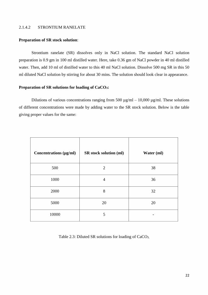
2.1.4.2 STRONTIUM RANELATE
Preparation of SR stock solution:
Strontium ranelate (SR) dissolves only in NaCl solution. The standard NaCl solution
preparation is 0.9 gm in 100 ml distilled water. Here, take 0.36 gm of NaCl powder in 40 ml distilled
water. Then, add 10 ml of distilled water to this 40 ml NaCl solution. Dissolve 500 mg SR in this 50
ml diluted NaCl solution by stirring for about 30 mins. The solution should look clear in appearance.
Preparation of SR solutions for loading of CaCO3:
Dilutions of various concentrations ranging from 500 µg/ml – 10,000 µg/ml. These solutions
of different concentrations were made by adding water to the SR stock solution. Below is the table
giving proper values for the same:
Concentrations (µg/ml)
SR stock solution (ml)
Water (ml)
500 2 38
1000 4 36
2000 8 32
5000 20 20
10000 5 -
Table 2.3: Diluted SR solutions for loading of CaCO3.

23
Drug loading of SR:
The sieved CaCO3 powder of the size below 50 microns was taken, 100 mg of which was
taken in five different 10 ml beakers and add the diluted aspirin solution to each of the beakers.
The amount of CaCO3 powder needs to be in a specific ratio with the concentration or
volume of the drug taken. This is for the drug to get entrapped within the nanoparticles it is
important to take same, double or multiple times more of the drug than the amount of CaCO3 powder
taken.
The solutions were mixed by shaking first then sonicated for about 15-30 mins. These
solutions were then kept for about 48 hours for the drug to get settled and entrapped in the CaCO3
powder. The water content evaporates slowly leaving the drug, NaCl and CaCO3 powder behind. It is
important to keep in mind that the solutions must not be disturbed or receive any sudden jerk, which
may sometimes cause the drug to come out of CaCO3.
CaCO3 powder (mg)
Diluted SR
solutions (ml)
SR quantity
(mg)
% of SR
100 5 2.5 2.5
100 5 5 5
100 5 10 10
100 5 25 25
100 5 50 50
Table 2.4: Drug loading of SR
24
After the solution is evaporated leaving only dry layer of powder at the bottom, scrape it and
collect in small 1 ml Eppendorf tubes. Close them tightly to avoid moisture content from increasing.
2.1.5 CHARACTERIZATION AND QUANTIFICATION
2.1.5.1 XRD
About 2-3 mg of CaCO3 powder, drug loaded CaCO3 powder of all the concentrations were
taken.
2.1.5.2 FE- SEM
Sample preparation:
The samples of CaCO3 powder and drug loaded CaCO3 powder of all the concentrations were
taken. Each powder was taken in very minute quantity, enough to be seen on a piece of butter paper.
Another small piece of butter paper was taken and rubbed in circular motion over the powder to get
rid of any agglomeration if present. This causes a thin film formation of the powder. The metal stubs
with a carbon tape on it having the sticky side up, is touched over this film causing the particles to
stick to the carbon tape.
These stubs with samples on them were then kept in the platinum coating machine. The machine
uses vacuum environment to bombard the sample particles with platinum particles. This after about
5-10 mins, there forms a coating of platinum metal over the sample. The main use of this is for better
conductivity for electrons to pass while viewing the images. The better the conductivity, better are
the images.
2.1.5.3 HR-LCMS
Liquid chromatography / Mass Spectroscopy (LC / MS) is a technique which combines high
performance liquid chromatography HPLC, a powerful analytical separation technique with mass
spectroscopy, a powerful analysis & detection technique. There are two common atmospheric
pressure ionization (API) LC/MS process: Electrospray Ionization (ESI) & Atmospheric Pressure
Chemical Ionization (APCI). Both are soft ionization technique. The technique combines highly

25
efficient electrospray ion generation and focusing of Agilent Jet Stream technology with a hexabore
capillary sampling array and dual-stage ion funnel for increased ion sampling and transmission.
Nano HPLC combined with mass spectrometer can analyzed small molecule as well large molecules
like proteins UHPLC separations can be detected by PDA & Mass spectrometer as different
detectors.
The samples mentioned in Buffer sample collection in section 3.1.6.1 and 3.1.6.2 were
collected in the small vials that have a rubbery entrance in the cap for sample injection. The solutions
were filtered before transferring them in the vials.
2.1.5.4 UV-Visible Spectroscopy
This technique was specially used for Strontium ranelate as it has proven to be of high
concentration for HR-LCMS. Less diluted samples are required for UV compared to the LCMS.
Here mainly this technique is used for checking drug release.
Different concentrations like 5 µg/ml, 25 µg/ml, 50 µg/ml, 100 µg/ml, 200 µg/ml of the
drug (SR) were prepared from the same stock solution. These were subjected to optical density
measurements at 323 nm by the UV-Vis spectrophotometer and a calibration curve was plotted.
Then the supernatant of the loaded mixture of SR and CaCO3 was filtered and diluted with distilled
water 5 times its volume and the filtrates were subjected to optical density tests at the same
wavelength. With the help of the resultant peaks, slope and intercept, the percent efficiency was
calculated.

26
2.1.6 DRUG RELEASE
Drugs that are once entrapped take different amount of time to get release with respect to
different concentrations. Dialysis technique was used for releasing the drug. Consideration of the
interactions of drugs and dialysis must include an understanding of the mechanism of transport
during dialysis, i.e., diffusion.
2.1.6.1 ASPIRIN
The drug was taken in the quantities mentioned in Table 2.5, inside the five separate dialysis
bags which were tied up on both the ends and hung in five separate 100 ml beakers. Take 50 ml of
PBS buffer in each of the beakers. The beakers contained small stirrer bars and kept on the magnetic
stirrers.
The aspirin now starts getting released through the permeable membrane into the buffer due
to diffusion. The buffer samples were collected after every one hour for 5 hrs straight. The last buffer
sample was collected after 19 hrs. Thus there were 5 samples of 5 different concentrations of 5 hrs
and also 5 samples of the last hour.
Buffer sample collection:
For every hour, 0.5 ml of sample was directly collected from the concentrations 500 µg/ml
and 1000 µg/ml. However, every hour collection for the concentrations 2000 µg/ml, 5000 µg/ml and
10000 µg/ml, only 0.1 ml of the buffer sample was taken and to this, 0.4 ml of fresh PBS buffer was
added. This was done to dilute the samples and keep the levels of NaCl low, so that it does not
interfere much during LCMS.

27
Sample
concentration
(µg/ml)
Quantity of
loadedCaCO3(mg)
Total initial
aspirin quantity
in loaded CaCO3
(mg)
PBS buffer in
beaker (ml)
500 40 1 50
1000 20 1 50
2000 10 2 50
5000 10 5 50
10000 10 10 50
Table 2.5: Aspirin dialysis
Preparation of standards for aspirin:
Concentration (µg) Aspirin (µg) PBS buffer (µl)
5 5 500
10 10 500
15 15 500
20 20 500
Table 2.6: Standards for aspirin
28
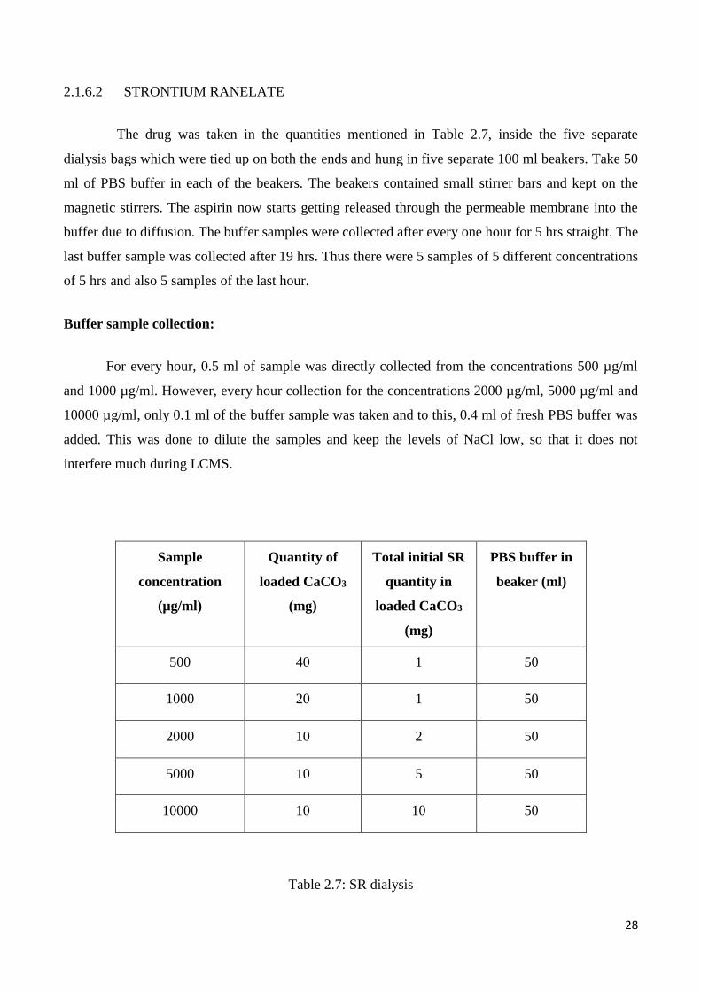
2.1.6.2 STRONTIUM RANELATE
The drug was taken in the quantities mentioned in Table 2.7, inside the five separate
dialysis bags which were tied up on both the ends and hung in five separate 100 ml beakers. Take 50
ml of PBS buffer in each of the beakers. The beakers contained small stirrer bars and kept on the
magnetic stirrers. The aspirin now starts getting released through the permeable membrane into the
buffer due to diffusion. The buffer samples were collected after every one hour for 5 hrs straight. The
last buffer sample was collected after 19 hrs. Thus there were 5 samples of 5 different concentrations
of 5 hrs and also 5 samples of the last hour.
Buffer sample collection:
For every hour, 0.5 ml of sample was directly collected from the concentrations 500 µg/ml
and 1000 µg/ml. However, every hour collection for the concentrations 2000 µg/ml, 5000 µg/ml and
10000 µg/ml, only 0.1 ml of the buffer sample was taken and to this, 0.4 ml of fresh PBS buffer was
added. This was done to dilute the samples and keep the levels of NaCl low, so that it does not
interfere much during LCMS.
Sample
concentration
(µg/ml)
Quantity of
loaded CaCO3
(mg)
Total initial SR
quantity in
loaded CaCO3
(mg)
PBS buffer in
beaker (ml)
500 40 1 50
1000 20 1 50
2000 10 2 50
5000 10 5 50
10000 10 10 50
Table 2.7: SR dialysis

29
Preparation of standards for SR:
Concentration (µg) SR (µg) NaCl solution
(µl)
PBS buffer (µl)
5 5 1 499
10 10 1 499
15 15 1 499
20 20 1 499
Table 2.8: Standards for SR

30
CHAPTER 3
3.1 RESULTS AND DISCUSSION

31
3.1.1 CHARACTERIZATION AND QUANTIFICATION
3.1.1.1 X-Ray Diffraction (XRD)
Calcium carbonate nanoparticles are abundant inorganic biomaterials with different
morphological structures that have attracted the interest of researchers in different fields. The
Calcium carbonate occurring in nature contains Aragonite. Aragonite is soluble and thus easier for
the animals or plant (there are calcareous algae, as well) to secrete. Fossil shells often have
“recrystallized” to calcite, although most sea shells originally are aragonite. X-ray diffraction is a
sensitive instrument used for the identification of crystalline phases of inorganic compounds. X-ray
powder diffraction analysis was performed.
X-ray diffraction analysis is the method by which multiple beams of X-ray create a three
dimensional picture of the density of electrons of any crystalline structure. The purpose is to identify
with a high degree of certainty, the composition of the molecules, on an atomic scale. This makes it
the most reliable method to determine the purity of calcium carbonate.
2 theta
Fig 3.1: Shows the XRD pattern of natural calcium carbonate obtained from the sea shells.
The XRD pattern of calcium carbonate obtained from the sea shells is shown in fig 3.1, with
blue colour. The blue pie diagram shows that it is 100% calcium carbonate.

32
3.1.1.2 Field Emission Scanning Electron Microscopy (FE-SEM) and (EDX)
In FE-SEM an electron beam is scanned across a sample's surface. When the electrons strike
the sample, a variety of signals are generated, and it is the detection of specific signals which
produces an image or a sample's elemental composition. The three signals which provide the greatest
amount of information in FE-SEM are the secondary electrons, backscattered electrons, and X-rays.
Secondary electrons are emitted from the atoms occupying the top surface and produce a readily
interpretable image of the surface. The contrast in the image is determined by the sample
morphology. A high resolution image can be obtained because of the small diameter of the primary
electron beam.
Backscattered electrons are primary beam electrons which are 'reflected' from atoms in the
solid. The contrast in the image produced is determined by the atomic number of the elements in the
sample. The image will therefore show the distribution of different chemical phases in the sample.
Because these electrons are emitted from a depth in the sample, the resolution in the image is not as
good as for secondary electrons. Interaction of the primary beam with atoms in the sample causes
shell transitions which result in the emission of an X-ray. The emitted X-ray has an energy
characteristic of the parent element. Detection and measurement of the energy permits elemental
analysis (Energy Dispersive X-ray Spectroscopy or EDS). EDS can provide rapid qualitative, or with
adequate standards, quantitative analysis of elemental composition with a sampling depth of 1-2
microns. X-rays may also be used to form maps or line profiles, showing the elemental distribution
in a sample surface. [21]
Fig 3.2: FE-SEM image of Calcium carbonate

33
Fig 3.3: FE-SEM images of Aspirin
Fig 3.4: FE-SEM images of Strontium ranelate
Energy dispersive X-Ray (EDX) composition analysis
Energy Dispersive X-Ray Analysis (EDX), referred to as EDS or EDAX, is an x-ray
technique used to identify the elemental composition of materials.
EDX systems are attachments to Electron Microscopy instruments like FE-SEM or
Transmission Electron Microscopy (TEM), where the imaging capability of the microscope identifies
the specimen of interest. The data generated by EDX analysis consist of spectra showing peaks
corresponding to the elements making up the true composition of the sample being analyzed.
Elemental mapping of a sample and image analysis are also possible.

34
In a multi-technique approach EDX becomes very powerful, particularly in contamination analysis
and industrial forensic science investigations. The technique can be qualitative, semi-quantitative,
and quantitative; also providing spatial distribution of elements through mapping. The EDX
technique is non-destructive and specimens of interest can be examined in situ with little or no
sample preparation. [20]
Fig 3.5: Shows the weight % of chemical composition of CaCO3
3.1.2 DRUG LOADING STUDIES
In recent years, the development of analytical methods for simultaneous determinations of
drugs has gained considerable attention due to their importance in quality control testing of drugs
and their products and in ordinary laboratories because of their wide availability and suitability.
Since no method is reported in literature for the simultaneous quantification of aspirin and strontium
in tablets for the routine quality control assay in ordinary laboratories, the development of such
method could be appreciable.

35
3.1.2.1 ASPIRIN
The drug content was evaluated for all the concentrations and it was observed that the
nanoparticles of only CaCO3 that showed inter-particle spaces and crevices were now appeared filled
up. The drug thus is entrapped within or between the nanoparticles. These results were mainly
attributed to aspirin powders increasing the charge density. Various solvent systems like distilled
water, ethanol and mixture of ethanol: water were tried and the ethanol: water proved to give the best
loading of drug. Maximum loading obtained in this work is 50%. The water is used because it
provides some hold to the ethanol moiety for better loading of drug. It makes the process of
evaporation slower to a good extent causing lesser harm to the nanoparticle’s structures. Below are
the FE-SEM images that show increase in the drug loading with an increase in the drug concentration
while keeping the quantity of calcium carbonate powder same for all formulations.
Fig 3.6: Aspirin loading of ratio 1:16 as per Table 4.2

36
Fig 3.7: Aspirin loading of ratio 1:4 as per Table 4.2
Fig 3.8: Aspirin loading of ratio 1:1 as per Table 4.2

37
Fig 3.9: Aspirin loading of ratio 5:2 as per Table 4.2
Fig 3.10: Aspirin loading of ratio 5:1 as per Table 4.2
38
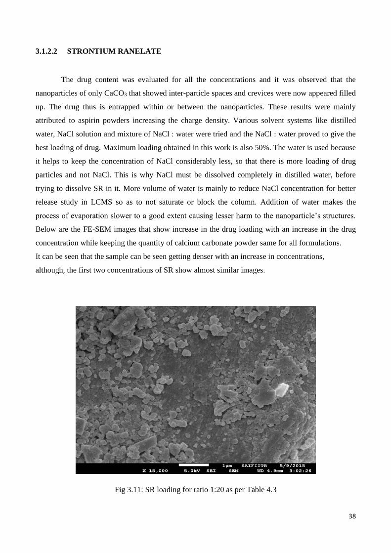
3.1.2.2 STRONTIUM RANELATE
The drug content was evaluated for all the concentrations and it was observed that the
nanoparticles of only CaCO3 that showed inter-particle spaces and crevices were now appeared filled
up. The drug thus is entrapped within or between the nanoparticles. These results were mainly
attributed to aspirin powders increasing the charge density. Various solvent systems like distilled
water, NaCl solution and mixture of NaCl : water were tried and the NaCl : water proved to give the
best loading of drug. Maximum loading obtained in this work is also 50%. The water is used because
it helps to keep the concentration of NaCl considerably less, so that there is more loading of drug
particles and not NaCl. This is why NaCl must be dissolved completely in distilled water, before
trying to dissolve SR in it. More volume of water is mainly to reduce NaCl concentration for better
release study in LCMS so as to not saturate or block the column. Addition of water makes the
process of evaporation slower to a good extent causing lesser harm to the nanoparticle’s structures.
Below are the FE-SEM images that show increase in the drug loading with an increase in the drug
concentration while keeping the quantity of calcium carbonate powder same for all formulations.
It can be seen that the sample can be seen getting denser with an increase in concentrations,
although, the first two concentrations of SR show almost similar images.
Fig 3.11: SR loading for ratio 1:20 as per Table 4.3

39
Fig 3.12: SR loading for ratio 1:4 as per Table 4.3
Fig 3.13: SR loading for ratio 1:2 as per Table 4.3

40
Fig 3.14: SR loading for ratio 1:1 as per Table 4.3
Fig 3.15: SR loading for ratio 2:1 as per Table 4.3

41
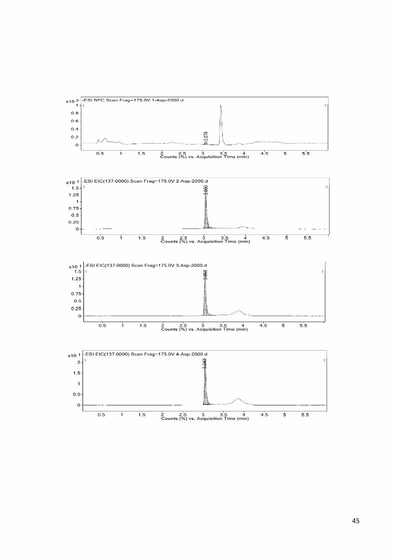
3.1.3 DRUG RELEASE STUDIES
3.1.3.1 ASPIRIN
The drug release for aspirin was done by HR-LCMS technique. A main objective of using
porous carriers as a drug delivery system is to improve dissolution rate. Dissolution experimental
methods have shown that drug loading into porous nanoparticles can accelerate drug release. The
faster drug dissolution of the loaded drug was attributed to the large surface area of the calcium
carbonate nanoparticles which are thus loaded efficiently. Increased crystallization pressure in pores
leads to locally increased solubility and might be an additional mechanism for faster drug release.
Drug release from drug-loaded calcium carbonate was investigated without specific formulation
strategies to eliminate influences altering the dissolution rate, and to have direct information on the
performance of drug-loaded particles.
Conc.
(µg/ml)
↓
1st hour
2nd hour
3rd hour
4th hour
5th hour
Last hour (after
19 hrs)
Peak Areas
500
1304054
1110467
1236587
9911476
1207767
1286887
1000
1674158
1414768
1314158
1360398
1471738
5406717
2000
1329527
2057347
2192447
2536127
2440587
1163997
5000
7416827
9145467
9363317
1016338
9608017
6097227
10000
8169697
9484467
9488767
1051198
1024598
5774576
Table 3.1: High-Resolution LCMS areas according to concentration and time

42

43
Fig 3.16: The results for the concentration 500 µg/ml show that aspirin released showed fast release
from the 2nd hour till the last hour. Although considering the peak areas, it can be seen that the
release shows exponential increase at the beginning of second hour and reduces as it moves towards
the last hour.

44
Fig 3.17: The HR-LCMS results for the concentration 1000 µg/ml show that aspirin released at a
rapid release right from the first hour till the last hour. Although considering the peak areas, it can be
seen that the release shows exponential increase at the beginning of second hour and reduces as it
moves towards the last hour.
45

46
Fig 3.18: The HR-LCMS results for the concentration 2000 µg/ml show that aspirin released at a
rapid release right from the second hour till the last hour. Although considering the peak areas, it can
be seen that the release shows exponential increase at the beginning of second hour and reduces as it
moves towards the last hour.

47
Fig 3.19: The HR-LCMS results for the concentration 5000 µg/ml show that aspirin released at a
rapid release right from the first hour till the last hour. Although considering the peak areas, it can be
seen that the release shows exponential increase at the beginning of second hour and reduces as it
moves towards the last hour.

48

49
Fig 3.20: The HR-LCMS results for the concentration 10,000 µg/ml show that aspirin released at a
rapid release right from the first hour till the last hour. But compared to first 5 hours, the release was
lesser. Although considering the peak areas, it can be seen that the release shows exponential
increase at the beginning of second hour and reduces as it moves towards the last hour.
4 6 8 10 12 14 16 18 20 22
15000000
20000000
25000000
30000000
35000000
40000000
45000000
Area
Linear Fit of Area
Are
a
Concentration (micro gram/ml)
Equation y = a + b*
Adj. R-Squar 0.99484
Value Standard Erro
Area Intercept 7.76562E 968452.7660
Area Slope 1.70272E 70725.79012
Fig 3.21: The calibration curve of linearity for Aspirin.

50
0 2000 4000 6000 8000 10000
0.00E+000
2.00E+007
4.00E+007
6.00E+007
8.00E+007
1.00E+008
1.20E+008
1.40E+008
1.60E+008
1.80E+008
Are
a
Concentration (micro gram/ml)
1st hour
2nd hour
3rd hour
4 hour
5 hour
Last hour
Fig 3.22: The graphical representation of the release of all the concentrations.
Concentrations (µg/ml) Drug : CaCO3
500 1 : 16
1000 1 : 5
2000 1 : 1
5000 2.5 : 1
10000 5 : 1
Table 3.2: The ratios of aspirin drug to calcium carbonate for loading

51
3.1.3.2 STRONTIUM RANELATE
Different concentrations of the drug (SR) were prepared from the stock solution, ranging
from 5 µg-100 µg/ml. Optical density measurements were taken at 323 nm by the UV-Vis
spectrophotometer and a calibration curve was plotted
Fig 3.23: Overlay spectra of linearity (5-100 μg/ml) of Strontium ranelate
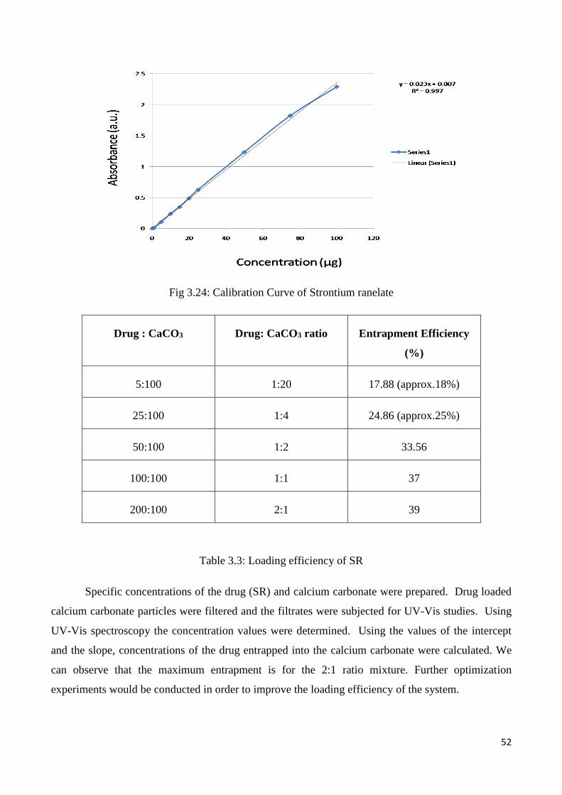
52
Fig 3.24: Calibration Curve of Strontium ranelate
Drug : CaCO3 Drug: CaCO3 ratio Entrapment Efficiency
(%)
5:100 1:20 17.88 (approx.18%)
25:100 1:4 24.86 (approx.25%)
50:100 1:2 33.56
100:100 1:1 37
200:100 2:1 39
Table 3.3: Loading efficiency of SR
Specific concentrations of the drug (SR) and calcium carbonate were prepared. Drug loaded
calcium carbonate particles were filtered and the filtrates were subjected for UV-Vis studies. Using
UV-Vis spectroscopy the concentration values were determined. Using the values of the intercept
and the slope, concentrations of the drug entrapped into the calcium carbonate were calculated. We
can observe that the maximum entrapment is for the 2:1 ratio mixture. Further optimization
experiments would be conducted in order to improve the loading efficiency of the system.

53
CHAPTER 4
CONCLUSION AND FUTURE SCOPE
54
CONCLUSION
Contrary to common belief, we demonstrated that drug loading by solvent evaporation is
simple, fast, and efficient. In particular, the solvent-evaporation method offers three advantages over
the widely used impregnation method: the resulting drug loading can be precisely determined in
advance, a high drug loading can be achieved in a short time, and the loading efficiency is good up to
the loading capacity of calcium carbonate or other porous carriers, so there is also no waste of
expensive drugs). The combination of qualitative FE-SEM analysis and HPLC quantification was
sufficient to estimate both loading efficiency and loading capacity. Nevertheless, drug loading up to
50% with good release was achieved. Proof of feasibility of the solvent-evaporation method was
mainly attributed to the unique properties of the model carrier used in this study. Drug-loading
capacity could possibly be increased by slowing evaporation even more, thus allowing enough time
for diffusion of drug into the core of the particles. Dissolution experiments proved that drug release
can be accelerated by loading poorly water-soluble drugs into nanoparticles by the solvent-
evaporation method. In contrast to the impregnation method based on adsorption, solvent
evaporation led to a low fraction of amorphous drug after drug loading. The faster drug release can
be explained by surface enlargement of the drug which may be caused due to heat during drying
even though less and locally increased drug solubility.
FUTURE SCOPE
Drug-loaded calcium carbonate may be used in capsules or tablets for oral delivery. Possible
applications include the formulation of poorly soluble compounds to accelerate the dissolution.
Sustained release formulations of highly soluble drugs can be obtained by coating of loaded
particles. Technical advantages include the formulation of drugs which are administered at low
doses, or the preparation of orally dispersible tablets. These findings are promising for the future
development of carrier-based drug delivery systems.
55
REFERENCES
1. Samuel A. Wickline, et al., Nanotechnology for Molecular Imaging and Targeted Therapy.
Circulation, 107: 1092-1095. (2003)
2. Small is the new trend in the medical industry, Digi News, from
http://www.digimouth.com/news/small-is-the-new-big-in-the-medical-world.html, (2011)
3. Frank X. Gu, et al., Targeted nanoparticles for cancer therapy. Nano Today, Volume 2, Issue
3, Pages 14-21. (2007)
4. Michael Goldberg, et al., Nanostructured materials for applications in drug delivery and
tissue engineering. Journal of Biomaterials Science Polymer Edition, 18(3): 241–268. (2007)
5. How nanotechnology is shaping stem cell research, The Guardian, from
http://www.theguardian.com/nanotechnology-world/nanotechnology-shaping-stem-cell-
research (2012)
6. Cuenca AG., et al., Emerging implications of nanotechnology on cancer diagnostics and
therapeutics. Cancer, 107(3):459-66, (2006)
7. Stephen Mahler, et al., Advances in drug delivery. Journal of Chemical Technology and
Biotechnology, DOI: 10.1002/jctb.4689 (2015)
8. Murugan Ramalingam, et al., Tissue Engineering and Regenerative medicine: A Nano
approach. CRC Press, 454-458, (2012)
9. Aspirin, NHS Choices, from http://www.nhs.uk/Conditions/Anti-platelets-aspirin-low-dose-
/Pages/Introduction.aspx
10. What is aspirin? What is aspirin for? from
http://www.medicalnewstoday.com/articles/161255.php
11. Aspirin use during pregnancy. Drugs.com., from
http://www.drugs.com/pregnancy/aspirin.html
12. 'Hands-on' self-test materials for organic structure solution from powder diffraction data,
International union of crystallography, http://www.iucr.org/resources/commissions/powder-
diffraction/projects/organic-datasets (2009)
13. A. Semalty, et al., Development and characterization of aspirin-phospholipid complex form
improved drug delivery. International Journal of Pharmaceutical Sciences and
Nanotechnology, Volume 3, Issue 2, (2010)

56
14. A. Salehi, et al., Simultaneous UV-VIS spectrophotometric determination of aspirin and
methocarbamol in tablets. Research in Pharmaceutical Sciences, 7(5), (2012)
15. Mancini Marisabel Mourelle, et al., New strontium salts, synthesis and use thereof in the
treatment of osteoporosis. EP 2 530 068 A1 (2012)
16. Find a vitamin or supplement strontium. WebMD - Therapeutic Research Faculty, from
http://www.webmd.com/vitamins-supplements/ingredientmono-1077-
strontium.aspx?activeingredientid=1077&activeingredientname=strontium (2009)
17. Reginster JY., Strontium ranelate in osteoporosis. Current Pharmaceutical Design,
8(21):1907-16, (2002)
18. Abdullahi Shafiu Kamba, et al., Synthesis and Characterisation of Calcium Carbonate
Aragonite Nanocrystals from Cockle Shell Powder (Anadara granosa). Journal of
Nanomaterials, Article ID 398357, 9 pages (2013)
19. Daniel Preisig, et al., Drug loading into porous calcium carbonate microparticles by solvent
evaporation. European Journal of Pharmaceutics and Biopharmaceutics, Volume 87, Issue 3,
Pages 548–558 (2014)
20. Y. Ueno, et al., Drug-incorporating calcium carbonate nanoparticles for a new delivery
system. Journal of Controlled Release, Volume 103, Issue 1, Pages 93–98, (2005)
21. SEM/EDS - Scanning Electron Microscopy with X-ray microanalysis, from
http://wings.buffalo.edu/faculty/research/scic/sem-eds.html

57
APPENDIX
1. HR-LCMS SOFTWARE
Agilent Technologies
Mass hunter workstation software. LCMS data acquisition for 6200 series TOF/6500 series
Q-TOF.
Version B.05.01
Build 5.01.5125.1
2. X-RAY DIFFRACTION
Philips powder diffractometer PW3040/60 X’pert pro
3. UV-VIS SPECTROPHOTOMETRY
Agilent Technologies
Cary 100 series
4. SCANNING ELECTRON MICROSCOPY
JSM – 7600 F
Software: PC-SEM
Sputtering machine: JEOL JFC – 1600 Auto fine coater

![Filler and Coating Pigments for Papermakers€¦ · includes SC, MFC, WFU, and WFC) are calcium carbonate (pre-cipitated calcium carbonate [PCC] and ground calcium carbonate [GCC]),](https://static.fdocuments.in/doc/165x107/5eae9d439cc5d419877523e0/filler-and-coating-pigments-for-papermakers-includes-sc-mfc-wfu-and-wfc-are.jpg)