Natroapophyllite, a new orthorhombic sodium … a new orthorhombic sodium analog of apophyllite I....
14
American Mineralogisl, Volume 66, pages410-423, l98I Natroapophyllite,a new orthorhombicsodium analog of apophyllite I. Description,occurrence, and nomenclature HtRou.lnu MATSUEDA Institute of Mining Geology Akita University, Akita 010, Japan YesuNoRr Mrun-e.' eNo JoHN RucrrDce Department of Geology, University of Toronto Toronto, Ontario MsS IAI, Canada Abstract NatroapophylliteNaCaoSirO2oF ' 8H2O, a sodium analogof apophyllite,hasbeenfound in white skarn at the SampoMine, Okayama, Japan.Electron microprobe analysis of the min- eralgives SiO252.79, CaO25.4l, NarO 3.05,K2O 0.33,F 2.27,HrO (by difference) l7.ll weight percent; (-O=F' : 0.96). Optical and crystallographic parameters are: a: 1.536(2), B : 1.538(2), y: l.5tA(2),2V(meas.):32(l)", r<v;a:8.875(4), D:8.881(6), c: 15.79(l)A, space group Pnnm,Z : 2; D obs. : 2.50,D calc. : 230 g/cm3. The crystal structure, refaed (R : 0.056) by least-squares methods from 563ditruseX-ray reflections collectedwith an automatic single-crystal difractometer, has rings of SiOotetra- hedra. Ca- and Na-polyhedra are more distorted in natroapophyllite comparedwith apo- phyllite because of the substitutionof Na for K. TGA shows discontinuous water lossof 16 weight percent,similar to apophyllite. DTA and IR data suggest that natroapophyllite has hydrogen atoms in three di-fferentstructural environments. A pseudo solid-solutionseries exists between natroapophylliteand fluorapophyllite which bridges the orthorhombic-tetragonalsymmetry change.Crystals are shown which display continuousand discontinuous zoning. Introduction The nomenclature of the apophyllite group is com- plex. Fluorapophyllite and hydroxyapophyllite have recently been defined by Dunn et al. (1978) as the valid names for the end membersof the tetragonal apophyllite series, in which K is the major alkali cat- ion. Orthorhombic varietiesof apophyllite have also beendescribed. Sahama (1965) gavedetailsof an or- thorhombic fluorine-rich apophyllite KCa.AlSi, OroF'8HrO, which contained significant Al. Although Na may substitute for K in apophyllite to a small degree, samples in which Na predominates over K have only recentlybeendescribed (Matsueda, 1975,1980; Miura et al., 1916; Miura, 1977; Miva I On leavefrom Departmentof MineralogicalSciences and Ge- ology, Faculty of Science, YamaguchiUniversity, Yamaguchi 753' Japan. and Rucklidge, 1979). The Na-rich examples are in- variably orthorhombic and fluorine-rich, and the name natroapophyllite has beenproposed for this so- dium analog of the orthorhombic apophyllite de- scribed originally by Sahama (1965) and Belsare (1969).It is not the sodium analog of ordinary tetra- gonal apophyllite. The name natroapophyllite,given in allusion to its compositionalrelation to apophyl- lite, was approvedby the IMA Commission on New Minerals and New Mineral Namesin October, 1976. Other additional prefixesfor natroapophyllitewould be in order, e.9., ortho- and fluor-, but in the absence of non-orthorhombicor other than fluorine-rich nat- roapophyllite,it seems unnecessary to burden the lit- erature with more complex names than natroapo- phyllite, which is defined as being the orthorhombic sodium end member of the apophyllite group miner- als with the formula NaCaoSi'OroF'8HrO. 0003-004x/8 I /030+04 l 0$02.00 410
-
Upload
trinhkhanh -
Category
Documents
-
view
222 -
download
1
Transcript of Natroapophyllite, a new orthorhombic sodium … a new orthorhombic sodium analog of apophyllite I....

American Mineralogisl, Volume 66, pages 410-423, l98I
Natroapophyllite, a new orthorhombic sodium analog of apophyllite
I. Description, occurrence, and nomenclature
HtRou.lnu MATSUEDA
Institute of Mining GeologyAkita University, Akita 010, Japan
YesuNoRr Mrun-e.' eNo JoHN RucrrDce
Department of Geology, University of TorontoToronto, Ontario MsS IAI, Canada
Abstract
Natroapophyllite NaCaoSirO2oF ' 8H2O, a sodium analog of apophyllite, has been found inwhite skarn at the Sampo Mine, Okayama, Japan. Electron microprobe analysis of the min-eralgives SiO2 52.79, CaO25.4l, NarO 3.05, K2O 0.33, F 2.27, HrO (by difference) l7.llweight percent; (-O=F' : 0.96). Optical and crystallographic parameters are: a: 1.536(2), B: 1.538(2) , y : l .5 tA(2) ,2V(meas.) :32( l ) " , r<v;a:8.875(4) , D:8.881(6) , c : 15.79( l )A,space group Pnnm, Z : 2; D obs. : 2.50, D calc. : 230 g/cm3.
The crystal structure, refaed (R : 0.056) by least-squares methods from 563 ditruse X-rayreflections collected with an automatic single-crystal difractometer, has rings of SiOo tetra-hedra. Ca- and Na-polyhedra are more distorted in natroapophyllite compared with apo-phyllite because of the substitution of Na for K. TGA shows discontinuous water loss of 16weight percent, similar to apophyllite. DTA and IR data suggest that natroapophyllite hashydrogen atoms in three di-fferent structural environments.
A pseudo solid-solution series exists between natroapophyllite and fluorapophyllite whichbridges the orthorhombic-tetragonal symmetry change. Crystals are shown which displaycontinuous and discontinuous zoning.
Introduction
The nomenclature of the apophyllite group is com-plex. Fluorapophyllite and hydroxyapophyllite haverecently been defined by Dunn et al. (1978) as thevalid names for the end members of the tetragonalapophyllite series, in which K is the major alkali cat-ion. Orthorhombic varieties of apophyllite have alsobeen described. Sahama (1965) gave details of an or-thorhombic fluorine-rich apophyllite KCa.AlSi,OroF'8HrO, which contained significant Al.
Although Na may substitute for K in apophylliteto a small degree, samples in which Na predominatesover K have only recently been described (Matsueda,1975, 1980; Miura et al., 1916; Miura, 1977; Miva
I On leave from Department of Mineralogical Sciences and Ge-ology, Faculty of Science, Yamaguchi University, Yamaguchi 753'Japan.
and Rucklidge, 1979). The Na-rich examples are in-variably orthorhombic and fluorine-rich, and thename natroapophyllite has been proposed for this so-dium analog of the orthorhombic apophyllite de-scribed originally by Sahama (1965) and Belsare(1969). It is not the sodium analog of ordinary tetra-gonal apophyllite. The name natroapophyllite, givenin allusion to its compositional relation to apophyl-lite, was approved by the IMA Commission on NewMinerals and New Mineral Names in October, 1976.Other additional prefixes for natroapophyllite wouldbe in order, e.9., ortho- and fluor-, but in the absenceof non-orthorhombic or other than fluorine-rich nat-roapophyllite, it seems unnecessary to burden the lit-erature with more complex names than natroapo-phyllite, which is defined as being the orthorhombicsodium end member of the apophyllite group miner-als with the formula NaCaoSi'OroF'8HrO.
0003-004x/8 I /030+04 l 0$02.00 410
MATSUEDA ET AL.: NATROAPOPHYLLITE
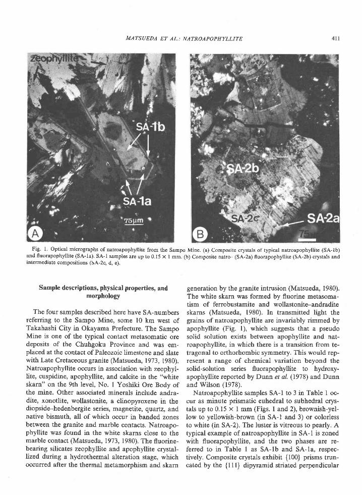
Fig' l. Optical micrographs of natroapophyllite from the Sampo Mine. (a) Composite crystals of typical natroapophyllite (SA-lb)and fluorapophyllite (SA-la). SA-l samples are up to 0.15 x I mm. (b) Composite natro- (SA-2a) fluorapophyllite (SA-2b) crystals andintermediate compositions (SA-2c, d, e).
Sample descriptions, physical properties, andmorphology
The four samples described here have SA-numbersreferring to the Sampo Mine, some l0 km west ofTakahashi City in Okayama Prefecture. The SampoMine is one of the typical contact metasomatic oredeposits of the Chuhgoku Province and was em-placed at the contact of Paleozoic limestone and slatewith Late Cretaceous granite (Matsueda, 1973, 1980).Natroapophyllite occurs in association with zeophyl-lite, cuspidine, apophyllite, and calcite in the 'hrhite
skarn" on the 9th level, No. I Yoshiki Ore Body ofthe mine. Other associated minerals include andra-dite, xonotlite, wollastonite, a clinopyroxene in thediopside-hedenbergite series, magnetite, quartz, andnative bismuth, all of which occur in banded zonesbetween the granite and marble contacts. Natroapo-phyllite was found in the white skarns close to themarble contact (Matsueda, 1973, 1980). The fluorine-bearing silicates zeophyllite and apophyllite crystal-lized during a hydrothermal alteration stage, whichoccurred after the thermal metamorphism and skarn
generation by the granite intrusion (Matsueda, 1980).The white skarn was formed by fluorine metasoma-tism of ferrobustamite and wollastonite-andraditeskarns (Matsueda, 1980). In transmitted light thegrains of natroapophyllite are invariably rimmed byapophyllite (Fig. l), which suggests that a pseudosolid solution exists between apophyllite and nat-roapophyllite, in which there is a transition from te-tragonal to orthorhombic symmetry. This would rep-resent a range of chemical variation beyond thesolid-solution series fluorapophyllite to hydroxy-apophyllite reported by Dunn et al. (1978) and Dunnand Wilson (1978).
Natroapophyllite samples SA-l to 3 in Table I oc-cur as minute prismatic euhedral to subhedral crys-tals up to 0.15 x I mm (Figs. I and2), brownish-yel-low to yellowish-brown (in SA-l and 3) or colorlessto white (in SA-2). The luster is vitreous to pearly. Atypical example of natroapophyllite in SA-l is zonedwith fluorapophyllite, and the two phases are re-ferred to in Table I as SA-lb and SA-la, respec-tively. Composite crystals exhibit {100} prisms trun-cated by the {l I l} dipyramid striated perpendicular

4t2 MATSUEDA ET AL: NATROAPOPHYLLITE
Table l. Occurrences, parag€nes€s, compositions, and optie axial angles of fluorapophyllite and natroapophyllite from the Sampo Mine
Spec iEen No. Occurrence Paragenes is A lka l j - con tenE* 2V(neas . )
SA- la
SA- Ib
SA-2a
SA-2b
SA-2c
SA-2d
SA-2e
SA-3a
SA-3b
SA-3c
sA-4
1 Whlte skarn' ( 9L No .1 Yosh i k iZeophy l l i te , ca lc i te ,andrad i te , f luor i te , cusp id ine
l lagnet i te , andrad i te ,f luor i te
F luor i te , xonoto l i te ,andradite, magnetite, cuspidlne
F luor i te , andrad i te
Kt . o8Nto. ozc"4. t3Ko.o6Nto .9oc"4 .13
Ko .03*"0 . 86c"4 . 08Kr . olNto. 02c"4 . 09Ko. 4oN"o. z8ct4 . ooKo. 85Nto. roca4. 16Kt . ogN"o. o3c"4. ol
Ko. o7N"o . 75c '4 . r2Ko.45Nuo.5 lc t4 . l2*o .95Nto .03c"4 . zo
"o .9oNto .o3c t4 . t3
ore Body) I
- ? l
> > 0
> 0
1
=30
1
> 0
Druse in white skarn(9L No.1 Yosh ik i Ore Body)
White skarn( 9 L W e s t N o , I Y o s h i k i O r eBody)
Spherical skarn j.n marble(10L l { res t . No. l Yosh ik iOre Body)
* Atonic z'atios of aLkali ions on the basis of Si = 8.00.
to the c axis (Fig. la). Specimen SA-2 is subdivided2a-2e for varying compositions in the range natro- tofluorapophyllite. It exhibits well-developed fl I U di-pyramids with {100} prisms which give the sample a"fish-eye" appearance (Figs. lb and2\.
The external morphology of the crystals is sinilar
to that offluorapophyllite (Figs. I andz) because, asshown below, the crystals are composites with therims being K-rich fluorapophyllite and the cores Na-rich natroapophyllite. Optical measurement could be
Fig. 2. Scanning electron micrographs of natroapophyllite (SA-2a). (a) Subhedral crystals of SA-2 showing 'fish eye' appearance. (b)
Euhedral crytsals of SA-2.

MATSUEDA ET AL.: NATROAPOPHYLLITE
o(41) o
ox)Qc'rz l
%.\
@
s i ( 1 )
,a Ca( l ) O.J o(41)O I
!"-els'
O o ( 4 1 )
Fig. 3. The Si6O2s sheet in natroapophyllite projected on (001).
made most reliably on specfunen SA-l in which the Kand Na phases are easily distinguished. The Na-richcomponent is colorless in thin section and distinctlybiaxial with 2V(meas.) : 3211;o, r 1 via : 1.536(2),B : 1.538(2) and y : 1.54r'r(2\. The K-rich com-ponent is uniaxid with the properties of normal fluor-apophyllite, ar : 1.536 and e : 1.539 (see Table l).Biaxial examples of the K-rich component are alsonoted in Table l, but the detailed description of thesephases will be reserved for a separate paper.
Cleavage is perfect on {001} and poor on [ll0].The Mohs hardness is 4 to 5, and the streak is lightgray. The mineral does not fluoresce in UV radia-tion. An IR spectrum of natroapophyllite is shown inPart IL The powder used for this was carefully sepa-rated from the K-rich fluorapophyllite parts of thecrystals. The spectrum shows two sharp peaks at3560 and 3420 cm-' and one broad peak at 3020cm-'. These correspond to two strong and one weakhydrogen bonds (see Part II). The specific gravity,measured by the suspension method in Thoulet's so-lution on somewhat impure materials insluding small
amounts of calcite and fluorite is 2.50, and the calcu-lated value is 2.30. Natroapophyllite is slightly sol-uble in l: I HCI and 1: I HNO3.
Chemistry
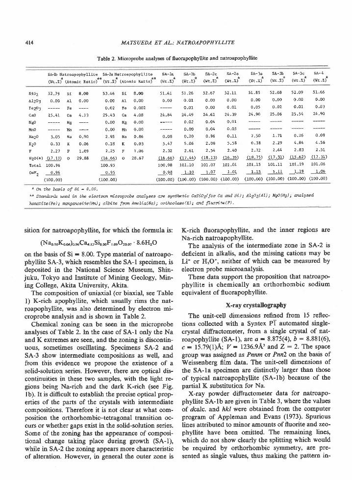
The chemical compositions of the natroapophylliteand fluorapophyllite crystals were determined withthe JXA-5A electron microprobe analyzer at KytrshtrUniversity. The analyses, which were performed atl5kV accelerating voltage and l5nA sample current,are shown in Table 2, along with a list of the stan-dards used. Water was determined by difference. Noother elements besides those listed were detected ineither form of apophyllite from the Sampo.Mine.Wet-chemical analysis of somewhat impure materialincluding a small amount of apophyllite from thesame locality was performed by Dr. K. Ishibashi ofFukuoka University, using weight loss on ignition.The results are HrO(+) : 15.83 and HrO(-) : 0.14weight percent. An analysis of the most Na-rich spec-imen (SA-lb) with 3.05 weight percent NarO is pre-sented in Table 2. We define this as the type compo-
414 MATSUEDA ET AL; NATROAPOPHYLLITE
Table 2. Microprobe analyses of fluorapophyllite and natroapophyllite
SA-lb NatroapophyLlite* **(I{t.Z) (Atonlc Ratio)
SA-2a Natroapophyllite SA-la SA-2b* * * * *
(wr.Z) (Atontc lratlo) (wt.U) (wt.Z)
SA-3a SA-3b SA-1c SA-4* * * *
( l l r .z) (wr.U ) (wr.Z) ( I . l t .U )
SA-2c
(wt. z)SA-2e
(wr .z )
slo2 52.79 s1
Al2O3 0 .00 A l
Fe2p3 Fe
CaO 25.4L Ca
Mso Mg
llno l4n
Na2O 3.05 Na
K2o 0 .33 K
E 2 . 2 7 F
Itzo(+) (I7.1r) oTota l I00 .96
0=F" 0 .96
(100.00)
8.00 51.46 Si 8,00
0 .oo 0 .00 A1 0 .00
0 .02 Fe 0 .002
4 .L3 25 .43 ca 4 .08
0 .00 Mg 0 .00
0.00 lh 0.00
0 .90 2 .95 Na 0 .85
0 . 0 5 0 . 1 8 K 0 . 0 3
1 .09 2 .25 F r . 06
29 .88 (16 .66 ) o 28 .67
100.95
0 . 95(100.00)
51,61 5L.26 52.67 52.Lr
0 .00 0 .01 0 .00 0 .00
0 .01 0 .00 0 .01
24.84 24.49 24.6L 24.39
0 .02 0 .04 0 .01
0 .00 0 .04 0 .05
0 .08 0 .20 0 .96 0 .11
5 . 47 5 . 06 2 .08 5 . 58
2 . 3 2 2 . 6 L 2 . 5 4 2 . 4 0
(16 .65 ) ( 17 .44 ) ( 18 ,13 ) ( 16 .35 )
100.98 101.10 101.07 101.01
0 .98 1 .10 1 07 1 .01
(100.00) (100.00) (100.00) (100.00)
51.85 52.08 52.O9 5L,66
0 .00 0 .00 0 .00 0 .00
0 . 0 5 0 , o 2 0 . 0 1 0 . 0 3
24.90 25.06 25,54 24.90
2 . 5 0 1 . 7 r
0 . 3 8 2 . 2 9
2 . 7 2 2 . 6 4
(18 .75 ) ( 17 ,31 )
101.15 101.11
1 ,15 1 .11
(r00.00) (100.00)
0 .26 0 .09
4 . 8 4 4 . 5 6
2 . 8 3 2 . 5 L
( r5 .62 ) ( 17 .31 )
101.19 10r.05
1 .19 1 .06
(10o.oo) (100.00)
1 0n the baeie of Si -- 8.00.
** Standrcds wed i.n tlp electm mi.crcprcbe aalyeee m syntletic CaSi?3(fon Ca and Si.); AL2og(AL); ag?(Mg); oatyzed
lurcttte(Fe); nwryorceite(I,ld; albite fzon AreLia(Nd; orbloclase(D; od f'Lrcrtte(F).
sition for natroapophyllite, for which the formula is:
(Nao.rolQ.*)o.ruCao., r Si, *F,.o"Or0..' 8.6HrO
on the basis of Si : 8.00. Type material of natroapo-phy[ite SA-3, which resembles the SA-l specimen, isdeposited in the National Science Museum, Shin-juku, Tokyo and Institute sf I\4ining Geology, Min-ing College, Akita University, Akita.
The composition of uniaxial (or biaxial, see Tablel) K-rich apophyllite, which usually rims the nat-roapophyllite, was also determined by electron mi-croprobe analysis and is shown in Table 2.
Chemical 2sning can be seen in the microprobeanalyses of Table 2. In the case of SA-l only the Naand K extremes are seen, and the zoning is discontin-uous, somet:mes oscillating. Specimens SA-2 andSA-3 show intermediate compositions as well, andfrom this evidence we propose the existence of asolid-solution series. However, there are optical dis-continuities in these two samples, with the light re-gions being Na-rich and the dark K-rich (see Fig.lb). It is difficult to establish the precise optical prop-erties of the parts of the crystals with intermediatecompositions. Therefore it is not clear at what com-position the orthorhombic-tetragonal transition oc-curs or whether gaps exist in the solid-solution series.Some of 1ls ssning has the appearance of composi-tional change taking place during growth (SA-l),while in SA-2 the 2ening appears more characteristicof alteration. However, in general the outer zone is
K-rich fluorapophyllite, and the inner regiorxi areNa-rich natroapophyllite.
The analysis of the intermediate zone in SA-2 isdeficient in alkalis, and the nissing cations may beLi* or HrO*, neither of which can be measured byelectron probe microanalysis.
These data support the proposition that natroapo-phyllite is chemically an orthorhombic sodiumequivalent of fluorapophyllite.
X-ray crystallography
The unit-cell dinensions refined from 15 reflec-tions collected with a Syntex PT automated single-crystal diffractometer, from a single crystal of nat-roapophyllite (SA-1), are a:8.875(4), b : 8.881(6),c : 15.79(l)A; V: 1236.943 andZ: 2' The spacegroup was assigned as Pnnm or Pnn2 on the basis ofWeissenberg film data. The unit-cell dimensions ofthe SA-la specimen are distinctly larger than thoseof typical natroapophyllite (SA-lb) because of thepartial K substitution for Na.
X-ray powder di-ffractometer data for natroapo-phyllite SA-lb are given in Table 3, where the valuesof dcalc. and hkl were obtained from the computerprogram of Appleman and Evans (1973). Spuriouslines attributed to minor amounts of fluorite and zeo-phyllite have been omitted. 1trs lsp4ining lines,which do not show clearly the splitting which wouldbe required by orthorhombic symmetry, are pre-sented as single values, llus 6aking the pattern in-
MATSUEDA ET AL.: NATROAPOPHYLLITE 4t5
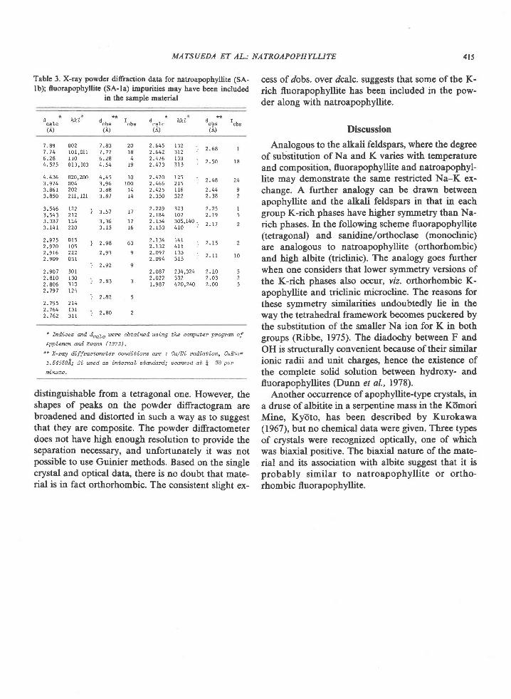
Table 3. X-ray powder diffraction data for natroapophyllite (SA-lb); fluorapophyllite (SA-la) impurities may have been included
in the sample material
cess of dobs. over dcalc. suggests that some of the K-rich fluorapophyllite has been included in the pow-der along with natroapophyllite.
Discussion
Analogous to the alkali feldspars, where the degreeof substitution of Na and K varies with temperatureand composition, fluorapophyllite and natroapophyl-lite may demonstrate the same restricted Na-K ex-change. A further analogy can be drawn betweenapophyllite and the alkali feldspars in that in eachgroup K-rich phases have higher symmetry than Na-rich phases. In the following scheme fluorapophyllite(tetragonal) and sanidine/orthoclase (monoclinic)are analogous to natroapophyllite (orthorhombic)and high albite (trictinic). The analogy goes furtherwhen one considers that lower symmetry versions ofthe K-rich phases also occur, viz. orthorhombic K-apophyllite and triclinic microcline. The reasons forthese symmetry similarities undoubtedly lie in theway the tetrahedral framework becomes puckered bythe substitution of the smaller Na ion for K in bothgroups (Ribbe, 1975). The diadochy between F andOH is structurally convenient because of their similarionic radii and unit charges, hence the existence ofthe complete solid solution between hydroxy- andfluorapophyllites (Dunn et al., 1978).
Another occurrence of apophyllite-type crystals, ina druse of albitite in a serpentine mass in the KdmoriMine, Kyoto, has been described by Kurokawa(1967), but no chemical data were given. Three typesof crystals were recognized optically, one of whichwas biaxial positive. The biaxial nature of the mate-rial and its association with albite suggest that it isprobably similar to natroapophyllite or ortho-rhombic fluorapophyllite.
carc
rilh.kL d - r -
oDs obs(A)
d . h k tcarc
t,&ld . r -
oDs 06(;')
7.89 0027 , 7 4 1 0 1 , 0 1 16 . 2 8 r 1 04 . 5 2 5 0 1 3 , 1 0 3
4.436 020,2003 . 9 7 4 0 0 43.861 2023.850 zrt,tzl
3 ,546 1223.543 2 t23.331 LL43.141 220
2 . 9 1 5 0 1 52 . 9 7 0 t 0 52 . 9 1 6 2 2 22.909 03r
2 .907 3012.8r0 r302 , 8 0 5 3 1 02.797 t24
2.795 2L42 . 7 6 4 1 3 12 . 7 6 2 3 1 1
2.545 1322.642 3r2 I z 'oo
2 . 4 7 6 1 3 3 r r < n2 - 4 7 3 3 1 3
2 . 4 7 0 1 2 5 \ . , . e2-466 2 t52 . 4 2 5 1 1 6 2 . 4 42 . 3 5 0 3 2 2 2 , 3 4
2.229 323 2 .252 . 1 4 4 1 0 7 2 . t 92 . t 5 4 3 0 5 , 1 4 0 1 ' ' "2 - r53 410
2 . 1 3 4 1 4 1 , , r <2 - 1 3 2 4 l r t - ' - '
2 .097 135 ] . r l2 . 0 9 4 3 1 5
2.087 234,324 2.L02.022 332 2 ,03I .987 420,240 2 .OO
7 . 8 37 . 7 16 . 2 84 . 5 4
4 . 4 53 . 9 63 . 8 83 . 8 7
\ 3 . 5 i
3 . 3 6
j 2 . 9 8
t o 1
j 2 . 9 2
j 2 . 8 3
j 2 . 8 2
] 2 . 8 0
20184
I9
IO100
T4
T 1
T1I6
63
I
1 8
92
I5
2
2
10
525
9
3
5
2
* fniLLeee md dcale uere obtqined using the cqrputer p"ogrm of
AWLaM md Eves (7973).
*r X-ruA dLffruetoneter conditions qe : Cu/Ni radi.atim, CuKqt=
l.seosoi; Si ueed ae intewl stmdard; 6cmed ot \ 20 per
frinute.
distinguishable from a tetragonal one. However, theshapes of peaks on the powder diffractogram arebroadened and distorted in such a way as to suggestthat they are composite. The powder diffractometerdoes not have high enough resolution to provide theseparation necessary, and unfortunately it was notpossible to use Guinier methods. Based on the singlecrystal and optical data, there is no doubt that mate-rial is in fact orthorhombic. The consistent slight ex-
4r6 MATSUEDA ET AL.: NATROAPOPHYLLITE
II. Crystal Structure
YesuNoRt Mlune2
Department of Geology, University of TorontoToronto, Ontario M5S IAl, Canada
Tosulo Kero
Institute of Earth SciencesYamaguchi University, Yamaguchi 753, fapan
JOHN RUCKLIDGE
Department of Geologt, University of TorontoToronto, O4tario MsS IAl, Canada
AND HIROHARU MATSUEDA
Institute of Mining GeologyAkita University, Akita 010, Japan
Introduction
The chemical and crystallographic data presentedin Part I suggest that fluorapophyllite and natroapo-phyllite are isostructural. Crystal structures of tetrag-onal potassium-rich fluorapophyllite have been de-tennined by Taylor and Ndray-Szab6 (1931), andrefined by Colville et al. (1971), Chao (1971), Prince(1971), and Bartl and Pfeifer (1976); and that ofte-tragonal potassium-rich hydroxyapophyllite byRouse et al. (1978). Crystal structures of ortho-rhombic potassium-rich apophyllite (Sahama, 1965;Belsare, 1969), however, have not been determinedbecause they are complexly twinnsd. It is well knownthat minor amounts of Na replace K in apophyllite,but no reports on the crystal structure of natural nat-roapophyllite with Na/K >> I have been published.
In this section the results of a crystal structureanalysis of natroapophyllite (SA-lb) are given. Thedifferential thermal analysis (DTA), thermogravi-metric analysis (TGA) and IR absorption spectrumof the new mineral are also reported. Comparisonsare made with tetragonal apophyllites of other work-ers. According to the redefinitions of Dunn et al.(1978) we refer to the latter as 'fluorapophyllite.'
2 On leave from Department of Mineralogical Sciences andGeology, Faculty of Science, Yamaguchi University, Yamaguchi753, Japan.
Experimental methods and results
A crystal 0.2 x 0.3 x 0.3 mm was mounted on aSyntex PT automated single-crystal ditrractometerequipped with a graphite monochromator, at KyUshtrUniversity. MoKa radiation was used to measure 984crystallographically-independent reflections up to 20: 60o, of which 563 reflections were judged to be sig-nificant (I -- 3). No absorption corrections weremade, since the size and nature of the crystal wassmall enough for the effect to be negligible. The unitcell parameters d : 8.875(4), D : 8.881(6), c :
15.79(l)A, V: 1263.9L'were refined fron tle datafor the 15 strong reflections used to orient the crystalon the Syntex Pl.
The crystal was from the Sampo Mine and wasdesignated SA-lb. This was the most Na-rich amongthe specirnens found there (Tables I and 2). Tenother specimens listed in Table I contain less Na,and are intermediate in composition between fluor-apophyllite and natroapophyllite. The specimenwhich gave the sharpest reflections on Weissenbergphotographs was SA-2, which originated in a drusein the white skarn. SA-l and SA-3 specimens, on theother hand, yielded slightly di-ffuse spots, but showedmore Na enrichment than SA-2. Figure 1(a) showsan optical nicrograph of SA-1, where zoning due toNa-K substitution can be seen. The crystal (SA-lb)from SA-1 used for the structure determination wasselected because it had the smallest cell parameters of

MATSUEDA ET AL.: NATROAPOPHYLLITE 417
the six measured, and hence the highest Na content.Althougb it appeared to be optically homogeneouswhen placed in an oil cell in the spindle stage of apolarizing microscope, we cannot exclude the possi-bility that submicroscopic K-rich regions exist, andthese might account for the diffuseness of the reflec-tions. The composition assumed for this crystal (SA-lb) was that of the most Na-rich analysis found inSA-l (see SA-lb in Table 2).
Starting parameters of the ions and water mole-cules in natroapophyllite were taken from the refine-ments of the fluorapophyllite structure by Colville etal. (1971) and Chao (1971). Their parameters weremodified appropriately to account for Na sub-stituting for K and the reduced symmetry (ortho-rhombic yr. tetragonal), which for most atoms causesa doubling of the number of independent atomic po-sitions in an asymmetric unit. Hence Si(l) and Si(2)were derived from Si, O(2) and O(21) from O(2) andso on. Atom O(l), however, does not split. The O(l)site in the tetragonal cell lies on a two-fold axis at z :
{ and has a multiplicity of 8. In the orthorhombic cellthis site becomes a general position, again with amultiplicity of 8, but z is no longer constrained to thevalue {.
The refinement was carried out with the full-ma-trix least-squares program FLS4 written by Sakurai(1967).'Neutral atom scattering factors for Na, Ca,Si, F, and O were taken from the International Tablesfor X-ray Crystallography VoL IV (1979; the 0.33weight percent K indicated by the electron micro-probe analysis (Table l) was neglected. The recipro-cal variances, l/d, of the structure-factor amplitudeswere used as weights in the refinement, except forunobserved reflections which were assigned zeroweight. A three-dimensional difference Fourier syn-thesis was made and the possible positions of hydro-gen atoms H(l), H(ll), H(2), and H(21) were foundin the map. However, the isotropic temperature fac-tor of hydrogen atom H(21) was rather large (about3), and hence this position is less well determinedthan the others.
The assignment of the space group of natroapo-phyllite to Pnnm was based on the results of runningthe data through the center of symmetry detectionprogram Rsws3 written by Sakurai (1967). The pres-
3 To obtain a copy of a list of the observed and calculatedstructure-factor amplitudes, order document AM-81-154 from theBusiness Office, Mineralogical Society of America, 2000 FloridaAve., N.W., Washington, D.C. 20009. Please remit $1.00 inadvance for the microfiche.
Table 4. Atomic coordinates and isotropic temperatur€ factors ofnatroapophyllite (SA- I b)
Atom
0 .0833 (3 )
0 .2708 (3 )
o.246s (3)
0 .1148 (3 )
0
0
0 .1322 (8 )
0, 1902 (8)
0 .4123 ( 8 )
0. 098 7 (8)
0.2305 (8)
o.4s0s (9)
0 .2253 (11 )
0 . 17 (1 )
0. s4 (1)
0 .4 3 ( 1 )
0 . 3 1 ( 2 )
0 . 1 8 9 0 ( 3 ) 1 . 1 2 ( 6 )
0 . 3 1 0 8 ( 3 ) 1 . 1 3 ( 5 )
0 1 . 2 0 ( 6 )
o 1 . 2 s ( 6 )
z 2.84(20'
o r . 8 7 ( 2 4 )
0 . 2 5 1 3 ( 6 ) 1 . 3 6 ( 1 3 )
o . 2 L 6 2 ( 6 ) 1 . 6 3 ( 1 5 )
0 . 2 8 3 9 ( 5 ) 1 . 5 9 ( 1 4 )
0 . 0 9 2 1 ( 6 ) 1 . 7 s ( 1 5 )
o ,4079 (6 ) L .66(74)
0 . 0 8 8 7 ( 6 ) 2 . 4 5 ( r 8 )
0 .0890(7) 2 . ,70(19)
0 . 0 s ( 1 ) 1 . 6
0 . 0 8 ( 1 ) L . 4
0 . 1 4 ( 1 ) 0 . 8
o , 1 4 ( 1 ) 3 . 2
ence of a center of symmetry is also supported by thefact that refinement converged in space gtotp Pnnmto give an unweighted final residual of 0.056 com-pared with 0.3 when space group Pnn2was tried. Thefinal atomic parameters with their standard devia-tions are given in Table 4. The calculated interatomicdistances and angles with errors are listed in Table 5.
Natroapophyllite is built of sheets of four-mem-bered rings of alternating tetrahedra (Fig. 3) with in-terleaving sheets containing the Ca and Na atoms.Natroapophyllite is essentially isostructural withapophyllite (Taylor and N6ray-Szab6, l93l) and hy-droxyapophyllite (Rouse et al., 1978), with Na sub-stituted for K and the ion pairs Si, Ca, O(2), O(3),O(4) having two independent sets of coordinates(Table 4) because of the lowering of symmetry fromtetragonal to orthorhombic.
Si-O tetrahedra
The tetrahedra formed around the two Si atomshave slightly different configurations. The Si(l) ionsare coordinated to O(1), O(2), O(21), and O(3). TheSi(2) ions are coordinated to O(l), O(2), O(21) andO(31) (Figs. 4 and 5).
Figure 6 shows the Si(l)- and Si(2)-tetrahedraforming rings of four Si tetrahedra around the two-fold axis. Eight-membered rings of tetrahedra arealso located in this plane, thus forming a layeredstructure parallel to the perfect {001} cleavage (Fig.
sr(1) o. zzSg(:)*
s i ( 2 ) 0 .4161 (3 )
ca (1 ) 0 .1143 (3 )
ca(z) 0.7s37 (3)
N a 0
F O
o(1) o .3677(7)
o ( 2 ) 0 . 0 8 8 2 ( 7 )
o ( 2 1 ) 0 . 3 0 9 9 ( 8 )
o ( 3 ) 0 . 2 6 9 6 ( 8 )
o ( 3 1 ) 0 . 4 0 1 6 ( 8 )
o(4) 0 .2286(9)
o ( 4 1 ) o . s 5 o 2 ( 9 )
n ( 1 ) 0 . 4 7 ( 1 )
H ( 1 1 ) 0 . 1 7 ( 1 )
H ( 2 ) 0 . 1 6 ( r )
H ( 2 r ) o . 5 6 < 2 )
418 MATSUEDA ET AL.: NATROAPOPHYLLITE
Table 5. Bond lenglhs and bond angles in natroapophyllite (SA-lb)
tond lengths (i)
Tetrahedra (O-O) Ca(O,F)7 Polyhedra
ca(1) -o (3) 2 .396(8)-o(31) 2.392(8)-o (4) 2 .49r (9 )
rr€Eln 2.426
ca(2) -o (3) 2 .399(11)-o(31) 2 .213(8)-o (41) 2 .490(10)
mean 2.367
ca( l ) -F 2 .473(3)ca(2) -F 2 .412(3)
I12O Mol-ecules
H(r ) -o (3) 2 .0 (1)-o (41) 1 .1 (1)
H(11) -o (3r - ) 1 .8 (1)- o ( 4 ) 1 . 0 ( 1 )
r r (2 ) -o (21) 2 .6 (L)- o ( 4 ) 1 . 0 ( 1 )
r r (21) -o (2) 2 .3 ( r )-o (4r_) r - .1 (1)
Tetrahedra (Si-O-Si)
SiO4 Tetrahedtg
s i (1 ) -o (1)-o (2)-o(21.)-o (3)
mean
s l (2 ) -o (1)-o(2)-o(2r_)-o( 3r_)
mean
1 ' .631(8)L.626(7)1 .616 ( 7 )1 .578 (10)1. 6l_3
1 . 6 0 8 ( 8 )7 .624(7)L . 6 2 7 ( 7 )1 . s 7 9 ( 9 )1 . 6 1 0
o(1) -o(2)-o (21)-o(3)
o(2) -o(21)-o(3)
o(21) -o(3)mean
2 . s 8 9 ( 9 )2 . s68 (10)2.675(L2)2.629 (Lo)2.662(17)2 .660 ( r_1)2 .63L
Si-Si
s i ( 1 ) . - s 1 ( 2 ) 3 . 0 3 8 ( s )s1(1) - -s i (2 ) 3 .064(4)
NaOg Polyhedron
Na-o(4) 2 .763(9)-o (41) 2 .8s4(10)
o(4)-o(41) 3.487(L2)
Tetrahedra (O-S1-O)
o(1) -s i (1 ) -o (2) 10s .3(4)-o (21) 104.s (4)-o (3) LL2.9(4)
o(2) -s i (1 ) -o (21) 108.4(4)-o(3) Lrz.4(4)
o(21) -s r (1 ) -o (3) 112.8(4)
o ( 1 ) - o ( 2 ) 2 . 5 6 9 ( 1 0 )-o(21) 2 .s90(10)-o(31) 2.64o(L2)
o(2) -o(21) 2.632(10)-o(31) 2 .660(11)
o(21) -o(3r . ) 2 .665(11)meao 2.626
Bond Angles (degrees)
o(11) -s l (2 ) -o (2)-o (21)-o(31)
o(2) -s l (2 ) -o (21)-o(31)
o(21) -s1 (2) -o(31)
sl (1) -o (1) -si (r_)139.4 (5)
si (r-) -o (2) -si (2)r41. 6 (6)
si(1) -o(2r) -si (2)r41 .3 (6 )
1 0 s . 3 ( 4 )106.4 (4 )11r.. 8 (4)108. 2 (4 )Lr2.3(4)Lr2.4(4)
t ?he syrtnetz,y tzwtsfonn is gLoen ae foLlous: t+c, t-A, i-2.
o ( 3 )
! s i t r ls i (2)o
o ( 1 )
S i ( 1 ) - t e t r a h e d r o n S i ( 2 ) - t e t r a h e d r o n
Fig. 4. Geometry of O-O distances of Si(l)- and Si(2)-tetrahedra in natroapophyllite.
o (31 )
MATSUEDA ET AL.: NATROAPOPHYLLITE 4t9
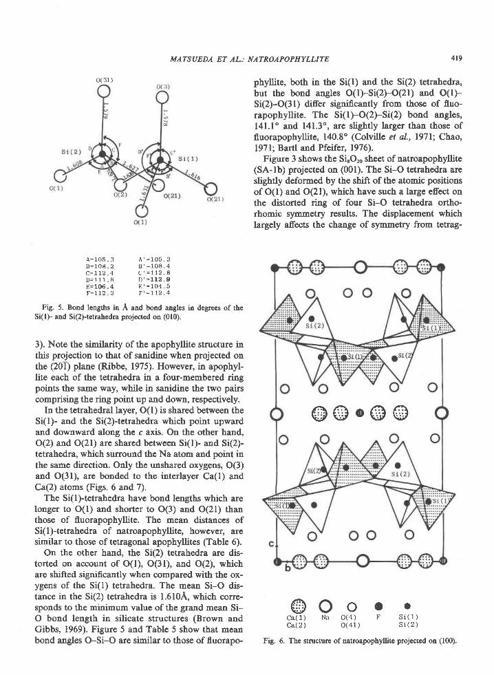
phyllite, both in the Si(l) and the Si(2) tetrahedra,but the bond angles O(l)-Si(2FO(21) and O(l)-S(2)-O(31) ditrer significantly from those of fluo-rapophyllite. The S(l)-O(2)-Si(2) bond angles,l4l.l' and l4l.3o, are slightly larger than those offluorapophyllite, 140.8o (Colville et al., l97l; Chao,l97l;Bartl and Pfeifer, 1976).
Figure 3 shows the SirOro sheet of natroapophyllite(SA-lb) projected on (001). The Si-O tetrahedra areslightly deformed by the shift of the atomic positionsof O(l) and O(21), which have such a large effect onthe distorted ring of four Si-O tetrahedra ortho-rhomic symmetry results. The displacement whichlargely affects the change of symmetry from tetrag-
A = 1 O 5 . 3B = 1 0 8 . 2C = 1 r 2 4D = 1 1 1 . 8ll= 106 . 4F = 1 1 2 3
A ' = 1 o 5 . 3B ' = 1 0 8 . 4
C ' = 1 1 2 . 8D ' = 1 1 2 . 9
E ' = 1 O 4 . 5F ' = r l 2 . 4
Fig. 5. Bond lengths in A and bond angles in degrees of ttreSi(l)- and Si(2)-tetrahedra projected on (010).
3). Note the similarity of the apophyllite structure inthis projection to that of sanidine when projected onthe (201) plane (Ribbe, 1975). However, in apophyl-lite each of the tetrahedra in a four-membered ringpoints the same way, while in sanidine the two pairscomprising the ring point up and down, respectively.
In the tetrahedral layer, O(l) is shared between theSi(l)- and the Si(2)+etrahedra which point upwardand downward along the c axis. On the other hand,O(2) and O(21) are shared between Si(l)- and Si(2)-tetrahedra, which surround the Na atom and point inthe same direction. Only the unshared oxygens, O(3)and O(31), are bonded to the interlayer Ca(l) andCa(2) atoms (Figs. 6 and 7).
The Si(l)-tetrahedra have bond lengths which arelonger to O(l) and shorter to O(3) and O(21) thanthose of fluorapophyllite. The mean distanc€s ofSi(1)-tetrahedra of natroapophyllite, however, aresimilar to those of tetragonal apophyllites (Table 6).
On the other hand, the Si(2) tetrahedra are dis-torted on account of O(l), O(31), and O(2), whichare shifted significantly when compared with the ox-ygens of the Si(l) tetrahedra. The mean Si-O dis-tance in the S(2) tetrahedra is l.610A, which corre-sponds to the minimum value of the grand mean Si-O bond length in silicate structures (Brown andGibbs, 1969). Figure 5 and Table 5 show that meanbond angles O-Si-O are similar to those of fluorapo-
@OooCa(1 ) Na O(4 ) FCa(2) o(41)
os i ( t )s i ( 2 )
@@o@@
Fig. 6. The structur€ ofnatroapophyllite projected on (100).
Ca(1)
H(2x)
o(3)
o(41)
'.ut rlI
MATSUEDA ET AL.: NATROAPOPHYLLITE
H( 11
rr(2)cat i
o(31)
o( 312) " l :
1 1 )
Ca(2)
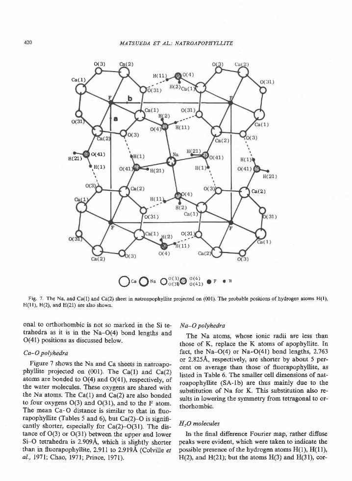
Na-O polyhedra
The Na atoms, whose ionic radii are less thanthose of K, replace the K atoms of apophyllite. Infact, the Na-O(4) or Na-O(41) bond lengths, 2.763or 2.8254, respectively, are shorter by about 5 per-cent on average than those of fluorapophyllite, aslisted in Table 6. The smaller cell dimensions of nat-roapophyllite (SA-lb) are thus mainly due to thesubstitution of Na for K. This substitution also re-sults in lowering the symmetry from tetragonal to or-thorhombic.
HrO molecales
In the final difference Fourier map, rather diffusepeaks were evident, which were taken to indicate thepossible presence of the hydrogen atoms H(l), H(l l),H(2), and HQD; but the atoms H(3) and H(31), cor-
O..QH. oSl i i@8li l , oF o H
Fig. 7. The Na, and Ca(l) and Ca(2) sheet in natroapophyllite projected on (001). The probable positions ofhydrogen atoms H(l),H(ll), H(2), and H(21) are also shown.
onal to orthorhombic is not so marked in the Si te-trahedra as it is in the Na-O(a) bond lengths andO(41) positions as discussed below.
Ca-O polyhedra
Figure 7 shows the Na and Ca sheets in natroapo-phyllite projected on (001). The Ca(l) and Ca(2)atoms are bonded to O(4) and O(41), respectively, ofthe water molecules. These oxygens are shared withthe Na atoms. The Ca(l) and Ca(2) are also bondedto four oxygens O(3) and O(31), and to the F atom.The mean Ca-O distance is similar to that in fluo-rapophyllite (Tables 5 and 6), but Ca(2)-O is signifi-cantly shorter, especially for Ca(2)-O(31). The dis-tance of O(3) or O(31) between the upper and lowerSi-O tetrahedra is 2.909A, which is slightly shorterthan in fluorapophyllite, 2.911 to 2.9194 (Colville eral., l97 l; Chao, l97 l; Prince, l97l).
MATSUEDA ET AL.: NATROAPOPHYLLITE 421
Table 6. Selected meatr interatomic distances (A) of natroapophyllite (SA-lb) compared with those in the apophyllites previouslyreported
Natroapophyllite(sA-3)
CoLvILLe et aL. Chao(1e71 ) ( 1e71 )
Prince Bartl and Pfeifer Rouse et aL.(1e7r-) (Le76) (1978)
s l - oC a - O
Ca - F,OH
Na ,K - O
o(4) - o(4r_)Tetrahedra
( o - o )
n(1) - o(3)- o(4)
H(2) - o(2)- o (4 )
t . 6 1 3 1 . 6 1 0
2 . 4 2 6 2 . 3 6 7
2.413 2 .4L2
2 . 7 6 3 2 . 8 s 4
3.487
2.63L 2 .626
2 . O 1 . 8
1 . 0 1 . 1
2 . 3 2 . 6
1 . 0 1 . 1
L .6L4 1 .615
2 . 4 2 2 2 . 4 3 0
2.478 2 .429
2 . 9 7 L 2 . 9 5 0
3 . 6 9 4
2 . 6 3 2
L.827 L .762
0 . 9 6 8 0 . 9 8 3
2.693 2 .269
o . 5 s 2 0 . 9 5 8
1 . 5 1 6
2 . 4 2 7
2.4L6
2 . 9 7 2
3 . 6 9 0
2.636
1. 615
2 . 4 2 2
2.4r4
2 . 9 6 5
L . 7 7 2
0. 983
2.250
0 . 9 5 3
1 . 6 1 6
2 . 4 3 s
2 . 4 3 5
2 . 9 s 4
3 . 6 7 2
2.637
j z . t t+ l
) r. srz
responding to the H(3) reported by Prince (1971) andBartl and Pfeifer (1976), could not be found in theexpected positions. Tables 5 and 6 show that the H-O bond lengths are similar to those of fluorapophyl-lite. But the relaxation allowed by the orthorhombicsymmetry results in slight changes in the geometryo(3)-H(l)-o(41)-H(21)-o(2) and o(3I)-H(l 1)-O(4)-H(21) con-figurations, which would be identicalin the case of tetragonal symms1ry. The angles H(l)-O(41)-H(21) and H(ll)-O(4)-H(2) are lll(7)' and94(9)", respectively; the angles O(2)-H(21)-O(41)and O(21)-HQ)-O(4) are 137(ll)o and 176(9)'; andthe angles O(3)-H(IFO(41) and O(3 l)-H(l 1)-o(4)arc 124(9)0 and 157(l l)'. That these differences maybe significant is supported by the independent DTAand IR data discussed below.
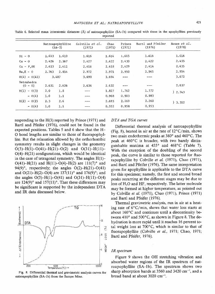
Fig. 8. Differential thermal and gravimetric analysis curves fornatroapophyllite (SA-lb) from the Sampo Mine.
DTA and TGA curves
Diferential thermal analysis of natroapophyllite(Fig. 8), heated in air at the rate of l2"C/min, showstwo main endothermic peaks at 380o and 460oC. Thepeak at 460"C is broader, with two barely distin-guishable maxima at 455" and 468oC (Table 7).With the exception of the doubling of the secondpeak, the curve is similar to those reported for fluo-rapophyllite by Colville et al. (1971), Chao (1971),and Bartl and Pfeifer (1976). The same interpretationgiven for apophyllite is applicable to the DTA curvefor this specimen; namely, the first and second broadpeaks occurring at the different stages may be due toloss of HrO and HF, respectively. The latter moleculemay be formed at higher temperature, as pointed outby Colville et al. (1971), Chao (1971), Prince (1971)and Bartl and Pfeifer (1976).
Thermal gravimetric analysis, run in air at a heat-ing rate of 6"C/min, shows that water loss starts atabout l60oC and continues until a discontinuity be-tween 4l0o and 500"C, as shown in Figure 8. The de-hydration is more rapid until it reaches 16 percent to-tal weight loss at 700oC, which is similar to that offluorapophyllite (Colviile et al., l97l; Chao, l97l;Bartl and Pfeifer, 1976).
IR spectrum
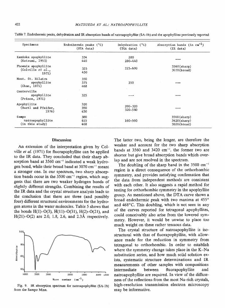
Figure 9 shows the OH stretching vibration andabsorbed water regions of the IR spectrum of nat-roapophyllite (SA-lb). The spectrum shows twosharp absorption bands at 3560 and3420 cm-', and abroad band at about 3020 cm-'.
>
F
6U=
MATSUEDA ET AL.: NATROAPOPHYLLITE
Table 7. Endothennic peaks, dehydration and IR absorption bands ofnatroapophyllite (SA-lb) and the apophyllites previously reported
Speclnens Endotherolc peaks ("C)(mA dara)
Dehydration (oC)(TGA date)
Absorptloo bands (ln cn-l)(IR data)
Kanioka apophyllite(Ko i zun i , 1953 )
Phoenix apophylllte(Colwt l le et aL. ,
L97L)
Mont. St. Hilalreapophylllte
( C h a o , 1 9 7 1 )
Ceatervllleapophyllite
(Pr lnce, 1971)
Apophylllte(Bsrtl and Pfeifer,
L976)
SaEponatroapophyllite
(in this study)
280280-440
334440
325450
350375458
320350450
380455468
225-600
250
200-320320-5 80
160-500
3560(sharp)3070(broad)
3560 (sharp)3420(sharp)3020(broad)
Discussion
An extension of the interpretation given by Col-ville et al. (1971) for fluorapophyllite can be appliedto the IR data. They concluded that their sharp ab-sorption band at 3560 cm-' indicated a weak hydro-gen bond, while their broad band at3O7O cm-' meanta stronger one. In our spectnrm, two sharp absorp-tion bands occur in the 3500 cm-' region, which sug-gests that there are two weaker hydrogen bonds ofslightly di:fferent strengths. Q6alfining the results ofthe IR data and the crystal structure analysis leads tothe conclusion that there are three (and possiblyfour) different structural environments for the hydro-gen atoms in the water molecules. Table 5 shows thatthe bonds H(l)-O(3), H(ll)-O(31), H(2)-O(21), andH(21)-O(2) are 2.0, 1.8, 2.6, and 2.3A respectively.
The latter two, being the longer, are therefore theweaker and account for the two sharp absorptionbands at 3560 and 3420 cm-'. the former two areshorter but give broad absorption bands which over-lap and are not resolved in the spectrum.
The doubling of the sharp band in the 3500 cm-'region is a direct consequence of the orthorhombicsymmetry, and provides satisfying confirmation thatthe data from independent methods are consistentwith each other. It also suggests a rapid method fortesting for orthorhombic symmetry in the apophyllitegroup. As mentioned above, the DTA curve shows abrsad endothermic peak with two maxima at 455"and 468oC. 1tris dsuUing, which is not seen in anyof the curves reported for tetragonal apophyllites,could conceivably also arise from the lowered sym-metry. However, it would be unwise to place toomuch weight on these rather tenuous data.
The crystal structure of natroapophyllite is iso-structural with that of fluorapophyllite, with allow-ance made for the reduction in symmetry fromtetragonal to orthorhombic. In order to establishwhere the symmetry change takes place in the K-Nasubstitution series, and how much solid solution ex-ists, systematic structure determinations and IRmeasurements of other samples with compositionsintermediate between fluorapophyllite andnatroapophyllite are required. In view of the diffuse-ness of the reflections from the most Na-rich crystals,high-resolution transmission electron microscopymay be informative.
o
a
1600 1500
trave nubcr ( o-1;
Fig. 9. IR absorption spectrum for natroapophy[ite (SA-lb)from the Sampo Mine.

AcknowledgmentsWe thank Dr. A. Kato of the National Scicnce Museum, Japan
for valuable suggestions regarding the naming of this new mineral;Dr. J. S. White, National Museum of Natural History, Smithso-dan Institutioq lv6shingto4 D.C. for valuable discussions; Dr. T.Watenabe of KyDshD University for his help in the DTA, TGAand IR measurements.
The calculations were carried out on the FAcoM M-200 at theComputer Center of Kyushu University and also on the HITAc880O computer at T6kyo University. Y. Miura acknowledges a fel-lowship from the National Sciences and Engineering ResearchCouncil of Canada under which this work was completed.
Drs. R. C. Ewing and P. Dunn are thanked for their criticalconrments on the manuscript.
ReferencesAppleman, D. E. and Evans, H. T. Jr. (1973) Job 9214: Indexing
and least-squares refinement of powder difraction data. U.S.Dept. of Commerce Technical Information Service, PB2l6, 188.
Bartl, H. and Pfeifer, G. (1976) Neutronenbeugungsanalyse desApophyllit KCa4(Si4O ro)2(F/OH) . 8H2O. Neues Jahrbuch fiirMineralogie Monatshefte, 58-65.
Belsare, M. R. (1969) A chemical study of apophyllite fromPoona. Mineralogical Magazine, 37, 288-289.
Brown, G. E. and Gibbs, G. V. (1969) Oxygen coordination andthe Si-O bond. American Mheralogist,54, 1528-1539.
Chao, G. Y. (1971) The refnement of the crystal structure of apo-phyllite. II. Determination of the hydrogen positions by X-raydiffraction. American Mineralogist, 56, 123+1242.
Colville, A. A., Anderson, C. P., and Black, P. M. (1971) Rcfine-ment ofthe crystal structure ofapophyllitc. I. X-ray diffractionand physical properties. American Mineralogist" 56, 1222-1233.
Dunn" P. J. and Wilsor; W. E. (1978) Nomenclature revisions inthe apophyllite group: hydroxyapophyllite, apophyllite, fluor-apophyllite. Mineralogical Record, 3, 95-98.
Dunn, P. J., Rouse, R. C. and Norberg, J. A. (1978) Hydroxy-apophyllite, a new mineral, and a redefinition of the apophyllitegroup. L Description, occurrenc€s and nomcnclature. AmericanMineralogist, 63, 196-199.
Koizumi, M. (1953) The differential thermal analysis curves andthe dehydration curves ofzeolites. Mineralogical Journal, l, 36-47.
Kurokawa, K. (1967) Apophyllite found in the Kdmori UltrabasicMass (in Japanese). Journal of Japanese Association of Miner-alogists, Petrologiss, and Economic Geologists, 57 (6), 232-237.
423
Matsueda, H. (1973) On the mode of occurrence and mineralparagenesis of iron-wollastonite (ferro-bustemite) skarn in theSampo Mine, Okayama Prefecture (in Japanese). Science Re-ports, Department of Geology, Kyusht University, 11,265-273.
Matsueda, H. (1975) "Na-apophyllite" from the skarn of theSampo Mine, Okayama Prefecture, Japan (abstract, in J.apa-nese). Annual Joint Meeting of Japanese Association of Miner-alogists, Petrologists, and Economic Geologists; MineralogicalSociety of Japan; and Mining Geology. Abstracts with Program(Kofu),7s.
Matsueda, H. (1980) Pyrometasomatic iron-copper ore deposits ofthe Sampo Mine, Okayama Prefecture. Part I. Geology andMineralogy. Journal of Mining College, Akita University, Se-ries A, 5(4), 15-77.
Miura, Y. (1977) Cation distribution and physical properties inapophyllite-type structure (abstract" in Japanese). Annual Meet-ing of Japanese Crystallographic Society. Abstracts with Pro-gram, lB-3.
Miura, Y. and J. C. Rucklidge (1979) Variations in apophyllitestructure and chemistry (abstract). American CrystallographicAssociation. Abstracts with Program (Honolulu), Series 2, 6(2)'89.
Miura, Y., Matsueda, H. and T. Kato (1976) Crystal structure ofNa-substituted apophyllite from the Sampo Mine, Okayama(abstract, in Japanese). Annual Meeting of Mineralogical So-ciety of Japan. Abstracts with Program, 130.
Prince, E. (1971) Refinement of thc crystal structure of apophyl-lite. III. Determination of the hydrogen positions by neutrondifraction. Anerican Mineralogist, 56, 1243-1251.
Ribbe, P. H. (1975) The chemistry, structure, and nomenclature offeldspars. In P. H. Ribb€, Ed., Feldspar Mineralogy. Mineralog-ical Society of America, Short Course Notes 2, Rl-R52.
Rouse, R. C., Peacor, D. R. and Dunn" P. J. (197E) Hydroxy-apophyllite, a new mineral, and a redefinition ofthc apophyllitegroup. IL Crystal structure. American Mineralogist" 63, 199-202.
Sahama, Th. G. (1965) Yellow apophyllite from Korsnas, Finland.Mineralogical Magazinc, 34, 406-415.
Sakurai, T. (1965) Universal Crystallographic Computation Pro-gram System. Crystallogr. Soc. Japan, Tokyo.
Taylor, W. H. and N6ray-Szab6, St. (1931) The structure of apo-phyllite. Zeitschrift fiir Kristallographie, 77, 146-158.
Manuscript received, October 15, 1979;acceptedfor publication, October 30, 1980.
MATSUEDA ET AL: NATROAPOPHYLLITE