NATIONAL UNIVERSITY OF LIFE AND ENVIRONMENTAL …
21
NATIONAL UNIVERSITY OF LIFE AND ENVIRONMENTAL SCIENCES OF UKRAINE CHAIR OF AGRICULTURAL BIOTECHNOLOGY «PLANT BIOTECHNOLOGY BASIS OF PLANT PROTECTION» Methodical instructions to implement laboratory work for students of the specialty 6.090105 “Plant Protection” Kyiv – 2014
Transcript of NATIONAL UNIVERSITY OF LIFE AND ENVIRONMENTAL …

NATIONAL UNIVERSITY OF LIFE AND ENVIRONMENTAL SCIENCES
OF UKRAINE
CHAIR OF AGRICULTURAL BIOTECHNOLOGY
«PLANT BIOTECHNOLOGY BASIS OF PLANT PROTECTION»
Methodical instructions to implement laboratory work
for students of the specialty
6.090105 “Plant Protection”
Kyiv – 2014

2
UDC 606:632
The basic guidelines for laboratory works of the subject "Plant
Biotechnology Basis of Plant Protection".
Considered and approved at the Department of Agricultural Biotechnology
protocol # 5 of December 5, 2013.
Recommended for publication by the Scientific Council of Educational and
research institute of environment and biotechnology, protocol #4 of December 19,
2013.
Compiled by:
Lobova Oksana, candidate of biological sciences, associate professor
Pilipchuk Elena, postgraduate student
Reviewer:
Kolomiets Ulia, candidate of biological sciences, associate professor
Haychenko Vitalyi, doctor of biological sciences, professor
Educational publication
Recommendations for laboratory works for students of 6.090105 "Plant
Protection"
Responsible for the issue: Lobova Oksana, candidate of biological sciences,
associate professor

3
CONTENT
LABORATORY WORK №1 .......................................................................................................... 4
ORGANIZATION OF BIOTECHNOLOGICAL LABORATORY ............................................... 4
1.1. Features of laboratory equipment ......................................................................................... 4
1.2.Methods of ware, tools, materials sterilization ...................................................................... 4
1.3.Rules of work in laminar - box .............................................................................................. 6
1.4.Keep of lab - journal .............................................................................................................. 6
LABORATORY WORK №2 .......................................................................................................... 7
MOTHER LIQUOR MACROSALTS AND MICROSALTS FOR MS’S NUTRIENT MEDIUM
......................................................................................................................................................... 7
LABORATORY WORK №3 ........................................................................................................ 10
PREPARATION OF NON-HORMONAL NUTRIENT MEDIUM ............................................. 10
LABORATORY WORK №4 ........................................................................................................ 12
SEEDS STERILIZATION AND GROWING IN ASEPTIC CONDITIONS .............................. 12
LABORATORY WORK №5 ........................................................................................................ 15
STERILIZATION OF COURGETTES AND RECEIVING ASEPTIC CULTURE OF PLANTS
....................................................................................................................................................... 15
LABORATORY WORK №6 ........................................................................................................ 16
PREPARATION OF NUTRIENT MEDIUM FOR CALLUS ..................................................... 16
LABORATORY WORK №7 ........................................................................................................ 18
RECEIVING PRIMARY CALLUS FROM DIFFERENT EXPLANTS OF ASEPTIC PLANTS
....................................................................................................................................................... 18
LABORATORY WORK №8 ........................................................................................................ 19
SUBCULTIVATION OF PRIMARY CALLUS TO INCREASE ITS MASS ............................. 19
LABORATORY WORK №9 ........................................................................................................ 20
GETTING OF CALLUS CULTURE OF PLANT TISSUES ....................................................... 20
LABORATORY WORK №10 ...................................................................................................... 21
PREPARATION OF PLANT REGENERANTS THROUGH DIRECT AND INDIRECT
MORPHOGENESIS ...................................................................................................................... 21

4
Laboratory work № 1. Organization of biotechnological laboratory
Required space isolated rooms, modern equipment and high-quality reagents
of biotech laboratory.
1.1. Features of laboratory equipment
For the biotech research is necessary to have a specialized laboratory, which
is equipped with special facilities and equipment.
The biotechnological laboratory consists of several rooms:
1. The room for glassware wash, here are washer with deep with acid-
resistant sinks, distiller, shelvings for drying of glassware.
2. The room for preparation of nutrient mediums, here are technical,
analytical, torsion balance, рН-meter, a refrigerator, cupboard for keep of clean
glassware, reagents.
3. The room for sterilization of nutrient mediums, tooling, glassware, here
are autoclaves, drying oven with an operating mode 160 - 180°С.
4. Laminar-boxing. Sterility in laminar boxing provides the bacterial filter,
the size of pores 0,2 - 0,4 microns.
5. Light culture room, here are shelvings with glass shelfs, conditioner (the
photoperiod 14-16 hour).
6. Dark culture room.
1.2.Methods of ware, tools, materials sterilization
Sterilization methods of glassware, nutrient mediums, plant material
• Sterilization of laminar-boxing: work surface of laminar-boxing wipes alcohol.
• Sterilization of hands: hands wash soap solution, then wipe 96° alcohol.
• Sterilization of tooling: scissors, tweezers, scalpels sterilize 96º alcohol and roast
on fire of a alcohol-lamp. Then tooling put on support and use for one
manipulation.
5
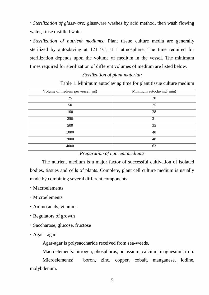
• Sterilization of glassware: glassware washes by acid method, then wash flowing
water, rinse distilled water
• Sterilization of nutrient mediums: Plant tissue culture media are generally
sterilized by autoclaving at 121 °C, at 1 atmosphere. The time required for
sterilization depends upon the volume of medium in the vessel. The minimum
times required for sterilization of different volumes of medium are listed below.
Sterilization of plant material:
Table 1. Minimum autoclaving time for plant tissue culture medium
Volume of medium per vessel (ml) Minimum autoclaving (min)
25 20
50 25
100 28
250 31
500 35
1000 40
2000 48
4000 63
Preparation of nutrient mediums
The nutrient medium is a major factor of successful cultivation of isolated
bodies, tissues and cells of plants. Complete, plant cell culture medium is usually
made by combining several different components:
• Macroelements
• Microelements
• Amino acids, vitamins
• Regulators of growth
• Saccharose, glucose, fructose
• Agar - agar
Agar-agar is polysaccharide received from sea-weeds.
Macroelements: nitrogen, phosphorus, potassium, calcium, magnesium, iron.
Microelements: boron, zinc, copper, cobalt, manganese, iodine,
molybdenum.
6
1.3.Rules of work in laminar - box
While working in aseptic room observe the following rules:
1. Before the work necessary to seal the doors and windows.
2. Dress sterile gown, cap or bandage.
3. Perform disinfection of hands (rub them 80% ethanol).
4. Wipe 80% ethanol surfaces of tables, closely spaced outlet burner.
5. Place on the table the necessary tools to work.
6. Avoid unnecessary movements of hands with open Petri dish.
7. Through land plant material, utensils kept at an angle to avoid direct contact
with dust.
8. After landing, before closing, the edges of dishes and plugs burn in a flame
burner.
9. Planting is carried out as soon as plant material, reducing to a minimum the
time in which the culture dishes still open.
10. At the end of the tube culture dishes coated cellophane caps. The side faces
Petri dishes glued with adhesive tape.
1.4.Keep of lab - journal
In order to control compilation and analysis of the correctness of the
results should lead laboratory journal. Entries are made in a specific order:
1 - date research;
2 - the object of research;
3 - the conditions of the experiment;
4 - basic methods of research and analysis;
5 - the results (usually drawn in the form of tables, graphs, charts).

7
LABORATORY WORK №2
MOTHER LIQUOR MACROSALTS AND MICROSALTS FOR MS’S
NUTRIENT MEDIUM
Uterine solution by Macrosalts MS where their concentration is increased in
10 times. Salts g/500 ml of uterine solution:
NH4NO3 8,25
KNO3 9,5
CaCl2 x 2H2O 2,2
NaH2PO4 0,85
MgSO4 x 7H2O 1,85
For 1 liter of medium selected 100 ml of uterine solution.
Uterine solution of Fe-chelate for MS: salts, g/200ml of uterine solution:
Na2EDTA x 2H2O 1,492
FeSO4 x 7H2O 1,112
For 1 liter of medium selected 5 ml of uterine solution.
Initial solution of Fe-chelate (200 ml) prepares, dissolves of 1,492 g of
NA2EDTA * 2H2O and 1.112 g of FE2SO4 * 7H2O, and then brings it to a boil.
Uterine solution of microsalts for MS, where their concentration is increased
in 100 times: salts, mg/200ml of uterine solution:
H3BO3 62
Na2MoO4 x 2H2O 2,5
KJ 8,3
MnSO4 x 4H2O 223
ZnSO4 x 7H2O 86
CuSO4 x 5H2O
CoCl2 x 6H2O
weigh up 10 mg of each salt and to
dissolve in 40 ml of water
For 1 liter of medium selected 1 ml of uterine solution.
Uterine solution of vitamins. Vitamins, mg/100 ml of uterine solution:
В1 (thiamine HCl) 10
В6 (pyrodoxine HCl) 50
РР(nicotinic acid) 50
For 1 liter of medium selected 1 ml of uterine solution. Solution with
vitamins (1.0 and 0.1 mg / ml) prepared and directly dissolved in bidistilled water.

8
Solutions with auxins 2,4-D, NAA, IAA, IMA and their analogues prepared by
dissolving 100 mg of the substance in 0.5 - 2 ml of ethanol, and add warm
water to 100 ml.
Kinetin, Zeatin, 6-BAP previously dissolved in a small amount of 0.5 N HCl
and heated at adding the right amount of water.
If you need to add to the nutrient medium abscisic acid (ABA), it dissolved in
70% of ethanol and adjusted to the desired volume.
Gibberellin acid dissolved directly in water and added to the culture media.
Table 2. Uterine solutions for preparates nutrient medium
Initial component Sample Necessary volume for
preparation of 1l of nutrient
medium
By MS By White
Macroelements, g/l
NH4NO3 16,5 –
100ml
KNO3 19.0 0,8
Ca(NO3)24H2O – 2,0
CaCl2 б/в 3,3 –
MgSO4 7H2O 3,7 3,6
KH2PO4 1,7 –
Na2EДТО 0,37 0,37
FeSO4 7H2O 0,28 0,28
KCl – 0,65
NaH2PO4 H2O – 0,165
Na2SO б/в – 2,00
Microelements, mg/100 ml
Н3BO3 620 150
1 ml
MnSO4 4H2O 2230 –
ZnSO4 7H2O – 150
ZnSO4 4H2O 860 –
KJ 83 –
Na2MoO4 2H2O 25 25
CuSO4 5H2O 2,5 4
CoCl2 6H2O 2,5 –
MnCl2 4H2O – 530
Вітаміни, мг/л
Тіамін HCl (В1) 10 10
1ml
Нікотинова к-та (РР) 50 50
Піридоксин HCl (В6) 50 10
9
Plant growth regulators
Plant growth regulators are the critical media components in determining
the developmental pathway of the plant cells. The plant growth regulators used
most commonly are plant hormones or their synthetic analogues.
There are five main classes of plant growth regulator used in plant cell
culture, namely:
• (1) auxins;
• (2) cytokinins;
• (3) gibberellins;
• (4) abscisic acid;
• (5) ethylene.
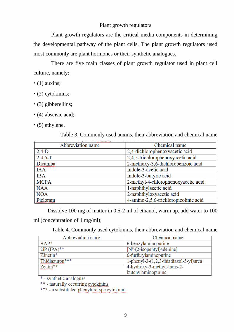
Table 3. Commonly used auxins, their abbreviation and chemical name
Dissolve 100 mg of matter in 0,5-2 ml of ethanol, warm up, add water to 100
ml (concentration of 1 mg/ml);
Table 4. Commonly used cytokinins, their abbreviation and chemical name

10
Dissolve 100 mg matter in 2 ml 0,5 НС1, warm up, add water to 100 ml
(concentration of 1 mg / ml);
― Gibberellins:
• GA - Gibberellin acid;
• Dissolve 100 mg matter in 100 ml water (concentration of 1 mg/ml);
― Abscisin:
• ABA - Abscisic acid;
• Dissolve in 3 ml of 70 % of ethanol, add water to 100 ml
• Regulators of growth keep in a refrigerator at temperature +4°С
To prepare solutions of vitamins
• Thiamine-HCl (В1), Pyridoxine-HCl (B6) nicotinic acid (РР), ascorbic acid
(С), dissolve in water (concentration of 1 mg/ml) Hydrocarbons and organic
supplements add in medium.
• The scheme of preparation of a nutrient medium: In a volumetric glass add
agar, saccharose, macroelements, microelements, vitamins, regulators of growth,
add distilled water up to the necessary volume and put on a magnetic mixer. The
agar to be dissolved, then add 1н КОН to finish рН medium with the necessary
value. Spill medium at a flask or test-tubes, close.
• Medium sterilize in an autoclave 20 - 25 mines at 1 atm.
LABORATORY WORK №3
PREPARATION OF NON-HORMONAL NUTRIENT MEDIUM
Nutrient medium - a major factor of successful cultivation of isolated organs,
tissues and cells of plants. Nutrient medium should include macrosalts, microsalts,
vitamins, carbohydrates, phytohormones. Some culture media include casein
hydrolyzate, certain amino acids.
Objective of work: To prepare the culture medium of Murasige-Skoog for
further culturing on them isolated tissues, cells and organs of plants.

11
Materials and equipment: macrosalts and microsalts, vitamins, growth
regulators, alcohol, scales, spatulas, tile, laboratory glassware, magnetic mixer,
HCl, KOH.
1. Preparation of nutrient medium - 0.5 liters:
1. In the flask: pour 50 ml of distilled water and add exactly measure of each
solution of macro- and microelements, Fe-chelate and vitamins.
• Weigh the required amount of mesoinozyt, add to the solution.
• Weigh the required amount of sucrose, add to the solution.
• Add distilled water to the required volume - 500 ml.
• Weigh agar-agar and add to the solution.
• pH is set on the pH meter, bring it to the desired value and add (dropwise)
0.1n KOH, 0.1 N NaOH or 0.1 N HCl.
2. Heat the culture medium to dissolve agar within 25-30 minutes.
3. Pour the medium in tubes, cover with foil and carefully wrapped around
the neck of the glass.
4. Sterilization of nutrient medium – in an autoclave under pressure 0.08 -
0.1 MPA (1 atm.), T = 115 - 120 °C for 15-20 minutes.
Table 5. The composition of the nutrient medium:
Elements of
nutrient medium 1 l 0,5 l 0,250 l 0,100 l
Macro MS 100 ml 50 ml 25 ml 10 ml
Micro MS 1 ml 0,5 ml 0,25 ml 0,1 ml
Fe-chelate 5 ml 2,5 ml 1,25 ml 0,5 ml
Mesoinosyte 0,1 g 0,05 g 0,025 g 0,01 g
Vitamins МS 1 ml 0,5 ml 0,25 ml 0,1 ml
Sacharose 30 g 15 g 7,5 g 3 g
Agar-agar 6,8 g 3,4 g 1,7 g 0,68 g
КОН 10-16 drops 8 drops 4 drops 2 drops
12
LABORATORY WORK №4
SEEDS STERILIZATION AND GROWING IN ASEPTIC CONDITIONS
Seeds sterilization and aseptic cultivation of plants
The surfaces of plants are usually contaminated with spores of various
microorganisms and fungi (endogenous and epiphytic microflora, viruses).
However, the internal tissues are sterile, though they may be in non-pathogenic
bacteria that are not always found.
The basic requirement for obtaining callus and suspension cultures is a
sterilization plant facilities. Type of sterilizing agents, concentration and duration
depend on the density and sensitivity of tissue that sterilized.
Standard requirements for sterilizing agents
Before sterilization tissue is pre-cleaned. For this storing organs (roots,
tubers), thick stems of plants, some seeds of woody species:
• thoroughly wash with soap under running water, while pre-flushing by
brush;
• remove the upper epidermis (in roots and tubers);
• washed with distilled water, laminar-box dipped for a few seconds in
absolute alcohol.
The surface of the shoots and leaves of trees rubbed with cotton wool soaked
with ethanol (alcohol), and seed and fruit rinsed with ethanol. Treatment of 70%
ethanol improves performance sterilizing solutions.
In order to more fully wetting the surface pubescent surface sterilizing
solution is added in recent detergents - surfactants, detergents (soap, detergent,
etc.) 4 - 5 drops to 1 liter of water.
Seed sterilization and aseptic cultivation of plants
Depending on the degree of seeds contamination divided into three groups:
- With minor surface contamination by microorganisms (seed wheat, sorghum,
cabbage);
13
- Infection of the outer surface of a seed (lettuce, spinach, radishes, tomatoes,
corn, carrots);
- The presence of microorganisms on the surface and in the middle of seeds (rice,
sunflower, soybean, pine).
Knowing which group belongs seed processing mode chosen seed, but
sterilizing substance, its concentration and the time of sterilization are selected for
each type and grade empirically.
Purpose of work. Select concentration of sterilizing solution and time of
sterilization of seeds that maximize the overall effectiveness of the process.
Receive sterile seeds and grow from it aseptic plants.
Materials and equipment. Soapy solution, 70% ethanol solution, cups with
sterile water, concentrated solution "bleaching powder“, cups for sterilizing
solutions, graduated cylinder, seeds, tubes with non-hormonal nutrient medium for
plants, sterile Petri dishes with a filter paper, sterile Petri dishes, gauze, forceps,
scalpels, alcohol lamp, ethyl alcohol for sterilization of instruments, laminar-box.
Progress of work:
1. Prepare a solution of "whiteness" of various concentrations.
I - 1 part of preparation and 4 of water (1:4)
II - 1 part of preparation and 3 of water (1:3)
III - 1 part of preparation and 2 of water (1:2)
IV - 1 part of preparation and 1 of water (1:1)
Solutions used once immediately after preparation.
2. Wash the seeds in soapy water.
3. In gauze bags puts 10 seeds.
4. Seeds 1 min placed in a beaker with 70% ethanol.
5. By sterile forceps carry bags of seeds in cups with the appropriate concentration
of sterilizing solution and maintain the appropriate time.
6. Sterilized seeds were washed three times for 10 min in sterile water. This sterile
tweezers carry bags of glass with sterilizing solution into a glass with sterile
distilled water. Withstand 10 min. Washing is repeated 3 times, using a new batch

14
of water.
7. Wash bags with seeds by sterile tweezers transferred to a sterile Petri dish with
filter paper.
8. By sterile forceps carry one of the bags to another Petri dish, roll him with
tweezers and roll seeds in a Petri dish.
9. By sterile forceps carry seeds on a nutrient medium in a test tube.
10. Test tubes with seeds placed in a thermostat at 25 ± 1 ° C.
11. After 4 - 6 days check the purity of seed germination.
12. After germination of seeds is transferred to a thermostat with lighting 400 lux
and a temperature of 25 ° ± 1 ° C.
Table 6. For checking the purity of seed germination
Concentration of
"whiteness" in
volume
Duration of
sterilization, min.
Total
quantity of
seeds,
pieces
Quantity of infected
seeds after 7 days
Germination of
seeds Effectiveness
of
sterilization
(%) pieces % pieces %
1:4
10
15
30
1:3
10
15
30
1:2
10
15
30
1:1
10
15
30

15
LABORATORY WORK №5
STERILIZATION OF COURGETTES AND RECEIVING ASEPTIC
CULTURE OF PLANTS
Purpose of work. Select concentration of sterilizing solution and time of
sterilization of seeds that maximize the overall effectiveness of the process.
Receive sterile seeds and grow from it aseptic plants.
Materials and equipment. Soapy solution, 70% ethanol solution, cups with
sterile water, concentrated solution "bleaching powder“, cups for sterilizing
solutions, graduated cylinder, seeds, tubes with non-hormonal nutrient medium for
plants, sterile Petri dishes with a filter paper, sterile Petri dishes, gauze, forceps,
scalpels, alcohol lamp, ethyl alcohol for sterilization of instruments, laminar-box.
Progress of work:
1. Prepare a solution of "whiteness" of various concentrations.
1 part of preparation and 2 of water (1:2)
Solutions used once immediately after preparation.
2. Wash the seeds in soapy water.
3. In gauze bags puts 10 seeds.
4. Seeds 1 min placed in a beaker with 70% ethanol.
5. By sterile forceps carry bags of seeds in cups with the appropriate concentration
of sterilizing solution and maintain the appropriate time.
6. Sterilized seeds were washed three times for 10 min in sterile water. This sterile
tweezers carry bags of glass with sterilizing solution into a glass with sterile
distilled water. Withstand 10 min. Washing is repeated 3 times, using a new batch
of water.
7. Wash bags with seeds by sterile tweezers transferred to a sterile Petri dish with
filter paper.
8. By sterile forceps carry one of the bags to another Petri dish, roll him with
tweezers and roll seeds in a Petri dish.
9. By sterile forceps carry seeds on a nutrient medium in a test tube.
10. Test tubes with seeds placed in a thermostat at 25 ± 1 ° C.

16
11. After 4 - 6 days check the purity of seed germination.
12. After germination of seeds is transferred to a thermostat with lighting 400 lux
and a temperature of 25 ± 1 ° C.
Table 7. For checking the purity of seed germination
Concentration
of "whiteness"
in volume
Duration of
sterilization,
min.
Total
quantity of
seeds,
pieces
Quantity of
infected seeds
after 7 days
Germination
of seeds
Effectivenes
s of
sterilization
(%) pieces % pieces %
1:2
10
15
30
LABORATORY WORK №6
PREPARATION OF NUTRIENT MEDIUM FOR CALLUS
Objective of work: To prepare the culture medium of Murasige-Skoog for
further culturing on them isolated tissues, cells and organs of plants for receiving
primary callus.
Materials and equipment: macrosalts and microsalts, vitamins, growth
regulators, alcohol, scales, spatulas, tile, laboratory glassware, magnetic mixer,
HCl, KOH.
Preparation of nutrient medium
Progress of work:
1. In the flask: pour 50 ml of distilled water and add exactly measure of each
solution of macro- and microelements, Fe-chelate and vitamins.
• Weigh the required amount of mesoinozyt, add to the solution.
• Weigh the required amount of sucrose, add to the solution.
• Add distilled water to the required volume - 500 ml.
• Weigh agar-agar and add to the solution.
• pH is set on the pH meter, bring it to the desired value and add (dropwise)
0.1n KOH, 0.1 N NaOH or 0.1 N HCl.
2. Heat the culture medium to dissolve agar within 25-30 minutes.

17
3. Pour the medium in tubes, cover with foil and carefully wrapped around
the neck of the glass.
4. Sterilization of nutrient medium – in an autoclave under pressure 0.08 -
0.1 MPA (1 atm.), T = 115 - 120 °C for 15-20 minutes.
Table 8. The composition of the nutrient medium
Elements 1 l
Macro MC 100 ml
Micro MC 1 ml
Fe-chelate 5 ml
Inositol 0,1 g
White’s vitamins
(nicotinic acid
0,05 g, tiamine
0,01 g for 100 ml)
1 ml
2,4 D 3 ml
Saccharose 30 g
Agar 6,8 g
КОН 10-16 drops
Callus cells differ by:
• intensity of growth,
• consistency
• color
• the ability to green at light and other properties.
Cell colonies in agar medium can be compact and firm, and plump that takes
in callus and break into separate pieces.
The last type of callus in liquid medium easily separated single cells and
gives rise to suspension culture. It is noted that the compact callus may give rise to
loose tissue, but not conversely.
Consistency of callus depends largely on the composition of the medium.
Anatomical structure is characterized by loose of many organized centers of
meristematic activities separated by large undifferentiated cells.
18
Thick callus less differentiated and contain many large vacuolised cells.
There is a difference between these types of callus and cell packing: thick callus
composed of tightly adjacent to each cell.
LABORATORY WORK №7
RECEIVING PRIMARY CALLUS FROM DIFFERENT EXPLANTS OF
ASEPTIC PLANTS
Receiving primary callus from different explants of aseptic plants
Purpose of work. Learn to chosen tissue explants and culturing conditions to
induce dedifferentiation and formation of callus.
Materials and equipment. Plants of different species, soap solution,
concentrated solution "whiteness", glasses with sterilizing solutions, graduated
cylinder, sterile distilled water, test tubes with nutrient medium, Petri dishes with
sterile filter paper, sterile Petri dishes, nylon, forceps, scalpels, alcohol, laminar
box.
Progress of work:
Explants are aseptic plants derived from seeds under in vitro (tobacco, pea,
carnation, sunflower, tomato).
1. Sterile forceps removed the plant from the tube and placed in a sterile Petri
dish.
2. Hold the tweezers, by sterile scalpel cutting the plant explants: cuttings, leaf
plates, fragments of stems, roots. The leaves make additional cuts in the
central vein.
3. Explants cultivated on callus medium in a bottle that sign with the name of
the type explants (petiole, leaf, stem, root).
4. Cultured in the dark at +25 - 26,5 °C. They spend subcultivation every 3 - 4
weeks and determine the frequency of callusgenesis as the number of
explants with callus to the total planted explants as a percentage. The data
recorded in the table.
+
19
Table 9. Testing of environments for induction of leaf callus explants
Nu
trie
nt
med
ia
Ty
pes
of
exp
lan
tats
Qu
anti
ty o
f
exp
lan
ts,
pie
ces
Dat
e of
pla
nti
ng
Dat
e of
ind
uct
ion
Per
cen
tag
e of
call
usg
enes
is
Co
nd
itio
n o
f
call
us*
* State of callus estimate as follows:
+ + + – white viable callus, growing;
+ + - – callus in good condition, yellow;
+ - - – flabby, dark yellow callus;
- - - – dark brown, viable callus.
LABORATORY WORK №8
SUBCULTIVATION OF PRIMARY CALLUS TO INCREASE ITS MASS
After the formation of callus aseptically separated and placed it on the
surface of fresh culture medium. As a result of receiving a sterile culture of callus
tissue, which can be maintained for a long time, occasionally dividing it into
fragments. Callus subcultivated on medium of the same composition as for
dedifferentiation or in medium with a reduced concentration of auxin to stimulate
the growth of callus mass.
Progress of work:
1. Explants with primary callus transferred to a sterile Petri dish.
2. Hold explants by tweezers, cut callus by scalpel.
3. By scalpel share callus tissue level fragments and transferred to fresh
culture medium.
4. Signs Petri dish and close by parafilm.
5. Petri dish cultured in an incubator at 25 ± 1 ° C.
6. After 4 - 6 weeks callus tissue is divided into equal pieces and then
transferred to fresh culture medium.
20
LABORATORY WORK №9
GETTING OF CALLUS CULTURE OF PLANT TISSUES
Purpose of work. Get callus culture of plant tissues.
Progress of work:
1. Conduct preliminary training of laminar-box to work, which involves
sterilizing by ultraviolet light for 30 min., followed by wiping work surfaces by
sterilizing solution (70 - 96% ethanol).
2. The basic requirement for operations is adherence to sterility, as in an oven
pre maintain tools and utensils for 2 - 2.5 hours at a temperature 180 - 210 °C or
sterilized by autoclaving for 25 min. at 0.1 - 0.12 MPa.
3. Immediately before working tools dipped in ethanol and burn surfaces in
alcohol lamp.
4. The starting material (any parts of plants) thoroughly washed in soapy water,
then rinse lightly pink solution of KMnO4 (potassium permanganate) and
transferred to a sterile working box.
5. By sterile instruments cut raw material into pieces of 2 - 3 cm and placed in
a pre- sterilized gauze bags for further immersion in the capacity of sterilizing
solution (a solution of mercuric chloride, bleach, ethanol).
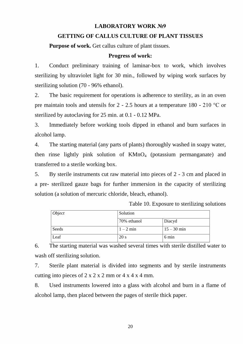
Table 10. Exposure to sterilizing solutions
Object Solution
70% ethanol Diacyd
Seeds 1 – 2 min 15 – 30 min
Leaf 20 s 6 min
6. The starting material was washed several times with sterile distilled water to
wash off sterilizing solution.
7. Sterile plant material is divided into segments and by sterile instruments
cutting into pieces of 2 x 2 x 2 mm or 4 x 4 x 4 mm.
8. Used instruments lowered into a glass with alcohol and burn in a flame of
alcohol lamp, then placed between the pages of sterile thick paper.
21
9. Source plant material (explants) are exposed on the surface of agar medium
in test tubes, flasks or Petri dish and placed in a growth chamber (in an
incubator). Grown in the dark at a temperature of 23 - 28 °C.
10. After 3 - 4 weeks on the cut surface marked the formation of primary callus
tissue. Preparation of callus tissue depends on the type of plant and tissue. The
cells of plants that are in the final stages of differentiation under the influence
of inducers of cell division - cytokinins and auxins, moving in dedifferentional
status and reduced meristematic activity. After the formation of primary callus
under aseptic conditions it is separated from the explants and transplanted to a
new agar nutrient medium.
LABORATORY WORK №10
PREPARATION OF PLANT REGENERANTS THROUGH DIRECT AND
INDIRECT MORPHOGENESIS
Explants are aseptic tobacco plants.
Progress of work:
1. Puff plate damaged by cutting central vein cultured on MC medium with the
addition of 2,4-D at a concentration of 4 mg / L in the dark at a temperature of 25 -
27 ° C.
2. After 7 - 10 days cell explants to form callus.
3. Clip morphogenic callus and cultivated it on MC medium with the addition
of 6-BAP at a concentration of 1 mg / l.
4. After 10 days produced optimally formed shoots.
5. For rhizogenesis - shoots transferred to culture medium without growth
regulators and inositol.