NATIONAL INSTITUTE OF GENETICS · 2015-02-25 · NATIONAL INSTITUTE OF GENETICS JAPAN ANNUAL REPORT...
156
NATIONAL INSTITUTE OF GENETICS JAPAN ANNUAL REPORT No. 17 1966 Published by THE NATIONAL INSTITUTE OF GENETICS KUJi1lUl, Sizuoka-ken, Japan 1967
Transcript of NATIONAL INSTITUTE OF GENETICS · 2015-02-25 · NATIONAL INSTITUTE OF GENETICS JAPAN ANNUAL REPORT...

NATIONAL INSTITUTE OF GENETICSJAPAN
ANNUAL REPORT
No. 17 1966
Published by
THE NATIONAL INSTITUTE OF GENETICSKUJi1lUl, Sizuoka-ken, Japan
1967

Annual Report
of the
National Institute of GeneticsNo. 17, 1966
Published by
The National Institute of Genetics, Japan1967

CONTENTS
General statement.. . . . . . . . . . . . . . . . . . . . . . . . . . . . . . . . . . . . . . . . . . . . . . . . . 1Staff 2Council............................................................ 5Projects of research for 1966 6Researches carried out in 1966 11
1. CytogeneticsInduction of plasma cell neoplasms in BALB/c mice, their
karyotypes and r-globulin specificity. YOSIDA, T. H., IMAI, H.T., MORIWAKI, K. and MIGITA, S. 11
Change of ploidy in plasma cell tumors (MSPC) in early transplantgenerations. IMAI, H. T., YOSIDA, T. H. and MORIWAKI. K. " 12
Comparative study of karyotypes in sublines producing and nonproducing r-globulin in mouse plasma cell tumors. YOSIDA, T.H., POTTER, M. and IMAI, H. T. 13
Karyotypes of mouse plasma cell tumor MOPC-31B before andafter in vitro cultivation. YOSIDA, T. H., IMAI, H. T., MASUDA,T. and NAMBA, Y. 14
Comparative study of mouse leukemias developed by treatmentwith chemicals and radiation. YOSIDA, T. H., TSURUTA, R. andKURITA, Y. 15
Alteration of karyotypes in a mouse leukemia strain DML.TSURUTA, R. and YOSIDA, T. H............................. 16
Changes in aggregate-forming activity of cells in carcinogenesis.KURODA, Y. 17
Difference in aggregate-forming activity between normal andmalignant cells. KURODA, Y. 18
II. Physiological and developmental geneticsBehavior of nuclei in germinating pollen grains of wheat, rice
and maize. KIHARA, H. and HORI, T. 19Photoperiodic response of various Oryza species. IX. KATAYAMA,
T. C ,. 19Anatomical studies on interior root found in root of rice plant.
KATAYAMA, T. C. 21A genetic study on skeleton-length in Japanese quail. ISOGAI, I.,
KAWAHARA, T. and SAKAI, K. I. 21Genetic changes in body weight caused by competition in chickens.
FUJISHIMA, T. 22

ii ANNUAL REPORT OF NATIONAL INSTITUTE OF GENETICS NO. 17
A genetical study on organ formation in Nicotiana tabacum L.HIGUCHI, S. and SAKAI, K. 1. 25
Major gene and polygenes governing the rachis deficiency inrice. WASANO, K. and SAKAI, K. 1. . . . . . . . . . . . . . . . . . . . . . . 26
Estimation of genetic parameters in Chamaecyparis forests.SAKAI, K. 1., HAYASHI, S. and MUKAIDE, H. 27
Developmental genetic study of panicle formation in rice. IyAMA,S. 28
A developmental genetic study in rice. BALAL, M. S. and SAKAI,K. 1. 30
Electrophoretic comparison of soluble proteins from differentorgans of tobacco plant. NARISE, S. and SAKAI, K. 1. 31
Analysis of genetic correlations between panicle, internode andleaf lengths among mutant strains of a rice variety, Norin 8.MORISHIMA, H. and OKA, H. I. 32
Analysis of growth curves for panicle and internode elongationin mutant strains of a rice variety, Norin 8. MORISHIMA, H. andOKA, H. 1. 33
Differentiation of aggregation-promoting materials from embryonicchick liver cells. KURODA, Y. 34
Characterization of tissue-specific materials with cell-bindingactivity obtained from embryonic chick cells. KURODA, Y. .. 35
In vitro cultivation of single cells from Drosophila melanogasterlarvae. KURODA, Y. 37
III. Biochemical geneticsSome notes on the chromogranules in hypodermal cells of silkworm
larvae. TSUJITA, M. 39Development of chromogranules in the larval skin of the silkworm.
TSUJITA, M. and SAKURAI, S. 40Some properties of nucleic acid in larval skin cells of the silkworm.
KOJIMA, K., TSUJITA, M. and SAKURAI, S. 42Incorporation of 14C-glycine into chromogranules of larval skin
cells of the silkworm. SAKURAI, S. and TSUJITA, M. 44Chemical characterization of chromogranule membrane in larval
skin cells of the silkworm. SAKURAI, S. and TSUJITA, M. .. 46Genetic effects of DNA in Ephestia. NAWA, S. and YAMADA, M. .. 48Peroxidase isozymes in leaves of Pharbitis nil. ENDO, T. 50Hormonal enzyme regulation in the cultured hypocotyl of Pharbits
nil. ENDO, T. 50Variation in peroxidase isozymes of Oryza perennis and O. sativa.
CHU, Y. E. 51

CONTENTS iii
Characterization of xanthine dehydrogenase from Drosophila.SHINODA, T. 53
Multiple molecular forms of xanthine dehydrogenase in Drosophila.SHINODA, T. 53
IV. Evolutionary geneticsAn intergeneric hybrid between Eremopyrum orientale and
Henrardia persica. SAKAMOTO, S. . . . . . . . . . . . . . . . . . . . . . . . . 55Three intergeneric hybrids among Heteranthelium piliferum,
Eremopyrum buonapartis and Hordeum sp. SAKAMOTO, S. .. 55Diallele crosses among Sikkimese rice types. III. KATAYAMA, T.
C. 56Further studies on embryo transplantation in the genus Oryza.
KATAYAMA, T. C. 57Geographical distribution of winter, intermediate and spring types
of common wheat. NAKAI, Y. 58Embryosac sterility of F 1 hybrids between strains of Oryza
perennis. CHU, Y. E. and OKA, H. I. 59Pattern analysis of character variations in Oryza perennis.
MORISHIMA, H. and OKA, H. I. . . . . . . . . . . . . . . . . . . . . . . . . . . . . 59Population survey of No.1 chromosome polymorphism of black
rats (Rattus rattus) collected in Japan and Korea. YOSIDA, T.H., MORIGUCHI, Y., KANG, Y. S. and SHIMAKURA, K. 61
Segregation of three chromosome types in black rats crossed inthe laboratory. YOSIDA, T. H. and MORIGUCHI, Y. 62
V. Mathematical and statistical studies on population geneticsSimulation studies on the number of neutral alleles maintained
in a finite population by mutation. KIMURA, M. 64Two loci polymorphism as a stationary point. KIMURA, M. 65The mutational load with epistatic gene interactions in fitness.
KIMURA, M. and MARUYAMA, T. . . . . . . . . . . . . . . . . . . . . . . . . . . 67Eigenvalues in a genetics problem. MARUYAMA, T. 68A diffusion process with heterosis. MARUYAMA, T. 70An application of Kimura's formulae to define the evolutionary
load in a small population. MARUYAMA, T. 72Dimensionality of human migration. YASUDA, N. 73Dimensionality and distance in human migration. YASUDA, N. .. 74A statistical singularity at the ABO blood group system. YASUDA,
N. 75
VI. Experimental studies on population geneticsDeleterious genes in the second chromosome concealed in natural

iv ANNUAL REPORT OF NATIONAL INSTITUTE OF GENETICS NO. 17
populations of Drosophila melanogaster. OSHIMA, C. andWATANABE, T. K. . . . . . . . . . . . . . . . . . . . . . . . . . . . . . . . . . . . . . . . . 77
Distribution of persistent lethal genes in natural populations.OSHIMA, C. and WATANABE. T. K. 78
Recessive visible mutant genes on the second chromosomeconcealed in natural populations. OSHIMA, C. and WATANABE,T. K. 79
Segregation distorter (SD) genes and their linked lethal genes inDrosophila melanogaster. WATANABE, T. K. and OSHIMA, C. .. 80
A mechanism of persistence of some lethal genes in naturalpopulations of Drosophila melanogaster. WATANABE, T. K. .. 80
Further study on chromosomal polymorphism in the Kofu andKatsunuma natural populations. WATANABE, T. and OSHIMA,C. 81
Lack of chromosomal interaction with respect to overdominancein Drosophila melanogaster. MUKAI, T. 83
The detrimental load to the lethal load ratio (D: L ratio) ofnewly arising mutations in Drosophila melanogaster. MUKAI,T. and CROW, J. F. 84
Studies on the competition between races lA and 21B of wheatleaf rust. KATSUYA, K. 85
Interaction among genotypes for migration in Drosophila melano-gaster. NARISE, T. 86
The relation between migratory activity and competitive abilityin Drosophila melanogaster. NARISE, T. 87
Experimental induction of bilateral asymmetry in wings ofDrosophila melanogaster. NARISE, T. and SAKAI, K. I. .... 88
VII. Radiation genetics and chemical mutagenesis in animalsPost-irradiation modification and mechanism of reverse dose-rate
effect on mutation induction in silkworm gonia. TAZIMA, Y.and SADO, T. 90
Repair of radiation induced premutational damages revealed byfractionated irradiation of silkworm spermatids. TAZIMA, Y.and ONIMARU, K. 91
Modification of r-ray-induced mutation frequencies in the silkwormby post-treatment of spermatids and spermatozoa with nitrogengas. TAZIMA, Y. and ONIMARU, K. 92
Mechanisms of mutation induction by mitomycin-C in the silkworm.T AZIMA, Y. and ONIMARU, K. 94
Mutagenicity of a nitrofuran derivative applied to silkworm germcells. TAZIMA, Y. and FUKASE, Y. 95

CONTENTS v
Changes in the mutation response of post-meiotic silkworm germcells to r-rays with the progressing spermiogenesis. T AZIMA,Y. 97
Studies on strain differences in radiosensitivity in the silkworm.I. Screening of sensitive and resistant strains to embryonicradiation killing. MURAKAMI, A. and TAZIMA, Y. 98
Studies on strain differences in radiosensitivity in the silkworm.II. Relation between sensitivity to embryonic killing andmutability. MURAKAMI, A. and TAZIMA, Y. 100
Relation between sensitivity to killing and mutation observedduring a mitotic cycle of silkworm cleavage nuclei. MURA-KAMI, A. 102
The effect of 5-bromodeoxyuridine (BUDR) on the frequency of14 MeV fast neutron induced mutations in the gonial cells ofthe silkworm. MURAKAMI, A. 103
Relative biological effectiveness of 14.1 MeV neutrons in theinduction of dominant lethal mutations in the mouse. TUTI-KAWA, K. 105
VIII. Radiation genetics in plantsRBE of radiations in E-1 hole of Kyoto University Reactor (KUR).
MATSUMURA, S., AMANO, E. and HAYASHI, M. 107Comparison of mutagenic efficiency between EMS and r-rays.
MATSUMURA, S. and FUJII, T. 109Comparison of the killing effect of i-rays and thermal neutrons.
FUJII, T. 110On the determination of absorbed dose in heavy ionizing particles.
FUJII, T. 111Photoreactivation of an UV-induced mutation in maize. MATSU-
MURA, S. and MABUCHI, T. 112Photoreactivation of UV-induced damage in maize pollen. AMANO,
E. and MABUCHI, T. 114On the somatic variations in corn and chrysanthemum under
chronic r-irradiation. MATSUMURA, S. and FUJII, T......... 115Endosperm mutations induced by UV in corn. FUJII, T. 117
IX. Microbial geneticsGenetic map of Hi gene III Salmonella. YAMAGUCHI, S. and
IINO, T. . . . . . . . . . . . . . . . . . . . . . . . . . . . . . . . . . . . . . . . . . . . . . . . . . . 119A straight flagellar mutant in Salmonella. IINO, T. and MITANI,
M 120
vi ANNUAL REPORT OF NATIONAL INSTITUTE OF GENETICS NO. 17
Flagellin biosynthesis in Salmonella spheroplasts. SUZUKI, H.and IINO, T. 121
Genetic fine structure of the mot loci in Salmonella typhimurium.ENOMOTO, M. 121
Mapping of three mot loci in Salmonella by linkage analysis.ENOMOTO, M. 122
Difference in frequencies of cotransduction of mot C with Higene in Salmonella. ENOMOTO, M. and YAMAGUCHI, S. 123
Infection of bacteriophage-chi to Serratia marcescens. IINo,T 124
Normal repressed level of ornithine transcarbamylase activity incrude extracts of an arginine sensitive mutant of Salmonellatyphimurium. ISHIDSU, J. . . . . . . . . . . . . . . . . . . . . . . . . . . . . . . . . . 125
x. Human geneticsEvaluation of the family planning programme in Japan. MATSU-
NAGA, E. 126Association of ear-wax types with susceptibility to arteriosclerosis
-A preliminary report. MIYAHARA, M. and MATSUNAGA, E. 127Maternal age of mosaics with Down's syndrome. MATSUNAGA,
E., TONOMURA, A., OISHI, H. and KIKUCHI, Y. 129Chromosome replication in Down's syndrome. KIKUCHI, Y. and
OISHI, H. 130Phenotypes and sex chromosomes in five patients with Turner's
syndrome. OISHI, H., KIKUCHI, Y. and MATSUDA, E. 131Clinical conditions of patients with apparently normal chromo-
somes. III. OISHI, H. and KIKUCHI, Y. 133Amino acid sequence around cystine residues of a lambda type
human Bence-Jones protein. SHINODA, T. 134Cellulose acetate electrophoresis and a2-lipoprotein of human
serum. OGAWA, Y. 135
Books and papers published in 1966 by staff members.............. 136Abstracts of diary for 1966 142Foreign visitors in 1966 .. . . . . . . . . . . . . . . . . . . . . . . . . . . . . . . . . . . . . . . . . . . 143Acknowledgment 145Author index 146

GENERAL STATEMENT
During this fiscal year there was no significant progress toward thecompletion of our Institute except for the establishment of an additionallaboratory in the Department of Population Genetics. We still want toadd three departments, namely for molecular genetics, biophysics, andfine structure.
A budget for our main building was not allocated this year. Howeverwe shall be able to complete the remaining one-third of the building bythe end of 1967.
This year Dr. T. H. Yosida received the award given by the GeneticsSociety of Japan for his paper on the relation between chromosomal alteration and development of tumors.
To our regret, death removed from our ranks Dr. YO Takenaka, headof the Department of Cytogenetics, who died of stomach cancer on the18th of March, 1966. He started his scientific career after graduatingfrom the University of Tokyo where he had majored in cytology underProf. K. Fujii in 1927. Immediately thereafter he went to Seoul University where he taught until 1946 botany courses for junior students. Afterthe end of World War II, he came back to Japan and became a memberof our Institute established in 1949. In 1953 he became the head of theDepartment of Cytogenetics. His main contributions in plant cytologywere studies on Rumex, Lilium and Nicotiana. Later he concentrated onPrunus (cherry trees) and became even well known among the citizens ofMisima as an enthusiastic investigator of cherry trees. According tohis opinion Prunus yedoensis, one of our most popular cherries, might bea hybrid between P. lannesiana and P. pendula. He also has bredseveral new strains. One of them, the Showa-Sakura cherry tree, is mostfamous. One hundred and twenty plants of this strain were planted inthe Garden of the Imperial Palace and will produce beautiful flowers intwo to three years. The grateful citizens of Misima are planning to erecta monument in recognition of his contributions in front of our Institutebordered by cherry trees which he loved so much.
When he was in Seoul, Dr. Takenaka was a mountaineer and a traveler.Almost all the high mountains were climbed by him. He has written abook entitled" The Mountains and Landscape of Korea (1938)." He traveledtwice to Inner Mongolia as leader of scientific expeditions.
Dr. Takenaka will not be forgotten by his pupils at the Seoul Universityas a teacher and also by his colleagues at the National Institute of Geneticsfor his endeavors in the early years of its establishment and development.
2 ANNUAL REPORT OF NATIONAL INSTITUTE OF GENETICS NO. 17
STAFF
(At the End of 1966)
Director
KIHARA, Hitoshi, D. Sc., Member of Japan Academy, Emeritus Professorof Kyoto University
Members
1. Department of Morphological GeneticsTAZIMA, Yataro, D. Ag., Head of the Department
The 1st LaboratoryTAZIMA, Yataro, D. Ag., Head of the LaboratoryMURAKAMI, Akio, D. Ag.
The 2nd LaboratoryKURODA, Yukiaki, D. Sc., Head of the LaboratorySADO, Toshihiko, D. Ag.
2. Department of CytogeneticsYOSIDA, Tosihide H., D. Sc., Head of the Department
The 1st LaboratoryYOSIDA, Tosihide H., D. Sc., Head of the LaboratoryMORIWAKI, Kazuo, D. Sc.; IMAI*, Hirotami; MASUJI*, Hiroshi;TSURUTA*, Reiko
The 2nd LaboratoryYOSIDA, Tosihide H., D. Sc., Head of the LaboratoryYONEDA, Yoshiaki, D. Sc.
3. Department of Physiological GeneticsOSHIMA, Chozo, D. Sc., Head of the Department
The 1st LaboratoryOSHIMA, Chozo, D. Sc., Head of the LaboratoryWATANABE, Takao K.
The 2nd LaboratoryKIHARA, Hitoshi, D. Sc., Head of the LaboratorySAKAMOTO, Sadao, D. Ag.; KATAYAMA*, Tadao C., D. Ag.;NAKAI*, Yasuo; HORI*, Tadaaki
* Research members under grant from other organizations or visiting researchers.

STAFF
4. Department of Biochemical GeneticsTSUJITA, Mitsuo, D. Ag., Head of the Department
The 1st LaboratoryNAWA, Saburo, D. Sc., Head of the LaboratoryYAMADA, Masa-aki
The 2nd LaboratoryOGAWA, Yoshito, D. Med., Head of the LaboratoryENDO, Toru, D. Ag.
The 3rd LaboratoryTSUJITA, Mitsuo, D. Ag., Head of the LaboratorySAKURAI, Susumu; KOJIMA*, Kunihiro
3
5. Department of Applied GeneticsSAKAI, Kan-Ichi, D. Ag., Head of the Department
The 1st LaboratorySAKAI, Kan-Ichi, D. Ag., Head of the LaboratoryKAWAHARA, Takatada, D. Ag. (in U.S.A.); FUJISHIMA, Tohru, D. Ag.;NARISE*, Takashi, D. Sc.; INOUE*, Teruo
The 2nd LaboratorylYAMA, Shin-ya, D. Ag., Head of the LaboratoryEL-BALAL*, Mohamed S.; HAYASHI*, Shigesuke; NARISE*, Sumiko,D. Med.; TOMITA*, Koji; WASANO*, Kikuo; HIGUCHI*, Seiichiro;KOIKE*, Tuneo
The 3rd LaboratoryOKA, Hiko-Ichi, D. Ag., Head of the LaboratoryMORISHIMA-OKINO, Hiroko, D. Ag.; CHU*, Yaw-En
6. Department of Induced MutationMATSUMURA, Seiji, D. Ag., Head of the Department
The 1st LaboratoryTUTIKAWA, Kiyosi, Acting Head of the LaboratoryMUKAI, Terumi, Ph. D., D. Sc. (in U.S.A.)
The 2nd LaboratoryMATSUMURA, Seiji, D. Ag., Head of the LaboratoryFUJII, Taro, D. Ag.
The 3rd LaboratoryMATSUMURA, Seiji, D. Ag., Head of the LaboratoryAMANO, Etsuo
7. Department of Human GeneticsMATSUNAGA, Ei, D. Med., D. Sc., Head of the Department
The 1st LaboratoryMATSUNAGA, Ei, D. Med., D. Sc., Head of the Laboratory

4 ANNUAL REPORT OF NATIONAL INSTITUTE OF GENETICS NO. 17
SHINODA, Tomotaka (in U.S.A.); MATSUDA, Ei
The 2nd LaboratoryMATSUNAGA, Ei, D. Med., D. Sc., Head of the LaboratoryKIKUCHI, Yasumoto, D. Sc.; OISHI, Hidetsune, D. Sc.;SHIBATA*, Kunihiko
8. Department of Microbial GeneticsIINO, Tetsuo, Ph. D., D. Sc., Head of the Department
The 1st LaboratoryIINO, Tetsuo, Ph. D., D. Sc., Head of the LaboratoryENOMOTO, Masatoshi, D. Sc.; YAMAGUCHI*, Shigeru
The 2nd LaboratoryIINO, Tetsuo, Ph. D., D. Sc., Head of the LaboratorySUZUKI, Hideho, D. Sc.; ISHIDSU, Jun-ichi; SUZUKI*, Yasuko
9. Department of Population GeneticsKIMURA, Motoo, Ph. D., D. Sc., Head of the Department
The 1st LaboratoryKIMURA, Motoo, Ph. D., D. Sc., Head of the LaboratoryHIRAIZUMI, Yuichiro, D. Sc. (in U.S.A.); MARUYAMA, Takeo, Ph. D.
The 2nd LaboratoryKIMURA, Motoo, Ph. D., D. Sc., Head of the LaboratoryYASUDA, Norikazu, Ph. D.
10. Experimental FarmMATSUMURA, Seiji, D. Ag., Head of the FarmMIYAZAWA, Akira
I-Ionorary Members and Part-Time Staff
KOMAI, Taku, D. Sc., Member of Japan Academy, Emeritus Professor ofKyoto University
KUWADA, Yoshinari, D. Sc., Member of Japan Academy, Emeritus Pro-fessor of Kyoto University
LILIENFELD, Flora A., Ph. D.OGUMA, Kan, D. Ag., Emeritus Professor of Hokkaido UniversityTANAKA, Yoshimaro, D. Ag., D. Sc., Member of Japan Academy, Emeritus
Professor of Kyushu University
Department of Administration
MORINAGA, Norihiro, Head of the Department

COUNCIL
KANAMORI, Shigeru, Chief of the General Affairs SectionTANAKA, Mutsuo, Chief of the Finance Section
Association for Propagation of the Knowledge of Genetics
5
KIHARA, Hitoshi, President, Director of the InstituteTAZIMA, Yataro, Managing Director, Head of the Morphological Genetics
DepartmentMATSUMURA, Seiji, Manager, Head of the Induced Mutation DepartmentMATSUNAGA, Ei., Manager, Head of the Human Genetics DepartmentSINOTO, Yosito, Manager, Professor of International Christian UniversityWADA, Bungo, Manager, Emeritus Professor of Tokyo University
COUNCIL
OKADA, YO, Chairman, Emeritus Professor of Tokyo UniversityMORIWAKI, Daigoro, Vice Chairman, Professor of Tokyo Metropolitan
UniversityFURUHATA, Tanemoto, Director of Scientific Research Institute of PoliceIMAI, Tomizo, Director of National Institute of Agricultural SciencesKAYA, Seiji, Emeritus Professor of Tokyo UniversityKIKKAWA, Hideo, Professor of Osaka UniversityMAKINO, Sajiro, Professor of Hokkaido UniversityMATSUO, Takane, Professor of Tokyo UniversityOCHI, Yuichi, President of Azabu University of Veterinary ScienceOGUMA, Kan, Emeritus Professor of Hokkaido UniversitySAITO, Toshio, Governor of Sizuoka PrefectureSAKATA, Takeo, President of T. Sakata CompanyT ACHI, Minoru, Director of Institute of Population ProblemsTSUDA, Kyosuke, Director of Institute of Applied Microbiology, Tokyo
UniversityTSUKAMOTO, Kempo, Director of National Institute of Radiological SciencesWADA, Bungo, Emeritus Professor of Tokyo University

6 ANNUAL REPORT OF NATIONAL INSTITUTE OF GENETICS NO. 17
PROJECTS OF RESEARCH FOR 1966
Depar~men~ of Morphological Gene~ics
Genetics of the silkworm (TAZIMA and ONIMARU)Repair processes in radiation mutagenesis (TAZIMA, SADO and ON1MARU)Genetic studies of radiosensitivity in the silkworm (MURAKAMI)Chemical mutagenesis in the silkworm (TAZIMA and ONIMARU)Genetic studies on insect cells in tissue culture (KURODA)Developmental genetic studies on carcinogenesis in tissue culture (KURODA)Biochemical genetics on tissue-specific materials with cell-binding activity
(KURODA)
Depar~men~ of Cy~ogene~ics
Cytogenetical and biochemical studies on tumor cells (YosmA, MORIWAKI,KURITA, MASUJI, OHARA, IMAI, FUKAYA and TSURUTA)
Mechanism of chromosomal abnormalities by treatment with chemicals(YosmA, KURITA and TSURUTA)
Studies on chromosomal polymorphism of Muridae (YosmA, MORIWAKI andMORIGUCHI)
Experimental breeding and genetics of mice and rats (YosmA, MORIWAKI,KURITA, SAKAKIBARA, MORIGUCHI and SONODA)
Correlation between taxonomy and karyology of ants (IMAI)Morphological and genetical studies on some plant tumors (YONEDA and
CHU)Cytogenetical and biochemical studies on morning glory (YONEDA)
Depar~men~ of Physiological Gene~ics
Genetic studies on insecticide resistance in Drosophila pseudoobscura(OSHIMA)
Population genetics of deleterious genes in natural populations of Drosophila melanogaster (OSHIMA and WATANABE, T. K.)
Studies on chromosomal aberrations of natural populations of Drosophilamelanogaster (OSHIMA and WATANABE, T. K.)
Nucleus substitution in wheat and related species (KIHARA and HORI)Comparative gene analysis with reference to the origin of wheat (KIHARA
and TSUNEWAKI)Geographical distribution of necrosis genes in wheat (TSUNEWAKI and
NAKAI)

PROJECTS OF RESEARCH FOR 1966
Cytogenetic studies in the tribe Triticeae (SAKAMOTO)Genetic bases of ecological differentiation in Agropyron (SAKAMOTO)Collection and preservation of Oryza species (KIHARA)Morphological studies of Oryza (KIHARA and KATAYAMA)Investigation of .photoperiodic responses of Oryza species (KATAYAMA)
Department of Biochemical Genetics
7
Studies on transformation in higher organisms (NAWA, YAMADA andTSUJITA)
Genetical and biochemical studies of pteridine metabolisms in insects(NAWA and TSUJITA)
Studies on a gene for retarded moulting (rm) in the silkworm (TSUJITA)Studies on the chromogranule formation in larval hypodermal cells of the
silkworm (TSUJITA and SAKURAI)Analysis of genetic action on cell differentiation in higher organisms
(TSUJITA and NAWA)Biochemical studies on the differentiation of muscle proteins in animals
(OGAWA)Genetical and biochemical studies of human serum proteins (OGAWA)Comparative studies on seed proteins of rice plant by electrophoretic
analysis (SAKURAI)Genetics on isozymes in plants (ENDO)Enzyme regulation in cultured organ of morning glory (ENDO)
Department of Applied Genetics
Studies on developmental instability in poultry (SAKAI, KAWAHARA andFUJISHIMA)
Quantitative genetic studies in poultry (KAWAHARA, FUJISHIMA and INOUE)Theoretical studies on breeding techniques (SAKAI and IYAMA)Studies on competition in plants and animals (SAKAI, IYAMA, FUJISHIMA
and NARISE, T.)Estimation of genetic parameters in forest trees (SAKAI, HAYASHI and
TOMITA)Developmental genetics of quantitative characters in plants (SAKAI, EL
BALAL, WASANO and HIGUCHI)Genetic studies on developmental instability in plants (SAKAI and SHIMA
MOTO)Studies on the effects of X-ray irradiation on quantitative characters of
rice (IYAMA)Biochemical studies on development of higher plants (SAKAI, NARISE, S.

8 ANNUAL REPORT OF NATIONAL INSTITUTE OF GENETICS NO. 17
and HONDA)Genetic studies of isolating barriers in Oryza (OKA and CHU)Survey of geographical variation in Oryza perennis (MORISHIMA and OKA)Experiments on natural selection in wild and cultivated rice forms (MORI-
SHIMA and OKA)Analysis of sterility genes in Oryza (OKA and MORISHIMA)Analysis of genetic plant types (MORISHIMA and OKA)
Department of Induced Mutation
Radiation genetics of mice (TUTIKAWA)Population genetics of Drosophila (MUKAI)Studies on the effects of irradiation on populations (MUKAI)Radiation genetics of cereals (MATSUMURA, FUJII and MABUCHI)Radiation genetics of Arabidopsis (FuJII)Radiation genetics and its practical application (MATSUMURA and MABUCHI)Radiation genetics of corn (FUJII and AMANO)Biophysical studies of radiation genetics (IKENAGA and KONDO)Radiation dosimetry (IKENAGA, AMANO and HAYASHI)
Department of Human Genetics
Genetic consequences of population trends (MATSUNAGA)Dermatoglyphics (MATSUNAGA and MATSUDA)Down's syndrome in Japan (MATSUNAGA, OISHI and KIKUCHI)Cytogenetics in man (OISHI, KIKUCHI and SHIBATA)DNA replication in human chromosomes (KIKUCHI and OISHI)Biochemical studies on plasma proteins and enzymes (SHINODA)Chemical modification of ribonucleic acid and their constituents (SHINODA)
Department of Microbial Genetics
Genetic fine structure analysis on microorganisms (IINO and YAMAGUCHI)Genetics of cellular regulatory mechanisms (SUZUKI, H., ISHIDSU and
SUZUKI, Y.)Genetics of bacterial flagella (IINO, ENOMOTO and SUZUKI, H.)Genetics of motility in bacteria (ENOMOTO)Genetics of host range in bacteriophages (IINO, ENOMOTO and YAMAGUCHI)
Department of Population Genetics
Theoretical studies of population genetics (KIMURA)

PROJECTS OF RESEARCH FOR 1966 9
Uses of computers in the theoretical studies of population genetics(KIMURA and MARUYAMA)
Effects of radiation-induced mutation on fitness (HIRAIZUMI)
Populational implications of meiotic drive with special reference to theSD locus in D. melanogaster (HIRAIZUMI)
Studies on the genetic structure of human populations (YASUDA)


RESEARCHES CARRIED OUT IN 1966
I. CYT06E:NE:TICS
Induction of Plasma Cell Neoplasms in BALB/c Mice, TheirKaryotypes and r-Globulin Specificity I)
Tosihide H. YOSIDA, Hirotami T. IMAl, Kazuo MORlWAK[
and Shunsuke MlGITA21
13 plasma cell tumors induced by Dr. M. Potter of National CancerInstitute, Bethesda, U.S.A., in BALB/c mice were characterized by neartetraploid stemline cells, except for one tumor which had a hyperdiploidkaryotype (Yosida et al1964 and 1966, this Ann. Rep. 14 and 16). Almost alltumors were observed through many transplant generations from 10th to70th. In order to ascertain the chromosomal condition of primary plasmacell tumors, we have studied 5 primary plasma cell neoplasms induced in thislaboratory. Among 35 BALB/c mice injected with complete Freund adjuvant five developed plasma cell tumors. They were named MSPC-1 toMSPC-5. The range of chromosome number distribution, modal chromosome number, ratio of cells at diploid(s), triploid(1.5s), tetraploid(2s) andoctoploid(4s) level, specificity of r-globulin in serum and urine in all fivetumors are given in Table 1. In all those neoplasms no marker chromosomes were observed in the primary state.
Table 1. Karyological and biochemical characteristics of 5plasma cell neoplasms (MSPC)
Name of Range of% of polyploid cells* No. of Specific protein
Mode cells ob-tumors chromo no. Is 1.5s 2s 4s served Serum Urine
MSPC-l 38-81 40 92 0 8 0 50 r-AMSPC-2 39-93 86 14 8 78 0 50 r-F
MSPC-3 35-94 39 24 2 64 0 50 r-F A-chainMSPC-4 39-168 78 12 2 84 2 50
MSPC-5 35-94 44 72 4 24 0 50 r-A
* Is, 1.5s, 2s and 4s denote respectively near-di-, near-trio, near-tetra- and nearoctoploid chromosome numbers.
11 This work was supported by a research grant from the National Cancer Institute (CA 07798-03), Public Health Service, U.S.A.
2) Virus Institute, Kyoto University, Kyoto.

12 ANNUAL REPORT OF NATIONAL INSTITUTE OF GENETICS NO. 17
Change of Ploidy in Plasma Cell Tumors (MSPC) in EarlyTransplant Generations l )
Hirotami T. IMAI, Tosihide H. YOSIDA and Kazuo MORIWAKI
In mouse plasma cell neoplasm MSPC-1, the majority of cells (92 percent) were characterized by having near-diploid chromosome number (srange) in the primary state. Among them, 68 per cent cells showedexactly diploid chromosome number (40). The tumor was transplanted to3 mice, in 2 of them successfully. But the development of the transplantedtumors was very slow. About four months after inoculation, a smalltumor was recognized in the site of inoculation. The chromosome numberin most cells of the tumor thus developed was reduced to 39. In the secondtransplant generation, 3 mice were successfully transplanted. Amongthem, one mouse (2a) was killed 29 days after inoculation and its tumorcells were examined. 48 per cent of them were characterized by havingnear-tetraploid chromosome (2s) number. The mode was at 78 chromosomes. In another mouse (2b) which was killed 43 days after transplantation,cells at 2s level were increased to 66 per cent. The mode, however, wasdecreased to 76 chromosomes. In the remaining mouse (2c) cells at 2slevel increased to 70 per cent, while the mode was reduced to 73 chromosomes.
Tumor 2a, one of the second transplant generation, was again successfully transplanted to three other mice (3a, 3b and 3c). Since this generation, two tumor lines, one with diploid and another near-tetraploidchromosomes, were established separately. The diploid line of MSPC-1,however, easily changed to tetraploid condition in the course of transplantations.
In the case of MSPC-3 plasma cell tumor, 14 and 34 per cent cells wereat diploid level in the primary solid and ascites tumors, respectively. Inthe first transplant generation of the solid tumor frequency of diploidcells was reduced to 6 per cent. Frequency of near-diploid cells in another MSPC-5 line which was characterized by 44 modal chromosomes was72 per cent in the primary tumor, but in the first transplant generationcells at near-diploid level were remarkably decreased to about 9 per cent,and in the second transplant generation no cells at diploid level could beobserved in the tumor cell population.
Based on the above results, we conclude that plasma cells can developto malignancy in diploid condition, but they easily change to tetraploidcondition in the course of cell multiplication.
11 This work was supported by a research grant from the National Cancer Institute (CA 07798-03), Public Health Service, U.S.A.

RESEARCHES CARRIED OUT IN 1966
Comparative Study of Karyotypes in Sublines Producingand Non-Producing r-Globulin in Mouse
Plasma Cell Tumors!)Tosihide H. YOSIDA, Michael POTTER and Hirotami T. IMAI
13
Chromosomes of r-globulin producing (positive) and non-producing (negative) sublines in three mouse plasma cell tumors, RPC-6A, RPC-20 andMOPC-70, were compared in order to ascertain whether a karyotypicdifference could be found between them. All negative lines were derivedfrom their positive parental lines in the course of serial transplantations.The RPC-6 positive and negative lines were examined at the 56th andthe 73rd transplant generations, respectively. In RPC-20 line, they wereat the 77th and 75th transplant generations, respectively, while, inthe MOPC-70A positive line cells at the 12th transplant generation wereobserved. 70A ·10A and 70A ·10E positive lines which were established by10 cell transplantations of the 70A line were also observed. They wereat the 69th and 59th transplant generations, respectively. In the r-globulinnegative line, the 82nd transplant generation was used (Table 1). Asthe table shows, karyotypes of all r-globulin non-producing (negative)sublines were remarkably different from those of their parental positivelines.
In the RPC-20 line the change of karyotypes in the negative line fromthat of the positive parental line was clear. In the positive line, 77 and76 chromosomes, among them as markers one submetacentric (SM) , onemetacentric (M) and one minute (m), were observed most frequently. Thelong telocentric chromosomes (TC) with secondary constriction near thecentromere were another marker in this line. They have the appearanceof SAT-chromosomes. In the negative subline, however, 75 and 74chromosomes, among them one SM-, one M- and two m-markers, wereusually observed. In the karyotype of this subline only one TC-elementwas found. One new minute marker found in the negative line was similarto the SAT-shaped element of TC-chromosome. Based on the above investigations, it is suggested that the karyotypes of the negative line haddeveloped by breakage at the secondary constriction of the TC-chromosomeincluded in the positive line karyotype, and the SAT-like element withcentromere which resulted from the breakage remained in the negativeline karyotype as a new minute, and then some telocentric chromosomewas lost, producing the negative line karyotype from that of the positiveline.
1) This work was supported by a research grant from the National Cancer Institute (CA 07798-03), Public Health Service, U.S.A.

14 ANNUAL REPORT OF NATIONAL INSTITUTE OF GENETICS NO. 17
Table l. Karyotypes in protein producing (positive) and non-producing(negative) sublines in mouse plasma cell tumors
Chromosome Marker TransplantTumor line Protein chromo- r-Globulinno. (Mode) some* generation
RPC-6A Positive 64-176 (68) 1M r-A serum 56
" Negative 53-145 (73) 2M,2SM None 70
RPC-20 Positive 67-154 (77) 1M, ISM, Lambda 692TC, 1m chain
" Negative 40-145 (75) 1M, ISM, None 70lTC, 2m
MOPC-70A Positive 37-128 (73) None r-F serum, 12excesskappa
MOPC·70A·10A " 57-68 (66) " " 69
MOPC-70A·lOE " 63-70 (68) " 59
MOPC-70A Negative 68-86 (77) 2L,lm None 82
* M=metacentric; SM = submetacentric; L=extremely long telocentric;TC=telocentric with secondary constriction near the centromere;m=minute.
The relation of karyotype change between positive and negative sublines in the other two lines (RPC-6A and MOPC-70A) was not clearlyrecognized, because the difference between their karyotypes was toocomplicated.
Karyotypes of Mouse Plasma Cell Tumor MOPC-31B beforeand after in Vitro Cultivation!)
Tosihide H. YOSIDA, Hirotami T. IMAI, Yujiro NAMBA2),Toru MAsuDA2), and Sunsuke MIGITA2)
Mouse plasma cell tumor MOPC-31B which was obtained from Dr. M.Potter of the National Cancer Institute, Bethesda, U.S.A., was characterized by producing r-F globulin and excess kappa chain. Number of chromosomes of the tumor cells at the 61st transplant generation ranged from76 to 81 showing the highest frequency at 80 (54 per cent). Among 50cells 90 per cent had one long telocentric and one minute as markerchromosomes, while only 8 per cent cells showed one metacentric markerin addition to the above two. These tumor cells were cultivated in vitro
11 This work was supported by a research grant from the National Cancer Institute (CA 07798-03), Public Health Service, U.S.A.
2) Virus Institute, Kyoto University, Kyoto.

RESEARCHES CARRIED OUT IN 1966 15
by Y. Namba, one of the authors. After 10 culture generations, chromosomes in 50 tumor ceIls were again analysed. Among them 64 per centceIls had three marker chromosomes (one long telocentric, one metacentric and one minute) which were observed rarely in the parental ascitesform. On the other hand, ceIls with two markers commonly observed inascites tumor were never found in the ceIls of the culture adapted line,although they produced r-F globulin and kappa chain as weIl as the ascites form.
Comparative Study of Mouse Leukemias Developed byTreatment with Chemicals and Radiation!)
Tosihide H. YOSIDA, Reiko TSURUTA and Yoshinori KURITA
In order to find a relation between the chromosomal condition of mouseleukemias and the source of carcinogenic agents, we have used methylchoranthren, DMBA, and r-radiation for the induction of leukemias. Inthe present experiments chromosomes of 28 leukemias were observed.Among them 11 leukemias were induced by treatment with methylchoranthren given to adult RF-strain mice, 3 by treatment with DMBA ofadult RF strain mice, 2 by DMBA given to newborn RF strain mice,2 by DMBA given to newborn Swiss albino (SWM) mice, 4 by treatmentwith r-rays of adult C57BL mice, and one by treatment with DMBA andr-rays of an RF adult mouse. Five spontaneous leukemias of RF-strainmice were also observed. The chromosomes of all leukemias developedprimarily were observed in various organs, such as bone marrow, spleen,thymus and lymphnodes. The results of observations are summarized asfollows:
1) Leukemias induced by chemicals and r-radiation showed a mode ofchromosome numbers varying from 39 to 48.
2) The modal chromosome number was different by the organ examined. Frequency (per cent) of cells with diploid 40 as modal chromosomenumber to those with chromosomes over and under 40 as the mode differs markedly by the organ examined; namely, in bone marrow, spleen,thymus, and lymphnodes it was 76.2, 66.7, 23.6 and 53.3 per cent,respectively. Cells with chromosome numbers outside of 40, mostly had41 chromosomes.
3) Among leukemias developed by treatment with DMBA three deve-
11 This work was supported by a Grant in Aid for Foundamental Scientific Research from the Ministry of Education in Japan (No. 94002, 1966), and by a researchgrant from the National Cancer Institute (CA 07798-03), Public Health Service,U.S.A.

16 ANNUAL REPORT OF NATIONAL INSTITUTE OF GENETICS NO. 17
loped by injection of the chemical at the adult stage, and other fourdeveloped by injection of the same drug to newborn mice. The formershowed normal diploid chromosome numbers in all organs examined, whilein the latter deviating chromosome numbers from normal karyotype werefound in many organs examined.
A clear relationship between chromosome alteration and developmentof mouse leukemias by various carcinogenic agents could not be found atpresent. The study will be continued.
Alteration of Karyotypes in a Mouse Leukemia Strain DMVIReiko TSURUTA and Tosihide H. YOSIDA
An ascites leukemia strain DML developed in an RF-strain mouse bytreatment with DMBA had 40 chromosomes in the primary tumor. Allchromosomes were rod-shaped like those of the normal karyotype of themouse. The chromosome number was reduced to 39 at the first transplantgeneration and was again reduced to 37 (mode) and 38 in the 5th transplant generation. Cells with 37 chromosomes were characterized by havingtwo submetacentric elements, one large and one small, and those with 38chromosomes had one small submetacentric chromosome. From the karyological analysis it is suggested that cells with 38 chromosomes havedeveloped from those with 37 chromosomes by breakage of the large submetacentric element at the centromere. After the 11th transplant generation cells with 38 chromosomes were observed most frequently in thecell population, and this condition was maintained until the present 20thtransplant generation.
In the 6th transplant generation the solid tumor developed at the siteof inoculation was accompanied by an ascites tumor in the peritonealcavity. The solid and ascites type tumors were transplanted separatelyand two tumor lines were established. Karyotypes of the solid type tumordid not change from those of the original ascites line. In mice bearingsolid type tumors, bone marrow, splean and mesentery lymphnodes wereexamined karyologically. In these organs cells with typical DML karyotypes were usually observed. This result means that the DML-cellstransplanted subcutaneously invaded easily those organs.
1) This work was supported by a Grant in Aid for Foundamental Scientific Research from the Ministry of Education in Japan (No. 94002, 1966), and by a researchgrant from the National Cancer Institute (CA 07798-03), Public Health Service,U.S.A.
RESEARCHES CARRIED OUT IN 1966
Changes in Aggregate-Forming Activity of Cellsin Carcinogenesis!)
Yukiaki KURODA
17
Dissociated cells from lO-day embryonic chick liver and heart werecultured in monolayer in standard culture medium. After 2 days of cultivation the cells were infected with Rous sarcoma viruses (RSV) for 50minutes at 38°C, washed and cultured for another two or five days inmonolayer. The cells were collected by treatment with trypsin and cellsuspensions containing each 3 x 10⁶ cells in 3 ml culture medium wererotated on a gyratory shaker by the standard procedure. After 24 hoursof rotation the aggregation patterns of RSV-infected cells were comparedwith those of non-infected control cells which had been cultured for corresponding day number in monolayer.
Aggregates formed from RSV-infected liver cells showed an increasein average diameter in comparison with those from non-infected liver cells.RSV-infected heart cells also formed larger aggregates than those fromnon-infected heart cells. These results indicate that embryonic chickcells transformed by infection with Rous sarcoma virus may have alteredtheir surface properties functioning in mutual cohesiveness in aggregateformation. The increase in adhesiveness in transformed cells coincideswith the previous findings of the piling-up behavior of RSV-infected transformed cells and loss of contact inhibition of neoplastic cells.
A malignant tumor that appeared spontaneously in mammary glands ofthe ddl mouse was dissociated by treatment with trypsin. Cell suspensionscontaining each 3 x 10⁶ cells in 3 ml culture medium were rotated bystandard procedure. Aggregation patterns obtained from rotation culturesof mammary tumor cells were compared with those from control culturesof normal mammary gland cells of the mouse.
Mammary tumor cells formed after 24-hour rotation some large aggregates 0.5 mm in diameter and many small aggregates. This was in clearcontrast with the complete absence of such aggregates in 24-hour controlcultures of normal mammary gland cells.
It has been reported that the embryonic cells showed age-dependentchanges in aggregation patterns and that cells maintained in monolayercultures showed a decline in aggregate-forming activity. Changes in aggregation patterns shown in the neoplastic transformed cells may havesome relation to changes accompanying differentiation which take placein the cells.
I) This work was supported by a Grant in Aid for Foundamental Scientific Research from the Ministry of Education in Japan.
18 ANNUAL REPORT OF NATIONAL INSTITUTE OF GENETICS NO. 17
Difference in Aggregate-Forming Activity between Normaland Malignant Cells!)
Yukiaki KURODA
Human cervical carcinoma, strain HeLa cells, were intermingled witha variety of normal cells from chick embryos and tested in rotation cuItures for their selective sorting-out property in co-aggregates with normalcells from tissues of various embryonic origins. It was previously foundthat cells of different histogenetic identities (heterotypic), when co-aggregates, tended to become sorted out into distinct, type-specific groupings,while cells with similar histogenetic functions (isotypic), though fromgenetically remote animals, remained interspersed within composite aggregates and formed chimaeric, mosaic tissues (Moscona, 1957).
When HeLa cells were intermixed with epidermal cells from 9-day embryonic chick dorsal skin and rotated for 24 hours, spherical or oval aggregates were formed. Internally, the cells were found grouped accordingto types; the aggregates consisted of a central distinct region of chickepidermal cells and of an outer region formed solely by HeLa cells. HeLacells intermixed with 7-day embryonic chick liver cells of endodermalorigin formed large aggregates with rough surface, which consisted solelyof HeLa cells. Chick liver cells in the mixed cell suspension formed aggregates of spherical shape, separated from HeLa-aggregates.
When HeLa cells were intermingled with dermal cells from 9-day embryonic chick dorsal skin or mesoblast cells from 6-day embryonic chicklimb-bud, aggregates formed after rotation for 24 hours consisted ofchimaeric structures of HeLa cells and either of chick cells of mesodermalorigin. In the aggregates HeLa cells and chick cells were interspersed andclosely associated with each other.
The fact that HeLa cells became sorted out with embryonic chick cellsof ectodermal and endodermal origin and formed chimaeric tissues interspersed by chick cells of mesodermal origin, suggests that HeLa cellsmight have originated from some mesodermal tissue of human cervix andmight have maintain their original property after a long period of cultivation. Selective affinity of HeLa cells for a specific type of normal cellsfound in the present experiment may explain the selective mechanism bywhich the original neoplastic cells metastasize to some specific types oftissues or organs, though in topographically remote sites of the animal body.
1) This work was supported by a Grant in Aid for Foundamental Scientific Research from the Ministry of Education in Japan.

RESEARCHES CARRIED OUT IN 1966
II. PHYSIOLOGICAL AND DEVELOPMENTALGENETICS
Behavior of Nuclei in Germinating PollenGrains of Wheat, Rice and Maize
Hitoshi KIHARA and Tadaaki HORI
19
Our microscopical studies on the behavior of the tube nucleus and twosperm nuclei in germinating pollen grains were carried out in three representative species of cereals, namely, Triticum aestivum, Oryza officinalis and Zea mays.
The germination of pollen grains was observed on self-pollinated stigmas. For staining of the tube nucleus and the male nuclei, acetocarminesolution was used. This was easy for wheat pollen grains, but very difficult for those of rice and maize. However if we strongly heat the pollengrains mounted in acetocarmine over the flame of an alcohol lamp, thetrinucleate condition can be clearly seen in all three materials.
Normal pollen grains of all three materials contain one tube nucleusand two sperm nuclei. In general all three species follow the same pattern of behavior of the three nuclei, i.e., two sperm nuclei enter the pollentube and the tube nucleus follows. This regular sequence in the movement of pollen nuclei is rarely disturbed.
Germination of the pollen grains on the stigma starts after 3-5 minutes.In wheat, the tube nucleus remains frequently in the pollen grain, whileit almost always emigrates in rice and maize.
It is likely that male gametes are transported passively by the cytoplasmic stream to the pollen tube during germination, since they lie nearerto the germ pore than to the tube nucleus. It is suggested that an autonomous movement of male gametes may act as an auxiliary agent intransportation. The tube nucleus seems to be intimately connected withthe cytoplasm and is located far from the germ pore. This may be themain reason why the tube nucleus enters the pollen tube later than thesperm nuclei.
Photoperiodic Response of Various Oryza Species. IXTadao C. KATAYAMA
One of the factors influencing photoperiodic sensitivity is the so-calledaccumulation effect. The accumulation effect is shown by the photoperiodic effectiveness of short day treatment interrupted by long day condition.Accumulation effect of strains of O. sativa, O. sativa var. spontanea, O.

20 ANNUAL REPORT OF NATIONAL INSTITUTE OF GENETICS NO. 17
perennis, O. glaberrima, O. breviligulata and O. stapfii was analyzed thisyear. A combination of 12h 30m light+ 1P30m dark periods was used asshort day condition, whereas natural day length from June to August wasused as long day treatment.
W0027, a strain of O. glaberrima, treated by 15S (15 short days followedby long day condition) headed 3.7 days earlier than the control plot; plantstreated by 5S+5L+10S (5 short days plus 5 long days plus 10 short days,followed by long day condition) headed 0.3 day earlier than the controlplot. This result indicates that the interposed 5 long days cancelled outthe effect of the first short day treatment. However, plants treated by5S+5L+ 15S headed 26.0 days earlier than the control plot. Thus, posttreatment by a longer period of short days could bring about recoveryfrom the cancelling effects of the interposed long day condition. On theother hand, plants treated by 10S+5L+5S headed 16.0 days earlier thanthe control plot. In this case the effect of a long initial period of shortdays could not be cancelled out by an interposed long day condition.From these results, the following conclusions are drawn. Even if thetotal number of given short days was the same, the longer was the initialshort day treatment, the larger was the accumulation effect. In otherwords, complete photoperiodic induction is easily achieved by an earlycontinuous short day treatment.
In order to clarify the differences between the accumulation effects invarious strains or species, the heading dates of each strain treated in thesame way were compared. Having received 15 short days as a total, fourplots, Le., 1) 15S, 2) 10S+5L+5S, 3) 5S+5L+ lOS and 4) 5S+5L+5S+5L+5S, were compared. For example Kyoto Asahi, a Japanese cultivatedvariety of O. sativa, headed respectively 17.7 days, 17.7 days, 15.3 daysand 14.7 days earlier than the control plot in 1), 2), 3) and 4) (5 per centl.s.d. = 1.06). Even in combined treatment, the effect of pre-treatmentwas not cancelled out.
WOlO6, an Indian strain of O. sativa var. spontanea, headed 23.3 days,26.7 days, 19.0 days and 1.3 days earlier than the control plot, in theabove 1), 2), 3) and 4), respectively (5 per cent l.s.d. =4.84). In this strain,the effect of pre-treatment was almost completely cancelled by the combined treatment as the result in plot 4) shows. It is clear that the accumulation effect varies widely by the strain. Moreover, the lower wasthe latitude from where the strain was obtained, the smaller was theaccumulation effect. This finding indicates adaptation to low latitudes,where the period of effective short days for photoperiodic induction islonger than in higher latitudes.

RESEARCHES CARRIED OUT IN 1966
Anatomical Studies on Interior Root Found in Root of Rice PlantTadao C. KATAYAMA
21
Frequently in rice root, a lateral root grows into the cortex and elongates downward when the aerenchymatous tissue is strongly developed.Such root can be called "interior root." Interior roots are often recognizedat the base of seminal and adventitious roots, but are never found inlateral roots. In many cases lateral roots, differentiated in mature plantsor initiated from old main roots, remain as interior roots. They can bedivided into the following six groups according to their development andshape. 1) Interior root elongates into the middle part of the cortex ofthe main root but not into exodermis; it bends there and extends downward. 2) It pushes into exodermis of the main root and remains there.3) It reaches to the exodermis of the main root and further extendsdownward along it. 4) It extends downward and then pushes into theexodermis of the main root. 5) It elongates straight and pushes into theexodermis of the main root, extending there downward. Shape of themain root is consequently changed. 6) It breaks the exodermis of themain root and extends downward. In all cases, the tip of an interiorroot never breaks out through the exodermis of the main root.
Interior roots differ from normal lateral roots in anatomical structure,especially that of a small central cylinder. In general, they have beenfound in old roots or in basal parts of a root. Formation of interior rootsmay be caused by the following factors; decreased cell divisions, andhardening of cell membranes in old plants.
A Genetic Study on Skeleton-Length in Japanese QuailIwahiro ISOGAI, Takatada KAWAHARA and Kan-Ichi SAKAI
Eighty pairs, each of one male and one female bird, all selected atrandom from a quail population maintained for three generations as aclosed flock in the National Institute of Genetics were propagated. Sevenhundred birds from those pairs were killed and their bone-samples werestained with alizarin-red. The length was recorded of skull, cervicalvertebrae, thoracic vertebrae, synsacrum plus caudal vertebrae, humerus,ulna, metacarpus, femur, tibia and tarsometatarsus.
By the analysis of variance and covariance, it was found that bones ofa female were always larger than those of a male except for skull lengthwhich was not different. Heritability of bone length of posterior extremities (femur, tibia and tarsometatarsus) was 0.84 on the average, whilethe bones of anterior extremities (humerus, ulna and metacarpus) had

22 ANNUAL REPORT OF NATIONAL INSTITUTE OF GENETICS NO. 17
the little lower value of 0.68. Lower heritabilities were found in trunkbone length (thoracic vertebrae, synsacrum and caudal vertebrae) withthe value of 0.38, cervical vertebrae with the value of 0.26, while thelowest value was obtained with the skull, the heritability being 0.21.Genetic correlations among different bones were generally higher thanphenotypic correlations. Comparison among trunk bones, anterior andposterior extremities showed that genetic correlations between the twolatter were very high with rg=0.87. The trunk tended to be more orless highly correlated with posterior than with the anterior extremities.Bones within each body part were highly correlated with each other incomparison with correlations between different body parts.
Genetic Changes in Body Weight Caused byCompetition in Chickens
Tohru FUJISHIMA
Three breeds of domestic fowl, White Leghorn, Rhode Island Red andBarred Plymouth Rock, were crossed by each other following the schemeof 3 x 3 diallel crosses, and the progeny were used to investigate theeffects of competition on growth, and to divide the body weight, whencompetition occurred, into the components of body weight and competitiveability.
At one week of age, the males and females obtained from those crosseswere divided into three paternal strain groups, three maternal straingroups and two mixed groups involving three different paternal strains.Each paternal and maternal strain group contained 27 birds comprising9 birds of each of three different dams in a paternal strain and threedifferent sires in a maternal strain respectively, while each mixed groupcontained a total of 27 birds consisting of 3 birds of each of three different dams in every paternal strain.
Therefore, it is probable that competition may occur among differentmaternal strains in a paternal strain group, among different paternalstrains in a maternal strain group, and among both combined in a mixedgroup. The experiments were conducted under restricted and full feedconditions in the mixed groups, but only under restricted conditions inthe paternal and maternal strain groups. The same amount of feedconsumed by the full-fed group in the previous week on a per bird basiswas fed to the restricted group every week during the experimentalperiod. Under these experimental conditions, the birds were reared until6 weeks of age and weighed every week.
The models for estimating the genetic parameters were as follows;

RESEARCHES CARRIED OUT IN 1966
Yij=M+Si+ Dj+(SD)ij+ Eij
23
where Yij=progeny mean of the cross of sire breed i and dam breed j.And, in paternal strain groups,
Si=si , Dj=dj+Cdj, (SD)ij = (sd)ij +Cdij
In maternal strain groups,
Si=si+Csi, Dj=dj, (SD)ij = (sd)ij+Csij ,
and in a mixed strain group,
Si=si+Csi, Dj=dj+Cdj, (SD)ij = (sd)ij+C(sd)ij ,
where
si =effect of body weight of sire breed i,Csi = effect of competitive ability of sire breed i,dj =effect of body weight of dam breed j,Cdj =effect of competitive ability of dam breed j,
(sd)ij=effect of interaction between si and dj,Cdij = effect of competitive ability of dam strain j within i paternal
strain group,Csij = effect of competitive ability of sire strain i within j maternal
strain groups,CCsd)ij = effect of interaction between Csi and Cdj,
Thus, in male progeny,
si=Ai+Li, dj=Aj+Lj+Mj, Csi=Aei+Lei, Cdj=Aej+Lej+Mej ,
and in female progeny,
si=Ai+Li, dj=Aj+Mj, Csi=Aei+Lci, Cdj=Aej+Mej,
where
Ai =cumulative additive effect of the autosomal genes for the bodyweight of breed i,
Li =cumulative additive effect of the sex-linked genes for the bodyweight of breed i,
Mj =average maternal effect of body weight of breed j,Aei =cumulative additive effect of the autosomal genes of competitive
ability for breed i,Lei =cumulative additive effect of the sex-linked genes for the compe
titive ability of breed i,Mej = average maternal effect of competitive ability of breed j,
22 Ai= 22 Mj= 22 Li= 22 Aei= 22 Lei= 22 Mej=O~ j iii J.
24 ANNUAL REPORT OF NATIONAL INSTITUTE OF GENETICS NO. 17
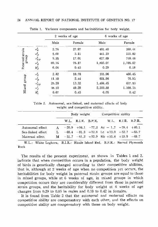
Table 1. Variance components and heritabilities for body weight.
2 weeks of age 6 weeks of age
Male Female Male Female
.: a1 2.76 27.97 481.40 268.44.;;;1-0 ,,2 5.61 3.11 441.19 133.82....,mo. D-::I
"~D 9.35 17.01 617.69 740.46roo.: 1-0I-ob.O 0
2 69.16 75.17 1,805.07 1, 785.62B FJro
h~ 0.06 0.45 0.29 0.180...
0.,,2 2.82 18.78 101.96 486.45s
::I ob 11.30 2.44 601.98 76.9301-0b.O 2 25.20 13.32 1,484.45 637.53
"0(JSD
<11,,~ 38.10 49.29 2,209.88 1,108.35x
~ h2 0.07 0.45 0.05 0.42s
Table 2. Autosomal, sex-linked, and maternal effects of bodyweight and competitive ability.
Body weight
W.L. R.I.R. B.P.R.
Competitive ability
W.L. R.I.R. B.P.R.
Autosomal effect
Sex-linked effect
Maternal effect
A -20.9 +98.1 -77.2 Ac - 1.7 -78.4 +80.1
L +68.4 -81.2 +12.8 Lc +13.0 +52.7 -65.7
M -31.7 -61.2 +92.9 Mc +35.8 +33.9 -69.7
W.L.: White Leghorn, R.I.R.: Rhode Island Red, B.P.R.: Barred PlymouthRock
The results of the present experiment, as shown in Tables 1 and 2,indicate that when competition occurs in a population, the body weightof birds is genetically changed according to their competitive abilities,that is, although at 2 weeks of age when no competition yet occurs, theheritabilities for body weight in paternal strain groups are equal to thosein mixed groups, while at 6 weeks of age, in mixed groups in whichcompetition occurs they are considerably different from those in paternalstrain groups, and the heritability for body weight at 6 weeks of agechanges from 0.29 to 0.05 in males and 0.18 to 0.42 in females.
It is found from Table 2 that the autosomal and maternal effects oncompetitive ability are compensatory with each other, and the effects oncompetitive ability are compensatory with those on body weight.

RESEARCHES CARRIED OUT IN 1966
A Genetical Study on Organ Formation in Nicotianatabacum L.
Seiichiro HIGUCHI and Kan-Ichi SAKAI
25
This study aimed at finding the genetical basis of organ formation inNicotiana tabacum L. Ten varieties were used for the study: Hicks,Connecticut Broad Leaf, Coker 139, Coker 316, Coker 319, Ibusuki, Daruma,Nicotin-free tobacco, T. I. 448A and Ambalema. Five plants selected atrandom from each variety were investigated for the size of vegetativeand reproductive organs. Genetic correlations among them are given inTable 1.
Table 1. Genetic correlation coefficients between the dimensions of vegetativeand reproductive parts of Nicotiana tabacum L.
Stipule Leaf
Longfilament
Style
Corolladiameter
Corollatube
Calyx
~ Upper~ Inter
.eo mediateen Lower
Upper'\;l Interj mediate
Lower
0.94
0.58 0.60
0.69 0.63 0.77
0.59 0.65 0.90 0.85
0.30 0.22 0.58 0.72 0.63
-0.01 0.08 0.18 0.16-0.11 0.69
-0.16-0.02 0.32 0.26 0.19 0.56 0.96
0.03 0.17 0.55 0.33 0.54 0.20 0.56 0.87
0.26 0.46 0.64 0.19 0.62-0.14-0.11 0.19 0.58
0.09 0.30 0.38 0.01 0.39-0.32-0.22-0.13 0.29 0.87
-0.02 0.09 0.37 0.30 0.48- 0.28-0. 33~ 0.12 0.40 0.68 0.86
It is found from Table 1 that the dimensions of flower parts, i. e. filaments, style and corolla, are mutually highly correlated, but not withthose of stipules and leaves. Calyx is moderately correlated with eitherflower organs or stipules. Stipules are highly correlated with each otherbut not with leaves, while leaves are highly correlated among themselves.Thus, according to size, organs of a tobacco plant may be divided into

26 ANNUAL REPORT OF NATIONAL INSTITUTE OF GENETICS NO. 17
three groups: 1) flower parts, i.e. stamens, pistil and corolla, 2) stipulesand 3) leaves. No conclusion can be drawn at present for the calyx.Within each group, genetic correlations were relatively high, whereasthose between different groups tended to be low. In addition, there wasa tendency to high correlation between adjacent parts. No definite tendencycould be seen in environmental correlations.
Major Gene and Polygenes Governing the RachisDeficiency in Rice
Kikuo WASANO and Kan-Ichi SAKAI
The present study aims at finding the role of polygenes affecting amajor gene-controlled character in rice. The character under consideration is the rachis deficiency controlled by a recessive sp-gene. The nature
Table 1. Effect of major gene, sp, and polygenes affecting the sp phenotype,on some quantitative characters in rice
sp sp effect Polygene RatioCharacter sp+ (M) effect (P) (P)j(M)L H m
Expressivity of sp gene 0 45.00 56.65 50.83 50.83** 5.83** 0.11
:e r,nt 40.45 42.16 35.66 38.91 - 1.54 -3.25** 2.11
.~~ Panicles 18.36 6.32 3.99 5.16 -13.20** -1.17** 0.09
:::: Straw 22.41 35.83 31.67 33.75 11.34** -2.08 -0.18
·rnm 11.23 12.41 11.98 12.19 0.96** -0.22** -0.23
a~ Panicles 11.04 11.37 9.95 10.66 0.38 -0.72* 1.89
Z Non-bearing tillers 0.19 1.03 2.02 1.53 1.34** 0.50** 0.37
rUlm 102.65 99.33 90.76 95.04 7.61** -4.29** 0.56
~ Panicle 21.14 15.89 14.12 15.01 - 6.13** -0.89** 0.15
'"' C' In",""'" 38.15 32.85 31.48 32.17 5.98** -0.69** 0.12
~ 2nd Internode 26.21 26.43 25.97 26.20 0.01 -0.23 />10.00.....l
3rd Internode 21.92 23.10 20.87 21.99 0.07 -1.12** />10.00
sp+ : Normal phenotype.L: Lines selected for low expressivity.H: Lines selected for high expressivity.m: (H+L)j2.**,*: Significant at 1% and 5% level, respectively.(M) : (H+L)j2-sp+.(P) : (H-L)j2./>10.00 means that polygene effect is very large in comparison with the effectof major gene.

RESEARCHES CARRIED OUT IN 1966 27
of the character is described briefly in the preceding issue, Ann. Rep. 16: 74.In the F2 population between a wild and asp-strain, 88 plants of 323 intotal were rachis deficient, homozygous for the recessive sp locus. Ofthese 88 segregants, selection for high as well as low expressivity wasconducted. The results are summarized in Table 1. From Table 1, wefind that, 1) the major gene, sp, not only governs the rachis deficiencybut also increases straw weight and number of immature tillers anddecreases weight of panicles per plant, panicle length and length of firstinternode or culm. 2) There are polygenes which affect the abovecharacter in addition to the major sp-gene. These polygenes also affectother characters pleiotropically. The effects, however, are not always thesame as those of the major gene. 3) Polygenes enhancing rachis deficiency operate toward decreasing other quantitative characters. 4) The intensityof polygenes in comparison with that of the major gene, sp, is found inthe last colmn of Table 1. It is given in the form of a ratio of polygeneeffect against the effect of the major gene. Of twelve ratios, straw weightand number of tillers show negative values indicating that the effect ofpolygenes is opposite to that of the major gene. It is also interestingthat 2nd and 3rd internodes are little affected by the sp-gene, whereasthey are more or less affected by polygenes.
Estimation of Genetic Parameters in ChamaecyparisForests
Kan-Ichi SAKAI, Shigesuke HAYASHI andHiromasa MUKAIDE
Hinoki, Chamaecyparis obtusa Sieb. et Zucc., is one of the most importanttree species in Japan. They are propagated only from seed. The presentstudy deals with investigations on inter-tree competition and estimationof genetic, environmental and competitional parameters in several forestsof the species.
The forests were artificially planted and were 20 to 60 years old.The adopted method of study will be published in detail in the coming
issues of Silvae Genetica (1967). The results are presented in Table 1.It is found from Table 1 that competition apparently occurs in Chamaecy
paris forests. It is of interest to find that an amount of genetic variationfor growth (h 2
) appears to be inversely correlated with the index ofcompetitive stress (c2
). This might suggest, though it is quite speculativeat present, that in this species the genotypic growth is negatively correlated with competitive ability.

28 ANNUAL REPORT OF NATIONAL INSTITUTE OF GENETICS NO. 17
Table 1. Genetic, environmental and competitional variance, heritability (h2),
index of competitive stress (c2) and correlation coefficient betweenadjoining two trees (ri, i+l)
-----
Strain
Okayama (1) Okayama (2) Gifu (1) Gifu (2) Oita (A)
Number of trees 200 200 207 191 246
Age 27 22 45 60 26
G 8.02±0.27 -2.35±1.61 6.83±0.12 11.82±6.57 -0.26±2.56
E 0.05±0.01 2.30±0.49 1.20±0.04 2.30±2.20 7.03±1.05
C 3.62±1.31 0.20±0.09 0.88±5.05 4.20±1. 74
h2 0.99 0 0.83 0.79 0
c2 0.61 0.02 0.06 0.37
r(i, i+l) +0.150 -0.190 -0.038 -0.122 -0.004
Developmental Genetic Study of Panicle Formation in RiceShin-ya IYAMA
Two populations of rice variety Norin No.8, one irradiated by 20,000 rX-rays and the other as untreated control, were maintained by one parentone offspring method for this investigation. At the fifth generation, 25lines were derived from the control and 35 lines from the X-rayed population. Ten plants from each line were chosen at random, and the length
Table 1. Mean lengths of the various parts of culm and paniclein the control and the X-rayed population
Mean length (em)Character
Control X-rayed
1. Third culm internode 16.758 16.368
2. Second culm internode 19.968 19.616
3. First culm internode 31.581 30.205
4. Panicle 18.245 17.555
5. Second rachilla 6.610 6.463
6. Third rachilla 7.015 6.812
7. Fourth rachilla 7.199 6.963
8. First rachis internode 2.473 2.376
9. Second rachis internode 1.316 1.297
10. Third rachis internode 1.818 1.734
RESEARCHES CARRIED OUT IN 1966 29
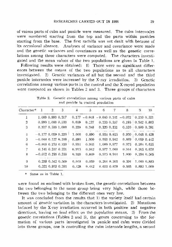
of vaious parts of culm and panicle were measured. The culm internodeswere numbered starting from the top and the parts within paniclesstarting from the base. The first rachilla was not dealt with because ofits occasional absence. Analyses of variance and covariance were madeand the genetic variances and covariances as well as the genetic correlations among those characters were computed. The characters investigated and the mean values of the two populations are given in Table 1.
Following results were obtained: 1) There were no significant differences between the means of the two populations as to all charactersinvestigated. 2) Genetic variances of all but the second and the thirdpanicle internodes were increased by the X-ray irradiation. 3) Geneticcorrelations among various parts in the control and the X-rayed populationwere computed as shown in Tables 2 and 3. Three groups of characters
Table 2. Genetic correlations among various parts of culmand panicle in control population
Character* 2 3 4 5 6 7 8 9 10
1 1.0000.990 0.317 -0.177 -0.048 -0.0400.141 -0.072 0.259 0.3212 0.990 1.000 0.533 0.059 0.127 0.233 0.357 0.195 0.542 0.8033 0.317 0.533 1.000 0.220 0.248 0.320 0.331 0.330 0.500 0.391
4 -0.1770.059 0.220 1.000 0.890 0.934 0.915 0.920 0.048 0.4285 -0.048 0.127 0.248 0.890 1.000 0.963 0.942 0.869 0.059 0.4426 - 0.040 0.233 0.320 0.934 0.903 1. 000 0.977 0.975 0.264 0.6557 0.141 0.3570.331 0.915 0.942 0.977 1.000 0.944 0.305 0.6788 -0.072 0.195 0.330 0.920 0.869 0.975 0.944 1.000 0.394 0.565
9 0.259 0.542 0.500 0.048 0.059 0.264 0.305 0.394 1.000 0.88310 0.321 0.803 0.391 0.428 0.442 0.655 0.678 0.565 0.883 1.000
* Same as in Table 1.
were found as enclosed with broken lines, the genetic correlations betweenthe two belonging to the same group being very high, while those between the two belonging to the different ones very low.
It was concluded from the results that 1) the variety itself had certainamount of genetic variation in the characters investigated. 2) Mutationsinduced by the X-ray irradiation occurred in both positive and negativedirections, having no final effect on the population means. 3) From thegenetic correlations (Tables 2 and 3), the genes concerning to the formation of various parts investigated in panicle and culm were dividedinto three groups, one is controlling the culm internode lengths, a second

30 ANNUAL REPORT OF NATIONAL INSTITUTE OF GENETICS NO. 17
Table 3. Genetic correlations among various parts of culmand panicle in X-rayed population
Character* 1 2 3 4 5 6 7 8 9 10
1 1.000 0.689 0.802 ·0.539 0.524 0.476 0.513 0.176 . 0.151 0.3462 0.689 1.000 0.783 .0.701 0.589 0.596 0.712 0.525 0.170 0.420
3 0.802 0.783 1.000 0.848 0.748 0.782 0.837 0.561 . 0.147 0.472
4 0.539 0.701 0.848 1.000 0.884 0.935 0.960 0.797 0.051 0.460
5 0.524 0.589 0.748 0.884 1.000 0.974 0.906 0.658 0.118 05486 0.476 0.596 0.782 0.935 0.974 1.000 0.970 0.747 0.140 0.579
7 0.513 0.712 0.837 0.960 0.906 0.970 1.000 0.804 0.079 0.559
8 0.176 0.525 0.561 0.797 0.658 0.747 0.804 1.000 0.317 0.634
9 0.151 0.170 0.147 0.051 0.118 0.140 0.079 0.317 . 1.000 0.718
10 0.346 0.420 0.472 0.460 0.548 0.579 0.559 0.634 ·0.718 1.000
* Same as in Table 1.
controlling the lengths of panicle, the lower rachillas of panicle and thefirst rachis internode, and a third responsible for the lower internodelengths of panicle. 4) In addition, it was suggested from the geneticcorrelations of the X-rayed population that most new mutations wouldhave been induced more or less pleiotropically for all the characters inthe same direction, making correlations between groups in the X-rayedpopulation higher than those in the control population.
A Developmental Genetic Study in RiceM. S. BALAL and Kan-Ichi SAKAI
Organ formation in rice was investigated from the standpoint of statistical-genetics. Materials used were 99 lines selected at random fromthe Norin No.8 variety established since 1959 and the same number ofX-irradiated lines of the same variety. Characters investigated werelength of panicle, first, second and third leaves counted from the topand first, second, third and fourth culm internodes. Analysis of variancebetween and within lines showed that both populations of 99 lines eachcontained significant genetic variation, indicating that in an establishedrice variety a certain amount of genetic variation was retained, on theone hand, and that the genetic variance in the X-rayed population waslarger than in the control population, on the other hand. Estimation ofgenetic correlation and degree of pleiotropic effect of polygenes (seeSakai and Suzuki 1964, in Rad. Bot. 4: 141~151) suggested that organs of

RESEARCHES CARRIED OUT IN 1966 31
rice could be divided into two developmental groups. One is the upperorgan group which includes panicle, first and second leaves and firstculm internode, which show high positive correlation among each other.The second is the basal organ group which includes third and fourthculm internodes which are highly positively correlated. The remainingtwo organs, third leaf and second culm internode, are moderately correlated with the organs of either upper or basal group. In general, geneticcorrelation between neighbouring organs, regardless whether they wereleaves, panicle or culm internodes, tended to be higher than that amongremote organs.
A selection experiment for the length of third culm internode showedthat the fourth was also affected by that selection, though it had littleeffect on panicle and first as well as second culm internode. The application of nitrogen at the time of panicle formation yielded about 40 per centincrease in length of third and fourth culm internodes but only a 10 percent or less increase in panicle and first culm internode. An anatomicalstudy indicated that the third and fourth culm internodes developed inadvance of panicle primordia, while the upper internodes (first and second)developed after them.
The foregoing evidences indicate that organs developing in rice are notcontrolled by the same genes but by genes locally controlling specificorgans.
Electrophoretic Comparison of Soluble Proteins fromDifferent Organs of Tobacco Plant
Sumiko NARISE and Kan-Ichi SAKAI
In order to inquire into the mechanism of gene control over the development of higher plants, an electrophoretic analysis was undertaken withsoluble proteins from different organs of Nicotiana tabaccum L.. Organsinvestigated were stamens, pistils, corollas, calyxes, nerves and mesophyllsof stipules and also those of leaves. They were separately homogenizedwith the 0.1 M phosphate buffer (pH 7.2) and centrifuged. The supernatants were precipitated with saturated ammonium sulfate and the precipitate was used for analysis after dialysis against distilled water.Electrophoresis was carried out in columns of polyacrylamide gel using amodified method of Orstein and Davis. Protein bands were stained withamido black. All protein bands separated were compared on the basisof their relative mobility measured as fractions to the distance the fastestband manifested. The experiment was repeated five to ten times foreach organ. By the (-test, twenty-eight different kinds of bands in all
32 ANNUAL REPORT OF NATIONAL INSTITUTE OF GENETICS NO. 17
were identified. Of those 28 bands, 14 were found in leaves from themiddle part of stem, 22 in leaves from the top, 17 in stipules, 7 in calyxes,10 in petals, 9 in pistils and only 5 in stamens. For the purpose ofcomparison among organs, it seemed appropriate to take into account notonly the distribution pattern of bands, but also quantitative measurementsof intensity of each band. The quantitative evaluation was made bygrading the intensity into six classes 0 to 5 by naked eye. Thus, theresults obtained from this combined comparison were as follows: Therewas a great deal of similarity among leaves in the number and the concentration of proteins of identical mobility, whereas not much similaritybetween leaves and reproductive organs. Furthermore, the similaritybetween stipules and leaves was relatively high and the same tendencywas shown between stipules and calyxes. These results may help tosolve the puzzle of genetic control over the development of higher plants.
Analysis of Genetic Correlations between Panicle, Internodeand Leaf Lengths among Mutant Strains of a
Rice Variety, Norin 8Hiroko MORISHIMA and Hiko-Ichi OKA
For a survey of genetic variations in the sizes of different organs, 33mutant strains induced from a rice variety, Norin 8, were grown in anexperimental plot, and were measured regarding the lengths at maturityof the panicle, the first to fifth (from the top) internodes, and the firstto fourth leaf sheaths and leaf blades. Those strains, obtained from theNational Institute of Agricultural Sciences through the kindness of Dr.T. Kawai, were normally fertile and their yields were comparable to thatof Norin 8.
Genetic correlations between the measured character were computed,and to find an integrated picture of character variations, the matrix ofgenetic correlations was studied by the method of principal componentanalysis. The first component appeared to represent the general size ofvarious organs, the vector having almost equal loadings on all the characters. The scores given by this vector showed the variation betweengenerally tall and generally short strains.
The second component vector had positive loadings on the panicle, theupper (first and second) internodes and the upper leaves (both sheath andblade; first and second), but negative loadings on the lower internodesand lower leaves. This indicates that the strains vary between a typehaving well-elongated upper organs and short lower organs, and the opposite type. These plant types may be called the "upward-elongation"

RESEARCHES CARRIED OUT IN 1966 33
and" basal-elongation" types. The former type appeared to have higheryielding potential than the latter, when the scores given by the secondcomponent vector were compared with grain yields of the strains, estimated as panicle number per plant x spikelet number per panicle x fertilityX mean grain weight.
We have formerly found from a segregating population of rice twolatent phases of character association which we considered as variationaxes of "genetic plant types" (genetically conditioned character association),one representing the variation between the" panicle-number" and" paniclelength" types, and the other representing the variation between the" internode-length" and" internode-number" types (Ann. Rep. 16: 68~69). Theabove-mentioned" upward-elongation" and" basal-elongation" types maybe compared with the" internode-length" and "internode-number" types,respectively, as the former would have fewer but longer elongated-internodesthan the latter. The occurrence of this plant-type variation in differentmaterials seems to suggest that it might result from a general trend ofthe developmental system to respond to genetic variations.
The third component vector seemed to distinguish between differentkinds of organs, i. e., internode, leaf sheath and leaf blade. There maybe a phase of correlation at which organs of the same kind are correlatedmore intimately than those of different kinds. The first to third components thus extracted could represent a 86 per cent portion of the totalmulti-dimensional variation.
Analysis of Growth Curves for Panicle and Internode Elongationin Mutant Strains of a Rice Variety, Norin 8
Hiroko MORISHIMA and Hiko-Ichi aKA
The growth and development of plants may be investigated fremdifferent viewpoints. This study is concerned with multivariate analysisof growth parameters separately obtained from different organs of ricestrains. In 23 mutant strains induced from Norin 8 (a part of the materialsmentioned in the previous report), the lengths of panicles and the firstto fourth internodes were measured five times before and after headingat one week interval and at maturity. In each strain, growth curves ofthese organs were estimated by fitting the data to Robertson's equation,log (xIA-x)=k(t-t1/2), where x is the size at time t, A is the final size,t1/2 is the time at which a half of the final size is reached, and k is aparameter for growth rate. The actual growth rate (dxldt) was computedfrom these values.
Regarding the variations in A, t1/2- H (number of days between the t1/2
34 ANNUAL REPORT OF NATIONAL INSTITUTE OF GENETICS NO. 17
time and heading time), and dx/dt of different organs, genetic variances andcorrelations were computed. To observe the pattern of association of thesevalues, principal components were extracted from phenotypic correlationmatrices of A, tl/Z- Hand dx/dt, respectively. Genetic contributions tothe components were then estimated in terms of the regression coefficientsof genetic values on the component axes by a method newly devised byHashiguchi et at. (in press). From the" genetic vectors" obtained, variationpatterns in the growth of those organs were estimated as follows: 1) Inthe final size (A), the first component (AI) showed the variation in generalsize of all the five organs, and the second component (All) showed thephase of variation between the "upward-elongation" and "basal-elongation"types, in the same manner as set forth in our previous report. 2) Thefirst component from the interval between (l/Z time and heading timerepresented the general earliness of panicle and internode elongationrelative to heading. 3) The first component from the growth rate (dx/dt)represented variation in the "general growth rate", while the secondcomponent represented a phase of variation at which the growth ratesof panicle and the third internode were negatively associated with thoseof the first and second internodes.
The component scores given by these vectors were computed in eachstrain. Correlations between the scores indicated that 1) strains havinggenerally long organs tended to have a generally high growth rate forthose organs, and 2) strains of the" upward-elongation" type tended to havea low rate of panicle elongation and a high rate of internode elongation,and vice versa. It seems that slow panicle growth and fast panicle emergence (due to fast internode elongation) bring about well developedpanicles, elongated upper internodes and relatively short lower internodes.Such a growth type may have high yielding potential.
Differentiation of Aggregation-Promoting Materialsfrom Embryonic Chick Liver Cells
Yukiaki KURODA
Trypsin-dissociated cells from avian and mammalian embryos whencultured by rotation-mediated procedure tend to aggregate and formhistoformative structures. Liver cells dissociated from chick embryos atthe ages of 7-, 8-, 10-, 14-, 18-, and 20-days were tested for their aggregability in rotation cultures. Liver cells dissociated from 7-day embryosformed two or three large aggregates after 24 hours of cultivation.Aggregates formed from liver cells of older embryos showed a gradualdecline in average diameter. Almost complete loss of aggregability was
RESEARCHES CARRIED OUT IN 1966 35
seen in liver cells dissociated from 20-day embryos.Mixed suspension of liver cells from both younger and older embryos
produced some aggregates which had an average diameter intermediatebetween those found for each of these ages separately. Histologically themajor part of those aggregates consisted of liver cells from youngerembryos.
Cell-free supernatants were obtained from reciprocation cultures ofembryonic chick liver cells, and tested at 28°C for their activity in promoting aggregation. Supernatants prepared from 7-day embryonic livercells were considerably enhancing aggregation of 7-day embryonic livercells, but had no effect on those from I8-day cells. Supernatants preparedfrom I8-day embryonic liver cells had no effect on liver cells either from7-day or I8-day embryos.
Supernatants prepared from 7-day embryonic liver cells promoted theformation of aggregates from I8-day embryonic liver cells by rotation at38°C for three or more days. The promoting activity of aggregate formation by the supernatant seems to be temperature-dependent, since at28°C this supernatant had no effect on the dissociated cells from I8-dayembryo. It seems likely that non-aggregation of cells from older embryosmight be related to changes accompanying progressing specialization.Incubation of such specialized cells with active supernatants may modifythe cells so that they produce some materials acting on cell cohesion.
Characterization of Tissue-Specific Materials with Cell-BindingActivity Obtained from Embryonic Chick Cells
Yukiaki KURODA
It has been previously found that in the process of aggregation ofdissociated embryonic cells some substances released from the cells intothe culture medium promoted the adhesive property of each cell. Theseaggregation-promoting substances were contained in cell-free supernatantsprepared from a culture medium in which dissociated cells were shakenby reciprocating motion at 100 rpm for 2 hours at 38°C. Supernatantsprepared from 7-day embryonic liver cells showed an activity in enhancingaggregation of 7-day embryonic liver cells at 28°C, but no effect on 7-dayembryonic heart cells, whereas supernatants prepared from embryonicheart cells acted only on embryonic heart cells in forming aggregates,but had no effect on embryonic liver cells; the aggregation-promotingactivity of the supernatants was tissue- or organ-specific. In the mediumadded with 0.5 flg/ml actinomycin D or 5 flg/ml puromycin dissociatedliver cells were inhibited from forming aggregates. These results suggest

36 ANNUAL REPORT OF NATIONAL INSTITUTE OF GENETICS NO. 17
that the production of aggregation-promoting materials by the cells mightbe mediated by DNA-dependent RNA synthesis and protein synthesis.
Some informations on the composition of the supernatants from 7-dayembryonic liver and heart cells were obtained. Both those supernatantshad UV absorption spectra with a peak at 280 mp and some amount ofprotein was detected by chemical determinations with slight amounts ofDNA and RNA. The supernatants lost their aggregation-promotingactivity by heat-treatment at 80 a C for 10 minutes.
Amino acid composition of proteins contained in supernatants from 7day embryonic liver and heart cells was compared. For the purpose ofthis analysis Tyrode's solution was used in which the cells were shakenon a reciprocating shaker at 38a C for 2 hours. Molar ratios of aminoacid components in acid hydrolyzates of supernatant proteins are given
Table 1. Molar ratios of amino acid components in proteins in supernatantsprepared from 7-day embryonic chick liver and heart cells
Amino acid Liver cell supernatant Heart cell supernatant
Acidic and neutralAlanine 0.83 0.78
Aspartic acid 1.08 1.03
Glutamic acid 1.07 1.05Glycine 0.97 0.84
Isoleucine 0.56 0.62
Leucine 1.00 1.00
Methionine 0.03 0.18
Phenylalanine 0.37 0.39
Proline 0.55 0.51
Serine 0.63 0.49Threonine 0.45 0.49Tyrosine 0.21 0.27
Valine 0.77 0.81
BasicAmmonia 1.40 0.96
Arginine 0.60 0.61
Histidine 0.32 0.30Lysine 1.00 1.00
Samples were hydrolyzed in 6N HCI for 40 hours at 105a C. A HitachiKLA-2 type amino acid analyzer was used. Amino acid componentsare expressed in molar ratios corresponding to 1.00 mole of leucineor lysine for acidic and neutral or basic amino acids respectively.
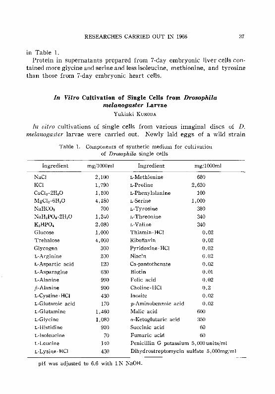
RESEARCHES CARRIED OUT IN 1966 37
in Table 1.Protein in supernatants prepared from 7-day embryonic liver cells con
tained more glycine and serine and less isoleucine, methionine, and tyrosinethan those from 7-day embryonic heart cells.
In Vitro Cultivation of Single Cells from Drosophilamelanogaster Larvae
Yukiaki KURODA
In vitro cultivations of single cells from various imaginal discs of D.melanogaster larvae were carried out. Newly laid eggs of a wild strain
Table 1. Components of synthetic medium for cultivationof Drosophila single cells
Ingredient mg/l000ml Ingredient mg/l000mI
NaCI 2,100 L-Methionine 680
KCI 1,790 L-Proline 2,620
CaC\z·2HzO 1,100 L-Phenylalanine 100
MgClz·6HzO 4,180 L-Serine 1,000
NaHCOa 700 L-Tyrosine 380
NaHzPO,·2HzO 1,240 L-Threonine 340
KzHPO, 2,080 L-Valine 340
Glucose 1,000 Thiamin· HCI 0.02
Trehalose 4,000 Riboflavin 0.02
Glycogen 300 Pyridoxine· HCI 0.02
L-Arginine 2;)0 Niacin 0.02
L-Aspartic acid 120 Ca-pantothenate 0.02
L-Asparagine 630 Biotin 0.01
L-Alanine 990 Folic acid 0.02
p-Alanine 900 Choline· HCI 0.2
L-Cystine· HCI 430 Inosite 0.02
L-Glutamic acid 170 p-Aminobenzoic acid 0.02
L-Glutamine 1,460 Malic acid 600
L-Glycine 1,080 a-Ketoglutaric acid 350
L-Histidine 920 Succinic acid 60
L-Isoleucine 70 Fumaric acid 60
L-Leucine 140 Penicillin G potassium 5,000 units/ml
L-Lysine·HCI 430 Dihydrostreptomycin sulfate 5, OOOmg/ml
pH was adjusted to 6.6 with 1 N NaOH.

38 ANNUAL REPORT OF NATIONAL INSTITUTE OF GENETICS NO. 17
(Oregon-R) were collected and sterilized in 70 per cent ethyl alcohol for 10minutes. The eggs were transferred to autoclaved food medium containedin bottles. Mature third-instar larvae (96 hours after hatching at 25°C)grown under those sterile conditions were dissected in order to isolate variousimaginal discs used for cultivation. Wing discs, eye-antennal discs, legdiscs, testes, and ovaries were cultured in a synthetic medium supplementedwith 10 per cent calf serum in TD-7 flasks at 25°C. The components of theculture medium are shown in Table 1. Detailed procedure for cultivationwill be described elsewhere.
Another attempt was made to dissociate the imaginal discs from maturelarvae into single cells by treatment with trypsin. Isolated imaginaldiscs were incubated in 1 per cent trypsin solution for 15 minutes. Afterrinsing with physiological saline the discs were dispersed into single cellsin the culture medium by flushing them briskly through the tip of a finepipette. The single cell suspension obtained in this waY,was distributedinto TD-7 flasks and incubated at 25°C. Establishment of cell lines having some distinct genetic markers and isolation of clones from a varietyof tissues or organs by colony formation procedure are carried out.

RESEARCHES CARRIED OUT IN 1966
III. BIOCHEMICAL GENETICS
Some Notes on the Chromogranules in Hypodermal Cellsof Silkworm Larvae
Mitsuo TSUJITA
39
When a part of white turbid cytoplasm of the hypodermis of 5th instarlarvae is placed on a seat mesh covered with a collodion film and electronmicroscopically examined, a number of elliptical, oval or spindle shapedchromogranules into which the electron-rays do not completely penetratecan be observed. The chromogranules in larval skin cells of the w-estrain have almost uniform size (1 x 1.5 ,u). On the contrary, in C-1OSstrain those having the usual size are mixed with small ones. In somechromogranules their granule (vesicle) membrane can be clearly discriminated. Their density is 1.4~1.7 which is higher than of any other cellorganelles.
In general, in the cytoplasm surrounding the chromogranules no structure interrupting intense electron rays can be observed. This seems toindicate that the chromogranules produced abundantly in the cytoplasmof larval skin cells of the normal strain are the organelles which bringabout the non-transparency of larval skin.
Each mature chromogranule consists of a vesicle and a thin membranesurrounding it. In the course of development, ribosomal particles, 150~200 A in diameter, arrange themselves in a uniform layer at the peripheryof the granules. It seems that they are intimately associated with thegranule membrane. Although those particles are apt to be lost after thematuration of the granule, it was often observed that most of them remain sticking to the membrane.
The chromogranules disintegrate after the loss of granule contents inthe following cases.
1) A part of chromogranules in hypodermal cells of larvae disintegratesin each instar, especially at the moulting stage. The granule membraneloses its normal function, the stored secretion products come out and thenit disintegrates.
2) All of chromogranules in hypodermal cells of the larvae disintegrateat the full grown larval stage: They begin to issue forth their secretionproducts through their membrane subjecting it to some change. Losing thesecretion products, the ghosts, i.e. the membranes, remain and then theyalso disappear. The density of chromogranules is remarkably reduced byloss of granule contents.
3) The fact that disintegration of chromogranules can be artificially
40 ANNUAL REPORT OF NATIONAL INSTITUTE OF GENETICS NO. 17
induced was reported (Tsujita and Sakurai, 1963). This disintegrationprocess was electron-microscopically studied.
When mulberry leaves painted with 0.3 per cent melamine solution werecontinuously fed to normal or lemon yellow larvae from the beginning oftheir instar, disintegration of chromogranules started 2~3 days after thetreatment, namely, the contents of granules were lost through theirmembranes and later the membranes disappeared.
All chromogranules disintegrate and pteridine compounds and uric acidare lost from hypodermal cells. Thus, larvae treated with melaminebecome highly transparent. Normal opaque hypodermis could develop inlarvae with transparent hypodermis that were artificially produced, whenthey were fed for about two days with fresh mulberry leaves withoutmelamine. In this recovery process a number of minute particles firstappeared in the cytoplasm and they gradually increased in size. Althoughthe skin of larvae in this stage exhibited semi-transparency, when thoseminute particles in hypodermal cells grew to usual size, the larvae showed white non-transparent skin.
4) It is difficult to fix the complete content of chromogranules becausealmost all of them are lost in the procedure of fixation and dehydration.For example, after fixation with osmic acid, dehydration was carried outthrough alcohol from low to high concentration. During this procedurethe materials were examined by electron microscope and it was foundthat the granule contents were almost all lost through the granule membranes during dehydration.
Development of Chromogranules in the Larval Skinof the Silkworm
Mitsuo TSUJITA and Susumu SAKURAI
The developmental process of the chromogranules in larval skin cellsof the silkworm was studied by electron microscopical observation of thinsections.
The larval skin cells of the silkworm larvae have generally the shapeof cylinders and the elliptical nucleus occupies a central position. Atfirst, numerous ribosomes, 150~200A in diameter, assemble here and therein the cytoplasm forming ribosomal aggregates. A small vesicle appearsin the center of each aggregate. The vesicles gradually increase in sizeand spherical, oval or elliptical granules are formed. The ribosomes arrange themselves uniformly in a layer surrounding the granule membraneand some proteinous substance can be seen inside the vesicle. It may be saidthat the precursor of chromogranules is a type of endoplasmic reticulum.
RESEARCHES CARRIED OUT IN 1966 41
It was substantiated (Tsujita and Sakurai, 1964) that soluble proteinscombining either with pteridine pigments or with uric acid are presentin the chromogranules. It may be safely said that those compounds arecontained in the granule vesicles and squeeze out when the chromogranules are pressed upon.
The well delimited precursor of the chromogranules can be precipitated by a procedure leading to the separation of the mitochondrial fraction.
It was stated above that the process of chromogranule development frominitial to mature stage can be followed up in the cytoplasm of hypodermal cells. The early steps of the same developmental process can befollowed up on the sections through the mitochondrial fraction.
Gene dependent variations were electron-microscopically observed as toshape and size of chromogranules produced in larval skin cells of normalstrains and mutants with transparent larval skin.
It may be inferred that a large amount of uric acid found in the larvalhypodermis of normal strains is mostly contained in the chromogranules.On the contrary, size and shape of chromogranules produced in thehypodermal cells of larvae with transparent hypodermis are abnormal andthe amount of mature granules is decreased. The larval skin becomestransparent to varying degree in accordance with the shape and theamount of chromogranules. For example, in hypodermal cells of larvaehomozygous for WO" or w-a no mature chromogranules are found. Consequently, uric acid or pteridine compounds can be scarcely detected intheir hypodermis. As for other mutant strains, transparency of larvalskin and the amount of uric acid and pteridine compounds varies fromstrain to strain according to the shape and amount of chromogranules,especially their amount.
In the course of granule formation, the development of vesicle (granule)membrane with attached to it numerous ribosomal particles, which playsome important role in the secretion of polypeptides, is the most important process. When it proceeds normally the granules have completeor sufficient ability for secretion and storage, and abundant chromogranulespacked compactly with secretion products are formed in larval skin cells.
So far as our experimental results are concerned it seems that uricacid production and pteridine metabolism are normally carried out in theemployed mutant strains with transparent larval skin. Therefore, it isinferred that in hypodermal cells of those larvae, the function of thegranules, i.e. secretion and storage, is defective owing to the abnormaldevelopment of the granules, especially of granule membrane.
It was found by us that the granule membrane protein consists of ahomogenous single unit protein. It seems that the granule membraneprotein is made up by polymerization of this unit protein which is the
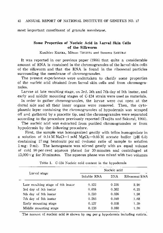
42 ANNUAL REPORT OF NATIONAL INSTITUTE OF GENETICS NO. 17
most important constituent of granule membrane.
Some Properties of Nucleic Acid in Larval Skin Cellsof the Silkworm
Kunihiro KOJIMA, Mitsuo TSUJITA and Susumu SAKURAI
It was reported in our previous paper (1966) that quite a considerableamount of RNA is contained in the chromogranules of the larval skin cellsof the silkworm and that the RNA is found in the ribosomal particlessurrounding the membrane of chromogranules.
The present experiments were undertaken to clarify some propertiesof the nucleic acid obtained from larval skin cells and from chromogranules.
Larvae at late moulting stage, on 3rd, 5th and 7th day of 5th instar, andearly and middle mounting stages of C-124 strain were used as materials.
In order to gather chromogranules, the larvae were cut open at thedorsal side and all their inner organs were removed. Then, the cytoplasmic layer containing the chromogranules of hypodermis was scrapedoff and gathered by a pincette tip, and the chromogranules were separatedaccording to the procedure previously reported (Tsujita and Sakurai, 1966).
The nucleic acid was extracted from purified chromogranules or fromhypodermis by the following procedure.
First, the sample was homogenized gently with teflon homogenizer ina solution of 0.14 M NaCI-1 mM MgCb-0.05 M acetate buffer (pH 6.0)containing 17 mg bentonite per ml (volume ratio of sample to solution1 mg: 9 mI). The homogenate was stirred gently with an equal volumeof cold 90 per cent aqueous phenol for 30 minutes and centrifuged at13,000 X g for 30 minutes. The aqueous phase was mixed with two volumes
Table 1. C-I24 Nucleic acid content in the hypodermis
Nucleic acidLarval stage
Late moulting stage of 4th instar
3rd day of 5th instar
5th day of 5th instar
7th day of 5th instar
Early mounting stage
Middle mounting stage
Soluble RNA
0.522
0.658
0.210
0.255
0.127
0.135
DNA
0.255
0.362
0.036
0.040
0.038
0.060
Ribosomal RNA
2.90
6.25
1.98
1.68
1.30
1.90
The amount of nucleic acid is shown by mg per g hypodermis including cuticle.
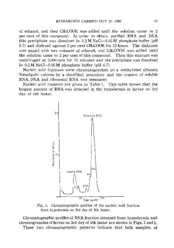
RESEARCHES CARRIED OUT IN 1966 43
of ethanol, and then CHaCOOK was added until the solution came to 2per cent of this compound. In order to obtain purified RNA and DNAthis precipitate was dissolved in 0.2 M NaCI-0.05 M phosphate buffer (pH6.7) and dialysed against 2 per cent CHaCOOK for 12 hours. The dialyzatewas mixed with two volumes of ethanol, and CHaCOOK was added untilthe solution came to 2 per cent of this compound. Then this mixture wascentrifuged at 3,000 rpm for 15 minutes and the precipitate was dissolvedin 0.2 M NaCl-0.05 M phosphate buffer (pH 6.7).
Nucleic acid fractions were chromatographed on a methylated albuminKieselguhr column by a simplified procedure and the content of solubleRNA, DNA and ribosomal RNA was measured.
Nucleic acid contents are given in Table 1. This table shows that thelargest amount of RNA was detected in the hypodermis in larvae on 3rdday of 5th instar.
1.0I~ibosomal Rr--;A
E
60sSoluble RNA
50 100Tube number
Fig. 1. Chromatographic profiles of the nucleic acid fractionfrom hypodermis on 3rd day of 5th instar.
Chromatographic profiles of RNA fraction obtained from hypodermis andchromogranules of larvae on 3rd day of 5th instar are shown in Figs. 1 and 2.
Those two chromatographic patterns indicate that both samples, of

44 ANNUAL REPORT OF NATIO~AL INSTITUTE OF GENETICS ~O. 17
Ribosomal RNA
1.0
~ 0.5
-e.§-<
60s405
O.O''-----..,,50~-----:l~OO~----Tube number
Fig. 2. Chromatographic profiles of the nucleic acid fractionsfrom chromogranules on 3rd day of 5th instar.
hypodermis and chromogranules, contained 4 S soluble RNA, DNA and40 S, 60 S ribosomal particles. Therefore, it may be said from these ex·perimental results that the numerous minute particles surrounding themembrane of chromogranules are ribosomal particles.
Incorporation of HC-Glycine into Chromogranulesof Larval Skin Cells of the Silkworm
Susumu SAKURAI and Mitsuo TSUJITA
In order to observe polypeptide synthesis in chromogranules, 0.3 pc ofHe-glycine was injected into the body fluid of the silkworm at 4thmoulting stage, on 3rd day and 5th day of 5th larval instar, and at fullgrown larval stage. The cytoplasm of the hypodermal layer was scrapedoff and gathered. The raw fresh cytoplasm was mixed with 0.25 Msucrose solution (volume ratio of sample to sucrose solution 1: 10) andcentrifuged at 900xg for 10 minutes.
The chromogranules were sedimented by centrifugation at 900xg for 10minutes. Microsomes and mitochondria were isolated as shown in Fig. 1.

RESEARCHES CARRIED OUT IN 1966
Raw fresh cytoplasm in 10 volumes of 0.25 M sucrose solution900 X g for 10 min
I
45
ICrude
chromogranules
ISupernatant layer
5,000 X g for 10 minI
iMitochondria
IPellet
(microsomes I)
ISupernatant layer
12,000 X g for 10 minI
Supernatant layer104,000xg for 60 min
II
Pellet(microsomes IV)
Fig 1. Isolation procedure of labeled cell fractions.
The supernatant layer was centrifuged at 5,000 X g for 10 minutes. Theresultant pellet was the fraction containing mitochondria and chromogranuleprecursors (Tsujita and Sakurai, 1966). The protein residues obtained fromthe labeled cell fractions according to the procedure of Schmidt-Thannhauser and Schneider (STS) were dissolved in 0.3 N-potassium hydroxideand the radioactivity was measured in a windowless gas flow counter.
C.P.M.
200
100B.F.
CH.G.
count per minutebody fluidchromogranules
OL..-_-~-~-_-.........-....-30 60 90 120 150 180
min.
Fig. 2. Time course of incorporating reaction of 14C-labeledamino acid into body fluid and soluble polypeptides ofchromogranules.
C.P.M.:H.F.CH. G.:
As shown in Fig. 2, maximum incorporation of 14e-glycine into chromogranules could be seen 2 hours after injection (Sakurai and Tsujita, 1966).

46 ANNUAL REPORT OF NATIONAL INSTITUTE OF GENETICS NO. 17
The chromogranules had higher radioactivity than the microsomes andmitochondria, and moreover HC was mostly distributed among polypeptidecomponents of secretion products within the granules after incorporationof HC-Iabeled amino acid into the granules.
Chemical Characterization of Chromogranule Membranein Larval Skin Cells of the Silkworm
Susumu SAKURAI and Mitsuo TSUJITA
Preparation and purification of insoluble protein from granule membrane-·Purified chromogranules were dissolved in 2 per cent deoxycholate. Thesolution was dialysed against distilled water for overnight at 5°C. Theprecipitate formed was collected by centrifugation at 5,000 x g for 10minutes, suspended in 0.25 per cent sucrose solution and again centrifuged at 1O,000xg for 10 minutes. The precipitate was dissolved in 8 Murea solution and 2-mercaptoethanol was added at pH 8.6 (volume ratioof sample to 2-mercaptoethanol 1 mg: 1 mI). After 1 or 2 hours, the reaction mixture was eluted through Sephadex G 25 1.4 x 100 em column whichhad been equillibrated with 0.001 M EDTA-0.5 per cent methylamine-HCIbuffer at pH 8, to remove excess urea and 2-mercaptoethanol. The reduced membrane was then treated with p-chloromercuribenzoic acid(PCMB)at room temperature for 30 minutes and adjusted to pH 8 (Anfinsen 1961).The final mixture was dialysed against 0.5 per cent methylamine- HCIbuffer at pH 8 and further dialysed against distilled water. The suspension of PCMB-membrane protein was lyophilized for storage.
Electrophoretic and ultracentrifugation analysis of peME-membraneprotein-After removal of the PCMB by suspension in 0.001 M EDTA0.5 per cent methylamine-HCI buffer at pH 8, 1 ml of mercaptoethanol permg of the derivative was added and the fully reduced membrane proteinwas applied to the Sephadex column as described above. The reducedsample thus obtained was examined for electrophoresis. Polyacrylamidegels were prepared with 6 per cent cyanogum 41 in Tris-HCI buffer atpH 9. Electrophoresis was carried out in vertical position for 7 hours at5 rna constant. Tris-glycine buffer at pH 8.7 was used as the electrolyte.The gels were stained with 0.1 per cent amido black 10 B in a solvent ofmethanol-water-acetic acid (5: 4: 1). Excess dye was removed by overnight washing the gel in a 10 per cent acetic acid.
As the electrophoretic pattern of Fig. la shows, purified granule membrane protein migrated as one clear-cut single band.
The mercaptoethanol reduced sample was dissolved in 5 per centmethylamine-HCI buffer at pH 8 and ultracentrifuged at 56,000 rpm.

RESEARCHES CARRIED OUT IN 1966
-Origin
- Spacer gel
+ +(a) (b)
Fig. 1. Disk electrophoresis of protein from granulemembrane of normal and oily mutant.a. Protein from normal granule membrane.b. Protein from granule membrane of oily mutant.
47
Fig. 2. Sedimentation diagrams of protein from normal granulemembrane. Ultracentrifugation at 56,000 rpm at 4°C, The 0.25per cent solution of the protein in methylamine-HCI buffer (pH 8).Photographs were taken at 8 minutes intervals.
As shown in Fig. 2, purified granule membrane protein of the normalstrain exhibited a single symmetrical boundary in the analytical ultracentrifuge in 5 per cent methylamine-HCI buffer.
Thus, it may be inferred from the above-mentioned experimental results that the membrane protein of the chromogranules is composed of ahomogeneous single unit protein.
According to our preliminary experimental results, the electrophoreticpattern of the membrane protein, obtained from chromogranules of hypodermal cells of 60CO r-ray induced w-b mutant with oily larval skin, exhibitedtwo or three minor bands, not shown by normal granule membrane, andelectrophoretic mobility of the major band differed from that of normal

48 ANNUAL REPORT OF NATIONAL INSTITUTE OF GENETICS NO. 17
granule membrane (Fig. Ib). This fact suggests that there is some defectin the formation of granule membrane protein and some important erroroccurs in the secretion and storage of polypeptides within the granules.
As to the constituent amino acids of the granule membrane protein,seventeen amino acids, lysine, arginine, histidine, aspartic acid, glutamicacid, threonine, serine, proline, glycine, alanine, cystine, valine, methionine, leucine, isoleucine, tyrosine and phenylalanine were detected usinga Hitachi's amino acid analyzer.
Genetic and biochemical studies of the amino acid sequence in the unitprotein of the granule membrane found in the present investigation andof the mode of polymerization of the unit protein in the formation ofchromogranule membrane is now under way.
Genetic Effects of DNA in EphestiaSaburo NAWA and Mas'laki YAMADA
1. The experiment has been designed for inducing DNA-mediatedgenetic transformation in Ephestia. An imago of the wild type strain(NCR, a+/a+) is black eyed, while a homozygote (a/a) for the recessivegene a, which controls the enzyme tryptophan pyrrolase, is red eyed.Larvae of a/a were injected with DNA extracted from NCR adults. Theinjected larvae were permitted to develop into adults and were examinedfor eye-color. Some of them were crossed to untreated a/a to score thechange in eye color in their progeny and in subsequent generation. Blackeyed animals have been obtained either among the treated animals or insubsequent generations. The results are summarized in Table 1. When
Table l. Frequency of mutations among treated animals and insubsequent generations
Ex-Treated generation B, B2
peri- No. of adults Black-eyed No. of adults Black-eyed No. of adults Black-eyedment examined mutants examined mutants examined mutants
1 105 0 6,655 0 1,196 12 140 0 4,010 0 2,156 2
3 214 0 9,425 0 1,974 14 220 1 9,105 2 13,980 4
5 54 0 3,398 4 4,168 06 85 0 7,039 0 14,699 1
Total 818 1 39,632 6 38,173 9

RESEARCHES CARRIED OUT IN 1966 49
transmission of the newly acquired black eye color has been followed forseveral generations by mating mutants with untreated a/a, it becameevident that there were several types of mutants. 1) Most of the black-eyedmutants have been confirmed to be a+/a heterozygotes. 2) One mutantmale, which appeared in B 2 in Table 1, gave a+/a heterozygotes and a/ahomozygotes in the proportion of 6: 1, when crossed with an a/a female,showing that the genotype of the mutant was not simply a+/a. It mayhave been a mosaic. 3) A few mutants behaved as being a+/a+ homozygotes, since they gave only a+/a progeny when crossed by a/a. Thesemutants appeared in the progeny (Bl and B 2 in Table 1) derived fromcrosses of treated animals with untreated a/a. It is difficult to imaginethe occurrence of a+/a+ homozygotes in the progeny from crosses witha/a. Special care has been taken to avoid contamination by migratinganimals. 4) An unusual mutant, a black-eyed female, was obtained in thetreated generation. When the female was crossed with an a/a male, allthe Bl animals were black, although only five were obtained, all females.All those females were crossed with a/a males. One pair gave no progeny.Three pairs gave in the total 356 imagos, all of which were black, but allwere a+/a heterozygotes. The fourth pair gave rise to 123 black-eyedoffsprings, half of which were a+/a heterozgotes, but the rest were not, andgave only black-eyed animals (a+/a) in crosses with a/a. These observationscould be interpreted on the assumption that a piece of DNA incorporatedinto the cell behaved as a particle which could reproduce itself, althoughits sudden disappearance cannot be plausibly explained. 5) One mutantfemale, which was apparently black-eyed, gave no black-eyed animals insubsequent generations when crossed with a/a. It seems likely that inthis female the mutation occurred in somatic cells but not in germ cells.
Treatment of eggs of a/a with NCR-DNA has also been carried out,giving similar results.
In a reverse type of experiment, eggs of NCR (a+/a+) were treatedwith DNA extracted from a/a adults. When imagos developed from treated eggs were crossed with a/a, a red-eyed mutant has been obtained.Further experiments are now in progress.
2. Some properties of DNA used in the experiments have been describedpreviously (Ann. Rep. No. 15). The molecular weight of the DNA wasestimated from its viscosity and sedimentation. The intrinsic viscosity[r;] ranged from 70 to 110 for different preparations, using a rotatingcylinder viscometer by Zimm and Crothers. The mean value of sedimentation coefficient (S~.,o) was 30. Using these values, we obtained 16'"'-'23 X 106 as molecular weights for DNA prepared by the same method,which was used to extract DNA in the transformation experiments.

50 ANNUAL REPORT OF NATIONAL INSTITUTE OF GENETICS NO. 17
Peroxidase Isozymes in Leaves of Pharbitis nilToru ENDO
Although the biological function of plant peroxidases is yet poorlyunderstood, it may be considered in relation to the role of indoleaceticacid oxidases. Peroxidases of intact cell sap were squeezed out fromleaves of the Japanese mornning glory, Pharbitis nil, and were zymographically examined by starch gel electrophoresis, pH 8.5 borate buffersystem. The enzymes were stained by slight modification of a methodreported before (Endo, Rad. Bot. 7: 35). The final concentrations of thereactants were 0.03 per cent hydrogene peroxide, 0.1 per cent benzidineacetate and 0.01 M tris-acetic acid buffer, pH 4.0. Several isozymes weredetected in six leaves of different age of a strain with normal leaves andblue flowers, TKS-046. The results showed that number and activity ofisozymes generally increased with age.
On the zymogram of the youngest leaf, two or three blurred anodalbands were observed and one sharp as well as one weak cathodal band.In the oldest leaf, at least six anodal and two cathodal bands, sharp andheavy, were found. The sharp cathodal band showed similar activity inthe course of development of the leaves examined. However, anothersimilar strain, TKS-036 lacked the sharp band. In F I hybrids, TKS-046xTKS-036 and reciprocal, the isozyme band always appeared at the samelocation on the zymogram as that of TKS-046. No hybrid enzymes weredetected.
Hormonal Enzyme Regulation in the CulturedHypocotyl of Pharbitis nil
Toru ENDO
The in vitro effects of plant hormones and antihormones on enzymesystems were zymographically studied in cultured hypocotyls of Pharbitisnil. The hypocotyls were excised from 7 day-old seedlings, cut intopieces 15 to 25 mm long and cultured for three weeks on Linsmaier-Skoogmedium (RM-1964) with or without hormones. The slant media, exceptfor control, contained one of the following optional constituents; 10 ppmof kinetin (KT), gibberellic acid (GA), indole-3-acetic acid (IAA), naphthaleneacetic acid (NAA) and 2,4-dichlorophenoxyacetic acid (2,4-D) or100 ppm of maleic hydrazide (MH), triiodebenzoic acid (TIBA), chlorocholine chloride (CCC), N-dimethylaminosuccinamide (B-9) and cyclohexaimide (CHI).
Morphologically, roots were formed on control, GA, CCC, B-9 and IAA

RESEARCHES CARRIED OUT IN 1966 51
medium. A large callus developed, especially at the base of the cutting,on KT and IAA medium. A comparatively smaller callus was also foundon NAA and 2,4-D, as well as, rarely, on control medium. No differentiation was observed on MH, TIBA and CHI media. Also, TIBA resultedin a browing of the cuttings except the parts near the ends.
The isozymes examined were those of peroxidase and esterase. Esterases were stained by a reacting mixture which contained 1 mM 1naphthylacetate, 0.02 M phosphate buffer, pH 7.0, and 1 mg/ml of FastBlue RR salt. Activities of both enzyme species were very weak in intacttissue, but in general increased considerably after culturing. The zymographic analyses showed, more or less, differences in enzyme pattern notonly among the hypocotyl parts, callus and root, but also among the mediacontaining different constituents. In the cultured hypocotyl separatedfrom roots or callus, TIBA, NAA, 2,4-D, IAA and MH accentuated, inthis order, the activity of several peroxidase isozymes, in comparison withthe control. For instance, a few cathodal bands were newly detected inTIBA-cultured material. IAA was unique in that one sharp cathodalband was considerably activated. The other optional constituents werenot very effective. Even CHI, inhibitor of protein systhesis, did not affectthe formation and/or activation of peroxidases. At least four anodal andone cathodal esterase bands were observed in the hypocotyl after culturing. MH and 2,4-D appreciably activated those isozymes. A significantobservation was made in TIBA-cultured material which did not show anyesterase isozymes.
Variation in Peroxidase Isozymes of Oryzaperennis and O. sativa
Yaw-En CHU
Peroxidase isozymes in various organs of a wild rice species, Oryzaperennis Moench, were investigated using the starch-gel electrophoresismethod. From leaf blade, leaf sheath, internode, basal node, and root,eight isozyme bands were found running toward the cathode and fivebands running toward the anode. Each organ showed a specific patternof band distribution, and gene dependent differences were found in thepresence or absence of certain bands. In the leaf blade, leaf sheath androot, the activities of the bands were found to differ with the age of theorgan.
In six perennis populations of different origin, within-populational variations in zymograms were examined. Populations of perennial habit, onefrom India and three from Africa, each showed several :different zymo-

52 ANNUAL REPORT OF NATIONAL INSTITUTE OF GENETICS NO. 17
grams, but a population from the Amazon basin, supposedly of annualhabit, was not polymorphic.
Further, the zymograms of leaf blade and leaf sheath were observedin 110 perennis strains collected from the natural habitats in differentcountries (each represented by one plant), 38 sativa strains from variousAsian countries, and the intermediate perennis-sativa strains fromJeypore Tract, India. The zymograms of perennis strains were quitevariable, and the frequencies of certain bands markedly differed amonggeographical strain-groups. Most African and American strains had bands4A and 5C, which were not frequent in the Asian material. Of severalzymogram types of leaf blade found among Asian perennis strains, two,peculiar to the Asian strain-group, were found to characterize sativavarieties, as shown in Table 1. Most Indica varieties had band 5C, which
Table l. Comparison of leaf-blade zymograms between Asian perennisand sativa strains
Asian perennis Intermediate perennis-sativa sativaType from ]eypore
no. ZymogramCultivatedPeren-
nial Annual WildIndica ]aponica
Indica ]aponica
1 2A 2 6 8 1 4 2 18
2 2A 5C 6 4 5 12 1 16 2
3 5C 1
4 2A 4C 5C 1
5 4A 7 5
6 4A 5C 3 6
7 4A 4C 5C
8 2A 3A 4A5C 1 1
9 4C 5C 1
No. of strains 19 23 15 13 5 18 20
most Japonica varieties did not show. The semi-wild strains from JeyporeTract showed a similar variation pattern as found in sativa varieties.
These isozyme variations seem to suggest genic differences that mayhave some bearing on populational variability as well as on racial differentiation. It may be inferred that cultivated rice might have arisenfrom wild forms having certain peroxidase isozymes.
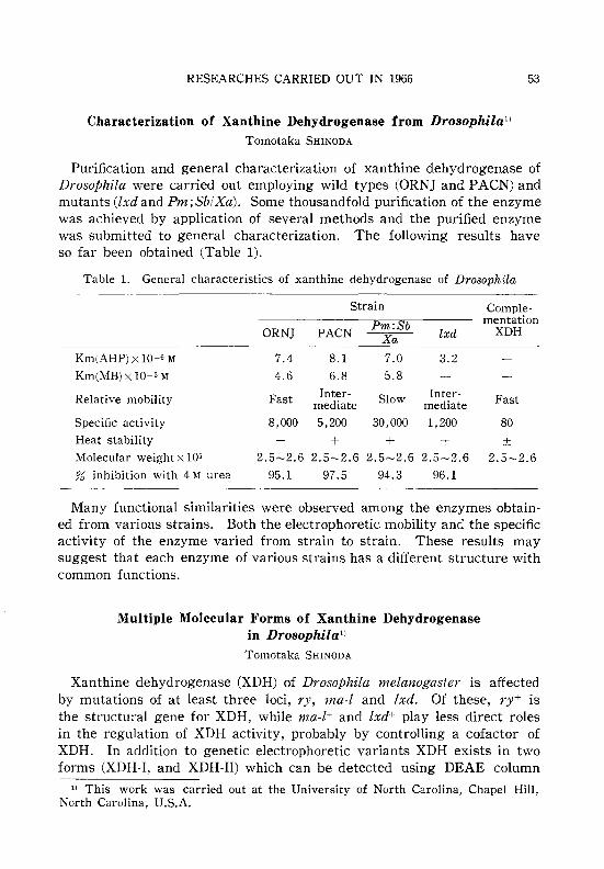
RESEARCHES CARRIED OUT IN 1966
Characterization of Xanthine Dehydrogenase from Drosophila!)Tomotaka SHINODA
53
Purification and general characterization of xanthine dehydrogenase ofDrosophila were carried out employing wild types CORN] and PACN) andmutants (lxd and Pm; Sb/Xa). Some thousandfold purification of the enzymewas achieved by application of several methods and the purified enzymewas submitted to general characterization. The following results haveso far been obtained (Table 1).
Table 1. General characteristics of xanthine dehydrogenase of Drosophila
Km(AHP) x 10-6 M
Km(MB) x 10-5 M
Relative mobility
Specific activity
Heat stability
Molecular weight x 105
;?is inhibition with 4 M urea
Strain Comple·
Pm:Sb mentationORNJ PACN Xa lxd XDH
7.4 8.1 7.0 3.2
4.6 6.8 5.8
Fast Inter· Slow Inter· Fastmediate mediate
8,000 5,200 30,000 1,200 80
+ + + + ±2.5-2.6 2.5-2.6 2.5-2.6 2.5-2.6 2.5-2.6
95.1 97.5 94.3 96.1
Many functional similarities were observed among the enzymes obtain·ed from various strains. Both the electrophoretic mobility and the specificactivity of the enzyme varied from strain to strain. These results maysuggest that each enzyme of various strains has a different structure withcommon functions.
:Multiple :Molecular Forms of Xanthine Dehydrogenasein Drosophila!)
Tomotaka SHINODA
Xanthine dehydrogenase (XDH) of Drosophila melanogaster is affectedby mutations of at least three loci, ry, ma·l and lxd. Of these, ry+ isthe structural gene for XDH, while ma·l+ and lxd+ play less direct rolesin the regulation of XDH activity, probably by controlling a cofactor ofXDH. In addition to genetic electrophoretic variants XDH exists in twoforms (XDH·I, and XDH·II) which can be detected using DEAE column
11 This work was carried out at the University of North Carolina, Chapel Hill,North Carolina, U.S.A.

54 ANNUAL REPORT OF NATIONAL INSTITUTE OF GENETICS NO. 17
chromatography or polyacrylamide gel electrophoresis. In 4 per cent gelthe XDH-I migrates approximately 8 per cent faster than the XDH-ILThe kinetic properties of these forms do not differ greatly. Sucrosegradient centrifugation indicates that they sediment at slightly differentrates. Conversion of XDH-I to XDH-II, but not vice versa, can be accomplished by incubation with Drosophila extracts that are deficient in XDH.Some properties of the enzyme are summarized in Table 1.
Table 1. Comparison of some properties of XDH-I and XDH-I1
Km(AHP) x 10-6 M
Km(MB) x 10-5 M
Sp. activity
Mobility
Mol. weight
% of remaining activity at 50°C, 1 h
Effect of ma-l+(or ry+) factor
% inhibition with 4 M urea
XDH-I
10.1
6.2
4,700
F
260,000
68
+97.1
XDH-I1
7.1
5.5
10,200
S
250,000
61.2
95.1

RESEARCHES CARRIED OUT IN 1966
IV. EVOLUTIONARY GENETICS
An Intergeneric Hybrid between Eremopyrumorientale and Henrardia persica
Sadao SAKAMOTO
55
Henrardia is a monotypic genus in the tribe Triticeae represented byone species, Hn. persica (Boiss.) C.E. Hubbard. Morphologically this genusis different from other members of the tribe. In order to find phylogeneticrelationships of this genus with other genera of the tribe, a strain of Hn.persica var. glaberrima (Hausskn.) c.E. Hubbard (2n=14, Iran) was crossedwith various species of Aegilops, Eyemopyrum, Heteranthelium, Taeniatherumand Triticum in 1965. From these crosses, 25 triploid F 1 hybrids wereproduced only when Er. orientale (Linn.) Jaub. et Spach. (2n=28, Iran)was used as the female parent.
Morphological characteristics of the Fl hybrids were of Eremopyrumtype, while the length of the spikes was intermediate. Disarticulation ofthe ripe spikelets was characterized by wedge type as that of the Eremopyrum parent. Average chromosome pairing per cell of the Fl was O.OOm+0.42II +20.20r . Bivalents ranging from 0 to 4 were all terminally associated.Thus, no genomic homology was found between Er. orientale and Hn.persica. Complete sterility was observed. Colchicine solution (0.5 per cent)was applied to the tillering clones of the F1 and 150 well-developed seedswere obtained in 1966.
Three Intergeneric Hybrids among Heteranthelium piliferum,Eremopyrum buonapartis and Hordeum sp.
Sadao SAKAMOTO
The genus Heteranthelium in the tribe Triticeae is monotypic with Ht.piliferum (Banks et Soland.) Hochst., having quite a specific spike characteristic, i. e. mixture of fertile and rudimental spikelets in one spike. In1965 a strain of Ht. piliferum (2n=14, Afghanistan) was crossed with variousspecies of Aegilops, Eremopyrum, Henrardia and Hordeum. The followingtwo intergeneric hybrids were obtained: 1) two diploid hybrids, Ht. PiliferumxEr. buonapartis (Spreng.) Nevski var. buonapartis (2n=14, Iran),and 2) a triploid hybrid, Ht. piliferum X Hordeum sp. (2n=28, material"No. 66w" was received from the All-Union Institute of Plant Industry,Leningrad, 1964). In the same year, 3) 19 triploid hybrids, Er. buonapartis var. buonapartisxHordeum sp., were also produced.
Growth of hybrids under 1) was subnormal and the shape of the spikes

56 ANNUAL REPORT OF NATIONAL INSTITUTE OF GENETICS NO. I7
was of Eremopyrum type having no rudimental spikelets which are thecharacter of Heteranthelium parent. Growth of the hybrid under 2) wasvigorous and the spike morphology was intermediate between the parents.The spikelets had two empty glumes and two florets like the Heterantheliumparent but no rudimental spikelets were found. Growth of all hybridsunder 3) was very vigorous and the shape of their spikes was of Hordeumtype. However, three spikelets with two florets. each and four emptyglumes were observed at most spikelet nodes. Thus, the numerical composition at the spikelet node of the Fl was intermediate between theparents. Sterility of all three hybrid combinations was complete.
Average chromosome pairing per cell of the Fl hybrids was in 1)0.0411(0-2)+ 13.91(10-14), in 2) 0.00m(0-1)+5.06n(2-8)+ 10.881(5-17) and in 3)0.00Iv(0-1)+0.00m(0-1)+5.5011(1-8)+9.97rC5-19). The parentheses indicatethe range of respective pairing. Judging from the chromosome pairing in1), 2) and 3), 5.011 and 5.511 observed in 2) and 3) indicate autosyndesis ofchromosomes derived from the Hordeum parent. The conclusion may bedrawn that the Hordeum parent used in this experiment was a tetraploidspecies having two partially homologous genomes.
The tillering clones of 3) were treated with 0.5 per cent colchicinesolution and 31 seeds were obtained in 1966.
Diallel Crosses among Sikkimese Rice Types. IIITadao C. KATAYAMA
Diallel crosses were carried out using sixteen strains, namely, fourteenSikkimese, one indica and one japonica strains. All crosses were successfuland the FI seeds obtained were sown in April, 1965. This year, thecharacters of intact and husked FI seeds, such as length, width, thickness,ratio of length to width and grain weight, were compared with those ofthe parents. Investigation of many other characters of the hybrids, suchas number of tillers, plant height, heading date, internode length, isunderway.
In the previous paper (Katayama 1966), the Sikkimese strains could bedivided into 10 indica type and 4 japonica type strains based on the pollenfertility of the FI's produced by diallel crosses. However, from the resultsobtained at present, 3 strains, C7725, C7727 and C7734, formerly classifiedas indica, were found to be of intermediate type between indica andjaponica.
Therefore, Sikkimese rice is composed of typical japonica, typical indicatypes and an intermediate type. It is assumed that Sikkim might be oneof the differentiation centers of cultivated Oryza sativa into japonica

RESEARCHES CARRIED OUT IN 1966
and indica types.
Further Studies on Embryo Transplantation in the Genus Oryza
Tadao C. KATAYAMA
57
In order to investigate the compatibility relation between embryo andendosperm in Oryza species, embryo transplantations were made betweenrice species. The method was described in a previous paper (Katayama1965). In the present article, two experiments are reported.
Four strains of O. sativa (two indica and two japonica types) and onestrain of each of the following nine species were used, namely, O. officinalis,O. minuta, O. eichingeri, O. latifolia, O. australiensis, O. granulata, O.ridleyi, O. brachyantha and O. subulata. Seedlings from intact seeds developed in addition to co1eopti1e, 1st, 2nd and 3rd leaves. Seedlings of O. sativadeveloped three to four roots and those of other species developed one totwo roots. Embryos alone without endosperm produced only a co1eopti1eand no root appeared except in a japonica type of O. sativa.
In heterogeneous transplantation, embryos of o. sativa were grafted uponthe endosperms of other species. In all cases, only a coleopti1e has developed. In all reciprocal transplantations, also only a co1eopti1e developed.This means that the grafted embryos failed to absorb nutrients from alienendosperms which suggests a distant relation to O. sativa.
In the previous report, it was strongly indicated that nutrient absorption from endosperms of indica type strains by embryos of the other closelyrelated species employed, was much better than from endosperms ofjaponica type strains. In order to study the details on this particularpoint, 10 strains of O. sativa (five japonica and five indica strains) astesters, and one strain of O. sativa var. spontanea, O. perennis, O. barthii,O. glaberrima and O. breviligulata, were used. When embryos of japonicastrains were grafted upon the endosperms of other species, co1eoptile and1st leaf developed in most cases. In all reciprocal transplantations, i. e.,when embryos of other species were grafted upon the endosperms ofjaponica strains, also co1eoptile and 1st leaf developed. When embryos ofindica strains were grafted upon the endosperms of other species, co1eoptileand 1st leaf appeared in most cases. However, in the reciprocal transplantation, i. e., when embryos of the other species were grafted upon theendosperms of indica strains, co1eoptile, 1st, 2nd and, in some cases, even3rd leaf, developed.
From these results, the previous finding was confirmed which indicatedthat indica type is taxonomically nearer related to the other employedspecies than japonica type.

58 ANNUAL REPORT OF NATIONAL INSTITUTE OF GENETICS NO. 17
Geographical Distribution of Winter, Intermediate andSpring Types of Common Wheat
Yasuo NAKAI
The growing habit of about 1400 varieties of common wheat collectedfrom various parts of the world was examined using growth chambersadjusted to a constant temperature, 20°C, with 16 hr illumination. Afterthree months from sowing, when all spring standard varieties wereheading, the growing habit of the tested varieties was determined; varietiesthat headed at that time were classified as spring, those with visible earsinside as intermediate and those showing no ear formation as winter types.The result is summarized in Table 1.
Table l. Geographical distribution of three growth habittypes in common wheat
Locality No. var. tested Spring Intermediate Winter(%) (%) (%)
Pakistan 38 58 18 24
Afghanistan 39 12 30 58
Iran 147 27 11 62
India 58 98 0 2
Tibet 19 100 0 0China 180 27 12 61Japan 169 37 11 52
Greece 9 44 12 44
Italy 54 52 2 46
Iberia 96 30 4 66
Russia 89 69 2 29
Sweden 34 100 0 0
Norway 13 100 0 0
Finland 15 100 0 0
U.S.A. 260 33 7 60
S. America 43 81 7 12
Australia 79 97 0 3
Total 1342
As to the varieties from Pakistan, India, Tibet, Russia, South Americaand Australia, many were of spring or intermediate type. In China, Japan,Mediterranean countries and U.S.A., both winter and spring types were

RESEARCHES CARRIED OUT IN 1966 59
equally frequent. Some strains of Afghanistan and most of eastern Iranwere of spring type. With respect to the Scandinavia strains used inthis examination they were of spring type. The distribution pattern ofdifferent growth habit types of Aegilops squarrosa in central Asia (Kiharaand Tanaka, 1958) is identical to that found here for common wheat.
Ernbryosac Sterility of F 1 Hybrids betweenStrains of Oryza perennis
Yaw-En CHU and Hiko-Ichi OKA
Embryosacs were observed in F1 hybrids between strains of Oryza perennis Moench, O. sativa 1., O. breviligulata Chev. et Roehr, and O. glaberrima Steud., which are pollen sterile in varying degree (Ann. Rep. 14: 74,15: 84 and 16: 71). An embryosac with an egg nucleus, two polar nucleiand antipodes were regarded as normal; this estimation was verified byback-crossing. Most Fl hybrids showing high pollen sterility had an appreciable percentage of normal embryosacs. For instance, the F1's betweenAmerican and Asian strains of O. perennis showed 45-70 per cent normalembryosacs, though their pollen fertility was almost zero.
In sterile F1 hybrids, the deterioration of embryosacs seemed to takeplace in three different stages, namely 1) deterioration of the tetrad immediately after meiosis, 2) failure of mitosis producing two to four nuclei,and 3) failure of nuclear differentiation into egg and polar nuclei. TheF1hybrids between the Oceanian strains and those of the other geographicalgroups of O. perennis gave relatively low embryosac fertilities, rangingbetween 25 per cent and 65 per cent. When the Oceanian perennis strainswere crossed with O. glaberrima or O. breviligulata, the F1 embryosacsshowed deterioration immediately after meiosis which was normal, andhad little or no normal embryosacs at the flowering time.
Pattern Analysis of Character Variations in Oryza perennisHiroko MORISHIMA and Hiko-Ichi OKA
Oryza perennis Moench is distributed throughout tropical countries ofthe world and comprises many varieties or forms. The Asian formstend to differentiate into the perennial (perennis or balunga) and annual(spontanea or fatua) types. Our collections from New Guinea are alsodivided into perennial and annual types. From 1958 to 1965, a numberof strains of this species, obtained from their natural habitats in differentcountries, were repeatedly grown in a greenhouse or in automaticallycontrolled short-day plots, and records were taken regarding 18 metric
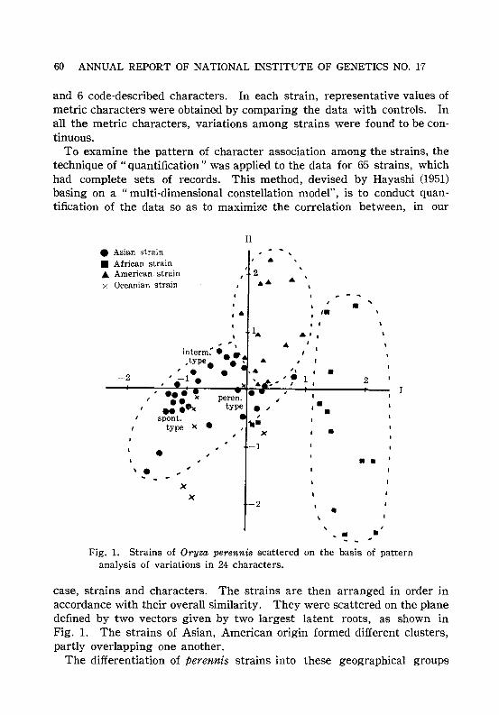
60 ANNUAL REPORT OF NATIONAL INSTITUTE OF GENETICS NO. 17
and 6 code-described characters. In each strain, representative values ofmetric characters were obtained by comparing the data with controls. Inall the metric characters, variations among strains were found to be continuous.
To examine the pattern of character association among the strains, thetechnique of "quantification" was applied to the data for 65 strains, whichhad complete sets of records. This method, devised by Hayashi (1951)basing on a "multi-dimensional constellation model", is to conduct quantification of the data so as to maximize the correlation between, in our
II
• Asian strain• African strainA American. strainx Oceanian strain
,.2••
-2
interm:- e• ~,type. .''. ., -1 •
,• 'II-
X
-1•\ .,
X
X-2 -
Fig. 1. Strains of Oryza perennis scattered on the basis of patternanalysis of variations in 24 characters.
case, strains and characters. The strains are then arranged in order inaccordance with their overall similarity. They were scattered on the planedefined by two vectors given by two largest latent roots, as shown inFig. 1. The strains of Asian, American origin formed different clusters,partly overlapping one another.
The differentiation of perennis strains into these geographical groups

RESEARCHES CARRIED OUT IN 1966 61
could also be found from their Fl sterility relationships (Ann. Rep. 14: 74and 15: 84). The above results of computation indicate that the geographical groups can be distinguished by character-association patterns, thoughnot by a single character. The figure also shows that in the Asian group,the perennial and annual types were separated, and in the Americangroup, strains from Cuba, Surinam and the Amazon basin could be separated. It seems that the varieties of O. perennis distributed in differentcountries have different patterns of character correlation, possibly inrelation to their different modes of adaptation to environment.
Population Survey of No.1 Chromosome Polymorphism of BlackRats (Rattus rattus) Collected in Japan and Korea
Tosihide H. YOSIDA, Yukuo MORIGUCHI,Yung Sun KANG1) ond Kyojiro SHIMAKURA2)
It was already reported that black rats (Rattus rattus) were characterizedby a polymorphism of the largest No.1 chromosome pair, present as atelocentric homomorphic pair (TIT), a telocentric and subtelocentric heteromorphic pair (TIS) and a subtelocentric homomorphic pair (SIS). Thefrequency of these three types in 124 rats collected in 7 localities ofJapan and 9 animals from Seoul, Korea, was already reported in this annual report (No. 16, 1966). Later we collected in total 236 rats in 9lacalities of Japan and 26 rats in Seoul, Korea. The results of observation are given in Table 1. As the table shows, all animals collected inSapporo, Niitsu (Niigata Pre£.) and Obama (Fukui Pref.) had the TIT pair
Table 1. Frequency of black rats (Rattus rattus) with three typesof No. 1 chromosomes, collected in Japan and Korea
Locality
Karyotype Niitsu Obama Sugadaira Hiratsuka MisimaSapporo (Niigata) (Fukui) (Nagano) (Kanagawa) (Sizuoka)
TT 100% 100 100 80 70 70
TS 0 0 0 20 30 30
SS 0 0 0 0 0
Total no. of 50 30 11 15 7 56animals
11 Seoul University, Korea.2) Hokkaido University, Sapporo.

62 ANNUAL REPORT OF NATIONAL INSTITUTE OF GENETICS NO. 17
LocalityTotal in Seoul
Karyotype Hamamatsu Ube Kusudomari Japan (Korea)(Sizuoka) (Yamaguchi) and Nagasaki
TT 57% 82 82 200 9(84.8%) (34.6%)
TS 29 18 18 35 13(14.8%) (50%)
1 4SS 14 0 0 (0.4%) (15.4%)
Total no. of 7 11 49 236 26animals (100.0) (100.0)
while in the other places in Japan, namely Sugadaira (Nagano Pref.),Hiratsuka (Kanagawa Pref.), Misima, Hamamatsu (Sizuoka Pref.) , Ube(Yamaguchi Pref.) and Nagasaki (included Kusudomari), animals with TISheteromorphic pair were observed at 18 to 33 per cent. Except for onlyone animal with SIS homomorphic pair which was found in Hamamatsupopulation, all the others showed the TIT homomorphic pair. Frequencyof animals with TIT, TIS and SIS chromosomes in all 236 was 84.8, 14.8and 0.4 per cent, respectively. On the other hand, among 26 animalscollected in Seoul, Korea, about 50 per cent had the TIT pair, and about15 per cent animals were of SIS type.
Based on the above investigations, it can be said that the frequencyof animals with No.1 TIT, TIS and SIS chromosomes was significantlydifferent in different localities of Japan and in Korea.
Segregation of Three Chromosome Types in BlackRats Crossed in the Laboratory
Tosihide H. YOSIDA and Yukuo MORIGUCHI
Segregation of three types (TIT, TIS, and SIS) of No.1 chromosomesin black rats, Rattus rattus, which were crossed in the laboratory wasalready reported for some animals (Ann. Rep. 16, 1966). We got moredata on the segregation of the three chromosome types from animals bredin our laboratory. The results of observations are shown in the followingtable (Table 1). Animals with SIS chromosomes were bred in the laboratorywith success. From crossing a TIS female by an SIS male we obtainedfour litters. Average litter size of the hybrids was four. Chromosomesof two litters were observed. They segregated into 5 animals with a TISchromosome type and 3 animals with an SIS type. On the other hand,by cmssing an SIS female by an SIT male only one baby rat was obtained,

RESEARCHES CARRIED OUT IN 1966 63
Table l. Segregation of three chromosome types in black rats(Rattus rattus) bred in the laboratory
Karyotypes of Segregation Total no. ofparents T/T T/S SIS animals
T/S(¥)xT/S(o) 18 43 12 73
T/T(¥)xT/S(o) 37 39 0 76
T/S(¥)xT/T(o) 59 61 0 120
T/T(¥)x T/T(o) 48 0 0 48
T/S(¥)x S/S(o) 0 3 2 5
but it died soon after birth. By mating an SIS female by a T/T maletwo baby rats were born, but they also died within one day after birth.Based on the above investigation, SIS females of the black rat may beunfit to bear and nurse their brood.

64 ANNUAL REPORT OF NATIONAL INSTITUTE OF GENETICS NO. 17
V. MATHEMATICAL AND STATISTICAL STUDIESON POPULATION GENETICS
Simulation Studies on the Number of Neutral AllelesMaintained in a Finite Population by Mutation
Motoo KIMURA
The number of isoalleles that may be maintained in a finite populationwas studied theoretically for neutral and overdominant mutations by Kimuraand Crow (1964), who derived, for neutral mutations, the following formulafor the equilibrium distribution of allelic frequencies;
(1)
where M=N,u in which N, stands for the effective population number andu stands for the mutation rate per generation. In deriving the aboveformula it was assumed that each mutant represents an allelic state notpreexisting in the population.
The above distribution has the meaning that <]J(x)dx gives an approximation to the expected number of alleles whose frequencies in the population lie within the range x to x+dx(0<x<1).
Using the above formula, the average and the effective number ofalleles in a population of size N may be given as follows:
na=\l <]J(x)dx= 4M il(1-X)4M-1X- I dx, (2)
J1/ (2NJ J1/ (2N)
n,=1/~: x 2<]J(x)dx=4M+1, (3)
The average number of alleles is defined as the reciprocal of the meanfrequency of alleles contained in a population, while the effective numberof alleles is defined as the reciprocal of the sum of squares of allelicfrequencies.
In order to test the validity of the above formulae, simulation studieswere carried out by using computer IBM 7090. Fig. 1 illustrates a resultof Monte Carlo experiment, assuming a population consisting of 50 malesand 50 females, of which only 25 males and 25 females actually participatein breeding (N=l00, N,=50). In the experiment, the process of mutationand random sampling of gametes was simulated by generating pseudorandom numbers in the computer. In each generation, 100 male and 100female gametes are randomly chosen respectively from 25 breeding malesand 25 breeding females to form the next generation. Mutation to a new(not pre-existing) allele is induced in each gamete with probability 0.005
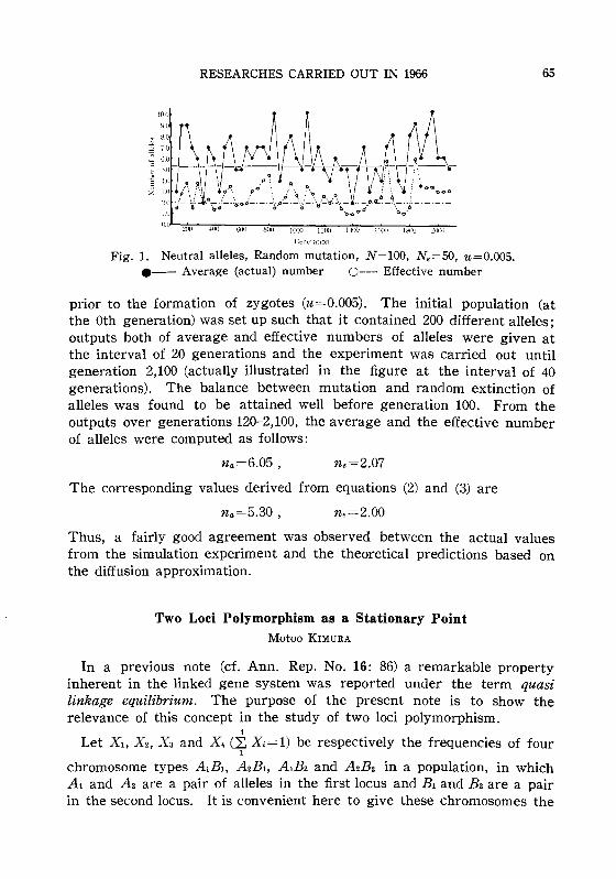
1.0
RESEARCHES CARRIED OUT IN 1966
.9.,fJ'~'/~~\~04·~~~.~\ ..rii. ~ ~~ '~tf ~ _ _t'.#L
iJ_ 6 _ .' _
bd a 0 8 bboOQ' h.,,.o
65
!l.O 21J0 100 600 SOl) 1(0) I:..'fXJ l-lOO llJi)O l.'\1)(J :;\100
Gt'l1lTMion
Fig. 1. Neutral alleles, Random mutation, N=I00, N e=50, u=O.OO5.e-- Average (actual) number 0--- Effective number
prior to the formation of zygotes (u='0.005). The initial population (atthe Oth generation) was set up such that it contained 200 different alleles;outputs both of average and effective numbers of alleles were given atthe interval of 20 generations and the experiment was carried out untilgeneration 2,100 (actually illustrated in the figure at the interval of 40generations). The balance between mutation and random extinction ofalleles was found to be attained well before generation 100. From theoutputs over generations 120-2,100, the average and the effective numberof alleles were computed as follows:
n a=6.05, ne=2.07
The corresponding values derived from equations (2) and (3) are
n a =5.30, ne=2.00
Thus, a fairly good agreement was observed between the actual valuesfrom the simulation experiment and the theoretical predictions based onthe diffusion approximation.
Two Loci Polymorphism as a Stationary PointMotoo KIMURA
In a previous note (d. Ann. Rep. No. 16: 86) a remarkable propertyinherent in the linked gene system was reported under the term quasilinkage equilibrium. The purpose of the present note is to show therelevance of this concept in the study of two loci polymorphism.
;
Let Xl, X2, Xa and X4 (2: X=1) be respectively the frequencies of four1
chromosome types AlBl, A2Bl, AlB2 and A2B2 in a population, in whichAl and A2 are a pair of alleles in the first locus and Bl and B2are a pairin the second locus. It is convenient here to give these chromosomes the

66 ANNUAL REPORT OF NATIONAL INSTITUTE OF GENETICS NO. 17
numbers, 1,2,3 and 4, so that the frequency of chromosome i is Xi (i=l,2, 3, 4) and the fitness (in selective values) of the genotype formed by theunion of chromosomes i and j may be denoted by Wij. In a large randommating population, if c is the recombination fraction between the two loci,the amount of change in· one generation of the chromosome frequenciesmay be given as follows:
where
.1Xl = {XI(WI. -w)-cDw}/W
.1XF {X2(W2. - wHcDw}/w
.1Xs={Xs(ws. -lv)+cDw}/w
.1X4={X4(W4. -w)-CDw}/lV l (1)
and
4
Wi. = 2: WijXj ,j=l
w= 2: WijXiXj ,ij
(2 )
Thus, at the genetic equilibrium in which .1Xi=O (i=l, 2, 3, 4), we have
XI(WI. -w)= -X2(W2. -w)= -Xs(ws. -w)=X4(W4. -w)=cDw .
The equilibrium chromosome frequencies Xi (i=l, 2, 3, 4) may be obtainedby solving the above set of equations.
I will now show that the equilibrium point (Xl, X2, Xs, X4) is a stationarypoint of w with the two side conditions,
4
2: Xi=l ,i=l
and
(XIX 4)/(X2XS)=Ro ,
where Ro is a constant giving the ratio between the frequencies of couplingand repulsion phases at equilibrium, i.e. RO=(XIX4)/(X2XS). The stationary point of w with these two restrictions is equivalent to the stationarypoint of
ifJ=w-2p (2: Xi-1)--2A (log XIX4_ log Ro) ,i X 2Xs
in which no restrictions are imposed among Xi's (-2p and -2A areLagrange multipliers). Thus

RESEARCHES CARRIED OUT IN 1966
at/J/aXl=2wl. -2p-(ZA/X1)=O ,
at/J/aX2=2w2. -2p+(2)./X2)=O ,
etc., giving
Xl(Uh. -p)= -X2(W2. -p)= -Xa(Wa. -p)=X.(W•. -p)=). ,
in which, it is easy to show that
p=iiJ,
and4
).=(Wl. -W2 -Wa. +wd/ 2: (1/X;) .i=l
Thus, the relation (3) together with
(XIX.)/(X2Xa)=Ro ,
should be equivalent to (2).
The Mutational Load with Epistatic GeneInteractions in Fitness
Motoo KIMURA and Takeo MARUYAMA
67
(3 )
The mutational load in a large population was first calculated byHaldane in 1937 without assuming an epistatic component in fitness.Later, a similar but more detailed calculation was carried out by Kimura(1961). Also, the mutational load in a small population was studied byKimura, Maruyama and Crow (1963).
In the present work, the effect of epistasis on the mutational load wasstudied using a model which assumes that fitness is a function of thenumber of mutant genes in an individual. The results are summarized asfollows:
1) If the deleterious effect of the mutant genes is proportional to thesquare of their number, the load under random mating becomes roughlyhalf as large as in the case of no epistasis, provided that the averagenumber of such genes per individual is fairly large. Under asexual reproduction, however, the epistasis has no effect in reducing the load. Thesituation is intermediate for a random mating population of a hypotheticalorganism having only one pair of chromosomes between which no crossingover takes place.
2) The mutational load may also be reduced under random mating iffitness is a threshold character.

68 ANNUAL REPORT OF NATIONAL INSTITUTE OF GENETICS NO. 17
3) Epistatic interaction in fitness among deleterious mutant genes maybe classified into two types, namely, the reinforcing type and the diminishing type. In the former, the deleterious effect becomes disproportionatelylarge as their number in an individual increases. On the other hand, inthe latter, the deleterious effect per mutant gene becomes smaller as theirnumber increases. Consideration of mutational load as well as developmental homeostasis involved suggests that the reinforcing type of epistasisamong deleterious mutant genes must be more common than the diminishing type in nature.
For the details, see Kimura and Maruyama (1966), Genetics 54: 13371351.
Eigenvalues in a Genetics ProblemTakeo MARUYAMA
The smallest and the second smallest eigenvalues in a genetics problemare important, for they determine respectively the asymptotic rate offixation in a case with absorbing boundaries and the asymptotic rate ofapproach to a steady state if it exists. Kimura has given a power seriesexpansion of the smallest eigenvalue for the general case with absorbingboundaries. Miller also gave an analytic method and numerical examplesin cases of heterosis. The purpose of the present work was to show thatan analogous method to the perturbation method in quantum mechanicscan be used to obtain all eigenvalues in a genetics problem, including ofcourse the first and second eigenvalues, when we know the characteristicpairs of unperturbed equation.
The mathematical model considered is
Genotype
Fitness
We assume no mutation and no migration, and that the effective size ofa population is equal to N. Then the diffusion equation is
aif> = _1_ ~{X(I-x)if>}+s~{(2x-I-2)x(l-x)if>} .at 4N ax2 ax
Letting if>(x;t)=T·X be the solution of the above equation, we have
T' 1 [ d2
d ]4Ny = X dx2 {x(l-x)X}+4Ns~{(2x-l-z)x(l-x)X} =-A,
where
(1)
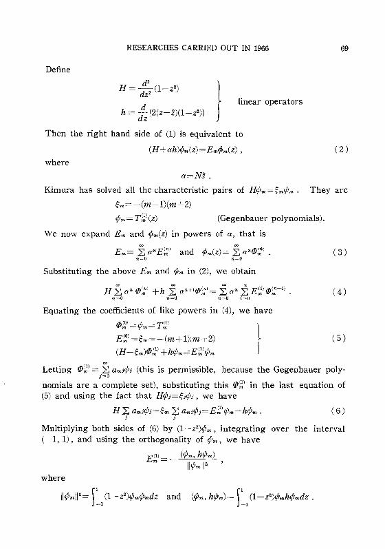
Define
RESEARCHES CARRIED OUT IN 1966 69
d2
H= -(1-z2)
dz2
h = d: {2(z-z)(1-z2)} J
linear operators
Then the right hand side of (1) is equivalent to
(H+ah)<Pm(z)=Em<Pm(z) ,
where
(2 )
(Gegenbauer polynomials).
a=NS.
Kimura has solved all the characteristic pairs of H¢m=~m¢m. They are
~m= --(m+ 1)(m+2)
¢m=Tr;.I(Z)
We now expand Em and <pm(Z) in powers of a, that is
E m= ~ anE;;:1 and <Pm(Z)= ~ antlJ):) .n=O n=O
Substituting the above Em and <pm in (2), we obtain
H ~ an tlJ',;," +h }2 an+1tlJ';:l = £ an i:. E;:/ tlJ;::-i) .n=O n=O n=O i=L1
Equating the coefficients of like powers in (4), we have
(3)
(4)
tlJ':.I=¢m=Tr;.1
E~) =~m=-(m+1)(m+2)
(H-~m)tlJ;;,1 +h¢m=Er;.I¢m I (5 )
Letting tlJ~) = ~ amj¢j (this is permissible, because the Gegenbauer polyj=O
nomials are a complete set), substituting this tlJr;.) in the last equation of(5) and using the fact that H¢j=~j¢j, we have
H 2: amj¢j-~m 2: amj¢;=Er;.I¢m-h¢m . (6)j j
Multiplying both sides of (6) by (1-z 2)¢m, integrating over the interval(-1, 1), and using the orthogonality of ¢m, we have
EO) __ (¢m, h¢m)m - II¢m11 2 '
where
II¢mIl 2 = L1 (1-z2)¢m¢mdz and (¢m, h¢m)= L1 (1-z2)¢mh¢mdz .

70 ANNUAL REPORT OF NATIONAL INSTITUTE OF GENETICS NO. 17
In a similar, but more complicated way, 2nd, 3rd, 4th terms in (3) can beobtained. They are
where
and
E;;) =.2: .2: (¢m, h¢i)(¢i, h¢j)(¢j, h¢m) ,ioem joem (~i-~m)(~j-~m)
h=h-(¢m, h¢m) ,
E~) = -.2: .2: .2: (¢m, h¢i)(¢i, h¢j)(¢j, h¢k)(¢k, h¢m)i,<m j'<m hm (~i-~m)(~j-~m)(~k-~m)
- E;;') .2: .2: (¢m, h¢J)(¢j, h¢m)ioem joem (~i-~m)(~j-~m)
A Diffusion Process with HeterosisTakeo MARUYAMA
As Wright has emphasized, finite populations may be important in thecourse of evolution because random drift in gene frequency can move thepopulation from one adaptive peak to another on a fitness surface diagram.Although it is true that random drift can eventually shift a finite population from one peak to any other peak, we would like to know the timerequired for it. As a first attempt to solve the problem, a simple modelwas constructed of a fitness surface with only one peak, representing apolymorphism due to heterosis, and an approximate solution of the genefrequency distribution was obtained.
The diffusion equation corresponding to heterotic selection in a finitepopulation is
(1)
where Va.=x(1-x)j2N and Ma.=x(1-x)(Sl-(Sl+S2)X). A solution to equation (1) with initial condition lim ¢J(P, x;t)=o(P-x) has not yet been solved.
t~O
However if we restrict the behavior of the process at the local neighborhood of XO=Sl!(Sl +S2) , the point where the process starts, we have roughlyVa.=xo(1-xo)!2N and Ma.=xo(1-xo){Sl-(Sl+S2)X}. Then (1) becomes

RESEARCHES CARRIED OUT IN 1966 71
where Y=SI-(SI +S2)X, I'=SIS2/(SI +S2) and a=Sls2/4N. If we assume thesolution of (2) to be ifJ= T(t) Y(y), then we have
_1_ dT(t) = _l_{a d2
1Y + f3 dY +I'Y}=JT(t) dt Y(y) dy 2 Y dy .
It is easy to see that T(t) = cAt . The right hand side of (3) becomes
a d2y +I'Y dY +(I'-J)Y=O.
dy 2 dy
If we let Z=..j ~y and Z(z)=Y(z) , we have
d2Z dZ ( J)-+z-+ 1-- Z=O,
dz2 dz I'
(3 )
(4 )
equation (4) is a Hermite differential equation and its integrable solutions
are Zn=C-z2/4Dn(z) n=O, 1,2,'" where - ~ =0,1,2,'" and
Hence the solution of (2) is
ifJ(t;x) = ~ ancl\ntc-z2/4Dn(z) .n=O
Now we want to determine the coefficients an such that limifJ(t;x)=o(xo-x).t~O
The principle in determining an is ) o(x-xo)f(x)dx=f(xo) for all function
f(x) and the orthogonatity of Dn(z). The final form of the solution is
rI.( t) [N(SI +52)J {( SI )2/ slsd }'t' X· = exp - ---X, SISZtrc SI +S2 2N(SI +S2)2 .
Although this solution is valid for only a short time, it has a pleasingform; ifJ(x;t) is a normal distribution (in particular it is the Wiener transition function) with variance proportional to time and the inverse of thepopulation size.

72 ANNUAL REPORT OF NATIONAL INSTITUTE OF GENETICS NO. 17
An Application of Kimura's Formulae to Define theEvolutionary Load in a Small Population
Takeo MARUYAMA
Gene frequencies or the distribution of gene frequencies are determinedby selection coefficients, population size, mutation rate etc. If the environment changes the relative fitness of genotypes, a new equilibrium will beattained by natural selection. If we assume for this discussion that thechange is such that the previously advantageous gene becomes disadvantageous, the shift of gene frequencies will be large. This case iscalled a gene substitution. If we further assume that there is neithermutation nor migration during the process, the substitution will be complete.This change in the relative fitnesses causes a genetic load, called theevolutionary load. Symbolically if we let let) be the genetic load at timet, then the evolutionary load for the whole process is given by
r=o l(t)dt .
It has been shown by Kimura that the load in an infinitely large population is
where h is the degree of dominance and Po is the initial gene frequency.The main trouble with finite populations is that, even if we assume Po tobe positive, some populations will loose the advantageous gene and willstay in the lost class until mutation occurs. Therefore it is not possibleto define the evolutionary load in analogy to the case of an infinite population. However we may measure the load as follows; for each samplepath we calculate the difference in fitnesses between the best genotype andthe average fitness at a given time, add them from time zero to infinityand take the expectation with respect to the collection of all sample paths.Because this is the expected total genetic death spent in the substitutionof the gene, we divide it by the fraction which went to fixation. We callit the evolutionary load. Applying Kimura's solution 1> (P, x; t) to thediffusion equation in genic selection and his solution u(P) to the probabilityof ultimate fixation we can write the load in a finite population, in a caseof genic selection, as
1)00 ~1-(-) dt sx¢(P, x;t)dx .u P 1=0 .=0
(1)

RESEARCHES CARRIED OUT IN 1966
The explicit formulae for ¢(P, x;t) and uCP) due to Kimura are
~
¢(P, x;t)= 2: Cke-l.-kt+2CzVlklll(z) ,k=O
73
where z=1-2x, c=Ns, and Ck and Ak are constants, Vlk llJ = 2:' Ink T~I\Z)n=O.l
where Ink are constants and T111 (Z) are Gegenbauer polynomials, and
1_e-4N8P
u(P)= 1_e-4N8 •
Therefore using the relation
I nm= (I T~J(z) T';;.I(Z) = {O if m-n=oddJ-I (n+ 1)(n+2) if m;;:;'n and m-n=even
(1) can be expressed as
where
[I (1-z2)(1-z)eczT~\z)dz
dn = IIT~)112 ,
IIT~1112 = [I (1-Z2){T~I)(Z)}2dZ=2{.(3t)-1)J-I n+- n!
2
Dimensionality of Human MigrationNorikazu YASUDA
A pattern of human migration has been considered as two-dimensional,that is, man moves in all directions from where he was born. However,this is not true in a population with barriers.
Suppose that an individual moves from his birthplace to a place to wherehe is attracted by job or mate. This exploration range will be proportionalto the inverse of dimensional power of marital distance (x), defined as thedistance between the birthplaces of consorts. An acceptable frequencydistribution is m(x)=ab/(l +ax)b+l, where b+1 represents dimension anda is a scale parameter or "attraction index".
Examples of estimated parameters are given as follows:
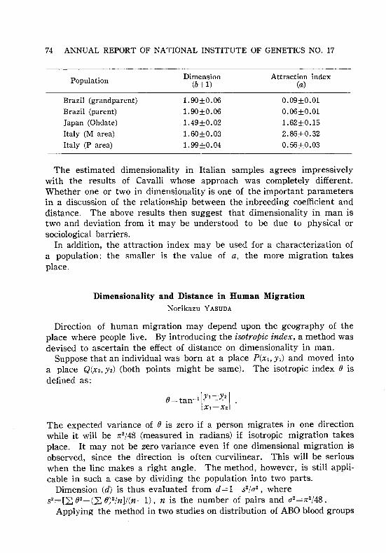
74 ANNUAL REPORT OF NATIONAL INSTITUTE OF GENETICS NO. 17
Population
Brazil (grandparent)
Brazil (parent)
Japan (Ohdate)
Italy (M area)
Italy (P area)
Dimension(b+l)
1.90±O.06
1.90±O.06
1.49±O.02
1.60±O.03
1.99±O.04
Attraction index(a)
O.09±O.OI
O.06±O.OI
1.62±O.152.86±O.32
O.56±O.03
The estimated dimensionality in Italian samples agrees impressivelywith the results of Cavalli whose approach was completely different.Whether one or two in dimensionality is one of the important parametersin a discussion of the relationship between the inbreeding coefficient anddistance. The above results then suggest that dimensionality in man istwo and deviation from it may be understood to be due to physical orsociological barriers.
In addition, the attraction index may be used for a characterization ofa population: the smaller is the value of a, the more migration takesplace.
Dimensionality and Distance in Human MigrationNorikazu YASUDA
Direction of human migration may depend upon the geography of theplace where people live. By introducing the isotropic index, a method wasdevised to ascertain the effect of distance on dimensionality in man.
Suppose that an individual was born at a place P(XI, YI) and moved intoa place Q(X2, Yz) (both points might be same). The isotropic index (j isdefined as:
The expected variance of (j is zero if a person migrates in one directionwhile it will be 1r2/48 (measured in radians) if isotropic migration takesplace. It may not be zero variance even if one dimensional migration isobserved, since the direction is often curvilinear. This will be seriouswhen the line makes a right angle. The method, however, is still applicable in such a case by dividing the population into two parts.
Dimension (d) is thus evaluated from d=1+s2/a2, wheres2=[22 (j2_(L. (j)2/nl/(n-l) , n is the number of pairs and a2=1r2/48.
Applying the method in two studies on distribution of ABO blood groups

RESEARCHES CARRIED OUT IN 1966 75
Distance (KM)
600 700500300 400.50 100. 200
2.0 ••-........- ....:.:::
. .. .S\viss •••••••
.2~u";:;;c:"§ 1.05
C<
(3087 communities)Sweden
(1071 communities)
1.01
Fig. 1. Dimensionality and distance in man.
in Switzerland (Rosin, 1956) and in Sweden (Beckman, 1959) where individuals are grouped by their birthplace so that the distribution ofcommunities may be considered approximately as an expected distributionof human migration, and making all possible pairwise combinations of 3087communities in Switzerland and of 1071 in Sweden, we found that theisotropic distances are about 100 km. in Switzerland and about ZOO km. inSweden, reflecting the geography of each country. Dimension becomesone for further remote distances (see figure).
A Statistical Singularity at the ABO Blood Group SystemNorikazu YASUDA
Suppose that the observed and the expected numbers of individuals inthe standard ABO blood group classification are given by:
Phenotype Genotype Observed Expected0 00 no N[r2+(r-r2)a]A AA,AO nA N[P2+Zpr+{P - (P2 +Zpr)}a]B BB,BO nB N[q2+Zqr+{q-(q2+Zqr)}a]
AB AB nAB N[Zpq-Zpqa]
where p, q and r are gene frequencies of A, Band 0, respectively, a isthe inbreeding coefficient, and N=nA+nB+n..IB+no. Then the maximumlikelihood scores under the null hypothesis that a=O are
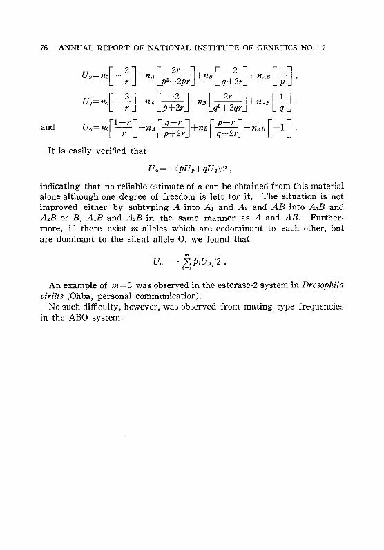
76 ANNUAL REPORT OF NATIONAL INSTITUTE OF GENETICS NO. 17
and
up=n{- ~]+nA~2~~p;]+nB[q~~r]+nAB[~]'
uq=no[- 2]+nA[~2]+nBr 2 2r ]+nAB [~] ,r P+ r Lq +2qr qUa=n{l-;r]+nA[;~;r]+nB[:;;r]+nAB[-1].
It is easily verified that
indicating that no reliable estimate of a can be obtained from this materialalone although one degree of freedom is left for it. The situation is notimproved either by subtyping A into Al and Az and AB into AlB andAzB or B, AlB and AzB in the same manner as A and AB. Furthermore, if there exist m alleles which are codominant to each other, butare dominant to the silent allele 0, we found that
mUa=- 2:.PiUp./2.i=l ~
An example of m=3 was observed in the esterase-2 system in Drosophilavirilis (Ohba, personal communication).
No such difficulty, however, was observed from mating type frequenciesin the ABO system.

RESEARCHES CARRIED OUT IN 1966
VI. EXPERIMENTAL STUDIES ONPOPULATION GENETICS
Deleterious Genes in the Second Chromosome Concealed inNatural Populations of Drosophila melanogaster1)
Chozo OSHIMA and Takao K. WATANABE
77
Among 905 second chromosomes isolated from natural populations at Kofuand Katsunuma territory in late October 1965, the frequency of lethalchromosomes was 15.47 per cent. The frequencies of lethal chromosomesdid not fluctuate to a remarkable extent during the past several years, butthose of semilethal and subvital chromosomes in the extracted chromosomesin 1965 were decreased as compared with their earlier frequencies, asshown in Table 1.
Table 1. Frequencies of the second chromosomes carrying deleterious genesin natural populations of the Kofu and Katsunuma territory
No. of the second Frequency ofYear chromosomes isolated Lethals Semilethals Subvitals Normals
1959 144 13.19±2.82 19.44±2.30 16.67±3.11 50.69±4.17
1963 668 20.21±1.55 18.11±1.49 25.15±1.68 36.53±1.86
1964 740 15.54±1.33 19.59±1.46 7.57±0.98 57.30±1.82
1965 905 15.47±1.20 6.85±0.84 3.76±0.63 73.92±1.46
A tester strain, having the dominant marker genes Cy and Pm on eachsecond chromosome, was used in the analysis, but its genetic background(X and third chromosomes) was previously substituted with normal chromosomes originated from the same natural population. Consequently, thegenetic background was assumed to be highly heterozygous. On thecontrary, the genetic background of the tester strain, which had been usedin the former experiments, was assumed to be homozygous, as it wassubstituted with homozygous chromosomes of an isogenic Samarkand strain.
The frequencies of chromosomes carrying deleterious genes except forcomplete lethals, could have been influenced by the state of geneticbackground of the tester strain. Therefore, the frequencies of semilethalsand subvitals, found in the past years (1959, 1963, 1964), would be nearer
I) The following six investigations were supported by a grant from the JapanSociety for the Promotion of Science as part of the Japan-U.S. Cooperative ScienceProgram.

78 ANNUAL REPORT OF NATIONAL INSTITUTE OF GENETICS NO. 17
to the real frequencies in the natural populations than those obtained in 1965.
Distribution of Persistent Lethal Genes in Natural PopulationsChozo OSHIMA and Takao K. WATANABE
From six different populations, distributed over the Kofu and Katsunumaterritory (15 X 3 km2), 132 lethal chromosomes were isolated, and allelismtests between lethal genes were performed. The results are representedin Table 2.
Table 2. Allelism tests between lethal genes of 132 lethal chromosomes
Whole Within Within BetweenKofu Katsunuma Kofu & Katsunumapopulations populations populations populations
No. of lethals 132 63 69 132
No. of crosses 8646 1953 2346 4347
No. of alleliccrosses 169 54 38 77
Allelic rate (%) 1.95 2.76 1.62 1. 77
Seventy-nine lethal chromosomes, isolated from the same populations in1964, have been maintained individually by the Cy-balanced condition.Allelism tests between newly isolated 132 lethal chromosomes and the oldones were completely performed by the diallel cross. Among the total of10428 crosses, 248 were allelic and allelic rate was determined to be 2.38per cent, being similar to that (2.35 per cent) between lethals isolated in1963 and 1964.
Table 3. Frequency of persistent lethals among all isolated lethalsand their persistence periods
Period No. of lethals No. of persistent Frequencytested lethals (%)
1 Year 1963-1964 100 35 35.0
1964-1965 140 52 37.1
2 Years 1963-1965 140 29 20.7
4 Years 1959-1963 114 10 8.8
5 Years 1959-1964 100 9 9.0
6 Years 1959-1965 140 10 7.1

RESEARCHES CARRIED OUT IN 1966 79
From the results of such successive allelism tests, some lethal genes wereconfirmed to have persisted for a long time in the Kofu and Katsunumapopulations, as shown in Table 3.
Eighty lethal genes among 132 lethals were assumed to be newly arisenlethals and the allelic rates between them were very low, as shown inTable 4.
Table 4. Allelic rates between newly arisen lethal genes
Whole Within Within BetweenKofu Katsunuma Kofu & Katsunumapopulation population population population
No. of lethals 80 38 42 80No. of crosses 3160 703 861 1596No. of allelic
crosses 15 3 7 5
Allelic rate (%) 0.47 0.43 0.81 0.31
Recessive Visible Mutant Genes on the Second ChromosomeConcealed in Natural Populations
Chozo OSHIMA and Takao K. WATANABE
Among 905 chromosomes, 89 (9.8 per cent) had a recessive visible mutantgene. A mutant gene, making bristles slender and short, was namedtentatively 'reduced' (rd) and similar mutant genes were found in 63chromosomes. Another mutant gene, manifesting brown eye, was identifiedto be the bzv gene (2-104.5); it was found in 12 chromosomes in additionto five kinds of mutant genes (tentatively named spread wing, excisedwing, upturned wing, reduced wing and small body) observed in a fewchromosomes.
For the past two years, many chromosomes, having the rd gene, wereisolated from the Kofu and Katsunuma populations. By the results ofallelism tests between them, these rd genes could be divided into fivegroups and the loci of rd-1 and rd-4 were determined to be 66.9± and51.8± respectively. The manifestation of rd gene is very similar to thatof Minute genes, of which about 20 loci have been recorded on the secondchromosome, most of them clustering near the centromere. It may beassumed that by some mechanisms a fairly number of rd genes have beenmaintained in the natural populations in spite of lower viability of heterozygous flies (rd/ + ).

80 ANNUAL REPORT OF NATIONAL INSTITUTE OF GENETICS NO. 17
Segregation Distorter (SD) Genes and Their LinkedLethal Genes in Drosophila melanogaster
Takao K. WATANABE and Chozo OSHIMA
Among 420 second chromosomes, which were extracted from naturalpopulations at Kofu and Katsunuma in late October of 1965,15 chromosomes(3.6 per cent) were found to have segregation distorter genes, as a result ofchecking segregation ratios in heterozygous males for each second chromosome and the cinnabar brown chromosome. Seven chromosomes amongthem had each a lethal gene. The lethal gene on five of them was clarifiedto be a persistent one (1401, locus: 47.9), which had been maintained in highfrequency in the Kofu and Katsunuma populations for at least two years.
Most SD chromosomes (86.7 per cent) had In~2R)C on the right arm,whose equilibrium frequency in the natural populations was about 25 percent. The SD gene was assumed to be located near the proximal end ofthe right arm. Then, the linkage between SD and the inversion could bestrong, but that between SD and [401 was weaker, because the lethalgene was located on the left arm.
An SD chromosome was marked with Lobe (locus: 72.0) gene and wasused to analyze the varying segregation ratio in heterozygous male fliesin about 390 chromosomes extracted from the natural population. Themean (k) segregation ratio was estimated to be 0.77, but it varied widelyfrom 0.4 to 1.0. At 0.5 as k value, the effect of the segregation distorterwas suppressed and at 1.0 as k value, complete expressibility of the genewas assumed.
The segregation of homozygous male flies for SD genes was foundnormal, but in the offspring the number was reduced. Several hypothesesillustrating the mechanism of segregation distortion have been proposed,but the problem still remains unsolved.
A Mechanism of Persistence of Some Lethal Genes inNatural Populations of Drosophila melanogaster
Takao K. WATANABE
Two lethal genes (1404, [407) on the second chromosome have beenmaintained at high frequency for at least 6 years in the same naturalpopulations. One hundred twenty female flies, heterozygous for Cy andthe persistent lethal chromosomes (Cy/[404 or Cy/[407) and the samenumber of female flies heterozygous for Cy and normal chromosomes (Cy/ni)were mated with male flies having two normal chromosomes in heterozygous condition.

RESEARCHES CARRIED OUT IN 1966 81
Each female was mated with three males in a vial and transferred everyday to a new vial containing the culture medium. In the course of ex·periments, the longevity of the two kinds of flies was examined, and thenumber of their progeny was compared as to productivity. The ratio ofnon·Cy flies was estimated. Then, the viability of lethal heterozygotes(llni) could be compared with that of normal heterozygotes (ndm).
The mean longevity of Cy·lethal heterozygous females was about 6 daysshorter than that of Cy-normal heterozygous females, but the formerproduced 509 flies on an average during 10 days after emergence and thelatter produced only 460 flies. The viability of lethal heterozygotes andnormal heterozygotes is given in Table 5.
Table 5. Comparison of viabilities (pooled basis) of lethal andnormal heterozygous flies
Period
1-5 days 6-10 days 11-41 days
No. of Viability* No. of Viability No. of Viability*flies flies flies
llni 28351 1.0035±0.01192 22861 1.0174±0.01346 20772 1.0377±0.01440
ndnj 27244 0.9519±0.01154 25383 1.0021±0.01258 30370 0.9867 ±0.01132
* 1% level significance.
The data were divided into three parts according to the period of egglaying. From the results, significantly more lethal heterozygous flies thannormal heterozygous flies were not only produced by younger parents butalso by older parents. This could be an effective mechanism of thepersistence for those lethal genes.
Further Study on Chromosomal Polymorphism inthe Kofu and Katsunuma Natural Populations
Taishu WATANABEll and Chozo OSHIMA
About 800 male flies were captured from two natural populations in theKofu and Katsunuma territory in late October of 1965. These male flieswere individually mated with several virgin females of an isogenic Sam·arkand strain. The salivary chromosomes of 8 or 10 Fl larvae of eachculture were observed. The chromosomal types of 573 male flies could bedetermined and the frequency of each kind of chromosomes was recorded.The expected numbers of all possible homozygous and heterozygous com·
1) Department of Biology, Faculty of Science, Kyushu University.
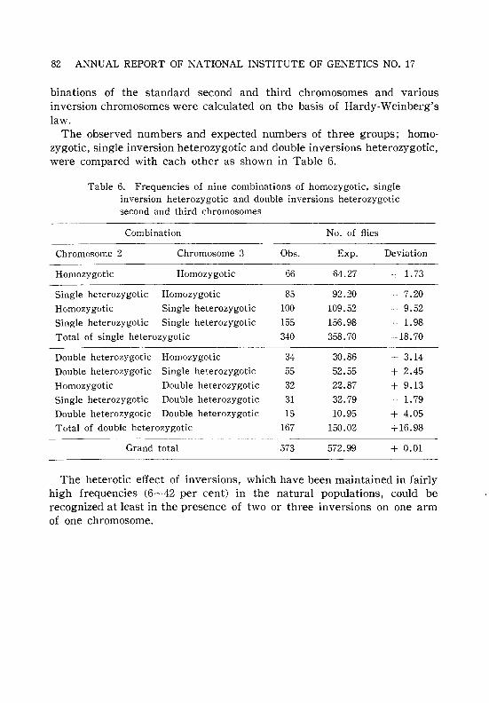
82 ANNUAL REPORT OF NATIONAL INSTITUTE OF GENETICS NO. 17
binations of the standard second and third chromosomes and variousinversion chromosomes were calculated on the basis of Hardy-Weinberg'slaw.
The observed numbers and expected numbers of three groups; homozygotic, single inversion heterozygotic and double inversions heterozygotic,were compared with each other as shown in Table 6.
Table 6. Frequencies of nine combinations of homozygotic, singleinversion heterozygotic and double inversions heterozygoticsecond and third chromosomes
Combination No. of flies
Chromosome 2 Chromosome 3 Obs. Exp. Deviation
Homozygotic Homozygotic 66 64.27 + 1.73
Single heterozygotic Homozygotic 85 92.20 7.20
Homozygotic Single heterozygotic 100 109.52 9.52
Single heterozygotic Single heterozygotic 155 156.98 1.98
Total of single heterozygotic 340 358.70 -18.70
Double heterozygotic Homozygotic 34 30.86 + 3.14
Double heterozygotic Single heterozygotic 55 52.55 + 2.45
Homozygotic Double heterozygotic 32 22.87 + 9.13
Single heterozygotic Double heterozygotic 31 32.79 - 1.79
Double heterozygotic Double heterozygotic 15 10.95 + 4.05Total of double heterozygotic 167 150.02 +16.98
Grand total 573 572.99 + 0.01
The heterotic effect of inversions, which have been maintained in fairlyhigh frequencies (6~42 per cent) in the natural populations, could berecognized at least in the presence of two or three inversions on one armof one chromosome.

RESEARCHES CARRIED OUT IN 1966
Lack of Chromosomal Interaction with Respect toOverdominance in Drosophila melanogaster ll
Terumi MUKAI
83
As previously reported (Genetics 50: 1-19, 1964), spontaneous mutantpolygenes controlling viability have been accumulated independently in104 second chromosome lines which originated from a single normalchromosome. Based upon the test for the heterozygous effects of thesemutant polygenes located only in one of the homologous chromosomes inGenerations 32, 60, and 78, we have proposed that an optimum level ofheterozygosity exists for manifestation of overdominance (Genetics 52:493-501, 1965; Ann. Rep. 16). Furthermore, it was reported that theoptimum level of heterozygosity in the second chromosomes was independent of the heterozygosity Df the third chromosomes (Sbj -T- vs. +! +third chromosome genetic background) (Ann. Rep. 16). In order to confirmthe second finding, an experiment was conducted in Generation 85.Homozygous and heterozygous viabilities of 50 chromosome lines selectedfrom the above 104 lines were tested both in homozygous and heterozygousgenetic backgrounds (SbjUbx third chromosome). The outline of the resultis presented in Table 1. Forty-six lines out of 50 showed overdominance
Table 1. Means of the standardized viabilities of homozygotesand heterozygotes and their correlation coefficients
Homozygousviability
Homoz>:gous 0.3329*** +0.0455genetIc background -
Heteroz,Ygous 0.3113*** +0.0468genetIc background -
Heterozygousviability
1.0611*** ±O. 0045
1.1007***±0.0074
Correlation coeff.between homo- and
heterozygotes
-0.1180
-0.2443
Notes 1. Number of chromosome lines=50.2. Control viability=1.0000.
*** Significantly different from 1.0000 (P<0.001).
in both genetic backgrounds, two showed it only in homozygous geneticbackground, and the remaining two only in heterozygous genetic background. Thus, it is confirmed that the manifestation of overdominancein the second chromosome is independent of the heterozygosity in the
1) This work was conducted in the Department of Medical Genetics, Universityof Wisconsin, Madison, Wisconsin, U.S.A. and was supported by a PHS GrantGM-07666.

8t ANNUAL REPORT OF NATIONAL INSTITUTE OF GENETICS NO. 17
third chromosome, contrary to the interaction among genes located withinchromosomes. If the second and the third chromosomes are not independent, overdominance is not to be expected in the second chromosomesunder the optimum heterozygosity hypothesis, because the level of heterozygosity of the third chromosomes (Sb/Ubx) is probably much higher thanthe optimum level for the second chromosomes.
The Detrimental Load to the Lethal Load Ratio (D:L Ratio) ofNewly Arising Mutations in Drosophila melanogaster ll
Terumi MUKAI and James F. CROW
Greenberg and Crow (1960) have proposed a method for testing thedegree of dominance of detrimental genes by comparing the D:L ratio inequilibrium population and that for newly arising mutations. The formerratio has been estimated to be 0.5~1.0 by Greenberg and Crow (1960),Band (1964), and Temin (1966).
In order to estimate the D:L ratio for newly arising mutations, spontaneous mutations controlling viability have been accumulated at a minimumpressure of natural selection in 150 second chromosomes which originated
Table 1. Summary of experimental results in Generations 10 and 20
(CH)** (PQ)** (RT)**
Gen.O Gen.10 Gen.20 Gen.O Gen.10 Gen.20 Gen.O Gen.10 Gen.20
Number oflethal lines o 1 3 o 1 2 o 2 6
Mean viabilityincludinglethals
Mean via·bilityexcludinglethals
D:L ratio
(34.10) 32.08
(34.10) 32.73
29.48
31.36
1.35
(33.51) 31.64
(33.51) 32.28
29.82
31.05
1.89
(28.11) 25.02
(28.11) 26.06
21.20
24.01
1.27
The number of chromosome lines is 50 in each group; each originated from asingle chromosome extracted from a natural population.** Stands for the original Madison chromosomes.
II This work was conducted in the Department of Medical Genetics, Universityof Wisconsin, Madison, Wisconsin, U.S.A. and was supported by a PHS GrantGM-07666.

RESEARCHES CARRIED OUT IN 1966 85
from 3 Madison chromosomes (50 from each Madison chromosome). Homozygous viability of each chromosome was tested in Generations 10 and 20.The result is presented in Table 1. The D: L ratio was calculated for eachchromosome group which originated from a single Madison chromosome.The results are 1.27~1.89 which are larger than those in equilibriumpopulations. This indicates that the degree of dominance of detrimentalgenes is larger than that of lethal genes which was estimated to be ii=0.02~0.04 by several workers (e.g. Crow and Temin 1964; Hiraizumiand Crow 1960; Stern, et at. 1952).
Studies on the Competition between RaceslA and 21B of Wheat Leaf Rust
Keizo KATSUYA
Physiologic races of various plant pathogenic fungi continuously vary inprevalence in nature. The change in prevalence of races 1A and 21B ofwheat leaf rust, Puccinia rubigo-vera Winter f. sp. tritici Mains, was investigated by comparing in growth cabinets aggressiveness of the two racesand the factors that might influence them.
When a mixture of urediospores of the two races was used to inoculate twosusceptible wheat varieties, Norin No. 61 and Shinchunaka, for five uredialgenerations race 21B quickly became predominant at 15° and 20°C, andafter four uredial generations race 1A was nearly eliminated from themixtures. Relative competitive abilities were not found to be influencedby the density of pustules on infected leaves. The rate of growth ofpustules on the two wheat varieties grown at 15° and 20°C was determinedby measurement with a microscope. Pustules of race 21B grew fasterthan those of race 1A at 15°C, and at twelve days after inoculation pustulesize of race 21B was about 1.6 times larger than that of race 1A. At20°C, however, pustules of race 21B grew almost the same as those ofrace 1A. Experimentation showed that the urediospores of race 21B weremore infective than those of race 1A on the two wheat varieties and thatthe incubation period of race 21B was remarkably. shorter than that ofrace 1A at 15°C. The predominance of race group 21 of wheat leaf rustin north Japan may have resulted from the greater aggressiveness ofthat race.

86 ANNUAL REPORT OF NATIONAL INSTITUTE OF GENETICS NO. 17
Interaction among Genotypes for Migration inDrosophila melanogaster
Takashi NARISE
In the previous migration experiment with wild and vestigial flies ofDrosophila melanogaster it was found that the migratory activity of thevestigial flies increased due to mixing them with wild flies (see this AnnualReport No. 12). It then become the immediate task to find out if the genevestigial itself was responsible for making sensitive to the stimulation, orwas an interaction between the gene and its genetic background responsible. The present experiment was conducted to resolve this problem.
Employed were five wild strains, MS-1, Niihama, Oshoro, Gifu, and Tosu,and their substitution vg strains. They were mixed one with another ina manner shown in Table 1. One hundred flies were introduced into themigration-tube in the either mixing or control experiment. The mixturewas composed of 50 flies of a wild strain and 50 flies of the vg-substitutedstrain. The experimental results are presented in Table 1 and Table 2.
Table l. Effect of vestigial genotype on migratory activity of five wild strains
Migratory activity of wild type strains
MS-l Tosu Gifu Niihama Oshoro
MS-l (vg) 13.2 22.0 25.6 20.8 31.2
Tosu (vq) 16.0 10.0 35.4 15.2 20.8
Gifu (vg) 19.8 12.4 22.0 16.0 22.4
Niihama (vg) 21.2 16.0 37.2 22.0 21.6
Oshoro (vg) 20.8 13.2 27.2 19.2 19.6
Single 26.8 28.4 21.6 28.8 24.6
Table 2. Effect of wild type on migratory activity of five vestigial strains
Migratory activity of vestigial substituted strains
MS-l (vg) Tosu (vg) Gifu (vg) Niihama (vg) Oshoro (vg)
MS-l 17.2 16.4 16.4 19.6 24.4Tosu 23.6 11.6 16.4 12.0 20.0Gifu 21.6 20.8 19.2 17.6 22.8Niihama 18.8 27.6 16.8 8.0 18.8Oshoro 24.8 14.4 18.8 15.6 13.6Single 11.0 11.0 18.8 5.8 13.5
RESEARCHES CARRIED OUT IN 1966 87
In these tables, MS-l (vg) means the MS-l strain substituted with thevestigial gene into the genetic background of MS-l and so on.
It is found from Table 1 and Table 2 that the high migratory activityof wild type flies generally decreases by being mixed with a vestigialstrain except for the Gifu strain. The activity of vestigial strains, however,increases due to being mixed with a wild strain except for Gifu (vg) strain.It is noteworthy that the migratory activity of a strain increased whenthe strain was mixed with strain having different genetic background incomparison with that having the same genetic background except forNiihama and Gifu (vg) strains.
The Relation between Migratory Activity and CompetitiveAbility in Drosophila melanogaster
Takashi NARISE
In order to find if migratory activity is in some way or other relatedto competitive ability in Drosophila melanogaster, an experimental studywas conducted with a wild strain, MS-l. A selection experiment startedwith two hundred flies of the strain for high migratory activity. Theselection was conducted using the migration-tube set, and the descriptionof the apparatus and method of experiment have been repeatedly reportedin the preceding issues of this Annual Report (see Annual Reports No.7and No.8).
The selection was continued through eight generations, when the migratory activity of the selected strain attained 34.8 per cent in comparisonwith the original population with 20.5 per cent migration.
Competitive ability of the selected strain was tested in the 8th selectiongeneration. Five pairs of flies of the selected strain were mixed with fivepairs of the tester, a wild type of D. simulans, and that were kept five daysin a half-pint milk bottle. Only emerged male flies of both species werecounted because of the difficulty in distinguishing female flies betweenthe two species. The number of all emerged male flies per bottle in asingle culture of selected and original melanogaster strains was 193.53 and193.68, respectively, suggesting that the two strains were not differentwith regard to propagating capacity. The capacity of D. simulans inthe single culture was as low as 68.20. Table 1 presents the number ofemerged male flies under mixed condition.
The findings from this experiment are : 1) the competitive ability ofthe selected strain was far lower than that of the original one, and 2) thenumber of emerged flies in the next generation was not different betweenthe original and the selected strain suggesting that the fitness of these two

88 ANNUAL REPORT OF NATIONAL INSTITUTE OF GENETICS NO. 17
Table 1. Number of all emerged male flies per bottle under mixed condition
Original
Selected
melanogaster
72.40
49.24
simulans
0.735.96
strains had little changed.From this experiment, the conclusion can be drawn that migratory
activity and competitive ability are negatively associated at least in thepresent fly population of D. melanogaster. For generalization of this fact,however, more study is without doubt required.
Experimental Induction of Bilateral Asymmetryin Wings of Drosophila melanogaster
Takashi NARISE and Kan-Ichi SAKAI
This study deals with an experimental induction of bilateral asymmetrtyin the wings of Drosophila melanogaster. It has been observed that thewings of wild flies are almost completely symmetrical. Accordingly, thereseemed to be little hope to obtain flies having asymmetrical wings bymere selection in wild populations. Finding, however, that the vestigialflies are almost more or less asymmetrical in their wings, selection forhigh as well as for low asymmetry was attempted in the population ofvestigial stock. It was found that the selection was successful only forhigher, but unsuccessful for lower asymmetry. The selected vestigial strainwith high asymmetry in wings or, for simplicity, the vg-As strain, wascrossed with wild type flies whose wings are completely symmetrical. Thecrossing was made between 25 wild type males and 125 vg-As females or25 sets were set up each of 1 wild male and 5 vg-As females, yielding125 families in total. The experiment was conducted at 25 0 ± 1°C.
Of the 25 sets of the hybridization experiment, 15 showed no sign ofasymmetry. In ten, however, one or more than two families gave riseto bilateral asymmetry in wings of some flies. Those ten sets are shownin Table l.
It is found from Table 1 that the complete symmetry in wings of normalwild flies can be upset by the effect of genetic system favoring asymmetry,integrated by selection in the vestigial flies.
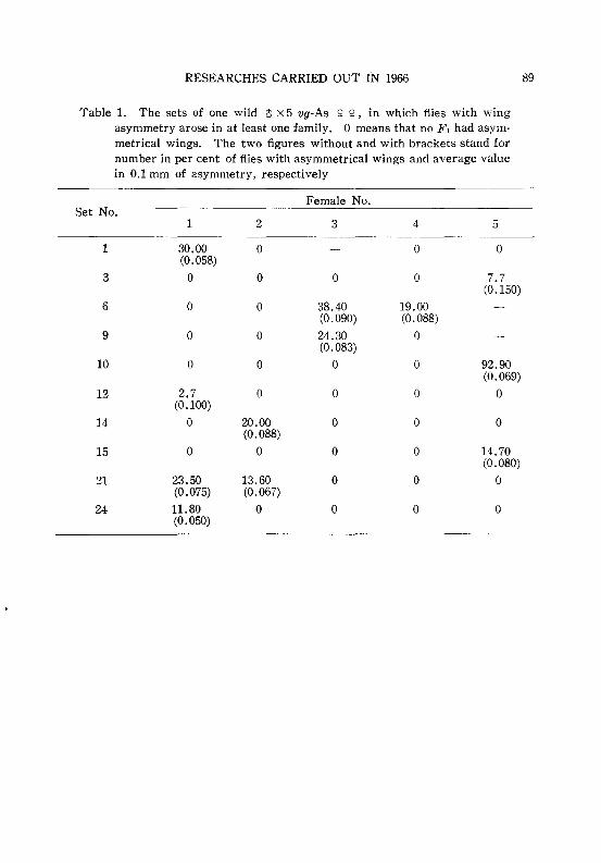
RESEARCHES CARRIED OUT IN 1966 89
Table l. The sets of one wild 0 x 5 vg-As ~ ~, in which flies with wingasymmetry arose in at least one family. o means that no F 1 had asym-metrical wings. The two figures without and with brackets stand fornumber in per cent of flies with asymmetrical wings and average valuein 0.1 mm of asymmetry, respectively
Female No.Set No.
1 2 3 4 5
1 30.00 0 0 0(0.058)
3 0 0 0 0 7.7(0.150)
6 0 0 38.40 19.00(0.090) (0.088)
9 0 0 24.30 0(0.083)
10 0 0 0 0 92.90(0.069)
12 2.7 0 0 0 0(0.100)
14 0 20.00 0 0 0(0.088)
15 0 0 0 0 14.70(0.080)
21 23.50 13.60 0 0 0(0.075) (0.067)
24 11.80 0 0 0 0(0.050)

90 ANNUAL REPORT OF NATIONAL INSTITUTE OF GENETICS NO. 17
VII. RADIATION GENETICS AND CHEMICALMUTAGENESIS IN ANIMALS
Post-Irradiation Modification and Mechanism of Reverse DoseRate Effect on Mutation Induction in Silkworm Gonia
Yataro TAZIMA and Toshihiko SADO
An enhancing effect on radiation-induced mutation was observed inthe silkworm after fractionated exposure of gonial cells. As a possibleinterpretation of this phenomenon, some kind of modification in cellularmetabolism was assumed. The modified state, which becomes detectableby increased mutation frequency, persists at least for 48 hours and ischaracterized by a peak appearing 18-24 hours after the initial exposure.The non-decay type curve suggested that this was not due to a biochemicaldisturbance but presumably to cell synchronization.
The results of an experiment, where two split doses were given to twoexperimental series in reciprocal order (250 R-L750 R and 750R+250R) 6hours up to 48 hours apart, showed that the peak appears earlier for theinitial 250 R than for the initial 750 R series. By labelling S phase cellswith H3-thymidine, Sado observed that irradiated cells are blocked intheir progression at Gz and their proportion increases remarkablly in thecell population. This observation favours the hypothesis of cell synchronization. If this were the case, a decrease in mutation frequency shouldalso be detected when second irradiation were given at an appropriateinterval after the initial exposure. This, however, was not observed upto 72 hour interval, suggesting that synchronization, if any, is destroyedimmediately in the subsequent cell cycle. Presumably the cells are releasedfrom the blockade at a reduced rate.
Based on those results, we examined the validity of the same mechanismfor the interpretation of the reverse dose-rate effect. Cytological observations revealed that the mitotic process of gonia at a late stage is inhibited,being blocked at G, and Gz, during chronic exposure. Furthermore, itwas confirmed by a new series of chronic exposure experiments lastingthree days which were started every 24 hours that the chronic treatmentbecomes more effective for mutation induction than the acute one as earlyas one day after hatching, coinciding with the initiation of cellular activity.This observation explains why the dose-rate effect is reversed within avery short period.
(Abstract of a paper presented at the third International Congress ofRadiation Research, Cortina D'Ampezzo)

RESEARCHES CARRIED OUT IN 1966
Repair of Radiation Induced Premutational Damages Revealedby Fractionated Irradiation of Silkworm Spermatids
Yataro TAZIMA and Kimiharu ONIMARU
91
The results of our study, for past six years, on dose-rate effect ofradiation, strongly suggested the existence of a mechanism of repair ofpremutational damages. Furthermore, the assumption of a repair phenomenon allowed a consistent interpretation of several kinds of experimentaldata obtained so far. Our knowledge, however, was rather scanty as tothe mechanism of repair of premutational damages in higher organisms.
A new project has, therefore, been undertaken in our laboratory for theinvestigation of such a mechanism. As a first step of this project effortshave been made to find out good evidences for the repair of premutationaldamages which could be utilized as an experimental system for furtherstudies.
For the elucidation of repair, two methods can be applied. One is asplit dose irradiation with an appropriate time interval. The other is apost-irradiation treatment with various agents. In both cases, repair ofpremutational damages, if any had occurred, would be detected by a decrease in observed mutation frequency.
As reported previously, a remarkable enhancing effect has been observedwith regard to mutation frequency when we irradiated silkworm spermatogonia with two split doses of 500 R+500 R. The mechanism was soonrevealed in a cytological study by Sado (1966). Namely, after initial exposure the proportion of G2 cells increased among the germ cell populationand if the second exposure was given to those cells an enhancement inmutation response was the result. Accordingly, those cases can not beregarded as due to true repair.
In order to obtain conclusive evidences for the repair, fractionationexperiments have been carried out with spermatids. Silkworm spermatidsappear most abundantly in full-grown larvae. Furthermore, at this stagethe cells are fairly homogeneous with respect to their developmental stage.Total dose of 1000 R was given to full-grown larvae in two fractions: 500 R+500 R. Intervals between the two exposures were 0, 6 and 12 hours.The results are given in Fig. 1.
It can be seen from the figure that split dose irradiation clearly reducesthe induced mutation frequency in comparison with the same dose givensingly.
Similar experiments were carried out at V-4.5 (fifth instar day 4.5) andmid-pupal stage, but the results were less conclusive for both stages.
Since irradiation of full-grown larvae almost completely excludes the

•
i2
92 ANNUAL REPORT OF NATIONAL INSTITUTE OF GENETICS NO. 17
tFractionation (500 R+500 R)
80661 Full-grown larvae
~ ::.[.----------- :_!' mO'
--............ ,
!::~~ p' ------:
, 3O~__ .. 't~ _~ remos
20l ---------...--------------..
lOt"o 6
Interval in hoursFig. 1. Results of split dose irradiation of full-grown
larvae: 500 R + 500 R with 0, 6 and 12 hour intervals.Heavy lines, solid and broken, represent the split doseirradiation and light lines denote single dose irradiation.
risk of contamination with cells irradiated at other stages than spermatids,the observed reduction in mutation frequency can be regarded as thereflection of a repair of premutational damages produced by radiation.
Modification of r·Ray-Induced Mutation Frequencies in the Silkwormby Post-Treatment of Spermatids and Spermatozoa
with Nitrogen GasYataro T AZIMA and Kimiharu ONIMARU
As reported in the foregoing article (this Annual Report), a direct evidence of a repair of premutational damages was obtained by fractionatedirradiation of spermatids. Another evidence has been obtained from thecomparison of the effects of post-irradiation treatment between nitrogenand oxygen.
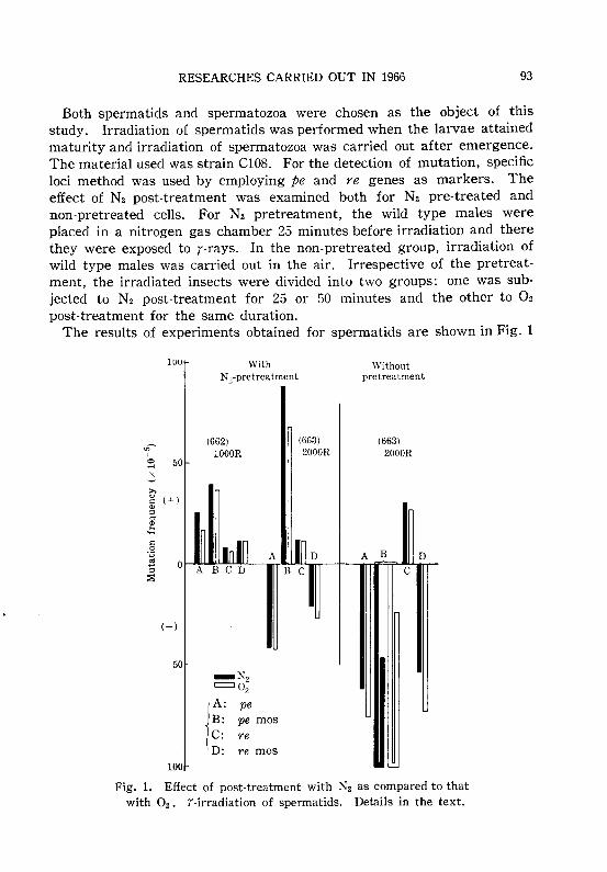
RESEARCHES CARRIED OUT IN 1966 93
Both spermatids and spermatozoa were chosen as the object of thisstudy. Irradiation of spermatids was performed when the larvae attainedmaturity and irradiation of spermatozoa was carried out after emergence.The material used was strain C108. For the detection of mutation, specificloci method was used by employing pe and re genes as markers. Theeffect of N2 post-treatment was examined both for N2 pre-treated andnon-pretreated cells. For N2 pretreatment, the wild type males wereplaced in a nitrogen gas chamber 25 minutes before irradiation and therethey were exposed to r-rays. In the non-pretreated group, irradiation ofwild type males was carried out in the air. Irrespective of the pretreatment, the irradiated insects were divided into two groups: one was subjected to N2 post-treatment for 25 or 50 minutes and the other to 02
post-treatment for the same duration.The results of experiments obtained for spermatids are shown in Fig. 1
IUU
;:; 50
WithNz-pretreatment
(662)1000R
(663)2000R
Withoutpretreatment
(663\2000R
(-)
50_Nz=Oz
jA: peB: pe masC: reD: re mas
100
Fig. 1. Effect of post-treatment with N2 as compared to thatwith O2 • r-irradiation of spermatids. Details in the text.

94 ANNUAL REPORT OF NATIONAL INSTITUTE OF GENETICS NO. 17
In this figure cases in which post-treatment with N2 was mutagenicallymore effective than with O2 are shown in the upward histogram and thosewhere N2 was less effective are represented in the downward histogram.
Our experiments show that post-treatment with N2 is in most casesmutagenically more effective than that with 02 for spermatids irradiatedunder anaerobic condition. However, the effect becomes reverse whenthe spermatids are irradiated under aerobic condition.
In contrast, N2 post-treatment of spermatozoa resulted in a lowermutagenic effectiveness than 02 post-treatment when the insects had beenpretreated with N2 and irradiated under N2.
Those results almost agree with those obtained by Sobels et al. forDrosophila and may be mentioned as another evidence of a repair ofpremutational damages.
Mechanisms of Mutation Induction by Mitomycin-C in the SilkwormYataro T AZIMA and Kimiharu ONIMARU
During the course of our experiments of post-irradiation treatment ofsilkworm spermatogonia with various metabolic inhibitors, we observedthat mitomycin-C enhanced markedly the mutagenic effect of r-rays. Theenhancing effect was more pronounced when the chemical had been administered 12 hours after irradiation.
Concerning the effect of mitomycin-C upon biological systems, numerouspapers have been published by many workers and at least three differentmechanisms have been assumed: i.e., 1) inhibition of DNA synthesis, 2)degradation of DNA and 3) cross-linking of DNA.
Since a strong mutagenic effect upon silkworm spermatozoa has beenelucidated for some alkylating agents which cross-link DNA (Nakao, 1961),experiments have been carried out to examine if mitomycin-C is mutagenically effective when given to mature spermatozoa.
The chemical was injected as a saline solution into wild type male pupaeof C108 strain two or three days before emergence. The administereddose of the chemical were 0.02~0.05mg per capita. Mutation frequencieswere estimated by specific loci method as usual. The results are shownin Table 1.
As may be clearly seen from the table, injection of mitomycin-C resultedin a high incidence of mosaic mutations as well as whole type recessivemutations. The most striking was an extraordinarily high incidence ofmosaics. Their frequency was from 10 to 20 times as high as that ofthe whole type.
Assuming that the incidence of mosaics was due to a single helix (of

RESEARCHES CARRIED OUT IN 1966
Table 1. Results of injection of mitomycin-C into male pupae (654) ofthe silkworm
pe re 'i' X treated++ 0
95
Injected dose Observed number of eggs* Mutants detected**
per capita Unferti- Early Sur· Total pe, + re, +lized dying viving pe mos re mos
0.05mg 3,174 2,225 49,005 54,404 46 471 30 661(5.8) (4.1) (90.1) (94) (951) (61) (1,349)
0.02mg 3,517 2,401 65,788 71,706 135 823 71 959(4.9) (3.4) (91.7) (205) (1,251) (108) (1,458)
Numerals in parentheses are (*) percentage of eggs observed and (**) mutationfrequency per surviving eggs represented in the order of 10-5•
DNA) type mutation and that of whole type mutations to a simultaneousoccurrence of two single helix type mutations at the same locus, theprobability of the incidence of whole type mutations could have beenexpected to be the square of the probability of the mosaics. The observedfrequency for the whole type was about one order higher than such expectation. This suggests that whole type mutants were also producedby a single mutational event provoked on a double helix of DNA.
Anyhow, the above results indicate that mitomycin-C can act on oneof the double helices of spermatozoa DNA, where DNA synthesis is nomore in progress.
Mutagenicity of a Nitrofuran Derivative Applied toSilkworm Germ Cells
Yataro T AZIMA and Yosoji FUKASE
It has been reported by Endo et al. (1963, 1966) that a nitrofuran derivative, 3-amino-6[2(5-nitro-2 furyl) vinyl]-l, 2, 4,-triazin hydrochloride,NFT, exhibits in several respects a similar action to that of mitomycin-C.According to their report, the chemical is a potent phage inducer inEscherichia coli K12 (i.) cells and inhibits DNA synthesis without affectingRNA and protein synthesis in non-lysogenic bacteria.
Since mitomycin-C has been proved to have a strong mutagenicity inthe silkworm, we thought that it would be interesting to examine themutagenic effect of NFT in comparison with that of mitomycin-C.
NFT used in this experiment was kindly supplied by Prof. H. Endo,Cancer Institute, Kyushu University. Two series of experiments havebeen carried out. Materials and methods used were almost the same asthose reported in the foregoing article if not otherwise specified.

96 ANNUAL REPORT OF NATIONAL INSTITUTE OF GENETICS NO. 17
1) Feeding experiment with third instar larvaeNFT was administered to the third instar larvae by painting an aqueous
solution of the chemical on mulberry leaves. The administered dosesare given in the foot notes of Table 1.
Table l. Results of feeding NFT to the third instar larvae (664)
pe re !f x treated++ 0
No. of mutants detected Mutation frequencyNo. of eggs (x 10-5)
Treatmentobserved pe pe, + re re, + pe pe, + re re, +
mas mas mas mas
None 134,116 31 6 0 2 23 5 0 2
*NFT (L) 123,483 28 10 1 4 23 8 1 3**NFT (H) 126,031 51 14 3 3 41 11 2 2
*NFT (L): 10 mg of NFT was given to 1,000 larvae at one time.**NFT (H): 50 mg of NFT was given to 1,000 larvae in five times.
It may be clearly seen from Table 1 that an increased dose of thechemical increases the mutation frequency both for whole and mosaicmutants.
2) Injection experiment at post pupal stageThe chemical was injected as a saline solution to wild type male pupae
of C108 strain two or three days before emergence. Injections with knownmutagenic agents, nitromine (NM) and ethyl methane sulphonate (EMS),were performed in parallel. The results are given in Table 2. In thisexperiment the mutagenic effect of NFT could be again observed. The
Table 2. Results of injection of NFT into pupae (663)
pe re !f x treated++ 0
No. of mutants detected Mutation frequencyNo. of eggs (x 10-5)
Treatmentpe, + re, + pe, +observed pe re pe re re, +mas mas mas mos
Control 149,908 42 12 2 2 28 8 1 1
NFT 0.01% 98,540 55 15 1 5 56 15 1 5
" 0.1% 121,026 80 11 4 5 66 9 3 4NM 0.025% 114,869 59 25 4 9 51 22 4 8
EMS 0.01% 135,386 50 55 4 43 40 44 3 34
" 0.1% 103,516 59 1,065 3 602 57 1,029 3 582
* Injected amount was 0.05 mL of physiological salt solution of the chemical atthe concentration specified.
RESEARCHES CARRIED OUT IN 1966 97
induced mutation frequencies were clearly enhanced for whole mutations,while those for mosaics were not significantly increased.
Those results are in sharp contrast to those obtained for NM, EMS aswell as mitomycin-C, which produced a high incidence of mosaics. NFTseems to have a specific action by attacking both DNA helices at oneevent.
Changes in the Mutation Response of Post-Meiotic SilkwormGerm Cells to r-Rays with Progressing Spermiogenesis
Yataro TAZIMA
The double helix model of DNA suggests that there could be two typesof mutations concerning the site of occurrence, i.e. those occ*ring inone of the double helices and those occurring in both helices. Identification of those two types may be possible if post-meiotic germ cells areirradiated. When mutants are obtained as mosaics, they may be attributedto the single helix type. Whereas, mutants in whose whole body a recessive character is expressed may correspond to the double helix typemutation.
It has often been observed in the silkworm that irradiation of spermatozoa resulted in the production of a higher proportion of mosaics thanwhen early spermatids were irradiated.
In order to know if mutation response varies according to progressingspermiogenesis, data obtained from different experiments have beenarranged in accordance with the development of the insects. Althoughthe extent of mutation response varies depending on the irradiated stage,those data can be compared so far as the ratio of mosaics to whole mutantsis concerned.
In Fig. 1 calculated ratios of mosaics to whole mutants at each locusare plotted against the progression of the developmental stages of theinsect. In the male, meiosis takes place most frequently around V-2(instar V day 2), and thereafter the germ cells enter into the spermatidstage. When the larva attains maturity (Full G.) fully formed spermatozoaappear in the testis and increase in number progressively thereafter un·til the mid-pupal stage (M.P.).
As can be clearly seen from the figure, the incidence of mosaics inrelation to whole mutation is very low before meiosis, reaches to theunity around V-4.5 and then increases rapidly with the progress ofspermiogenesis.
The relation can be more intuitively understood from the curves markedwith rightward arrows. Those curves represent changes in induced fre-

98 ANNUAL REPORT OF NATIONAL INSTITUTE OF GENETICS NO. 17
7
rtl
I:: 6n:l
a 5CI)
"0..c:::: 4
ErtlU
3"@rtl0a
..... 200
'';:ro
Ct::
I 10 7
10
~
,/~/ 5,,/
" re,- ,--------'-'c:::·· __--,,--- JMothM.P.lJI-o V-4.5 Full G. Pupa
Developmental stage
Fig. 1. Changes in the mutation response of silkworm spermatogenic cellswith the progress of development. Curves with rightward arrows are tobe read against the right ordinate and vice versa.
quencies per R for whole and mosaic mutants, in an average for two loci.It can be inferred from those results that the site of mutation response
becomes reduced in size with the progression of spermiogenesis. Presumably this change might be correlated with a decreasing facility to repairpremutational damages.
Studies on Strain Differences in Radiosensitivity in the Silkworm
I. Screening of Sensitive and Resistant Strains toEmbryonic Radiation Killing
Akio MURAKAMI and Yataro TAZIMA
A comparison of radiation effects between strains of different radiosensitivity may help to understand better the mechanism of radiationeffects on living organisms. From this view point, experiments have beencarried out in order to select sensitive and resistant strains to embryonicradiation killing. A worldwide collection of silkworm strains has beenmaintained for nearly 50 years at the Sericultural Experiment Station,Tokyo. Among 250 strains, 30 representative strains were chosen andtheir radiosensitivity to embryonic killing was examined. X-irradiation

RESEARCHES CARRIED OUT IN 1966 99
was applied to over-wintered eggs on the 30th of March, 1966. Irradiationconditions were: 173kVp, 25 rnA and 1.0 mm Al filter. Four different doses,800, 1,200, 1,600 and 2,000 R, were given at dose-rate of 250 R/min. Afterhatching of the surviving embryos, hatchability was calculated and LD50
was estimated from dose-response curves. The results are shown inTable 1. The average LD50 for 30 strains was 910 R. It may be clearly
Table l. Differences in radiosensitivity among 30 strains
Race Strain LD50 (R) Race Strain LD50 (R)
Aojuku 1,460 fDai-anky6 860
Akajuku 1,320 I Dai-k6jy6 1,050
Kojiki 230 Chinese , Ben-y6 800Japanese
Tadamisan 940 IS t A• 800I an o-sanmm
Matamukasi 1,320 \ Sisen-sanmin 980
Chusu 890 China-southern f Daiz6 930
Korean K6rai-sanmin 650 Asian \ Ringetu 670
Ryukyuan Ryukyu-tasanken 1,110 IToruko-6ken 1,100
Kansen 1,580 Ascoli 140Kin-6 800 Biohne 520
Seikyo 1,160 Maruke-6ken 610
Chinese Sekko 800European
Maiera-zebra 760
Daient6 800 Rosa 1,020
Hik6 1,340 Var 1,280
Sy6k6 1,080 \ Kokuga 1,500
seen from the table that there were striking differences among strainswith regard to radiosensitivity. The LD50 values for the most radioresistant three strains were approximately 1,500 R. In contrast, twostrains were extremely sensitive, i.e., LD50 was 140 R for Ascoli and 240 Rfor Kojiki. These differences in radiosensitivity might have been, in part,due to different dosages of the relevant genes accumulated in those strains.
Table 2. LD50 's obtained for 16 combinations of diallelic crosses
FemaleMale
Kansen Aojuku Kojiki Ascoli
Kansen (1,580) 1,580 1,700 1,480 1,750
Aojuku (1,460) 1,750 1,540 1,060 1,660
Kojiki (230) 1,750 1,460 230 1,600
Ascoli (140) 1,750 1,625 1,600 140

100 ANNUAL REPORT OF NATIONAL INSTITUTE OF GENETICS NO. 17
In order to investigate the mechanism, the radiosensitivity of F1 eggswas examined in all combinations of diallelic crosses between some resistantand sensitive strains. Those eggs were irradiated one day after Heltreatment for artificial hatching. The results obtained are given in Table2. LDso values thus obtained for self-bred batches were almost the sameas that observed for over-wintered eggs. In cross-bred batches, however,LDso's were roughly the same as those observed in inbred batches of theresistant strains. Those results suggest that the radiosensitivity of silkworm embryos is controlled by two major genes at least, and that theeffect of maternal or cytoplasmic factors is relatively small.
Studies on Strain Differences in Radiosensitivity in the Silkworm
II. Relation between Sensitivity to Embryonic Killing and MutabilityAkio MURAKAMI and Yataro T AZIMA
As reported in the foregoing article, some strains sensitive or resistantto radiation were discovered with regard to embyronic killing. Usingthose strains, an investigation has been carried out to ascertain if sensitivity to mutation runs parallel to sensitivity to killing. The strains usedwere: (resistant) Kansen, Aojuku and Hiko, (moderate) Sekko and (sensitive)Ascoli and Kojiki. The mutability was measured by the specific locusmethod, using egg colour markers. Irradiation conditions of X-rays were:173kVp, 25mA and 1.0 mm Al filter throughout this study. Doses deliveredwere 250 R per minute.Irradiation of gonial cells: Both spermatogonia and oogonia were irradiated in the larvae after hatching. The dose applied was 1,000 R only.The results are given in Table 1 a and b, separately for spermatogoniaand oogonia, respectively.
Table 1. Mutation rates for the goniaI cells in five representative strainsa. Spermatogonia
Mutation-rate
Strain
Kansen
Aojuku
Sekko
Ascoli
Kojiki
pe locus
Whole
xlO 8
37.4
58.5471.3
232.1135.4
Mosaics
x10 8
1.6
5.8
3.12.2
15.6
re locus
Whole
x10 8
6.213.0
144.9
49.1
41.6
Mosaics
xlO 8
1.3
0.04.1
0.0
10.4

b. Oogonia
RESEARCHES CARRIED OUT IN 1966 101
Strain
Kansen
Aojuku
Sekko
Ascoli
Kojiki
Mutation-rate
pe locus re locus
Whole Mosaics Whole Mosaics
X 10-8 X 10-8 X 10-8 x10⁻⁸
2.4 0.019.0 9.0 1.4 0.0
173.5 12.7 122.5 7.8109.8 3.9
35.0 7.0 19.6 1.4
It can be seen from the table that the mutation rates are higher forspermatogonia than for oogonia in all strains studied. It is of interest tonote that the mutation rate of a Chinese strain, Sekko, was extraordinarilyhigh, although as to embryonic killing it belonged to the moderate group.Except for Sekko, it may be roughly inferred that sensitive strains toembryonic killing are also sensitive to mutation in both sexes.Irradiation of oiJ"cytes: The sensitivity of oocytes and mature spermatozoawas tested after irradiation of newly emerged adults. The results formature oocytes are given in Table 2. In this experiment X-ray doses
Table 2. Mutation rates for the mature oJcytes in five representative strains
Mutation-rate
Strain
Kansen
Aojuku
Hiko
Ascoli
Kojiki
pe locus
Whole
X 10-8
470.6386.0634.8
1,012.41,111.7
Mosaics
x10-8
28.430.2
35.5130.372.3
re locus
Whole
x10-8
500.6372.3766.1447.9732.0
Mosaics
X 10-8
27.014.9
23.177.755.6
given were 1,000 Rand 2,000 R. The mutation-rates are shown in anaverage for the two doses. It appears that sensitive strains for embryonickilling, Ascoli and Kojiki, were also sensitive with respect to mutation.Irradiation of mature spermatozoa: The results of the experiment withmature spermatozoa are presented in Table 3. In this experiment X-raydoses given were 3,000 Rand 6,000 R. The mutation rate is expressedas an average of the two doses. No distinct correlation was found be-

102 ANNUAL REPORT OF NATIONAL INSTITUTE OF GENETICS NO. 17
Table 3. Mutation rates for the mature sperms in five representative strains
Strain
Kansen
Aojuku
Hiko
Ascoli
Kojiki
Mutation·rate
pe locus re locus
Whole Mosaics Whole Mosaics
x 10 B x10 B x10 8 x10 8
16.7 37.6 8.9 12.4
16.1 26.3 8.7 14.2
22.8 34.3 4.7 18.7
26.9 24.2 8.9 22.8
19.9 38.8 5.8 18.2
tween radiosensitivity to embryonic killing and mutability. This findingis in sharp contrast to those described above for the three types of germcells. The discrepancy might be due to either to the lack of recovery ofradiation damage or to a different repair system in mature spermatozoa.
Relation between Sensitivity to Killing and Mutation Observedduring a Mitotic Cycle of Silkworm Cleavage Nuclei
Akio MURAKAMI
Cyclic change in radiosensitivity to killing has been revealed for earlycleavage nuclei of the silkworm (Murakami, 1966). The most sensitivephase within a cell cycle was found to be the metaphase/early telophase,whereas the least sensitive was the interphase. This finding suggestedthat dividing chromosomes are sensitive.
The purpose of this study was to investigate the change in mutationresponse in relation to the progress of the mitotic cycle during earlycleavage. Materials used were eggs laid by ++/++ females mated withpe re/pe re males. These eggs were collected every 10 minutes and allowedto develop for a definite duration until they were irradiated. From 170minute to 230 minute old eggs after oviposition were irradiated at 10 minuteintervals. Radiation dose of 1,000 R was delivered in one minute. Mutation frequencies were estimated by specific locus method using the pe andre genes. The lethality was also examined at the same time. The mitoticphase of the irradiated cells was determined cytologically. The resultsare shown in Fig. 1. Mutations most frequently found in this experimentwere fractionals. The highest peak for embryonic killing was observedat 190 minute age. In contrast, the highest mutation frequency wasobserved at 200 minute age. Cytological observation indicated that the

RESEARCHES CARRIED OUT IN 1966 103
('·;0I.";> J~cE
"""0,.,,...0
E 30'"
<10·
3000
Cc'""2000 0-
!;Cc
".:3~
1000 ":>l
170 180 190 200 210 220 (min)Time after oviposition
Fig. 1. Changes in sensitivity to killing and mutation during amitotic cycle as expressed after oviposition as time function.
phase of maximum embryonic killing corresponded to metaphase/earlytelophase (or M +Gl) and the highest mutation response appeared at latetelophase/early interphase (or Gl +early S). The discrepancy between thepeak stages for cell killing and for mutation response suggests that themechanisms involved are different. Presumably we may assume that thetarget or targets responsible for cell killing are chromosomes and thosefor mutation response are DNA strands.
The Effect of 5-Bromodeoxyuridine (BUDR) on the Frequency of14 MeV Fast Neutron Induced Mutations in
the Gonia} Cells of the SilkwormAkio MURAKAMI
It was reported in our previous paper (Murakami and Tazima, 1964)that a post-irradiation treatment of silkworm spermatogonia with BUDRor 5-Bromouracil (BU) increases the frequency of specific locus mutationsby about two or three times at the maximum (Murakami and Tazima,1964). This enhancing effect was assumed to be caused by an inhibitionwith such chemicals of the recovery process of pre-mutational lesions.
It was thought, therefore, to be of interest to study whether the radiation lesions produced by higher LET (Linear Energy Transfer) radiationscould be modified by a similar treatment with those chemicals. Hence,the effect of BUDR treatment of 14 MeV fast neutron induced mutationswas studied in gonial cells of the silkworm.
Materials and method used were the same as described in the previouspaper (Murakami and Tazima, 1964). BUDR was administered to newly

104 ANNUAL REPORT OF NATIONAL INSTITUTE OF GENETICS NO. 17
ha tched larvae of a wild type by feeding them on leaves supplementedwith the chemical (20 mg per 1,500 heads) immediately after 14 MeVneutron irradiation.
The results are given in Table 1 a and b, for males and females, sepa-rately.
Table l. Effect of BUDR on 14 MeV fast neutron-induced mutationfrequency in silkworm gonia
a. Males
pe locus re locus
Treatment Total No. Whole Mosaics Whole Mosaicsobserved
Mut. Mut. Mut. Mut. Mut. Mut. Mut. Mut.dltd. freq. dltd. freq. dltd. freq. dltd. freq.
xl0 5 xl0 5 xl0 5 xl0 5
Control 176,245 58 32.9 3 1.7 4 2.3 0 0.0BUDR* 145,184 48 33.1 5 3.4 4 2.8 1 0.714 MeV neutrons 219,521 172 78.4 6 2.7 33 15.0 2 0.91,000 rad14 MeV neutrons 147,976 133 89.9 8 5.4 40 27.0 2 1.41,000 rad+BUDR*
b. Females
xl0- 5 xl0-5 xl0-5 xl0-5
Control 212,420 21 9.9 8 3.8 14 6.6 0 0.0BUDR* 177,346 8 4.5 8 4.5 3 1.7 1 0.614 MeV neutrons 244,530 68 27.8 9 3.7 28 11.5 2 0.81,000 rad
14 MeV neutrons 147,212 17 11.6 14 9.5 22 14.9 2 1.71,000 rad+BUDR*
* BUDR: 20 mg per 1, 500 heads.
The mutagenicity of BUDR treatment alone is not clear. However,when combined with 14 MeV neutrons, mutation frequencies were slightlyenhanced in the experiment with spermatogonia. Whereas, no enhancingeffect was observed in the experiment with oogonia. In the former theenhancing effect was higher at the re locus than at the pe locus. Asto the enhancing effect of BUDR, neutron induced mutations respondedto a lesser extent than X-ray induced mutations. This might have beendue to the fact that neutron induced pre-mutational damages are lessresponsive to repair than those produced by low LET radiation.
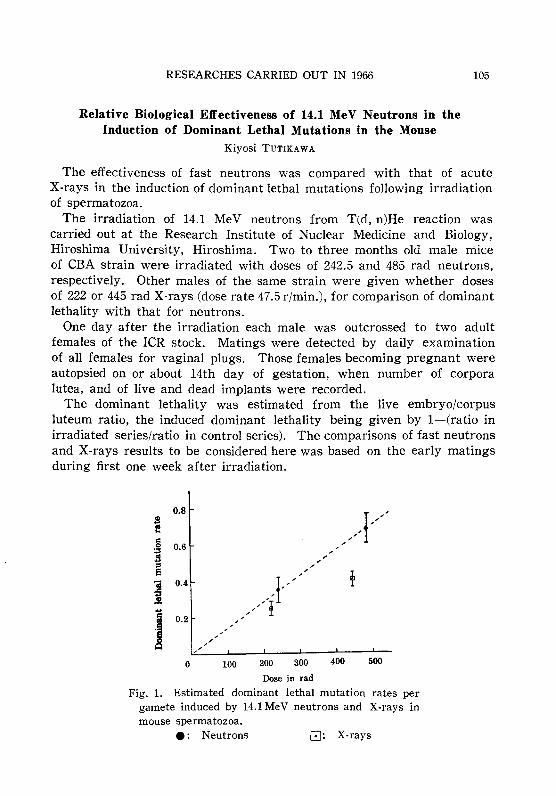
RESEARCHES CARRIED OUT IN 1966
Relative Biological Effectiveness of 14.1 MeV Neutrons in theInduction of Dominant Lethal Mutations in the Mouse
Kiyosi TUTIKAWA
105
The effectiveness of fast neutrons was compared with that of acuteX-rays in the induction of dominant lethal mutations following irradiationof spermatozoa.
The irradiation of 14.1 MeV neutrons from T(d, n)He reaction wascarried out at the Research Institute of Nuclear Medicine and Biology,Hiroshima University, Hiroshima. Two to three months old male miceof CBA strain were irradiated with doses of 242.5 and 485 rad neutrons,respectively. Other males of the same strain were given whether dosesof 222 or 445 rad X-rays (dose rate 47.5 r/min.), for comparison of dominantlethality with that for neutrons.
One day after the irradiation each male was outcrossed to two adultfemales of the ICR stock. Matings were detected by daily examinationof all females for vaginal plugs. Those females becoming pregnant wereautopsied on or about 14th day of gestation, when number of corporalutea, and of live and dead implants were recorded.
The dominant lethality was estimated from the live embryo/corpusluteum ratio, the induced dominant lethality being given by l-(ratio inirradiated series/ratio in control series). The comparisons of fast neutronsand X-rays results to be considered here was based on the early matingsduring first one week after irradiation.
0.8 /I'/,oS.......0 0.6 .-
:;3 ,.:l , ,::I .-e , .-
~iii 0.4
/"iI'"~.!l
j 0.2 ,,, ,
~,
.- ,, ,,
0 100 200 300 400 500
Dose in rad
Fig. 1. Estimated dominant lethal mutation rates pergamete induced by 14.1 MeV neutrons and X-rays inmouse spermatozoa.
• : Neutrons 8: X-rays
106 ANNUAL REPORT OF NATIONAL INSTITUTE OF GENETICS NO. 17
Using-loge (l-P) as an estimation of the dominant lethal mutationrates per gamete induced by two kinds of radiation, it was shown thatthe mutation frequency from neutrons is higher than that from X-rays,and the dose-frequency curves are in linear relationship (Fig. 1). If weequate the mutation rate of 445 rad X-rays with that of 242.5 rad neutronsaround the 50 per cent level of dominant lethality, results suggest that14.1 MeV neutrons are about 1.8 times as effective as acute X-rays forthe induction of dominant lethal mutations in mouse spermatozoa.

RESEARCHES CARRIED OUT IN 1966
VIII. RADIATION GENETICS IN PLANTS
RBE of Radiations in E-I Hole of Kyoto UniversityReactor (KUR)
Seiji MATSUMURA, Etsuo AMANa and Masaru HAYASHI
107
To obtain basic data for future use of Kyoto University Reactor (KUR),dry seeds of rice plant and Einkorn wheat were irradiated in E-l exposurehole of KUR under nitrogen gas or air atmosphere. As preliminary experiments, in June, 1966, hulled seeds of Oriza sativa were sealed inpolyethylene vials after being flushed with nitrogen gas so that the desired atmospheres were obtained, and, three days later, they were irradiated in E-l hole for 180 minutes at 1,000 kW. Radiation dosage of eachsample was adjusted by varying the distance from the core side end, andthermal neutron doses were calculated from activation of gold foils andatomic composition of the material. r-ray from 6,000 Ci mcs source ofNational Institute of Genetics was used as standard radiation.
Results of seedling height measurement of the reactor radiations irradiated seeds showed that 50 per cent seedling height reducing doseswere obtained at 21 cm for nitrogen gas and 22 cm for air atmosphere,from core side end of the E-l sample holding boat, revealing distinctprotective effect of nitrogen gas. Existence of such oxigen effect stronglysupport the heavy contamination of r-rays measured by physical dosimetry(d. KURRI-TR-16).
In October, 1966, seeds of Triticum monococcum were prepared as samples sealed in specially designed two celled air tight acryl resin containers.Nitrogen atmospheres were achieved by several times flushings of nitrogengas after reducing gas pressure by a vacuum pump each time, and sealedin nitrogen gas stream. Exposure to reactor radiation was done threedays later in E-l hole of KUR. As T. monococcum seeds are more radiation sensitive than O. sativa seeds, 90 minutes of exposure at the same1,000 kW gave 50 per cent seedling height reducing dose at about 24 cmfor air preparation and 22 cm for nitrogen atmosphere, from the core endof the boat. Seedling height measurement on seventh day after sowingagain revealed oxigen effect as shown in Figure 1 in which E-l radiationdose was shown by thermal neutron component. Calculations of RBE ofthe reactor thermal neutrons free of contaminating r-rays were tried atthe 50 per cent seedling height reducing dose, assuming that the contaminating r-rays in E-l radiations might have the same RBE as that of 137CSr-ray, and that the difference of dosages of the two r-rays might beattributed to thermal neutrons.

108 ANNUAL REPORT OF NATIONAL INSTITUTE OF GENETICS NO. 17
100
\\
10' 103
Radiation dose (rad)
10" 10'
Fig. 1. Effect on seedling height of T. monococcum of KUR E-1thermal neutrons and 137Cs r ray.
Under air atmosphere, the difference in r-rays was 2,000 rads, effect ofwhich was comparable to 131 rads of thermal neutrons, giving RBE value15.3. Since efficiency of the r-rays might be reduced under nitrogen gasatmosphere the difference in r-rays was 5,000 rads and equivalent effectmight be brought about by 181 rads of thermal neutrons, giving RBE value28.3. Although fluctuation of r-ray contamination and difficulty of itsdosimetry are the problems still to be worked out about the reactor radiations, following calculations were tried further. With the same assumptionabout efficiency of r-rays, it might be considered that 131 rads of thermalneutrons reduced final 13 per cent of the seedling height to reach 50 percent height of control in case of air, and 181 rads reduced final 17 percent in case of nitrogen atmosphere, to get to 50 per cent height, suggestingthe same efficiency of thermal neutrons in seedling height reduction regardless of the atmosphere.
As for RBE values of overall E-1 radiations to 187Cs r-ray, 1.12 forexposure in air and 1.35 in nitrogen atmosphere were obtained.
Since contribution of thermal neutrons to the effect on the botanicalmaterials used was only 13.6 per cent (air) or 26.7 per cent (nitrogen),RBE being considered, the values estimated may subject to be correctedwhen advanced dosimetry for contaminating r-rays can supply furtherinformation.

RESEARCHES CARRIED OUT IN 1965
Comparison of Mutagenic Efficiency between EMS and r-RaysSeiji MATSUMURA and Taro FUJII
109
Seeds of a normal strain and heterozygotic F, seeds from a cross between normal and chlorina mutant of Triticum monococcum were soakedin water for two hours and then treated with 0.1, 0.3 and 0.5% EMSsulutions for 22 hours. For comparison, the seeds soaked for 24 hourswere subjected to 0.5, 1.0 and 1.5 kR of r-rays. Moreover, EMS andr-ray treatments were combined to examine the synergestic effect of bothtreatments.
In the normal strain, several types of stripes were observed in manyplants during the growth period of EMS treated lots, while they appearedvery seldom in r-ray lots. About 20 per cent survived plants had chlorinalike yellowish green stripes in 0.3% lot, and a similar frequency was alsoobserved in EMS 0.3% +r-ray 1.0 kR lot, while such type of stripes wasnever seen in the three r-ray lots, as shown in Table 1.
Table l. Appearance of chlorina-like streaks in the treated generation
No. of Total Chlorina or chlorina-like streaksStrain Treatment seeds No. of No. of No. ofsown spikes plants spikes (%)
Control 300 0
EMS 0.1% 200 7183 2 3 (0.042)
" 0.3% 200 4185 23 42 (1. 004)
Normal r-ray 0.5 kR 200 7990 0
" 1.0 kR 200 5887 0
1.5 kR 200 5226 0
EMS 0.3% +r·ray 1 kR 200 4270 18 42 (0.984)
Control 300
EMS 0.1% 400 7447 0
" 0.3% 400 4703 20 46 (0.978)
F, r-ray 0.5 kR 348 6965 1 3 (0.043)
" 1.0kR 400 7138 2 4 (0.056)II 1.5 kR 400 5987 2 6 (0.100)
EMS 0.3% +r·ray 1 kR 400 1607 9 24 (1.493)
Seeds from some tillers with stripes and from normal green tillers of thesame individual were sown and segregation of chlorina seedlings wasexamined. Albina or other kinds of chlorophyll mutants were segregatedamong these X2 seedlings, but no chlorina-like segregants was observed.
110 ANNUAL REPORT OF NATIONAL INSTITUTE OF GENETICS NO. 17
From this fact, we may say that no somatic mutations from dominantgreen to recessive chlorina occurred in F 1 by EMS treatment.
In the F 1 strain, number of chlorina or chlorina-like stripes in EMS 0.3%lot amounted to 0.98 per cent and the frequency was almost the same asin the normal strain. Rates of striping in r-ray lots increased with increase of dosage but they were much lower than those of EMS 0.3% lot.The rate in the EMS 0.3% +r-ray 1.0 kR lot was 1.49 per cent which ishigher than the sum from EMS 0.3% and r-ray 1.0 kR lots.
Segregation of several kind of chlorophyll mutants were observed inthe X2 generation of the normal strain. Chlorophyll mutation rate wassimilar in EMS 0.1% and r-ray 1.0 kR lot, but the in EMS 0.3% lot wasvery high. These results show that the mutagenicity of EMS is veryhigh in high dosages when the mutability is compared on the basis oftheir survival rates.
Comparison of the Killing Effect of r-Raysand Thermal Neutrons
Taro FUJII
Dry and 48 hour steeped seeds were used and exposed to thermal neutrons were done by the pneumatic tube which was provided in the reactorof Kyoto University; the length of irradiation time were 7 sec in minimumand 180 sec in maximum at the out put of 1 MW. The 7 sec irradiationof dry seeds showed merely 19 per cent of survival rate, and no survivalplants were observed in other lots.
From the above experience, out put of reactor was diminished to 50 kWin the second experiment and similar observation was repeated; the detailsof irradiation and survival rates were given in Table 1 with the resultsof r-ray irradiations. r-ray irradiations for dry seeds had almost no effect,but slight decrease of survival rate was observed in 100 kR lot. Decreaseof survival rate was marked in wet seeds lots. In the neutron lots, killingeffects were severe and no survival plant was observed by 180 sec irradiation of dry seeds while contaminated r-ray was 45 kR. Higher killingeffects in the wet seeds were also seen in the neutron lots, but it mightbe the results of contaminated r-rays because the environmental modificationof radiation effect is very small in high LET radiations, while it is ratherlarge with sparsely ionizing radiations. These results show that the killingeffect of thermal neutrons was very high. RBE of thermal neutrons insomatic mutations will be examined in the next step of experiment andRBE versus LET relations should be determined.
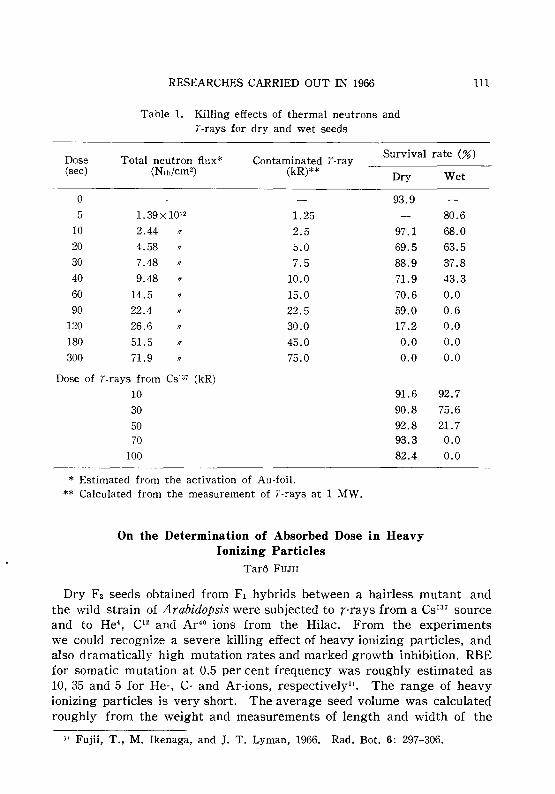
RESEARCHES CARRIED OUT IN 1966
Table 1. Killing effects of thermal neutrons andT-rays for dry and wet seeds
111
Dose Total neutron f1ux* Contaminated T-raySurvival rate (%)
(sec) (Nth/cm2) (kR)** Dry Wet
0 93.95 1.39x10¹² 1.25 80.6
10 2.44 1/ 2.5 97.1 68.020 4.58 1/ 5.0 69.5 63.530 7.48 1/ 7.5 88.9 37.840 9.48 1/ 10.0 71.9 43.360 14.5 !I 15.0 70.6 0.090 22.4 1/ 22.5 59.0 0.6
120 26.6 1/ 30.0 17.2 0.0180 51.5 1/ 45.0 0.0 0.0300 71.9 II 75.0 0.0 0.0
Dose of T-rays from CS'37 (kR)
10 91.6 92.730 90.8 75.650 92.8 21.770 93.3 0.0
100 82.4 0.0
* Estimated from the activation of Au-foil.** Calculated from the measurement of T-rays at 1 MW.
On the Determination of Absorbed Dose in HeavyIonizing Particles
Tarc~ FUJII
Dry F2 seeds obtained from F1 hybrids between a hairless mutant andthe wild strain of Arabidopsis were subjected to r-rays from a CS137 sourceand to He', 0 2 and Ar'o ions from the Hilac. From the experimentswe could recognize a severe killing effect of heavy ionizing particles, andalso dramatically high mutation rates and marked growth inhibition. RBEfor somatic mutation at 0.5 per cent frequency was roughly estimated as10,35 and 5 for He-, C- and Ar-ions, respectivelyll. The range of heavyionizing particles is very short. The average seed volume was calculatedroughly from the weight and measurements of length and width of the
11 Fujii, T., M. Ikenaga, and J. T. Lyman, 1966. Rad. Bot. 6: 297-306.

112 ANNUAL REPORT OF NATIONAL INSTITUTE OF GENETICS NO. 17
seeds, to be 20.5 x 10⁻³ mm3• From this calculation, it was concluded that
Ar-ions must be stopped within the seeds but He- and C-ions certainlycould penetrate and pass through seed tissue. Moreover the energytransfer varies according to the position of the range or tissue whichmay be seen from the Bragg Curve21
•
Further experiment about killing efficiency of heavy ionizing particleswas done according to above questions. Dry seeds were irradiated withC-ions in five different lots. Each lot received the same number of par-
Table 1. Relation cetween the survival rates ardthe thickness of acsorbers
--_._---- -~.~_._~
Acsorber Range Survival rate(mg/cm 2) (micron) (%)
-------_ .. -
Control 71.6
Lot 1 0 525 66.0
2 10.2 400 64.8
3 19.7 315 57.6
4 29.9 215 46.7
5 39.3 125 46.8
ticles per cm2 , but the energies of the ions were varied for the differentlots by different thickness of absorbers and lot 1 received a surface doseof 400 rads (Table 1). Survival rates decreased with the increase of absorber's thickness. The results suggest that the irradiation of C-ions withmaximum stopping power will show more severe killing effect, becausethe maximum of the stopping power is 6 X 10³ Mev/g/cm2 at the range of45 mg/cm2
• Namely, RBE value of C-ions should be more larger than thatobtained in the previous experiment when the embryo was attacked bythe ions with maximum stopping power. Further studies are now underexamination.
Photoreactivation of an UV-Induced Mutation in MaizeSeiji MATSUMURA and Tomoo MABUCHI
A normal maize strain (Su) concerning the sugary endosperm gene ofa local variety and a recessive line (su) of Golden Cross Bantam wereused in this study. Table 1 summarizes the data obtained from the experiments with UV- or r-ray induced mutations at Su locus after visible
21 Brustad, T., P. Ariotti and J. T. Lyman, 1950. Lawrence Rad. Lab. Rep. UCRL9454.

RESEARCHES CARRIED OUT IN 1966 113
Table 1. Frequencies of mutations from Su to 8U in maize pollengrains induced by UV and r-rays treated with
VL and dark after irradiation
No. ofNo. of endosperm mutants
Irradia- Post- No. of seeds % of theDose irradiation seeds Whole Chimerastion treatment set set Obs. Obs. wholeper ear
(%) (%) muta-tions
Control 0 VL 4164 134 1(0.024) 3(0.072) 25.00 Dark 4023 108 1 (0.025) 1 (0.025) 50.0
UV 2.7 X 10³ ergs VL 3420 148 6(0.17) 15 (0 .44) 28.55.4x 103 ergs VL 4556 85 18(0.39) 35 (0. 76) 33.9
/I Dark 2333 41 22(0.94) 23(0.98) 48.88.1x103 ergs VL 913 43 5 (0. 55) 11 (1.20) 31.2
/I Dark 1557 45 25 (1.60) 19 (1.22) 56.8
r-rays 550R VL 4122 132 25(0.60) 11(0.27) 69.4/I Dark 5579 143 35(0.62) 13(0.23) 72.9
HOOR VL 2134 152 44(2.06) 18 (0.84) 70.9Dark 4154 122 122 (2.40) 22 (0. 52) 81.9
light (VL) and dark post treatments. The percentage of seed set washigh after the two doses of r-rays, while it decreased with the increaseof UV-dose to about 50 per cent of the control lot at the highest dose.Induced whole (endosperm wholly deficient for the dominant character)and chimeral mutants (endosperm consisting partially of su and Su phenotypes) were scored separately for VL and dark posttreatment. No marked differences in the frequency of whole and chimeral mutants betweenVL and dark posttreatments were observed. As to the frequency of thetwo types, whole mutation was more frequent than the chimeral one.The frequency of chimeras fits roughly a linear relation with increasingdoses, while it is rather exponential than linear for whole mutation.
It is noted that the frequency of mutations induced by r-irradiationwas not affected by subsequent VL treatment either for whole or chimeralmutants. On the other hand, the frequency of whole and chimeral mutations from UV-irradiation showed almost linear relation. The frequencyof whole mutation was considerably decreased to about one third by exposure to visible light, while there was no marked difference in the frequency of chimeras between VL and dark posttreatment.
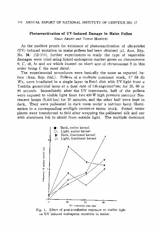
114 ANNUAL REPORT OF NATIONAL INSTITUTE OF GENETICS NO. 17
Photoreactivation of UV-Induced Damage in Maize PollenEtsuo AMANa and Tomoo MABUCHI
As the positive proofs for existence of photoreactivation of ultraviolet(UV) induced mutation in maize pollens had been obtained (d. Ann. Rep.No. 16: 112-114), further experiments to study the type of repairabledamages were tried using linked endosperm marker genes on chromosome9, C, sh, bz and wx which located on short arm of chromosome 9 in thisorder being C the most distal.
The experimental procedures were basically the same as reported before (Ann. Rep. ibid.). Pollens of a multiple dominant stock, CI Sh BzWx, were irradiated in a single layer in Petri dish with UV light from aToshiba germicidal lamp at a dose rate of 136 ergs/mm2/sec for 30, 60 or90 seconds. Immediately after the UV treatments, half of the pollenswere exposed to visible light from two 400 W high pressure mercury fluorescent lamps (6,400 lux) for 30 minutes, and the other half were kept indark. They were pollinated in dark room under a natrium lamp illumination to a corresponding multiple recessive tester stock. Potted testerplants were transferred to field after wrapping the pollinated silk and earwith aluminum foil to shield from outside light. The multiple dominant
.: Dark, entire kernel0: Light, entire kernelt: Dark, fractional kernelD: Light, fractional kernel
,
9060
i, .~.~;!~~/ly
o ~ •o 30
UV irradiation time (sec)
Fig, 1. Effect of post-irradiation exposure to visible lighton UV induced endosperm mutation in maize.
RESEARCHES CARRIED OUT IN 1966 115
stock used as male parent carried also a dominant yellow endospermgene Y on chromosome 6 besides the aforementioned genes, and this Ywas useful to confirm fertilization by the treated pollen to y (white endosperm) carrying tester, in case of loss of all the four chromosome 9 markerphenotypes.
Results of experiment are shown in Figure 1, in which B-F-B typemosaic kernels were included in either whole or fractional (more than 1/8of surface area of the kernel) kernel mutation, according to the area ofmosaics. In this experiment, photoreactivations of UV induced damageswere observed as lowered mutation frequencies in the groups which weretreated with visible light after UV irradiations compared to the groupswithout the post exposure to visible light.
Contrary to the previously reported results on su locus, in which wholekernel type showed more photoreactivation, present results showed morephotoreactivation in fractional or sectorial loss .of the dominant phenotypesthan in whole kernel type. This discrepancy might be due to the difference of the stocks used, commercial variety vs. genetical pure strain, orthe systems used, single specific locus or multiple loci, but, for a conclusive explanation, this should be studied further.
As for the type of damages which could be repaired by the post exposureto visible light, simultaneous loss of a few neighboring marker genes andB-F-B type mosaics were reduced by the post exposure. This stronglysuggests that some of the UV induced damages which would result inchromosome breakage might be repaired by post exposure to visible light.
Since the number of mutated kernels at a single locus were not manyenough, conclusion about repairability of the damages which would resultin so called point mutations should be reserved until further data will beaccumulated.
Distribution of initiating points of the B-F-B cycles seemed to be proportional to map distances.
On the Somatic Variations in Corn and Chrysanthemumunder Chronic r-Irradiation
Seiji MATSUMURA and Taro FUJII
Heterozygotic F1 seeds from the cross between normal green (Yg 2) andyellow green mutant (yg2) of corn, and white flowering variety of chrysanthemum were planted in the r-greenhouse with four different lots onJuly 18, and relation between the frequency of somatic mutation or variagation and dose rate effect were examined. Radiation intensity was 0.293.06 Rjhr, total irradiation time being 1741 hours; therefore, total dosage

116 ANNUAL REPORT OF NATIONAL INSTITUTE OF GENETICS NO. 17
in each lot was 430, 910, 2370 and 4500 R.In corn, Yg2-7yg2 mutation was detected by yellow green stripes on
leaves. Lengths of mutated stripes were different according to the developmental stage and occurrence of mutation; the number of the stripeslonger than 1 cm was counted in the present experiment. Dose versusmutation frequency shows almost linear relations when the results shownin Table 1 are plotted on the figure.
Table 1. Somatic mutation frequency in corn underchronic r -irradiations
Dose rate(R/h)
0.29
0.62
1.38
3.06
Total dcsage(R)
o430
910
2470
4500
No. of examinedleaves
109
294
223
318
199
No. of stripes(frequency)
o5(0.050)
9 (0.132)
30 (0.266)
28 (0.429)
Somatic variegation in chrysanthemum from white to yellow was observed in many flowers. When the variegation occurred in early stage,yellow sectors appeared on several ray flowers and when it occurred inlate stage streak appeared on single ray flower. Dose versus variegationcurve also showed almost linear relation like that in corn. From theseresults, no dose rate effect was observed in the present experiment,namely 0.29-3.06 R/hr, because when the recovery of radiation damageoccurred at low dose rate range, effect at higher dose rate range mustbe much higher than the present results.
Table 2. Somatic variegations in chrysanthemum underchronic r -irradiations
Dose rate(R/h)
0.29
0.62
1.38
3.06
Total dosage(R)
o430
910
2470
4500
No. of examined No. of streaksflowers (frequency)
46 5 (0.11)
50 18(0.36)
50 48 (0.96)
50 50(1.00)
50 153(3.06)
No. of sectors(frequency)
o4(0.08)
3(0.06)
4(0.08)
29 (0.58)

RESEARCHES CARRIED OUT IN 1965
Endosperm Mutations Induced by UV in CornTaro FUJII
117
A wild strain (Bz) with respect to the bronz color of endosperm and arecessive homozygotic strain (bz) was used. Pollen grains of the domim.ntline were irradiated by UV-rays, emitting primary 2537 A, at a dose rateof about 140 ergs/mm2/sec. Just after the irradiation, a half of the irradiated pollen grains was exposed to visible light of 6500 lux for 30minutes, the pollen was crossed with recessive strain under the sun light(PR treatment). The other half of the irradiated pollen was held for 30minutes in darkness and pollinations with recessive strain were done indark room with natrium lamp (Dark treatment). Plants of the darktreatment lot were kept under the dark condition for 24 hours and removed to the field. For comparison, r-ray irradiated pollen grains werepollinated after PR or dark treatments with the same manners as those
Table 1. Frequencies of Bz---.bz mutations induced by UV andr-rays with PR and dark treatment
Post-No. of mutation
No. of (frequency)Treatment Dose irradiation seedstreatment Whole Fractional
Control PR 1246 0 0
Dark 5052 0 0
UV 15 sec PR 6673 1 (0.02) 8 (0.12)
Dark 2733 4 (0.15) 17 (0. 62)
30 sec PR
Dark 371 2(0.54) 4 (1. 08)
60 sec PR 4289 2(0.05) 5 (0.12)
Dark 389 5 (1. 29) 5 (1. 29)
9J sec PR 4007 2(0.05) 17(0.42)
Dark 3035 20(0.66) 64(2.11)
r-ray 300R PR 3211 5(0.16) 2 (0.06)
Dark 4200 6(0.14) 8 (0.19)
600R PR 4823 30 (0.62) 16(0.33)
Dark 4680 21 (0.45) 7(0.15)
900R PR 5017 47 (0.94) 23 (0.46)
Dark 3841 25 (0. 65) 28(0.72)
of UV-irradiations. Results are summarized in the Table 1; the wholemutation means that the endosperm is wholly deficient for the dominant
118 ANNUAL REPORT OF NATIONAL INSTITUTE OF GENETICS NO. 17
character, and fractional mutation means that the endosperm has partiallyBz and bz characters. In the r-ray treated lots, mutation rate at higherdosage lot was rather higher than the linear relation. Frequency offractional and whole mutations were almost same in each lot and thesefrequencies were not much different between PR and dark treatment. Onthe other hand, dose versus mutation frequency was almost linear in UVtreated lots, and frequency of fractional mutation was higher than thatof the whole one in each lot in dark treatment. Frequency was markedlydecreased according to the PR treatment but rate of fractional mutationis still high. From the experiments, photorecovery of UV-damage couldbe observed in Bz gene like that of Su gene by Matsumura and Mabuchi,and further detailed experiments in relation to the sector size of fractionalmutation are now underway.
RESEARCHES CARRIED OUT IN 1966
IX. MICROBIAL GENETICS
Genetic Map of HI Gene in SalmonellaShigeru YAMAGUCHI and Tetsuo IINO
119
Flagellar protein, flagellin, of Salmonella has complex antigenicities; thatis, there are several serologically different antigenic determinants on oneflagellin molecule. The set of the specificities of this complex antigenis determined as a whole by one of the allelic genes at the two loci,HI and H2, which are the structure genes for flagellin. Based on theassumption that the antigenic specificity-determing regions correspondingto the respective antigenic determinants distribute linearly within H gene,it was attempted to assign the positions of these regions on the geneticmap of H gene, HI in the present experiment, by establishing the systemof intra-HI recombination. Three derivatives of 8J241, a stable phase-1strain of Salmonella abortus-equi, given different alleles of HI-g.. bytransduction were used. Their designations and their compositions of theantigenic specificities are as follows; Tr6 gl, g2, gs, t, Tr16 gs, g.,j, and Tr17gl, g2, g., gs, m.
The establishment of the system of intra-HI recombination was achievedby use of HI-linked fia- mutants. The principle of the system is thatoccurrence of a fia+ transductant in transduction between a pair of fiamutants, whose mutation sites are closely linked to HI and are on theopposite sides of it, should require a crossover at the region between the twofia- sites which includes HI locus; therefore the closer the fia- sites areto HI, the larger is the relative frequency of the occurrence of intra-HIrecombinants among the fia+ transductants.
About fifty fia- mutants from each of Tr6, Tr16, and Tr17 were isolatedby selection in which phage M8, a host-range mutant of bacteriophagechi, was used as selecting agent. In several mutants showing high frequencyof co-transduction of fia- with HI, the relative positions of their fia- siteswith respect to HI locus were examined by carrying out the reciprocaltransductions with a standard HI-linked fia- strain, a fia- mutant of 8J241 ,and then comparing the ratios of the number of fia+ transductants havingthe donor-type flagellar antigen to that of fia+ transductants having therecipient-type between each pair of reciprocal crosses. Two classes of fiamutants which have their fia- sites on the opposite sides of HI locus werefound among the mutants originated from each of Tr6, Tr16, and Tr17.
Transductions were then carried out in all pair-wise combinations amongthese mutants which have different alleles of HI-g.. each, and the fiasites are on the opposite sides of HI. Antigenic compositions of fia+
120 ANNUAL REPORT OF NATIONAL INSTITUTE OF GENETICS NO. 17
transductant clones were examined by use of anti-factor(s) serums. Amon.g1266 fla+ transductant clones examined, 26 clones showed the antigeniccompositions of recombinant-types. From the consideration of the antigeniccompositions of these intra-HI recombinants it was shown that each ofantigenic specificity-determining sites corresponding to respective antigenicdeterminants locates as a unit and as a whole they are in a linear arraywithin Hl. The arrangement established so far is as follows.
~(f)~-
--g2--g1--g4--t--(g3, g5, m)--'-- --'
HI
f is somewhere right of g4. Relative order of the regions in parentheseshas not yet been determined.
A Straight Flagellar Mutant in SalmonellaTetsuo IINO and Michiko MITANI
A mutant of Salmonella typhimurium was found to produce straightflagella in phase-2 (antigen-l.2) and normal flagella in phase-1 (antigen-i).The straight flagella were observed by both light and electron microscopyeither with or without formalin fixation. Flagellar bundles of the mutantbacteria prepared in 0.25 per cent methylcellulose (w/v) and examined bydark field microscopy were also found to be straight. It was shown byelectron microscopy that the component flagella of the straight flagellarbundles were in most instances irregularly twisted around each other.Heteromorphous cells which had both straight flagella and either normalor mini-smaIl-amplitude flagella were seen at the frequency of about 10per cent among the bacteria in phase-2.
The bacteria with straight flagella were non-motile but they weresensitive to bacteriophage-chi, which has been known to infect motilebacteria of salmonella species.
In transduction with P22 phage from a normal flagellar strain to thephase-2 straight strain, transductional clones with normal flagella in bothphase-1 and phase-2 were obtained. The transductional clones showedthe antigen of the recipient in phase-1 and that of the donor in phase-2.This indicates that the straight mutant originated by a mutation of thestructural gene of phase-2 flagellin.
In absorption-agglutination experiments with antisera prepared againstflagena of either normal-I.2 or straight-l.2, no antigenic difference between normal and straight flagella could be detected.

RESEARCHES CARRIED OUT IN 1966
Flagellin Biosynthesis in Salmonella SpheroplastsHideho SUZUKI and Tetsuo lINO
121
Salmonella typhimurium grown in a modified 3XD medium (Na2HP0414.3 mM, KH2P046 mM, NH4CI 20 mM, MgS04 2 mM, CaCh 0.2 mM, glycerol300 mM, Casamino acid 1.5 per cent, Gelatin 0.002 per cent) was washedwith 5 mM tris-buffer (adjusted to pH 8 at 30 0 with HCI) and suspendedin 50 mM tris-buffer containing 10 per cent sucrose at a concentration of2~4 X 10⁹ cells/m!. The cell suspension was treated with lysozyme(100 pg/ml) for ten minutes and then EDTA (5 mM) was added. Afteranother ten minutes, not less than 99 per cent of cells were converted tospheroplasts as determined by microscopic observation and viable counts.Then BSA (2 mg/m!) was added and the spheroplasts were centrifuged,suspended in a small amount of saline containing 10 per cent sucrose anddispersed into minimal medium supplemented with 10 per cent sucroseand 2 mM amino acids to give a concentration of approximately 5 X 10⁸
cells/m!. At starting incubation, HC-alanine (0.2 pc/ml) or 3H-Ieucine wasadded. Flagellin fraction was prepared from cells which incorporatedradio active amino acid for thirty to sixty minutes, purified through reconstitution and analyzed with starch-gel electrophoresis. The resultsindicated that the spheroplasts do synthesize flagellin to an extent comparable to the control cells. A microscopic observation of stained preparation, however, revealed that the spheroplasts still retained flagella. Asthe intracellular pool is considered to be extremely small, if any, inSalmonella, active flagellin synthesis of the spheroplasts could be interpreted as a reflex of intactness of flagella forming apparatus on thepersistent cell wall rather than an accumulation of flagellin in the cytoplasmof the spheroplasts.
Genetic Fine Structure of the mot Loci inSalmonella typhimurium
Masatoshi ENOMOTO
Flagella of mot (motility) mutants which though flagellated are nonmotiledo not differ from those of the wild-type motile cell in antigenicity, numberper bacterium, shape under electron microscope and configuration observedby X-ray diffraction. Both the wild type and the mot mutants show flagellarantigenic phase variation. These facts suggest that the paralysis is notdue to any defect of flagella themselves, but to a defect of the flagellumactivating mechanism within the bacteria. As a part of investigationsto clarify this mechanism, genetic analysis of mot loci was carried out.
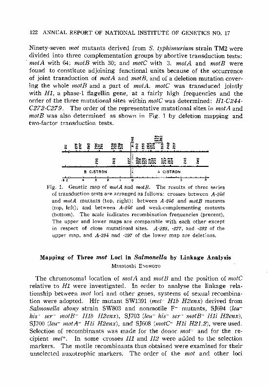
122 ANNUAL REPORT OF NATIONAL INSTITUTE OF GENETICS NO. 17
Ninety-seven mot mutants derived from S. typhimurium strain TM2 weredivided into three complementation groups by abortive transduction tests:motA with 64; motB with 30; and motC with 3. motA and motB werefound to constitute adjoining functional units because of the occurrenceof joint transduction of motA and motB, and of a deletion mutation covering the whole motB and a part of motA. motC was transduced jointlywith HI, a phase-l flagellin gene, at a fairly high frequencies and theorder of the three mutational sites within motC was determined: HI-C244C272-C279. The order of the representative mutational sites in motA andmotB was also determined as shown in Fig. 1 by deletion mapping andtwo-factor transduction tests.
......,,.,.. ::
B CISTRON A CISTRON
Fig. 1. Genetic map of motA and motB. The results of three seriesof transduction tests are arranged as follows: crosses between A -246and motA mutants (top, right); between A-246 and motB mutants(top, left); and between A-246 and weak-complementing mutants(bottom). The scale iildicates recombination frequencies (precent).The upper and lower maps are comparable with each other exceptin respect of close mutational sites. A-238, -277, and -282 of theupper map, and A-294 and -297 of the lower map are deletions.
Mapping of Three mot Loci in Salmonella by Linkage AnalysisMasatoshi ENOMOTO
The chromosomal location of motA and motB and the position of motCrelative to HI were investigated. In order to analyse the linkage relationship between mot loci and other genes, systems of sexual recombination were adopted. Rfr mutant SWl39l (met- Hib H2enx) derived fromSalmonella abony strain SW803 and nonmotile F- mutants, SJ694 (leuhis- ser- motB- Hib H2enx), SJ703 (leu- his- ser- motB- Hli H2enx) ,SHOO (leu- motA- Hli H2enx) , and SJ608 (motC- Hli H21.2) , were used.Selection of recombinants was made for the donor mot+ and for the recipient met+. In some crosses HI and H2 were added to the selectionmarkers. The motile recombinants thus obtained were examined for theirunselected auxotrophic markers. The order of the mot and other loci

RESEARCHES CARRIED OUT IN 1966 123
involved in this work was inferred to be met-ser-H2-his-motC-HI-motBmotA-leu.
Difference in Frequencies of Cotransduction of motewith HI Gene in Salmonella
Masatoshi ENOMOTO and Shigeru YAMAGUCHI
In P22 mediated transduction from a motile wild-type strain to nonmotilemutants of Salmonella, motC (motility) and some fla (flagellation) genes aretransduced jointly with HI (phase-1 flagellin) gene. When motile donorstrains having HI which was introduced from a certain Salmonella speciesare used in transduction to a nonmotile mutant serotypically different fromthe donor, frequencies of cotransduction (percent fraction of motile transductant with donor HI allele) are different from each other among thecombinations, though the donors are phenotypically indistinguishable. Inorder to elucidate this phenomenon, the following experiment was carriedout. Salmonella dublin strain SJll (HIgp motC+ flaK+), mutants motC-244(HIi) and flaK-48 (latent HIi) derived from S. tYPhimurium strain TM2were used. The order of the genes in this chromosomal region is flaKHI-motC. By means of transduction from SJll to motC-244 and flaK-48,motile transductants motC+ HIgp and flaK+ HIgp were isolated. Theformer transductants received at least the chromosomal region between HIand motC-244 from SJ11 and the latter received the region between flaKand HI. Using these transductants as donors and motC-244 as the recipient,transduction tests were carried out and the frequencies of cotransductionwere examined. With donors obtained from motC-244 the frequencies ofcotransduction were 32 to 44 per cent and with those obtained from flaK48 they were 4.5 to 16 per cent, whereas the frequency with SJll was19 per cent. In the cotransduction of motC with HI, a crossover betweenflaK and HI is required as well as a crossover at the right region ofmotC (This Ann. Rept. 16: 91). It is inferred that the decreased frequencyof cotransduction with the donors obtained from flaK-48 is the result ofreduced crossover between flaK and HIgp, due to the heterogeneity ofthe chromosomal region between S. tYPhimurium and S. dublin, and thatthe increased frequency with the donors obtained from motC-244 is theresult of relative increase in the crossover between flaK and HIgp, dueto the reduced crossover between HI and motC.

124 ANNUAL REPORT OF NATIONAL INSTITUTE OF GENETICS NO. 17
Infection of Bacteriophage-Chi to Serratia marcescensTetsuo lING
It was found that bacteriophage-chi, which has been known to attackmotile cells of both Salmonella and Escherichia species, is also able toinfect motile strains of Serratia marcescens. The Serratia strains used wereB-181-2 through B-181-10, B-182-1 through B-182-9, B-183-2 and B-183-3.One of these 20 strains, B-181-6, was non-flagellate and consequently nonmotile. The remaining 19 strains were motile having peritrichous flagellawith normal curvature.
Sensitivities of those Serratia strains to chi and its host range mutantM8, which is able to attack Salmonella possessing the g-complex antigen,were examined by the spotting method of E. W. Meynell. The bacteriophageused was propagated on Salmonella abortus-equi strain SJ241 which issensitive to both chi and M8. The non-motile strain, B-181-6, was completely resistant to both chi and M8, whereas the remaining 19 motilestrains were lysed by both chi and M8. The plaques formed with thelatter strains were clear and indistinguishable in size and shape fromthose formed with Salmonella strain SJ241.
A spontaneous non-flagellate mutant was isolated from motile strain B181-8 by screening on semi-solid nutrient agar medium. The mutant wasfound to be resistant to both chi and M8. Chi-phage can be used as aselective agent for the isolation of non-motile mutants from actively motilestrains of Serratia by the same procedure which has been applied toSalmonella and Escherichia coli.
Chi-phages were propagated in S. marcescens B-181-7, S. abortus-equiSJ241 and E. coli W3110, and the stocks were designated chi (Ser), chi (Sal)and chi (Kc) respectively. The combinations of chi (Sal) and Serratia, andchi (Ser) and Salmonella, gave efficiencies of plating (EOP) not significantlydifferent from those of the homospecific combinations, Le. the combinationsin which the indicator strain used was the same as the propagating one.However, when chi (Sal) or chi (Ser) were tested with Escherichia, theEOP decreased markedly. The infection by chi (E.c) of either Salmonellaor Serratia also resulted in decreased EOP as compared with the homospecific combinations. When chi (E.c) was grown on S. abortus-equi SJ241or S. marcescens B-181-7, the progeny phages then exhibited high EOP :onthese bacteria while the EOP on E. coli W3110 decreased markedly. Thissuggests the presence of host induced modification in the system.
Anti-chi serum, prepared by immunization of a rabbit with a phagestock of chi (Sal), was absorbed with the propagating strain, diluted 1000fold and neutralization :velocity constants with chi (Sal), chi (Ser) and
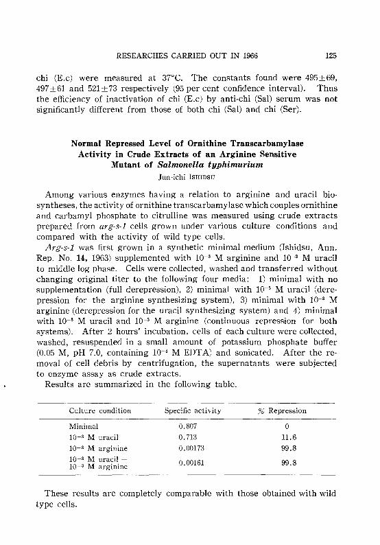
RESEARCHES CARRIED OUT IN 1966 125
chi (E.c) were measured at 37°C. The constants found were 495±69,497±61 and 521±73 respectively (95 per cent confidence interval). Thusthe efficiency of inactivation of chi (E.c) by anti-chi (Sal) serum was notsignificantly different from those of both chi (Sal) and chi (Ser).
Normal Repressed Level of Ornithine TranscarbamylaseActivity in Crude Extracts of an Arginine Sensitive
Mutant of Salmonella typhimuriumJun-ichi ISHIDSU
Among various enzymes having a relation to argmme and uracil biosyntheses, the activity of ornithine transcarbamylase which couples ornithineand carbamyl phosphate to citrulline was measured using crude extractsprepared from arg-s-l cells grown under various culture conditions andcompared with the activity of wild type cells.
Arg-s-l was first grown in a synthetic minimal medium (Ishidsu, Ann.Rep. No. 14, 1963) supplemented with 10-3 M arginine and 10-3 M uracilto middle log phase. Cells were collected, washed and transferred withoutchanging original titer to the following four media: 1) minimal with nosupplementation (full derepression), 2) minimal with 10-3 M uracil (derepression for the arginine synthesizing system), 3) minimal with 10-3 Marginine (derepression for the uracil synthesizing system) and 4) minimalwith 10-3 M uracil and 10-3 M arginine (continuous repression for bothsystems). After 2 hours' incubation, cells of each culture were collected,washed, resuspended in a small amount of potassium phosphate buffer(0.05 M, pH 7.0, containing 10-4 M EDTA) and sonicated. After the removal of cell debris by centrifugation, the supernatants were subjectedto enzyme assay as crude extracts.
Results are summarized in the following table.
Culture condition
Minimal
10-3 M uracil
10-3 M arginine
10-3 M uracil +10-3 M arginine
Specific activity
0.807
0.713
0.00173
0.00161
% Repression
o11.6
99.8
99.8
These results are completely comparable with those obtained with wildtype cells.
126 ANNUAL REPORT OF NATIONAL INSTITUTE OF GENETICS NO. 17
x. HUMAN GENETICS
Evaluation of the Family Planning Programme in JapanEi MATSUNAGA
An official programme on family planning in Japan was launched in1952, in order to replace the increasing practice of artificial abortion bycontraceptive methods. Placing the emphasis on the guidance of low incomefamilies, local health centers have been playing a leading and supervisingrole in the programme, with the aid of some ten thousand public healthnurses and private work midwives. Besides, a number of voluntary organi·zations have assisted the government's programme, and mass communication by women's magazines has contributed greatly towards thepopularization of the knowledge about contraception. As a result, presentusers of some kind of contraceptive measures have been rapidly increasingamong every social stratum during a short period.
On the other hand, the decline in births has still been accompanied bya tremendous number of notified abortions. Taking into account the likewisetremendous number of unnotified ones-presumably some 60 to 100 per centof the notified cases-, a question has occasionally been raised as to theeffectiveness of the family planning programme as described above.However, when measured per 1,000 women aged 15-49, the number ofnotified abortions has steadily been decreasing from the maximum of 50 in1955 to 30 in 1965, while the number of births dropped to 65 in 1957-justhalf the number of 1948-and remained thereafter almost constant, implyingthis being the number of "wanted births" of the present Japanese.
Under these demographic trends, a method was devised to evaluate therespective roles in the lowering of birth rate of 1) induced abortions, 2)contraceptive practice, 3) legalized sterilization, and 4) postponement ofmarriage. The result showed that, while the family planning programmehad scarcelly been successful during the first few years, the abortions havebeen, even allowing for as many unnotified as for the notified ones, increasingly replaced by contraception since 1957. It should be noted, however,that even in 1965 about two thirds of the reduction in births was presumablybased on induced abortions.
Details of this work will be published elsewhere.

RESEARCHES CARRIED OUT IN 1966
Association of Ear-Wax Types with Susceptibility toArteriosclerosis-A Preliminary Report!)
Mitsuo MIYAHARA' and Ei MATSUNAGA
127
It is now well known that the ear-wax types in man are determined bya simple genetic mechanism, the wet type being inherited dominantly overthe dry type. There is a marked variation in the incidence of the respectivetypes among various ethnic groups; more than 80 per cent of Japanesebelong to dry type, whereas this type of cerumen is exceptional amongboth Caucasians and Negroes. Since there is an apparent associationbetween wet cerumen and development of apocrine sweat glands or axillaryodour, it may be assumed that the effect of the allele pair responsible forear-wax types is concerned with certain metabolic patterns presumably oflipid material. It has been hoped that investigations will be carried outto seek for an association between ear-wax types and some internaldiseases, such as arteriosclerosis and other related diseases (Matsunaga,"11962).
Data on ear-wax types have been collected from in- and outpatients inthe Department of Internal Medicine of the Sapporo Medical CollegeHospital, Sapporo, during the last two years from January 1964 to December1966. The diagnosis of "arteriosclerosis" of our patients was made, inmost instances, if they were affected with coronary arterial sclerosis orischemic heart disease, and in the remaining cases, if they had a historyof either apoplectic stroke or encephalomalacia, or they showed calcification of the arc of the aorta as revealed by roentgenography. Theconditions were then divided into two groups, i.e., arteriosclerosis withand without hypertension. Patients with other diseases were also includedin the study. In the classification of the disease entries, those with uncertain diagnosis were all included in the "miscellaneous" group.
Of 2,448 patients so far examined, there were 39 or 1.6 per cent whoseear-wax type could not be determined with certainty. Although ourexperience suggests that ambiguous cases are most likely to be of wettype, they were excluded from the subsequent analysis. The incidenceof wet cerumen among the remaining 2,409 was 16.9 per cent, which wasnot significantly different from the 19.0 per cent (X'=2.92, d.f.=I, P>0.05)previously obtained by a survey (Matsunaga et al. 41 , 1954) of 1,639 pupils
II This work was supported by a grant from the Toyo Rayon Foundation for thePromotion of Science and Technology.
21 Department of Internal Medicine, Sapporo Medical College, Sapporo.31 Matsunaga, E., 1962. The dimorphism in human normal cerumen. Ann.
Human Genet. 25: 273-286.

128 ANNUAL REPORT OF NATIONAL INSTITUTE OF GENETICS NO. 17
of a middle school in Sapporo.The distribution of the patients differed between sexes according to the
disease, but for each disease entry there was no significant difference inthe distribution of ear-wax types according to sex, so that the data werepooled for male and female patients, and the results are summarized inTable 1.
Table I. Association of wet cerumen with a variety of diseases
Groups of patients No. Wet type Relativerisk for X2
affected with examined No. % wet type
I. Arteriosclerosis 96 29 30.2 1.85 7.05*2. Arteriosclerosis with 138 19 13.8 0.68 2.26hypertension
3. Essential hypertension 29J 50 17.2 0.89 0.494. Diabetes meIlitus 47 4 8.5 0.40 3.085. Endocrine disorders 80 16 20.0 1.07 0.056. Heart diseases 122 17 13.9 0.69 1.897. Kidney diseases 70 13 18.6 0.97 0.018. Liver diseases 90 15 16.7 0.85 1.899. Blood diseases 30 3 10.0 0.47 1.49
10. Gastrointestinal diseases 429 77 17.9 0.93 0.24II. Lung diseases 45 6 13.3 0.66 0.9012. Miscellaneous 972 158 16.3 0.83 3.08
Total 2,409 407 16.9Control 1,638 311 19.0
* Significant at the 1 per cent level.
From the table it can be seen that the incidence of wet cerumen amongthe patients with arteriosclerosis, not accompanied by hypertension, wasstrikingly high-as high as 30.2 per cent-, whereas it was only 13.8 percent among the arteriosclerotic patients with hypertension, the differencebeing significant at the 1 per cent level (x2 =9.39, d.f.=I, P<O.01). Inother groups of patients the incidences of wet cerumen were all below20 per cent. Using the figure of 19.0 per cent as control, the risks of thewet type relative to the dry in the susceptibility to various diseases havebeen calculated by means of Woolf's methodS I (1955). The item for which
41 Matsunaga, E., S. Itoh, T. Suzuki, and S. Sugimoto, 1954. Incidence andinheritance of the ear-wax types. Sapporo Med. J. 6: 1-4.
SI Woolf, B., 1955. On estimating the relation between blood group and disease.Ann. Human Genet. 19: 251-253.

RESEARCHES CARRIED OUT IN 1966 129
significance was shown was only arteriosclerosis without hypertension;for this condition the risk of individuals with wet cerumen was apparentlyincreased as much as 85 per cent as compared with individuals with drycerumen.
It has been widely recognized that there is a variation in the incidenceof arteriosclerosis among various ethnic groups; for example, coronaryarterial sclerosis is much more common in the United States than inJapan. Since the Japanese immigrants in the United States show higherincidence of coronary arterial sclerosis than the Japanese in Japan, environmental factors such as dietary habits seem to predominate in the causationof the disease, but genetic factors underlying racial differences cannot beexcluded. The present study indicates that, while there is no associationbetween ear-wax types and arteriosclerosis accompanied by hypertension,individuals with wet cerumen are more susceptible to arteriosclerosiswithout hypertension. This association may account, to some extent, forthe observed difference in the incidence of arteriosclerosis among variousethnic groups.
This work is still in progress and the data will be analyzed with referenceto cholesterol level of the blood in the patients.
Maternal Age of Mosaics with Down's SyndromeEi MATSUNAGA, Akira TONOMURAl) Hidetsune OISHI
and Yasumoto KIKUCHI
The overwhelming majority of cases with Down's syndrome is now knownto be primary G-trisomics, and the etiological importance of maternal ageis well established. For the cases with translocations, maternal age isgenerally accepted to be of no significance, while less attention has beenpaid to maternal age of the mosaics.
In order to test for a possible effect of maternal age upon mitoticnon-disjunction, data have been collected from the literature, includingour own cases, concerning maternal age of 31 cases of mosaicism withvarying degree of clinical signs of Down's syndrome. When they weredivided into two classes, class A with predominantly normal cells andclass B with predominantly G-trisomic or tetrasomic cells as revealed byeither blood or skin culture, the mean maternal age was 35.1±1.6 forclass B mosaics, while it was 29.4±2.0 for class A mosaics, the differencebeing significant at the 5 per cent level (t=2.24, d.f.=29, P<O.05). Sinceclass A mosaics are most likely to have developed from fertilized eggswith normal karyotype, it appears that mitotic non-disjunction may not
1) Department of Cytogenetics, Tokyo Medical and Dental University.

130 ANNUAL REPORT OF NATIONAL INSTITUTE OF GENETICS NO. 17
be affected by maternal age. However, more data are needed to draw adefinite conclusion.
Chromosome Replication in Down's SyndromeYasumoto KIKUCHI and Hidetsune OISHI
Autoradiographic techniques have been used mainly for the study ofDNA replication pattern of human chromosomes. This technique maybe utilized also for chromosome identification. We report here a preliminary study of autoradiographic analysis of the G group chromosomesin patients with Down's syndrome and in normal subjects.
Four female patients with the standard type of G-trisomy, two normalfemales, and two normal males were studied. Leucocytes obtained fromvenous blood of each subject were cultured according to standard technique.Chromosome replication at the end of the DNA synthesis period (S period)was analysed by means of autoradiography, tritiated thymidine and continuous labeling technique.
Metaphase plates selected for analysis were scored according to thenumber of small acrocentrics labeled per cell. The results are shown inTable 1. In normal subjects, cells with 4 labeled G chromosomes occurred
Table 1. Number of cells showing the distribution of labelingamong acrocentrics of the G group
No. of G chromosomes labeled Total No. ofSubjects cells examined0 1 2 3 4 5
Normal males 12 11 24 22 31 100
Normal females 5 14 27 18 37 100
Female patients with 10 8 18 32 19 18 100Down's syndrome
most frequently (31 per cent in males and 37 in females); cells with 2labeled G chromosomes were found less frequently, while the frequenciesof cells with 1 or 3 labeled G chromosomes were still lower. This indicatesthat most of the homologous pairs of G group replicate synchronously.In female patients with Down's syndrome, 32 out of 100 cells were observedto have 3 labeled and 2 unlabeled G chromosomes, while there were only18 cells with 2 labeled and 3 unlabeled G chromosomes. The results maysuggest that the sequence of replication of the extra chromosome is similarto the original homologous pair.
For detail analysis of grain distribution, 20 metaphase plates were

RESEARCHES CARRIED OUT IN 1966 131
observed from a normal female and the same number from a femalepatient with Down's syndrome. In 12 cells from the normal female, 2 Gchromosomes were generally labeled over the long arm (late replicatingpair), but chromosomes of another pair were completely unlabeled orlightly labeled only over the centromere region or the short arm (earlyreplicating pair). The remaining cells did not show a consistent pattern.In the patient with Down's syndrome, 12 cells had 3 late replicating Gchromosomes and 2 early replicating ones. On the other hand, 5 cells had2 late and 3 early replicating G chromosomes. In the remaining cells,different patterns were observed.
In the autoradiographs of chromosomes from each patient other thanthe G group, the characteristic pattern of late replication was observedin 1 X chromosome, in 2 of the D group (Nos. 13-15) and in Nos. 16 and18.
The above results suggest that the extra chromosome associated withDown's syndrome belongs to the late replicating pair of G group, andthat its presence does not affect DNA synthesis of chromosomes other thanG group.
Phenotypes and Sex Chromosomes in Five Patientswith Turner's Syndrome
Hidetsune OISHI, Yasumoto KIKUCHI and Ei MATSUDA
It is now widely accepted that monosomy of X or! X deletion is responsible for clinical conditions called Turner's syndrome. However,individuals with this chromosome aberration do not always show all ofthe classical signs of Turner's syndrome (shortness of stature with sexualinfantilism, cubitus valgus, and webbing of the neck). During our currentsurvey, in collaboration with some University Hospitals in Tokyo, ofhuman chromosomes in various congenital disorders and sex anomalies,we examined the chromosomes in four cases of XO patients and in onewith XO/XXX mosaicism. The results of clinical and laboratory examinations are summarized in Table 1. In all cases, short stature, shieldchest, cubitus valgus without webbing of the neck, lacking or scanty pubichair, and a small, scarcely palpable uterus were noticed.
Analyses of dermal patterns of fingers and hands were performed onthree patients, and the formulas are given in Table 2. Since the totalridge counts of the finger tips in Japanese females are on the average137.70±41.09 (Matsunaga), our patients seem to have higher ridge counts.The characteristic t"s were observed in the left palms of cases Nos. 4and 5, while the axial triradius was absent in both hands of case No.3.

132 ANNUAL REPORT OF NATIONAL INSTITUTE OF GENETICS NO. 17
Table 1. Some clinical data in five patients under study
Case No. 1 2 3 4 5
Sex chromosome constitution XO XO XO XO XO/XXXDrumstick +(14/500)*Maternal age at the birth of 37 27 30 24 24the patientPaternal age at the birth of 45 35 35 28 28the patient
Birth weight (kg) 3.10 3.18 2.15 2.45Age examined (years) 17 16 15 10 10Height (cm) 143 134 130 106 121Weight (kg) 41.5 34.7 40.0 23.5 25.5Arm span (cm) 141 123.7 131.8 94.7 120IQ (or intelligence) 99 Normal Normal 49 123Short neck + + + +Webbed neck
Shield chest + + + + +Cubitus valgus + + + + +Menstruation
Development of the breasts +Pubic and axillary hairs Scanty
Small uterus + + + + +Gonadotropin (unit/day) >32 >32 <32 <4 <417·KS (mg/day) 4.37 1.6 0.9 0.61 1.0
* Including two cells with two drumsticks.
Table 2. Dermatoglyphics of the patients
Case No. Finger patterntypes (I-V)*
Totalridgecount
Palmar pattern formulae** atd angle
3{Right U. U. U. U. Lp l1.X.7.1- 0- Ar.O.O.l.O200
Left W. U. U. U. W 7.7.5".1-0- Ar.O.O.O.L/L
4{Right U. At. U. U. U l1.X.7.3- t-Au.O.O.V.O 42°151 g" 78°Left U. R. Lp. Lp. U 9.7.5".3-tt" - LUV.V/V.O.O.V 43°
5{Right U. Ucp. Ucp. W. W 11.9.8.5' -t-V.V/V.O.L.O 45°150 g" 75°Left U. W. W. W. W 9.9.5".1-tt" - Ws.V/V.O.L.V 45°
* At; tented arch, W; whorl, R; radial loop, Ucp; ulnar central pocket loop,U; ulnar loop, Lp; lateral pocket loop.
** Cummins-Midlo's formula.

RESEARCHES CARRIED OUT IN 1966 133
Thenar patterns as seen occasionally in patients with this syndrome werenoticed in cases Nos. 4 and 5. In addition, all three patients had palmartransverse creases.
Further studies are required for the discrimination of some metricalcharacters genetically controlled by sex chromosomes.
Clinical Conditions of Patients with Apparently NormalChromosomes, III
Hidetsune OISHI and Yasumoto KIKUCHI
Based on leucocyte cultures of peripheral blood, patients with variouspathological conditions were found to have 46 chromDsomes of apparentlynormal karyotype. Cases of some interest are listed below.
Name Age Legal sex Clinical conditions
M
M
FF
M
FM
2 years
2 years
2 years
5 months F
5 months M
4 years3 years
1 year1 year
54 S. T.
56 H. T.
55 Y.S.
48 M.O.49 Y.M.
52 A.O.53 Y.M.
51 T.O.
50 S.K.
a) Multiple deformations with mental retardation
47 N.1. 3 years F Hypertelorism; antimongoloid slant eyes;low-set ears; webbing of the neckMicrocephaly; exophthalmos; low-set earsMicrocephaly; hypertelorism; epicanthus;micrognathia (" Cri du chat" syndrome?)Exophthalmos; large deformed lobes of theear; bifid tongue; high-arched palateOblique palpebral fissures; low-set ears;polydactylyCongenital glaucoma; aphasiaOsteogenesis imperfecta; hydrocephalus;small deformed lobes of the ear; congenitalheart diseaseHypertelorism; antimongoloid slant eyes;hydrocephalusHypertelorism; syndactyly; antimongoloidslant eyes; epicanthusHypertelorism; antimongoloid slant eyes
b) Sex anomalies
57 H. S. 36 years
58 E. M. 5 years
M
F
Slender body; funnel-shaped chest; smallpenis; cataract; low excretion of 17KS;high excretion of gonadotropin (Klinefelter's syndrome?)Female pseudohermaphroditism
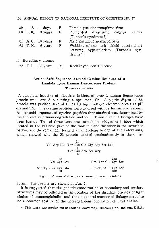
134 ANNUAL REPORT OF NATIONAL INSTITUTE OF GENETICS NO. 17
59 -.S. 11 days F Female pseudohermaphroditism60 K.K. 9 years F Primordial dwarfism; cubitus
(Turner's syndrome?)61 A.G. 16 years F Male pseudohermaphroditism62 Y.K. 6 years F Webbing of the neck; shield chest;
stature; hypertelorism (Turner'sdrome?)
c) Hereditary disease
63 T. I. 23 years M Recklinghausen's disease
valgus
shortsyn-
Amino Acid Sequence Around Cystine Residues of aLambda Type Human Bence·Jones Protein ll
Tomotaka SHINODA
A complete location of disulfide bridges of type L human Bence-Jonesprotein was carried out using a specimen, Sh. A peptic digest of Shprotein was purified several times by high voltage electrophoresis at pH6.5 and 3.5. The cystine peptides were oxidized with performic acid vapour.Amino acid sequence of cystine peptides thus obtained was determined bythe subtractive Edman degradation method. Three disulfide bridges havebeen found; Two of these were the intrachain bridges-a bridge whichlocated in the variable part of the molecule and the other in the invariantpart-, and the remainder formed an interchain bridge at the C-terminal,which showed why the Sh protein existed predominantly in the dimer
21Val-Arg-ILu-Thr-Cys-Gln-Gly-Asp-Ser-Leu
ITyr-Cys-Asn-Ser-Arg
86
135Val-Cys-Leu
ISer-Tyr-Ser-Cys-Gln
194
212Pro-Thr-Glu-Cys-Ser
IPro-Thr-Glu-Cys-Ser
212
Fig. 1. Amino acid sequence around cystine residues.
form. The results are shown in Fig. 1.It is suggested that the genetic conservation of secondary and tertiary
structures may be reflected in the location of the disulfide bridges of lightchains of immunoglobulin, and that a general manner of linkage may alsobe a common feature of the heterogeneous population of light chains.
1) This work was carried out at Indiana University, Bloomington, Indiana, U.S.A.

RESEARCHES CARRIED OUT IN 1966 135
Cellulose Acetate Electrophoresis and az-Lipoprotein of Human SerumYoshito OGAWA
When electrophoretic analysis of human serum is carried out usingcellulose acetate strips as a supporting medium, a fine new band oftenappears in the part of IXz-globulin pattern (Fig. 1, arrow). The nature of
+
Fig. 1. Separation pattern of human serum on cellulose acetate strip(Separax).
this new fraction was not yet studied. The present writer has thoroughlyexamined this fraction and succeeded to confirm its identity with IXz'lipoprotein fraction because of its affinity to Sudan Black B (Swahn'smethod) and the results of immunochemical examination, immunoelectrophoresis and absorption test, applying anti IXz-lipoprotein rabbit serum(Behring-Werke).
Details of this works had been published in Physico-Chemical BiologyVol. 12.

136 ANNUAL REPORT OF NATIONAL INSTITUTE OF GENETICS NO. 17
BOOKS AND PAPERS PUBLlS~ED IN 1966BY STAFF MEMBERS
ASAKURA, S., G. EGUCHI and T. IINO, 1966. Salmonella flagella: in vitro reconstitution and over-all shapes of flagellar filaments. J. Mol. BioI. 16: 302-316.
CAVALLI-SFORZA, L. L., M. KIMURA and I. BARRAI, 1966. The probability of consanguineous marriages. Genetics 54: 37-60.
CHUNG, C. S., N. E. MORTON and N. YASUDA, 1966. Genetics of interracial crosses.Ann. N. Y. Acad. Sci. 134: 666-687.
ENDO, T., 1966. Cytolasm and conformation of proteins (in Japanese). Nucleus andCytoplasm 9: 6-10.
ENDO, T., 1966. Application of zymography to plant sciences (in Japanese). SABCOJ. 2: 120-126.
ENDO, T. and D. SCHWARTZ, 1966. Tissue specific variations in the urea sensitivity of the E , esterase in maize. Genetics 54: 233-239.
ENDO, T. and D. SCHWARTZ, 1966. Regulation of cathecol oxidase. Maize GeneticsCoop. News Letter 40: 165-166.
ENOMOTO, M., 1966. Genetic studies of paralyzed mutants in Salmonella. I. Genetic finestructure of the mot loci in Salmonella typhimurium. Genetics 54: 715-726.
ENOMOTO, M., 1966. Genetic studies of paralyzed mutants in Salmonella. II. Mapping of three mot loci by linkage analysis. Genetics 54: 1069-1076.
ENOMOTO, M. and T. IINO, 1966. The comparison of normal and curly flagella inSalmonella abortus-equi by two-dimensional separation of peptides. Japan. J.Genetics 41: 131-139.
FUJII, T., M. IKENAGA and J. T. LYMAN, 1966. Radiation effects on Arabidopsisthaliana. II. Killing and mutagenic efficiencies of heavy ionizing particles.Radiation Botany 6: 297-306.
FUJII, T. and S. MATSUMURA, 1966. Comparison of mutagenic efficiency betweenethyl methanesulfonate and ionizing radiations in dipliod wheat (Preliminaryreport). Seiken Zih6 18: 23-31.
GALSSMAN, E., T. SHINODA, E. J. DUKE and J. F. COLLINS, 1966. Interconversion ofmolecular forms of xanthine dehydrogenase from Drosophila. Ann. N. Y. Acad.Sci. 130: (in press).
GALSSMAN, E., T. SHINODA, H. M. MOON and J. D. KALAM, 1966. In vitro complementation between non-allelic Drosophila mutants deficient in xanthine dehydrogenase. IV. Molecular weight. J. Mol. BioI. 20: 419-422.
IINO, T., 1966. Bacterial flagella (in Japanese). Protein Nucleic Acid Enzyme(Tokyo) 11: 41-49.
IINO, T., 1966. Morphogenesis of bacterial flagella (in Japanese). Kagaku ToSeibutsu 4: 345-350.
IINO, T. and M. ENOMOTO, 1966. Genetical :studies of non-flagellate mutants ofSalmonella. J. Gen. Microbiol. 43: 315-327.
IINO, T. and M. MITANI, 1966. Flagella-shape mutants in Salmonella. J. Gen.Microbiol. 44: 27-40.

BOOKS AND PAPERS PUBLISHED IN 1966 BY STAFF MEMBERS 137
IINO, T., M. MITANI and S. ASAKURA, 1966. Formation of bacterial flagella (inJapanese). Inst. Appl. Microbiol. Tokyo Univ. Symp. 7: 113-128.
IKENAGA, M. and T. MABUCHI, 1966. Photoreactivation of endosperm mutationsinduced by ultraviolet light in maize. Radiation Botany 6: 165-169.
KATAYAMA, T. C., 1966. Anatomy of the root of Oryza sativa with emphasis ondevelopmental processes. Seiken Ziho 18: 6-22.
KIHARA, H., 1966. Factors affecting the evolution of common wheat. Indian J.Genetics Plant Breeding 26A (Special Symp. No.): 14-28.
KIHARA, H. and T. HORI, 1966. The behavior of nuclei in germinating pollen grainsof wheat, rice and maize. Der Ztichter 36: 145-150.
KIHARA, H. and K. TSUNEWAKI, 1966. Basic studies on hybrid wheat breeding,carried out at the National Institute of Genetics. Seiken Ziho 18: 55-63.
KIKUCHI, Y., 1966. The pattern of chromosome replication in man (in Japanese).Kagaku 36: 65-69.
KIKUCHI, Y., 1966. Analysis of chromosomes in newborn infants (in Japanese). Clin.Gynecol. Obstet. 20: 887-892.
KIMURA, M., 1965. Evolutionary considerations on spontaneous mutation rates(Preliminary report). Gamma Field Symp. 4: 85-96.
KIMURA, M. and T. MARUYAMA, 1966. The mutational load with epistatic geneinteractions in fitness. Genetics 54: 1337-1351.
KIRITANI, K., S. NARISE and R. P. WAGNER, 1966. The reductoisomerase of Neurospora crassa. J. BioI. Chern. 240: 2047-2051.
KIRITANI, K., S. NARISE and R. P. WAGNER, 1966. The dihydroxy acid dehydrataseof Neurospora crassa. J. BioI. Chern. 241: 2042-2046.
KURODA, Y., 1966. Sorting-out mechanisms of animal tissues and cells (in Japanese).Inst. Appl. Microbiol. Tokyo Univ. Symp. 7: 42-55.
KURODA, Y., 1966. Tissue organization from single cells (in Japanese). Heredity(Tokyo) 20: 8-12.
KURODA, Y., 1966. Differentiation and tissue specificity of aggregation-promotingmaterials from chick embryos (in Japanese). Japan. J. Exptl. Morphol. 20: 93.
KURODA, Y., 1966. Sorting-out phenomena of animal tissues and cells (in Japanese).Kagaku To Seibutsu 4: 394-400.
MABUCHI, T. and S. MATSUMURA, 1966. Oxygen and storage effects on radiationdamage in einkorn wheat seed. W. I. S. 21: 10.
MABUCHI, T. and S. MATSUMURA, 1966. Radiosensitivity in pollen grains of Triticumand Aegilops. W. I. S. 22: 20.
MATANO, Y. and T. H. YOSIDA, 1966. Alteration of stemline chromosomes in hypotetraploid Ehrlich carcinoma. Proc. Japan Acad. 42: 404-407.
MATSUMURA, S., 1966. Differences in effects of r-rays and fast neutrons on wheat.W. I. S. 21: 3-6.
MATSUMURA, S., 1966. Radiation genetics in wheat. IX. Differences in effects ofgamma-rays and 14 MeV, fission and fast neutrons from Po-Be. RadiationBotany 6: 275-283.
MATSUMURA, S. and T. FUJII, 1966. Radiosensitivity of wheat seeds. W. I. S.22: 21.
MATSUMURA, S. and T. MABUCHI, 1966. Photoreactivation of an ultraviolet light

138 ANNUAL REPORT OF NATIONAL INSTITUTE OF GENETICS NO. 17
induced mutation in maize pollen. Seiken Ziho 18: 1-5.MATSUNAGA, E., 1966. Genetic epidemiology of sporadic retinoblastoma (in Japanese).
Igaku No Ayumi 59: 208-212.MATSUNAGA, E., 1966. Causes of Down's syndrome (in Japanese). Heredity (Tokyo)
20(11): 33.MATSUNAGA, E., 1966. Possible genetic consequences of family planning. J. Amer.
Med. Assoc. 198: 533-540.MATSUNAGA, E., 1966. Down's syndrome and maternal inbreeding. Acta Genet.
Med. Gemell. 15: 224-230.MATSUNAGA, E., 1966. Some remarks on the biology of twins. Japan. J. Human
Genet. 11: 227-228.MORTON, N. E., M. P. MI and N. YASUDA, 1966. Bivalent alleles. Amer. J. Human
Genet. 18: 233-242.MORTON, N. E., M. P. MI and N. YASUDA, 1966. A special theory of hemagglutina
tion. Vox. Sang. 11: 12-20.MORTON, N. E., M. P. MI and N. YASUDA, 1966. A study of the SIt alleles in nor
theastern Brazil. Vox. Sang. 11: 194-208.MUKAI, T., 1966. Further studies on the optimum heterozygosity hypothesis in
Drosophila melanogaster. Genetics 54: 350-351.MUKAI, T., I. YOSHIKAWA and K. SANO, 1966. The genetic structure of natural
populations of Drosophila melanogaster. IV. Heterozygous effects of radiationinduced mutations on viability in various genetic backgrounds. Genetics 53:513-527.
MURAKAMI, A., 1966. Relative biological effectiveness of 14 MeV neutrons to gamma-rays for inducing mutations in mature sperm of the silkworm. Japan. J.Genetics 41(1): 17-26.
MURAKAMI, A., 1966. Relationship between radiosensitivity and mitotic cycle during early developmental stages of silkworm eggs (in Japanese). Radiation BioI.Res. Comm. 1(1): 29-42.
NARISE, T., 1965. The effect of relative frequency of species in competition.Evolution 19: 350-354.
NARISE, T., 1966. The mode of migration of Drosophila ananassae under competitiveconditions. Univ. Texas Publ. 6615: 121-131.
NARISE, S. and J. L. HUBBY, 1966. Purification of esterase-5 from Drosophilapseudoobscura. Biochim. Biophys. Acta 122: 281-288.
OGAWA, Y., 1966. Cellulose acetate electrophoretic technique (in Japanese). Japan.J. Clin. Pathol. 11: 46-63.
OGAWA, Y.. 1966. Ponceau 3R staining of serum proteins on Separax (in Japanese).Physico-Chern. BioI. (Tokyo) 11: 224-225.
OGAWA, Y., 1966. Cellulose acetate electrophoresis of human serum proteins. I.(in Japanese). Japan. J. Med. Tech. 15: 147-153.
OGAWA, Y., 1966. Cellulose acetate electrophoresis of human serum proteins. II.(in Japanese). Japan. J. Med. Tech. 15: 197-204.
OGAWA, Y., 1966. Human serum protein electrophoresis on cellulose acetate strips(in Japanese). Pathol. Bacteriol. (Tokyo) 48: 5-12.
OGAWA, Y., 1966. The cellulose acetate electrophoretic technique. III. Ponceau 3R

BOOKS AND PAPERS PUBLISHED IN 1966 BY STAFF MEMBERS 139
staining of albumin in human serum on Separax (in Japanese). Med. BioI.(Tokyo) 73: 125-127.
OGAWA, Y., 1966. Growth, differentiation and regeneration. XLII. Effect of glucurono-Iactone on the development of skeletal myosin in X-irradiated Triturusembryos (in Japanese). Med. BioI. (Tokyo) 73: 130-133.
OGAWA, Y., 1966. The cellulose acetate electrophoretic technique. V. Ponceau 3Rstaining of p- and r-globulin in human serum on Separax (in Japanese). Med.BioI. (Tokyo) 74: 169-172.
OGAWA, Y., 1966. The cellulose acetate electrophoretic technique. VI. On theconcentration of trichloroacetic acid in staining and acetic acid in washingsolutions (in Japanese). Med. BioI. (Tokyo) 73: 198-202.
OGAWA, Y., 1966. The cellulose acetate electrophoretic technique. VII. Conditionsof current supply (in Japanese). Med. BioI. (Tokyo) 73: 228-231.
OGAWA, Y. and H. MIYAUCHI,1966. The cellulose acetate electrophoretic technique.IV. Ponceau 3R staining of a-globulin in human serum on Separax (in Japanese)Med. BioI. (Tokyo) 73: 134-136.
OGAWA, Y., M. ABE, M. KITAMURA, N. KOSAK AI, K. SHIMAO, H. TOMITA H. HIRAIand K. MOMMA, 1966. On the standard procedure for the analysis of serumprotein fractions by cellulose acetate electrophoresis (in Japanese). PhysicoChern. BioI. (Tokyo) 11: 351-356.
OGAWA, Y. and T. HASEGAWA, 1966. Normal value of serum protein fractions ofJapanese population on Separax, a new cellulose acetate membrane for electrophoretic analysis (in Japanese). Physico-Chern. BioI. (Tokyo) 11: 319.
OISHI, H., 1966. Human chromosomes and its analysis (in Japanese). Japan. J.Clin. Pathol. 14: 780-784.
OISHI, H., 1966. Human diploid cell strains (in Japanese). Heredity (Tokyo) 20(7):55-57.
OKA, H. I., 1966. Growing season of rice in the tropics (in Japanese). Japan. J.Trop. Agr. 9: 195-199.
OSHIMA, C., 1966. "Mechanisms of micro- and meso-evolution" in Modern Biology9: 185-215, "Ecology and Evolution" (in Japanese). Iwanami-Shoten, Tokyo.
OSHIMA, C., 1966. Persistence of some recessive lethal genes in natural populationsof Drosophila melanogaster. Proc. Symp. held in Prague, August 9-11, 1965,Mutation in Population: 41-48.
SAKAI, K. I. and S. IYAMA, 1966. Studies on competition in plants and animals.XI. Competitive ability and density response in barley. Japan. J. Breeding16: 1-9.
SAKAMOTO, S., 1966. Cytogenetic studies in the tribe Triticeae. IV. Natural hybridization among Japanese Agropyron species. Japan. J. Genetics 41: 189-201.
SAKAMOTO, S., 1966. Intergeneric hybrids between two Eremopyrum and Agropyronspecies. W. I. S. 21: 22.
SAKAMOTO, S., 1966. An intergeneric hybrid between Eremopyrum orientale (Linn.)Jaub. et Spach. and Aegilops squarrosa Linn. W. I. S. 22: 6.
SAKAMOTO, S. and M. MURAMATSU, 1966. Cytogenetic studies in the tribe Triticeae.II. Tetraploid and hexaploid hybrids of Agropyron. Japan. J. Genetics 41: 155168.

140 ANNUAL REPORT OF NATIONAL INSTITUTE OF GENETICS NO. 17
SAKAMOTO, S. and M. MURAMATSU, 1966. Cytogenetic studies in the tribe Triticeae.III. Pentaploid Agropyron hybrids and genomic relationships among Japaneseand Nepalese species. Japan. J. Genetics 41: 173-187.
SCHWARTZ, D. and T. ENDO, 1966. Alcohol dehydrogenase polymorphism in maize-Simple and compound loci. Genetics 53; 709-715.
SHINODA, T. and E. GLASSMAN, 1966. Multiple molecular forms of xanthine dehydrogenase in Drosophila (submitted for publication).
SUZUKI, H. and T. IINO, 1966. An assay for newly-synthesized intracellular flagel·lin. Biochim. Biophys. Acta 124: 212-215.
TANAKA, Y. and F. GOTO, 1965. Growth-accelerating action of K (Knobbed) genein the silkworm. III. Size and number of knobs with respect to the growthaccelerating action (in Japanese). Rept. Silk Sci. Res. Inst. 14: 12-16.
TANAKA, Y. and F. GOTO, 1965. Single gene action of some dominant charactersin the silkworm. I. (in Japanese). Rept. Silk Sci. Res. Inst. 14: 17-19.
TANAKA, Y. and F. GOTO, 1965. Experimental studies on the characteristics ofvarious genes in the silkworm. I. (in Japanese). Rept. Silk Sci. Res. Inst. 14:20-25.
TAZIMA, Y., 1966. How mutations occur? (in Japanese). Section II, Chapter 5 in"Gendai Seibutsugaku Taikei" (D. MORIWAKI and K. ONO, ed.) pp. 248-276.Nakayama-shoten, Tokyo.
T AZIMA, Y., 1966. Radiations and Radiosotopes in Sericulture (in Japanese). KagakuTo Gijitsu 5(1): 48-51, (2): 38-41, (3): 36-39, (4): 62-65, (5): 26-29, (6): 30-33,(7): 38-41, (8): 48-52, (9): 48-52, (10): 64-67, (11): 22-25, (12): 34-37, 6(1): 6265, (2): 34-37, (3): 52-55. Zenkoku Yosan Kyodokumiai Rengokai, Tokyo.
TAZIMA, Y., 1966. A report from the Third International Congress of RadiationResearch (in Japanese). Rad. BioI. Res. Comm. 1(1): 61-68.
TAZIMA, Y., 1966. A report from the Conference on Space Radiation Biology (inJapanese). Iden (Tokyo) 20(1): 45-46.
TONOMURA, A., H. OISHI, E. MATSUNAGA and T. KURITA, 1966. Down's syndrome:A cytogenetic and statistical survey of 127 Japanese patients. Japan. J. HumanGenet. 11: 1-16.
TSAI, K. H. and H. I. OKA, 1966. Genetic studies of yielding capacity and adaptability in crop plants. 2. Analysis of genes controlling heading time in Taichung 65 and other rice varieties. Bot. Bull. Acad. Sinica 7: 54-70.
TSUJITA, M. and S. SAKURAI, 1966. Structure of chromogranules in the hypodermalcells of silkworm larvae. Proc. Japan Acad. 42: 950-955.
TSUJITA, M. and S. SAKURAI, 1966. Chemical composition of chromogranules produced in the hypodermal cells of silkworm larvae. Proc. Japan Acad. 42:956-959.
TSUJITA, M. and S. SAKURAI, 1966. Development of chromogranules in the larvalskin of the silkworm. Proc. Japan Acad. 42: 960-965.
YONEDA, Y. and T. STONIER, 1966. Elongation of stem internodes in the Japanesemorning glory Pharbitis nil in relation to auxin destruction. Physiol. Plant.19: 977-981.
YOSIDA, T. H., 1966. Chromosomal alteration and the development of tumors.XIV. Comparative idiogram analysis of several sublines of mouse lymphocytic
BOOKS AND PAPERS PUBLISHED IN 1936 BY STAFF MEMBERS 141
neoplasm, P388, growing in vivo and in vitro. Japan. J. Genetics 41: 43-58.YOSIDA, T. H., 1956. Chromosomal alteration and the development of tumors. XV.
Change of chromosome pattern in 8-azaguanine and amethopterin-resistantsublines of mouse lymphocytic neoplasm, P388, cultured in vitro. Japan. J.Genetics 41: 59-74.
YOSIDA, T. H., 1966. Relation between chromosomal alteration and developmentof tumors (Prize Lecture of the Genetics Society of Japan) (in Japanese).Japan. J. Genetics 41: 439-451.
YOSIDA, T. H., 1956. Method for chromosome analysis in Muridae, with specialregards to the chromosomal polymorphism (in Japanese). SABCO J. 2: 1-7.
YOSIDA, T. H., 1966. Part of chromosomes to induction of tumor and its growth(in Japanese). Igaku No Ayumi 59: 205-207.
YOSIDA, T. H., 1966. Karyology of malignant growths (in Japanese). Japan. J.Cancer Clin. 12: 431-437.
YOSIDA, T. H., H. IMAI and H. SATO, 1966. Chromosomal alteration and the development of tumors. XII. Differentiation of karyotypes in mouse asciteshepatoma MH-134 and MH-129P maintained in different locations. Gann 57:9-17.
YOSIDA, T. H., Y. KURITA and K. MORIWAKI, 1966. Chromosomal alteration andthe development of tumors. XIII. Karyotype difference in solid and ascites typetumors of mouse plasma cell neoplasm X5563, with special regard to the serumelectrophoretic pattern of gamma globulin. Japan. J. Genetics 41: 9-19.
WATANABE, T. K. and C. OSHIMA, 1966. Distribution of natural lethal genes onthe second chromosomes of Drosophila melanogaster. Japan. J. Genetics 41;367-378.
142 ANNUAL REPORT OF NATIONAL INSTITUTE OF GENETICS NO. 17
ABSTRACTS OF DIARY FOR 1966
January 21January 25January 26February 18March 1March 24April 8April 20April 22May 20June 4June 24July 18July 29September 12
September 30October 27, 28November 2November 4, 5
November 10November 11
November 25December 16
141st Meeting of Misima Geneticist' Club69th Biological Symposium70th Biological Symposium142nd Meeting of Misima Geneticist' Club71st Biological Symposium143rd Meeting of Misima Geneticist' Club16th Meeting of the Society of Electrophoresi::;72nd Biological Symposium144th Meeting of Misima Geneticist' Club145th Meeting of Misima Geneticist' Club26th Meeting of the Board of Councillors146th Meeting of Misima Geneticist' Club73rd Biological Symposium147th Meeting of Misima Geneticist' Club4th Meeting of the Organizing Committee of the 12th
International Congress of Genetics148th Meeting of Misima Geneticist' ClubMeeting of Directors of the National Institutes74th Biological Symposium18th Meeting of the Tokai Branch of the Sericultural
Society of Japan75th Biological SymposiumPublic Lectures on Genetics (at the Daiichi-Seimei Hall,
Tokyo)149th Meeting of Misima Geneticist' Club150th Meeting of Misima Geneticist' Club

FOREIGN VISITORS IN 1966
FOREIGN VISITORS IN 1966
143
Jan. 2
Jan. 24
Feb. 28
Apr. 2
Apr. 16-21
Apr. 19
May 4
Jun. 30
JuI. 18
JuI. 19
JuI. 29
Aug. 9
Aug. 15
Aug. 25
Aug. 28
STROMBERGER, H. G., Pasadena High School and JuniorCollege, Pasadena, California, U.S.A.
TOKIN, B. P., Department of Zoology, University of Leningrad, Leningrad, U.S.S.R.
NEWCOMBE, H. B., Biology Branch, Atomic Energy of CanadaLimited, Chalk River, Ontario, Canada
KANG, Y. H., Department of Botany, Yonsei University,Seoul, Korea
SCHMIDT, J, W., Department of Agronomy, University ofNebraska, Lincoln, Nebraska, U.S.A.
WAYLAND, H., Department of Engineering Science, California Institute of Technology, Pasadena, California, U.S.A.
JORDAN, H. 0., Economics Development Institute, Universityof Nigeria, Enugu, East Nigeria
TAKAHASHI, T., Department of Geology, University ofRochester, Rochester, New York, U.S.A.
DEGENHARDT, K. H., Institut fur Humangenetik und Vergleichende Erbpathologie der UniversWit Frankfurt amMain, Germany
DAGG, C. P., The Jackson Laboratory, Bar Harbor, Maine,U.S.A.
GAFFNEY, L., c/o A. A. Tegel Pty. Ltd., Leppington, NewSouth Wales, Australia
CARTER,O., Department of Agronomy, University of Sydney,New South Wales, Australia
CHO, W. K., Department of Zoology, Seoul National University, Korea
CHAKRAVARTTI, M. R., Indian Statistical Institute, Calcutta,India
MAVALWALA,]., Department of Anthropology, Universityof California, U.S.A.
SWAMINATHAN, M. S., Division of Botany, Indian AgriculturalResearch Institute, New Delhi, India
KIM, Y. C., College of Agriculture, Korea University, Seoul,Korea
SHAHI, B. B., Seed Testing Laboratory, Department of Agriculture, H. M. G., Kathmandu, Nepal
YO, C. J., Department of Botany, College of Science,National Taiwan University, Taipei, Taiwan, China
144 ANNUAL REPORT OF NATIONAL INSTITUTE OF GENETICS NO. 17
Aug. 28 HOUSE, L. R., The Rockefeller Foundation, New Delhi, IndiaCHAKRABANDHU, M.C., Department of Agriculture, Bangkok,
ThailandCARANGAL, A. R. Jr., Department of Research and Develop
ment, San Miguel Corporation, Manila, PhilippinesKu, Y., College of Agriculture, National Taiwan Uni
versity, Taipei, Taiwan, ChinaCREECH, J. L., New Crops Research Branch, Beltsville,
Maryland, U.S.A.CALDECOTT, R. S., Department of Agronomy and Plant
Genetics, University of Minnesota, Minnesota, U.S.A.NATARAJAN, A. T., Institute of Radiobiology, Stockholm
University, Stockholm, SwedenSIGURBJORSSON, B., Plant Breeding and Genetics Section,
I. A. E. A., Vienna, AustriaAug. 29 CLAUSEN, J., Department of Plant Biology, Carnegie Insti-
tution of Washington, Stanford, California, U.S.A.Aug. 30 LIDICKER, W. Z., Jr., Museum of Vertebrate Zoology, Uni-
versity of California, Berkeley, U.S.A.Aug. 31 KANG, Y. S., Department of Zoology, Seoul National Uni-
versity, Seoul, KoreaAug. 31-Sep. 1
BROCK, R.O., Division of Plant Industry, CommonwealthScientific and Industrial Research Organization, Canberra,Australia
FRYXELL, P. A., Soil Crop Sciences Department, Texas A.and M. University (U.S. Department of Agriculture),Texas, U.S.A.
LEWIS, H., University of California, Los Angeles, California, U.S.A.
GRANT, W. F., Department of Genetics, McGill University,Montreal, Canada
ASTAUROV, B. L., Department of Experimental Embryology,A.N. Severtzov Institute of Animal Morphology, Moscow,U.S.S.R.
Sep. 1 CARSON, H. L., Department of Zoology, Washington Uni-versity, St. Louis, Missouri, U.S.A.
LOVE, A., and LOVE, D., Department of Biology, Universityof Colorado, Boulder, Colorado, U.S.A.
Sep. 2-3 HUNZIKER, J. H" Department of Biological Sciences, Uni-versity of Buenos Aires, Buenos Aires, Argentina
FOREIGN VISITORS IN 1966 145
Sep. 6 MEYER, G. F., Max-Plank-Institute fUr Biologie, Tiibingen,Germany
Sep. 9 PAIK, Y. K., Department of Biology, School of Science,Yonsei University, Seoul, Korea
GIESBRECHT, P., Molecular Biology Division, Robert KochInstitute, Berlin, West Germany
Sep. 20-21 BHATIA, C., Indian Agricultural Research Institute, NewDelhi, India
Oct. 27 LEJEUNE,]., Institut de Progenese, Faculte de Medecine,Universite de Paris, Paris, France
Oct. 28-29 LAW, L. W., Carcinogenesis Section, National Cancer Institute" N. I. H., Bethesda, Maryland, U.S.A.
ROOSA, R. A., Wistar Institute, Philadelphia, Pennsylvania,U.S.A.
STUDZINSKI, G., Jefferson Medical College, Philadelphia,Pennsylvania, U.S.A.
PLEIBEL, N., Wistar Institute, Philadelphia, Pennsylvania,U.S.A.
Oct. 29-31 BORGHESE, E., Istituto di Anatomia Topografica, Dell'Universita di Napoli, Napoli, Italy
Nov. 2 KOLLER, P. C., Department of Cytogenetics, Chester BeattyResearch Institute, Institute of Cancer Research, RoyalCancer Hospital, London, England
POGGENDORFF, W. H. G., Department of Agriculture, FanerPlace, Sydney, New South Wales, Australia
Nov. 10 BAPAT, C. V., and BHISEY, A. N., Biology Division, IndianCancer Research Centre, Parel, Bombay, India
Nov. 11 GERHARTZ, H., Medizinische Klinik der Freien UniversiUit,Berlin, Germany
ACKNOWLEDGMENT
The editor wishes to express his sincere gratitude to Dr. F. A. LILIENFELDfor her kindness in reading the original manuscripts. (Yukiaki KURODA)
146 ANNUAL REPORT OF NATIONAL INSTITUTE OF GENETICS NO. 17
AUTHOR INDJ:X
AMANO, E 107,114BALAL, B. S. . . . . . . . . . . . . . . . . . . . . . . .. 30CHU, Y. E 51, 59CROW, J. F ••.......................• 84ENDO, T ••.......................... 50ENOMOTO, M 121, 122, 123FUJII, T. . 109, 110, 111, 115, 117FUJISHIMA, T. 22FUKASE, Y 95HAYASHI, M ••...................... 107HAYASHI, S•........................ 27HIGUCHI, S. •. . . . . . . . . . . . . . . . . . . . . . .. 25HORI, T ••.......................... 19IINO, T 119, 120, 121, 124IMAI, H. T ••.............. 11, 12, 13, 14ISHIDSU, J. . 125ISOGAI, 1. •.......................... 21IYAMA, S•.......................... 28KANG, Y. S. 61KATAYAMA, T. C•......... 19,21,56,57KATSUYA, K •....................... 85KAWAHARA, T 21KIHARA, H. •. . . . . . . . . . . . . . . . . . . . . . .. 19KIKUCHI, Y •.......... 129,130, 131, 133KIMURA, M. . 64, 65, 67KOJIMA, K. •.•...................... 42KURITA, Y. •..................... . .. 15KURODA, Y •............ 17,18, 3i, 35, 37MABUCHI, T 112,114MARUYAMA, T •............ 67,68,70,72MASUDA, T •••...................... 14MATSUDA, E. . 131MATSUMURA, S ••. '" .. 107, 109, 112, 115MATUNAGA, E. ..........• 126, 127, 129MIGITA, S. 11, 14MITANI, M. • •...................... 120MIYAHARA, M. . 127
MORIGUCHI, Y •.................• 61,62MORISHIMA, H. . 32, 33, 59MORIWAKI, K 11,12MUKAI, T ••....................• 83,84MUKAIDE, H.. . . . . . . . . . . . . . . . . . . . . . .. 27MURAKAMI, A. . 98, 100, 102, 103NAKAI, Y •.......................... 58NAMBA, Y •......................... 14NARISE, S. 31NARISE, T 86,87,88NAWA, S 48OGAWA, Y 135OISHI, H 129, 130, 131, 133OKA, H.1. ................• 32, 33, 59, 60ONIMARU, K •................. 91, 92, 94OSHIMA, C•...........• 77, 78, 79, 80, 81POTTER, M. 13SADO, T •........................... 90SAKAI, K. 1. ..... 21, 25, 26, 27,30, 31, 88SAKAMOTO, S. •. . . . . . . . . . . . . . . . . . . . .. 55SAKURAI, S•............... 40, 42, 44, 46SHIMAKURA, K 61SHINODA, T 53,134SUZUKI, H. • 121TAZIMA, Y•.. 90,91,92,94,95,97,98,100TONOMURA, A 129TSUJITA, M 39, 40, 42,44,46TSURUTA, R 15,16TUTIKAWA, K. .,105W ASANO, K. .. . . . . . . . . . . . . . . . . . . . . .. 26WATANABE, T •..................... 81WATANABE, T. K 77,78,79,80YAMADA, M. . . . . . . . . . . . . . . . . . . . . . . .. 48YAMAGUCHI, S. . ..............• 119, 123YASUDA, N 73, 74, 75YOSIDA, T. H .
.......... 11, 12, 13, 14, 15, 16, 61, 62

I~
fjJftJlta~liif~miF~ m 17 i}
1lH;fll42:¥ 7 ~ 20 El i=[HilU1lH;fll42 :¥ 7 ~ 25 El ~rr
~rr:lf * ~ ~m3ZiI~~liif~mr*.J
••~ m. ill fi lIEm3ZlI~~liif~mr*.J
ffi AilU~ ~ :tt: .* JmJkJjl;fi~ml8:llI~1IlT 184
ffiJilUm :~ mt~~~!=IJAilUM:JkJj(fi~ml8:llI~1IlT184
~ rr m IE iL iI iii;~ if 3't fiJi"1Ml~=:Szm~EE 1,111'i[~·(=:Sz) (75) 0771, 0772, 4228
(<<rd'l) 3492

..-... " .
,
q 100 ~ '00 eqo eoo IC\OOnt
"


![Intrinsic Radiosensitivity of Normal Human Fibroblasts and ... · (CANCER RESEARCH 52. 6348-6352. November 15. 1992] Intrinsic Radiosensitivity of Normal Human Fibroblasts and Lymphocytes](https://static.fdocuments.in/doc/165x107/60cc08f35a119f051502c1e0/intrinsic-radiosensitivity-of-normal-human-fibroblasts-and-cancer-research.jpg)