NANOTOXICOLOGY: An Emerging Discipline Evolving … · NANOTOXICOLOGY: An Emerging Discipline...
44
NANOTOXICOLOGY: An Emerging Discipline Evolving from Studies of Ultrafine Particles Supplemental Web Sections Günter Oberdörster 1 , Eva Oberdörster 2 , Jan Oberdörster 3 1 University of Rochester Department of Environmental Medicine Rochester, NY 2 Southern Methodist University Department of Biology Dallas, TX 3 Bayer CropScience Toxicology Department Research Triangle Park, NC Corresponding Author: Dr. Günter Oberdörster University of Rochester Department of Environmental Medicine 575 Elmwood Avenue, MRBx Bldg., Box 850 Rochester, NY 14642 USA e-mail: [email protected] fax: 585-256-2631; tele: 585-275-3804
Transcript of NANOTOXICOLOGY: An Emerging Discipline Evolving … · NANOTOXICOLOGY: An Emerging Discipline...

NANOTOXICOLOGY:An Emerging Discipline Evolving from Studies of Ultrafine Particles
Supplemental Web Sections
Günter Oberdörster1, Eva Oberdörster2, Jan Oberdörster3
1University of Rochester
Department of Environmental Medicine
Rochester, NY2Southern Methodist University
Department of Biology
Dallas, TX3Bayer CropScience
Toxicology Department
Research Triangle Park, NC
Corresponding Author:Dr. Günter Oberdörster
University of Rochester
Department of Environmental Medicine575 Elmwood Avenue, MRBx Bldg., Box 850
Rochester, NY 14642 USAe-mail: [email protected]
fax: 585-256-2631; tele: 585-275-3804

2
Outline of Nanotoxicology manuscript - WEB SECTIONS
1. Introduction
1.1 Naturally-Occurring and Anthropogenic Nano-Sized Materials1.2 Physico-Chemical Characteristics as Determinants of Biological Activity
1.3 Human Exposure to Nano-Sized Materials
1.4 Manufactured Nanomaterials in the Environment2. Review of Toxicology of Airborne Ultrafine Particles
3. Concepts of Nanotoxicology3.1 Laboratory Rodent Studies
3.2 Ecotoxicological Studies
3.3 ROS Mechanisms of Nano-Sized Particle Toxicity3.4 Exposure-Dose-Response Considerations
4. Portals of Entry and Target Tissues
4.1 Respiratory Tract4.1.1 Efficient Deposition of Inhaled Nano-Sized Particles
4.1.2 Disposition of NSP in the Respiratory TractClassical Clearance Pathways
Epithelial Translocation
Translocation to the Circulatory SystemNeuronal Uptake and Translocation
4.2 GI Tract and Skin5. Risk Assessment
6. Summary and Outlook
7. References8. Figure Legends
9. Tables

3
SUPPLEMENTAL INFORMATION – WEB SECTIONS
1. Introduction1.1 Naturally-Occurring and Anthropogenic Nano-Sized Materials
1.2 Physico-Chemical Characteristics as Determinants of Biological Activity
1.3 Human Exposure to Nano-Sized MaterialsA few examples of NSP at the workplace and in the environment in the context of
an exposure–dose–response paradigm are summarized in Table S-1. Some obvious andnot so obvious sources for UFP include tailpipe emissions that, after dilution in highways
can reach very high number concentrations of up to ten million particles per cm3
(Kittelson et al. 2001), UFP in ice rinks from the exhaust of resurfacing machines, evenfrom natural gas-powered equipment (Rundell 2003); generation of UFP during waxing
of skis with the potential to result in acute lung injury (Bracco and Favre 1998; Dahlquist
et al. 1992); welding fumes (Zimmer et al. 2002); and emissions of power plants, whetherfired by coal, oil, or natural gas (Chang et al. in press). Some of many other sources are
listed in Table 1, indicating that indeed UFP are ubiquitous in indoor and outdoor air.Indeed, a source emission inventory for the South Coast Air Basin surrounding Los
Angeles (USA) estimated a primary UFP emission rate of 13 tons per day (Cass et al.
2000). However, due to heterogeneous and homogeneous coagulation, UFP numbersdecrease at higher concentrations rapidly by factors of 10 and more; for example, with
increasing distances from highways (Zhu et al. 2002), or with increasing aging times ofaerosols.
However, since heterogeneous coagulation of UFP onto accumulation mode
particles is a more efficient mechanism for removal of UFP than homogeneousaccumulation of same sized particles, therefore the presence of particles in the
accumulation mode becomes important. Cleaning up the air by reducing the number oflarger accumulation mode particles significantly may cause a longer persistence and
thereby increase of the ultrafine mode since the sink for their effective elimination is no
longer present. This mechanism is thought to be responsible for an increase in UFP inambient air of Erfurt, Germany, after the air in that part of the country was cleaned after

4
the reunification of Germany in order to comply with a lower mass standard for
particulate matter in ambient air (Tuch et al. 1997). Specific considerations with regardto the exposure-dose-response paradigm for NP are summarized in Table S-2
1.4 Manufactured Nanomaterials in the Environment
It is estimated that thousands of tons of engineered nanomaterials, primarily
fullerenes (at least 1500 tonnes per year) and single-walled and multi-walled nanotubes(SWNT, MWNT; at least 120 tonnes per year), will be produced annually by 2007 (NNI
2004). The uses of these nanomaterials are in a wide range of products, including in fueland solar cells (fullerenes), as stabilizers in tires (MWNT), in personal care products, and
in some plastic products (sunglasses, tennis balls, car-body parts) to name just a few.
When considering the nanomaterial life-cycle, one would expect some human exposuresduring manufacture, use, and disposal of the nanomaterial. In each of these steps,
environmental contamination is likely. For example, MWNT are used in the manufacture
of tires, and would be expected to be worn off along with the rest of the tread duringnormal use.
2. Review of Toxicology of Airborne Ultrafine ParticlesIn fact, if the hypothesis is correct that ambient UFP are toxicologically more
active than particles of the larger modes, one would expect the exposure–responserelationship between adverse effects and particulate mass concentrations to be bi-phasic
or curvilinear. The slope should be steeper at lower mass concentrations and flatter athigher mass concentrations when ultrafine particles are removed by heterogeneous
coagulation. Coagulation of ultrafine particles onto accumulation mode particles is 10-
100 times faster than homogeneous coagulation within the ultrafine mode (NRC 1979).The exposure–response relationship for daily mortality observed in the old data of the
London smog episodes of the 1950’s through the 1970’s displays precisely thiscurvilinear form, indicating that at lower airborne mass concentrations the slope is
steeper than at higher concentrations. Figure S-1 summarizes those data with respect to
daily mortality observed during these episodes (Schwartz and Marcus 1990). Thisbehavior is consistent with a greater reactivity of ultrafine particles, i.e., at lower ambient

5
particle mass concentrations below ~100 µg/m3 ultrafine particles persist much longer
whereas at higher concentrations coagulation to the accumulation mode occurs. Maynardand Maynard (Maynard and Maynard 2002) provided a more detailed analysis of the
London smog mortality data. They concluded that particle surface area is a better doseparameter to describe these data, emphasizing the involvement of ambient ultrafine
particles, due to their larger specific surface area and the phenomenon of coagulation at
higher concentrations. There may be other explanations for the curvilinear behavior,however, the ultrafine particle hypothesis is consistent with the observed data.
While epidemiological studies obviously are based on realistic ambient exposuresof the species of interest (humans), they indicate primarily associations, although
causality can be assumed if confounders are excluded and if plausible mechanisms can be
identified. With respect to the toxicological in vivo studies, the use of mostly highexposure concentrations and of model particles can make it difficult to extrapolate to
relevant lower environmental concentrations. Even when inhaled ambient UFP are used
it seems that only artificially elevated concentrations of ambient UFP – using respectiveconcentrators (Demokritou et al. 2002; Sioutas et al. 1999) – can induce effects in
healthy subjects. Although effects of UFP were consistently observed, establishing aseparate ambient air standard for UFP is still under debate. There are, though, a number
of characteristics that are unique for NSP, relating to their behavior in the air and in the
respiratory tract, giving them a distinctly different potential to cause adverse healtheffects than larger sized particles. In addition to normalizing pulmonary responses by
particle surface area, normalization based on lung weight can provide additionalnormalization across species (Fig. S-3)
3. Concepts of Nanotoxicology
3.1 Laboratory Rodent Studies
The significance of a large surface area for catalytical reactions is well known (Fig. 2); it
is, therefore, quite plausible that particle surface area is an appropriate dose parametergiven that cell membranes and subcellular structures interact with the surface of solid

6
particles rather than their mass, which results in biological/toxicological responses. Of
course, this is likely to change if a particle surface is modified, due to solubility or otheralteration (coating; charge) or if chemically different particles are compared. For
example, 20 nm TiO2 and 20 nm Al2O3 particles induce both a significantly greaterinflammatory response when 500 µg are administered intratracheally to the lung of rats
compared to the same mass dose of larger-sized TiO2 and Al2O3 particles, corresponding
to the larger specific surface area of the ultrafine particles. However, when thepersistence of the inflammatory response was evaluated it turned out that the nano-sized
TiO2-exposed animals had reached control levels much earlier than the Al2O3-exposedrats (Fig. S-3) (Oberdörster et al. 1990).
Another interesting finding of the PTFE fume studies was that rats could
be adapted to these highly toxic NSP by pre- exposures for 5 minutes on each of 3consecutive days, followed on day 4 by a 15-minute exposure. Compared to non-adapted
rats, which received only clean air sham-exposures on 3 days and were then exposed
together with the adapted animals to the 15-minute PTFE fume exposures, pre-exposurehad induced a state of tolerance: There were no clinical signs of toxicity and no
significant increases in inflammatory lung lavage parameters, such as increased lavageprotein and increased lavage neutrophils, as opposed to the non-adapted rats which
showed severe pulmonary inflammation and all died within 3 hours (Fig. S-4) (Johnston
et al. 2000).
3.2 Ecotoxicological Studies:Glutathione depletion can be an indicator of oxidative stress, and the decreased
LPO in gill and liver of largemouth bass after nC60 exposure could be indicative of tissue
repair. Initial suppressive subtractive hybridization of pooled control fish vs. pooled 0.5ppm fullerene-exposed largemouth bass liver mRNA (web-section Table S-2) showed
some indications of an inflammatory response. Enzymes related to repair of tissues wereupregulated in liver (e.g., putative hepatocyte growth factor activator), supporting the
tissue repair hypothesis. Genes related to an inflammation response were up-regulated
(e.g., Macrophage Stimulating Factor), and also immunosuppressive proteins (e.g.,
lipocalins) were found to be upregulated, with a concomitant decrease in some

7
inflammatory cytokines (e.g., several COX genes). Clearly the immune system was
responding to fullerene-exposure. In addition, proteins important in homeostasis andmetabolism were suppressed (e.g., glucokinase and hexokinase; Fatty Acid Binding
Protein; Warm-Temperature Acclimation Related Protein). A cytochrome P450(CYP2K1) possibly involved in fatty acid metabolism was upregulated, and is being
further studied as a potential enzyme of LPO tissue repair or fullerene metabolism. Future
studies will follow up with these findings in gene expression changes.
Not all nanomaterials are bactericidal, and one must be careful to not lump allnanomaterials into the same category. For example, un-coated and peptide-wrapped
(Dieckmann et al. 2003) and ssDNA-wrapped Single-Walled Carbon Nanotubes (SWNT)
(Zheng et al. 2003) are not toxic to E. coli up to ppm levels, which are the limits of theirwater solubility (Dr. Rockford Draper, University of Texas, personal communication).
There are several groups of organisms that represent unique targets for nano-sized
materials. Micron-sized zooplankton and larger filter-feeding organisms make up thebasis of aquatic food webs, and these organisms can selectively filter particles based on
both size and surface chemistry (Conova 1999). Many filtering apparatuses of filterfeeders do not selectively strain items from the water, rather they take all nano-sized
materials and materials of specific surface chemistries (the specific chemistries are
species-dependent). Changing nanomaterial surface chemistry to make them morebiocompatible could ultimately lead to selective filtering and uptake by filter feeding
invertebrates such as the mole crab studied by Conova (1999). Special considerations interms of safety assessment should be made for the ability of filter-feeding invertebrates to
consume nano-sized materials, which would ultimately mobilize these materials up the
food chain, including to humans. In addition, many benthic and soil invertebratesspecialize in ingesting sediment and extracting organic material, and the chemistry of
many nano-materials predicts that engineered nanomaterials will tend to sorb tosediments (Lecoanet et al. 2004; Lecoanet and Wiesner 2004).
Another potential target group is chlorophyll-containing organisms. More
efficient solar cells are being produced based on synthetic chlorophyll donating electronsto fullerenes in a carbon paste (Kureishi et al. 1999). It is unknown whether natural

8
chlorophyll can also donate electrons to fullerenes, and subsequently fullerenes may be
able to deplete the organisms’ ability to store energy. This, however, is very speculativeand requires further research.
Web-Section Table S-3: Up- and down-regulated genes from initial largemouth basssuppressive subtractive hybridization studies, after fish were exposed to 0.5 ppm nC60 for48 hours.
some UP-regulated genesgene functionalpha-2-HS-glycoprotein differentiation of monocytes and macrophagesAMBP protein precursor is a lipocalin with immunosuppressive propertiescomplement component C5-1 classical complement pathwayCYP 2K4 oxidoreductase, on ERfibrinogen beta chain blood clottingG proteins cell signalingheart-type fatty acid bindingprotein
involved in lipid metabolism; growth inhibition anddifferentiation
macrophage stimulating 1 similar to hepatocyte growth factor; response toinflammation; released in response to tissue damage;activates macrophages; reduces NO production(negative feedback?)
plasma hyaluronan-bindingprotein precursor
serine protease; possibly regulates Hepatocyte growthfactor activator?regulates immune cell adhesion and activation
putative hepatocyte growthfactor activator (GRAAL)
regeneration of tissue damage; activated by thrombin intissues; regulated by serine proteases
similar to 4-hydroxyphenylpyruvatedioxygenase
oxido-reductase; Catalysis of the reaction: 4-hydroxyphenylpyruvate + O2 = homogentisate + CO2.
some DOWN-regulated genesgene functionalpha-2-macroglobulin-2 a plasma proteinase inhibitor;
a member of the complementfamily of serum proteins;may also function as a potentadjuvant in eliciting immuneresponses; potent immuneenhancement
apolipoprotein A1 precursor Apolipoprotein (apo) A1 plays acentral role in the metabolism ofHDL
apolipoprotein H defense/immunity; heparinbinding

9
bindingchemotaxin activates macrophagescomplement component C3 immune system opsonincytochrome c oxidase subunit II (COX 2) reduction of O2; involved in
inflammatory pathwaydifferentially regulated trout protein upregulation of immune responseelastase 4 precursor Polymorphonuclear (PMN)
granulocytes contain elastase 4;PMN elastase in conjunctionwith oxyradicals can cause tissuedamage; elastase is a proteolyticenzyme that is used by PMNs todestroy invaders
fatty acid binding protein-2, hepatic involved in lipid metabolism;growth inhibition anddifferentiation
ferritin, middle subunit iron storageglucokinase Catalysis of the reaction: ATP +
D-glucose = ADP + D-glucose 6-phosphate;critical role in sensinghypoglycemia;control of glucose metabolism
hepcidin precursor innate immune system as anti-microbial agent; iron absorption
organic solute transporter beta estrone 3-sulfate transportactivity; inhibited by anionicdrugs; also transportstaurocholate, digoxin, andprostaglandin E2 but not ofestradiol 17beta-d-glucuronide orp-aminohippurate
prostaglandin D synthase role of regulating bodytemperature and also promoteswakening; glutathione-dependent; role in reproduction
related to verrucotoxin-a haemolysis, hypotensive andcytolytic factor
ribosomal protein L26 expressed during anoxia;stabilizes mRNA
saxitoxin and tetrodotoxin binding protein 1precursor
glycoprotein; involved inaccumulation and/or excretion oftoxins in puffer fish.
warm-temperature-acclimation-related-65kDa-protein acclimation response
10
3.3 ROS Mechanisms of Nano-Sized Particle Toxicity
3.4 Exposure-Dose-Response Considerations
4. Portals of Entry and Target Tissues4.1 Respiratory Tract
The ICRP model predictions are based on experimental evidence of numerous
well-conducted studies in humans for particles above 0.1 µm for all three regions of therespiratory tract (for review see EPA 1996). With respect to NSP, experimental data for
nasal deposition in humans have been published (Cheng et al. 1996; Swift et al. 1992)which served as a basis for the ICRP model shown in Figure 8. There are also deposition
data for inhaled NSP in humans for tracheobronchial and the alveolar region of the
respiratory tract (Jaques and Kim 2000), and there are human deposition data for totalrespiratory tract deposition (Daigle et al. 2003; Jaques and Kim 2000; Schiller et al.
1988). All of these agree reasonably well with the ICRP model. Additional experimental
data for deposition of particle sizes below 50 nm in the human tracheobronchial andalveolar regions are still needed. However, since the predictions of the ICRP (1994) and
other models are based on experimental data from several groups using most of thedifferent particle sizes, as well as on validated mathematical descriptions of a particle’s
inertial, gravimetric and diffusional behavior in anatomical replicas, these models are
well accepted and widely used for dosimetric purposes.The information about the fraction of inhaled particles depositing in different
regions of the respiratory tract provided in Figure 8 should not be misinterpreted byassuming that a high deposition fraction in one region of the respiratory tract also implies
a high dose to individual cells in that region. Considering the large differences in
epithelial surface area between the different regions, just the opposite could be true. Forexample, inhaled 20 nm particles are predicted to have the highest deposition efficiency
in the alveolar region of the lung. When modeling the deposition of such particles alongthe individual generations of the lower respiratory tract, one can see that most of the mass
of these particles is depositing beyond generation 16 of the tracheobronchial region, i.e.,
in the alveolar region (Fig. S-5a). However, taking into account that the epithelialsurface areas in tracheobronchial and alveolar regions have vastly different sizes, the

11
deposited dose normalized per unit surface area or per cell is quite different. Figure S-5b
shows that in that case, the upper generations of the tracheobronchial region receive thehighest doses per unit surface area. An additional factor that needs to be considered is
that during deposition hot spots of deposition occur at bifurcational junctions (Balásházyet al. 1999, 2003). These hot spots comprising about 100 cells or areas of 100 x 100 µm
on cranial ridges, can increase focal concentrations by 1-2 orders of magnitude.
An example may serve to illuminate further differences between regional andsurface area deposition, by comparing the predicted deposition of inhaled poly-disperse
ultrafine (CMD 20 nm) and fine (CMD 250 nm) particles in the three regions of thehuman respiratory tract during nose breathing (Figure S-5). Assumed is an inhaled
concentration of 100 µg/m3 over a 6-hr. exposure period. The geometric standard
deviation of the particle size distribution is 1.7. Deposited amounts, or dose, per regionand per unit surface area in all three regions of the respiratory tract are shown, as
predicted by a Multiple Path Particle Deposition (MPPD) model (Asgharian et al. 1999).
The dose deposited per region increases from the nasal to the tracheobronchial to thealveolar region for both particle sizes. However, the ultrafine particle deposition on a
mass basis is more than twice in each of the three regions compared to the fine particles.When expressing the deposited dose per unit surface area, a different picture emerges.
While the more than 2-fold greater deposition of the ultrafine aerosol is unchanged, the
highest surface area dose is now received by the naso-pharyngeal area followed by thetracheobronchial region, and the least is deposited in the alveolar region (Figure S-5).
Expressed in terms of number of particles per unit surface area, it is about 5,000 timeshigher for the ultrafine particles compared to the fine particles. This may have significant
implications for the likelihood of NSP to cause effects and to be translocated to
extrapulmonary sites as will be discussed in the section 4.1.2.
4.1.1 Efficient Deposition of Inhaled Nano-Sized Particles
4.1.2 Disposition of NSP in the Respiratory Tract
Classical Clearance Pathways

12
Nasal mucosa and tracheobronchial region are supplied with an effective
clearance mechanism consisting of ciliated cells forming a mucociliary escalator to movea mucus blanket towards the pharynx (posterior nasal region and tracheobronchial
region). This constitutes a very fast clearance for solid particles which, in thetracheobronchial region, removes most of them within 24 hours. It operates most likely
also for deposited ultrafine particles (Kreyling et al. 2002). Studies by Schürch et al.
(1990) show, however, that surface tension lowering forces of a thin surfactant layer inthe bronchial tree act on particles to submerge them into the mucus and sol phase of the
airway fluid which may result in a prolonged retention of particles in this region asobserved by Stahlhofen et al. (1995). From the oropharynx, particles are then swallowed
into the GI tract thus being eliminated from the respiratory tract. A more detailed review
of these mechanisms is provided by Kreyling and Scheuch (2000).The involvement of different surface receptors for particle phagocytosis by
alveolar macrophages and subsequent events of macrophage activation and cytokine and
chemokine release have been reviewed elsewhere (Dörger et al. 2000; Valberg andBlanchard 1991).
The results from the study of macrophage lavage recovery of NSP vs. larger sizedparticles imply that NSP – inhaled and deposited as singlets in the alveolar space – are
not efficiently phagocytized by alveolar macrophages. This could be either due to an
inability of macrophages to phagocytize these small particles; or due to a lack of thedeposited singlet NSP to generate a chemotactic signal at the site of their deposition, or
caused by surfactant action facilitating epithelial cell uptake. Studies supporting the firsthypothesis show that an optimal particle size for phagocytosis by alveolar and other
macrophages is between 1-3 µm, and that beyond these sizes phagocytosis rates become
progressively slower (Green et al. 1998; Hahn et al. 1977; Tabata and Ikada 1988).However, in vitro dosing of alveolar macrophages with ultrafine particles indicates that
they are phagocytized by macrophages and activate these cells (Brown et al. 2001;Donaldson et al. 2002; Li et al. 2003; Stone et al. 1998), and it has been shown that
macrophages can sense nano-scale grooves down to a depth of 71 nm under cell culture
conditions (Wojciak-Stothard et al. 1996). Obviously, in vitro studies using monolayersof alveolar macrophages or macrophage cell-lines with direct application of the particles

13
onto the cells do not mimic realistic in vivo conditions, a chemotactic gradient is not
needed in a dense cell culture to encounter the particles. However, one can safely assumethat, even if the phagocytosis rate for NSP is much slower, internalization would have
occurred by 24 hours post-deposition in vivo, provided the alveolar macrophages cameinto contact with the deposited NSP. Therefore, the second and third hypotheses are
more likely to explain the result in Figure 10, that the macrophages do not “sense” the
deposited NP since they do not generate a chemotactic signal or only a very weak one,and that the alveolar epithelial surfactant layer accelerates contact of the particles with
epithelial cells (Schürch et al., 1990). This suggestion is also supported by the lowdeposited dose per unit alveolar surface area even for the 20 nm ultrafine particles which
have the highest deposition efficiency there (Fig. S-5b). Still, experimental proof for this
hypothesis is needed to explain the results of Figure 10 that only a low percentage of NSPdeposited by inhalation in the alveolar region is taken up by alveolar macrophages.
Epithelial TranslocationSince the different particle types had been intratracheally instilled as aggregates
rather than inhaled as singlet particles, the authors suggested that i) TiO2 in contrast tocarbon black disaggregated to a greater degree than carbon black which led to
endocytosis into epithelial cells and translocation to the pulmonary interstitium; or ii) that
the large interstitial dose gave rise to a shift of the inflammatory response (chemotacticstimuli) from the alveolar space to the interstitium such that elicited inflammatory
neutrophils were not attracted into the alveolar space. No histological examination of thelung tissue was performed to confirm the shift of inflammation towards the interstitium.
However, earlier as well as later studies substantiated the propensity of NSP to
translocate across epithelial layers and reach remote extrapulmonary sites (see section4.2).
In general, interstitial translocation constitutes a translocation pathway for thoseparticles which are not phagocytized by alveolar macrophages, either due to their small
size – as is the case for NSP – or due to an overloading of the alveolar macrophage
capacity to phagocytize particles. A state of particle overload has been induced in anumber of chronic rat inhalation studies with very high particle concentrations leading to

14
increased translocation of the larger fine particles into the interstitium (ILSI 2000). Once
in the interstitium, translocation to regional lymph nodes can occur either as free particlesor after phagocytosis by interstitial macrophages. Some engineered NP, specifically
fullerene derivatives, have been shown to be antigenic (Braden et al. 2000; Chen et al.1998; Erlanger et al. 2001), raising the possibility of humoral immune responses after
exposure to these NP. It is unknown at this point whether other NP are also antigenic.
Translocation to regional lymph nodes of larger-sized, respirable, poorly solubleparticles in a particle overload situation is a well-known phenomenon. However, such
translocation occurs with NSP at much lower lung doses expressed as mass. NSPaccumulation in the local lymph nodes is even more pronounced in a situation where
exposure to these particles results in higher lung burdens. This was seen in a study in
which rats were exposed for 12 weeks to high concentrations of ultrafine (20 nm) or fine(250 nm) TiO2 particles. TiO2 lung burdens as well as TiO2 content in bifurcational
lymph nodes were determined at the end of exposure and 7 months post-exposure
(Oberdörster et al. 1994). Because of the high exposure concentration of ~20 mg/m3, theultrafine particles as well as the fine particles were inhaled as aggregates with similar
aerodynamic diameter of 0.7 – 0.8 µm which resulted in similar deposition of bothparticle types throughout the respiratory tract. As was found in prior studies (Fig. 5a,b),
the inflammatory response with concomitant increase in alveolar neutrophil numbers was
much greater for ultrafine TiO2 and correlated with the larger particle surface area. Thereis evidence that disaggregation of the aggregated ultrafine TiO2 particles occurred which
facilitated translocation across alveolar epithelium and to regional lymph nodes.Subsequently, there was an almost 6-fold higher accumulation of ultrafine TiO2 in the
regional thoracic lymph nodes compared to the fine TiO2 by mass.
Translocation to the Circulatory System
Some particles after accumulation in lymph nodes will also translocate furtherinto post-nodal lymph and enter the blood circulation. This mechanism was
demonstrated for fibrous particles in dogs: Intrabronchially instilled amosite fibers were
found in post-nodal lymph samples of the right thoracic duct collected in the neck areabefore entering the venous circulation (Oberdörster et al. 1988). There was an obvious

15
size limitation in that only the shorter and thinner fibers (< 500 nm diameter) appeared in
the post-nodal lymph. A lympho-hematogeneous pathway was also suggested in non-human primates to explain the translocation of fine crystalline silica particles to the livers
of exposed monkeys, following chronic inhalation of high concentrations (Rosenbruch1990; Rosenbruch and Krombach 1992). Thus, the clearance pathway from local lymph
nodes to the blood circulation is not restricted to NSP but occurs also for fine particles,
although rates are likely different. The initial step, however, involving transcytosisacross the alveolar epithelium into the pulmonary interstitium, seems to occur with larger
particles only under high lung load situations or with cytotoxic particles (e.g., crystallineSiO2) when the phagocytic capacity of alveolar macrophages is overwhelmed and the
particles are present in the alveoli as free particles for an extended period of time.
Several recent studies in rodents and humans indicate that rapid translocation ofinhaled NSP into the blood circulation occurs. Nemmar et al. (2002) reported findings in
humans that inhalation of 99mTc-labelled ultrafine carbon particles (Technegas®) resulted
in the rapid appearance of the label in the blood circulation shortly after exposure andalso in the liver. They suggested that this at least partly indicated translocation of these
particles into the blood circulation. In contrast, other studies in humans with 99mTc-labeled carbon particles (33 nm) by Brown et al. (2002) did not confirm such uptake into
the liver, and the authors cautioned that the findings by Nemmar et al. (2002) were likely
due to soluble pertechnetate rather than labeled ultrafine particles. Inhalation studies inrats have shown that ultrafine elemental 13C particles (CMD ~30 nm) had accumulated to
a large degree in the liver of rats by 24 hours after exposure, indicating efficienttranslocation into the blood circulation (Oberdörster et al. 2002). These NSP were
generated in an argon atmosphere by electric spark discharge between two elemental 13C
electrodes, with subsequent addition of diluting air. Suggested pathways into the bloodcould be across the alveolar epithelium as well as across intestinal epithelium from
particles cleared via the mucociliary escalator and swallowed into the GI tract. On theother hand, using a method of intratracheal inhalation of ultrafine 192Ir particles in rats,
Kreyling et al. (2002) found only minimal translocation (<1%) from the lung to
extrapulmonary organs, although they reported 10-fold greater translocation of thesmaller (15 nm) compared to the larger (80 nm) ultrafine particles. As an explanation for

16
the apparent difference between the behavior of ultrafine iridium and carbon particles the
authors discuss the possibility of their binding to proteins which may either facilitate orimpede translocation across epithelia.
The conclusion from these studies is that translocation of inhaled NSP from therespiratory tract into the blood circulation can occur, although the efficiency of such
translocation may well depend on the chemical and surface characteristics of the
particles. Kato et al. (2003) reported in a most recent study in rats that even particles aslarge as 240 nm translocated from the alveoli into intravascular spaces by transcytosis
across Type I and Type II cells, provided the particles were coated with lecithin, whichindicates that surface properties may play a crucial role. It is conceivable that differences
in surface coating of the aforementioned studies with nano-sized elemental 13C and 192Ir
can account for the observed differences to translocate: Most recent analyses about thechemical composition of the electric spark discharge generated 13C particles revealed that
they have a coating of organics, most likely coming from impurities of the diluting air,
which adsorb on the large surface area of the particles (Su et al. 2004). Additionalstudies are needed to determine particle size and surface chemistry dependent
translocation rates across the alveolar epithelial/endothelial cell barrier. The results of Nemmar et al. (2002; 2003) provide further indirect evidence that
NSP are translocating from the lung into the blood circulation and affect blood
coagulation. These authors saw in an experimental thrombosis model in hamsters that i.v.
injected and intratracheally instilled positively, but not negatively, charged 60 nm
polystyrene particles accelerated thrombus formation acutely in a peripheral vein. Larger400 nm positively charged particles did not increase acute thrombus formation when
intratracheally instilled at a dose which induced pulmonary inflammation equivalent to
the dose of thrombus-inducing 60 nm particles. Although high doses were used, theseresults are consistent with the concept that i) nano-sized but not fine particles deposited in
the lung translocate to the blood circulation and can affect endothelial function; ii) thatsurface properties – including charge and also chemistry (Kato et al. 2003; Oberdörster et
al. 2000) – are important determinants of effects of NSP.
Caveolae existing in alveolar epithelial and endothelial cell membranes(Oberdörster and Utell 2002) mediate translocation as discussed above, but also clathrin-

17
coated pits may be involved. Virologists have investigated the various modes that viruses
(biological NSP) gain entry into cells and interact with cytoskeletal and other subcellularstructures during their intracellular pathways (Bantel-Schaal et al. 2002; Helenius et al.
1980; Lakadamyali et al. 2003; Pelkmans et al. 2001; Seisenberger et al. 2001;Suomalainen et al. 1999). A recent review (Smith and Helenius 2004) summarizes these
processes of endocytosis and intracellular distribution. Receptors and Attachment
Factors are the first lines of interaction between viruses and cells, after which viruses areendocytosed and “given a free ride deep into the cytoplasm”. Endocytosis is mediated
via clathrin-coated pits or via caveolae, followed by transport of the virus particle into thecytoplasm and ultimately into the nucleus, which has a 39 nm nuclear pore size. These
simple mechanisms require no macromolecular assemblies to pass the nano-sized particle
(virus) through the plasma membrane to the nucleus. Based on the studies summarized inTable 4, these endocytotic pathways are very likely also operational for NP, dependent on
surface properties as discussed.
Neuronal Uptake and Translocation
Day 1 13C concentrations of cerebrum and cerebellum were also significantlyincreased, but the increase was inconsistent, significant only on one additional day of the
post-exposure period, possibly reflecting translocation across the blood-brain barrier in
certain brain regions. The increases in olfactory bulbs are consistent with the earlierstudies in non-human primates which had demonstrated that intranasally-instilled solid
NSP translocate along axons of the olfactory nerve into the CNS. These studiesdemonstrated that the CNS can be targeted by airborne solid NSP and that the most likely
mechanism is translocation from deposits on the olfactory mucosa of the nasopharyngeal
region of the respiratory tract via the olfactory nerve. Translocation of NSP from airway mucosa along sensory nerves in non-human
primates and rodents is little recognized as a mechanism of importance in the area ofhealth effects of inhaled particles. Questions about the relevance for humans and about
underlying mechanisms of neuronal uptake and transport arise. Figure 13 shows
schematically the gross and fine anatomical structure of the human olfactory mucosa witholfactory neuronal dendrites and axons and its proximity to the olfactory bulb of the

18
brain. It has been well demonstrated in rodents in numerous studies that inhaled or
intranasally instilled soluble metal compounds are translocating via olfactory neurons tothe olfactory bulb (Brenneman et al. 2000; Dorman et al. 2002; Gianutsos et al. 1997;
Tjälve and Henriksson 1999). Dorman et al. (2002) pointed out the importance ofsolubility for the translocation of inhaled Mn compounds along the olfactory route.
Significantly less Mn from inhaled poorly soluble 1.6 µm Mn-phosphate particles
compared to soluble Mn-sulfate accumulated in the olfactory bulb of rats. The largeparticle size of 1.6 µm ruled out neuronal translocation as solid particles. The olfactory
nerve pathway has also been demonstrated in fish using soluble Mn (Tjälve et al. 1995),and could have played a role in fullerene-induced Lipid Peroxidation in largemouth bass
brain described in section 3.2 (Oberdörster 2004).
The following comparison between rats and humans is an attempt to quantify thehuman CNS exposure from inhaled NSP via the olfactory neuronal pathway using data
from the rat study with inhaled nano-sized carbon 13C. From the results of this study, it
was estimated that ~20% of the deposited amount translocated to the olfactory bulbwithin 7 days after the exposure (Oberdörster et al. 2004). The airflow directed to the
human nasal olfactory mucosa (5% of total nasal mucosa) is 10% of the total nasalairflow (Keyhani et al. 1997). A much larger relative olfactory mucosa of 50% of the
nasal mucosa in the rat receives only 15% of the total nasal airflow (Kimbell et al. 1997),
so that the relative deposited amount per unit surface area of the olfactory mucosa may benot that different between rats and humans. Indeed, the Multiple Path Particle Deposition
(MPPD) model (Asgharian et al. 1999) predicts that deposition of inhaled 20 nmparticles in the nasopharyngeal region is about 5 times greater per unit nasal surface area
in humans than in rats, i.e., ~60 ng/cm2 for rats compared to ~300 ng/cm2 in humans,
assuming a 6-hr. exposure at 100 µg/m3 under normal resting breathing conditions forboth species. Assuming even distribution across the nasal mucosa – which is most likely
not quite correct – rats would deposit ~480 ng on their olfactory mucosa and humansabout ~1,575 ng. Using the 20% translocation value estimated in the 13C translocation
study, the rats would translocate 96 ng to their olfactory bulbs (85 mg organ weight), and
humans would translocate 315 ng to their olfactory bulbs (168 mg organ weight(Turetsky et al. 2003); that is 1.1 ng/mg olfactory bulb in rats and 1.9 ng/mg olfactory

19
bulb in humans, a 1.6-fold higher concentration in humans. This is a rough estimate only
and should not be over-interpreted and generalized for all NSP since neuronaltranslocation rates may be very dependent on particle size, surface chemistry and other
parameters, and may also be different between rodents and humans.If – instead of assuming even distribution of deposited NSP – one takes a different
approach and assumes that inhaled 20 nm particles are depositing on the olfactory
mucosa proportionately to the airflow directed to that region, the result changes evenmore towards a higher surface area dose in the human olfactory mucosa and a lower one
in rats. In rats it would be 20 ng/cm2 (160 ng on total olfactory mucosa), and in humans600 ng/cm2 (3150 ng on total olfactory mucosa), a 30-fold difference. The olfactory bulb
would then be dosed with 32 ng in rats and 630 ng in humans (20% translocation of
deposited amount), equivalent to olfactory bulb concentrations of 0.4 ng/mg in rats and3.8 ng/mg in humans, an almost 10-fold higher concentration in humans.
Of course, these modeling exercises do not prove that efficient olfactory
translocation of inhaled NSP in humans occurs, but the evidence in non-humanprimates including chimpanzees strongly support the existence of this mechanism in
humans despite a rudimentary olfaction system; and the dosimetric calculations indicatethat this pathway could be significant, especially considering accumulations during
repeated, even low level, exposures to airborne NSP. Final confirmation of nano-sized
particle neuronal translocation obviously can only come from human evidence. Even ifthe surface loading of the human olfactory mucosa is much less than the model predicts,
and therefore the amount being translocated is much less, an exposure over many years ordecades conceivably could result in significant accumulation of biopersistent nano-size
particles in the olfactory bulb.
The chemistry of ambient UFP depend on particle size and source of origin. Thesmaller ambient ultrafine particles approaching 10 nm consist increasingly of organic
compounds (Kittelson 1998) which are lipid or water soluble. It is conceivable that these,too, could be translocated via neurons since neuronal transport of proteins, lipids, and
cellular organelles is a well known phenomenon (Grafstein and Forman 1980). A likely
mechanism for neuronal transport of solid NSP is the cell’s shuttle system of axonal anddendritic microtubuli (Hirokawa 1998), involving binding proteins of the Kinesin

20
Superfamily. Numerous open questions are still to be answered, including questions
about uptake and transport mechanisms, influence of physico-chemical properties of NSPon translocation, translocation into deeper CNS structures, adverse effects on the CNS
from translocated NSP, and, most importantly, what is the extent of neuronaltranslocation of NSPin humans? Again, insights from virology on intracellular
movements of these nano-sized viruses could guide efforts in nanotoxicology to
determine intracellular and transcellular NSP movements.Exposure to airborne NSP at certain occupational settings should also be
considered in the context of CNS effects. For example, arc welding generates largenumber concentrations of ultrafine and fine metal oxide particles (Zimmer et al. 2002),
and a recent epidemiological study suggested an increased risk of welders to develop
Parkinson’s Disease almost 20 years earlier than the general population (Racette et al.2001) . Manganese in welding fumes is a known neurotoxicant (Aschner 2000), and its
ultrafine particle size in welding fumes could potentially facilitate translocation to the
CNS. Studies in rats show efficient translocation of inhaled nano-sized Mn-oxide to theolfactory bulb in rats (Fig. 12). A link between these two findings still needs to be
established in future studies.
4.2 GI Tract and Skin
Various viruses (biological NSP) have long been known to travel via neuronalpathways, including viral meningitis, herpes virus (producing cold sores), and chicken
pox (producing shingles). The neuronal uptake mechanism has been describedspecifically for the respiratory tract, and it is not known whether it exists for other
exposure routes as well. However, the same or similar translocation mechanisms may
apply to dermal and intestinal exposures once penetration across the epidermis into thecorium or across intestinal epithelium has occurred (see section 4.2).
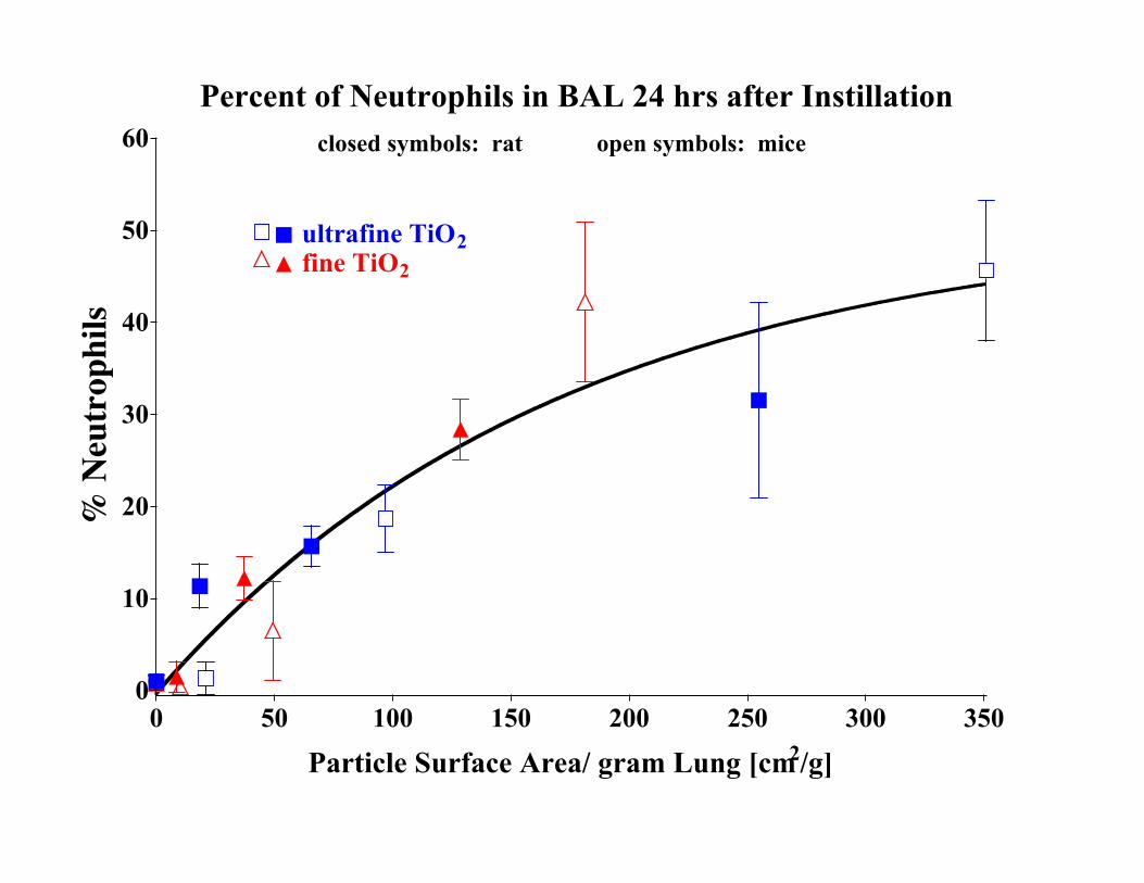
4.2.2 Epithelial TranslocationInstilled doses ranged from 65 µg to 1000 µg, yet the highest doses – in terms of
particle surface area retained in the lung – resulted in fewer inflammatory neutrophils in
the lung lavage (Figure 7a). In contrast, a similarly high dose (in terms of particlesurface) of 20 nm carbon black showed the expected greater response at higher doses.

21
On the other hand, lung lavage protein – another inflammatory indicator for increased
epithelial permeability – showed a steady dose dependent increase for all TiO2 sizes aswell as carbon black as would have been expected (Figure 7b). An additional finding
was a particle size and dose dependent translocation for TiO2 towards the pulmonaryinterstitium, whereas such translocation was only minimal for carbon black by 24 hours
post-dosing (Figure 7c,d). Indeed, at the highest doses of the 12 and 20 nm TiO2 about
52 and 50%, respectively, of the retained lung burden was “interstitial”, that is inepithelial or interstitial sites.
These studies with ultrafine and fine TiO2 particles showed a strongly particle sizedependent translocation to epithelial and interstitial sites: 24 hours after intratracheal
instillation of 500 µg into rats, more than 50% of the 12 nm sized TiO2 had translocated,
whereas it was only 4% and less for 220 nm and 250 nm TiO2 (Figure 7d).
5. Risk Assessment
6. Summary & OutlookNumerous anthropogenic — mainly combustion related — and natural sources
generate airborne ultrafine particulate matter (<100 nm). The ubiquitous occurrence of
the ultrafine particles results in significant human exposures under environmental and
certain occupational conditions. Several epidemiological studies have found adverserespiratory and cardiovascular health effects in susceptible parts of the population to be
associated with these particles. Controlled clinical studies with 10-50 µg/m3 ultrafineelemental carbon particles as surrogates for ambient particles did induce cardiovascular
effects in healthy adults. Studies in rodent models of a compromised respiratory or
cardiovascular system also showed mild inflammatory effects and oxidative stresssystemically and in the respiratory tract, using either laboratory-generated ultrafine
particles at high concentrations or ambient ultrafine particles. In addition, high dose invitro studies confirmed that the likely mechanism for their effects is via induction of
oxidative stress. Collectively, these results show that inhaled ambient ultrafine particles
can elicit significant effects.

22
Lacking, however, are long-term exposure studies and additional data about short-
term effects of ambient ultrafine particles at realistic concentrations. Inhaled ultrafineparticles deposit in all three regions of the respiratory tract via diffusional processes, with
the nose being a very effective filter and deposition site for the smallest (<5 nm) NSP,and tracheobronchial and alveolar regions being mainly targeted by about 5 and 20 nm
particles, respectively. Once deposited, the disposition of these particles appears to be
unique: In addition to the classical clearance processes known to exist for fine and coarseparticles, solid NSP can translocate to extrapulmonary organs across epithelial layers and
also are taken up via sensory nerve endings in the nasal and tracheobronchial regions.Particularly, translocation of nasally-deposited NSP along olfactory nerve axons into the
olfactory bulb has been demonstrated in non-human primates and rodents, thereby
circumventing the tight blood-brain barrier for accessing CNS structures. Depending ontheir chemical composition and bioavailability of particle components to neuronal cells, it
is conceivable that NSP reaching the CNS via this neuronal pathway will cause adverse
effects. Although this pathway is likely to exist in humans as well, conclusive evidenceis still lacking. Thus, there is a need to determine the potential of airborne NSP to cause
adverse CNS effects in laboratory animals and in humans. Figure S-8 summarizeshypotheses underlying effects caused by ambient ultrafine particulate matter, including
systemic acute phase responses induced by alveolar inflammation or through particles
reaching the blood circulation leading directly to cardiovascular effects; indirectcardiovascular effects via the autonomic nervous system from neuronally translocated
particles to respective ganglia and CNS structures; and potentially direct effects oftranslocated NSP on CNS functions. Modifying factors for these events most likely
include age, underlying disease and other co-airpollutants.
Although some of these emerging principles of nano-toxicology are specific forexposure via the respiratory tract (e.g., deposition of airborne NP), the fate and effects of
nanoparticles once taken up into the organism by varying routes are likely to be governedby the same mechanisms. For example, following inhalation exposure, local portal of
entry effects as well as effects in extrapulmonary organs (e.g., cardiovascular, liver,
CNS) due to the propensity of nano-sized particles to translocate, can be expecteddepending on modifying particle parameters.

23
Specific examples of translocation and effects of nano-sized particles and
presumed mechanisms are highlighted in this manuscript. They illustrate, on the onehand, that we need to be aware of possible acute adverse effects and potential long-term
consequences from nanoparticle exposures; on the other hand, the findings also give usinsights about the intriguing possibilities that engineered nanoparticles offer for potential
use as diagnostic tools or as therapeutic delivery systems. Obviously, a thorough
evaluation of desirable vs. adverse effects is required for the safe use of engineerednanoparticles. Thus, major challenges lie ahead to answer key questions of
nanotoxicology in order to characterize and predict potential risks, foremost being theassessment of human and environmental exposure, the identification of potential short
and long-term hazards. Furthermore, it is essential to understand more about the
biopersistence in cells and subcellular structures, the correlation betweenphysicochemical and biological/toxicological properties, and defining and characterizing
the biokinetics of nanoparticles (i.e., translocation pathways to sensitive structures within
organs, mechanisms of uptake and translocation at the organ/cellular/molecular levels) aswell as environmental effects such as persistence of coatings or covalent modifications
under weathering (UV, microbial degradation etc.), potential for bioaccumulation bybenthic fauna or filter feeders, and potential movement through the food chain. Research
to obtain answers to these questions requires an interdisciplinary team approach
involving toxicology, materials science, medicine, molecular biology, bioinformatics andtheir subspecialties.
7. References for web sectionAschner M. 2000. Manganese: brain transport and emerging research needs.
Environmental Health Perspectives 108(Supplement 3):429-432.Asgharian B, Miller F, Subramaniam rP. 1999. Dosimetry software to predict particle
deposition in humans and rats. CIIT Activities 19(No. 3).Balásházy I, Hofmann W, Heistracher T. 1999. Computation of local enhancement
factors for the quantification of particle deposition patterns in airway bifurcations.J Aerosol Science 30(No. 2):185-203.
Balásházy I, Hofmann W, Heistracher T. 2003. Local particle deposition patterns mayplay a key role in the development of lung cancer. Journal of Applied Physiology94:1719-1725.

24
Bantel-Schaal U, Hub B, Kartenbeck J. 2002. Endocytosis of adeno-associated virus type5 leads to accumulation of virus particles in the Golgi Compartment. J Virology76(5):2340-2349.
Berry JP, Arnoux B, Stanislas G, Galle P, Chretien J. 1977. A microanalytic study ofparticles transport across the alveoli: Role of blood platelets. Biomedicine 27:354-357.
Bracco D, Favre J-B. 1998. Pulmonary injury after ski wax inhalation exposure. Annalsof Emergency Medicine 32(5):616-619.
Braden B, Goldbaum F, Chen B, Kirschner A, Wilson S, Erlanger B. 2000. X-ray crystalstructure of an anti-Buckminsterfullerene antibody Fab fragment: Biomolecularrecognition of C60. PNAS 97(22):12193-12197.
Brenneman KA, Wong BA, Buccellato MA, Costa ER, Gross EA, Dorman DC. 2000.Direct olfactory transport of inhaled manganese (54MnCl2) to the rat brain:Toxicokinetic investigations in a unilateral nasal occlusion model. Toxicol ApplPharmacol 169:238-248.
Brown DM, Wilson MR, MacNee W, Stone V, Donaldson K. 2001. Size-dependentproinflammatory effects of ultrafine polystyrene particles: A role for surface areaand oxidative stress in the enhanced activity of ultrafines. Toxicology andApplied Pharmacology 175:191-199.
Brown JS, Zeman KL, Bennett WD. 2002. Ultrafine particle deposition and clearance inthe healthy and obstructed lung. Am J RespirCrit Care Med 166:1240-1247.
Cass GR, Hughes LA, Bhave P, Kleeman MJ, Allen JO, Salmon LG. 2000. The chemicalcomposition of atmospheric ultrafine particles. Phil trans R Soc Lond A358:2581-2592.
Chang OMC, Yi S-MY, Hopke PK, England GC, Chow JC, Watson JG. in press.Measurement of ultrafine particle size distributions from stationary combustionsources of coal, oil, and gas. J of the Air & Waste Management Association00(00):00-00.
Chen B, Wilson S, Das M, Coughlin D, Erlanger B. 1998. Antigenicity of fullerenes:Antibodies specific for fullerenes and their characteristics. PNAS 95:10809-10813.
Cheng YS, Yeh HC, Guilmette RA, Simpson SQ, Cheng KH, Swift DL. 1996. Nasaldeposition of ultrafine particles in human volunteers and its relationship to airwaygeometry. Aerosol Science & Technology 25:274-291.
Conova S. 1999. Role of particle wettability in capture by suspension-feeding crab(Emerita talpoida). Marine Biology 133:419-428.
Dahlquist M, Alexandersson R, Andersson K, Kolmodin-Hedman B, Malker H. 1992.Exposure to ski-wax smoke and health effects in ski waxers.ApplOccupEnvironHyg 7(10):689-693.
Daigle CC, Chalupa DC, Gibb FR, et al. 2003. Ultrafine particle deposition in humansduring rest and exercise. Inhalation Toxicology 15(No. 6):539-552.
deLorenzo A. 1970. The olfactory neuron and the blood-brain barrier. In: Taste and smellin vertebrates (Wolstenholme G, Knight J, eds). London:J. & A. Churchill, 151-176.
Demokritou P, Gupta T, Koutrakis P. 2002. A high volume apparatus for thecondensational growth of ultrafine particles for inhalation toxicological studies.

25
Aerosol Science and Technology 36:1061-1072.Dieckmann G, Dalton A, Johnson P, et al. 2003. Controlled assembly of carbon
nanotubes by designed amphiphilic Peptide helices. J Am Chem Soc 125(7):1770-1777.
Donaldson K, Brown D, clouter A, et al. 2002. The pulmonary toxicology of ultrafineparticles. J Aerosol Medicine - Deposition Clearance & Effects in the Lung15(2):213-220.
Dörger M, Münzing S, Allmeling A-M, Krombach F. 2000. Comparison of thephagocytic response of rat and hamster alveolar macrophages to man-madevitreous fibers in vitro. Human & Experimental Toxicology 19:635-640.
Dorman DC, Brenneman KA, McElveen AM, Lynch SE, Roberts KC, Wong BA. 2002.Olfactory transport: A direct route of delivery of inhaled manganese phosphate tothe rat brain. J Toxicology & Environmental Health (Part A: Current Issues)65(No. 20):1493-1511.
EPA. 1996. Air quality criteria for particulate matter (Vol. III) 600/P-95-001cF.Washington, DC 20460: Office of Research and Development.
Erlanger B, Chen B, Zhu M, Brus L. 2001. Binding of an anti-fullerene IgG monoclonalantibody to single wall carbon nanotubes. Nano Letters 1(9):465-467.
Gianutsos G, Morrow GR, Morris JB. 1997. Accumulation of manganese in rat brainfollowing intranasal administation. Fundamental and Applied Toxicology37(Article No. FA972306):102-105.
Grafstein B, Forman DS. 1980. INTRACELLULAR TRANSPORT IN NEURONS.Physiological Reviews 60(NO. 4):1167-1283.
Green TR, Fisher J, Stone M, Wroblewski BM, Ingham E. 1998. Polyethylene particlesof a "critical size" are necessary for the induction of cytokines by macrophages invitro. Biomaterials 19:2297-2302.
Hahn FF, Newton GJ, Bryant PL. 1977. In vitro phagocytosis of respirable-sizedmonodisperse particles by alveolar macrophages. In: Pulmonary Macrophagesand Epithelial Cells (Sanders CL, Schneider RP, Dagle GE, Ragen HA,eds):ERDA Symposium Series, 424-435.
Helenius A, Kartenbeck J, Simons K, Fries E. 1980. On the entry of semliki forest virusinto BHK-21 cells. J Cell Biol 84:404-420.
Hirokawa N. 1998. Kinesin and dynein superfamily proteins and the mechanism oforganelle transport. Science 279:519-526.
ICRP. 1994. Human Respiratory Model for Radiological Protection. Annals of the ICRP24:ICRP publication # 66.
ILSI. 2000. ILSI Risk Science Institute Workshop: The relevance of the rat lungresponse to particle overload for human risk assessment: A workishop consensusreport (ILSI risk Science Inst. Workshop Participants). Inhalation Toxicology12(Nos. 1-2):1-17.
Jaques PA, Kim CS. 2000. Measurement of Total Lung Deposition of Inhaled UltrafineParticles in Healthy Men and Women. Inhalation Toxicology 12:715-731.
Johnston CJ, Mango GW, Finkelstein JN, Barry BR. 1997. Altered pulmonary responseto hyperoxia in Clara cell secretory protein deficient mice. American JournalRespiratory Cell Molecular Biology 17:147-155.
Johnston CJ, Finkelstein JN, Mercer P, Corson N, Gelein R, Oberdorster G. 2000.

26
Pulmonary effects induced by ultrafine PTFE particles. Toxicol Appl Pharmacol168:208-215.
Kato T, Yashiro T, Murata Y, et al. 2003. Evidence that exogenous substances can bephagocytized by alveolar epithelial cells and transported into blood capillaries.Cell Tissue Research 311:47-51.
Keyhani K, Scherer PW, Mozell MM. 1997. A numerical model of nasal odoranttransport for the analysis of human olfaction. J theor Biol 186:279-301.
Kimbell JS, Godo MN, Gross EA, Joyner DR, Richardson RB, Morgan KT. 1997.Computer simulation of inspiratory airflow in all regions of the F344 rat nasalpassages. Toxicology & Applied Pharmacology 145:388-398.
Kittelson DB, Watts W, Johnson J. 2001. Fine Particle (nanoparticle) emissions onMinnesota highways.: Mn/DOT Report No. 2001-12.
Kittelson DV. 1998. Engines and nanoparticles: A Review. 29(5,6):575-588.Kreyling W, Semmler M, Erbe F, et al. 2002. Translocation of ultrafine insoluble iridium
particles from lung epithelium to extrapulmonary organs is size dependent butvery low. J Toxicology & Environmental Health 65A(20):1513-1530.
Kreyling WG, Scheuch G. 2000. Chapter 7: Clearance of Particles Deposited in theLungs. In: Particle-Lung Interactions (Gehr P, Heyder J, eds). New York -Basel:Marcel Dekker, Inc., 323-376.
Kureishi Y, Tamiaki H, Shiraishi H, Maruyama K. 1999. Photoinduced electron transferfrom synthetic chlorophyll analogue to fullerene C60 on carbon paste electrode.Preparation of a novel solar cell. Bioelectrochem Bioenerg 48(1):95-100.
Lakadamyali M, Rust M, Babcock H, Zhuang X. 2003. Visualizing infection ofindividual influenza viruses. PNAS 100:9280-9285.
Lecoanet H, Bottero J, Wiesner M. 2004. Laboratory Assessment of the Mobility ofNanomaterials in Porous Media. Environ Sci Technol 38:5164-5169.
Lecoanet H, Wiesner M. 2004. Velocity effects on fullerene and oxide nanoparticledeposition in porous media. Environ Sci Technol 38(16):4377-4382.
Li N, Sioutas C, Cho A, et al. 2003. Ultrafine particulate pollutants induce oxidativestress and mitochondrial damage. Environmental Health Perspectives 111(No.4):455-460.
Maynard AD, Maynard RL. 2002. A derived association between ambient aerosol surfacearea and excess mortality using historic time series data. AtmosphericEnvironment 36:5561-5567.
Nemmar a, Hoet PHM, Vanquickenborne B, et al. 2002. Passage of inhaled particles intothe blood circulation in humans. Circulation 105:411-414.
Nemmar A, Hoylaerts MF, Hoet PHM, Vermylen J, Nemery B. 2003. Size effect ofintratracheally instilled particles on pulmonary inflammation and vascularthrombosis. Toxicology and Applied Pharmacology 186:38-45.
NNI. 2004. What is Nanotechnology? http://www.nsf.gov/search97cgi/vtopic.NRC. 1979. Airborne Particles. Subcommittee on Airborne Particles, Committee on
Medical and Biologic Effects of Environmental Pollutants. Baltimore:UniversityPark Press.
Oberdörster E. 2004. Manufactured Nanomaterials (Fullerenes, C60) Induce OxidativeStress in Brain of Juvenile Largemouth Bass. Environ Health Perspect112(10):1058-1062.

27
Oberdörster G, Morrow PE, Spurny K. 1988. Size dependent lymphatic short termclearance of amosite fibers in the lung. Ann Occup Hyg 32((Supplement, InhaledParticles VI)):149-156.
Oberdörster G, Ferin J, Finkelstein J, Wade P, Corson N. 1990. Increased pulmonarytoxicity of ultrafine particles? II. Lung lavage studies. 21:384-387.
Oberdörster G, Ferin J, Lehnert BE. 1994. Correlation between particle size, in vivoparticle persistence, and lung injury. Environmental Health Perspectives 102(Suppl. 5):173-179.
Oberdörster G, Finkelstein JN, Johnston C, et al. 2000. HEI Research Report: AcutePulmonary Effects of Ultrafine Particles in Rats and Mice HEI Research Report.No. 96: Health Effects Institute.
Oberdörster G. 2001. Pulmonary effects of inhaled ultrafine particles (Review). Int ArchOccup Environ Health 74:1-8.
Oberdörster G, Sharp Z, Atudorei V, et al. 2002. Extrapulmonary transloction ofultrafine carbon particles following whole-body inhalation exposure of rats. JToxicology and Environmental Health 65A:1531-1543.
Oberdörster G, Utell MJ. 2002. Editorial: Is the central nervous system yet another targetorgan for ultrafine particles? Environ Health Perspect 110(No. 8):a440-A441.
Oberdörster G, Sharp Z, atudorei V, et al. 2004. Translocation of inhaled ultrafineparticles to the brain. Inhalation Toxicology 16(No. 6/7):437-445.
Pelkmans L, Kartenbeck J, Helenius A. 2001. Caveolar endocytosis of simian virus 40reveals a new two-step vesicular-transport pathway to the ER. Nature CellBiology 3:473-483.
Racette BA, McGee-Minnich L, Moerlein SM, Mink JW, Videen TO, Perlmutter JS.2001. Welding-related parkinsonism. Clinical features, treatment, andpathophysiology. Neurology 56:8-13.
Rosenbruch M. 1990. Experimentally induced liver granulomas after long-terminhalation of quartz in non-human primates. 132:469-470.
Rosenbruch M, Krombach F. 1992. Structural and functional hepatic changes afteerexperimental long-term inhaation of silica in non-human primates. Am RevRespir Dis 145(No. 4 (Intl. Conf. Suppl. - abstracts)):A91.
Rundell KW. 2003. High levels of airborne ultrafine and fine particulate matter in indoorice arenas. Inhalation Toxicology 15:237-250.
Schiller CF, Gebhart J, Heyder J, Rudolf G, Stahlhofen W. 1988. Deposition ofmonodisperse insoluble aerosol partilces in the 0.005 to 0.2 µm size range withinthe human respiratory tract. Ann occup Hyg 32(Supplement 1):41-49.
Schürch S, Gehr P, Hof VI, Geiser M, Green F. 1990. Surfactant displaces particlestoward the epithelium in airways and alveoli. Respiration Physiology 80:pp.17-32.
Schwartz J, Marcus a. 1990. Mortality and air pollution in London: A time seriesanalysis. American Journal of Epidemiology 131 (1):185-194.
Seisenberger G, Ried M, Endreb T, Büning H, Hallek M, Bräuchle C. 2001. Real-timesingle-molecule imaging of the infection pathway of an adeno-associted virus.Science 294:1929-1932.
Sioutas C, Kim S, Chang M. 1999. Development and evaluation of a prototype ultrafineparticle concentrator. JAerosol Science 30(8):1001-1012.

28
Smith AE, Helenius A. 2004. How viruses enter animal cells. Science 304:237-242.Stahlhofen W, Scheuch G, Bailey MR. 1995. Investigations of retention of inhaled
particles in the human bronchial tree. Radiation Protection Dosimetry 60 (No.4):311-319.
Stone V, Shaw J, Brown D, MacNee W, Faux S, Donaldson K. 1998. The role ofoxidative stress in the prolonged inhibitory effect of ultrafine carbon black onepithelial cell function. Toxicol In Vitro 12:649-659.
Su Y, Sipin M, Prather K, Gelein R, Lunts A, Oberdörster G. 2004. ATOFMScharacerization of individual model aerosol particles for expoure studies. AerosolScience and Technology submitted.
Suomalainen M, Nakano M, Keller S, Boucke K, Stidwill R, Greber U. 1999.Microtubule-dependent plus- and minus end - directed motilities are competingprocesses for nuclear targeting of adenovirus. J Cell Biol 144(4):657-672.
Swift DL, Montassier N, Hopke PK, et al. 1992. Inspiratory deposition of ultrafineparticles in human nasal replicate cast. 23:65-72.
Tabata Y, Ikada Y. 1988. Effect of the size and surface charge of polymer microsphereson their phagocytosis by macrophage. Biomaterials 9:356-362.
Tjälve H, Mejare C, Borg-Neczak K. 1995. Uptake and transport of manganese inprimary and secondary olfactory neurones in pike. Pharmacol Toxicol 77(1):23-31.
Tjälve H, Henriksson J. 1999. Uptake of metals in the brain via olfactory pathways.NeuroToxicology 20((2-3)):181-196.
Tuch T, Brand P, Wichmann HE, Heyder J. 1997. Variation of particle number and massconcentration in various size ranges of ambient aerosols in Eastern Germany.Atmospheric Environment 31(No. 24):4193-4197.
Turetsky BI, Moberg PJ, Arnold SE, Doty RL, Gur RE. 2003. Low olfactory bulb volumein first-degree relatives of patients with Schizophrenia. Am J Psychiatry160(4):703-708.
Valberg PA, Blanchard JD. 1991. Chapter 36, Pulmonary macrophage physiology:Origin, motility, endocytosis. In: Treatise on Pulmonary Toxicology --. In:Treatise on Pulmonary Toxicology--Comparative Biology of the Normal Lung.
Wojciak-Stothard B, Curtis A, Monaghan W, MacDonald K, Wilkinson C. 1996.Guidance and activation of murine macrophages by nanometric scale topography.Exp Cell Res 223:426-435.
Zheng M, Jagota A, Strano M, et al. 2003. Structure-based carbon nanotube sorting bysequence-dependent DNA assembly. Science 302:1545-1548.
Zhu Y, Hinds WC, Kim S, Shen SK, sioutas C. 2002. Study of ultrafine particles near amajor highway with heavy-duty diesel traffic. Atmospheric Environment36:4323-4335.
Zimmer AT, Baron PA, Biswas P. 2002. The influence of operating parameters onnumber-weighted aerosol size distribution generated from a gas metal arc weldingprocess. J Aerosol Science 33:519-531.

29

30
8. Figure Legends
Figure S-1: Correlation between daily mortality rate and urban particle concentrations
during the London smog episodes in the winters of 1958-1972 (data from
Schwartz and Marcus 1990). Also shown are the regression lines for thesteep slope and the shallow slope and the mathematically determined
inflection point at ~125 µg/m3. This could be explained by the presence of
singlet ultrafine particles at lower mass concentrations in the air andaggregated particles at higher mass concentrations (from Oberdörster 2001).
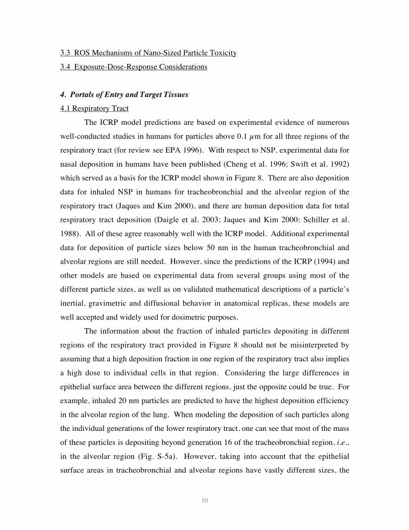
Figure S-2: Lung lavage neutrophils 24 hrs. after intratracheal dosing with ultrafine (20nm) and fine (250 nm) TiO2 in rats and mice. Normalization for different
particle surface areas and lung weights shows that acute pulmonary cell
response is essentially the same in both species (based on data from
Oberdörster et al., 2000).
Figure S-3: Chemically different NSP can have different effects: Whereas 20 nmparticles of both TiO2 and Al2O3 show the significantly greater inflammatory
response in the lung when compared to the same mass dose of larger
particles, nano-sized Al2O3 induces a more persistent effect which has notreturned to control levels by day 59 post-exposure.
Figure S-4: Inflammatory lung lavage parameters (neutrophils [PMN], protein) in ratswithin 4 hours after a 15-min. inhalation exposure to ultrafine PTFE fumes.
The “adapted” rats had been exposed for 5 minutes on each of 3 days prior to
the 15-min. exposure, whereas the “non-adapted” group was sham-exposedto filtered air for 3 days prior to exposure and the “sham” group was exposed
to filtered air throughout. The deposited alveolar dose of ultrafine PTFE wasestimated to be just ~65 ng (Johnston et al. 1997).
Figure S-5: Deposition of inhaled singlet 20 nm particles in different generations of the
lower human respiratory tract, based on predictions by the MPPD model(Asgharian et al. 1999). Generation 1 (trachea) to generation 16 (terminal
bronchioles) represent the tracheobronchial region, generations 16 to 26 are
the alveolar region. Deposited mass is expressed as relative units per airwaygeneration (a) or per m2 of each generation (b). Although 20 nm particles

31
have the highest deposition efficiency in the alveolar region (see also Figure
8), the tracheobronchial region receives the highest dose per unit surfacearea.
Figure S-6: Deposited doses of polydisperse particles with CMD of 20 nm and 250 nm(GSD = 1.7) during a 6-hr. inhalation at 100 µg/m3 (predicted by MPPD
model) in naso-pharyngeal, tracheo-bronchial, and alveolar region of the
human respiratory tract during nasal breathing. Although the alveolar regionreceives the highest dose and the nasal region the lowest, this is reversed
when the dose is expressed per unit surface area: the nose, – in this case, hasthe highest and the alveolar region the lowest dose. Note the consistently
higher deposited dose for the polydisperse 20 nm aerosol in all regions.
Figure S-7: Pulmonary inflammation and interstitial particle translocation 24 hrs. afterinstillation of 12 - 250 nm TiO2 particles in rats; carbon black (20 nm) used
as comparison. (a) Particle size and dose-dependent inflammatory cells in
lung lavage: note the lower response of the two larger doses of 12 and 20nm-sized TiO2, in contrast to 20 nm carbon black particles; (b) particle size
and dose-dependent protein in lung lavage: note the linear increase of thisinflammatory parameter with increasing doses for all particle types; (c) size
and dose-dependent interstitial translocation; note the lower translocation of
carbon black particles; (d) particle size dependent translocation of TiO2
particles (Oberdörster et al., 1992).
Figure S-8: Potential target sites and effects of inhaled NSP based on experimentalresults with ambient UFP inducing oxidative stress. Translocation and
effects are highly dependent on particle surface properties, biopersistence and
bioavailability of particle constituents, and are further modified by hostfactors such as age, disease state, and co-pollutants.
9. TablesTable S-1: Airborne Ultrafine/Nanoparticles: Workplace and Environment.
Table S-2: Up- and down-regulated genes from initial largemouth basssuppressive subtractive hybridization studies, after fish were exposed to 0.5ppm nC60 for 48 hours.

Sources Exposure Dose ResponseWhat are they? What levels? How much is retained? Physiological or adverse?
Indoorsheated surfacesfryingbroilinggrillingelectric motors
Outdoorsurban airinternal combustionpower plantsincineratorsgas-to-particle convers.forest firesairplane jetsrecreation (ski waxing)
Workplacemetallurgy (fumes)weldingpolymer fumesnanotechnology (biomed. electronics)nanotubes
Exposure Routesinhalationingestiondermal
Concentrationng/m3 - mg/m3
102 - >106 part./cm3
Durationminuteshoursdayscontinuous/peak
Locationdistance from sourcewind direction
Dosemetricmassnumbersurface
Depositionrespiratory tract: ventilatory and anatomic parameters
Dispositionwithin respiratory tractextrapulmonary organsdisease state
Physico-chemical Propertiesorganics, inorganicsmetalscrystalline, amorphoussurface areasolubility (water, lipid)
Epidemiologic Studiesambient UFPsusceptibles only?mortality/morbidity (respiratory, cardiovascular)
Clinical Studieslab. generated UFPambient UFPhealthy/susceptibles (respiratory, cardiovascular)
Animal Studieslab. generated UFPAmbient and occupational UFPcompromised animal models (respiratory, cardiovascular, CNS)mechanisms
In vitro Studiesmechanismsoxidative stresscellular/molecular
Airborne Ultrafine/Nano Particles: Workplace and EnvironmentTable S-1

TOXICOLOGY OF ENGINEERED NANOPARTICLES (NP)
Exposure Dose ResponseMechanisms of
Intake, UptakeMechanisms of
Cellular, Molecular Events
Which medium? air water food
What concentration?
Which route? inhalation ingestion dermal injection
How much?
Which dosemetric? mass number surface (size, chemistry, charge, shape)
Where retained? (Disposition) organ cell
How persistent?
Portal of entry effects vs.Remote organ effects?
Neutral?
Desirable? therapeutic diagnostic
Toxic? oxidative stress disrupting function: organ cell molecular pathway immune function
Modifying Factors: Age, Disease (Susceptibility)
Determining Dose
Biokinetics
Table S-2

0 200 400 600 800 1000 1200 1400240
260
280
300
320
340
360
mg/m3 (British Smoke)
Dai
ly M
orta
lity
Figure S-1
Percent of Neutrophils in BAL 24 hrs after Instillation
0 50 100 150 200 250 300 3500
10
20
30
40
50
60
ultrafine TiO2fine TiO2
closed symbols: rat open symbols: mice
Particle Surface Area/ gram Lung [cm2/g]
% Neutrophils
% N
eutr
ophi
ls

1 29 590
10
20
30
40
50
TiO2 20 nm
TiO2 250 nm
Days post-instillation1 29 59
0
10
20
30Al2O3 20 nm
Al2O3 500 nm
Days post-instillation
Lavage neutrophils following i.t. instillation of ultrafine and fine TiO2 and Al2O3 particles
(Oberdörster et al., 1990)
Figure S-3

0
20
40
60
80
100
PMNProtein
0
2
4
6
8* *
% P
MN
Protein, mg/m
l
Sham Adapted Non-adapted* P<0.05(ANOVA)
n=5 n=6 (all died within 3 hours) n=6 Fig. S-4
0 0

1
M
ass U
nits
Generation Number Generation Number
Particle Mass per Generation Particle Mass per Unit Surface Area
0
2
4
8
6
10
16 16 0
7 0
140
Mas
s Uni
ts/m
2
210
280
350
1
Figure S-5

Nose Tr-Br Alv0
20
40
60 Humanmg per Region
250 nm
Nose Tr-Br Alv0.01
0.1
1
10
100 Human
ng per cm2 Surface Area
20 nm
Deposited Doses in Naso-pharyngeal, Tracheobronchial and AlveolarRegion of Aerosol with 20 and 250 nm (CMD) Particles (GSD = 1.7)
Inhaled Over 6 hrs. at 100 µg/m3 (predicted by MPPD model)
Figure S-6
10 100 10000
10
20
30
40
250 nm
20 nm220 nm
20 nm
12 nm
Carbon
TiO2
Retained Particle Surface, cm2
Lav
age
Neu
trop
hils
(x
10-6
)
TiO2
Figure S-7a

10 100 10000.1
0.2
0.3
0.4
250 nm
20 nm220 nm
20 nm
12 nm
Carbon
TiO2
Retained Particle Surface, cm2
Lav
age
Prot
ein
mg/
ml TiO2
Figure S-7b

10 100 10000
20
40
60
250 nm
20 nm220 nm
20 nm
12 nm
Carbon
TiO2
Retained Particle Surface, cm2
% I
nter
stit
ial T
rans
loca
tion
Figure S-7c

10 1005 50 5000
20
40
60
12 nm20 nm220 nm250 nm
Particle size, nm
% I
nter
stit
ial T
rans
loca
tion
Oberdörster et al, 1992 Figure S-7d

Inhalation
Ultrafine Particles
Respir. Tract Deposition
LungInflammation
SystemicInflammation
Heart Effects
CentralNervousSystem
Blood VesselDysfunction
White BloodCell Activation
Extrapulmonary Organs
Liver Heart Bone Marrow
NeuronsCirculation
Inters
titium
Autonomic Nervous System
Particle translocation
Mediators
Modifying factors: Gender, underlying disease, co-pollutantsFigure S-8