Nanotechnology in Drug Delviery and Tissue Engineering - From Discovery to Applications
8
Nanotechnology in Drug Delivery and Tissue Engineering: From Discovery to Applications Jinjun Shi, †,§ Alexander R. Votruba, § Omid C. Farokhzad, †,§ and Robert Langer* ,†,‡ † MIT-Harvard Center for Cancer Nanotechnology Excellence and ‡ Department of Chemical Engineering, Massachusetts Institute of Technology, Cambridge, Massachusetts 02139 and § Laboratory of Nanomedicine and Biomaterials, Brigham and Women’s Hospital and Harvard Medical School, Boston, Massachusetts 02115 ABSTRACT The application of nanotechnology in medicine, referred to as nanomedicine, is offering numerous exciting possibilities in healthcare. Herein, we discuss two important aspects of nanomedicine, drug delivery and tissue engineering, highlighting the advances we have recently experienced, the challenges we are currently facing, and what we are likely to witness in the near future. KEYWORDS Nanotechnology, nanomedicine, drug delivery, tissue engineering I n pharmaceutical industries, a new molecular entity (NME) that demonstrates potent biological activity but poor water solubility, or a very short circulating half- life, will likely face significant development challenges or be deemed undevelopable. On the other hand, less active but pharmaceutically optimal compounds may become more suitable candidates for development. In either case, there is always a degree of compromise, and such trade- offs may inevitably result in the production of less-ideal drugs. However, with the emerging trends and recent ad- vances in nanotechnology, it has become increasingly possible to address some of the shortcomings associated with potential NMEs. By using nanoscale delivery ve- hicles, the pharmacological properties (e.g., solubility and circulating half-life) of such NMEs can be drastically im- proved, essentially leading to the discovery of optimally safe and effective drug candidates. This is just one ex- ample that demonstrates the degree to which nanotech- nology may revolutionize the rules and possibilities of drug discovery and change the landscape of pharmaceuti- cal industries. Indeed, current nanotechnology-based therapeutic products have been validated through the im- provement of previously approved drugs, and many novel classes of nanotherapeutics are now underway. 1-3 Drug discovery is only one of the many areas in health- care that nanotechnology is now benefiting. The current and promising applications of nanomedicine include, but are not limited to, drug delivery, in vitro diagnostics, in vivo imaging, therapy techniques, biomaterials, and tissue engineering. Summarized in Table 1 are some important opportunities that nanotechnology may afford in each research area. Some of these opportunities are becoming realities or are actually being used today, while others are generating promise in their early phases of development and are expected to experience vigorous growth in the foreseeable future. As recognition of the importance of this exciting field, it is expected that the global market of nanoscale applications in the medical field could grow to $70-160 billion by 2015. 4,5 In this perspective, we dis- cuss the applications of nanomedicine with specific focus on drug delivery and tissue engineering. Drug Delivery. Since liposomes were first described in the 1960s and proposed as carriers of proteins and drugs for disease treatment, 6 nanotechnology has made a sig- nificant impact on the development of drug delivery sys- tems. A variety of organic/inorganic nanomaterials and devices have been used as delivery vehicles to develop effective therapeutic modalities (Figure 1). So far, there are over two dozen nanotechnology-based therapeutic products approved by Food and Drug Administration (FDA) for clinical use, and more are in clinical trials. 1-3 Of these products, the majority are composed of a nontarget- ed delivery system (e.g., liposomes and polymers) and a drug, and are therefore considered first generation nano- therapeutics. 7 Compared to conventional drug delivery, the first gen- eration nanosystems provide a number of advantages. In particular, they can enhance the therapeutic activity by *To whom correspondence should be addressed. E-mail: [email protected]. Published on Web: 08/20/2010 Nanotechnology may revolutionize the rules and possibilities of drug discovery and change the landscape of pharmaceutical industries. PERSPECTIVE pubs.acs.org/NanoLett © 2010 American Chemical Society 3223 DOI: 10.1021/nl102184c | Nano Lett. 2010, 10 , 3223 –323 0
-
Upload
curlycutie -
Category
Documents
-
view
218 -
download
0
Transcript of Nanotechnology in Drug Delviery and Tissue Engineering - From Discovery to Applications

8/3/2019 Nanotechnology in Drug Delviery and Tissue Engineering - From Discovery to Applications
http://slidepdf.com/reader/full/nanotechnology-in-drug-delviery-and-tissue-engineering-from-discovery-to 1/8
Nanotechnology in Drug Delivery and TissueEngineering: From Discovery to Applications
Jinjun Shi,
†,§
Alexander R. Votruba,
§
Omid C. Farokhzad,
†,§
and Robert Langer*
,†,‡
† MIT-Harvard Center for Cancer Nanotechnology Excellence and ‡ Department of Chemical Engineering,Massachusetts Institute of Technology, Cambridge, Massachusetts 02139 and § Laboratory of Nanomedicine andBiomaterials, Brigham and Women’s Hospital and Harvard Medical School, Boston, Massachusetts 02115
ABSTRACT The application of nanotechnology in medicine, referred to as nanomedicine, is offering numerous exciting possibilitiesin healthcare. Herein, we discuss two important aspects of nanomedicine, drug delivery and tissue engineering, highlighting theadvances we have recently experienced, the challenges we are currently facing, and what we are likely to witness in the near future.
KEYWORDS Nanotechnology, nanomedicine, drug delivery, tissue engineering
In pharmaceutical industries, a new molecular entity(NME) that demonstrates potent biological activity but
poor water solubility, or a very short circulating half-life, will likely face significant development challenges or
be deemed undevelopable. On the other hand, less active
but pharmaceutically optimal compounds may become
more suitable candidates for development. In either case,
there is always a degree of compromise, and such trade-
offs may inevitably result in the production of less-ideal
drugs. However, with the emerging trends and recent ad-
vances in nanotechnology, it has become increasingly
possible to address some of the shortcomings associated
with potential NMEs. By using nanoscale delivery ve-hicles, the pharmacological properties (e.g., solubility and
circulating half-life) of such NMEs can be drastically im-
proved, essentially leading to the discovery of optimally
safe and effective drug candidates. This is just one ex-
ample that demonstrates the degree to which nanotech-
nology may revolutionize the rules and possibilities of
drug discovery and change the landscape of pharmaceuti-
cal industries. Indeed, current nanotechnology-basedtherapeutic products have been validated through the im-
provement of previously approved drugs, and many novel
classes of nanotherapeutics are now underway.1-3
Drug discovery is only one of the many areas in health-care that nanotechnology is now benefiting. The current
and promising applications of nanomedicine include, but
are not limited to, drug delivery, in vitro diagnostics, in
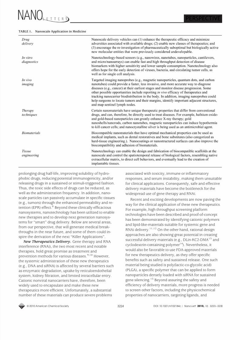
vivo imaging, therapy techniques, biomaterials, and tissueengineering. Summarized in Table 1 are some important
opportunities that nanotechnology may afford in each
research area. Some of these opportunities are becoming
realities or are actually being used today, while others are
generating promise in their early phases of development
and are expected to experience vigorous growth in the
foreseeable future. As recognition of the importance of
this exciting field, it is expected that the global market of nanoscale applications in the medical field could grow to
$70-160 billion by 2015.4,5 In this perspective, we dis-
cuss the applications of nanomedicine with specific focus
on drug delivery and tissue engineering.
Drug Delivery. Since liposomes were first described in
the 1960s and proposed as carriers of proteins and drugs
for disease treatment,6 nanotechnology has made a sig-
nificant impact on the development of drug delivery sys-
tems. A variety of organic/inorganic nanomaterials anddevices have been used as delivery vehicles to develop
effective therapeutic modalities (Figure 1). So far, there
are over two dozen nanotechnology-based therapeutic
products approved by Food and Drug Administration
(FDA) for clinical use, and more are in clinical trials.1-3 Of
these products, the majority are composed of a nontarget-
ed delivery system (e.g., liposomes and polymers) and a
drug, and are therefore considered first generation nano-
therapeutics.7
Compared to conventional drug delivery, the first gen-
eration nanosystems provide a number of advantages. In
particular, they can enhance the therapeutic activity by*To whom correspondence should be addressed. E-mail: [email protected].
Published on Web: 08/20/2010
Nanotechnology may revolutionize
the rules and possibilities of drug
discovery and change the
landscape of pharmaceutical
industries.
PERSPECTIVE
pubs.acs.org/NanoLett
© 2010 American Chemical Society 3223 DOI: 10.1021/nl102184c | Nano Lett. 2010, 10 , 3223–3230
8/3/2019 Nanotechnology in Drug Delviery and Tissue Engineering - From Discovery to Applications
http://slidepdf.com/reader/full/nanotechnology-in-drug-delviery-and-tissue-engineering-from-discovery-to 2/8
prolonging drug half-life, improving solubility of hydro-
phobic drugs, reducing potential immunogenicity, and/orreleasing drugs in a sustained or stimuli-triggered fashion.
Thus, the toxic side effects of drugs can be reduced, as
well as the administration frequency. In addition, nano-
scale particles can passively accumulate in specific tissues(e.g., tumors) through the enhanced permeability and re-
tention (EPR) effect.8 Beyond these clinically efficacious
nanosystems, nanotechnology has been utilized to enable
new therapies and to develop next generation nanosys-
tems for “smart” drug delivery. Below are several areas,
from our perspective, that will generate medical break-
throughs in the near future, and some of them could in-
spire the derivation of the next “Killer Applications”.New Therapeutics Delivery. Gene therapy and RNAinterference (RNAi), the two most recent and notable
therapies, hold great promise as treatment and
prevention methods for various diseases.9,10 However,
the systemic administration of these new therapeutics
(e.g., DNA and siRNA) is affected by several barriers such
as enzymatic degradation, uptake by reticuloendothelial
system, kidney filtration, and limited intracellular entry.
Cationic nonviral nanocarriers have, therefore, beenwidely used to encapsulate and make these new
therapeutics more efficient. Unfortunately, a substantial
number of these materials can produce severe problems
associated with toxicity, immune or inflammatory
responses, and serum instability, making them unsuitablefor clinical applications. Consequently, safe and effective
delivery materials have become the bottleneck for the
widespread use of gene therapy and RNAi.
Recent and exciting developments are now paving the
way for the clinical application of these new therapeutics.
For example, high throughput screening platform
technologies have been described and proof-of-concept
has been demonstrated by identifying cationic polymers
and lipid-like-materials suitable for systemic gene and
RNAi delivery.11,12 On the other hand, rational design
approaches are also showing great potential in creating
successful delivery materials (e.g., DLin-KC2-DMA13 andcyclodextrin-containing polymer14). Nevertheless, it
would also be favorable to use FDA-approved materials
for new therapeutics delivery, as they offer specific
benefits such as safety and sustained release. One such
material being studied is poly(lactic-co-glycolic acid)
(PLGA), a specific polymer that can be applied to form
nanoparticles densely loaded with siRNA for sustained
gene silencing.15 Beyond assuring the safety and
efficiency of delivery materials, more progress is needed
to screen other factors, including the physicochemical
properties of nanocarriers, targeting ligands, and
TABLE 1. Nanoscale Application in Medicine
PERSPECTIVE
© 2010 American Chemical Society 3224 DOI: 10.1021/nl102184c | Nano Lett. 2010, 10 , 3223-–3230
8/3/2019 Nanotechnology in Drug Delviery and Tissue Engineering - From Discovery to Applications
http://slidepdf.com/reader/full/nanotechnology-in-drug-delviery-and-tissue-engineering-from-discovery-to 3/8
formulation methods, all of which can affect the delivery
of new therapeutics in vivo.Targeted Delivery. It is widely believed that active
targeting, through the modification of nanoparticles with
ligands, has the potential to enhance the therapeutic
efficacy and reduce the side effects relative to
conventional therapeutics.16 The ability to actively target
specific cells rather than tissues also allows ligand-
conjugated nanocarriers to outperform first generation,nontargeted nanosystems. While the necessity of targeted
delivery depends on various factors (e.g., delivery
vehicles, drugs, and diseases), a myriad of importantbenefits have been demonstrated.3,16-19
In cancer therapy, the presence of targeting ligands
can greatly enhance the retention and cellular uptake of
nanoparticles via receptor-mediated endocytosis, even
though tumor accumulation is largely determined by the
physicochemical properties of nanoparticles.16,17 This can
then lead to higher intracellular drug concentration and
increase therapeutic activity, which is particularly
important for bioactive macromolecules (e.g., DNA andsiRNA) that require intracellular delivery for bioactivity.16
In the case of endothelial targeting for cardiovascular
diseases or immunological tissue targeting, nanoparticle
localization is guided by ligand-receptor interactions
rather than EPR.18 Similarly, ligand-mediated targeting is
of importance for the transcytosis of nanodrugs across
tight epithelial and endothelial barriers (e.g., blood-brain
barrier).19 Additionally, targeted delivery has beenapplied, in some instances, to combat multidrug
resistance (MDR).3 It is also envisioned that long-
circulating targeted nanoparitcles may be able to locate
and fight migrating cancer cells.
Despite three targeted nanoparticle systems now in
phase I/II clinical trials,3 the clinical translation of targeted
delivery is not as smooth as we expect. One possiblebarrier stems from the complexity behind manufacturing
viable targeted nanoparticles. Targeted nanoparticle
fabrication usually requires multiple steps, including
biomaterial assembly, ligand coupling/insertion, and
purification, which could cause batch-to-batch variation
and quality concerns. The recent development of single-step synthesis of targeted nanoparticles by self-
assembling prefunctionalized biomaterials provides a
simple and scalable manufacturing strategy.14,20 Anotherimportant consideration is targeting ligands. Among
others, some variables that must be considered include
ligand biocompatibility, cell specificity, binding affinity,
mass production, and purity.21 For example, to achieve
maximal specificity, the ideal ligand would be able to
recognize the most overexpressed receptors on target
cells relative to healthy ones. Other factors that could also
affect cell targeting include ligand surface density and
arrangement, as well as spacer type and length dividingligand molecules and nanoparticles.22 Nevertheless, with
advances in ligand engineering and screening, and
nanoparticle optimization, targeted delivery will become a
mainstay in the next generation of drug therapy.Co-delivery. Combination therapy has shown several
potential advantages (e.g., synergistic effects and reversal
of drug resistance) and may prove more effective than
single drug therapy.23 However, due to the distinctpharmacokinetic profiles of individual drugs, the
synergistic drug ratio optimized in vitro will undoubtedly
change after the conventional administration of drug
“cocktails”, an outcome that could in turn lead to
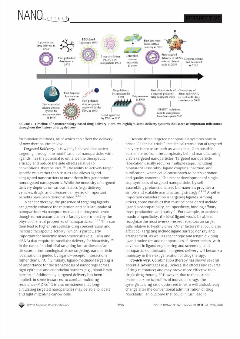
FIGURE 1. Timeline of nanotechnology-based drug delivery. Here, we highlight some delivery systems that serve as important milestonesthroughout the history of drug delivery.
PERSPECTIVE
© 2010 American Chemical Society 3225 DOI: 10.1021/nl102184c | Nano Lett. 2010, 10 , 3223-–3230
8/3/2019 Nanotechnology in Drug Delviery and Tissue Engineering - From Discovery to Applications
http://slidepdf.com/reader/full/nanotechnology-in-drug-delviery-and-tissue-engineering-from-discovery-to 4/8
insufficient therapeutic results in vivo. To this end, lipid-
and/or polymer-based nanoscale systems, previously
developed for single drug delivery, have been applied to
facilitate co-delivery. For some drug combinations, we
can successfully tune the relative dosage of various drugs
in single particle levels, and simultaneously deliver them
to target sites with a maintained drug ratio.24 For othercombinations, we need to develop novel delivery vehicles
with desired functionalities that enable co-encapsulation
of hydrophobic and hydrophilic drugs, active targeting,
and/or temporally controlled release.25,26 Such features
are especially essential for the co-delivery of drugs and
nucleic acids, which requires intracellular delivery to elicit
bioactivity.27,28 For example, in the case of co-delivering
chemotherapy and RNAi therapy to treat multidrugresistant cancers, the ideal nanoparticle would be
expected to first release siRNA to reduce the expression
of MDR transporters, followed by the release of
anticancer drugs.Another exciting advancement in co-delivery is the
ability to combine targeted imaging and therapeutic
agents within the same particle, allowing us to visualize
sites of targeted drug delivery and deliver therapeutics
simultaneously (“theranostics”).29 This technology is
innovative in concept and holds significant promise formaking large medical impacts within the next few
decades. It could provide us with critical information on
intracellular targets, ensure that therapeutic agents are
efficiently reaching their target sites, and enable the
effective early detection and treatment of diseases.
Current research is primarily focused on the design of such multifunctional nanosystems and proof-of-concept
tests,30-33 but more systematic in vivo studies areneeded.
Future research in this arena will also likely help us
trace the absorption, distribution, metabolism, and
excretion of nanoparticles in vivo. It is important that we
understand the pharmacokinetics of a given drug delivery
system to improve upon formulations, estimate clinical
doses, and guarantee safety.34 Currently, radionuclide
labeling is the only technique that can be used to provide
in situ quantitative information; but radio emitters may
be too unstable to conjugate with nanomaterials. With thehelp of recently developed in vivo imaging probes like
magnetic nanoparticles,7 quantum dots,35 gold
nanoparticles,36 and carbon nanotubes,37 more imaging
modalities may become available to track the distribution
of nanotherapeutics in the body.MEM/NEM Devices for Drug Delivery. Parallel to the
development of particulate delivery systems, significant
headway has also been made in the field of
micro/nanoelectromechanical (MEM/NEM) device-baseddrug delivery over the past decade. In particular,
implantable microchips containing nanosized reservoirs
have been developed to deliver drugs for long time
periods in a precisely controlled manner; microneedles
are being tested in painless transdermal drug delivery;
and the incorporation of nanofeatures (e.g., nanopores,
nanochannels, and nanoparticles) in microfabricated
systems are perfecting drug delivery and immunoisolation
techniques.38-41 Intriguingly, these devices can be further
modified to deliver new therapeutics, achieve targeteddelivery, and co-deliver multiple agents.41,42 Substantial
efforts are also being put into creating intelligent devices
that could potentially sense when and how much dose is
needed and then automatically release it from
reservoirs.42 To do this, one feat that must be met is the
continuous and stable monitoring of physical and
biochemical conditions in situ. The recent development of
nanotechnology-based sensors (e.g., nanowire andnanotube) may offer new ways to address this concern
and could even facilitate device miniaturization.43
In addition to drug delivery, micro- and nanofabricated
devices have shown potential in developing nanocarrierswith controlled physicochemical properties (e.g., size,
shape, and surface chemistries). By using
perfluoropolyether molds, fabricated by traditional top-
down approaches, polymeric particles at the
submicrometer scale can be replicated with variable
shapes and controllable surface chemistries.44 Comparedto bulk synthesis, microfluidic devices have also recently
been used for nanoprecipitation synthesis of smaller and
more homogeneous PLGA-PEG nanoparticles.45
Nonetheless, micro- and nanotechnologies, which can be
universally used to control the biophysicochemical
properties of various nanosystems, are still in greatdemand.
Beyond the aforementioned opportunities and
challenges, the following are also essential for thedevelopment of next generation drug delivery systems:
(1) detailed physicochemical characterization of
nanosystems (which will require sophisticated and state-
of-art techniques); (2) restriction of undesired uptake by
nontarget organs (e.g., liver and spleen); (3) improvement
of stimuli-triggered or programmable drug release
systems (e.g., pH, temperature, light, enzyme, ultrasound,
magnetic field, and electric current); (4) furthering the
means by which we can understand the biologicalprinciples of disease and its microenvironment, the
biological barriers that hinder drug delivery, and
endosomal trafficking pathways; and (5) identification of
biological markers attributable to diseased cells. With
continual advancement in these areas, we can expect that
the field of drug delivery will benefit greatly from the
emergence of finely engineered nanomaterials and
devices.Tissue Engineering. Tissue engineering is an evolving
interdisciplinary field integrating biology, engineering,
materials science, and medicine, that focuses on the de-
velopment of biological substitutes to restore, replace,
PERSPECTIVE
© 2010 American Chemical Society 3226 DOI: 10.1021/nl102184c | Nano Lett. 2010, 10 , 3223-–3230

8/3/2019 Nanotechnology in Drug Delviery and Tissue Engineering - From Discovery to Applications
http://slidepdf.com/reader/full/nanotechnology-in-drug-delviery-and-tissue-engineering-from-discovery-to 5/8
maintain, or enhance tissue and organ function.46 Over
the past few decades, continued progress in this specific
field has led to the creation of implantable tissues, some
of which are already used in humans (e.g., skin and carti-
lage) or have entered clinical trials (e.g., bladder and
blood vessels).47 Nevertheless, most tissue engineering
strategies rely on the principle that under appropriatebioreactor conditions, cells seeded or recruited into three-
dimensional (3D) biocompatible scaffolds are able to reas-
semble into functional structures resembling native tis-
sues. Early artificial scaffolds were designed to provide
cells structural integrity on a macroscopic level, but only
achieved moderate success. It is now widely accepted
that to recapitulate proper tissue functionality, scaffolds
should also establish a tissue specific microenvironment
to maintain and regulate cell behavior and function.48
Within tissues, cells are surrounded by extracellular
matrix (ECM) which is characterized by a natural web of
hierarchically organized nanofibers.49 This integralnanoarchitecture is important because it provides cell
support and directs cell behavior via cell-ECM interac-
tions. Furthermore, ECM plays a vital role in storing, re-
leasing, and activating a wide range of biological factors,
along with aiding cell-cell and cell-soluble factor interac-
tions.50 Thus, the ability to engineer biomaterials thatclosely emulate the complexity and functionality of ECM
is pivotal for successful regeneration of tissues. Recent
advances in nanotechnology, however, have enabled the
design and fabrication of biomimetic microenvironment
at the nanoscale, providing an analog to native ECM.48
Notably, these technologies have been applied to createnanotopographic surfaces and nanofeatured scaffolds,
and to encapsulate and control the spatiotemporal releaseof drugs (e.g., growth factors). In turn, these nanodevices
offer a means to direct cellular behaviors that range from
cell adhesion to gene expression.Cell-Nanotopography Interactions. Living cells are
highly sensitive to local nanoscale topographic patterns
within ECM.49 In the pursuit to control cell function by
underlying nanotopographic cues, engineered substrates
with different nanofeatures have become rapidly adopted
(Figure 2). Top-down lithographic techniques are nowutilized to create various nanopatterns, such as gratings,
pillars, and pits, in a precisely controlled manner.51
Techniques like micelle lithography, anodization, and
electrospinning can also be used to create an array of
nanospheres, vertical nanotubes, and nanofibers.52-54
Additionally, less-ordered nanotopographies are now
being fabricated by polymer demixing, chemical etching,
electrospinning, and phase separation processes.55
These advances in the design of nanoscale substrates
have enabled investigators to explore cell-nanotopographyinteractions and have allowed for the manipulation of cell
morphology, signaling, orientation, adhesion, migration,
proliferation, and differentiation. In particular, it has been
determined that nanogratings and aligned nanofibers can
govern the alignment and elongation of many differentcell types;54,56-59 the differentiation of mesenchymal
stem cells can be regulated by polymer nanogratings,58
disordered nanopits,60 or vertically aligned TiO2
nanotubes;53,61 endothelial cells cultured on nanogratings
can be organized into multicellular band structures,
forming aligned capillary-like tubes;56 and cell adhesionstrength and apopotosis can be controlled by the
combination of cell signaling epitopes and
nanotopography.52,62 In the near future, we can expect
the emergence of more striking results from different
combinations of cell type, topography geometry and
scale, and substrate material. Nonetheless, the design andfabrication of next-generation nanotopographic substrates
will be guided by a greater understanding of the
mechanisms by which cells respond to and sensenanofeatures.
Nanofabricated Scaffolds. While critical insights into
2D cell-nanotopography interactions are now enabling us
to direct cell behavior, considerable efforts have been
made to develop 3D artificial scaffolds at the nanoscale
for tissue engineering applications. Nanofibrous scaffolds
are now under wide investigation as they exhibit a very
similar physical structure to protein nanofibers in ECM.48
Among the three dominant nanofabrication methods,electrospinning is a very simple and practical technique,
suitable for the creation of aligned and complex 3D
structures; self-assembly technology emulates the process
of ECM assembly and can thus produce very thin
nanofibers; and phase separation allows for continuous
fiber network fabrication with tunable pore structure and
the formation of sponge-like scaffolding.63 On the other
hand, nanocomposite-based scaffolds (e.g.,nanohydroxyapatite/collagen) are very popular in hard-
tissue engineering, particularly for the reconstruction of
bone tissue.64 Beyond nanofibers and nanocomposites,
carbon nanotubes have also attracted attention due to
FIGURE 2. Schematic of fabricated nanotopographic features usedto guide cell behaviors via cell-nanotopography interactions.
PERSPECTIVE
© 2010 American Chemical Society 3227 DOI: 10.1021/nl102184c | Nano Lett. 2010, 10 , 3223-–3230
8/3/2019 Nanotechnology in Drug Delviery and Tissue Engineering - From Discovery to Applications
http://slidepdf.com/reader/full/nanotechnology-in-drug-delviery-and-tissue-engineering-from-discovery-to 6/8
their mechanical strength and electrical conductivity, and
because they can be readily incorporated into 3D
architectures.65
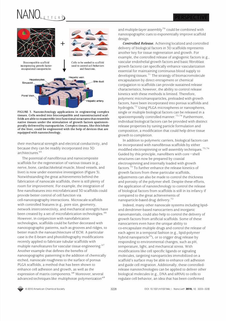
The potential of nanofibrous and nanocomposite
scaffolds for the regeneration of various tissues (e.g.,
nerve, bone, cardiac/skeletal muscle, blood vessels, andliver) is now under extensive investigation (Figure 3).
Notwithstanding the great achievements behind the
fabrication of nanoscale scaffolds, there is still plenty of
room for improvement. For example, the integration of
fine nanofeatures into microfabricated 3D scaffolds could
provide better control of cell function via
cell-nanotopography interactions. Microscale scaffolds
with controlled features (e.g., pore size, geometry,
network interconnectivity, and mechanical strength) have
been created by a set of microfabrication technologies.66
However, in conjunction with nanofabrication
technologies, scaffolds could be further decorated withnanotopographic patterns, such as grooves and ridges, to
better match the nanoarchitecture of ECM. A particular
case is the E-beam and photolithography modifications
recently applied to fabricate tubular scaffolds with
multiple nanofeatures for vascular tissue engineering.67
Another example that defines the benefits of
nanotopographic patterning is the addition of chemically
etched, nanoscale roughness to the surface of porous
PLGA scaffolds, a method that has been shown to
enhance cell adhesion and growth, as well as the
expression of matrix components.68 Moreover, several
advanced techniques like multiphoton polymerization69
and multiple-layer assembly70 could be combined with
nanotopographic cues to exponentially improve scaffold
design.Controlled Release. Achieving localized and controlled
delivery of biological factors in 3D scaffolds represents
another key for tissue regeneration and growth. For
example, the controlled release of angiogenic factors (e.g.,vascular endothelial growth factors and basic fibroblast
growth factors) can specifically enhance vascularization
essential for maintaining continuous blood supply to
developing tissues.71 The strategy of biomacromolecule
encapsulation by direct entrapment or chemical
conjugation to scaffolds can provide sustained release
characteristics; however, the ability to control release
kinetics with these methods is limited. Therefore,polymeric micro/nanoparticles, preloaded with growth
factors, have been incorporated into porous scaffolds and
hydrogels.72 Using PLGA microspheres or nanospheres,
single or multiple biological factors can be released in aspatiotemporally controlled manner.73,74 Furthermore,
individual biological factors can be provided with distinct
release properties by tuning particle formulation and
composition, a modification that could help drive tissue
growth to completion.
In addition to polymeric carriers, biological factors canbe incorporated with nanofibrous scaffolds by either
modified electrospinning or self-assembly techniques.75,76
Guided by this principle, nanofibers with core-shell
structures can now be prepared by coaxial
electrospinning and internally loaded with growth
factors.75
To further enhance the release kinetics of growth factors from these particular scaffolds,
adjustments can also be made to control the thicknessand porosity of the polymer shell. Despite these efforts,
the application of nanotechnology to control the release
of biological factors from scaffolds is still in its infancy if
compared to the great achievements of
nanoparticle-based drug delivery.72
Indeed, many other nanoscale systems including lipid-
and dendrimer-based nanocarriers and inorganic
nanomaterials, could also help to control the delivery of
growth factors from artificial scaffolds. Some of these
nanocarriers even have the unique ability toco-encapsulate multiple drugs and control the release of
each agent in a temporal fashion (e.g., lipid-polymer
hybrid nanoparticle25), or to trigger drug release by
responding to environmental changes, such as pH,
temperature, light, and mechanical stress. With
modifications like cell specific ligands or signaling
molecules, targeting nanoparticles immobilized on a
scaffold’s surface may be able to enhance cell adhesion
and guide cell migration. Additionally, these controlled-release nanotechnologies can be applied to deliver other
biological molecules (e.g., DNA and siRNA) to cells to
regulate cell behavior, an idea that has been confirmed
FIGURE 3. Nanotechnology applications in engineering complextissues. Cells seeded into biocompatible and nanostructured scaf-folds are able to reassemble into functional structures that resemblenative tissues under the stimulation of growth factors spatiotem-porally delivered by nanoparticles. Complex tissues, like this lobuleof the liver, could be engineered with the help of devices that areequipped with nanotechnology.
PERSPECTIVE
© 2010 American Chemical Society 3228 DOI: 10.1021/nl102184c | Nano Lett. 2010, 10 , 3223-–3230
8/3/2019 Nanotechnology in Drug Delviery and Tissue Engineering - From Discovery to Applications
http://slidepdf.com/reader/full/nanotechnology-in-drug-delviery-and-tissue-engineering-from-discovery-to 7/8
following the very recent development of poly( β-amino
esters)-DNA nanoparticles used to genetically engineering
stem cells for enhanced angiogenesis.77
Aside from the benefits discussed above,
nanotechnology is also expected to play an important role
in creating novel tissue regeneration strategies (e.g., cell
sheet engineering78) and in surmounting other importantobstacles in tissue engineering, such as the development
and characterization of new biomaterials and stem cell
engineering. For example, high throughput assays based
on nanoliter-scale synthesis have emerged for the cell-
based screening of biomaterials.79 Despite the ongoing
challenges, we can imagine that the achievement of
functional, artificial tissues/organs will have extremely
strong impact on regenerative medicine, as well as othermedical fields. One exciting opportunity that lies ahead is
the idea of “organ-on-a-chip”80 that, in the future, could
replace the expensive and life-costing animal testing used
for drug development and for evaluation/optimization of nanoparticulate systems for drug delivery.
Summary. Nanotechnology is becoming the driving
force behind a variety of evolutionary and revolutionary
changes in the medical field. The impact of nanotechnolo-
gy on drug delivery has helped to improve the efficacy of
available therapeutics and will likely enable the creationof entirely new therapeutic entities. For tissue engineer-
ing, nanotechnology has also opened the door to new ap-
proaches that could stimulate the reconstruction of com-
plex tissue architectures. We are optimistic about the
future of nanomedicine, given the wide array of innova-
tive nanoscale materials and technologies that stand on
the horizon. With the clarification of nanotechnology-spe-
cific medical regulations and a continued influx of invest-
ments and time, we believe that nanomedicine will notonly improve conventional therapies, but also bridge the
shortcomings of conventional medicine to help people on
both global and individual levels.
Acknowledgment. This work was supported by Na-tional Institutes of Health Grants U54-CA119349,DE013023, DE016516, EB000244, and EB006365; andthe Koch-Prostate Cancer Foundation Award in Nanother-apeutics.
NotesO.C.F. and R.L. have financial interest in BIND Bioscienc-es and Selecta Biosciences.
REFERENCES AND NOTES
(1) Wagner, V.; Dullaart, A.; Bock, A. K.; Zweck, A. Nat. Biotechnol.2006, 24, 1211–1217.
(2) Zhang, L.; Gu, F. X.; Chan, J. M.; Wang, A. Z.; Langer, R. S.;Farokhzad, O. C. Clin. Pharmacol. Ther. 2008, 83, 761–769.
(3) Davis,M. E.; Chen, Z.; Shin, D.M. Nat. Rev. Drug Discovery 2008,7 , 771–782.
(4) Jain, K. K. The Handbook of Nanomedicine; Humana Press: Totowa,2008; p 353.
(5) Occupational Health & Safety Report: Nanomedicine Market toSurpass $160 Billion by 2015. http://ohsonline.com/articles/2009/06/29/report-on-nanomedicine-market.aspx (accessed 6/1/10).
(6) Bangham, A. D.; Horne, R. W. J. Mol. Biol. 1964, 8, 660–668.(7) Riehemann, K.; Schneider, S. W.; Luger, T. A.; Godin, B.; Ferrari,
M.; Fuchs, H. Angew. Chem., Int. Ed. 2009, 48, 872–897.(8) Matsumura, Y.; Maeda, H. Cancer Res. 1986, 46, 6387–6392.(9) Putnam, D. Nat. Mater. 2006, 5 , 439–451.(10) Whitehead, K. A.; Langer, R.; Anderson, D. G. Nat. Rev. Drug
Discovery 2009, 8, 129–138.(11) Anderson, D. G.; Peng, W.; Akinc, A.; Hossain, N.; Kohn, A.;
Padera, R.; Langer, R.; Sawicki, J. A. Proc. Natl. Acad. Sci. U.S.A.2004, 101, 16028–16033.
(12) Akinc, A.; Zumbuehl, A.; et al. Nat. Biotechnol. 2008, 26, 561–569.
(13) Semple, S. C.; Akinc, A.; et al. Nat. Biotechnol. 2010, 28, 172–176.(14) Davis, M. E. Mol. Pharm. 2009, 6, 659–668.(15) Woodrow, K. A.;Cu, Y.; Booth, C. J.; Saucier-Sawyer, J. K.;Wood,
M. J.; Saltzman, W. M. Nat. Mater. 2009, 8, 526–533.
(16) Bartlett, D. W.; Su, H.; Hildebrandt, I. J.; Weber, W. A.; Davis,M. E. Proc. Natl. Acad. Sci. U.S.A. 2007, 104, 15549–15554.
(17) Pirollo, K. F.; Chang, E. H. Trends Biotechnol. 2008, 26, 552–558.(18) Farokhzad, O. C.; Langer, R. ACS Nano 2009, 3, 16–20.(19) Gabathuler, R. Neurobiol. Dis. 2010, 37 , 48–57.(20) Gu, F.; Zhang, L.; Teply, B. A.; Mann, N.; Wang, A.; Radovic-
Moreno, A. F.; Langer, R.; Farokhzad, O. C. Proc. Natl. Acad. Sci.U.S.A. 2008, 105 , 2586–2591.
(21) Allen, T. M. Nat. Rev. Cancer 2002, 2, 750–763.(22) Jung, H.; Yang,T.; Lasagna, M. D.; Shi, J.; Reinhart, G. D.; Cremer,
P. S. Biophys. J. 2008, 94, 3094–3103.(23) Greco, F.; Vicent, M. J. Adv. Drug Delivery Rev. 2009, 61, 1203–
1213.(24) Mayer, L. D.; Janoff, A. S. Mol. Interventions 2007, 7 , 216–223.(25) Sengupta, S.; Eavarone, D.; Capila, I.; Zhao, G. L.; Watson, N.;
Kiziltepe, T.; Sasisekharan, R. Nature 2005, 436, 568–572.
(26) Zhang,L.; Radovic-Moreno, A. F.;Alexis,F.; Gu, F. X.;Basto, P. A.;Bagalkot, V.; Jon, S.; Langer, R. S.; Farokhzad, O. C. ChemMed-Chem 2007, 2, 1268–1271.
(27) Wang, Y.; Gao, S.; Ye, W. H.; Yoon, H. S.; Yang, Y. Y. Nat. Mater.2006, 5 , 791–796.
(28) Saad, M.; Garbuzenko, O. B.; Minko, T. Nanomedicine 2008, 3,761–776.
(29) Debbage, P.; Jaschke, W. Histochem. Cell Biol. 2008, 130, 845–875.
(30) Bagalkot, V.; Zhang, L.; Levy-Nissenbaum, E.; Jon, S.; Kantoff,P. W.; Langer, R.; Farokhzad, O. C. Nano Lett. 2007, 7 , 3065–3070.
(31) Park, J. H.; von Maltzahn, G.; Ruoslahti, E.; Bhatia, S. N.; Sailor,M. J. Angew. Chem., Int. Ed. 2008, 47 , 7284–7288.
(32) Nasongkla, N.; Bey, E.; Ren, J. M.; Ai, H.; Khemtong, C.; Guthi, J. S.; Chin, S. F.; Sherry, A. D.; Boothman, D. A.; Gao, J. M. Nano Lett. 2006, 6, 2427–2430.
One exciting opportunity is the
idea of “organ-on-a-chip”80 that
could replace the expensive and
life-costing animal testing used for
drug development and for evaluation/
optimization of nanoparticulate
systems for drug delivery.
PERSPECTIVE
© 2010 American Chemical Society 3229 DOI: 10.1021/nl102184c | Nano Lett. 2010, 10 , 3223-–3230
8/3/2019 Nanotechnology in Drug Delviery and Tissue Engineering - From Discovery to Applications
http://slidepdf.com/reader/full/nanotechnology-in-drug-delviery-and-tissue-engineering-from-discovery-to 8/8
(33) McCarthy, J. R.; Weissleder, R. Adv. Drug Delivery Rev. 2008, 60,1241–1251.
(34) Sanhai, W. R.; Sakamoto, J. H.; Canady, R.; Ferrari, M. Nat.Nanotechnol. 2008, 3, 242–244.
(35) Dubertret, B.;Skourides, P.; Norris, D. J.; Noireaux, V.;Brivanlou,A. H.; Libchaber, A. Science 2002, 298, 1759–1762.
(36) Chanda, N.; Kattumuri, V.; Shukla, R.; Zambre, A.; Katti, K.;Upendran,A.; Kulkarni, R. R.; Kan, P.; Fent, G. M.; Casteel,S. W.;
Smith, C. J.; Boote, E.; Robertson, J. D.; Cutler, C.; Lever, J. R.;Katti, K. V.; Kannan, R. Proc. Natl. Acad. Sci. U.S.A. 2010, 107 ,8760–8765.
(37) De La Zerda, A.; Zavaleta, C.; Keren, S.;Vaithilingam, S.; Bodapati,S.; Liu, Z.; Levi, J.; Smith, B. R.; Ma, T. J.; Oralkan, O.; Cheng, Z.;Chen, X. Y.; Dai, H. J.; Khuri-Yakub, B. T.; Gambhir, S. S. Nat.Nanotechnol. 2008, 3, 557–562.
(38) Santini,J. T., Jr.;Cima,M. J.; Langer, R. Nature 1999, 397 , 335–338.(39) Ainslie, K. M.; Desai, T. A. Lab Chip 2008, 8, 1864–1878.(40) Prausnitz, M. R. Adv. Drug Delivery Rev. 2004, 56, 581–587.(41) Tasciotti, E.; Liu, X.; Bhavane, R.; Plant, K.; Leonard, A. D.; Price,
B. K.;Cheng, M. M.;Decuzzi, P.;Tour, J. M.; Robertson,F.; Ferrari,M. Nat. Nanotechnol. 2008, 3, 151–157.
(42) Staples, M.; Daniel, K.; Cima, M. J.; Langer, R. Pharm. Res. 2006, 23, 847–863.
(43) Patolsky, F.; Zheng,G.; Lieber, C.M. Nanomedicine 2006, 1, 51–65.
(44) Gratton, S. E.; Williams, S. S.; Napier, M. E.; Pohlhaus, P. D.;Zhou, Z.; Wiles, K. B.; Maynor, B. W.; Shen, C.; Olafsen, T.;Samulski, E. T.; Desimone, J. M. Acc. Chem. Res. 2008, 41,1685–1695.
(45) Karnik, R.;Gu, F.; Basto, P.; Cannizzaro, C.; Dean, L.; Kyei-Manu,W.; Langer, R.; Farokhzad, O. C. Nano Lett. 2008, 8, 2906–2912.
(46) Langer, R.; Vacanti, J. P. Science 1993, 260, 920–926.(47) Khademhosseini, A.;Vacanti, J. P.; Langer, R. Sci. Am. 2009, 300,
64–71.(48) Goldberg, M.; Langer, R.; Jia, X. J. Biomater. Sci. 2007, 18, 241–
268.(49) Stevens, M. M.; George, J. H. Science 2005, 310, 1135–1138.(50) Taipale, J.; Keski-Oja, J. FASEB J. 1997, 11, 51–59.(51) Bettinger, C. J.; Langer, R.; Borenstein, J. T. Angew. Chem., Int.
Ed. 2009, 48, 5406–5415.(52) Huang, J.; Grater, S. V.; Corbellini, F.; Rinck, S.; Bock, E.;
Kemkemer, R.; Kessler, H.; Ding, J.; Spatz, J. P. Nano Lett.2009, 9, 1111–1116.(53) Park, J.; Bauer, S.; von der Mark, K.; Schmuki, P. Nano Lett. 2007,
7 , 1686–1691.(54) Xu, C. Y.;Inai, R.;Kotaki, M.;Ramakrishna, S. Biomaterials 2004,
25 , 877–886.(55) Norman, J. J.; Desai, T. A. Ann. Biomed. Eng. 2006, 34, 89–101.(56) Bettinger, C. J.; Zhang, Z.; Gerecht, S.; Borenstein, J. T.; Langer,
R. Adv. Mater. 2008, 20, 99–103.(57) Gerecht, S.; Bettinger, C. J.; Zhang, Z.; Borenstein, J. T.; Vunjak-
Novakovic, G.; Langer, R. Biomaterials 2007, 28, 4068–4077.
(58) Yim, E. K.; Pang, S. W.; Leong, K. W. Exp. Cell Res. 2007, 313,1820–1829.
(59) Kim, D. H.; Lipke, E. A.; Kim, P.; Cheong, R.; Thompson, S.;Delannoy,M.; Suh, K. Y.; Tung, L.;Levchenko,A. Proc. Natl. Acad.Sci. U.S.A. 2010, 107 , 565–570.
(60) Dalby, M. J.; Gadegaard, N.; Tare, R.; Andar, A.; Riehle, M. O.;Herzyk, P.; Wilkinson, C. D.; Oreffo, R. O. Nat. Mater. 2007, 6,
997–1003.(61) Oh, S.; Brammer, K. S.; Li, Y. S.; Teng, D.; Engler, A. J.; Chien, S.; Jin, S. Proc. Natl. Acad. Sci. U.S.A. 2009, 106, 2130–2135.
(62) Ranzinger, J.; Krippner-Heidenreich, A.; Haraszti, T.; Bock, E.;Tepperink, J.; Spatz, J. P.; Scheurich, P. Nano Lett. 2009, 9, 4240–4245.
(63) Madurantakam, P. A.; Cost, C. P.; Simpson, D. G.; Bowlin, G. L.Nanomedicine 2009, 4, 193–206.
(64) Murugan, R.; Ramakrishna, S. Compos. Sci. Technol. 2005, 65 ,2385–2406.
(65) Edwards, S. L.; Werkmeister, J. A.; Ramshaw, J. A. Expert Rev.Med. Devices 2009, 6, 499–505.
(66) Borenstein, J. T.; Weinberg, E. J.; Orrick, B. K.; Sundback, C.;Kaazempur-Mofrad, M. R.; Vacanti, J. P. Tissue Eng. 2007, 13,1837–1844.
(67) Seunarine, K.;Meredith, D. O.;Riehle, M. O.; Wilkinson, C. D. W.;
Gadegaard, N. Microelectron. Eng. 2008, 85 , 1350–1354.(68) Pattison, M. A.; Wurster, S.; Webster, T. J.; Haberstroh, K. M.
Biomaterials 2005, 26, 2491–2500.(69) LaFratta,C. N.;Fourkas,J. T.; Baldacchini, T.;Farrer, R. A. Angew.
Chem., Int. Ed. 2007, 46, 6238–6258.(70) Bettinger,C. J.; Weinberg, E. J.; Kulig,K. M.; Vacanti,J. P.; Wang, Y.D.;
Borenstein, J. T.; Langer, R. Adv. Mater. 2006, 18, 165–169.(71) Langer, R. ACS Nano 2009, 3, 756–761.(72) Zhang, S. F.; Uludag, H. Pharm. Res. 2009, 26, 1561–1580.(73) Ma, P. X. Adv. Drug Delivery Rev. 2008, 60, 184–198.(74) Richardson, T. P.; Peters, M. C.; Ennett, A. B.; Mooney, D. J. Nat.
Biotechnol. 2001, 19, 1029–1034.(75) Sun, Z. C.; Zussman, E.; Yarin, A. L.; Wendorff, J. H.; Greiner, A.
Adv. Mater. 2003, 15 , 1929–1932.(76) Hosseinkhani, H.; Hosseinkhani, M.; Khademhosseini, A.;
Kobayashi, H.; Tabata, Y. Biomaterials 2006, 27 , 5836–5844.
(77) Yang, F.;Cho, S. W.;Son, S. M.;Bogatyrev, S. R.; Singh, D.;Green, J. J.; Mei, Y.; Park, S.; Bhang, S. H.; Kim, B. S.; Langer, R.;Anderson, D. G. Proc. Natl. Acad. Sci. U.S.A. 2010, 107 , 3317–3322.
(78) Elloumi-Hannachi, I.; Yamato, M.; Okano, T. J. Intern. Med. 2010, 267 , 54–70.
(79) Anderson, D. G.; Levenberg, S.; Langer, R. Nat. Biotechnol. 2004, 22, 863–866.
(80) Huh, D.; Matthews, B. D.; Mammoto, A.; Montoya-Zavala, M.;Hsin, H. Y.; Ingber, D. E. Science, 2010, 328, 1662-1668.
PERSPECTIVE
© 2010 American Chemical Society 3230 DOI: 10.1021/nl102184c | Nano Lett. 2010, 10 , 3223-–3230