Nanotechnology in Biomaterials: Nanoparticulates as Drug...
20
227 11 Nanotechnology in Biomaterials: Nanoparticulates as Drug Delivery Systems Birsen Demirbag, Sinem Kardesler, Arda Buyuksungur, Aysu Kucukturhan, Gozde Eke, Nesrin Hasirci, and Vasif Hasirci Departments of Biological Sciences and Chemistry Middle East Technical University, Ankara, Turkey 11.1 Drug Delivery Systems A drug delivery system (DDS) can be dened as the system that achieves the administra- tion of a therapeutic agent to the patient and improves the drug’s efcacy and safety by controlling the concentration, rate, time, and place of release of drugs in the body. 1 The primary purpose of drug delivery systems is to deliver the drug efciently and precisely to a targeted site in an appropriate period of time, while maintaining a high concentration of the drug in the diseased site and as low as possible in the healthy tissue. 2 In conventional systems plasma drug levels increase after administration to a patient, and then decrease to an ineffective level; however, the concentration should be in the ther- apeutic window. When a new administration is made, the same rise and fall happens. To overcome this low dose problem, higher drug concentrations can be applied, but this increases the risk of toxic effects of the drug and the treatment cost. For many diseases, the therapeutic agent should reach a certain concentration and remain constant at the site of action. In conventional systems, however, the plasma drug levels do not remain constant, CONTENTS 11.1 Drug Delivery Systems ................................................................................................... 217 11.2 Nanoparticulates for Drug Delivery Systems ............................................................. 219 11.2.1 Liposomes as Drug Delivery Systems ............................................................ 222 11.2.1.1 Methods of Liposome Preparation Based on Variations in the Replacement of Organic Solvent(s) by the Aqueous Media .............................................................................................. 224 11.2.1.2 Applications of Liposomes .............................................................. 224 11.2.2 Polymeric Nanoparticles as Drug Delivery Systems .................................... 227 11.2.2.1 Some Polymers Used in Nanoparticle Preparation ..................... 227 11.2.2.2 Nanoparticles .................................................................................... 228 11.3 Conclusion ........................................................................................................................ 233 Acknowledgments ...................................................................................................................... 233 References..................................................................................................................................... 233 K10254_C011.indd 227 5/12/11 7:19:34 PM
Transcript of Nanotechnology in Biomaterials: Nanoparticulates as Drug...
227
11Nanotechnology in Biomaterials: Nanoparticulates as Drug Delivery Systems
Birsen Demirbag, Sinem Kardesler, Arda Buyuksungur, Aysu Kucukturhan, Gozde Eke, Nesrin Hasirci, and Vasif HasirciDepartments of Biological Sciences and Chemistry Middle East Technical University, Ankara, Turkey
11.1 Drug Delivery Systems
A drug delivery system (DDS) can be de!ned as the system that achieves the administra-tion of a therapeutic agent to the patient and improves the drug’s ef!cacy and safety by controlling the concentration, rate, time, and place of release of drugs in the body.1 The primary purpose of drug delivery systems is to deliver the drug ef!ciently and precisely to a targeted site in an appropriate period of time, while maintaining a high concentration of the drug in the diseased site and as low as possible in the healthy tissue.2
In conventional systems plasma drug levels increase after administration to a patient, and then decrease to an ineffective level; however, the concentration should be in the ther-apeutic window. When a new administration is made, the same rise and fall happens. To overcome this low dose problem, higher drug concentrations can be applied, but this increases the risk of toxic effects of the drug and the treatment cost. For many diseases, the therapeutic agent should reach a certain concentration and remain constant at the site of action. In conventional systems, however, the plasma drug levels do not remain constant,
CONTENTS11.1 Drug Delivery Systems ................................................................................................... 21711.2 Nanoparticulates for Drug Delivery Systems ............................................................. 219
11.2.1 Liposomes as Drug Delivery Systems ............................................................22211.2.1.1 Methods of Liposome Preparation Based on Variations
in the Replacement of Organic Solvent(s) by the Aqueous Media .............................................................................................. 224
11.2.1.2 Applications of Liposomes .............................................................. 22411.2.2 Polymeric Nanoparticles as Drug Delivery Systems ....................................227
11.2.2.1 Some Polymers Used in Nanoparticle Preparation .....................22711.2.2.2 Nanoparticles ....................................................................................228
11.3 Conclusion ........................................................................................................................233Acknowledgments ......................................................................................................................233References .....................................................................................................................................233
K10254_C011.indd 227 5/12/11 7:19:34 PM
228 Bionanotechnology II: Global Prospects
but "uctuate because of distribution, metabolism and excretion. Drug delivery systems try to overcome this problem by releasing the drug with zero-order kinetics. These systems attain desired plasma levels and sustain the release of the drug for a given period of time. Zero-order release has the advantage of maintaining constant plasma levels, and therefore decreases the risks of toxicity and ineffectiveness. Another problem associated with the conventional systems is exempli!ed in cancer therapy. Most conventional chemotherapies are relatively nonspeci!c; for example, any chemotherapeutic drug that is administered intravenously enters systemic distribution, and this results in the exposure of healthy cells to the toxic chemical alongside the target cells.3 Targeted DDS have therefore been devel-oped to solve these problems by providing drugs with the predetermined concentration mainly at the target site.
Another reason for constructing a drug delivery system is that sometimes the bioactive agent is very unstable and has the risk of losing its bioactivity due to the environmental conditions.
The therapeutic strategy of drug delivery systems has to take into account many factors, such as the disease type, the route of administration, the properties of the drug, and the target site to achieve the highest ef!cacy with the therapeutic agent. The main aims for a drug delivery system are to4
Protect the drug from the host by minimizing its degradation, prolonging its bio-availability after administrationProtect the host from the toxic effects and side effects of the drug due to accumula-tion at the desired site via speci!c or nonspeci!c interactionsUse responsiveness to local stimuli, such as abnormal local pH values or local temperatures, to achieve the kinetics needed
Delivery can be made more effective by targeting actively or passively. For active target-ing, the carrier system or the therapeutic agent can be conjugated with a ligand speci!c to the target tissue or target cell type. One of the mechanisms of passive targeting to a tumor site utilizes the properties of vasculature at the tumor. Tumor vasculature has a chaotic structure, which consists of various loops, dead ends, and openings, and has increased leakiness. Additionally, because the lymphatic drainage is hampered, the drug carrier construct stays longer at the tumor site.5 These two factors are combined and result in an enhanced permeability and retention (EPR) effect.6 Macromolecules and nanoparticles take advantage of the EPR effect and can passively be targeted to the tumor site. Targeting of the therapeutic agent reduces systemic levels, and thus systemic toxicity, and achieves high drug levels at the desired site.1
Responsive drug delivery systems use the advantage of on-demand or passive triggering of the release of therapeutic agents using a local characteristic of tissues (low pH, high tem-perature) or an external stimulus (heat, light, temperature, or magnetic !eld).4 For example, in cancer therapy the tumor microenvironment has a reduced pH,7 increased temperature, and increased oxidative potential,8 and these environmental factors are used to trigger the release of drug at the tumor site.
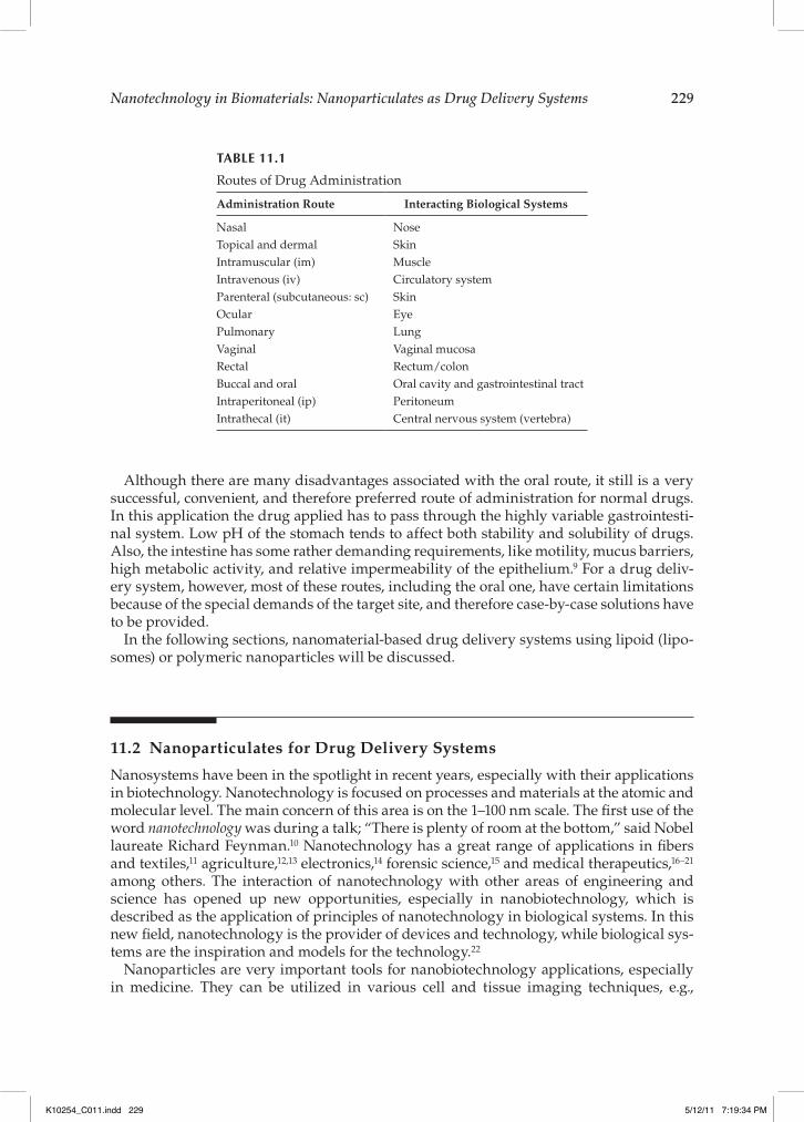
Drug delivery systems need to take into account the route of administration in order to maximize the amount of drug at the site of action. Different routes of administration require different conditions for successful drug delivery. Table 11.1 shows the routes for the administration of drug delivery systems and the biological systems they interact with.
K10254_C011.indd 228 5/12/11 7:19:34 PM
Nanotechnology in Biomaterials: Nanoparticulates as Drug Delivery Systems 229
Although there are many disadvantages associated with the oral route, it still is a very successful, convenient, and therefore preferred route of administration for normal drugs. In this application the drug applied has to pass through the highly variable gastrointesti-nal system. Low pH of the stomach tends to affect both stability and solubility of drugs. Also, the intestine has some rather demanding requirements, like motility, mucus barriers, high metabolic activity, and relative impermeability of the epithelium.9 For a drug deliv-ery system, however, most of these routes, including the oral one, have certain limitations because of the special demands of the target site, and therefore case-by-case solutions have to be provided.
In the following sections, nanomaterial-based drug delivery systems using lipoid (lipo-somes) or polymeric nanoparticles will be discussed.
11.2 Nanoparticulates for Drug Delivery Systems
Nanosystems have been in the spotlight in recent years, especially with their applications in biotechnology. Nanotechnology is focused on processes and materials at the atomic and molecular level. The main concern of this area is on the 1–100 nm scale. The !rst use of the word nanotechnology was during a talk; “There is plenty of room at the bottom,” said Nobel laureate Richard Feynman.10 Nanotechnology has a great range of applications in !bers and textiles,11 agriculture,12,13 electronics,14 forensic science,15 and medical therapeutics,16–21 among others. The interaction of nanotechnology with other areas of engineering and science has opened up new opportunities, especially in nanobiotechnology, which is described as the application of principles of nanotechnology in biological systems. In this new !eld, nanotechnology is the provider of devices and technology, while biological sys-tems are the inspiration and models for the technology.22
Nanoparticles are very important tools for nanobiotechnology applications, especially in medicine. They can be utilized in various cell and tissue imaging techniques, e.g.,
TABLE 11.1
Routes of Drug Administration
Administration Route Interacting Biological Systems
Nasal NoseTopical and dermal SkinIntramuscular (im) MuscleIntravenous (iv) Circulatory systemParenteral (subcutaneous: sc) SkinOcular EyePulmonary LungVaginal Vaginal mucosaRectal Rectum/colonBuccal and oral Oral cavity and gastrointestinal tractIntraperitoneal (ip) PeritoneumIntrathecal (it) Central nervous system (vertebra)
K10254_C011.indd 229 5/12/11 7:19:34 PM
230 Bionanotechnology II: Global Prospects
quantum dots,23 as growth factor delivery materials for tissue engineering,24,25 and for gene26 and drug27 delivery and transport purposes.
In drug delivery applications, nanoparticles are preferred mainly due to their improved bioavailability, solubility, and retention time.28 Encapsulation of drugs in nanoparticles increases the ef!ciency of the drug due to improved localization, especially after extrava-sation, a capability that the microparticles do not have. Moreover, an encapsulated drug is protected from early degradation while in the circulatory system, before reaching the target organ or tissue. Like microparticles, nanoparticles can be designed to target a par-ticular organ or tissue so that the drug accumulates at the predetermined area without excessive dilution along the route.29 In addition to targeted release, controlled or respon-sive release can also be achieved with nanoparticles. The control factors can be pH,30 temperature,31 and light exposure.32 In brief, utilization of nanoparticles in drug delivery increases the therapeutic value of the drug, which then can lead to lower risks and hos-pital costs.
Although nanoparticles are widely utilized in many areas, they may also pose risks to both human health and the environment. These risks have led to the development of a research area called nanotoxicology, which deals with the toxic effects of nanomaterials on living organisms. Although nanoparticles have been found to be dispersed everywhere (air, soil, and water), no exhaustive study has been carried out concerning the long-term detrimental effects of nanoparticles on the environment. However, it is known that there are various adverse health effects of nanoparticles. For example, it is reported that nano-particles can easily enter the body through inhalation, injection, dermal route, and inges-tion, and these particles can be accumulated in the tissues upon entering the body.33 They can also migrate from these entry points to the circulatory and lymphatic systems and ultimately reach organs and tissues. The deposition of these nanoparticles might lead to in"ammation or severe neurological disorders, such as Parkinson’s or Alzheimer’s dis-ease.34 Nanoparticles can also have other effects as a result of accumulation in organs and tissues. It would be ideal if the nanoparticles that have accomplished their function were removed from the body. Some can be removed without any degradation process (exhalation, excretion), while others are metabolized in the body. As a consequence of the degradation of these nanoparticles, several by-products, which can also lead to numerous complications, are formed.33
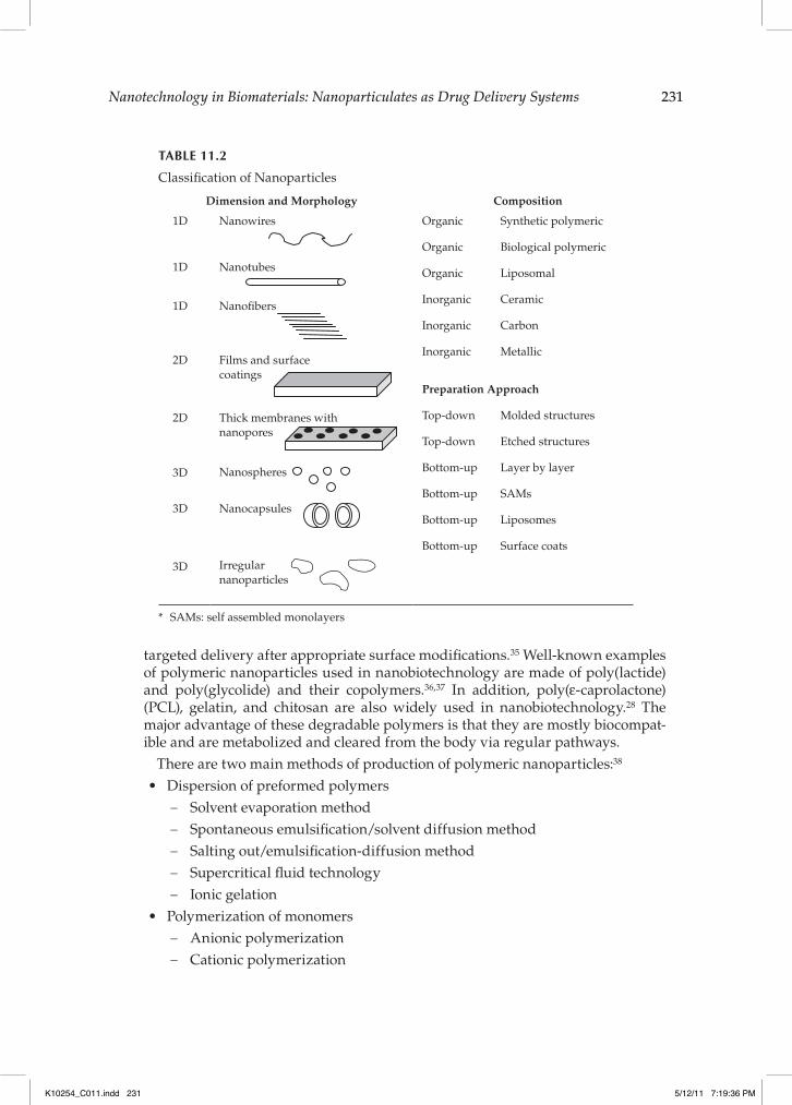
Depending on their application areas, nanosystems can be found in different forms and shapes. They are classi!ed in terms of dimension, morphology, composition, and prepara-tion approach. Table 11.2 presents a schematic summary of nanoparticles classi!ed accord-ing to these three categories.
The nanoparticles used in nanobiotechnology can also be classi!ed according to the materials they are made of, such as polymeric, lipid based, and inorganic:
Polymeric nanoparticles: These nanoparticles can be made of either homo- or copolymers, and their blends in different shapes (e.g., rod, !ber, sphere, etc.). They can be made of degradable or nondegradable, hydrophilic or hydrophobic poly-mers, and can constitute the bulk or the coat of the nanoparticles. Due to their nano size, they have a high surface area-to-volume ratio, indicating that there are more molecules on the surface of a nanoparticle than in the core. Since the sur-face is part of the interface with the tissue, these molecules have the potential to react with the surrounding molecules, which makes nanoparticles proper tools for
K10254_C011.indd 230 5/12/11 7:19:34 PM
Nanotechnology in Biomaterials: Nanoparticulates as Drug Delivery Systems 231
targeted delivery after appropriate surface modi!cations.35 Well-known examples of polymeric nanoparticles used in nanobiotechnology are made of poly(lactide) and poly(glycolide) and their copolymers.36,37 In addition, poly( -caprolactone) (PCL), gelatin, and chitosan are also widely used in nanobiotechnology.28 The major advantage of these degradable polymers is that they are mostly biocompat-ible and are metabolized and cleared from the body via regular pathways.
There are two main methods of production of polymeric nanoparticles:38
Dispersion of preformed polymersSolvent evaporation method !Spontaneous emulsi!cation/solvent diffusion method !Salting out/emulsi!cation-diffusion method !Supercritical "uid technology !Ionic gelation !
Polymerization of monomersAnionic polymerization !Cationic polymerization !
TABLE 11.2
Classi!cation of Nanoparticles
Dimension and Morphology Composition
1D Nanowires
Organic Synthetic polymeric
Organic Biological polymeric
Organic Liposomal
Inorganic Ceramic
Inorganic Carbon
Inorganic Metallic
Preparation Approach
Top-down Molded structures
Top-down Etched structures
Bottom-up Layer by layer
Bottom-up SAMs
Bottom-up Liposomes
Bottom-up Surface coats
1D Nanotubes
1D Nano!bers
2D Films and surfacecoatings
2D Thick membranes withnanopores
3D Nanospheres
3D Nanocapsules
3D Irregularnanoparticles
* SAMs: self assembled monolayers
K10254_C011.indd 231 5/12/11 7:19:36 PM
232 Bionanotechnology II: Global Prospects
Bulk polymerization !Emulsion polymerization !Suspension polymerization !
By using these routes, polymeric nanoparticles carrying bioactive or imaging agents could be prepared.Lipid-based nanoparticles: Lipid-based nanoparticles are useful tools for drug delivery and gene transfer since they protect their content from being cleared by the circulatory system while also protecting the nontarget tissues from adverse effects of the bioactive agent. More importantly, they can be metabo-lized and removed from the body without leaving any by-products behind. Nanoparticles made of phospholipids can be spontaneously self-assembled or prepared by similar processes. Depending on their preparation method, lipid nanoparticles can take different shapes and forms, such as micelles, bilayers, and hexagonal vesicles.39 Lipid-based nanoparticles can have a wide range of sizes (i.e., 20–1000 nm), because of the different methods used in their preparation. Liposomes are a special category of lipid nanoparticles, and their !eld of use in biotechnology is determined by physicochemical characteristics such as com-position, size, loading, and stability, in addition to their interaction with cells. For drug delivery purposes liposomes can be formulated in a suspension, as an aerosol, or in a semisolid form, such as cream, gel, or dry powder.40 In addition to different methods of lipid-based nanoparticle preparation, there also are large numbers of methods for active and passive loading of drugs in these nanopar-ticles. For example, hydrophilic drugs can be encapsulated in the aqueous core while hydrophobic ones can be embedded in the bilayer of the phospholipids. For speci!c purposes, conventional liposomes can be modi!ed; they can be PEGylated (poly(ethylene glycol) modi!ed) or targeted or designed to be charged through the use of functional groups or molecules. These modi!cations alter their interac-tions and control their distribution in the body.Inorganic nanoparticles: Inorganic nanoparticles are preferred in nanobio-technology due to their durability, magnetic properties, "uorescence, and radio opaqueness.41 Calcium phosphate, gold, carbon materials, silicon oxide, iron oxide, cadmium selenide, and zinc selenide are some examples of inorganic nanopar-ticles. Among the metallic nanoparticles, gold nanoparticles are very commonly used for delivery purposes since they can be shaped and brought to the desired size, and their surface can be modi!ed by coating with a variety of molecules. All inorganic nanoparticles can be surface modi!ed to be multifunctional and used in targeting a tissue in drug delivery, to avoid the immune system, and also to "uoresce for imaging.42
11.2.1 Liposomes as Drug Delivery Systems
Liposomes form spontaneously when phospholipids are hydrated. Bangham et al. in 1965 demonstrated that phospholipids form closed structures when dispersed in water, and these structures are relatively impermeable to entrapped material.43 After Bangham et al., there was a great expansion in the use of liposomes as models for biomembranes, for gene delivery, and as drug delivery systems.44 The main constituents of liposomes are phospho-lipids, which have an amphiphilic structure; they have polar and nonpolar regions. The polar regions contact with the aqueous phase inside and outside the liposomes, and the
K10254_C011.indd 232 5/12/11 7:19:36 PM
Nanotechnology in Biomaterials: Nanoparticulates as Drug Delivery Systems 233
nonpolar region is oriented away from the aqueous phase. Liposomes are biocompatible, mask the toxicity of the drug, and increase its time in the circulation, and thus the ef!cacy. Since their discovery, a number of them have been tested in clinical trials, and some of them were approved by the U.S. Food and Drug Administration (FDA).
The properties of liposomes (i.e., size, charge, release kinetics, etc.) depend on their for-mulation and the preparation methods. The spontaneous (and uncontrolled) formation of liposomes leads to a population of vesicles ranging from 10 nm to 10 µm in diameter. Liposomes can entrap hydrophilic and hydrophobic molecules in their aqueous compart-ment and within their membrane, respectively. One has to consider the phase transition temperature, chain length, and degree of saturation of the phospholipids, and the perme-ability of the vesicles toward various compounds. Although liposomes are formed spon-taneously, there are several methods to control their properties and obtain a population with predetermined features.
Liposomes can be designed to be neutral or charged. The major constituents of neutral liposomes are uncharged phosphatidyl cholines (PCs) such as dipalmitoyl phosphatidyl glycerol (DPPG), dipalmitoyl phosphatidyl choline (DPPC), and cholesterol. Phosphatidyl ethanolamine (PE) is used for positively and dipalmitoyl phosphatidic acid (DPPA) for negatively charged liposomes. Sterols, and especially cholesterol, are very important for liposome stability. They themselves cannot form the bilayer structures, but they can be incorporated into the liposomes up to 1:1 molar ratio. Cholesterol presence in the lipid bilayer has a signi!cant effect on the phase transition temperature. As the concentration of cholesterol in the bilayer membrane increases, the phase transition temperature also increases, up to a certain degree. This indicates that cholesterol incorporation increases the stability (and rigidity) of the bilayer. In addition, permeability and "uidity of the mem-brane are also affected by cholesterol.
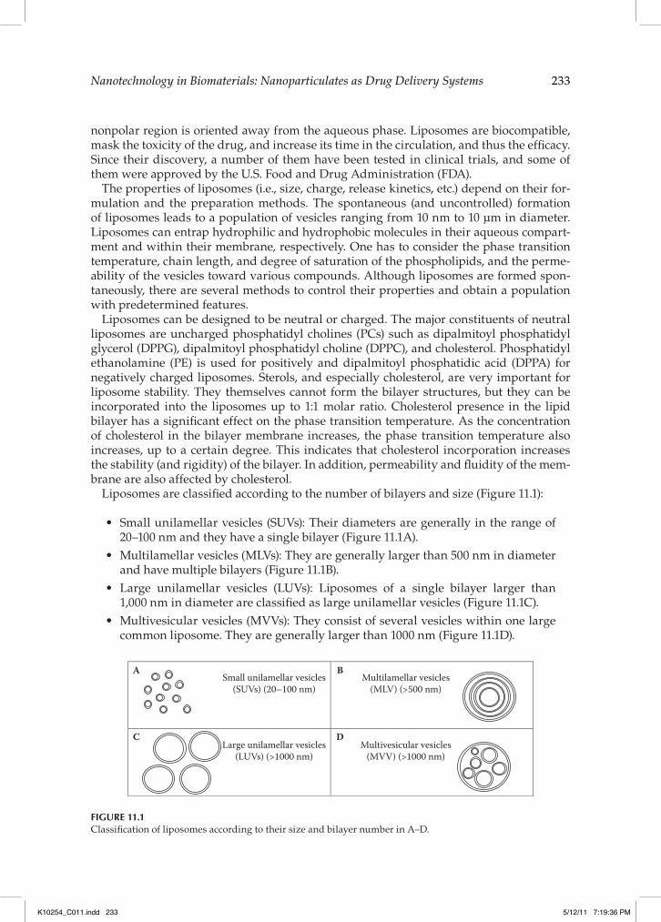
Liposomes are classi!ed according to the number of bilayers and size (Figure 11.1):
Small unilamellar vesicles (SUVs): Their diameters are generally in the range of 20–100 nm and they have a single bilayer (Figure 11.1A).Multilamellar vesicles (MLVs): They are generally larger than 500 nm in diameter and have multiple bilayers (Figure 11.1B).Large unilamellar vesicles (LUVs): Liposomes of a single bilayer larger than 1,000 nm in diameter are classi!ed as large unilamellar vesicles (Figure 11.1C).Multivesicular vesicles (MVVs): They consist of several vesicles within one large common liposome. They are generally larger than 1000 nm (Figure 11.1D).
A
C
BSmall unilamellar vesicles(SUVs) (20–100 nm)
Large unilamellar vesicles(LUVs) (>1000 nm)
Multivesicular vesicles(MVV) (>1000 nm)
Multilamellar vesicles(MLV) (>500 nm)
D
FIGURE 11.1Classi!cation of liposomes according to their size and bilayer number in A–D.
K10254_C011.indd 233 5/12/11 7:19:36 PM

234 Bionanotechnology II: Global Prospects
Liposomes are prepared with different methods used in various preparation steps. Following is a list of methods with parameters that could change the type of liposomes obtained as a result.45
11.2.1.1 Methods of Liposome Preparation Based on Variations in the Replacement of Organic Solvent(s) by the Aqueous Media
11.2.1.1.1 Removal of Organic Solvent(s) before HydrationDuring liposome preparation all lipid-soluble membrane ingredients are dissolved in an organic solvent in order to ensure a homogeneous membrane. In the removal of this organic solvent a rotary evaporator is the most common device used.46 The organic solvent is evaporated and the lipid is dried on the walls of the glass "ask. Then this dried lipid is hydrated and used in liposome production. Compounds to be carried in the lipoid mem-brane are added during the solution phase, and those to be carried in the aqueous part of the liposomes are added in the hydration step. This leads to small unilamellar vesicles (SUVs) and multilamellar vesicles (MLVs).
11.2.1.1.2 Reverse Phase EvaporationIn this type of liposome preparation, the !rst step is removal of the organic solvent, as above with a rotary evaporator. The lipids are then redissolved in the organic phase, the aqueous phase is added, and the resulting two-phase system is sonicated until the mixture becomes a homogeneous suspension of oil in water. The organic solvent is removed under vacuum in a rotary evaporator, and the aqueous suspension obtained at the end is called the reverse phase evaporation vesicle (REV).47 This method leads to very high encapsula-tion ef!ciencies and large unilamellar vesicles (LUVs).
11.2.1.1.3 Use of Water-Immiscible SolventsThis technique involves injection of an immiscible organic solution of the phospholipid through a needle into the aqueous phase in a very slow fashion while the organic phase is removed by vaporization. Large vesicles are formed as a result of this process. Ether injec-tion is a common example for this method.48 This method has very little risk of oxidative degradation of the phospholipids.
11.2.1.1.4 Use of Water-Miscible SolventsEthanol injection is a good example for this method. In ethanol injection lipid solution is prepared in ethanol and injected into an aqueous medium through a !ne needle. The force of injection also achieves mixing, resulting in an evenly dispersed phospholipid solution.49 With this technique small unilamellar vesicles (SUVs) are obtained.
Thus, even with a single parameter change, a large variety of liposomes are produced, proving the versatility of liposomes.
11.2.1.2 Applications of Liposomes
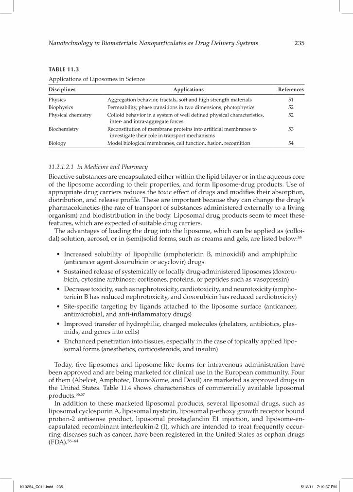
Liposomes are widely used in cosmetic products and pharmaceuticals, and they are also used as analytical tools in many disciplines of science50 (Table 11.3).
Liposomes are used in drug delivery, as controlled and sustained release systems, and in medical diagnostics and gene therapy. They are also used as a model in cell recognition and interaction studies, and in the investigation of the mode of action of active substances in pharmacology and medicine.
K10254_C011.indd 234 5/12/11 7:19:36 PM
Nanotechnology in Biomaterials: Nanoparticulates as Drug Delivery Systems 235
11.2.1.2.1 In Medicine and PharmacyBioactive substances are encapsulated either within the lipid bilayer or in the aqueous core of the liposome according to their properties, and form liposome-drug products. Use of appropriate drug carriers reduces the toxic effect of drugs and modi!es their absorption, distribution, and release pro!le. These are important because they can change the drug’s pharmacokinetics (the rate of transport of substances administered externally to a living organism) and biodistribution in the body. Liposomal drug products seem to meet these features, which are expected of suitable drug carriers.
The advantages of loading the drug into the liposome, which can be applied as (colloi-dal) solution, aerosol, or in (semi)solid forms, such as creams and gels, are listed below:55
Increased solubility of lipophilic (amphotericin B, minoxidil) and amphiphilic (anticancer agent doxorubicin or acyclovir) drugsSustained release of systemically or locally drug-administered liposomes (doxoru-bicin, cytosine arabinose, cortisones, proteins, or peptides such as vasopressin)Decrease toxicity, such as nephrotoxicity, cardiotoxicity, and neurotoxicity (ampho-tericin B has reduced nephrotoxicity, and doxorubicin has reduced cardiotoxicity)Site-speci!c targeting by ligands attached to the liposome surface (anticancer, antimicrobial, and anti-in"ammatory drugs)Improved transfer of hydrophilic, charged molecules (chelators, antibiotics, plas-mids, and genes into cells)Enchanced penetration into tissues, especially in the case of topically applied lipo-somal forms (anesthetics, corticosteroids, and insulin)
Today, !ve liposomes and liposome-like forms for intravenous administration have been approved and are being marketed for clinical use in the European community. Four of them (Abelcet, Amphotec, DaunoXome, and Doxil) are marketed as approved drugs in the United States. Table 11.4 shows characteristics of commercially available liposomal products.56,57
In addition to these marketed liposomal products, several liposomal drugs, such as liposomal cyclosporin A, liposomal nystatin, liposomal p-ethoxy growth receptor bound protein-2 antisense product, liposomal prostaglandin E1 injection, and liposome-en-capsulated recombinant interleukin-2 (1), which are intended to treat frequently occur-ring diseases such as cancer, have been registered in the United States as orphan drugs (FDA).56–64
TABLE 11.3
Applications of Liposomes in Science
Disciplines Applications References
Physics Aggregation behavior, fractals, soft and high strength materials 51Biophysics Permeability, phase transitions in two dimensions, photophysics 52Physical chemistry Colloid behavior in a system of well de!ned physical characteristics,
inter- and intra-aggregate forces 52
Biochemistry Reconstitution of membrane proteins into arti!cial membranes to investigate their role in transport mechanisms
53
Biology Model biological membranes, cell function, fusion, recognition 54
K10254_C011.indd 235 5/12/11 7:19:37 PM

236 Bionanotechnology II: Global Prospects
11.2.1.2.2 In CosmeticsIn cosmetics, liposomes’ moisturizing and restoring properties are generally exploited.
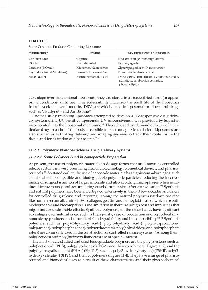
The !rst liposomal cosmetic products Capture (C. Dior) and Niosomes (L’Oréal) were intro-duced in 1987. Today, many more liposome-containing products, such as Hydra-Radiance Liposome Cream (Avon), Revision Liposomal Cream (Revision SkinCare), and Lipo"ow Forte plus C (Lipo"ow), have been developed and are sold in the marketplace. Liposomal cosmetic products are used for hydrating skin, reducing skin dryness (the main reason for aging and wrinkles), and replacing skin lipids.56 Liposomes are also known to contain recombinant proteins for wound or sunburn healing. Not only antiaging skin creams, but also unrinsable sunscreens, long-lasting perfumes, hair conditioners, aftershaves, and similar products have liposomes in their formulations. Table 11.5 shows some of these products.66
Liposomes provide many advantages in cosmetic products, such as shorter applica-tion periods, decreased side effects, delayed washing out (water resistant), increased skin hydration and increased penetration in to the deeper layers of the epidermis which are absorbed in, etc.
At our research center (BIOMATEN), different types of liposomes are being developed recently. In one study dehydration-rehydration vesicles (DRVs) were developed for use as a drug delivery system to achieve personalized treatment of patients with dermal ail-ments such as sunburn and psoriasis.67 DRVs were selected because they have a speci!c
TABLE 11.4
Characteristics of Commercially Available Liposomal Products
Trade Name, Bioactive Agent, Type of Dosage Form, Company
Phospholipids and Drug/Lipid Ratio
Liposome Type
Particle Size References
AmBisome®, amphotericin B, freeze-dried liposomes, Gilead Sciences/Fujisawa Healthcare
HSPCDSPGCHOL
-TocopherolDrug:lipid 1:6 (w:w)
SUV 100 nm 58, 59
Albelcet®, amphotericin B, liposomal suspension, Enzon
DMPCDMPGDrug:lipid 1:1 molar
MLV 5 #m 60
DaunoXome®, daunorubicin, liposomal suspension, Gilead Sciences
DSPCCHOLDrug:lipid 18.7:1 (w:w)
SUV ca. 45 nm 61
Doxil®, doxorubicin, liposomal suspension, J&J ALZA
MPEG-DSPEHSPCDrug:lipid 1:6 (w:w)
LUV 100 nm 62
Myocet®, doxorubicin, liposomal suspension, Elan
Egg-PCCHOLDrug:lipid 1:4 (w:w)
MLV 180 nm 63
Visudyne™, Vertepor!n®, benzoporphyrin, freeze-dried liposomes, QLT/Novartis
Egg-DMPCAscorbyl palmitateBHTDrug:lipid 1:7.5–15 (w:w)
SUV 100 nm 64
Abbreviations: SUV, small unilamellar vesicle; MLV, multilamellar vesicle; LUV, large unilamellar vesicle; HSPC, hydrogenated soy phosphatidyl choline; PC, phosphatidyl choline; DSPG, distearoylphosphatidyl glycerol; CHOL, cholesterol; DSPC, distearoylphosphatidyl choline; PG, phosphatidyl glycerol; DMPC, dimyris-toylphosphatidyl choline; MPEG-DSPE, N-(carbonylmethoxypoly(ethylene glycol) 2000)-1, 2-distearoyl-sn- glycero-3-phosphoethanolamine; BHT, butylated hydroxytoluene.
K10254_C011.indd 236 5/12/11 7:19:37 PM
Nanotechnology in Biomaterials: Nanoparticulates as Drug Delivery Systems 237
advantage over conventional liposomes; they are stored in a freeze-dried form (in appro-priate conditions) until use. This substantially increases the shelf life of the liposomes from 1 week to several months. DRVs are widely used in liposomal products and drugs such as Visudyne™ and AmBisome®.
Another study involving liposomes attempted to develop a UV-responsive drug deliv-ery system using UV-sensitive liposomes. UV responsiveness was provided by Suprofen incorporated into the liposomal membrane.68 This achieved on-demand delivery of a par-ticular drug in a site of the body accessible to electromagnetic radiation. Liposomes are also studied as both drug delivery and imaging systems to track their route inside the tissue and for detection of disease sites.69,70
11.2.2 Polymeric Nanoparticles as Drug Delivery Systems
11.2.2.1 Some Polymers Used in Nanoparticle Preparation
At present, the use of polymeric materials in dosage forms that are known as controlled release systems is a very promising area of biotechnology, biomedical devices, and pharma-ceuticals.71 As stated earlier, the use of nanoscale materials has signi!cant advantages, such as injectable biocompatible and biodegradable polymeric particles, reducing the inconve-nience of surgical insertion of larger implants and also avoiding macrophages when intro-duced intravenously and accumulating at solid tumor sites after extravasation.72 Synthetic and natural polymers have been investigated extensively in the last few decades as carriers for controlled drug release and targeting. Among the natural polymers used are proteins like human serum albumin (HSA), collagen, gelatin, and hemoglobin, all of which are both biodegradable and biocompatible. One limitation in their use is high cost and impurities that might induce undesirable effects. Synthetic polymers, on the other hand, have signi!cant advantages over natural ones, such as high purity, ease of production and reproducibility, nontoxic by-products, and controllable biodegradability and biocompatibility.72–74 Synthetic polymers such as poly( -hydroxy acids), poly( -hydroxy acids), poly(!-caprolactone), poly(amides), poly(phosphazenes), poly(orthoesters), poly(anhydrides), and poly(phosphate esters) are commonly used in the construction of controlled release systems.75 Among them, poly(lactides) and poly(hydroxyalkanoates) are of special interest.
The most widely studied and used biodegradable polymers are the poly( -esters), such as poly(lactic acid) (PLA), poly(glycolic acid) (PGA), and their copolymers (Figure 11.2), and the poly(hydroxyalkanoates) (PHAs) (Fig 11.3), such as poly(3-hydroxybutyrate) (P3HB), poly(3-hydroxyvalerate) (P3HV), and their copolymers (Figure 11.4). They have a range of pharma-ceutical and biomedical uses as a result of these characteristics and their physicochemical
TABLE 11.5
Some Cosmetic Products Containing Liposomes
Manufacturer Product Key Ingredients of Liposomes
Christian Dior Capture Liposomes in gel with ingredientsL’Oréal Efect du Soleil Tanning agentsLancome (L’Oréal) Niosomes, Nactosomes Glyceropolyether with moisturizerPayot (Ferdinand Muehlens) Formule Liposome Gel Thymoxin, hyaluronic acidEstee Lauder Future Perfect Skin Gel TMF, (Methyl trimethicone) vitamins E and A
palmitate, cerebroside ceramide, phospholipids
K10254_C011.indd 237 5/12/11 7:19:37 PM

238 Bionanotechnology II: Global Prospects
properties. These polymers and their degradation products have been shown to be nontoxic and biocompatible, and products made using these polyesters have been awarded FDA approval.
For more than three decades, linear polymers of lactide and glycolide have been used in various medical applications. Use of these polymers as carriers for controlled delivery of a wide range of bioactive agents for human and animal use has been an attractive research area.75 The properties of these linear polyesters have been modi!ed by making copoly-mers and by varying the ratios of the monomers in the copolymer with respect to each other. Therefore, copolymers such as poly(lactide-co-glycolide) (PLGA) (Figure 11.2) have been extensively investigated for various medical applications, including drug delivery devices, scaffolds for tissue engineering, surgical sutures, bone pins, and stents.76 PLGA was shown to be biocompatible in many applications and degrades into the nontoxic and natural products lactic acid and glycolic acid, which can be eliminated from the body as they are or after conversion into CO2 and H2O.77
PHAs (Figure 11.3), on the other hand are the most commonly used natural biotechno-logical polymers, because they are made by bacteria as well as by other organisms, some of which are transgenic.78 They are rapidly hydrolyzed in the environment, but more slowly in the human body, and this rate could be regulated by varying the composition, form, and size of the biomedical device.
The best known polymer of the PHA family is poly( -hydroxybutyrate) (P3HB). The mechanical properties, crystallinity, and solubility of the polyesters can also be controlled by their composition, molecular weight, and heterogeneity index (the molecular weight distribution in a given batch).74
11.2.2.2 Nanoparticles
Nanoparticles have a diameter in the range of 5–1000 nm. They can be irregular in shape or spherical. Spherical nanoparticles can be full in the interior with the matrix material or empty. Nanospheres are nanoparticles that are spherical and full, and hollow spherical sys-tems are called nanocapsules, where a core is surrounded by a membrane (Figure 11.5).71
O
x y
CH3
CH2CH2
GA LA
C O
O
C O
FIGURE 11.2Molecular structure of poly(lactic acid-co-glycolic acid) block copolymer. x and y can be in the range of several thousands, and the x:y ratio could vary between 100:0 and 0:100.
O
C
R
CH (CH2)m O
n
FIGURE 11.3Molecular structure of poly(hydroxyalkanoates). When m 1 and R CH3, the product is poly(3-hydroxybu-tyrate) (P3HB).
K10254_C011.indd 238 5/12/11 7:19:37 PM

Nanotechnology in Biomaterials: Nanoparticulates as Drug Delivery Systems 239
In the current research activities in many biomaterials laboratories, both types of these important biomedical polyesters are used to construct carriers for various bioactive agents. The followings are results from studies carried out in our laboratories.
11.2.2.2.1 Preparation and Characterization of Nano-Microspheres Designed for Controlled Release Purposes
11.2.2.2.1.1 Preparation PHBV and PLGA nano-microspheres were prepared by an oil-in-water (o/w) emulsion technique. These polymers are dissolved in an organic solvent such as dichloromethane, which is immiscible with water. Bioactive agents are dissolved in the organic solvent, and this organic solution is added to an aqueous solution to form an o/w emulsion, and thus the droplets. The aqueous phase generally contains an emulsi!er such as poly(vinylalcohol) (PVA), Tween, or Span to provide stabilization of the droplets. Emulsi!cation is achieved by sonication, and continuous mixing prevents cluster formation before the solvent evaporates at room temperature. Ultracentrifugation is generally used to collect the nano-microspheres. Excess aqueous and organic solvents are removed by wash-ing the nano-microparticles, and they are dried in a vacuum oven or by lyophilization.
11.2.2.2.1.2 Characterization of PLGA and PHBV Nano-Microspheres The various nano-microspheres of PHBV and PLGA were characterized for their size, size distribution, and surface topography using various methods such as a particle size analyzer, scanning electron microscopy (SEM), and an image analysis program (e.g., Scion Image of National Institutes of Health (NIH)), and "uorescence microscopy for "uorescent agent-loaded nano-microspheres.
In Figure 11.6, the in"uence of the drying method on the quality of recovery of PLGA (50:50) nano-microspheres is presented. It can be seen that even a simple stage of the pro-cess has a signi!cant in"uence on the nano-microspheres obtained.
O
O
O
OC C
3-HB x y3-HV
CH
CH3
CH2
CH3
CH2
CH2CH
FIGURE 11.4Molecular structure of PHBV. x and y can be in the range of several thousands.
Nanosphere Nanocapsule
FIGURE 11.5Schematic representation of nanospheres and nanocapsules carrying drugs in their structure.
K10254_C011.indd 239 5/12/11 7:19:38 PM
240 Bionanotechnology II: Global Prospects
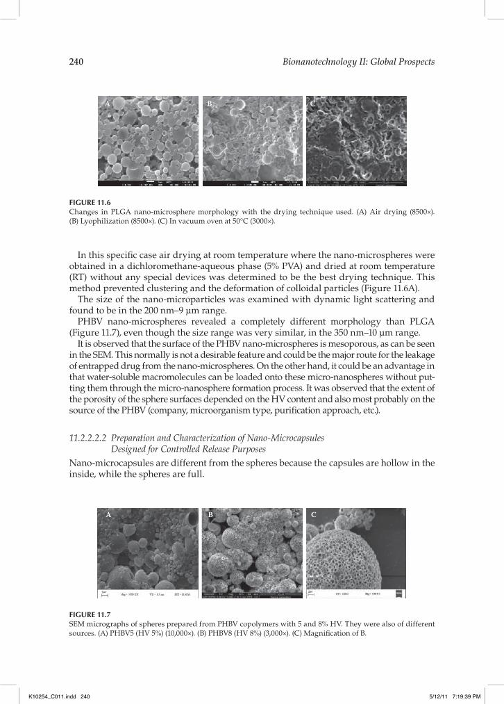
In this speci!c case air drying at room temperature where the nano-microspheres were obtained in a dichloromethane-aqueous phase (5% PVA) and dried at room temperature (RT) without any special devices was determined to be the best drying technique. This method prevented clustering and the deformation of colloidal particles (Figure 11.6A).
The size of the nano-microparticles was examined with dynamic light scattering and found to be in the 200 nm–9 µm range.
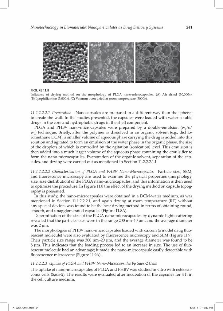
PHBV nano-microspheres revealed a completely different morphology than PLGA (Figure 11.7), even though the size range was very similar, in the 350 nm–10 µm range.
It is observed that the surface of the PHBV nano-microspheres is mesoporous, as can be seen in the SEM. This normally is not a desirable feature and could be the major route for the leakage of entrapped drug from the nano-microspheres. On the other hand, it could be an advantage in that water-soluble macromolecules can be loaded onto these micro-nanospheres without put-ting them through the micro-nanosphere formation process. It was observed that the extent of the porosity of the sphere surfaces depended on the HV content and also most probably on the source of the PHBV (company, microorganism type, puri!cation approach, etc.).
11.2.2.2.2 Preparation and Characterization of Nano-Microcapsules Designed for Controlled Release Purposes
Nano-microcapsules are different from the spheres because the capsules are hollow in the inside, while the spheres are full.
A B C
FIGURE 11.6Changes in PLGA nano-microsphere morphology with the drying technique used. (A) Air drying (8500$). (B) Lyophilization (8500$). (C) In vacuum oven at 50 C (3000$).
A B C
FIGURE 11.7SEM micrographs of spheres prepared from PHBV copolymers with 5 and 8% HV. They were also of different sources. (A) PHBV5 (HV 5%) (10,000$). (B) PHBV8 (HV 8%) (3,000$). (C) Magni!cation of B.
K10254_C011.indd 240 5/12/11 7:19:39 PM
Nanotechnology in Biomaterials: Nanoparticulates as Drug Delivery Systems 241
11.2.2.2.2.1 Preparation Nanocapsules are prepared in a different way than the spheres to create the wall. In the studies presented, the capsules were loaded with water-soluble drugs in the core and hydrophobic drugs in the shell component.
PLGA and PHBV nano-microcapsules were prepared by a double-emulsion (w1/o/w2) technique. Brie"y, after the polymer is dissolved in an organic solvent (e.g., dichlo-romethane DCM), a smaller volume of aqueous phase carrying the drug is added into this solution and agitated to form an emulsion of the water phase in the organic phase, the size of the droplets of which is controlled by the agitation (sonication) level. This emulsion is then added into a much larger volume of the aqueous phase containing the emulsi!er to form the nano-microcapsules. Evaporation of the organic solvent, separation of the cap-sules, and drying were carried out as mentioned in Section 11.2.2.2.1.1.
11.2.2.2.2.2 Characterization of PLGA and PHBV Nano-Microcapsules Particle size, SEM, and "uorescence microscopy are used to examine the physical properties (morphology, size, size distribution) of the PLGA nano-microcapsules, and this information is then used to optimize the procedure. In Figure 11.8 the effect of the drying method on capsule topog-raphy is presented.
In this study, the nano-microcapsules were obtained in a DCM-water medium, as was mentioned in Section 11.1.2.2.2.1, and again drying at room temperature (RT) without any special devices was found to be the best drying method in terms of obtaining round, smooth, and unagglomerated capsules (Figure 11.8A).
Determination of the size of the PLGA nano-microcapsules by dynamic light scattering revealed that the particle sizes were in the range 200 nm–10 µm, and the average diameter was 2 µm.
The morphologies of PHBV nano-microcapsules loaded with calcein (a model drug "uo-rescent molecule) were also evaluated by "uorescence microscopy and SEM (Figure 11.9). Their particle size range was 300 nm–20 µm, and the average diameter was found to be 8 µm. This indicates that the loading process led to an increase in size. The use of "uo-rescent molecule had an advantage; it made the nano-microcapsule easily detectable with "uorescence microscope (Figure 11.9A).
11.2.2.2.3 Uptake of PLGA and PHBV Nano-Microcapsules by Saos-2 CellsThe uptake of nano-microcapsules of PLGA and PHBV was studied in vitro with osteosar-coma cells (Saos-2). The results were evaluated after incubation of the capsules for 4 h in the cell culture medium.
A B C
FIGURE 11.8In"uence of drying method on the morphology of PLGA nano-microcapsules. (A) Air dried (30,000$). (B) Lyophilization (3,000$). (C) Vacuum oven dried at room temperature (3000$).
K10254_C011.indd 241 5/12/11 7:19:39 PM

242 Bionanotechnology II: Global Prospects
After the nano-microcapsules of PHBV and PLGA were stained with Nile red 0.1% (v/v), which has a red emission under "uorescence microscopy, Saos-2 cells were incu-bated with the nano-microparticles for 4 h. Before examination, the cells were stained with FITC ("uorescein isothio cyanate)-labeled phalloidin and 4’,6-diamidino-2-phenylindole (DAPI), respectively, dyes that stain the cell nuclei blue and the cell cytoskeleton green. The cells were then examined under "uorescence microscope (Figure 11.10). It has to be remembered that the sizes of most capsules are lower than the resolution of the "uorescent microscope (in the nano-range), so some of the red regions probably represent clusters of nanocapsules, while the others that are seen as individual specks could be micron sized. It can be observed that PHBV and PLGA nano-microcapsules are taken up by Saos-2 cells. An interesting !nding is that the capsules are generally located near or on the cell nuclei, implying that they could serve as carriers of agents for gene therapy because they seem to be able to avoid lysosomes and accumulate in the vicinity of the nuclei.
A B
FIGURE 11.9Fluorescence and SEM micrographs of calcein-loaded PHBV nano-microcapsules. (A) Scale bar is 100 µm. (B) 30,000$.
A B
FIGURE 11.10(See color insert.) Fluorescence micrographs of nano-microcapsules taken up by Saos-2 cells. (A) PHBV, 40$. (B) PLGA, 20$. Cell nuclei were stained with DAPI (blue), cell cytoskeletons were stained with FITC-labeled phal-loidin (green), and nano-microcapsules were stained with Nile red (red).
K10254_C011.indd 242 5/12/11 7:19:40 PM

Nanotechnology in Biomaterials: Nanoparticulates as Drug Delivery Systems 243
11.3 Conclusion
Nano- and low-micron-sized particles, whether polymeric or lipid based, are very versa-tile tools for drug delivery applications. They can be made in various sizes, from various materials, can carry molecules of different chemistries, and can be modi!ed for targeting purposes. As such, they are indispensable as drug carriers. And with the advent of nano-technology, which provides tools such as quantum dots, nanowires, and nanorods, the !eld has become more exciting than ever.
Acknowledgments
We gratefully acknowledge the support by METU (BAP 07.02.2009.00.01) through which part of this study was conducted.
References 1. Jain, K.K., Drug delivery systems Methods in molecular biology, Vol. 437, Humana Press, New York,
2008, chap. 1. 2. Uekama, K., Hirayama, F., and Irie, T., Cyclodextrin drug carrier systems, Chem. Rev., 98,
2045, 1998. 3. Pankhurst, Q.A., et al., Applications of magnetic nanoparticles in biomedicine, J. Phys. D Appl.
Phys., 36, R167, 2003. 4. Torchillin, V., Multifunctional pharmaceutical nanocarriers, Vol. 4, Springer, New York, 2008. 5. Carmeliet, P., and Jain, R.K., Angiogenesis in cancer and other diseases, Nature, 407, 249, 2000. 6. Maeda, H., et al., Tumor vascular permeability and the EPR effect in macromolecular therapeu-
tics: a review, J. Control. Release, 65, 271, 2000. 7. Gerweck, L.E., and Seetharaman, K., Cellular pH gradient in tumor versus normal tissue:
potential exploitation for the treatment of cancer, Cancer Res., 56, 1194, 1996. 8. Cook, J.A., et al., Oxidative stress, redox, and the tumor microenvironment, Semin. Radiat.
Oncol., 14, 259, 2004. 9. Malmsten, M., Surfactants and polymers in drug delivery, Marcel Dekker, New York, 2002. 10. Feynman R.P., There is plenty of room at the bottom, paper presented at Annual Meeting of the
American Physical Society, California Institute of Technology, December 29, 1959. 11. Dubas, S.T., Kumlangdudsana, P., and Potiyaraj, P., Layer-by-layer deposition of antimicrobial
silver nanoparticles on textile !bers, Colloids Surf. A, 289, 105, 2006. 12. Navrotsky, A., Nanomaterials in the environment, agriculture, and technology (NEAT), J.
Nanoparticle Res., 2, 321, 2000. 13. González-Melendi, P., et al., Nanoparticles as smart treatment-delivery systems in plants:
assessment of different techniques of microscopy for their visualization in plant tissues, Ann. Bot., 101, 187, 2008.
14. Kruis, F.E., Fissan, H., and Peled, A., Synthesis of nanoparticles in the gas phase for electronic, optical and magnetic applications—a review, J. Aerosol Sci., 29, 511, 1998.
15. Choi, M.J., et al., Metal-containing nanoparticles and nano-structured particles in !ngermark detection, Forensic Sci. Int., 179, 87, 2008.
K10254_C011.indd 243 5/12/11 7:19:40 PM

244 Bionanotechnology II: Global Prospects
16. Bonduelle, S., et al., Association of cyclosporin to isohexylcyanoacrylate nanospheres and sub-sequent release in human plasma in vitro, J. Microencapsul., 9, 173, 1991.
17. Bender, A.R., et al., Ef!ciency of nanoparticles as a carrier system for antiviral agents in human immunode!ciency virus-infected human monocytes/macrophages in vitro, Antimicrob. Agents Chemother., 40, 1467, 1996.
18. Kawashima, Y., et al., Mucoadhesive D,L-lactide/glycolide copolymer nanospheres coated with chitosan to improve oral delivery of elcatonin, Pharm. Dev. Technol., 5, 77, 2000.
19. Salata, O.V., Applications of nanoparticles in biology and medicine, J. Nanobiotechnol., 2, 2004. 20. Rieux, A.D., et al., Nanoparticles as potential oral delivery systems of proteins and vaccines: a
mechanistic approach, J. Control. Release, 116, 1, 2006. 21. Jahanshahi, M., and Babaei, Z., Protein nanoparticle: a unique system as drug delivery vehicles,
Afr. J. Biotechnol., 7, 4926, 2008. 22. Roco, M.C., Nanotechnology: convergence with modern biology and medicine, Curr. Opin., 14,
337, 2003. 23. Michalet, X., et al., Quantum dots for live cells, in vivo imaging, and diagnostics, Science, 307,
538, 2005. 24. Basmanav, F.B, Kose, G.T., and Hasirci, V., Sequential growth factor delivery from complexed
microspheres for bone tissue engineering, Biomaterials, 29, 4195, 2008. 25. Yilgor, P., et al., Incorporation of a sequential BMP-2/BMP-7 delivery system into chitosan-
based scaffolds for bone tissue engineering, Biomaterials, 30, 3551, 2009. 26. Mao, H.-Q., et al., Chitosan-DNA nanoparticles as gene carriers: synthesis, characterization and
transfection ef!ciency, J. Control. Release, 70, 399, 2001. 27. Panyam, J., and Labhasetwar, V., Biodegradable nanoparticles for drug and gene delivery to
cells and tissue, Adv. Drug Deliver. Rev., 55, 329, 2003. 28. Kumari, A., Yadav, S.K., and Yadav, S.C., Biodegradable polymeric nanoparticles based drug
delivery systems, Colloid Surf. B, 75, 1, 2010. 29. Alexis, F., et al., Factors affecting the clearance and biodistribution of polymeric nanoparticles,
Mol. Pharm., 5, 505, 2008. 30. Potineni, A., et al., Poly(ethylene oxide)-modi!ed poly( -amino ester) nanoparticles as a pH-
sensitive biodegradable system for paclitaxel delivery, J. Control. Release, 86, 223, 2003. 31. Sershen, S.R., et al., Temperature-sensitive polymer-nanoshell composites for photothermally
modulated drug delivery, J. Biomed. Mater. Res., 51, 293, 2000. 32. Cirli, O.O., and Hasirci, V., UV-induced drug release from photoactive REV sensitized by supro-
fen, J. Control. Release, 96, 85, 2004. 33. Oberdorster, G., Oberdorster, E., and Oberdorster, J., Nanotoxicology: an emerging discipline
evolving from studies of ultra!ne particles, Environ. Health Perspect., 113, 823, 2005. 34. Peters, A., et al., Translocation and potential neurological effects of !ne and ultra!ne particles
a critical update, Particle Fibre Toxicol., 3, 2006. 35. Hans, M.L., and Lowman, A.M., Biodegradable nanoparticles for drug delivery and targeting,
Curr. Opin. Solid State Mater., 6, 319, 2002. 36. Stolnik, S., et al., Surface modi!cation of poly(lactide-co-glycolide) nanospheres by biodegrad-
able poly(lactide)-poly(ethylene glycol) copolymers, Pharmaceut. Res., 11, 1800, 1994. 37. Katanec, D., Paveli, B., and Ivasovi, Z., Ef!ciency of polylactide/polyglycolide copolymers
bone replacements in bone defects healing measured by densitometry, Collegium Antropol., 28, 331, 2004.
38. Soppimath, K.S., et al., Biodegradable polymeric nanoparticles as drug delivery devices, J. Control. Release, 70, 1, 2001.
39. Israelachvili, J.N., Marcelja, S., and Horn, R.G., Physical principles of membrane organization, Q. Rev. Biophys., 13, 121, 1980.
40. Zhang, Y.P., Ceh, B., and Lasic, D.D., Liposomes in drug delivery, in Polymeric biomaterials, 2nd ed., Bumitriu S., ed., Marcel Dekkar Inc., New York, 2002, p. 783.
41. Xu, Z.P., et al., Inorganic nanoparticles as carriers for ef!cient cellular delivery, Chem. Eng. Sci., 61, 1027, 2006.
K10254_C011.indd 244 5/12/11 7:19:40 PM
Nanotechnology in Biomaterials: Nanoparticulates as Drug Delivery Systems 245
42. Liong, M., et al., Multifunctional inorganic nanoparticles for imaging, targeting, and drug delivery, Am. Chem. Soc., 2, 889, 2008.
43. Bangham, A.D., Standish, M.M., and Watkins J.C., The !rst description of liposomes, J. Mol. Biol., 13, 238, 1965.
44. Wang, B., Siahaan, T., and Soltero, R., Drug delivery: principles and applications, 1st ed., John Wiley & Sons, New Jersey, 2005.
45. Verma, A.M.L., et al., Liposomes as carrier systems, InPharm Communique, 2, 20, 2009. 46. Torchillin, V., and Weissig, V., Liposomes: a practical approach, 1st ed., Oxford University Press,
Oxford, 1990. 47. Szoka, F., Jr., and Papahadjopoulos, D., Procedure for preparation of liposomes with large inter-
nal aqueous space and high capture by reverse-phase evaporation, Proc. Natl. Acad. Sci. USA, 9, 4194, 1978.
48. Deamer, D., and Bangham, A.D., Large volume liposomes by an ether vaporization method, BBA Biomembranes, 443, 629, 1976.
49. Batzri, S., and Korn, E.D., Single bilayer liposomes prepared without sonication, BBA Biomembranes, 298, 1015, 1973.
50. Barenholz, Y., Liposome application: problems and prospects, Curr. Opin. Colloid Interface, 6, 66, 2001.
51. Pajic-Lijakovic, I., et al., Rheological quanti!cation of liposomes aggregation, Minerva Biotechnol., 17, 245, 2005.
52. Ulrich, S.A., Biophysical aspects of using liposomes as delivery vehicles, Bioscience Rep., 22, 129, 2002.
53. Jemioat-Rzeminska, M., Latowski, B., and Strzalka, K., Incorporation of plastoquinone and ubiquinone into liposome membranes studied by HPLC analysis. The effect of side chain length and redox state of quinine. Chem. Phys. Lipids, 110, 85, 2001.
54. Yuba, E., et al., pH-sensitive fusogenic polymer-modi!ed liposomes as a carrier of antigenic proteins for activation of cellular immunity, Biomaterials, 31, 943, 2010.
55. Lasic, D.D., Liposomes, Am. Sci., 80, 20, 1992. 56. Janoff, S.A., Liposome: rational design, 1st ed., Marcel Dekker, New York, 1999. 57. Barenholz, Y., Relevancy of drug loading to liposomal formulation therapeutic ef!cacy, J.
Liposome Res., 13, 1, 2003. 58. Linden, P.K., Amphotericin B lipid complex for the treatment of invasive fungal infections,
Expert Opin. Pharmacother., 4, 2099, 2003. 59. Adler-Moore, J., and Prof!tt, R.T., AmBisome: liposomal formulation, structure, mechanism of
action and pre-clinical experience, J. Antimicrob. Chemother., 49, 21, 2002. 60. Veerareddy, P.R., and Vobalaboina, V., Lipid-based formulations of amphotericin B, Drugs
Today, 40, 33, 2004. 61. Hempel, G., et al., Population pharmacokinetics of liposomal daunorubicin in children. Br. J.
Clin. Pharmacol., 56, 370, 2003. 62. Waterhouse, D.N., et al., A comparison of liposomal formulations of doxorubicin with drug
administered in free form: changing toxicity pro!les, Drug Safety, 24, 903, 2001. 63. Allen, T.M., and Martin, F.J., Advantages of liposomal delivery systems for anthracyclines,
Semin. Oncol., 31, 5, 2004. 64. Chowdhary, R.K., Shariff, I., and Dolphin, D., Drug release characteristics of lipid based benzo-
porphyrin derivative, J. Pharm. Pharm. Sci., 6, 13, 2003. 65. www.fda.gov/orphan/designat/list.htm. 66. Lasic, D.D., and Barenholz, Y., Handbook of nonmedical applications of liposomes: theory and basic
sciences, Vol. 1, CRC Press, Boca Raton, FL, 1996. 67. Kardesler, S., et al., Topical delivery systems for the treatment of skin diseases, in International
Symposium on Biotechnology: development and Trends, Ankara, Turkey, September 27–30, 2009, p. 112. 68. Demirbag, B., and Hasirci, V., UV responsive drug delivery from suprofen incorporated lipo-
somes, in 15th International Biomedical Science and Technology Symposium, Guzelyurt, TRNC, August 16–19, 2009, p. 84.
K10254_C011.indd 245 5/12/11 7:19:40 PM
246 Bionanotechnology II: Global Prospects
69. Buyuksungur, A., and Hasirci, V., Bioactive agent carrying quantum dot labeled liposomes, in 15th International Biomedical Science and Technology Symposium, Guzelyurt, TRNC, August 16–19, 2009, p. 112.
70. Buyuksungur, A., Padeste, C., and Hasirci V., Entrapment in liposome prevents toxicity of quantum dots, in International Symposium on Biotechnology: Development and Trends, Ankara, Turkey, September 27–30, 2009, p. 113.
71. Barratt, G., et al., Polymeric micro and nanoparticles as drug carriers, in Polymeric biomaterials, 2nd ed., Dumitriu, D., ed., Marcel Dekker, New York, 2002, chap. 28.
72. Jain, R.A., The manufacturing techniques of various drug loaded biodegradable poly(lactide-co-glycolide) (PLGA) devices, Biomaterials, 21, 2475, 2000.
73. Astete, C.E., and Sabliov, C.M., Synthesis and characterization of PLGA nanoparticles, J. Biomater. Sci. Pol. Ed., 17, 247, 2006.
74. Nair, L.S., and Laurencin, C.T., Polymers as biomaterials for tissue engineering and controlled drug delivery, Adv. Biochem. Eng./Biotechnol., 102, 47, 2006.
75. Domb, J.A., et al., Biodegradable polymers as drug carrier systems, in Polymeric biomaterials, 2nd ed., Dumitriu, D., ed., Marcel Dekker, New York, 2002, chap. 4.
76. Jain, R.A., et al., Controlled delivery of drugs from a novel injectable in situ formed biodegrad-able PLGA microsphere system, J. Microencapsul., 17, 343, 2000.
77. Stevanovic, M., and Uskokovic, D., Poly(lactide-co-glycolide)-based micro and nanoparticles for the controlled drug delivery of vitamins, Curr. Nanosci., 5, 1, 2009.
78. Errico, C., et al., Poly(hydroxyalkanoates)-based polymeric nanoparticles for drug delivery, J. Biomed. Biotechnol Vol 2009, p. 1–10, 2009.
K10254_C011.indd 246 5/12/11 7:19:40 PM