Nanomaterials with Antimicrobial Properties: Applications...
12
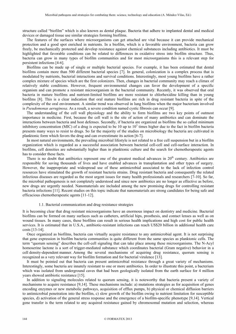
Nanomaterials with Antimicrobial Properties: Applications in Health Sciences C. L. Santos 1 , A. J. R. Albuquerque 2 , F. C. Sampaio 2 and D. Keyson 3 1 Post-graduation in Chemistry, Federal University of Paraiba, Centre for Natural and Exact Sciences, Campus I, Joao Pessoa, Paraiba, 58051-900, Brazil 2 RENORBIO, Northeast Network of Biotechnology, Federal University of Paraiba, Biotechnology Centre, Campus I, Joao Pessoa, Paraiba, 58051-900, Brazil 3 IDEP UFPB Institute for Development of Paraiba, Federal University of Paraíba, Campus I, Joao Pessoa, Paraiba, 58051-900, Brazil Drug resistant microorganisms are a serious and increasing public health problem. New strategies for controlling bacteria activity are urgently needed and nanomaterials can be a very promising approach. This review focuses on nanotechnology strategies for inhibiting cellular adhesion and attachment, interfering in bacterial physiology and communication (quorum sensing), and avoiding biofilm development. It is well established that metallic compounds can have or may increase antimicrobial activity. However, the antimicrobial potential of metallic nanostructured particles and their mechanisms of action are still on debate. For instance, recent studies showed that nanostructured surfaces can neutralize these electrostatic forces reducing the cellular adhesion and as consequence the chances of biofilm formation. This is particular interesting for dental materials since persistent oral bacterial adhesion takes place and for medical devices where resistant microorganisms can form pathogenic biofilms. The technology behind the nanomaterial synthesis is a key point to consider. Several techniques are available exploring reactions in solid state and reactions that involve chemical methods for wet-chemical synthesis such as: sol-gel, co-precipitation, polymeric precursor method, hydrothermal methods. These nanostructure particles can be of controllable size with large surface area in relation to their molecular weight. These features have led to different approaches (e.g. incorporating nanoparticles in solid materials or diffusing them into medicines) and many hypotheses about the mechanism of action have been proposed: a) Release of toxic ions (Cd2+, Zn2+, Ag+ ) that can bind to sulfur-containing proteins of the cell membrane and interfere in cell permeability; b) Toxic ions that can cause DNA damage; c) Interruption of electron transport, protein oxidation and membrane potential collapse due to its contact with CeO2 or nC60; d) Generation of ROS (Reactive Oxygen Species) that can cause disruption of cell membrane. These mechanisms may not operate separately suggesting that more than one mechanism can occur simultaneously. Another point to consider is the difference in effectiveness against some bacteria and fungi species. For instance, it is accepted that inhibitory activity of silver ions is higher for Gram negative bacteria than for Gram positive species of similar behavior. Several examples of nanomaterials with antimicrobial properties can be found in dentistry and medicine. These nanostructured particles are already present in restorative materials, in catheters and curatives. Fluorescent staining plus microscopic investigation and Zeta potentials are relevant techniques for evaluation of bacteria viability. It must be pointed out that nanotechnology is also available for other health applications such as biological markers for cancer treatments. Finally, the adverse effects of nanomaterials cannot be ruled out. Thus, more clinical trials as well as research in the level of molecular biology are needed for a safely use. Moreover, the production and synthesis of the nanomaterials have to consider the application of green nanotechnology principles assessing the environmental risks of manufactured nanomaterials. Keywords Nanomaterials; antimicrobials agents; biofilms; drug resistance 1. Biofilms: a successful microbial lifestyle Bacteria are the smallest organisms that contain all the structures and organelles required for growth and reproduction. Although very small, with a diameter around 10 -3 mm, bacterium has an effective osmotic barrier provided by a cell membrane and a cell wall that avoids mechanical damage. Some bacteria have substantially differences in their wall structure, components, and functions. For instance, the thickness of a layer of peptidoglycan divides bacteria into two classes according to their ability to retain a basic dye (crystal violet). This observation was first described by Hans Christian Joachim Gram 130 years ago and it is still valid. Basically, Gram positive bacterium has a very thick layer of peptidoglycan (20-80 nm) whereas Gram negative bacterium has a thin peptidoglycan layer (8 nm) with an outer membrane [1]. In addition to cell wall and membrane, some bacteria may present another important structure: the capsule. This bacterial cover can act as a barrier to toxic hydrophobic molecules, such as detergents, and can promote adherence to other bacteria or to host tissue surfaces. For cariogenic bacteria (microorganisms related to dental caries development) such as Streptococcus mutans, the polysaccharide capsules are the means by which the bacteria attach and stick to the tooth enamel [2-3]. These extracellular polysaccharides (mainly dextran and levan), are synthesized by the enzymes dextransucrase and levansucrase which are present on or near the bacterial cell surface. After bacteria get attached to the tooth surface, the polysaccharides can favor bacterial interactions and aggregation leading to a rich bacterial living Microbial pathogens and strategies for combating them: science, technology and education (A. Méndez-Vilas, Ed.) © FORMATEX 2013 ____________________________________________________________________________________________ 143
Transcript of Nanomaterials with Antimicrobial Properties: Applications...

Nanomaterials with Antimicrobial Properties: Applications in Health Sciences
C. L. Santos1, A. J. R. Albuquerque2, F. C. Sampaio2 and D. Keyson3 1Post-graduation in Chemistry, Federal University of Paraiba, Centre for Natural and Exact Sciences, Campus I, Joao
Pessoa, Paraiba, 58051-900, Brazil 2RENORBIO, Northeast Network of Biotechnology, Federal University of Paraiba, Biotechnology Centre, Campus I,
Joao Pessoa, Paraiba, 58051-900, Brazil 3IDEP UFPB Institute for Development of Paraiba, Federal University of Paraíba, Campus I, Joao Pessoa, Paraiba,
58051-900, Brazil
Drug resistant microorganisms are a serious and increasing public health problem. New strategies for controlling bacteria activity are urgently needed and nanomaterials can be a very promising approach. This review focuses on nanotechnology strategies for inhibiting cellular adhesion and attachment, interfering in bacterial physiology and communication (quorum sensing), and avoiding biofilm development. It is well established that metallic compounds can have or may increase antimicrobial activity. However, the antimicrobial potential of metallic nanostructured particles and their mechanisms of action are still on debate. For instance, recent studies showed that nanostructured surfaces can neutralize these electrostatic forces reducing the cellular adhesion and as consequence the chances of biofilm formation. This is particular interesting for dental materials since persistent oral bacterial adhesion takes place and for medical devices where resistant microorganisms can form pathogenic biofilms. The technology behind the nanomaterial synthesis is a key point to consider. Several techniques are available exploring reactions in solid state and reactions that involve chemical methods for wet-chemical synthesis such as: sol-gel, co-precipitation, polymeric precursor method, hydrothermal methods. These nanostructure particles can be of controllable size with large surface area in relation to their molecular weight. These features have led to different approaches (e.g. incorporating nanoparticles in solid materials or diffusing them into medicines) and many hypotheses about the mechanism of action have been proposed: a) Release of toxic ions (Cd2+, Zn2+, Ag+ ) that can bind to sulfur-containing proteins of the cell membrane and interfere in cell permeability; b) Toxic ions that can cause DNA damage; c) Interruption of electron transport, protein oxidation and membrane potential collapse due to its contact with CeO2 or nC60; d) Generation of ROS (Reactive Oxygen Species) that can cause disruption of cell membrane. These mechanisms may not operate separately suggesting that more than one mechanism can occur simultaneously. Another point to consider is the difference in effectiveness against some bacteria and fungi species. For instance, it is accepted that inhibitory activity of silver ions is higher for Gram negative bacteria than for Gram positive species of similar behavior. Several examples of nanomaterials with antimicrobial properties can be found in dentistry and medicine. These nanostructured particles are already present in restorative materials, in catheters and curatives. Fluorescent staining plus microscopic investigation and Zeta potentials are relevant techniques for evaluation of bacteria viability. It must be pointed out that nanotechnology is also available for other health applications such as biological markers for cancer treatments. Finally, the adverse effects of nanomaterials cannot be ruled out. Thus, more clinical trials as well as research in the level of molecular biology are needed for a safely use. Moreover, the production and synthesis of the nanomaterials have to consider the application of green nanotechnology principles assessing the environmental risks of manufactured nanomaterials.
Keywords Nanomaterials; antimicrobials agents; biofilms; drug resistance
1. Biofilms: a successful microbial lifestyle
Bacteria are the smallest organisms that contain all the structures and organelles required for growth and reproduction. Although very small, with a diameter around 10-3 mm, bacterium has an effective osmotic barrier provided by a cell membrane and a cell wall that avoids mechanical damage. Some bacteria have substantially differences in their wall structure, components, and functions. For instance, the thickness of a layer of peptidoglycan divides bacteria into two classes according to their ability to retain a basic dye (crystal violet). This observation was first described by Hans Christian Joachim Gram 130 years ago and it is still valid. Basically, Gram positive bacterium has a very thick layer of peptidoglycan (20-80 nm) whereas Gram negative bacterium has a thin peptidoglycan layer (8 nm) with an outer membrane [1]. In addition to cell wall and membrane, some bacteria may present another important structure: the capsule. This bacterial cover can act as a barrier to toxic hydrophobic molecules, such as detergents, and can promote adherence to other bacteria or to host tissue surfaces. For cariogenic bacteria (microorganisms related to dental caries development) such as Streptococcus mutans, the polysaccharide capsules are the means by which the bacteria attach and stick to the tooth enamel [2-3]. These extracellular polysaccharides (mainly dextran and levan), are synthesized by the enzymes dextransucrase and levansucrase which are present on or near the bacterial cell surface. After bacteria get attached to the tooth surface, the polysaccharides can favor bacterial interactions and aggregation leading to a rich bacterial living
Microbial pathogens and strategies for combating them: science, technology and education (A. Méndez-Vilas, Ed.)
© FORMATEX 2013
____________________________________________________________________________________________
143
structure called “biofilm” which is also known as dental plaque. Bacteria that adhere to implanted dental and medical devices or damaged tissue use similar strategies forming biofilms. The features of the surfaces where microorganisms can get attached are vital because it can provide mechanical protection and a good spot enriched in nutrients. In a biofilm, which is a favorable environment, bacteria can grow freely, be mechanically protected and develop resistance against chemical substances including antibiotics. It must be highlighted that favorable conditions can be related to differences in oxidative stress into biofilm structure. Thus, bacteria can grow in many types of biofilm communities and for most microorganisms this is a relevant step for persistent infections [4-6]. Biofilms can be made up of single or multiple bacterial species. For example, it has been estimated that dental biofilms contain more than 500 different bacterial species [7]. In general, colonization is a complex process that is modulated by nutrients, bacterial interactions and survival conditions. Interestingly, most young biofilms have a rather complex mixture of species which are the first colonizers. Then, changes in bacterial community may reach a climax of relatively stable conditions. However, frequent environmental changes can favor the development of a specific organism and can promote a resistant microorganism in the bacterial community. Recently, it was observed that oral bacteria in mature biofilms and nutrient-limited biofilms are more resistant to chlorhexidine killing than in young biofilms [8]. This is a clear indication that oral mature biofilms are rich in drug resistant bacteria in spite of the complexity of the oral environment. A similar trend was observed in lung biofilms when the major bacterium involved is Pseudomonas aeruginosa. As a result, a severe condition named cystic fibrosis can occur [9]. The understanding of the cell wall physiology and the ability to form biofilms are two key points of outmost importance in medicine. First, because the cell wall is the site of action of many antibiotics and can dominate the interactions between bacteria and host defenses. Secondly, if bacteria are organized as biofilms the so called minimum inhibitory concentration (MIC) of a drug is expected to be 10 up to 103 times higher due to the fact in biofilm bacteria presents many ways to resist to drugs. So far the majority of the studies on microbiology the bacteria are cultivated in planktonic form which favors the drug and can overestimate its action [6-7]. In most natural environments, the prevailing microbial lifestyle is not related to a free cell suspension but in a biofilm organization which is regarded as a successful association between bacterial cell-cell and cell-surface interaction. In biofilms, cell densities are substantially higher than in planktonic culture and the search for chemotherapeutic agents has to consider these facts. There is no doubt that antibiotics represent one of the greatest medical advances in 20th century. Antibiotics are responsible for saving thousands of lives and have enabled advances in transplantation and other types of surgery. However, the inappropriate and widespread use of these antimicrobial associated to the lack of infection control resources have stimulated the growth of resistant bacteria strains. Drug resistant bacteria and consequently the related infectious diseases are regarded as the most urgent issues for many health professionals and researchers [7-10]. So far, the microbial pathogenesis is not completely understood and since new antibiotics are no longer as effective as before, new drugs are urgently needed. Nanomaterials are included among the new promising drugs for controlling resistant bacteria infections [11]. Recent studies on this topic indicate that nanomaterials are strong candidates for being safe and efficacious chemotherapeutic agents [11-12].
1.1. Bacterial communication and drug resistance strategies
It is becoming clear that drug resistant microorganisms have an enormous impact on dentistry and medicine. Bacterial biofilms can be formed on many surfaces such as catheters, artificial hips, prosthesis, and contact lenses as well as on wound tissues. In many cases, these biofilms can result in serious health implications and high cost for public health services. It is estimated that in U.S.A., antibiotic-resistant infections can reach US$20 billion in additional health care costs [13-14]. Once organized as biofilms, bacteria can virtually acquire resistance to any antimicrobial agent. It is not surprising that gene expression in biofilm bacteria communities is quite different from the same species as planktonic cells. The term “quorum sensing” describes the cell-cell signaling that can take place among these microorganisms. The N-Acyl homoserine lactone is a sort of trigger-mediated substance which coordinates bacterial (Gram negative) behavior in a cell density-dependent manner. Among the several mechanisms of acquiring drug resistance, quorum sensing is recognized as a very relevant way for biofilm formation and for bacterial virulence [13]. It must be pointed out that bacteria can present antimicrobial resistance through a great variety of mechanisms. Interestingly, some bacteria are innately resistant to one or more antibiotics. In order to illustrate this point, a bacterium which was isolated from underground caves that had been geologically isolated from the earth surface for 4 million years showed antibiotic resistance [15]. In addition to signaling molecules related to quorum sensing, it is noteworthy that bacteria present a variety of mechanisms to acquire resistance [9,14]. These mechanisms include: a) mutations strategies as for acquisition of genes encoding enzymes or new metabolic pathways, acquisition of efflux pumps, b) physical or chemical diffusion barriers to antimicrobial penetration into the biofilm, c) slow growth of the biofilm owing to nutrient limitation for susceptible species, d) activation of the general stress response and the emergence of a biofilm-specific phenotype [9,14]. Vertical gene transfer is the term related to any acquired resistance gained by chromosomal mutation and selection, whereas
Microbial pathogens and strategies for combating them: science, technology and education (A. Méndez-Vilas, Ed.)
© FORMATEX 2013
____________________________________________________________________________________________
144

horizontal gene transfer stands for resistance obtained by acquisition of new genetic material from resistant microorganisms [16].
2. Antimicrobial agents and nanotechnology
While bacteria reach a size of 10-6 m, nanotechnology deals with structures as small as 10-9 m. In spite of this 10-3 m difference, the development of many technologies in the 1980s made possible the combination of these two worlds. As a result, nanotechnology evolved to a relevant branch of nanomedicine providing ideas of several potential applications in health sciences. Recent applications in biomedical research include a great variety of features and applications in nanobiotechnology and nanomedicine [12]. The development of materials or devices designed to interact with the body at sub-cellular scales imply some degree of specificity. Therefore, nanobiotechnology is a science under construction and for the sake of developing a drug with maximal therapeutic effects with limited adverse-effects a significant volume of research is still necessary. At least eight broad areas of nanotechnology are regarded as pertinent for developing research in biomedicine: synthesis and use of nanostructures, applications of nanotechnology to therapy, biomimetic and biologic nanostructures, electronic-biology interface, devices for early detection of disease, tools for the study of single molecules, nanotechnology and tissue engineering [17]. Taking into account the applications of nanotechnology to therapy, there is an important topic which is the potential application of nanotechnology for developing effective antimicrobials. It has been suggested that nanoparticles rely on very different mechanisms of antimicrobial activity when compared to antibiotics. For instance, Staphylococcus aureus is a bacterium that is naturally susceptible to virtually all antibiotics developed. Resistance is often acquired by horizontal transfer to genes from outside sources and may be related to their ability to adapt to beta-lactamase enzymes neutralizing penicillin due to the cleavage of the beta-lactam ring [18-19]. As for the antimicrobial mechanism of the nanoparticles against this microorganism and many others, it is known that it is probably related to its surface area. In other words, the smallest nanoparticles possess the strongest antimicrobial effect [18]. So, there is no doubt that size matters, and this is one of the reasons why nanoparticles are regarded as a real improvement in antimicrobial strategy. Although antibiotic molecules are smaller than any nanoparticle, one must bear in mind that small proteins size 5-10 nm are in the range of some nanoparticles that can vary from 1 to 10 nm. Taking into account this scale, bacteria are around 5000 nm which means 50 up to 5000 times bigger than nanoparticles [20-23]. Figure 1 illustrates the impact of size when comparing these entities.
Fig. 1 The sizes of biologically relevant entities. Left to right: molecule of cerium oxide, antibiotic molecule, ceria nanoparticle, virus and bacteria.
For the antibiotic molecule, size is important but several chemical and physical properties of antibiotic molecules have to be considered when evaluating its efficacy and ability to penetrate bacteria (e.g. hydrophobicity, stoichiometry and charge). Therefore, the rate of permeation of antibiotic through bacteria porin channels is not only a matter of size [24]. This might also be true for nanoparticles. Actually, the mechanism of antimicrobial activity of nanoparticles is still under debate though several hypotheses have been proposed [12,17].
3. Antimicrobial potential of nanomaterials
There are a substantial number of articles about the potential antimicrobial activity of nanoparticles, particularly testing metallic nanoparticles [12, 18, 20-22, 25-32, 34, 36-41]. The antimicrobial activity of many types of nanoparticles is certainly a function of their size but other features are important such as high surface area, unusual crystal morphologies (edges and corners) and reactive sites [26]. It is recognized that the main mechanism or the pools of mechanisms behind the antimicrobial activity of these nanostructures are not fully elucidated. Hence, several studies focusing on the antimicrobial activity of different metals and metallic nanoparticles against many species of bacteria and fungi have to be carried out for a clear picture on this matter [30-31].
Microbial pathogens and strategies for combating them: science, technology and education (A. Méndez-Vilas, Ed.)
© FORMATEX 2013
____________________________________________________________________________________________
145

Metals have been utilized as potent antimicrobials for centuries [33-34]. However, for biological systems as bacteria and mammalian cells, metals ions are also vital for many physiological processes. Obviously, only trace amounts of metal ions (e. g. copper, iron, cobalt) are required by both prokaryotic and eukaryotic cells [35]. For instance, iron is a cofactor of many enzymes and, as such, plays a vital role in several cell physiological processes, such as DNA replication, DNA transcription and central metabolism [36]. Thus, excessive levels of essential metals are harmful for living organisms [37]. Nanoparticles present different chemical features when compared to their bulk equivalent. In other words, materials in a nanostructure size may have the most favorable form since at least 50% of the molecules present in the surface of particles will eventually react to the microorganism. Therefore, the small size of the particle gives large surface area and consequently reactivity (and in many cases toxicity) will increase substantially [38-40]. The most tested metallic nanoparticles are silver, copper, gold, aluminum, titanium, iron, zinc, and more recently, the list can include: ceria, bismuth and others [25-32, 36-41]. It must be pointed out that some of these metals have been coated onto several materials. Another strategies are to incorporated these metals into a substrate such as poly-methylmethacrylate (PMMA) or dental restorative materials [26,38,39,42]. With respect to bacteria and fungi, the most frequent candidates for microbial experiments are: Staphylococcus aureus, Pseudomonas aeruginosa, Escherichia coli, Klebsiella pneumonia, Bacillus subtilis among other species [19,21,23,26,28,29,43]. A serious point of discussion is the evaluation of the antimicrobial activity of metallic nanoparticles between Gram positive and Gram negative bacteria [26,43]. Several aspects have to be considered when performing a comparison of metals activity and bacteria species. First, the results and understandings of bulk material do not apply for particles at nanoscale level. Second, there are a great variety of methods which partially explains the variation in the results. Third, the peptidoglycan plays an important role on the activity of the nanoparticles, but this effect might be modulated by other features of the milieu, such as pH or the metal structure or carrier [18,26,43]. For instance, it is has been observed that inhibitory activity of silver ions is higher for oral Gram-negative bacteria than for Gram-positive species of similar behavior [44]. However, there are studies that found different results including the observation of similar antimicrobial effect regardless the fact that the bacteria tested was Gram-positive or Gram-negative [23]. As an interesting example, there is the evaluation of the antimicrobial activity of CuO nanoparticles. It was found to be size-dependent as expected by many other studies. But the copper oxide nanoparticles synthesized during this study demonstrated a significant increase in antibacterial activities against both Gram-positive and -negative bacterial strains. Nevertheless, it is suggested that a reduction in the peptidoglycans in the Gram-negative species may increase their susceptibility when exposed to nanoparticles [44-46]. The effects of silver nanoparticles at a concentration of 10 μg cm−3 inhibited Escherichia coli growth by 70%. There was a significant reduction in the size of bacterial colonies when E. coli was cultivated on plates with more than 20 μg cm−3 of nanoparticles and there was no grow for 50–60 μg cm−3. The formation of “pits” in the cell wall was observed by image techniques suggesting the accumulation of the nanoparticles in the bacterial membrane [45]. However, penetration of silver nanoparticles into the cytoplasm of Gram-negative bacteria (E. coli) cannot be ruled out [46]. Conversely, under similar conditions, the thick peptidoglycan layer of Staphylococcus aureus (Gram-positive) compared to E. coli, prevented the penetration of silver particles into bacteria. Hence, the antimicrobial effect of nanoparticles can be related to the interaction with the bacterial surfaces when dealing with Gram-positive strains, whereas for Gram-negative the penetration of the particles can be expected to take place depending on size, charge and other features of the material [47,48-50]. Taking into account the future of antimicrobial nanomaterials and their health applications, it can be recognized that so far the studies have given a clear indication that nanoparticles have a promising future. These nanostructures alone or incorporated into materials can improve the performance of many products. Compared to antibiotics, there is the advantage of more efficiently facilitate the contact of drug-target sites, deliver other drugs or mixture of substances or antibiotics that have diffusion limitations. Moreover, nanostructures can be more safely recommended since it appears that many microorganisms are more likely to acquire resistance against conventional antibiotics than from nanoparticles [47]. Although this is not a consensus since there is a publication of possible development of horizontal gene transfer (P. aeruginosa) from silver nanoparticles [42]. Although some limitations will certainly come over time (e.g. toxicological aspects, adverse effects), nanoparticles are still opening a new universe of possibilities in the medical field. Current and future applications are innumerous when considering the specific field of antimicrobials. For instance, the strategies to incorporate nanoparticles in dental materials are related to ameliorate physical properties and also antimicrobial activity reducing the potential of bacterial get attached to dental filling surfaces [26]. The same principle is applied in medical devices such as catheters [49]. In recent years, in the literature, has described with success the preparation of several nanostructures, such like CuO, Cu2O, Cu, Se, CdS, Bi2S3, SnS2, γ-Fe2O3, ZnO and CeO2 that can be incorporated in many materials [51-54].
4. Nanomaterials synthesis
Metal particles are particularly interesting for nanoscale systems due to the fact that they can be chemically synthesized and modified using different techniques. In addition to the variety of synthetic routes, nanoparticles can offer the advantage of some similar features (optical or dielectric constants) as those of the bulk metal. Moreover, simplification
Microbial pathogens and strategies for combating them: science, technology and education (A. Méndez-Vilas, Ed.)
© FORMATEX 2013
____________________________________________________________________________________________
146

of methods has been reported. For instance, synthesis of nanospheres processed in a domestic microwave-hydrothermal oven is already possible. The reactions for obtaining nanoparticles are classified in agreement with the physical state of the used precursory materials. The methods of synthesis of nanoparticles can include attrition and pyrolysis. While some methods are bottoms up, some are called top down. The top down methods involve breaking the larger materials into nanoparticles [55]. Finally, the reactions can be classified into two major groups: a) reactions in solid state (e.g.: ceramic method or mixture of oxides) and b) the reactions that involve chemical methods for wet-chemical synthesis (e.g.: sol-gel, co-precipitation, polymeric precursors methods, hydrothermal methods) [56-64].
4.1. Reactions in solid state
Among the common methods for nanoparticles preparation which involves the reaction in the solid state, mechanical synthesis is a remarkable one. The Mechanical Synthesis (MS) it is a technique of processing ceramic powders. It allows producing a great variety of crystalline materials, amorphous and solid solutions, with rather stable nanometric and metastable structures. MS appeared in the end of the sixties, focusing on the production of nickel superalloy. It is a method of great interest, because it allows the reduction of the granulometry of the phases involved in the material, favoring a considerable chemical interaction among them and reducing problems related to the stoichiometry of the material under mild temperatures [65]. The process is accomplished when a solid mixture is submitted to a physical treatment of grinding that involves random collisions of particles in high energy. For that, several types of mills are employed, being the mill of balls the most common. As a good example, nanostructures of Ce1-xZrxO2 (x≤0.2) were obtained starting from the mixtures of CeCl3 and ZrCl4 in sodium hydroxide, for treatment of MS at different times of grinding (5 and 15 hours). It was obtained nanoparticles with a final composition of Ce0.8Zr0.2O2. The synthesized solid solution presented good thermal stability, high superficial area and grains of nanometric dimensions after elimination of NaCl produced in the reaction, and drying to 600 ºC [65,66]. Another MS for ceria production is related to the following reaction : 2CeCl3
. 6H2O(s) + 3Na2CO3.10H2O(s) → Ce2(CO3)nH2O(s) + 6 NaCl (s) + (42 - n) H2O
This synthesis was induced starting from the grinding process and a subsequent calcination of the precursory powders that, together with the by-product NaCl, took to the formation of ceria nanoparticles of 40-70 nm [66-67].
4.2. Reactions for wet-chemical synthesis
Aiming a better control of some properties of nanoparticles such a size, morphology and stoichiometry of the process, new methods of production have been proposed. The crystallization dynamics in wet-chemical synthesis is the key parameter for controlling morphology of metallic nanoparticles. The wet-chemical methods are preferable than MS because their final product present better characteristic (e.g.: larger homogeneity among the reagents, high purity of the products and low processing temperature) [68]. The co-precipitation method and sol-gel routes have been gradually substituting more conventional methods like MS for synthesis of high temperature superconducting oxides. Three different kinds of sol-gel techniques have been used: a) colloidal sol-gel, b) inorganic polymeric gel derived from organometallic compounds (Fig. 2) and c) gel routes involving formation of organic polymeric glass. The primary goal in all sol-gel techniques is the preparation of a homogeneous precursor solution. Taking into account that a homogeneous precursor is obtained, it is expected that a semi-rigid gel can be isolated with an atomic level homogeneity [68,69]. The sol-gel is particularly important for medicine because it made possible the development of techniques for entrapment of enzymes, methods for medical tests and coated implants [70]. The gel routes involving formation of organic polymeric net, also known as Pechini method, are of particular interest due to the fact that it is rather simple, versatile and with good cost-benefit ratio. Moreover, it uses a route with low temperature for burning which is in tune with green chemistry principles [69]. The form and the size of the particles of the ceramic powders produced by Pechini method are determined by the morphology of the precursor. Hence, precursors of high porosity and fragility are preferable for production of mixed oxides without great agglomerates.
Fig. 2 Proposed reactions for the preparation of CeO2 using polymeric percursors.
Microbial pathogens and strategies for combating them: science, technology and education (A. Méndez-Vilas, Ed.)
© FORMATEX 2013
____________________________________________________________________________________________
147

Applications of this method for coating of antimicrobials are many, and some are already available. Recently, ZnO nanoparticles were prepared by the Pechini method from a polyester by reacting citric acid with ethylene glycol in which the metal ions are dissolved, and incorporated into blend films of chitosan and poly (vinyl alcohol) (PVA). This material proved to have antimicrobial activity toward Staphylococcus aureus [71]. In dentistry and medicine, ultrasound radiation is widely used mostly for diagnosis. However, ultrasound can be used for cleaning materials, chemical processes, preparation of emulsions and suspensions and also for non-destructive evaluation of materials. Also known as sonochemistry, the chemical decomposition is regarded to be an excellent method for preparing nanoparticles. Recently, many types of nanoparticles have been prepared with a variety of metals [40,57]. Basically, the synthesis consists of the use of sound waves with superior lengths to 20 KHZ and is receiving attention of many research groups due to the simplified experimental process [40]. Some facilities in sonochemistry are the possibility of using small amounts of reagents and temperatures below 200°C and consequent low energy consumption (Green Chemistry). The theory behinds sonochemistry is explained by the effect of sonic radiation. This radiation can break chemical bonds and as a result there is the creation, growth and collapse of a bubble that is formed in the liquid. This phenomenon is known as “acoustic cavitation”. The estimated size of the collapsing bubble varies from ten to a few hundred microns and these bubbles favor the orientation of growth of the materials starting from smaller energy plans [40]. The precipitation process, used for the production of several types of materials consists in the formation of an insoluble solid product. Several parameters are of great importance in this stage (temperature, pH of the solution and concentration of the reagents) in order to guarantee the formation of fine particles with large surface area and chemical homogeneity [72]. After the precipitation stage, the intermediate product is submitted to a washing phase, until pH stability. Then it is filtered, dried and calcined. Another important route for nanoparticles synthesis is the microwave-hydrothermal method. This process have called attention of researchers groups because both inorganic as well as organic materials can be prepared. In addition, the whole process is carried out in closed systems (isolated) and using low temperatures. Thus, there are both environmental and economical advantages [73]. The microwave-hydrothermal method can promote the formation of materials of good chemical homogeneity, satisfactory chemical yield and reproducibility with low costs. Therefore, several publications are supporting this technique for preparation of nanoparticles which is taking place over conventional hydrothermal methods [61, 73].
5. Nanomaterials as antimicrobials: mechanisms of action
A classical antimicrobial agent needs to have the ability to reach vital molecular target sites involved in bacterial metabolism such as cell wall synthesis. Besides, it needs avoid ejection by efflux bombs and molecule modification by enzymes. Several natural and engineered nanomaterials have demonstrated strong antimicrobial properties through diverse mechanisms including photocatalytic production of reactive oxygen species that damage cell components and viruses, compromising the bacterial cell capsule, interruption of energy transduction and inhibition of enzyme activity and DNA synthesis. Some of these mechanisms are summarized and illustrated in figure 3. Basically, six major mechanisms have received more attention though more mechanisms have been proposed [12,23,26,74-83].
5.1. Cell membrane damage
The mechanism of action of nanoparticles on cell membranes is recognized as non-specific and it is not known if polymixins has any involvement on this process. Polymixins antibiotics can interact on cell membranes breaking vital barriers of microorganisms [42]. There is no doubt that cell permeability is altered when in contact to nanoparticles. Experiments indicated the formation of a “hole” or “pore” in the living cell membranes as a possible mechanistic hypothesis [84]. Although the meaning of the term “hole” or “pore” still requires clarification, images of cell damage has given clear evidence of this effect. In more serious or extreme cases a literal hole in the bilayer membrane exists which promotes the complete loss of the plasma membrane [84].
5.2. Release of toxic ions
It has been demonstrated that Cd2+, Zn2+, Ag+ ions can react in bacteria with different groups of proteins. The ability of Ag+ in forming sparingly soluble salts is also considered as one of its major mechanisms for attacking bacteria cells. For instance, cell respiration is inhibited when the chloride ions precipitate as silver chloride in the cytoplasm of the cells. The antimicrobial efficiency of silver nanoparticles is also well-known, especially against Gram-negative bacteria such as E.coli. Hence, the silver nanoparticles act as antimicrobial releasing silver ions and also by penetrating cells interfering in their metabolic systems [82]. Although silver has received much attention, Cd2+ and Zn2+ ions can also bind to sulfur-containing proteins of the cell membrane and interfere in cell permeability. There are good evidences that Ag+ ions can also damage DNA by inhibiting its replication. The concentrations required for bactericidal activity of silver nanoparticles are low (in the range 10(-9) mol/l) [82].
Microbial pathogens and strategies for combating them: science, technology and education (A. Méndez-Vilas, Ed.)
© FORMATEX 2013
____________________________________________________________________________________________
148
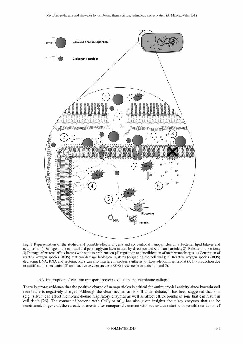
Fig. 3 Representation of the studied and possible effects of ceria and conventional nanoparticles on a bacterial lipid bilayer and cytoplasm. 1) Damage of the cell wall and peptidoglycan layer caused by direct contact with nanoparticles; 2) Release of toxic ions; 3) Damage of protons efflux bombs with serious problems on pH regulation and modification of membrane charges; 4) Generation of reactive oxygen species (ROS) that can damage biological systems (degrading the cell wall); 5) Reactive oxygen species (ROS) degrading DNA, RNA and proteins, ROS can also interfere in protein synthesis; 6) Low adenosintriphosphat (ATP) production due to acidification (mechanism 3) and reactive oxygen species (ROS) presence (mechanisms 4 and 5).
5.3. Interruption of electron transport, protein oxidation and membrane collapse
There is strong evidence that the positive charge of nanoparticles is critical for antimicrobial activity since bacteria cell membrane is negatively charged. Although the clear mechanism is still under debate, it has been suggested that ions (e.g.: silver) can affect membrane-bound respiratory enzymes as well as affect efflux bombs of ions that can result in cell death [26]. The contact of bacteria with CeO2 or nC60 has also given insights about key enzymes that can be inactivated. In general, the cascade of events after nanoparticle contact with bacteria can start with possible oxidation of
Microbial pathogens and strategies for combating them: science, technology and education (A. Méndez-Vilas, Ed.)
© FORMATEX 2013
____________________________________________________________________________________________
149

respiratory enzymes, and so help facilitate the production of ROS (Reactive Oxygen Species) and radical species that will eventually affect cell physiology and promote DNA degradation [44,79].
5.4. Generation of ROS (Reactive Oxygen Species)
The oxygen is a powerful oxidant agent and although it corresponds to the best acceptor of electrons during respiration it can be lethal for some bacteria. Triplet oxygen (or 3O2) is the ground state of the oxygen molecule and can be extremely poisonous for cells, but singlet oxygen (1O2) can also be deadly for bacteria. It is well known that the generation of singlet oxygen can lead to peroxidation of cellular constituents such as proteins and lipids [85]. Singlet oxygen is a potent reagent promoting spontaneous and undesirable oxidations inside the cell. During many processes, H2O2 is formed by the respiratory burst which consumes O2 with the production of free radicals. Hence, the production of free hydroxyl radicals is certainly the basis of hydrogen peroxide action and evidence exists for this reaction leading to oxidation of DNA, proteins and membrane lipids [85]. Bacteria affected by ROS lose the integrity of their membranes progressively, disabling them of adhering to the surfaces, to maintain an appropriate communication with other bacteria, or to express other functions with efficiency. Many theories for explaining the mechanisms of action of nanoparticles involve the liberation of ROS and inhibition of the cellular adhesion. Several microorganisms can fight back as a response to ROS by producing enzymes, such as superoxide dismutase. This strategy can be a successful way to neutralize oxidative stress. In addition, coping with extreme oxidative stress, bacteria can present two important systems: SoxRS (responding to superoxide) and the OxyR (responding to hydrogen peroxide). These systems can be effective ways to repair damaged cell components and regulate reducing conditions [18, 26,42]. Finally, it is well documented that bacteria exposed to nanoparticles present oxidative stress related to ROS. What is still an open field of investigation is the mechanism behind the whole process and if ROS generated by nanoparticles can result in more damage to the cell.
6. Role of antimicrobial Nanomaterials in Dentistry and Medicine
Table 1 summarizes the potential applications for some nanoparticles that have received some attention in research. Unlike antibiotics that may have only one mechanism of action, nanomaterials can be related to multiple cell processes. Table 1 Some nanoparticles with their possible antimicrobial mechanism and current or future applications in health sciences. The codes of antimicrobial mechanism can be seen in Figure 3.
Nanomaterial Antimicrobial mechanism
Applications References
Silver (Ag)
1, 2, 3, 4, 5, 6 Potable water filters, clothing, medical devices, coatings for washing, refrigerators, food containers.
74, 75, 80
ZnO
2, 4, 5
Antibacterial creams, lotions and ointment, deodorant, self-cleaning glass and ceramics.
74, 77
Cu/CuO
4, 5 Medical devices. 77, 78, 81
TiO2
1,4, 5, Air purifiers, water treatment systems for organic contaminant degradation, biofouling-resistant surfaces.
18, 74
Al2O3
5 Coating surfaces. 76, 75
CeO2
5 Modify the material to exert antioxidant
effects through altered electronic states. 79
In spite of the success of nanoparticles in effectively kill planktonic bacteria, the same is not always valid when come to the subject of bacteria biofilms. The incorporation of nanoparticles in a substrate can be an interesting strategy for promoting antimicrobial activity when biofilm formation is a crucial characteristic to be achieved. However, the ideal concentration of nanoparticles and the exposure time that can guarantee the antimicrobial effect within a young or mature biofilm has never been established [26]. Nanomaterials or incorporation of nanoparticles in dental materials are very important issues for developing modern restorative techniques. Oral biofilms are formed over teeth and restorative materials in short periods of time. The strategies to have materials that can avoid bacterial adhesion are relevant for controlling shortcomings in restorations. In addition to restorative materials, oral liquids (mouthwashes) and pastes (dentifrices) that contain nanoparticles (nano-
Microbial pathogens and strategies for combating them: science, technology and education (A. Méndez-Vilas, Ed.)
© FORMATEX 2013
____________________________________________________________________________________________
150

apatites) for controlling biofilm formation has also been proposed. The potential of nanoparticles incorporated in dentures and composite adhesives have come to a point of great development. However, the treatment of cavities with these nanomaterials is still at the research stage and clinical trials are needed [86]. In the medical field, catheters that were incorporated with nanoparticles showed very interesting results in preventing biofilm formation [49,87]. Experimental catheter coated with nanoparticles were prepared in one-step sonication (using sonochemistry) in which the nanoparticles of yttrium fluoride are synthesized and subsequently “thrown” at the solid surface present in the sonication cell. This material showed antibacterial properties against Escherichia coli and Staphylococcus aureus and proved to prepare sterile surfaces that can be very useful for various medical applications [87]. Drugs such as antibiotics are generally administered by intravenous or oral routes. In some cases low or incomplete absorption of the drug is a problem. The use of nanoparticles as drug delivery is arising great interest in pharmaceutical and medical fields. However, some specific points of caution have also been pointed out. For instance, the absorbed nanoparticles by inhalation may also influence its potential toxicity in a body. On the other hand, nanoparticles have the potential to cross the blood brain barrier and if not toxic, it may open new ways for drug delivery [88]. As a result, notable effects in killing bacteria may be accompanied by some toxic effect since nanoparticles can easily transpose cell barriers.
7. Adverse effects of nanomaterials
Metal oxide nanoparticles show promise for many kinds of applications. In addition to antimicrobials, many applications can be listed: catalysis, medical diagnosis and therapy, sensors, cosmetics, solar cells and coatings. The unique physicochemical properties of nanoparticles provide interesting features that are not present in conventional bulk materials. These desired physicochemical characteristics such as small size and large surface area of nanoparticles are also responsible for their toxicity [26, 42, 88]. It is well documented that silver particles can accumulate within the normal body but very little is understood on silver nanoparticles physiology effects. Information on ADME (absorption, distribution, metabolism and excretion) is urgently needed. Taking into account that many nanoparticles can be coated with flexible hydrophilic polymers, particularly polymers containing polyethylene glycol (PEG), it can be speculated that these particles will remain in the circulation for much longer periods than expected [89]. However, promising results of some studies indicated that silica and silicon can be relatively safe to be used in oral route. Moreover, silver nanoparticles may have minimum inhibitory concentration for some microorganism far below the toxic dose for human fibroblast [90]. In any case, more research is necessary for a better understanding of the toxic effects of metallic nanoparticles.
8. Future perspectives for antimicrobial nanomaterials
In recent years, biosynthesis of nanoparticles has received considerable attention due to the growing need to develop clean and nontoxic chemicals, environmentally friendly solvents, and renewable materials. Although the wet-chemical synthesis have been accepted as low cost and simple, the green method of nanoparticle synthesis employing plant extracts has been used as a viable alternative to chemical procedures and physical methods [91]. The chemical methods are regarded as harmful because the chemicals used are toxic, dangerous because can be flammable, and are not easily disposed in the environment. In spite of possible limitations, nanotechnology represents an innovative strategy to develop and test new pharmaceutical formulations based on metallic nanoparticles with efficacious antimicrobial properties. Silver nanoparticles are the most studied metal and have several potential applications in medicine and dentistry. Other metallic nanoparticles and metal oxides are presenting interesting features that may compensate drawbacks from other species. Characteristics of nanoparticles such as size and morphology are important not only for their antimicrobial activity, but also for reducing tissue and eukaryotic cell toxicities. The possibility of developing microbial resistance cannot be ruled out. In fact, induction of horizontal gene transfer in environmental systems has been suggested [42]. Therefore, pre-clinical and clinical trials are urgently needed for a better understanding of potentiality and limitations when using metallic nanoparticles and elucidate the mechanisms involved with the antimicrobial activity of these particles. Finally, this is an important area of research that deserves our attention owing to its potential application in the fight against multi-drug resistant microorganisms.
References
[1] Davis BD, Dulbecco R, Eiser HN, Grinsberg HS. Microbiology. 3rd ed. Harper and Row, New York. 1980 [2] Murray PR, Baron EJ, Jorgensen JH, Pfaller MA, Yolken RH. Manual of Clinical Microbiology. 8th ed. American Society for
Microbiology Press. Washington, DC. 2003.
Microbial pathogens and strategies for combating them: science, technology and education (A. Méndez-Vilas, Ed.)
© FORMATEX 2013
____________________________________________________________________________________________
151

[3] Silva ACB, Souza, DCC, Portela G, Araújo DAM, Sampaio, FC. Microbial Dynamics and Caries: The Role of Antimicrobials. In: Ming-yu L, ed. Contemporary Approach to Dental Caries. InTech. Croacia. 2012:203-220.
[4] O'Toole G, Kaplan HB, Kolter R. Biofilm Formation as Microbial Development. Annu Rev Microbiol. 2000;32:49-79. [5] Whittaker CJ, Klier CM, Kolenbrander PE. Mechanisms of adhesion by oral bacteria. Annu Rev Microbiol. 1996;50:513–552. [6] Gristina AG, Hobgood CD, Webb LX, Myrvik QN. Adhesive colonization of biomaterials and antibiotic resistance.
Biomaterials. 1987;8:423–426. [7] Marsh PD. Contemporary perspective on plaque control. Br Dent J. 2012; 22:601-6 [8] Shen Y, Stojicic S, Haapasalo M. Antimicrobial efficacy of chlorhexidine against bacteria in biofilms at different stages of
development. J Endod. 2011;37:657-61. [9] Mah TC, O’Toole GA. Mechanisms of biofilm resistance to antimicrobial agents. TRENDS in Microbiol. 2001;9:34-9. [10] Riley MA, Robinson SM, Roy CM, Dennis M, Liu V, Dorit RL. Resistance is futile: the bacteriocin model for addressing the
antibiotic resistance challenge. Biochem. Soc. Trans. 2012;40:1438-42. [11] Ernest H, Shetty R. Impact of Nanotechnology on Biomedical Sciences: Review of Current Concepts on Convergence of
Nanotechnology with Biology. J Nanotech online. 2005. Available at: http://www.azonano.com/article.aspx?ArticleID=1242 [12] Rai VR, Bai AJ. Nanoparticles and their potential application as antimicrobials. In: Méndez-Vilas A. ed. Science against
microbial pathogens: Communicating current research and technological advances. Formatex, Microbiology Series n3, vol.1, Badajoz, Spain. 2011;197-209.
[13] Rutherford ST, Bassler BL. Bacterial Quorum Sensing: Its Role in Virulence. Cold Spring Harb Perspect Med. 2012;2:a012427.
[14] Tenover FC. Mechanisms of antimicrobial resistence in bacteria. Am J Med. 2006;119:S3-S10. [15] Bhullar K, Waglechner N, Pawlowski A, et al. Antibiotic resistance is prevalent in an isolated cave microbiome. PLoS One
2012;7(4):e34953. [16] Lawrence JG. Horizontal and Vertical Gene Transfer: The Life History of Pathogens. In: Russell W, Herwald H (eds): Concepts
in Bacterial Virulence. Contrib. Microbiol. Basel, Karger, 2005:255-271. [17] BECON Nanoscience and Nanotechnology Symposium Report, June (2000). National Institutes of Health Bioengineering
Consortium, 2000. National Institute of Health. [18] Seil TS, Websters TJ. Antimicrobial applications of nanotechnology: methods and literature. Inter J Nanomed. 2012;7:2767-
2781. [19] Chambers HF, DeLeo FR. Waves of Resistance: Staphylococcus aureus in the Antibiotic Era. Nat Rev Microbiol. 2009;7:629–
641. [20] Morones JR, Elechiguerra JL, Camacho A, Holt K, Kouri JB, Ramírez JT, Yacaman MJ. The bactericidal effect of silver
nanoparticles. Nanotechnol. 2005;16:2346-53. [21] Verran J, Sandoval G, Allen NS, Edge M, Stratton J. Variables affecting the antibacterial properties of nano and pigmentary
titania particles in suspension. Dyes Pigm. 2007;73:298-304. [22] Suh WH, Suslick KS, Stucky GD, Suh YH. Nanotechnology, nanotoxicology, and neuroscience. Prog Neurobiol. 2009;87;133-
70. [23] Azam A, Ahmed AS, Oves M, Khan MS, Memic A. Size-dependent antimicrobial properties of CuO nanoparticles against
Gram-positive and -negative bacterial strains Int J Nanomedicine. 2012;7:3527–3535. [24] James CE, Mahendran KR, Molitor A, Bolla J, Bessonov AN, Winterhalter M, Pagès J. How β-Lactam Antibiotics Enter
Bacteria: A Dialogue with the Porins. PLoS ONE. 2009; 4: e5453. [25] Mukherjee A, Mohammed Sadiq I, Prathna TC, Chandrasekaran N. "Antimicrobial activity of aluminium oxide nanoparticles
for potential clinical applications." In: Méndez-Vilas A. ed. Science against microbial pathogens: Communicating current research and technological advances. Formatex, Microbiology Series n3, vol.1, Badajoz, Spain. 2011;245-51.
[26] Allaker RP. The use of nanoparticles to control oral biofilm formation. J dent Res. 2010;89:1175-85. [27] Hernandez-Delgadillo R, Velasco-Arias D, Diaz D, Arevalo-Niño K, Garza-Enriquez M, De la Garza-Ramos MA, Cabral-
Romero C. Zerovalent bismuth nanoparticles inhibit Streptococcus mutans growth and formation of biofilm. Int J Nanomedicine. 2012; 7: 2109–2113.
[28] Leid JG, Ditto AJ, Knapp A, Shah PN, Wright BD, Blust R, Christensen L, Clemons CB, Wilber JP, Young GW, Kang AG, Panzner MJ, Cannon CL, Yun YH, Youngs WJ, Seckinger NM, Cope EK. In vitro antimicrobial studies of silver carbene complexes: activity of free and nanoparticle carbene formulations against clinical isolates of pathogenic bacteria. J Antimicrob Chemother. 2012;67:138–148.
[29] Brown AN, Smith K, Samuels TA, Lu J, Obare SO, Scott ME. Nanoparticles Functionalized with Ampicillin Destroy Multiple-Antibiotic-Resistant Isolates of Pseudomonas aeruginosa and Enterobacter aerogenes and Methicillin-Resistant Staphylococcus aureus. Appl Environ Microbiol. 2012;78: 2768–2774.
[30] Chwalibog A, Sawosz E, Hotowy A, Szeliga J, Mitura S, Mitura K, Grodzik M, Orlowski P, Sokolowska A. Visualization of interaction between inorganic nanoparticles and bacteria or fungi. Int J Nanomedicine.2010; 5: 1085–1094.
[31] Chávez de Paz LE, Resin A, Howard KA, Sutherland DS, Wejse PL. Antimicrobial Effect of Chitosan Nanoparticles on Streptococcus mutans Biofilms. Appl Environ Microbiol. 2011;77: 3892–3895.
[32] Sweet MJ, Chesser A, Singleton I. Review: metal-based nanoparticles; size, function, and areas for advancement in applied microbiology. Adv Appl Microbiol. 2012;80:113-42.
[33] McDonnell G, Russell AD. Antiseptics and disinfectants: activity, action, and resistance. Clin Microbiol Rev. 1999;12:147-79. [34] Geethalakshmi R, Sarada DVL. Gold and silver nanoparticles from Trianthema decandra: synthesis, characterization, and
antimicrobial properties. Int J Nanomedicine. 2012;7: 5375–5384. [35] Hood MI, Skaar EP. Nutritional immunity: transition metals at the pathogen–host interface. Nature Rev Microbiol 2012;10:525-
537. [36] Andreini C, Bertini I, Cavallaro G, Holliday GL, Thornton JM. Metal ions in biological catalysis: from enzyme databases to
general principles. J Biol Inorg Chem. 2008;13:1205-1218.
Microbial pathogens and strategies for combating them: science, technology and education (A. Méndez-Vilas, Ed.)
© FORMATEX 2013
____________________________________________________________________________________________
152

[37] Botella H, Stadthagen G, Lugo-Villarino G, de Chastellier C, Neyrolles O. Metallobiology of host–pathogen interactions: an intoxicating new insight. Trends Microbiol. 2012;20:106–112.
[38] Stoimenov PK, Klinger RL, Marchin GL, Klabunde KJ. Metal Oxide Nanoparticles as Bactericidal Agents. Langmuir. 2002;18:6679–6686.
[39] Boldyryeva H, Umeda N, Plaskin OA, Takeda Y, Kishimoto N. High-influence implantation of negative metal ions into polymers for surface modification and nanoparticle formation. Sur Coat Tech. 2005;196:373-377.
[40] Yin L, Wang Y, Pang G, Koltypin Y, Gedanken A. Sonochemical synthesis of cerium oxide nanoparticles - effect of additives and quantum effect. J Colloid Interf Sci. 2002;246:78–84.
[41] Ivask A, George S, Bondarenko O, Kahru A. Metal-Containing Nano-Antimicrobials: Differentiating the Impact of Solubilized Metals and Particles. In: Cioffi N, Rai M, eds. Nano-Antimicrobials Progress and Prospect. Springer Berlin Heidelberg 2012;253-290.
[42] Aruguete DM, Bojeong K, Michael FH, Yanjun M, Yingwen C, Andy H, Jie L, Amy P. Antimicrobial nanotechnology: its potential for the effective management of microbial drug resistance and implications for research needs in microbial nanotoxicology. Environ Sci: Processes Impacts. 2013;15:93-102.
[43] Wang C, Wang L, Wang Y, Liang Y, Zhang J. Toxicity effects of four typical nanomaterials on the growth of Escherichia coli, Bacillus subtilis and Agrobacterium tumefaciens. Environ. Earth Sci. 2012;65:1643-1649.
[44] Spacciapoli P, Buxton D, Rothstein D, Friden P. Antimicrobial activity of silver nitrate against periodontal pathogens. J period res. 2001;36:108-113.
[45] Sondi I, Branka S. Silver nanoparticles as antimicrobial agent: a case study on E. coli as a model for Gram-negative bacteria. J colloid interface sci. 2004;1:177-182.
[46] Taglietti A, Fernandez YAD, Amato E, Cucca L, Dacarro G, Grisoli P, Necchi V, Pallavicini P, Pasotti L, Patrini M. Antibacterial Activity of Glutathione-Coated Silver Nanoparticles against Gram Positive and Gram Negative Bacteria Langmuir.2012;28:8140–8148.
[47] Taylor E, Webster TJ. Reducing infections through nanotechnology and nanoparticles. Int J Nanomedicine. 2011;6:1463 – 1473.
[48] Pal S, Tak YK, Song JM. Does the antibacterial activity of silver nanoparticles depend on the shape of the nanoparticle? A study of the gram-negative bacterium Escherichia coli. Appl Environ Microbiol. 2007;73:1712-1720.
[49] Lellouche J, Friedman A, Lahmi R, Gedanken A, Banin E. Antibiofilm surface functionalization of catheters by magnesium fluoride nanoparticles. Int J Nanomedicine. 2012;7:1175-87.
[50] Shankar SS, Rai A, Ahmad A, Sastry M. Rapid synthesis of Au, Ag, and bimetallic Au core-Ag shell nanoparticles using neem (Azadirachta indica) leaf broth. J Colloid Interface Sci. 2004;75:496–502.
[51] Yang Z, Xu J, Zhang W, Liu A, Tang S. Controlled Synthesis of CuO nanostructures by a simple solution Route. J Solid State Chem. 2007;180:1390-1396.
[52] Jia B, Gao L. Synthesis and characterization of single crystalline PbO nanorods via a facile hydrothermal method. Mater Chem Phys. 2006;100:351-354.
[53] Liu X, Geng B, Du Q, Ma J, Liu X. Temperature-controlled self-assembled synthesis of CuO, CuO2, and Cu nanoparticles through a single-precursor route. Mater Sci En: A. 2007;448:7-14.
[54] Wu C, Qiao X, Chen J, Wang H. Controllable ZnO morphology via simple template-free solution route. Mater Chem Phys. 2007,102:7-12.
[55] Xu C, van Zalinge H, Pearson JL, Glidle A, Cooper JM, Cumming DRS, Haiss W, Yao J, Schiffrin DJ, Proupın-Pérez M, Cosstick R, Nichols RJ. A combined top-down bottom-up approach for introducing nanoparticle networks into nanoelectrode gaps. Nanotechnology. 2006;17:3333–3339.
[56] Livage J, Henry M, Sanchez C. sol-gel chemistry of transition metal oxides. Prog Solid St Chem. 1988;18:259-341. [57] Rangari VK, Srivastava DN, Gedanken A. Preparation of ceria nanoparticles embedded in PMMA using sonochemical
technique. Mat letters. 2006;60:3766-3768. [58] Rangari VK, Mohammad GM, Jeelani S, Hundley A, Vig K, Singh SR, Pillai S. Synthesis of Ag/CNT hybrid nanoparticles and
fabrication of their nylon-6 polymer nanocomposite fibers for antimicrobial applications. Nanotechnology. 2010;21:095102. [59] Maliszewska I, Sadowski Z. Synthesis and antibacterial activity of of silver nanoparticles. 2nd National Conference on
Nanotechnology ‘NANO 2008’ IOP Publishing. J. Phys. 2009; Conf. Ser. 146: 012024. [60] Dudek M, Rapacz-Kmita A, Mroczkowska M, Mosiałek M, Mordarski G. Co-doped ceria-based solid solution in the CeO2–
M2O3–CaO, M=Sm, Gd system. Electrochim Acta. 2010;55:4387-4394. [61] Araújo VD, Avansi W, Carvalho HB, Moreira ML, Longo E, Ribeiro C, Bernardia MIB. CeO2 nanoparticles synthesized by a
microwave-assisted hydrothermal method: evolution from nanospheres to nanorods. Cryst Eng Comm, 2012,14, 1150-1154. [62] Martins TS, Hewer TLR, Freire RS. Cério: propriedades catalíticas, aplicações tecnológicas e ambientais. Quim Nova. 2007;8
2001-2006. [Portuguese] [63] He X, Zhang D, Li H, Fang J, Shi L. Shape and size effects of ceria nanoparticles on the impact strength of ceria/epoxy resin
composites. Particuology. 2011;9: 80–85. [64] Gao F, Lu Q, Komarneni S. Fast synthesis of cerium oxide nanoparticles and nanorods. J Nanosci Nanotechnol. 2006;12:
3812-9. [65] Carbajal-Ramos IA, Andrade-Gamboa J, Gennari FC. Nanostructured Ce1-xZrxO2 solid solutions produced by mechanochemical
Processing. Mater Chem Phys. 2013;137:1073-1080. [66] Li YX, Chen WF, Zhou XZ, Gu ZY, Chen CM. Synthesis of CeO2 nanoparticles by mechanochemical processing and the
inhibiting action of NaCl on particle agglomeration. Mater Lett. 2005:59:48-52. [67] Tsuzuki T, McCormick PG. Synthesis of ultrafine ceria powders by mechanochemical processing. J Am Ceram Soc.2001;
84:1453–58. [68] Kakihana M. “Sol-gel preparation of high temperature super conducting oxides.” J Sol-gel Sci Tech.1996; 6:7-55.
Microbial pathogens and strategies for combating them: science, technology and education (A. Méndez-Vilas, Ed.)
© FORMATEX 2013
____________________________________________________________________________________________
153
[69] Kakihana M. Invented rewiew - Sol-Gel preparation of high temperature suoer conducting oxides. J. Sol-Gel Sci. And Technol., v. 6, n.7, 1967
[70] Arcos D, Vallet-Regí M. Sol–gel silica-based biomaterials and bone tissue regeneration. Acta Biomaterialia. 2010;82874–88. [71] Vicentini DS, Smania AJ, Laranjeira MC. Chitosan/poly (vinyl alcohol) films containing ZnO nanoparticles and plasticizers.
Mater. Sci. Eng., C. 2010;30:503-508. [72] Chen HI, Chang, HY. Synthesis of nanocrystalline cerium oxide particles by the precipitation method. Ceram In. 2005;31:795–
802. [73] Deus RC, Cilense M, Foschini CR, Ramirez MA, Longo E. Simões AZ, Influence of mineralizer agents on the growth of
crystalline CeO2 nanospheres by the microwave-hydrothermal method. J Alloys Compd. 2013;550:245–251. [74] Li Q, Mahendra S, Lyon DY, Brunet L, Liga MV, Li D, Alvarez PJJ. Antimicrobial nanomaterials for water disinfection and
microbial control: Potential applications and implications. Water Res. 2008;42:4591–4602 [75] Oesterling E, Chopra N, Gavalas V, Arzuaga X, Lim EJ, Sultanab R, Butterfield DA, Bachas L, Hennig B. Alumina
nanoparticles induce expression of endothelial cell adhesion molecules. Toxicol Lett. 2008;178:160-166. [76] Dey S, Bakthavatchalu V, Tseng MT, Wu P, Florence RL, Grulke EA, Yokel RA, Dhar SK, Yang HS, Chen Y, St Clair DK.
Interactions between SIRT1 and AP-1 reveal a mechanistic insight into the growth promoting properties of alumina (Al2O3) nanoparticles in mouse skin epithelial cells. Carcinogenesis. 2008;29:1920–1929.
[77] Chang YN, Zhang M, Xia L, Zhang J,Xing G. The Toxic Effects and Mechanisms of CuO and ZnO Nanoparticles. Materials. 2012;5:2850-2871.
[78] Karlsson HL, Cronholm P, Gustafsson J, Moller L. Copper oxide nanoparticles are highly toxic: A comparison between metal oxide nanoparticles and carbon nanotubes. Chem Res Toxicol. 2008;21:1726–1732.
[79] Xia T, Kovochich M, Liong M, Mädler L, Gilbert B, Shi H, Yeh JI, Zink JI, Nel AE. Comparison of the mechanism of toxicity of zinc oxide and cerium oxide nanoparticles based on dissolution and oxidative stress properties. ACS Nano. 2008;23:2121-2134.
[80] Hussain SM, Hess KL, Gearhart JM, Geiss KT, Schlager JJ. In vitro toxicity of nanoparticles in BRL 3A rat liver cells. Toxicol In Vitro. 2005;19:975–983.
[81] Ren G, Hu D, Cheng EW, Vargas-Reus MA, Reip P, Allaker RP. Characterisation of copper oxide nanoparticles for antimicrobial applications. J Antimicrob Agents. 2009;33:587-90.
[82] Niskanen J, Shan J, Tenhu H, Jiang H, Kauppinen E, Barranco V, Pico, F, Yliniemi K, Kontturi K. Synthesis of copolymer-stabilized silver nanoparticles for coating materials. Colloid Polym Sci. 2010; 288, 543–553.
[83] Guggenbichler JP, Böswald M, Lugauer S, Krall T. A new technology of microdispersed silver in polyurethane induces antimicrobial activity in central venous catheters. Infection. 1999;27 Suppl 1:S16-23.
[84] Leroueil PR, Hong S, Mecke A, Baker JR Jr, Orr BG, Banaszak Holl MM. Nanoparticle interaction with biological membranes: does nanotechnology present a Janus face? Acc Chem Res. 2007;40:335-42.
[85] Bronshteint I, Aulova S, Juzeniene A, Iani V, Ma LW, Smith KM, Malik Z, Moan J, Ehrenberg B. In vitro and in vivo photosensitization by protoporphyrins possessing different lipophilicities and vertical localization in the membrane. Photochem Photobiol. 2006;82:1319-25.
[86] Hannig M, Hannig C. Nanomaterials in preventive dentistry. Nat Nanotechnol. 2010;5:565-9. [87] Lellouche J, Friedman A, Gedanken A, Banin E. Antibacterial and antibiofilm properties of yttrium fluoride nanoparticles Int J
Nanomedicine. 2012;7:5611–5624. [88] De Jong WH, Borm PJA. Drug delivery and nanoparticles: Applications and hazards. Int J Nanomedicine. 2008;3:133–149. [89] Garnett MC, Kallinteri P. Nanomedicines and nanotoxicology: some physiological principles. Occupational
Medicine.2006;56:307–311. [90] PanáčekA, Kolář M, Večeřová Renata, Prucek R, Soukupová J, Kryštof V, Hamal P, Zbořil R, Kvítek L. Antifungal activity of
silver nanoparticles against Candida spp. Biomaterials. 2009;30:6333–6340. [91] Shankar SS, Rai A, Ahmad A, Sastry M. Rapid synthesis of Au, Ag, and bimetallic Au core-Ag shell nanoparticles using neem
(Azadirachta indica) leaf broth. J Colloid Interface Sci. 2004;75:496–502.
Microbial pathogens and strategies for combating them: science, technology and education (A. Méndez-Vilas, Ed.)
© FORMATEX 2013
____________________________________________________________________________________________
154