Nanocarbons in Electrospun Polymeric Nanomats for Tissue ...
35
polymers Review Nanocarbons in Electrospun Polymeric Nanomats for Tissue Engineering: A Review Roberto Scaffaro * ,† , Andrea Maio † , Francesco Lopresti † and Luigi Botta † Department of Civil, Environmental, Aerospace, Materials Engineering, RU INSTM, University of Palermo, Viale delle Scienze, Ed. 6, 90128 Palermo, Italy; [email protected] (A.M.); [email protected] (F.L.); [email protected] (L.B.) * Correspondence: [email protected]; Tel.: +39-0912-386-3723 † These authors contribute equally to this work. Academic Editor: Gary L. Bowlin Received: 23 December 2016; Accepted: 17 February 2017; Published: 21 February 2017 Abstract: Electrospinning is a versatile process technology, exploited for the production of fibers with varying diameters, ranging from nano- to micro-scale, particularly useful for a wide range of applications. Among these, tissue engineering is particularly relevant to this technology since electrospun fibers offer topological structure features similar to the native extracellular matrix, thus providing an excellent environment for the growth of cells and tissues. Recently, nanocarbons have been emerging as promising fillers for biopolymeric nanofibrous scaffolds. In fact, they offer interesting physicochemical properties due to their small size, large surface area, high electrical conductivity and ability to interface/interact with the cells/tissues. Nevertheless, their biocompatibility is currently under debate and strictly correlated to their surface characteristics, in terms of chemical composition, hydrophilicity and roughness. Among the several nanofibrous scaffolds prepared by electrospinning, biopolymer/nanocarbons systems exhibit huge potential applications, since they combine the features of the matrix with those determined by the nanocarbons, such as conductivity and improved bioactivity. Furthermore, combining nanocarbons and electrospinning allows designing structures with engineered patterns at both nano- and microscale level. This article presents a comprehensive review of various types of electrospun polymer-nanocarbon currently used for tissue engineering applications. Furthermore, the differences among graphene, carbon nanotubes, nanodiamonds and fullerenes and their effect on the ultimate properties of the polymer-based nanofibrous scaffolds is elucidated and critically reviewed. Keywords: graphene; CNTs; nanodiamonds; fullerene; biopolymer; tissue engineering; electrospinning; mechanical properties; electrical properties; antimicrobial properties 1. Introduction Injury of organ and/or tissues has an important impact on quality of life and involves large social and economic costs. Using allogeneic grafts or other traditional treatments presents particular disadvantages such as risk of infection and immune rejection, as well as the limited availability of appropriate donor organs. Tissue engineering (TE) is an emerging area that gathers engineering and biological knowledge to create or restore injured tissues and organs by combining three basic tools: cells, biomaterials and biomolecules. In this context, TE requires scaffolds able to temporary replace the function of a living tissue from mechanical and physiological point of view exhibiting, at the same time, several physiochemical properties as well as biocompatibility [1–3]. Among the different approaches proposed for fabricating porous scaffolds for TE, electrospinning is one of the most investigated [4–7]. In fact, electrospinning is a versatile process technology exploited Polymers 2017, 9, 76; doi:10.3390/polym9020076 www.mdpi.com/journal/polymers
Transcript of Nanocarbons in Electrospun Polymeric Nanomats for Tissue ...

polymers
Review
Nanocarbons in Electrospun Polymeric Nanomats forTissue Engineering: A Review
Roberto Scaffaro *,†, Andrea Maio †, Francesco Lopresti † and Luigi Botta †
Department of Civil, Environmental, Aerospace, Materials Engineering, RU INSTM, University of Palermo,Viale delle Scienze, Ed. 6, 90128 Palermo, Italy; [email protected] (A.M.);[email protected] (F.L.); [email protected] (L.B.)* Correspondence: [email protected]; Tel.: +39-0912-386-3723† These authors contribute equally to this work.
Academic Editor: Gary L. BowlinReceived: 23 December 2016; Accepted: 17 February 2017; Published: 21 February 2017
Abstract: Electrospinning is a versatile process technology, exploited for the production of fiberswith varying diameters, ranging from nano- to micro-scale, particularly useful for a wide rangeof applications. Among these, tissue engineering is particularly relevant to this technologysince electrospun fibers offer topological structure features similar to the native extracellularmatrix, thus providing an excellent environment for the growth of cells and tissues. Recently,nanocarbons have been emerging as promising fillers for biopolymeric nanofibrous scaffolds. In fact,they offer interesting physicochemical properties due to their small size, large surface area,high electrical conductivity and ability to interface/interact with the cells/tissues. Nevertheless,their biocompatibility is currently under debate and strictly correlated to their surface characteristics,in terms of chemical composition, hydrophilicity and roughness. Among the several nanofibrousscaffolds prepared by electrospinning, biopolymer/nanocarbons systems exhibit huge potentialapplications, since they combine the features of the matrix with those determined by thenanocarbons, such as conductivity and improved bioactivity. Furthermore, combining nanocarbonsand electrospinning allows designing structures with engineered patterns at both nano- andmicroscale level. This article presents a comprehensive review of various types of electrospunpolymer-nanocarbon currently used for tissue engineering applications. Furthermore, the differencesamong graphene, carbon nanotubes, nanodiamonds and fullerenes and their effect on the ultimateproperties of the polymer-based nanofibrous scaffolds is elucidated and critically reviewed.
Keywords: graphene; CNTs; nanodiamonds; fullerene; biopolymer; tissue engineering; electrospinning;mechanical properties; electrical properties; antimicrobial properties
1. Introduction
Injury of organ and/or tissues has an important impact on quality of life and involves largesocial and economic costs. Using allogeneic grafts or other traditional treatments presents particulardisadvantages such as risk of infection and immune rejection, as well as the limited availability ofappropriate donor organs. Tissue engineering (TE) is an emerging area that gathers engineering andbiological knowledge to create or restore injured tissues and organs by combining three basic tools:cells, biomaterials and biomolecules.
In this context, TE requires scaffolds able to temporary replace the function of a living tissuefrom mechanical and physiological point of view exhibiting, at the same time, several physiochemicalproperties as well as biocompatibility [1–3].
Among the different approaches proposed for fabricating porous scaffolds for TE, electrospinningis one of the most investigated [4–7]. In fact, electrospinning is a versatile process technology exploited
Polymers 2017, 9, 76; doi:10.3390/polym9020076 www.mdpi.com/journal/polymers

Polymers 2017, 9, 76 2 of 35
for the production of fibers with varying diameters, ranging from nano- to micro-scale. The electrospunnanofibers show high specific surface area, high porosity and tunable mechanical properties [7,8]. Thus,wide ranges of electrospun polymers have been extensively studied for application in catalysis [9],oil spill remediation [10], food packaging [11] and drug delivery [12]. TE pays particular attentionon this technology since electrospun fibers offer topological structure features similar to the nativeextracellular matrix (ECM), thus providing an excellent environment for the growth of cells and tissuessuch as skin [13], bone [7,14], nerve [15] and vascular systems [16].
It is well known that, depending on the target tissue and/or on the type of disease, scaffoldsfor TE require specific features [2,7,17–19]. For this reason, in order to improve the physiochemicalproperties of biopolymeric electrospun structures, several scientific studies focused on the post-processsurface modifications [20], blends with other biopolymers [21] and/or the use of nanofillers [7,22–24].
In particular, the scientific literature reports a wide number of nanoparticles (NPs) used to improvethe bio/mechanical performance of nanofibrous mats such as biopolymeric NPs [24], nanoclays [25],saccharides nanocrystals [26], and nano-hydroxyapatite (HA) [27]. Among these, nanocarbons arenow considered the most promising fillers for the development of high performance materials [28–34].
In fact, among the several fibers prepared by electrospinning, biopolymer/nanocarbons fibersexhibit huge potential applications combining the features of the biopolymeric matrix with theproperties determined by the nanocarbons, i.e., conductivity and improved bioactivity. Furthermore,combining nanocarbons and electrospinning allows designing structures with nano- and microscaleengineered patterns thus increasing the potential synergy between the matrix and the filler [7,35].
Many excellent and recent review articles have focused on the preparation and applicationsof electrospun fibers for tissue engineering have been published [5,36–39]. Most of them mainlysummarized the exciting works emerging in a specific kind of polymer [40–43] or a specific TEapplication [4,44–49]. At the same time, several interesting reviews are focused on the preparationand characterization of nanocarbons used as filler for polymers [50–57]. However, to the best ofour knowledge, no review papers deal with the synergistic effect of nanocarbons and polymericelectrospun fibers on the performance of nanofibrous scaffolds for tissue engineering. Moreover,the structure-property relationship for this novel class of systems is still far from being fullyelucidated. This article aims to overview the most recent advances on electrospun polymeric nanomatscontaining nanocarbons for tissue engineering applications, paying particular attention to theirstructure-property relationship.
2. Electrospinning for Tissue Engineering
Electrospinning is a simple and versatile method to prepare ultra-thin fibers from polymersolutions or melts [58].
In Figure 1A–C we schematically represented three different electrospinning setups. Conventionalelectrospinning (Figure 1A); Parallel electrodes setup for aligned fibers (Figure 1B) and coaxialelectrospinning setup for core-shell structured fibers (Figure 1C).
The conventional electrospinning setup consists of three fundamental components: a high-voltagepower source, a collector and a spinneret, as schematized in Figure 1A. The spinneret is usuallyconnected to a syringe that is fed through the spinneret with a syringe pump. In front of the spinneret,at an appropriate distance, the collector is positioned so it can be either static or rotating. In order toconvert the polymer solution to a charged polymer jet, a high-voltage/low-current power system isrequired (usually up to 30 kV) to charge the jet from the spinneret tip toward the surface of the fibercollector [59].
The electrospinning process can be controlled by several variables able to affect the fibers diameterand their surface topology, i.e., polymer molecular weight, applied voltage, solution flow rate, polymerconcentration and electrode-collector distance [58,59].

Polymers 2017, 9, 76 3 of 35Polymers 2017, 9, 76 3 of 35
Figure 1. Schematic representations of (A) Conventional electrospinning setup; (B) parallel electrodes setup for aligned fibers; (C) coaxial electrospinning setup for core shell fibers.
The electrospinning process can be controlled by several variables able to affect the fibers diameter and their surface topology, i.e., polymer molecular weight, applied voltage, solution flow rate, polymer concentration and electrode-collector distance [58,59].
Electrospinning is one of the most widely studied processing techniques used to produce porous biomaterials to be cultivated with cells and it has also been demonstrated as giving the most promising results in terms of TE applications [5]. In fact, electrospun scaffolds are able to mimicking the architecture of the ECM thus showing great advantages for TE. The ECM has a structure consisting of a 3D fiber network; it surrounds the cells in tissues and mechanically supports them. An ideal scaffold should mimic as much as possible the structure and function of the natural ECM, until the seeded cells have formed their own ECM [5].
Electrospinning allows the preparation of fibrous mats from a broad range of materials of natural and synthetic origin. Furthermore, the electrospun scaffolds show a total porosity up to 90% that is highly required in many TE applications [60].
Moreover, electrospinning is a versatile way to prepare scaffold mimicking functional gradients of living tissues. For example, layer-by-layer electrospinning permits to prepare scaffolds exhibiting gradients in composition, microstructure and porosity [19].
For some TE applications, specific fiber arrangements could be more appropriate than conventional random mats. For instance, in nerve or muscle regeneration, the cells should grow along specific directions [61,62]. In these cases, the scaffolds may be required to display aligned fibers, parallel to each other, in order to guide the cell morphology, as shown in Figure 2. Yang et al. demonstrated that the neuronal stem cells elongated and their neurite outgrew along with the fiber direction for the aligned scaffolds, whereas the neurites were randomly orientated in scaffolds without fiber alignment [62].
The easiest way to obtain aligned fibrous structures exploits the presence of two parallel electrodes, able to break the axial symmetry of the deposition, thus causing the formation of parallel fibers, as shown in Figure 1B [63]. Otherwise, the fibers alignment may be achieved by using a cylindrical collector rotating at high speeds.
Unconventional electrospinning setups are also required in such cases where electrospun mats are designed to carry specific functional materials for drug delivery, e.g., for biological activities, etc. [63–68]. In this context, the preparation of core-shell fibers may be requested. Among these, coaxial electrospinning can be conveniently adopted for the fabrication of core-shell fibers composed of different polymers, as well as hollow fibers [63–68]. This particular technical set-up, as clearly visible in Figure 1C, involves the presence of two concentric dies, respectively connected to two syringes containing different solutions.
Figure 1. Schematic representations of (A) Conventional electrospinning setup; (B) parallel electrodessetup for aligned fibers; (C) coaxial electrospinning setup for core shell fibers.
Electrospinning is one of the most widely studied processing techniques used to produce porousbiomaterials to be cultivated with cells and it has also been demonstrated as giving the most promisingresults in terms of TE applications [5]. In fact, electrospun scaffolds are able to mimicking thearchitecture of the ECM thus showing great advantages for TE. The ECM has a structure consisting ofa 3D fiber network; it surrounds the cells in tissues and mechanically supports them. An ideal scaffoldshould mimic as much as possible the structure and function of the natural ECM, until the seeded cellshave formed their own ECM [5].
Electrospinning allows the preparation of fibrous mats from a broad range of materials of naturaland synthetic origin. Furthermore, the electrospun scaffolds show a total porosity up to 90% that ishighly required in many TE applications [60].
Moreover, electrospinning is a versatile way to prepare scaffold mimicking functional gradientsof living tissues. For example, layer-by-layer electrospinning permits to prepare scaffolds exhibitinggradients in composition, microstructure and porosity [19].
For some TE applications, specific fiber arrangements could be more appropriate thanconventional random mats. For instance, in nerve or muscle regeneration, the cells should growalong specific directions [61,62]. In these cases, the scaffolds may be required to display alignedfibers, parallel to each other, in order to guide the cell morphology, as shown in Figure 2. Yang et al.demonstrated that the neuronal stem cells elongated and their neurite outgrew along with the fiberdirection for the aligned scaffolds, whereas the neurites were randomly orientated in scaffolds withoutfiber alignment [62].
The easiest way to obtain aligned fibrous structures exploits the presence of two parallel electrodes,able to break the axial symmetry of the deposition, thus causing the formation of parallel fibers, asshown in Figure 1B [63]. Otherwise, the fibers alignment may be achieved by using a cylindricalcollector rotating at high speeds.
Unconventional electrospinning setups are also required in such cases where electrospun matsare designed to carry specific functional materials for drug delivery, e.g., for biological activities,etc. [63–68]. In this context, the preparation of core-shell fibers may be requested. Among these,coaxial electrospinning can be conveniently adopted for the fabrication of core-shell fibers composed ofdifferent polymers, as well as hollow fibers [63–68]. This particular technical set-up, as clearly visiblein Figure 1C, involves the presence of two concentric dies, respectively connected to two syringescontaining different solutions.
McKeon-Fischer et al. used this approach to prepare a core-shell structure constituted by an innerfiber made of a conductive polycaprolactone-carbon nanotubes nanocomposite and an outer sheath

Polymers 2017, 9, 76 4 of 35
based on a biocompatible hydrogel. In this way, the authors were able to fabricate a self-containedactuating scaffold for skeletal muscle TE [35].Polymers 2017, 9, 76 4 of 35
Figure 2. Laser scanning confocal microscopy micrographs of immunostained neurofilament 200 kD in neuronal stem cells after 2 days of culture; (a) on aligned nanofibers, low magnification (×200); (b) on aligned nanofibers, high magnification (×400); (c) on aligned microfibers; low magnification (×200) and (d) on aligned microfibers, high magnification (×400) Reprinted from [62] with permission from Elsevier.
McKeon-Fischer et al. used this approach to prepare a core-shell structure constituted by an inner fiber made of a conductive polycaprolactone-carbon nanotubes nanocomposite and an outer sheath based on a biocompatible hydrogel. In this way, the authors were able to fabricate a self-contained actuating scaffold for skeletal muscle TE [35].
The choice of polymer matrix for electrospun TE scaffolds depends on the final application, the nature of the tissues to be regenerated and their regeneration time. In fact, each material exhibits specific mechanical properties, wettability, bioactivity and degradation rates [69].
Usually, polymers for TE applications are designed to match the regeneration rate of tissue in order to disappear when the cells begin to regenerate it. Biocompatible and biodegradable natural and synthetic polymers such as polyglycolides (PGA) [70], polylactides (PLA) [1,2,17,26,71], polycaprolactone (PCL) [7,17,18,72], various copolymers [43], polyurethanes (PU) [73], collagens [74,75], gelatin [76], chitosans [77], silk fibroin (SF) [41,73], and alginates [78] are extensively investigated for this purpose. NPs are likely able to affect these features of the polymer matrices thus giving the designer the possibility to tune specific properties or even endow the materials with additional features [7].
3. Nanocarbons for Tissue Engineering
The terms nanocarbons, nanostructured carbons and carbon-based nanomaterials are commonly used to indicate an extremely wide and variegated range of carbon materials possessing
Figure 2. Laser scanning confocal microscopy micrographs of immunostained neurofilament 200 kDin neuronal stem cells after 2 days of culture; (a) on aligned nanofibers, low magnification (×200);(b) on aligned nanofibers, high magnification (×400); (c) on aligned microfibers; low magnification(×200) and (d) on aligned microfibers, high magnification (×400) Reprinted from [62] with permissionfrom Elsevier.
The choice of polymer matrix for electrospun TE scaffolds depends on the final application,the nature of the tissues to be regenerated and their regeneration time. In fact, each material exhibitsspecific mechanical properties, wettability, bioactivity and degradation rates [69].
Usually, polymers for TE applications are designed to match the regeneration rate oftissue in order to disappear when the cells begin to regenerate it. Biocompatible andbiodegradable natural and synthetic polymers such as polyglycolides (PGA) [70], polylactides(PLA) [1,2,17,26,71], polycaprolactone (PCL) [7,17,18,72], various copolymers [43], polyurethanes(PU) [73], collagens [74,75], gelatin [76], chitosans [77], silk fibroin (SF) [41,73], and alginates [78] areextensively investigated for this purpose. NPs are likely able to affect these features of the polymermatrices thus giving the designer the possibility to tune specific properties or even endow the materialswith additional features [7].

Polymers 2017, 9, 76 5 of 35
3. Nanocarbons for Tissue Engineering
The terms nanocarbons, nanostructured carbons and carbon-based nanomaterials are commonlyused to indicate an extremely wide and variegated range of carbon materials possessing at least atailored nanoscale dimension and physical-chemical features significantly affected by their nanoscalecharacteristics. Carbon nanotubes (CNTs) and graphene-based nanocarbons belong to this class ofmaterials comprising even other forms of nanostructured materials, including fullerene, nanofibers,-diamonds, -horns, -onions, -coils and so on [79].
Figure 3 illustrates some forms of nanocarbons, each one being characterized by different geometryand degree of sp2 (or sp3) hybridization. These latter features can be used to categorize them intodifferent classes and, on the other hand, can be adjusted or tuned in order to meet broad-spectrumrequirements that enable them to be used in an extremely wide range of application fields. In fact, dueto their unique structure and excellent mechanical, optical and electrical properties, briefly summarizedin Table 1, as well as their outstanding lightness, nanocarbons are fast emerging as zero-, one- andtwo-dimensional wonder materials. Due to the possibility to achieve an extremely wide range oftailored properties upon varying their structure, nanocarbons are extensively studied in applicationsgoing from photonics and optoelectronics to biotechnology and nanomedicine, advanced electrodes,supercapacitors and polymer composites [80]. Indeed, emerging trends show that their exceptionalproperties can be exploited for biomedical applications, especially in drug delivery and TE [81].In this context, nanocarbons offer intriguing physic-chemical and biological features for biomedicalapplications due to their nanometric size, large specific area and ability to interface/interact with thecells/tissues [82].
Polymers 2017, 9, 76 5 of 35
at least a tailored nanoscale dimension and physical-chemical features significantly affected by their nanoscale characteristics. Carbon nanotubes (CNTs) and graphene-based nanocarbons belong to this class of materials comprising even other forms of nanostructured materials, including fullerene, nanofibers, -diamonds, -horns, -onions, -coils and so on [79].
Figure 3 illustrates some forms of nanocarbons, each one being characterized by different geometry and degree of sp2 (or sp3) hybridization. These latter features can be used to categorize them into different classes and, on the other hand, can be adjusted or tuned in order to meet broad-spectrum requirements that enable them to be used in an extremely wide range of application fields. In fact, due to their unique structure and excellent mechanical, optical and electrical properties, briefly summarized in Table 1, as well as their outstanding lightness, nanocarbons are fast emerging as zero-, one- and two-dimensional wonder materials. Due to the possibility to achieve an extremely wide range of tailored properties upon varying their structure, nanocarbons are extensively studied in applications going from photonics and optoelectronics to biotechnology and nanomedicine, advanced electrodes, supercapacitors and polymer composites [80]. Indeed, emerging trends show that their exceptional properties can be exploited for biomedical applications, especially in drug delivery and TE [81]. In this context, nanocarbons offer intriguing physic-chemical and biological features for biomedical applications due to their nanometric size, large specific area and ability to interface/interact with the cells/tissues [82].
Figure 3. Schematic illustration of some nanocarbon. Reprinted with permission from [79]. Copyright (2013) American Chemical Society.
Figure 3. Schematic illustration of some nanocarbon. Reprinted with permission from [79]. Copyright(2013) American Chemical Society.

Polymers 2017, 9, 76 6 of 35
Table 1. Nanocarbons examined in this review: some properties of interest.
Nanocarbons for TE Mechanical properties Electrical properties Biological properties
Class and Geometry Type E (TPa) TS (GPa) CCM (cm2/V·s) Band gap (eV) Conductivity (S/cm) Cytotoxicity Antibacterial activity
CNT-family (1D)SWCNTs 1–1.3 [83] 13–52 [83] 1 × 10⁵ [84] 0.01–0.5 [84] 102–103 [79] Strong [85] Strong [86]DWCNTs 1.25 [83] 45 [83] 1 × 10⁵ [84] 0.01–0.5 [84] 102–103 [79] Strong [85] Strong [87]MWCNTs 0.2–0.9 [88,89] 1.7 [83] 1 × 10⁵ [84] 0.01–0.5 [84] 102–103 [79] Moderate [85] Moderate [87]
Graphene family (2D)Graphene ~1 [90] 130 [91] 2 × 10⁵ [91] 0 [91] 104 [82] High [92] Moderate [93]
GO 0.25–0.4 [90,91,94] 30–60 [90,91] Var [95,96] Var [95,96] 10−1 [82] Low [92] Strong [97]RGO 0.1–0.4 [94] 30–99 [90,91] 1 × 10⁵ [95] 0.01–0.05 [95] 102–104 [82] Moderate [92] Moderate [93]
Other nanocarbons (0D) Fullerenes N/A N/A 6 [98] 1.5–2.3 [99,100] 102–104 [100] Moderate [85] N/ANDs 1–1.3 N/A 103–104 [101] 5.5 [102,103] 10−2 [102] Low [104] N/A
N/A: Not available; Var: Variable; CCM: Charge carrier mobility.

Polymers 2017, 9, 76 7 of 35
3.1. Carbon Nanotubes
Carbon nanotubes (CNTs) can be thought of as long, slender fullerenes, where the walls ofthe tubes are hexagonal carbon (sp2 hybridized) and often capped at each end [105]. A typicalcategorization of CNTs takes into account the number of the walls constituting the nanostructure.Therefore, we can distinguish among single-walled (SWCNTs), double-walled (DWCNTs) andmulti-walled carbon nanotubes (MWCNTs), whose properties are found to vary depending on theirnature and chirality [106–108].
A SWCNT is formed by a graphene layer rolled-up along a given axis, defined as lattice vector,whose components determine the two key parameters of a nanotube, i.e., diameter and chirality [105].In fact, depending on the chirality (i.e., the angle between hexagons and the tube axis), SWCNTsdisplaying the same diameter can be either metals or semiconductors, with band gaps that can varyup to 2 or 3 orders of magnitude [105]. For nanotubes displaying the same chirality, the band gap isinversely proportional to the diameter. Thus, each nanotube could display distinct properties [105].
Regardless of the intrinsic differences among various types of CNTs, they possess superiormechanical, thermal and electric properties. In fact, they show an elastic modulus close to that ofpure diamond (around 1 TPa), thermal stability up to 2800 ◦C in vacuum, thermal conductivity abouttwice as high as diamond, electric-current-carrying capacity 3 orders of magnitude higher than copperwires [105]. Furthermore, electrical conductivity of CNTs may vary (up to 2 orders of magnitude)under mechanical bending or strain, and this electromechanical behavior is fully reversible [105].
Moreover, the recent use of CNTs for biological applications has made the development ofseveral functionalization routes—including bio-functionalization—necessary, aiming at improvingthe interactions between biological molecules and nanomaterials. In fact, the cytotoxicity of CNTsis a key-issue still unchallenged, since their 1D geometry (similar to asbestos) and the stronghydrophobicity pose several issue in terms of biocompatibility. Surface modification of CNTs representa successful strategy to tailor both bioactivity and dispersability of nanotubes but, conversely, someapproaches may affect the electrical conductance of CNTs, due to the introduction of sp3 discontinuitieswithin the sp2 framework [109,110]. Among the functionalization routes, covalent and non-covalentpathways were proposed, either in the presence or absence of solvents [110–113]. The choice of thetype of derivatization obviously depends on the target application since, for instance, in nerve tissueengineering the electrical properties are crucial whereas for repairing other kinds of tissue one canprefer to covalently introduce hydrophilic moieties or even bioactive compounds to remarkably reducethe risks of cytotoxicity [92].
3.2. Graphene-Based Nanocarbons
The definition of graphene refers to a one-atom thick honeycomb-like carbon sheet [79]. However,it is often present even under a few-layered form, being indicated as graphene nanoplatelets (GNP)or graphene nanosheets (GNS) [114]. Graphene, as well as GNP and GNS, are characterized by thestrong prevalence of sp2 hybridized atoms, which result in a 2D planar, aromatic structure [114,115].GNP, GNS and their multilayered counterpart, i.e., graphite, can be exfoliated into graphene by usingorganic solvents [116] or oxidized into graphene oxide (GO) [80]. This latter one, featuring a doublehoneycomb, constituted by the tunable presence of both sp2 and sp3 carbons, as well as aromaticand oxygenated domains, attracted enormous interest especially for biological applications [117].In fact, the biocompatibility of graphene-based materials was found to increase upon increasing thehydrophilicity, that is the O/C ratio [92]. Highly oxygenated samples of GO were found to ensuregood cytocompatibility and to promote cell adhesion, signaling and differentiation, presumablyowing to the combination of hydrophilic moieties and wrinkled texture, since the presence ofoxygen-containing functional groups are found to deform the graphenic planar lattice into a crumpledsheet-like configuration, as visible in Figure 4 [118,119].

Polymers 2017, 9, 76 8 of 35
Polymers 2017, 9, 76 8 of 35
Figure 4. AFM images of the as prepared graphene oxide (GO) sample. (a) top view; (b) height profile of the region marked by the white line through the crosses in panel (a); (c) 3D view evidencing the wrinkling size. Reprinted from [118] with permission from Elsevier.
Recent studies have been focused on demonstrating strong antimicrobial and antioxidant activities of GO, thus paving the pathway to the development of multifunctional biomaterials [92]. Of course, the presence of either aromatic and oxygenated groups make the GO lamellae highly dispersible in many solvents and provide the possibility to easily derivatize GO with a wide range of compounds in order to meet different demands [117]. Although the biocompatibility of graphene-based materials is found to increase as a function of O/C ratio, the mechanical and electrical properties are found to decrease upon increasing oxygen content. A defect-free graphene nanosheets possesses an elastic modulus equal to about 1 TPa, whereas it may decrease up to 200 GPa for GO [26]. Analogously, the electrical conductivity of defect-free single layer graphene is 104
Figure 4. AFM images of the as prepared graphene oxide (GO) sample. (a) top view; (b) height profileof the region marked by the white line through the crosses in panel (a); (c) 3D view evidencing thewrinkling size. Reprinted from [118] with permission from Elsevier.
Recent studies have been focused on demonstrating strong antimicrobial and antioxidant activitiesof GO, thus paving the pathway to the development of multifunctional biomaterials [92]. Of course,the presence of either aromatic and oxygenated groups make the GO lamellae highly dispersible inmany solvents and provide the possibility to easily derivatize GO with a wide range of compounds inorder to meet different demands [117]. Although the biocompatibility of graphene-based materialsis found to increase as a function of O/C ratio, the mechanical and electrical properties are found
Polymers 2017, 9, 76 9 of 35
to decrease upon increasing oxygen content. A defect-free graphene nanosheets possesses an elasticmodulus equal to about 1 TPa, whereas it may decrease up to 200 GPa for GO [26]. Analogously, theelectrical conductivity of defect-free single layer graphene is 104 S/cm (at room temperature) whereasthat of graphene oxide is approximately 10−1 S/cm. However, it has to be taken into account that theamount of defects, as well as the mean dimension of lateral size and the content and type of oxygenfunctionalities may vary, depending on the kind of graphite source and the oxidation method used,with obvious influences on final properties of GO flakes. It is worth noting that GO can be even treatedwith reducing agents and converted into reduced graphene oxide (RGO), which physical-chemicalproperties are intermediate between those of graphene and GO.
3.3. Other Nanocarbons for Tissue Engineering
Among the other forms of nanocarbon materials currently employed in the fabrication ofelectrospun mats for tissue engineering, few papers report on the use of fullerenes and nanodiamonds.
Fullerenes are cage-like structures of carbon atoms comprising hexagonal and pentagonalfaces [120]. The first type of fullerene discovered was the C60 molecule, i.e., a hollow sphere composedby 60 carbon atoms where each side of a pentagon coincides with the adjacent side of a hexagon,thus being the nanosized analogous of a soccer ball [120,121]. Fullerenes show an extremely strongreactivity, with characteristics close to those of alkenes [121]. Indeed, they are involved in a widevariety of reactions, such as cycloaddition, nucleophilic and electrophilic substitution, thus beingrelatively easy to be functionalized [121]. In this context, an emerging trend is currently focused on thedesign and development of water-soluble fullerenes, particularly promising for studying the cellularuptake within the scaffolds, as well as the biodistribution, and even to perform organ/target bindingtests. Among the fullerenes, those containing fluorescent nanoparticles have offered a high potentialfor bioimaging application due to their unique properties in terms of fluorescence emission, excellentsolubility in water, good cell permeability, and high biocompatibility [104].
Nanodiamonds (ND), i.e., nanoscale diamond particles, are gaining a significant concern for manybiological applications and only in the latest years, they are being considered as promising fillers forthe fabrication of tissue engineering scaffolds.
The structure of NDs mainly depends on the technique used for their preparation. When NDsare achieved by the destruction of bigger (natural or artificial) diamond crystals, they display thesame surface features as their bulk counterparts, whereas those obtained via detonation possesssignificantly different features [122]. In fact, the drastic conditions of the detonation environment leadto a large variety of surface functional groups on the particle surface. Furthermore, during this processre-graphitization phenomena may occur and they are usually prevented by using a cooling gas (CO2,H2O or inert gases), which obviously interacts/reacts with dangling bonds of NDs, thus influencingsurface chemistry of the resulting nanoparticles [122]. As a consequence, the sp2/sp3 ratio in NDs mayextremely vary, as already seen for GO. The bare (non-functionalized) surfaces of cubic crystals exhibitstructures similar to bulk diamond, whereas the surfaces of octahedral, cuboctahedral and sphericalclusters exhibit a transition from sp3 carbon to sp2 carbon (re-graphitization) [103].
NDs display a variety of surface functionalities, particularly suitable to adsorb or graft functionalgroups or much more complex moieties, for example, proteins or DNA, onto their surface. Differentlyfrom graphene and carbon nanotubes, NDs dispersions show strong colloidal stability in aqueousor polar media [103]. Furthermore, good biocompatibility and low cytotoxicity enable their use in abroad range of biological applications. Indeed, when they are used as fillers for tissue engineeringapplications, they can endow the resulting scaffolds with additional properties or functions, such asadsorptive separation, purification and analysis of proteins, vehicles for drugs, genes and antibodiesand fluorescence labeling [122]. Shin and coworkers demonstrated the possibility to stimulating themyogenesis of C2C12 myoblasts via the incorporation of GO into PLGA either decorated with a peptide(RGD-peptide) [123] and hybridized with collagen [124]. When GO is added to PLGA/RGD matrix,

Polymers 2017, 9, 76 10 of 35
the cell adhesion and proliferation is ensured by RGD peptides, whereas GO serves as promoter formyoblast differentiation [123].
4. Electrospun Polymeric Nanomats Containing Nanocarbons for Tissue Engineering
4.1. Polymeric Nanomats Containing Carbon Nanotubes
The use of CNTs in combination with biocompatible polymers offers attractive properties thatmake them suitable for biomedical applications. Attempts in this sense rely on the use of CNTs asreinforcements or additives to improve material physicochemical properties (e.g., strength, stiffness,electrical conductivity) or to achieve new functionalities. Moreover, the use of polymers allows theachievement of a wide collection of mechanically stable scaffold structures while reducing CNTcytotoxicity in the resulting materials [125]. Indeed, electrospun scaffolds containing CNTs havebeen extensively explored for TE applications, particularly for the regeneration of neural [125–129],muscle [35,130–132] and bone tissues [133–135] as reported in Table 2. Several polymers have beenused as matrix of electrospun mats, including natural polymers such as gelatin [131] and silk [135,136].Furthermore, the applicability of synthetic polymers for CNT dispersion has been also exploited,including polycaprolactone (PCL) [35,137], polyurethanes (PU) [138–140], poly(lactic-co-glycolicacid) (PLGA) [126,127,132], and especially polylactic acid (PLA) [125,129,134,141–143]. In thiscontext, the functionalization of pristine CNTs could represent a key step for the preparation ofCNT-nanomats [125]. Indeed, the decoration of the pristine nanotubes with organic moieties allowedovercoming the main problem related to their use as nanofillers in polymer matrices, which isthe pronounced tendency to form aggregates and bundles, because of the strong van der Waalsmutual interactions between their sp2-carbon networks. In addition, from a toxicological perspective,functionalization can prevent the formation of intracellular aggregates, i.e., potentially dangerousstructures, in case some cell eventually incorporate CNTs released from polymer matrix.
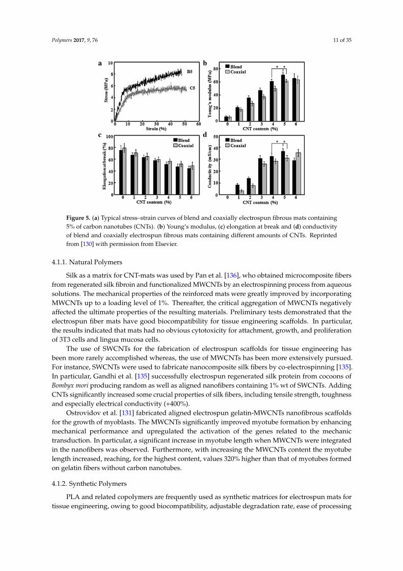
The fiber morphology and topography as well as the incorporated CNT amount can play acrucial role in tuning the mechanical and electrical properties and consequently the biocompatibilityof polymeric nanomats containing CNTs [130,134]. Generally, electrospun aligned mats display highermechanical properties in comparison with random ones [134,135,144]. Moreover, the alignment canpromote the proliferation of specific cells [134]. Regarding the CNT amount, usually, on increasingthe CNT loading, the mechanical and electrical properties can dramatically improve, as visible inFigure 5, which reports the main results related to blended and coaxially electrospun fibrous matscontaining different amounts of CNTs [130]. In particular, Young's modulus (Figure 5b) significantlyincreased up to 5% of CNTs, while when the CNT content reached 6%, there was no apparent changefor coaxial fibers and a slight decrease for blend fibers, due to the filler re-aggregation. The elongationat break of mats (Figure 5c) was found to decrease from around 75% to 45% upon increasing theCNT concentration both in blend and coaxial fibers, because of the stiffening effect of CNTs. Theconductivity of fibrous mats (Figure 5d) as a function of CNT content displayed the same behaviour asYoung’s modulus. In fact, electrical conductivity of coaxial fibers increased linearly with CNT content,whereas in the case of blended system this property reached a maximum when CNT content was equalto 5%, thereafter it was found to decrease, presumably due to re-aggregation phenomena. However,for both systems the electrical percolation threshold was observed at around 3%.
The diameter of electrospun fibers can depend on the CNT concentration [130,134]. In particular,thinner fibers are usually obtained with the increase in the CNT contents due to the increasedconductivity of electrospinning suspensions [130].
Polymers 2017, 9, 76 11 of 35
Polymers 2017, 9, 76 11 of 35
Figure 5. (a) Typical stress–strain curves of blend and coaxially electrospun fibrous mats containing 5% of carbon nanotubes (CNTs). (b) Young’s modulus, (c) elongation at break and (d) conductivity of blend and coaxially electrospun fibrous mats containing different amounts of CNTs. Reprinted from [130] with permission from Elsevier.
4.1.1. Natural Polymers
Silk as a matrix for CNT-mats was used by Pan et al. [136], who obtained microcomposite fibers from regenerated silk fibroin and functionalized MWCNTs by an electrospinning process from aqueous solutions. The mechanical properties of the reinforced mats were greatly improved by incorporating MWCNTs up to a loading level of 1%. Thereafter, the critical aggregation of MWCNTs negatively affected the ultimate properties of the resulting materials. Preliminary tests demonstrated that the electrospun fiber mats have good biocompatibility for tissue engineering scaffolds. In particular, the results indicated that mats had no obvious cytotoxicity for attachment, growth, and proliferation of 3T3 cells and lingua mucosa cells.
The use of SWCNTs for the fabrication of electrospun scaffolds for tissue engineering has been more rarely accomplished whereas, the use of MWCNTs has been more extensively pursued. For instance, SWCNTs were used to fabricate nanocomposite silk fibers by co-electrospinning [135]. In particular, Gandhi et al. [135] successfully electrospun regenerated silk protein from cocoons of Bombyx mori producing random as well as aligned nanofibers containing 1% wt of SWCNTs. Adding CNTs significantly increased some crucial properties of silk fibers, including tensile strength, toughness and especially electrical conductivity (+400%).
Ostrovidov et al. [131] fabricated aligned electrospun gelatin-MWCNTs nanofibrous scaffolds for the growth of myoblasts. The MWCNTs significantly improved myotube formation by enhancing mechanical performance and upregulated the activation of the genes related to the mechanic transduction. In particular, a significant increase in myotube length when MWCNTs were integrated in the nanofibers was observed. Furthermore, with increasing the MWCNTs content the myotube length increased, reaching, for the highest content, values 320% higher than that of myotubes formed on gelatin fibers without carbon nanotubes.
4.1.2. Synthetic Polymers
PLA and related copolymers are frequently used as synthetic matrices for electrospun mats for tissue engineering, owing to good biocompatibility, adjustable degradation rate, ease of processing and excellent mechanical properties of these polymers, further enhanced by the incorporation of CNTs, even at low concentrations [125–127,129,132,134,141–143].
Figure 5. (a) Typical stress–strain curves of blend and coaxially electrospun fibrous mats containing5% of carbon nanotubes (CNTs). (b) Young’s modulus, (c) elongation at break and (d) conductivityof blend and coaxially electrospun fibrous mats containing different amounts of CNTs. Reprintedfrom [130] with permission from Elsevier.
4.1.1. Natural Polymers
Silk as a matrix for CNT-mats was used by Pan et al. [136], who obtained microcomposite fibersfrom regenerated silk fibroin and functionalized MWCNTs by an electrospinning process from aqueoussolutions. The mechanical properties of the reinforced mats were greatly improved by incorporatingMWCNTs up to a loading level of 1%. Thereafter, the critical aggregation of MWCNTs negativelyaffected the ultimate properties of the resulting materials. Preliminary tests demonstrated that theelectrospun fiber mats have good biocompatibility for tissue engineering scaffolds. In particular,the results indicated that mats had no obvious cytotoxicity for attachment, growth, and proliferationof 3T3 cells and lingua mucosa cells.
The use of SWCNTs for the fabrication of electrospun scaffolds for tissue engineering hasbeen more rarely accomplished whereas, the use of MWCNTs has been more extensively pursued.For instance, SWCNTs were used to fabricate nanocomposite silk fibers by co-electrospinning [135].In particular, Gandhi et al. [135] successfully electrospun regenerated silk protein from cocoons ofBombyx mori producing random as well as aligned nanofibers containing 1% wt of SWCNTs. AddingCNTs significantly increased some crucial properties of silk fibers, including tensile strength, toughnessand especially electrical conductivity (+400%).
Ostrovidov et al. [131] fabricated aligned electrospun gelatin-MWCNTs nanofibrous scaffoldsfor the growth of myoblasts. The MWCNTs significantly improved myotube formation by enhancingmechanical performance and upregulated the activation of the genes related to the mechanictransduction. In particular, a significant increase in myotube length when MWCNTs were integratedin the nanofibers was observed. Furthermore, with increasing the MWCNTs content the myotubelength increased, reaching, for the highest content, values 320% higher than that of myotubes formedon gelatin fibers without carbon nanotubes.
4.1.2. Synthetic Polymers
PLA and related copolymers are frequently used as synthetic matrices for electrospun mats fortissue engineering, owing to good biocompatibility, adjustable degradation rate, ease of processing
Polymers 2017, 9, 76 12 of 35
and excellent mechanical properties of these polymers, further enhanced by the incorporation of CNTs,even at low concentrations [125–127,129,132,134,141–143].
Shao et al. successfully fabricated random oriented and aligned PLA/MWCNTs nanofiber meshesby electrospinning [134]. They showed that average diameter of nanofibers can be controlled byadjusting the amount of MWCNTs. Moreover, the incorporation of CNTs strongly enhanced both themechanical and electrical properties. Furthermore, these conductive nanofibrous scaffolds paved theway to study the synergistic effect of topographic signals and electrical stimulation on osteoblastsgrowth, with potential applications in bone tissue engineering. The results showed that the alignednanofibers were more efficient than their random counterparts in osteoblasts signaling and directioning.
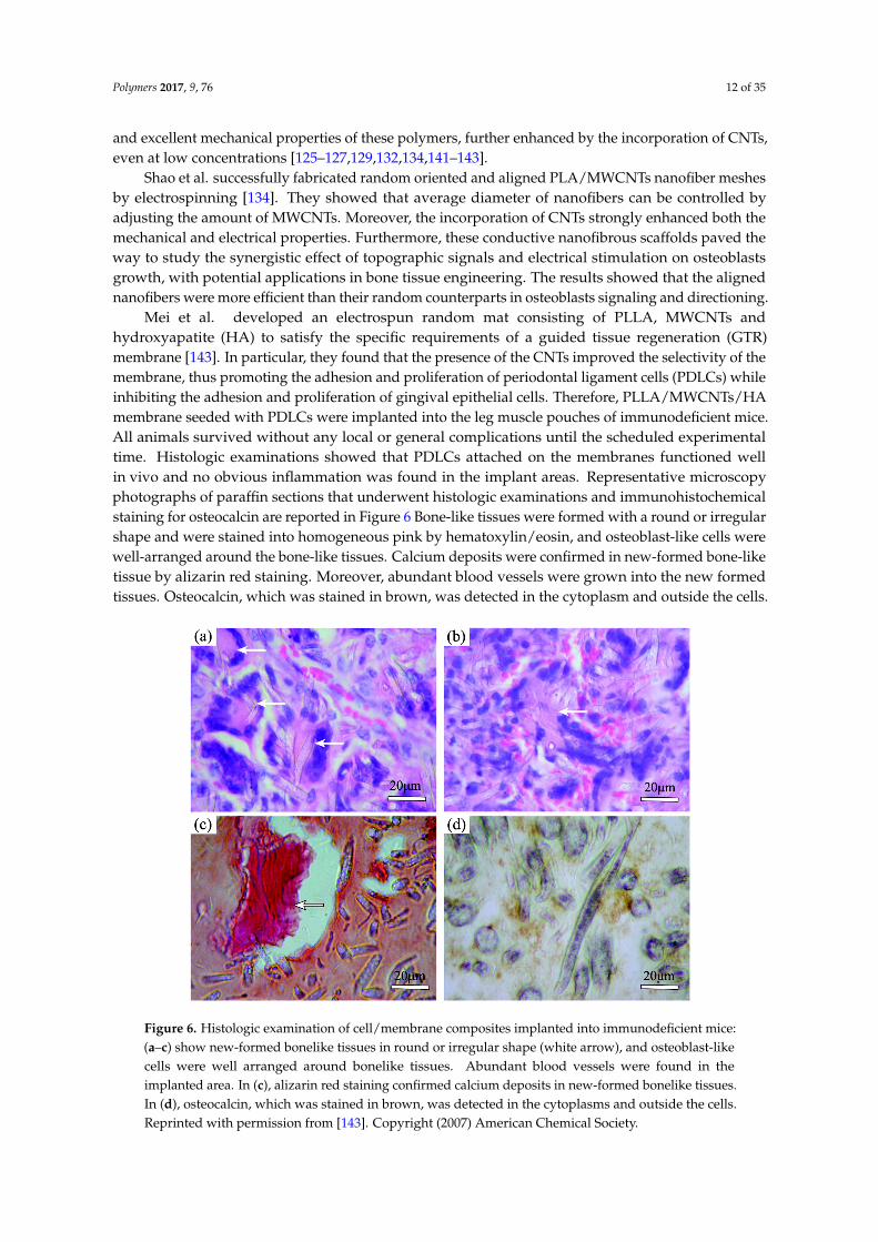
Mei et al. developed an electrospun random mat consisting of PLLA, MWCNTs andhydroxyapatite (HA) to satisfy the specific requirements of a guided tissue regeneration (GTR)membrane [143]. In particular, they found that the presence of the CNTs improved the selectivity of themembrane, thus promoting the adhesion and proliferation of periodontal ligament cells (PDLCs) whileinhibiting the adhesion and proliferation of gingival epithelial cells. Therefore, PLLA/MWCNTs/HAmembrane seeded with PDLCs were implanted into the leg muscle pouches of immunodeficient mice.All animals survived without any local or general complications until the scheduled experimentaltime. Histologic examinations showed that PDLCs attached on the membranes functioned wellin vivo and no obvious inflammation was found in the implant areas. Representative microscopyphotographs of paraffin sections that underwent histologic examinations and immunohistochemicalstaining for osteocalcin are reported in Figure 6 Bone-like tissues were formed with a round or irregularshape and were stained into homogeneous pink by hematoxylin/eosin, and osteoblast-like cells werewell-arranged around the bone-like tissues. Calcium deposits were confirmed in new-formed bone-liketissue by alizarin red staining. Moreover, abundant blood vessels were grown into the new formedtissues. Osteocalcin, which was stained in brown, was detected in the cytoplasm and outside the cells.
Polymers 2017, 9, 76 12 of 35
Shao et al. successfully fabricated random oriented and aligned PLA/MWCNTs nanofiber meshes by electrospinning [134]. They showed that average diameter of nanofibers can be controlled by adjusting the amount of MWCNTs. Moreover, the incorporation of CNTs strongly enhanced both the mechanical and electrical properties. Furthermore, these conductive nanofibrous scaffolds paved the way to study the synergistic effect of topographic signals and electrical stimulation on osteoblasts growth, with potential applications in bone tissue engineering. The results showed that the aligned nanofibers were more efficient than their random counterparts in osteoblasts signaling and directioning.
Mei et al. developed an electrospun random mat consisting of PLLA, MWCNTs and hydroxyapatite (HA) to satisfy the specific requirements of a guided tissue regeneration (GTR) membrane [143]. In particular, they found that the presence of the CNTs improved the selectivity of the membrane, thus promoting the adhesion and proliferation of periodontal ligament cells (PDLCs) while inhibiting the adhesion and proliferation of gingival epithelial cells. Therefore, PLLA/MWCNTs/HA membrane seeded with PDLCs were implanted into the leg muscle pouches of immunodeficient mice. All animals survived without any local or general complications until the scheduled experimental time. Histologic examinations showed that PDLCs attached on the membranes functioned well in vivo and no obvious inflammation was found in the implant areas. Representative microscopy photographs of paraffin sections that underwent histologic examinations and immunohistochemical staining for osteocalcin are reported in Figure 6 Bone-like tissues were formed with a round or irregular shape and were stained into homogeneous pink by hematoxylin/eosin, and osteoblast-like cells were well-arranged around the bone-like tissues. Calcium deposits were confirmed in new-formed bone-like tissue by alizarin red staining. Moreover, abundant blood vessels were grown into the new formed tissues. Osteocalcin, which was stained in brown, was detected in the cytoplasm and outside the cells.
Figure 6. Histologic examination of cell/membrane composites implanted into immunodeficient mice: (a–c) show new-formed bonelike tissues in round or irregular shape (white arrow), and osteoblast-like cells were well arranged around bonelike tissues. Abundant blood vessels were found in the implanted area. In (c), alizarin red staining confirmed calcium deposits in new-formed bonelike tissues. In (d), osteocalcin, which was stained in brown, was detected in the cytoplasms and outside the cells. Reprinted with permission from [143]. Copyright (2007) American Chemical Society.
Figure 6. Histologic examination of cell/membrane composites implanted into immunodeficient mice:(a–c) show new-formed bonelike tissues in round or irregular shape (white arrow), and osteoblast-likecells were well arranged around bonelike tissues. Abundant blood vessels were found in theimplanted area. In (c), alizarin red staining confirmed calcium deposits in new-formed bonelike tissues.In (d), osteocalcin, which was stained in brown, was detected in the cytoplasms and outside the cells.Reprinted with permission from [143]. Copyright (2007) American Chemical Society.
Polymers 2017, 9, 76 13 of 35
Vicentini et al. reported a study on the use of 4-methoxyphenyl functionalized MWCNTs asnanofiller into a PLLA matrix for the preparation of electrospun fibrous scaffold boosting neuriteoutgrowth and neuronal cell differentiation [125]. The tailored covalent functionalization of nanotubesurfaces allowed a homogeneous dispersion of the nanofillers within the polymer matrix, diminishingtheir natural tendency to aggregate and form bundles. Furthermore, TEM images showed carbonnanotubes anisotropically aligned along the fiber axes. The scaffolds prepared were tested in termsof biocompatibility and neuritogenesis and those containing CNTs gave the best results in neuriteoutgrowth, likely due to the nanocarbons-induced neuronal differentiation.
Other papers reported studies on the use of lactide polymers and CNTs for the fabrication ofdevices useful for neural tissue engineering [126,127,129]. In particular, Edwards et al. combinedthe properties of PLGA and MWCNTs not incorporating the nanofiller into the polymeric fibers,but electrospinning PLGA nanofibers onto a tubular MWCNT knitted scaffold [126].
In this case, the presence of electrospun PLGA led to the formation of small pores that enabledthe spanning and uniform distribution of cells, thus avoiding the formation of cell clusters irregularlydistributed on the surface, otherwise found in knitted tubular scaffolds only.
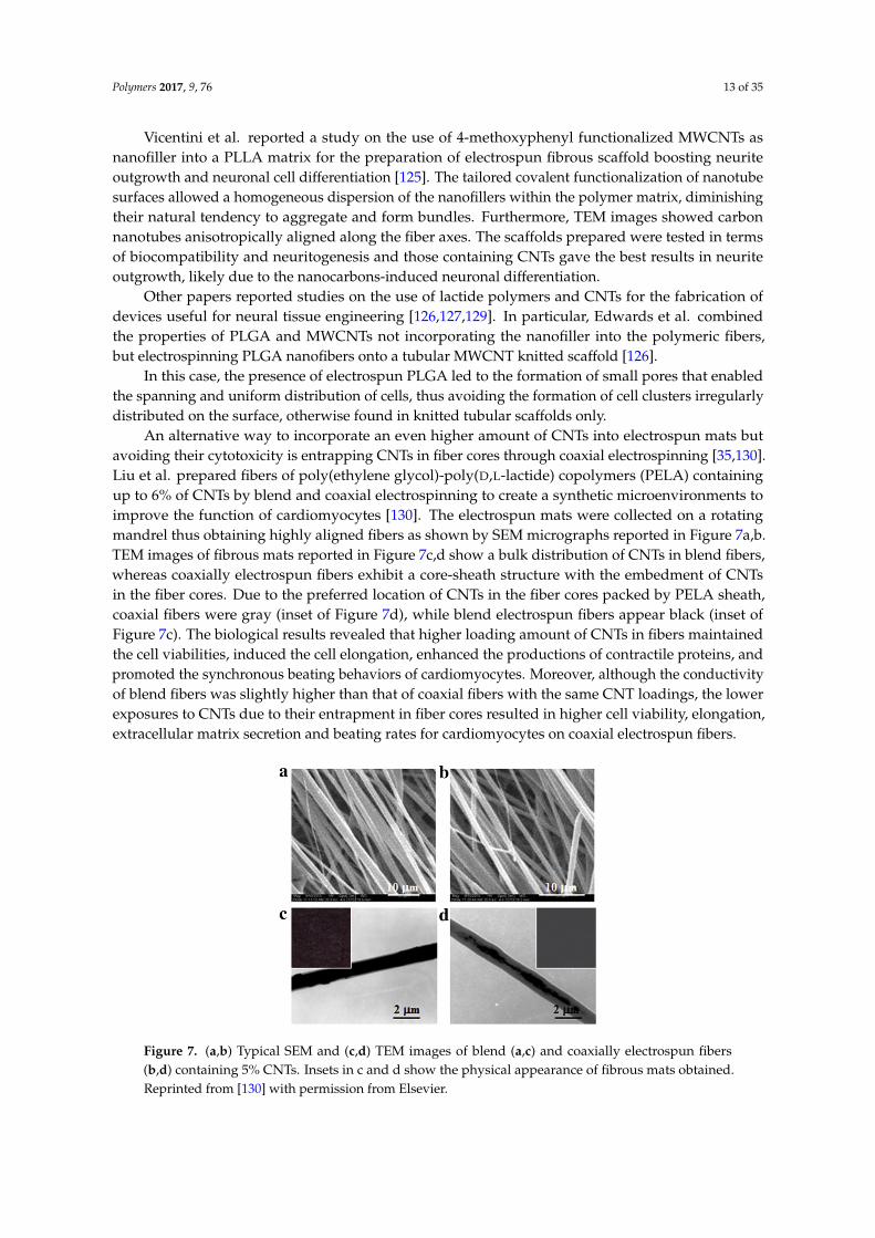
An alternative way to incorporate an even higher amount of CNTs into electrospun mats butavoiding their cytotoxicity is entrapping CNTs in fiber cores through coaxial electrospinning [35,130].Liu et al. prepared fibers of poly(ethylene glycol)-poly(D,L-lactide) copolymers (PELA) containingup to 6% of CNTs by blend and coaxial electrospinning to create a synthetic microenvironments toimprove the function of cardiomyocytes [130]. The electrospun mats were collected on a rotatingmandrel thus obtaining highly aligned fibers as shown by SEM micrographs reported in Figure 7a,b.TEM images of fibrous mats reported in Figure 7c,d show a bulk distribution of CNTs in blend fibers,whereas coaxially electrospun fibers exhibit a core-sheath structure with the embedment of CNTsin the fiber cores. Due to the preferred location of CNTs in the fiber cores packed by PELA sheath,coaxial fibers were gray (inset of Figure 7d), while blend electrospun fibers appear black (inset ofFigure 7c). The biological results revealed that higher loading amount of CNTs in fibers maintainedthe cell viabilities, induced the cell elongation, enhanced the productions of contractile proteins, andpromoted the synchronous beating behaviors of cardiomyocytes. Moreover, although the conductivityof blend fibers was slightly higher than that of coaxial fibers with the same CNT loadings, the lowerexposures to CNTs due to their entrapment in fiber cores resulted in higher cell viability, elongation,extracellular matrix secretion and beating rates for cardiomyocytes on coaxial electrospun fibers.
Polymers 2017, 9, 76 13 of 35
Vicentini et al. reported a study on the use of 4-methoxyphenyl functionalized MWCNTs as nanofiller into a PLLA matrix for the preparation of electrospun fibrous scaffold boosting neurite outgrowth and neuronal cell differentiation [125]. The tailored covalent functionalization of nanotube surfaces allowed a homogeneous dispersion of the nanofillers within the polymer matrix, diminishing their natural tendency to aggregate and form bundles. Furthermore, TEM images showed carbon nanotubes anisotropically aligned along the fiber axes. The scaffolds prepared were tested in terms of biocompatibility and neuritogenesis and those containing CNTs gave the best results in neurite outgrowth, likely due to the nanocarbons-induced neuronal differentiation.
Other papers reported studies on the use of lactide polymers and CNTs for the fabrication of devices useful for neural tissue engineering [126,127,129]. In particular, Edwards et al. combined the properties of PLGA and MWCNTs not incorporating the nanofiller into the polymeric fibers, but electrospinning PLGA nanofibers onto a tubular MWCNT knitted scaffold [126].
In this case, the presence of electrospun PLGA led to the formation of small pores that enabled the spanning and uniform distribution of cells, thus avoiding the formation of cell clusters irregularly distributed on the surface, otherwise found in knitted tubular scaffolds only.
An alternative way to incorporate an even higher amount of CNTs into electrospun mats but avoiding their cytotoxicity is entrapping CNTs in fiber cores through coaxial electrospinning [35,130]. Liu et al. prepared fibers of poly(ethylene glycol)-poly(D,L-lactide) copolymers (PELA) containing up to 6% of CNTs by blend and coaxial electrospinning to create a synthetic microenvironments to improve the function of cardiomyocytes [130]. The electrospun mats were collected on a rotating mandrel thus obtaining highly aligned fibers as shown by SEM micrographs reported in Figure 7a,b. TEM images of fibrous mats reported in Figure 7c,d show a bulk distribution of CNTs in blend fibers, whereas coaxially electrospun fibers exhibit a core-sheath structure with the embedment of CNTs in the fiber cores. Due to the preferred location of CNTs in the fiber cores packed by PELA sheath, coaxial fibers were gray (inset of Figure 7d), while blend electrospun fibers appear black (inset of Figure 7c). The biological results revealed that higher loading amount of CNTs in fibers maintained the cell viabilities, induced the cell elongation, enhanced the productions of contractile proteins, and promoted the synchronous beating behaviors of cardiomyocytes. Moreover, although the conductivity of blend fibers was slightly higher than that of coaxial fibers with the same CNT loadings, the lower exposures to CNTs due to their entrapment in fiber cores resulted in higher cell viability, elongation, extracellular matrix secretion and beating rates for cardiomyocytes on coaxial electrospun fibers.
Figure 7. (a,b) Typical SEM and (c,d) TEM images of blend (a,c) and coaxially electrospun fibers (b,d) containing 5% CNTs. Insets in c and d show the physical appearance of fibrous mats obtained. Reprinted from [130] with permission from Elsevier.
Figure 7. (a,b) Typical SEM and (c,d) TEM images of blend (a,c) and coaxially electrospun fibers(b,d) containing 5% CNTs. Insets in c and d show the physical appearance of fibrous mats obtained.Reprinted from [130] with permission from Elsevier.

Polymers 2017, 9, 76 14 of 35
The incorporation of MWCNTs into conducting polymers such as polyaniline [145,146] gaverise to electrospun fibers suitable as scaffolds in cell culture studies. Indeed, the presence of CNTsimproved cell growth and proliferation on the surface of the conducting nanofibers because of theirconductivity and mechanical strength provided by the PANI and CNTs.
Rodrigues et al. [133] proposed the use of electrospun poly (butylene adipate-co-terephthalate)(PBAT)-based fibers for bone regeneration, in spite of the poor mechanical resistance of neat PBAT.The authors demonstrated the possibility to overcome this drawback by adding low contents ofsuperhydrophilic MWCNTs (0.1–0.5 wt %), owing to their remarkable strengthening and stiffeningeffect. All samples showed cytocompatibility with MG63 osteoblast-like cells and in particular, onincreasing the MWCNTs content increased the cellular viability, thus indicating that the incorporationof 0.5% of MWCNTs increased its biocompatibility. Moreover, MG63 cells osteogenic differentiationshowed that mineralized nodules formation was increased in PBAT/0.5% MWCNTs when comparedto control group and neat PBAT.
Another possible way to combine the properties of electrospun mats and CNTs, different from theconventional incorporation, is the nanofiller coating on the surface of the nanomats, as proposed byJin et al. [128]. In particular, PLCL electrospun fibers were coated with ad hoc functionalized MWCNTsin order to provide better environments for cell adhesion and neurite outgrowth. The results revealedthat MWCNT-coated PLCL scaffolds exhibit improved adhesion, proliferation and neurite outgrowthof PC-12 cells in comparison with uncoated PLCL scaffolds.

Polymers 2017, 9, 76 15 of 35
Table 2. Examples of polymer-CNTs electrospun scaffolds for tissue engineering.
Polymers(and additives) Solvents Nanocarbons Nanocarbons
loading (wt %) Experimental setup Structure Main improvements Targettissue Refs.
CA/CS Acetone/DMF (2:1) MWCNT N/A electrospinning pluslayer-by-layer self-assembly Random, D = 305 ± 128 nm
Mechanical properties; cellattachment, spreading
and proliferation
Notspecified [147]
Gelatin Water MWCNT N/A Electrospinning followed bycrosslinking with GA vapor Aligned, D = 296 nm Mechanical properties; cell
alignment and differentiation Muscle [131]
PANI/PNIPAm-co-MAA HFIP/DMF (8:2) PANI-MWCNT N/A Conventional electrospinning Random , D = 500–600 nm Cell growth and viability Notspecified [145]
PANI/PNIPAm HFIP/DMF (8:2) HOOC-MWCNT N/A Conventional electrospinning Random , D = 400–500 nm Cell proliferation and viability Notspecified [146]
PBAT Chloroform/DMF(3:2)
MWCNT(plasma treated
with O2)0.1%–0.5% Conventional electrospinning Random, D = 250 ± 52
nm–272 ± 79 nm Mechanical properties Bone [133]
PCL DCM/methanol(3:1)
MWCNT(acid-treated) 0.1%–5% Conventional electrospinning Random, D = 117±45–
252 ± 146 nmAccelerating degradationbehavior; biocompatibility
Notspecified [137]
PCL–PAA/PVA DMF/DCM(1:1)–EtOH/H2O
MWCNT(acid-treated) 0.05% Coaxial electrospinning Random,
D = 1.861 ± 0.693 µmMechanical and electrical
properties; biocompatibilitySkeletalmuscle [35]
PELA DMF/DCM MWCNT 0%–6% Coaxial electrospinning Aligned, D = 2–3 µm Mechanical and electricalproperties; cell morphology Myocardial [130]
PLA Chloroform/DMF MWCNT 0%–1% Conventional electrospinning Random, D = 0.55–0.96 µm Mechanical andelectrical properties
Notspecified [141]
PLA DCM/DMF (3:1) MWCNT 1% Conventional electrospinning Random,D = 2.08 ± 0.13 µm
Mechanical andelectrical properties Cartilage [142]
PLA DMF/DCM MWCNT(acid-treated) 0%–5% Conventional electrospinning Random, D = 243–425 nm
Aligned, D = 232–402 nmMechanical and electrical
properties; cell morphology Bone [134]
PLCL DCM/EtOH (4:1) MWCNT-tartrate N/A MWCNT coating onelectrospun PLCL
Aligned,D = 1.30 ± 0.46 µm,
Cell adhesion, proliferationand neurite outgrowth Nerve [128]
PLGA DMF/THF (3:1) MWCNT 0.1%–1% Conventional electrospinning Random, D = 0.4–1.6 µm Electrical properties;myotube formation
Skeletalmuscle [132]
PLGA DMFA MWCNT N/A electrospinning ontoMWCNT knitted scaffold Random D = N/A Cell spanning Nerve [126]
PLGA/SF/catalpol HFIP MWCNT N/A Conventional electrospinning Random, D = 577 ±360–810 ± 270 nm N/A Nerve [127]
PLLA Chloroform/DMF(9:1) MWCNT-PhOMe 0.25% Conventional electrospinning Random, D = 200–600 nm Neurite outgrowth and
neuronal cell differentiation Nerve [125]
PLLA Chloroform/DMF(8.5:1.5) SWCNT 3% Conventional electrospinning Aligned, D = 430 nm Cell adhesion, growth,
survival and proliferation Nerve [129]
PLLA/HA DCM/1,4-dioxaneMWCNT(anodic
oxidated)0.3% Conventional electrospinning Random, D = 1 µm Cell adhesion
and proliferation.Periodontalligament [143]

Polymers 2017, 9, 76 16 of 35
Table 2. Cont.
Polymers(and additives) Solvents Nanocarbons Nanocarbons
loading (wt %) Experimental setup Structure Main improvements Targettissue Refs.
PU THF/DMF (1:1) MWCNT 0.1%–1% Conventional electrospinning Random, D = 600 ±300–1000 ± 400 nm Mechanical properties Not
specified [138]
PU DMAc MWCNT(acid-treated) 3% Conventional electrospinning Random, D = 300–500 nm Cell adhesion, proliferation,
migration and aggregationNot
specified [139]
PU DMAc MWCNT(acid-treated) 3% Conventional electrospinning Aligned, D = 300–500 nm
Cell proliferation,extracellular
collagen secretionVascular [140]
PVA/CS AA/water (70 wt %) MWCNT 0.99% Electrospinning followed bycrosslinking with GA vapor
Random , D = 157 ± 40 nm(non-crosslinked);
170 ± 43 nm (crosslinked)
Cell proliferation; proteinadsorption capability
Notspecified [148]
SF WaterMWCNT
(functionalizedwith SDBS)
0.25%–1.5% Conventional electrospinning Random, D = 3 µm Mechanical properties Notspecified [136]
SF Formic acid SWCNT 1%Co-electrospinning plustreatment with methanol
and/or stretching
Random , D = 153 ± 99 nmAligned, D = 147 ± 41 nm
Mechanical andelectrical properties Bone [135]
SEBS Toluene/THF (1:1) MWCNT 1.5% Conventional electrospinning Random, D = 12.3 ± 3.6 µmAligned, D = 10.2 ± 2.7 µm
Mechanical hysteresis andelectrical conductivity
Notspecified [144]
N/A: Data not available; D: Diamater; The other acronyms are available in the acronym list.

Polymers 2017, 9, 76 17 of 35
4.2. Polymeric Nanomats Containing Graphene-Based Nanocarbons
The main works on electrospun nanomats containing graphenic compounds for tissue engineeringare listed in Table 3. Among the graphenic compounds, GO is the most widespread one, because of itsbetter biocompatibility. In fact, its unique chemical-physical features, such as the high hydrophilicityensured by the presence of a wide range of oxygenated moieties and the wrinkled texture that results ina high roughness, are useful to provide cell proliferation and attachment, respectively. The choice of thematrix for GO-containing electrospun mats mainly depends on the target tissue for which the materialsare proposed. Naturally derived as well as synthetic biodegradable polymers are the most widespread.The former enable high degrees of cells adhesion, the latter provide better mechanical performance.
Lactide polymers, such as PLA, PLLA, PLGA, are frequently used for bone tissue engineering,owing to the excellent mechanical properties of these polymers, further enhanced by the incorporationof GO, even at small concentrations. Moreover, GO allows increasing surface wettability of PLA, whichis found to change from a hydrophobic to hydrophilic character, with positive repercussions on celladhesion and proliferation.
As previously discussed, the mean diameter of the nanofibers can be tuned by varying severalprocessing parameters. For the system PLA-GO, the nanofibers diameter distribution was foundto vary from hundreds of nanometers to few microns. The PLA-GO bionanomats for bone tissueengineering are composed by randomly oriented nanofibers, since these particular structures provideattractive ECM conditions for the anchorage, migration and differentiation of tissue cells, includingthose responsible for the regeneration of bone [149]. Moreover, GO was found to promote both cellsignaling and differentiation due to its wrinkled texture [150].
4.2.1. Natural Polymers
Massoumi et al. prepared electrospun nanofibrous scaffolds based on gelatin and afunctionalized GO [151]. The authors covalently attached a copolymer (poly(2-hydroxyethylmethacrylate)-graft-poly(ε-caprolactone)) onto an acylated sample of GO via atom transfer radicalpolymerization (ATRP). The electrical conductivity of the electrospun nanofibers obtained was in thescale of 10−5 S/m, which represents proper conductivity for scaffolds addressed to repair injurednerve tissues [151].
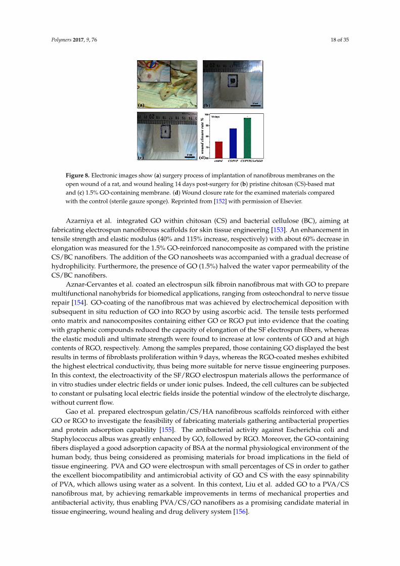
Nafiseh and Simchi fabricated nanofibrous scaffolds by electrospinning blended solutions ofchitosan (80 vol %), polyvinyl pyrrolidone (15 vol %), polyethylene oxide (5 vol %) containing GOnanosheets (0–2 wt %) [152]. GO significantly increased the conductivity and viscosity of highlyconcentrated chitosan solutions, thus enabling the spinnability of ultrafine and uniform fibers with anaverage diameter of 60 nm. The GO-reinforced nanofibers displayed enhanced elastic modulus andtensile strength (150%–300%) with a controllable water permeability to meet the required properties ofnatural skins. Furthermore, the nanofibrous structure was found to promote the cell attachment, bymaintaining characteristic cell morphology and viability up to 72 h. The nanofibrous membranes basedon neat CS and containing 1.5% GO were implanted on open wounds, as shown in Figure 8a. In vivoevaluations in rats showed a faster and more efficient wound closure rate in the case of nanofibrousmembranes and those loaded with 1.5% GO gave the best results, as clearly visible by comparingthe electronic images of the examined rat and the area of open wound after 14 days post-surgery inthe case of neat CS (Figure 8b) and CS containing 1.5% GO (Figure 8c). The wound closure rate wasevaluated by image analysis and shown in Figure 8d. The polymeric nanofibrous membrane promotedthe healing process compared with the control (sterile gauze sponge). This feature was attributedto the ultrastructure of the dressing materials together with the inherent healing abilities of CS foropen wounds. GO doping results in a further enhancement of wound closure ability (about 33% ascompared with sterile gauze sponges). The presence of GO nanosheets led to further advantages,including higher strength, adapted permeability, better cell attachment, and total absence of scarand/or inflammation owing to its antibacterial activity.
Polymers 2017, 9, 76 18 of 35
Polymers 2017, 9, 76 18 of 35
Figure 8. Electronic images show (a) surgery process of implantation of nanofibrous membranes on the open wound of a rat, and wound healing 14 days post-surgery for (b) pristine chitosan (CS)-based mat and (c) 1.5% GO-containing membrane. (d) Wound closure rate for the examined materials compared with the control (sterile gauze sponge). Reprinted from [152] with permission of Elsevier.
Azarniya et al. integrated GO within chitosan (CS) and bacterial cellulose (BC), aiming at fabricating electrospun nanofibrous scaffolds for skin tissue engineering [153]. An enhancement in tensile strength and elastic modulus (40% and 115% increase, respectively) with about 60% decrease in elongation was measured for the 1.5% GO-reinforced nanocomposite as compared with the pristine CS/BC nanofibers. The addition of the GO nanosheets was accompanied with a gradual decrease of hydrophilicity. Furthermore, the presence of GO (1.5%) halved the water vapor permeability of the CS/BC nanofibers.
Aznar-Cervantes et al. coated an electrospun silk fibroin nanofibrous mat with GO to prepare multifunctional nanohybrids for biomedical applications, ranging from osteochondral to nerve tissue repair [154]. GO-coating of the nanofibrous mat was achieved by electrochemical deposition with subsequent in situ reduction of GO into RGO by using ascorbic acid. The tensile tests performed onto matrix and nanocomposites containing either GO or RGO put into evidence that the coating with graphenic compounds reduced the capacity of elongation of the SF electrospun fibers, whereas the elastic moduli and ultimate strength were found to increase at low contents of GO and at high contents of RGO, respectively. Among the samples prepared, those containing GO displayed the best results in terms of fibroblasts proliferation within 9 days, whereas the RGO-coated meshes exhibited the highest electrical conductivity, thus being more suitable for nerve tissue engineering purposes. In this context, the electroactivity of the SF/RGO electrospun materials allows the performance of in vitro studies under electric fields or under ionic pulses. Indeed, the cell cultures can be subjected to constant or pulsating local electric fields inside the potential window of the electrolyte discharge, without current flow.
Gao et al. prepared electrospun gelatin/CS/HA nanofibrous scaffolds reinforced with either GO or RGO to investigate the feasibility of fabricating materials gathering antibacterial properties and protein adsorption capability [155]. The antibacterial activity against Escherichia coli and Staphylococcus albus was greatly enhanced by GO, followed by RGO. Moreover, the GO-containing fibers displayed a good adsorption capacity of BSA at the normal physiological environment of the human body, thus being considered as promising materials for broad implications in the field of tissue engineering. PVA and GO were electrospun with small percentages of CS in order to gather the excellent biocompatibility and antimicrobial activity of GO and CS with the easy spinnability of PVA, which allows using water as a solvent. In this context, Liu et al. added GO to a PVA/CS nanofibrous mat, by achieving remarkable improvements in terms of mechanical properties and antibacterial activity, thus enabling PVA/CS/GO nanofibers as a promising candidate material in tissue engineering, wound healing and drug delivery system [156].
Figure 8. Electronic images show (a) surgery process of implantation of nanofibrous membranes on theopen wound of a rat, and wound healing 14 days post-surgery for (b) pristine chitosan (CS)-based matand (c) 1.5% GO-containing membrane. (d) Wound closure rate for the examined materials comparedwith the control (sterile gauze sponge). Reprinted from [152] with permission of Elsevier.
Azarniya et al. integrated GO within chitosan (CS) and bacterial cellulose (BC), aiming atfabricating electrospun nanofibrous scaffolds for skin tissue engineering [153]. An enhancement intensile strength and elastic modulus (40% and 115% increase, respectively) with about 60% decrease inelongation was measured for the 1.5% GO-reinforced nanocomposite as compared with the pristineCS/BC nanofibers. The addition of the GO nanosheets was accompanied with a gradual decrease ofhydrophilicity. Furthermore, the presence of GO (1.5%) halved the water vapor permeability of theCS/BC nanofibers.
Aznar-Cervantes et al. coated an electrospun silk fibroin nanofibrous mat with GO to preparemultifunctional nanohybrids for biomedical applications, ranging from osteochondral to nerve tissuerepair [154]. GO-coating of the nanofibrous mat was achieved by electrochemical deposition withsubsequent in situ reduction of GO into RGO by using ascorbic acid. The tensile tests performedonto matrix and nanocomposites containing either GO or RGO put into evidence that the coatingwith graphenic compounds reduced the capacity of elongation of the SF electrospun fibers, whereasthe elastic moduli and ultimate strength were found to increase at low contents of GO and at highcontents of RGO, respectively. Among the samples prepared, those containing GO displayed the bestresults in terms of fibroblasts proliferation within 9 days, whereas the RGO-coated meshes exhibitedthe highest electrical conductivity, thus being more suitable for nerve tissue engineering purposes.In this context, the electroactivity of the SF/RGO electrospun materials allows the performance ofin vitro studies under electric fields or under ionic pulses. Indeed, the cell cultures can be subjectedto constant or pulsating local electric fields inside the potential window of the electrolyte discharge,without current flow.
Gao et al. prepared electrospun gelatin/CS/HA nanofibrous scaffolds reinforced with eitherGO or RGO to investigate the feasibility of fabricating materials gathering antibacterial propertiesand protein adsorption capability [155]. The antibacterial activity against Escherichia coli andStaphylococcus albus was greatly enhanced by GO, followed by RGO. Moreover, the GO-containingfibers displayed a good adsorption capacity of BSA at the normal physiological environment of thehuman body, thus being considered as promising materials for broad implications in the field oftissue engineering. PVA and GO were electrospun with small percentages of CS in order to gatherthe excellent biocompatibility and antimicrobial activity of GO and CS with the easy spinnabilityof PVA, which allows using water as a solvent. In this context, Liu et al. added GO to a PVA/CSnanofibrous mat, by achieving remarkable improvements in terms of mechanical properties andantibacterial activity, thus enabling PVA/CS/GO nanofibers as a promising candidate material intissue engineering, wound healing and drug delivery system [156].

Polymers 2017, 9, 76 19 of 35
Table 3. Examples of polymer-graphene electrospun scaffolds for tissue engineering.
Polymers(and additives) Solvents Nanocarbons Filler loading (wt %) Structure Main improvements Target tissue Refs
CS/GEL/HA AA/H2O GO; RGO 2% Random Bioactivity, antibacterial andmechanical properties Bone [155]
CS/PEO/BC AA/H2O GO 0–2 Random D = 145–254 nm Mechanical properties Skin [153]
CS/PVP/PEO AA/H2O GO 0–2 Random, D = 80–200 nm Mechanicalproperties, bioactivity Skin/bone [152]
GEL DMSO GO-g-[P(HEMA-g-CL)] 2–3 Random, D = 100–200 nm Mechanical and electrical properties,wettability Not specified [151]
PAN DMF GO; RGO N/A Random Mechanical, electrical properties Not specified [157]PCL CHCl3 GO N/A Random, D = N/A Mechanical, electrical, cell signaling Skeletal muscle [158]PCL CHCl3 GO 0.3–2 Random, D = 0.1–8 µm Mechanical, electrical properties, bioactivity Muscle [159]PCL DMF GO 0.3–0.5 Random; D = 1–3 µm Cell differentiation Nerve/cartilage [8]
PCL DMF GO 0.5–2 Random, D = 0.2–2.5 µm Mechanical properties,bioactivity, biodegradability Bone [160]
PCL DCM/EtOH 4:1 GO; GO-g-PEG 0.25–2 Random, D = 200–1000 nm Mechanical, wettability, cell adhesion Osteochondral [7]PCL AA GO; RGO 0–1 Aligned, D = 100–400 nm Mechanical properties Not specified [161]PLA CHCl3/DMF GO; GO-g-PEG 2 Random, D = 500–1000 nm Mechanical properties Osteochondral [28]
PLA/HA DCM/DMF GO 1–3 D = 412–516 nm Mechanical, bioactivity Bone [162]PLA/PU 4:1 DMF/DCM 2:3 GO 5 Random, D ~1 µm Biocompatibility, antimicrobial properties Cartilage [163]
PLGA THF/DMF GO 1 D = 783–1461 nm Wettability, bioactivity Bone [164]PLGA/Col HFIP GO 4 Random, D = 100–950 nm Cell proliferation, mechanical properties Bone/muscle [124]
PLGA/RGD HFIP GO N/A Random, D = 200–1440 nm myogenic differentiation Bone/muscle [123]PLGA/SF HFIP GO 1 Random, D = 130–280 nm Mechanical, wettability, cell differentiation Bone [165]
PLLA HFIP GO N/A Aligned; D = 680 nm Cell differentiation and growth Nerve [166]PU DMF GO 0.5-2 D = 290–400 nm Mechanical properties, bioactivity Osteochondral [167]
PVA H2O GNS 1%–7% Random, D = 200-800 nm Electrical properties Cartilage [168]PVA H2O GO 0-5 Random, D < 1 µm Mechanical properties, bioactivity Bone [169]
PVA/CS AA/H2O GO 0.05–0.6 D = 123–200 nm Mechanical properties Skin [156]PVC; FN THF/DMF (4:1) GO; RGO N/A N/A Mechanical, electrical properties, bioactivity Nerve [170]
SF H2O GO; RGO N/A D = 3.9–5.2 µm Electrical properties Nerve [154]
N/A: Data not available; D: Diamater; The other acronyms are available in the acronym list.
Polymers 2017, 9, 76 20 of 35
4.2.2. Synthetic Polymers
Liu et al. prepared electrospun mats based on PLA, HA (which content was kept constant at15 wt %) and GO, showing that the addition of small amounts of GO (from 1 to 3 wt %) caused adramatic stiffening and strengthening of PLA/HA [162]. The authors reported that the mechanicalproperties were found to increase upon GO content up to a loading level of 2%. Indeed, the nanomatscontaining 2 wt % GO displayed values of elastic modulus and tensile strength practically two-foldwith respect to PLA or PLA/HA, whereas those containing 3% GO showed mechanical propertiesworse than neat polymer. This issue is mainly due to the extremely high aspect ratio of GO, which tendsto self-aggregate and crumple after a certain percolation threshold, as already evidenced by severalstudies performed onto different matrices [119]. From a biological point of view, the combination of HAwith GO, especially when GO is loaded at 1%, exerts a remarkable effect on the adhesion and long-termproliferation of osteoblastic cells on the fibrous scaffolds. Furthermore, the authors demonstrated thatGO acts synergistically with calcium phosphate, thereby increasing alkaline phosphatase activity andthe calcium deposition of osteoblasts [162].
PLGA has been extensively investigated in the biomedical field because of excellentbiocompatibility, biodegradability and processability, since glycolic acid units endow higherhydrophilicity and ductility than PLA. However, its mechanical stiffness is unsatisfactory for bonetissue engineering. Incorporating GO could enable the possibility of achieving structurally stablenanocomposites, capable to support osteoblast growth and proliferation. Luo et al. preparedelectrospun nanofibrous scaffolds based on GO-doped PLGA. The results highlighted that—evenif bioactivity of nanocomposites was found to be higher than neat copolymer—the mechanicalperformance was found to decrease [164]. The authors ascribed this issue to the 2D topologicalplane structure of GO, which is supposed to tend to be vertical to the fibers, thus being unable totransfer the stress [164]. However, it has to be taken into account that polyesters undergo a such rapidhydrolytic degradation in acidic environment and in some cases GO might have a pro-degradativeeffect, as reported for similar systems [7,171]. Indeed, depending on the synthesis conditions and thetype of graphite source, some properties of GO, including acidity, C/O ratio (i.e., overall oxidationlevel) and relative surface content of –COOH and epoxy moietiesis, may vary considerably [7,171].
In some cases, PLGA was used in combination with natural polymers for GO-containingelectrospun fibrous scaffold addressed to tissue engineering [123,124,165].
Shao et al. incorporated GO into a blend made of PLGA and SF in order to develop electrospunnanomats for bone tissue engineering. The results put into evidence that 1 wt % GO led to thesimultaneous enhancement of mechanical and biological properties of the material. Figure 9 reportsthe representative tensile stress-strain curves (panel A) together with tensile strength (panel B),elastic modulus (panel C) and elongation at break (D) of electrospun fibrous PLGA, PLGA/SF andPLGA/SF/GO. As one can see, if compared to PLGA or PLA/SF, elastic modulus and tensile strengthof the materials containing GO were found to be 5-fold and 3-fold, respectively [165]. The authorsattributed the increase of tensile strength to the strong interfacial interactions between GO and PLGA(mainly due to hydrogen bonding), whereas the enhancement observed in the modulus was ascribedto the highly specific surface area of nanosheets.

Polymers 2017, 9, 76 21 of 35Polymers 2017, 9, 76 21 of 35
Figure 9. Mechanical properties of the electrospun fibrous pure poly(lactic-co-glycolic acid) (PLGA), PLGA–tussah, and GO-doped PLGA–tussah mats (n = 10 for each type of nanofibers) tested at room temperature. (A) Typical stress–strain curves; (B) tensile strength; (C) Young’s modulus; and (D) strain at break (* p < 0.05, ** p < 0.01). Reprinted from [165] with permission from Elsevier.
The addition of GO to a PLGA/collagen matrix dramatically increased the mechanical performance (up to +500%) and wettability, whereas mean diameter and roughness of the nanofibers were found to decrease [124]. Studies on the bioactivity were performed onto PLGA, PLGA/collagen and PLGA/collagen/GO nanomats and the presence of GO gave the best results either on cells initial attachment and cell proliferation within 7 days.
A useful strategy to avoid pro-degradative effects of GO on PLA and to improve interfacial adhesion is the covalent functionalization of GO. Zhang et al. carried out a comparative analysis between GO and GO-g-PEG as fillers for electrospun PLA [28]. The authors reported that GO-g-PEG was more effective than GO in enhancing thermal stability, mechanical properties, wettability. Furthermore, due to the presence of PEG chains, GO-g-PEG was able to remain dispersed within the polymer matrix even at high loadings (i.e., 5 wt %), differently from the pristine GO, which well-known tendency to self-aggregate was already discussed. Furthermore, the PLA cytocompatibility, investigated by studies on cell proliferation within 10 days, remained substantially unaltered after addition of either GO or GO-g-PEG.
An et al. fabricated PLA/PU blends filled with 3 and 5 wt % GO [163]. The authors reported that the materials displayed excellent biocompatibility together with a strong antimicrobial activity, imparted by GO, thus being suitable for cartilage and bone repairing while simultaneously reducing the threat of chronic infection of surrounding tissues.
GO, especially at high loadings, endows the nanomats with electrical properties, which are very suitable for nerve/neuronal tissue engineering. When applied to neuronal injuries repair, GO is preferentially coated onto electrospun polymers by a post-electrospinning treatment [166]. This two-step technique creates a conductive surface, particularly attractive for neuronal cells while maintaining the bulky properties of the polymer and using extremely lower contents of conductive fillers with respect to bulk inclusion-based techniques. Zhang et al. produced aligned PLLA nanofibrous scaffolds that were then surface modified via aminolysis in order to provide the surface with hydrophilic moieties, such as −OH, −COOH and −NH2, able to ensure a stable GO-coating [166]. The GO-coated nanofibrous scaffolds enabled the proliferation of Schwann cells cultured on the
Figure 9. Mechanical properties of the electrospun fibrous pure poly(lactic-co-glycolic acid) (PLGA),PLGA–tussah, and GO-doped PLGA–tussah mats (n = 10 for each type of nanofibers) tested at roomtemperature. (A) Typical stress–strain curves; (B) tensile strength; (C) Young’s modulus; and (D) strainat break (* p < 0.05, ** p < 0.01). Reprinted from [165] with permission from Elsevier.
The addition of GO to a PLGA/collagen matrix dramatically increased the mechanicalperformance (up to +500%) and wettability, whereas mean diameter and roughness of the nanofiberswere found to decrease [124]. Studies on the bioactivity were performed onto PLGA, PLGA/collagenand PLGA/collagen/GO nanomats and the presence of GO gave the best results either on cells initialattachment and cell proliferation within 7 days.
A useful strategy to avoid pro-degradative effects of GO on PLA and to improve interfacialadhesion is the covalent functionalization of GO. Zhang et al. carried out a comparative analysisbetween GO and GO-g-PEG as fillers for electrospun PLA [28]. The authors reported that GO-g-PEGwas more effective than GO in enhancing thermal stability, mechanical properties, wettability.Furthermore, due to the presence of PEG chains, GO-g-PEG was able to remain dispersed withinthe polymer matrix even at high loadings (i.e., 5 wt %), differently from the pristine GO, whichwell-known tendency to self-aggregate was already discussed. Furthermore, the PLA cytocompatibility,investigated by studies on cell proliferation within 10 days, remained substantially unaltered afteraddition of either GO or GO-g-PEG.
An et al. fabricated PLA/PU blends filled with 3 and 5 wt % GO [163]. The authors reportedthat the materials displayed excellent biocompatibility together with a strong antimicrobial activity,imparted by GO, thus being suitable for cartilage and bone repairing while simultaneously reducingthe threat of chronic infection of surrounding tissues.
GO, especially at high loadings, endows the nanomats with electrical properties, which arevery suitable for nerve/neuronal tissue engineering. When applied to neuronal injuries repair,GO is preferentially coated onto electrospun polymers by a post-electrospinning treatment [166].This two-step technique creates a conductive surface, particularly attractive for neuronal cells whilemaintaining the bulky properties of the polymer and using extremely lower contents of conductivefillers with respect to bulk inclusion-based techniques. Zhang et al. produced aligned PLLA

Polymers 2017, 9, 76 22 of 35
nanofibrous scaffolds that were then surface modified via aminolysis in order to provide the surfacewith hydrophilic moieties, such as −OH, −COOH and −NH2, able to ensure a stable GO-coating [166].The GO-coated nanofibrous scaffolds enabled the proliferation of Schwann cells cultured on thestructure. In this case, the alignment of PLLA fibers promoted the neurite growth along the nanofibers,thus resulting in high cytoskeleton directioning and differentiation. The presence of GO led toremarkable increments in terms of cell proliferation (+40%), neurite length (+60%) and neurite bearingcells (+50%).
Another polyester commonly adopted for tissue engineering is polycaprolactone (PCL). Owing toits high resilience and ductility [172], PCL can be used either for cartilage and neuronal regeneration [8].In this case, the addition of GO provides high strengthening and toughening, thus making PCL/GOnanocomposites suitable even for bone regeneration [160]. Indeed, systematic study on the effect ofGO and RGO on the rheological and mechanical properties of electrospun PCL-based nanocompositesput into evidence that RGO provides better adhesion and nucleating effect than GO, presumably dueto its higher hydrophobic character and its higher availability of sp2 domains that can interact with–CH2 moieties of PCL chains [161]. However, GO has higher bioactivity than RGO and for biomedicalpurposes GO is more suitable. Often, the GO is surface functionalized with other compounds ormolecules in order to enhance its affinity to PCL without compromising its bioactivity [7,151]. Generally,the increments observed after addition of GO to a PCL matrix are dramatically higher than thosereported for PLA-based nanomats. This feature mainly depends on the higher matrix-filler stiffnesscontrast [29]. Scaffaro et al. noticed that varying the loading level and the surface chemistry of GOenables the tuning of the mechanical properties of electrospun PCL-GO nanomats, thus allowingthe prepared materials to be used either for bone and cartilage injuries by appropriately changingthe preparation conditions [7]. The authors synthesized GO grafted with amino-terminated PEGmoieties (GO-g-PEG), with the latter endowing more hydrophilicity and less acidity [81]. The increaseof complex viscosity in PCL/GO-g-PEG suspensions caused an increase in the average fiber diameterif compared with PCL mats. In contrast, the complex viscosity of the PCL/GO suspensions was lowerif compared with that of the PCL solution thus causing a reduction of PCL/GO nanofiber diameters,as visible from Figure 10A,B [7]. The authors related the reduction of the complex viscosity to thepro-degradative effect of GO due to the oxygenated functional groups of GO that can interact with thePCL chains [7].
GO-g-PEG was more effective in reinforcing PCL than GO, in particular at low concentration,likely due to the grafted PEG chains, which improved the filler dispersion. At higher filler loadings,instead, the presence of PEG determined a plasticizing effect, thereby enhancing the ductility. Moreover,biological tests demonstrated that PCL/GO-g-PEG enhanced the capability of supporting osteoblasticcells attachment and growth.
Mohammadi et al. demonstrated the effectiveness of PCL-GO nanomats to develop implantsfor bone tissue engineering [160]. In particular, the nanocomposite scaffolds containing 2 wt % of GOexhibited superior mechanical properties and faster biodegradability than pure matrix. Furthermore,the presence of GO promoted in vitro biomineralization, indicating bioactive features of the PCL-GOscaffolds, which showed better ability even in protein adsorption and in myoblasts adhesionand proliferation.
Atel et al. designed PCL-GO electrospun cell-instructive materials capable of controlling andguiding cellular behavior to promote skeletal muscle tissue regeneration [159]. The authors reportedthat the presence of GO, ranging from 0.3 to 2 wt %, increased wettability, hydration and electricalconductivity of PCL. However, the mechanical properties were found to be higher than those of PCLin nanocomposites containing 0.3% GO, whereas for higher loadings a slight worsening of elasticmodulus and ultimate tensile stress was observed. Nevertheless, GO-containing nanocompositescaffolds were found to promote myoblast differentiation without differentiation media and thisproperty was found to monotonically increase upon GO content, as found by Chaudhuri et al. on ananalogous system but with different fibroblasts [158].
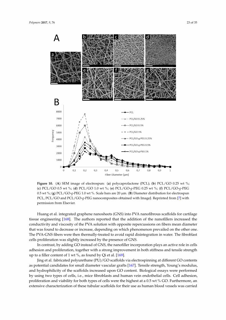
Polymers 2017, 9, 76 23 of 35Polymers 2017, 9, 76 23 of 35
Figure 10. (A) SEM image of electrospun: (a) polycaprolactone (PCL); (b) PCL/GO 0.25 wt %; (c) PCL/GO 0.5 wt %; (d) PCL/GO 1.0 wt %; (e) PCL/GO-g-PEG 0.25 wt %; (f) PCL/GO-g-PEG 0.5 wt %; (g) PCL/GO-g-PEG 1.0 wt %. Scale bars are 20 μm. (B) Diameter distribution for electrospun PCL, PCL/GO and PCL/GO-g-PEG nanocomposites obtained with ImageJ. Reprinted from [7] with permission from Elsevier.
Huang et al. integrated graphene nanosheets (GNS) into PVA nanofibrous scaffolds for cartilage tissue engineering [168]. The authors reported that the addition of the nanofillers increased the conductivity and viscosity of the PVA solution with opposite repercussions on fibers mean diameter that was found to decrease or increase, depending on which phenomenon prevailed on the other one. The PVA-GNS fibers were then thermally-treated to avoid rapid disintegration in water. The fibroblast cells proliferation was slightly increased by the presence of GNS.
In contrast, by adding GO instead of GNS, the nanofiller incorporation plays an active role in cells adhesion and proliferation, together with a strong improvement in both stiffness and tensile strength up to a filler content of 1 wt %, as found by Qi et al. [169].
Jing et al. fabricated polyurethane (PU)/GO scaffolds via electrospinning at different GO contents as potential candidates for small diameter vascular grafts [167]. Tensile strength, Young’s modulus, and hydrophilicity of the scaffolds increased upon GO content. Biological essays were performed by using two types of cells, i.e., mice fibroblasts and human vein endothelial cells. Cell adhesion, proliferation and viability for both types of cells were the highest at a 0.5 wt % GO. Furthermore, an extensive characterization of these tubular scaffolds for their use as human blood vessels was carried out, by highlighting that tubular PU/GO scaffolds containing 0.5 wt % GO meet the requirements to be used in vascular tissue engineering.
Figure 10. (A) SEM image of electrospun: (a) polycaprolactone (PCL); (b) PCL/GO 0.25 wt %;(c) PCL/GO 0.5 wt %; (d) PCL/GO 1.0 wt %; (e) PCL/GO-g-PEG 0.25 wt %; (f) PCL/GO-g-PEG0.5 wt %; (g) PCL/GO-g-PEG 1.0 wt %. Scale bars are 20 µm. (B) Diameter distribution for electrospunPCL, PCL/GO and PCL/GO-g-PEG nanocomposites obtained with ImageJ. Reprinted from [7] withpermission from Elsevier.
Huang et al. integrated graphene nanosheets (GNS) into PVA nanofibrous scaffolds for cartilagetissue engineering [168]. The authors reported that the addition of the nanofillers increased theconductivity and viscosity of the PVA solution with opposite repercussions on fibers mean diameterthat was found to decrease or increase, depending on which phenomenon prevailed on the other one.The PVA-GNS fibers were then thermally-treated to avoid rapid disintegration in water. The fibroblastcells proliferation was slightly increased by the presence of GNS.
In contrast, by adding GO instead of GNS, the nanofiller incorporation plays an active role in cellsadhesion and proliferation, together with a strong improvement in both stiffness and tensile strengthup to a filler content of 1 wt %, as found by Qi et al. [169].
Jing et al. fabricated polyurethane (PU)/GO scaffolds via electrospinning at different GO contentsas potential candidates for small diameter vascular grafts [167]. Tensile strength, Young’s modulus,and hydrophilicity of the scaffolds increased upon GO content. Biological essays were performedby using two types of cells, i.e., mice fibroblasts and human vein endothelial cells. Cell adhesion,proliferation and viability for both types of cells were the highest at a 0.5 wt % GO. Furthermore, anextensive characterization of these tubular scaffolds for their use as human blood vessels was carried
Polymers 2017, 9, 76 24 of 35
out, by highlighting that tubular PU/GO scaffolds containing 0.5 wt % GO meet the requirements tobe used in vascular tissue engineering.
Interestingly, electrospinning was even used to generate a polymer template for achievingfree-standing GO films for neuronal tissue engineering [170]. In their work, Jin et al. developeda novel fabrication method of free-standing reduced graphene oxide (RGO) films by vacuum filteringGO aqueous solutions through a nanofibrous membrane [170]. The GO was subsequently reduced intoRGO and finally functionalized with fibronectin (FN). The presence of electrospun PVC mat endowedgraphene films with a controllable patterning that increased the RGO roughness, particularly suitableto increase cell attachment and proliferation, due to the synergistic effects of the reactive functionalgroups in the FN protein and the large surface roughness and area of the graphene films.
The same authors reported the fabrication of PAN/RGO nanofibrous scaffold by combiningelectrospinning process and chemical reduction [157]. PAN/RGO displayed high mechanicalproperties (tensile strain = 18.5% and tensile stress = 1.38 MPa). Biological test results showedthat nanocomposites exhibited excellent biocompatibility, however very close to that of PAN,and high electrical conductivity, thus showing potential in the field of cell culture scaffolds forelectrical stimulation.
4.3. Polymeric Nanomats Containing Other Nanocarbons
In the scientific literature, few papers report on the use of 0D nanocarbons for the fabricationof electrospun polymeric scaffolds for tissue engineering. Among these, nanodiamonds were addedto PLA and PLGA for bone tissue repairs, and to chitosan for skin tissue engineering/woundhealing [173–176], whereas water-soluble fullerenes were used to endow PLLA nanomats withfluorescent properties for bioimaging [104,177].
Nanodiamonds were integrated into a polymer solution and electrospun to develop ananocomposite scaffold containing PLGA loaded with diamond nanoparticles [173]. Thecytocompatibility experiments, carried out by seeding primary human mesenchymal stem cells(hMSCs) on the scaffolds, showed that addition of diamond nanoparticles did not affect cellproliferation, and no cytotoxic cellular response was detected within 9 days in culture. Nanoindentationmeasurements revealed the presence of higher hardness zones at certain points on the fibers ofPLGA-nanodiamonds composites, likely owing to nanoparticles reinforcing mechanism. This studydemonstrates that PLGA nanofibers can be reinforced with nanodiamonds without adversely affectingcell behavior, thus displaying a good potential for future application in bone tissue engineering.Parizek and coworkers [174] investigated the structure-property relationship of PLGA/nanodiamondssystem in the case of high nanofiller loadings (23 wt %). The authors reported that ND particleswere either found to be arranged in a bead-like configuration in the central part of the fibers or toform clusters protruding from the fibers. The presence of ND resulted in meshes constituted bythicker fibers (with a +25% relative increase in diameter), however displaying smaller areas of pores(−64%). The PLGA-ND samples showed higher mechanical performance and improved bioactivity onhuman osteoblast-like MG-63 cells, which were found to be polygonal or spindle-like in shape, anddistributed homogeneously onto the membranes. Within 7 days after seeding, cell number was foundto rise continuously, and at the end of the experiment, these cells were able to create a confluent layer.The cell viability ranged from 92% to 97% and no significant inflammatory activity was detected.
Pereira et al. prepared electrospun nanomats based on PLA and low loadings of nanodiamonds(from 0.1 to 1 wt %) for bone tissue engineering [176]. Substantially, the results achieved are ingood agreement with those reported for PLGA/ND systems: incorporating NDs into the biopolymerdetermined the enhancement of hydrophilicity with positive effects on cell adhesion and proliferation,while no cytotoxic effects were recognized in osteoblasts (L929 cells).
Mahdavi et al. [175] reported the successful electrospinning of chitosan-based biopolymerscontaining bacterial cellulose (33 wt %) and medical grade nanodiamonds (3 nm; up to 3 wt %) for skintissue engineering. Morphological analysis put into evidence that introducing diamond nanoparticles

Polymers 2017, 9, 76 25 of 35
facilitated the electrospinning process with a decrease in the size of fibers (from 180 to 70 nm) anda highly uniform distribution of diameters and lengths. The authors found that beyond 1 wt %,percolation networks of nanoparticles were formed thus improving the properties of the nanofibrousmats: the enhancement of tensile strength (from 13 to 25 MPa) and hydrophilicity (favorablefor cell attachment), as well as the possibility to tune the water vapor permeability (from 342 to423 µg·Pa−1·s−1·m−1) suggest that nanodiamonds-modified mats are potentially suitable for woundhealing applications.
Liu et al. reported the preparation of PLLA nanofibers encapsulated with water-soluble fullerenenanoparticles by blend electrospinning [104]. The nanofibers were uniform, with average diametersranging from 300 to 600 nm. Water-soluble fullerene nanoparticles were encapsulated within thenanofibers, forming a core–shell structure and thereby endowing the resulting nanocomposite matswith excellent hydrophilicity. The mechanical properties, although lower than those of the pure matrix,were however desirable to tissue-engineered materials. The in vitro biological imaging experimentsindicated that fullerene nanoparticles were released from the nanofibers and penetrated into humanliver carcinoma HepG-2 cells. The integration of water-soluble fullerene nanoparticles within PLLAnanofibers allows us to achieve fluorescent biomaterials that are particularly promising for applicationsin bioimaging and drug delivery.
The same authors prepared a similar system by adding paclitaxel [177]. The delivery testsperformed in vitro demonstrated that the fullerene content greatly influenced the release rate ofpaclitaxel. Furthermore, while the drug paclitaxel inhibited the proliferation of HepG-2 cells effectively,the fluorescent signal of fullerenes in HepG-2 cells reflected the growth state of cells clearly. Hence,these ternary scaffolds could have a significant potential for tissue engineering, drug delivery andbioimaging applications.
5. Conclusions
Most of the electrospun scaffolds containing nanocarbons summarized in this review have provedto be good substrates for the adhesion, growth and differentiation of cells and stem cells.
Among the nanocarbons, 1D CNTs and 2D graphene-based nanomaterials are the mostwidespread. The use of 0D nanocarbons, such as nanodiamonds and fullerenes, is less investigated.
Generally speaking, either CNTs or graphene were used with a wide range of polymers,thus generally leading to a remarkable enhancement of mechanical and electrical properties.
In particular, CNTs are successfully used, especially for repairing injured nerve tissues,since they possess a unique combination of high electrical conductivity and strong ability to align alongthe fibers. These two features are particularly required for neuronal and muscle tissue engineeringapplications. However, due to their cytotoxicity, often CNTs have to undergo derivatization reactionswith hydrophilic moieties or biomolecules.
Among the graphene-family, GO is very promising in regenerating cartilage, bone, skin and nervetissues. In particular, due to its good cytocompatibility, GO can be used even without any furtherfunctionalization route and its surface texture offers a suitable environment for cells adhesion andproliferation, whereas its antimicrobial properties prevent the risk of inflammatory processes and itselectrical properties are found to be suitable for neuronal cells. Conversely, due to their geometry,2D lamellae display a strong tendency to re-aggregate after a certain concentration threshold, rangingfrom 2% to 5% weight content, depending on the processing conditions adopted, as well as hostpolymer properties.
On the other hand, nanodiamonds and fullerenes determine mechanical properties lowerthan CNTs and graphene but still desirable for certain applications, such as skin tissue repair.Nanodiamonds are mainly used to enhance the hydrophilicity of lactides, aiming at improvingbioactivity of the scaffolds.
Modified fullerenes are able to endow the scaffolds with additional properties, such asfluorescence, particularly useful for bioimaging, and controlled drug release. In fact, fullerenes

Polymers 2017, 9, 76 26 of 35
display a broad-spectrum reactivity that allows achieving water-soluble, fluorescent nanocarbons.Moreover, they are promising as carriers for a wide range of bioactive agents, owing to their strongability to penetrate into cells. They can be useful in the development of advanced scaffolds, whereregionally selective cell adhesion and directed growth is required, and in genomics and proteomics.
Author Contributions: Roberto Scaffaro, Andrea Maio, Francesco Lopresti and Luigi Botta conceived the idea forthis review; all the authors contributed equally to write the paper.
Conflicts of Interest: The authors declare no conflict of interest.
Abbreviations
AA Acetic acidBC Bacterial celluloseCA Cellulose acetateCCM Charge carrier mobilityCNT Carbon nanotubesCol CollagenCS ChitosanDCM DichloromethaneDMAc N,N-dimethylacetamideDMF N,N-dimethyl-formamideDMFA DimethylformaldehydeDMSO DimethylsulfoxideDWCNT Double-walled carbon nanotubeE Elastic modulusECM Extracellular matrixEtOH EthanolFN FibronectinGA GlutaraldehydeGEL GelatinGNS Graphene nanosheetsGO Graphene oxide
GO-g-[P(HEMA-g-CL)] Graphene oxide functionalized with poly(2-hydroxyethylmethacrylate)-graft-poly(e-caprolactone)
GO-g-PEG Pegylated graphene oxideHA HydroxyapatiteHFIP 1,1,1,3,3,3-hexafluoro-2-propanolMWCNT Multiwalled carbon nanotubeND NanodiamondNP NanoparticlePAA Polyacrylic acidPAN PolyacrylonitrilePANI PolyanilinePBAT Poly(butylene adipate-co-terephthalatePCL PolycaprolactonePELA Poly(ethylene glycol)-poly(D,L-lactide) copolymerPEO Polyethylene oxidePGA PolyglicolidePhOMe 4-methoxyphenylPLA Polylactic acidPLCL Poly(L-lactic acid-co-3-caprolactone)PLGA Poly(lactic-co-glycolic acid)PLLA Poly-L-lactidePNIPAm Poly(N-isopropyl acrylamide)PNIPAm-co-MAA Poly(N-isopropyl acrylamide-co-methacrylic acidPU PolyurethanePVA Poly(vinyl alcohol)PVC PolyvinylchloridePVP Polyvinyl pyrrolidoneRGD Arginine-glycine-aspartic acid peptideRGO Reduced graphene oxideSBF Simulated body fluidSDBS Sodium dodecyl benzene sulfonateSEBS Styrene–ethylene/butylene–styreneSF Silk fibroinSWCNT Single-walled carbon nanotubeTE Tissue engineeringTHF TetrahydrofuranTS Tensile strength

Polymers 2017, 9, 76 27 of 35
References
1. Lo Re, G.; Lopresti, F.; Petrucci, G.; Scaffaro, R. A facile method to determine pore size distribution in porousscaffold by using image processing. Micron 2015, 76, 37–45. [CrossRef] [PubMed]
2. Scaffaro, R.; Lopresti, F.; Botta, L.; Rigogliuso, S.; Ghersi, G. Preparation of three-layered porous PLA/PEGscaffold: Relationship between morphology, mechanical behavior and cell permeability. J. Mech. Behav.Biomed. Mater. 2016, 54, 8–20. [CrossRef] [PubMed]
3. Murphy, C.M.; Haugh, M.G.; O’Brien, F.J. The effect of mean pore size on cell attachment, proliferationand migration in collagen-glycosaminoglycan scaffolds for bone tissue engineering. Biomaterials 2010, 31,461–466. [CrossRef] [PubMed]
4. Kong, B.; Mi, S. Electrospun scaffolds for corneal tissue engineering: A review. Materials 2016, 9. [CrossRef]5. Braghirolli, D.I.; Steffens, D.; Pranke, P. Electrospinning for regenerative medicine: A review of the main
topics. Drug Discov. Today 2014, 19, 743–753. [CrossRef] [PubMed]6. Huang, Z.-M.; Zhang, Y.-Z.; Kotaki, M.; Ramakrishna, S. A review on polymer nanofibers by electrospinning
and their applications in nanocomposites. Compos. Sci. Technol. 2003, 63, 2223–2253. [CrossRef]7. Scaffaro, R.; Lopresti, F.; Maio, A.; Botta, L.; Rigogliuso, S.; Ghersi, G. Electrospun PCL/GO-g-PEG structures:
Processing-morphology-properties relationships. Compos. Part A Appl. Sci. Manuf. 2017, 92, 97–107.[CrossRef]
8. Song, J.; Gao, H.; Zhu, G.; Cao, X.; Shi, X.; Wang, Y. The preparation and characterization ofpolycaprolactone/graphene oxide biocomposite nanofiber scaffolds and their application for directingcell behaviors. Carbon 2015, 95, 1039–1050. [CrossRef]
9. Luo, Y.; Wang, K.; Chen, Q.; Xu, Y.; Xue, H.; Qian, Q. Preparation and characterization of electrospunLa(1-x)Ce(x)CoO(δ): Application to catalytic oxidation of benzene. J. Hazard. Mater. 2015, 296, 17–22.[CrossRef] [PubMed]
10. Sarbatly, R.; Krishnaiah, D.; Kamin, Z. A review of polymer nanofibres by electrospinning and theirapplication in oil-water separation for cleaning up marine oil spills. Mar. Pollut. Bull. 2016, 106, 8–16.[CrossRef] [PubMed]
11. Wen, P.; Zhu, D.-H.; Wu, H.; Zong, M.-H.; Jing, Y.-R.; Han, S.-Y. Encapsulation of cinnamon essential oil inelectrospun nanofibrous film for active food packaging. Food Control 2016, 59, 366–376. [CrossRef]
12. Cui, W.; Zhou, Y.; Chang, J. Electrospun nanofibrous materials for tissue engineering and drug delivery.Sci. Technol. Adv. Mater. 2016, 11, 014108. [CrossRef] [PubMed]
13. Ghosal, K.; Manakhov, A.; Zaj’ıcková, L.; Thomas, S. Structural and Surface Compatibility Study of ModifiedElectrospun Poly(ε-caprolactone)(PCL) Composites for Skin Tissue Engineering. AAPS PharmSciTech 2016,18, 72–81. [CrossRef]
14. Frohbergh, M.E.; Katsman, A.; Botta, G.P.; Lazarovici, P.; Schauer, C.L.; Wegst, U.G.K.; Lelkes, P.I. Electrospunhydroxyapatite-containing chitosan nanofibers crosslinked with genipin for bone tissue engineering.Biomaterials 2012, 33, 9167–9178. [CrossRef] [PubMed]
15. Jiang, X.; Mi, R.; Hoke, A.; Chew, S.Y. Nanofibrous nerve conduit-enhanced peripheral nerve regeneration.J. Tissue Eng. Regen. Med. 2014, 8, 377–385. [CrossRef] [PubMed]
16. Hinderer, S.; Layland, S.L.; Schenke-Layland, K. ECM and ECM-like materials—Biomaterials for applicationsin regenerative medicine and cancer therapy. Adv. Drug Deliv. Rev. 2015, 97, 260–269. [CrossRef] [PubMed]
17. Scaffaro, R.; Lopresti, F.; Botta, L.; Rigogliuso, S.; Ghersi, G. Integration of PCL and PLA in a monolithicporous scaffold for interface tissue engineering. J. Mech. Behav. Biomed. Mater. 2016, 63, 303–313. [CrossRef][PubMed]
18. Scaffaro, R.; Lopresti, F.; Botta, L.; Rigogliuso, S.; Ghersi, G. Melt Processed PCL/PEG Scaffold With DiscretePore Size Gradient for Selective Cellular Infiltration. Macromol. Mater. Eng. 2016, 301, 182–190. [CrossRef]
19. Scaffaro, R.; Lopresti, F.; Maio, A.; Sutera, F.; Botta, L. Development of polymeric functionally graded scaffold:A brief review. J. Appl. Biomater. Funct. Mater. 2016, in press. [CrossRef] [PubMed]
20. Surucu, S.; Masur, K.; Sasmazel, H.T.; Von Woedtke, T.; Weltmann, K.D. Atmospheric plasma surfacemodifications of electrospun PCL/chitosan/PCL hybrid scaffolds by nozzle type plasma jets for usage ofcell cultivation. Appl. Surf. Sci. 2016, 385, 400–409. [CrossRef]
21. Bauer, A.J.; Wu, Y.; Li, B. Electrospun Poly(ε-caprolactone)/Polyhedral Oligomeric Silsesquioxane-BasedCopolymer Blends: Evolution of Fiber Internal Structures. Macromol. Biosci. 2016. [CrossRef] [PubMed]
Polymers 2017, 9, 76 28 of 35
22. Ghorbani, F.M.; Kaffashi, B.; Shokrollahi, P.; Seyedjafari, E.; Ardeshirylajimi, A. PCL/chitosan/Zn-dopednHA electrospun nanocomposite scaffold promotes adipose derived stem cells adhesion and proliferation.Carbohydr. Polym. 2015, 118, 133–142. [CrossRef] [PubMed]
23. Liao, G.-Y.; Zhou, X.-P.; Chen, L.; Zeng, X.-Y.; Xie, X.-L.; Mai, Y.-W. Electrospun alignedPLLA/PCL/functionalised multiwalled carbon nanotube composite fibrous membranes and theirbio/mechanical properties. Compos. Sci. Technol. 2012, 72, 248–255. [CrossRef]
24. Zhu, W.; Masood, F.; O’Brien, J.; Zhang, L.G. Highly aligned nanocomposite scaffolds by electrospinningand electrospraying for neural tissue regeneration. Nanomed. Nanotechnol. Biol. Med. 2015, 11, 693–704.[CrossRef] [PubMed]
25. Nitya, G.; Nair, G.T.; Mony, U.; Chennazhi, K.P.; Nair, S.V. In vitro evaluation of electrospun PCL/nanoclaycomposite scaffold for bone tissue engineering. J. Mater. Sci. Mater. Med. 2012, 23, 1749–1761. [CrossRef][PubMed]
26. Scaffaro, R.; Botta, L.; Lopresti, F.; Maio, A.; Sutera, F. Polysaccharide nanocrystals as fillers for PLA basednanocomposites. Cellulose 2017, 24, 447–478. [CrossRef]
27. Asmatulu, R.; Patrick, S.; Ceylan, M.; Ahmed, I.; Yang, S.-Y.; Nuraje, N. AntibacterialPolycaprolactone/Natural Hydroxyapatite Nanocomposite Fibers for Bone Scaffoldings. J. Bionanosci.2015, 9, 120–126. [CrossRef]
28. Zhang, C.; Wang, L.; Zhai, T.; Wang, X.; Dan, Y.; Turng, L.-S. The surface grafting of graphene oxide withpoly(ethylene glycol) as a reinforcement for poly(lactic acid) nanocomposite scaffolds for potential tissueengineering applications. J. Mech. Behav. Biomed. Mater. 2016, 53, 403–413. [CrossRef]
29. Maio, A.; Fucarino, R.; Khatibi, R.; Rosselli, S.; Bruno, M.; Scaffaro, R. A novel approach to prevent grapheneoxide re-aggregation during the melt compounding with polymers. Compos. Sci. Technol. 2015, 119, 131–137.[CrossRef]
30. Gu, H.; Tadakamalla, S.; Zhang, X.; Huang, Y.; Jiang, Y.; Colorado, H.A.; Luo, Z.; Wei, S.; Guo, Z. Epoxyresin nanosuspensions and reinforced nanocomposites from polyaniline stabilized multi-walled carbonnanotubes. J. Mater. Chem. C 2013, 1, 729–743. [CrossRef]
31. Wei, H.; Ding, D.; Wei, S.; Guo, Z. Anticorrosive conductive polyurethane multiwalled carbon nanotubenanocomposites. J. Mater. Chem. A 2013, 1, 10805–10813. [CrossRef]
32. Wei, H.; Gu, H.; Guo, J.; Wei, S.; Guo, Z. Electropolymerized polyaniline nanocomposites from multi-walledcarbon nanotubes with tuned surface functionalities for electrochemical energy storage. J. Electrochem. Soc.2013, 160, G3038–G3045. [CrossRef]
33. Gu, H.; Guo, J.; He, Q.; Jiang, Y.; Huang, Y.; Haldolaarachige, N.; Luo, Z.; Young, D.P.; Wei, S.; Guo, Z.Magnetoresistive polyaniline/multi-walled carbon nanotube nanocomposites with negative permittivity.Nanoscale 2014, 6, 181–189. [CrossRef] [PubMed]
34. Zhang, X.; Yan, X.; He, Q.; Wei, H.; Long, J.; Guo, J.; Gu, H.; Yu, J.; Liu, J.; Ding, D.; et al. Electricallyconductive polypropylene nanocomposites with negative permittivity at low carbon nanotube loading levels.ACS Appl. Mater. Interfaces 2015, 7, 6125–6138. [CrossRef] [PubMed]
35. McKeon-Fischer, K.D.; Flagg, D.H.; Freeman, J.W. Coaxial electrospun poly(e-caprolactone), multiwalledcarbon nanotubes, and polyacrylic acid/polyvinyl alcohol scaffold for skeletal muscle tissue engineering.J. Biomed. Mater. Res. A 2011, 99, 493–499. [CrossRef] [PubMed]
36. O’Connor, R.A.; McGuinness, G.B. Electrospun nanofibre bundles and yarns for tissue engineeringapplications: A review. Proc. Inst. Mech. Eng. Part H J. Eng. Med. 2016, 230, 987–998. [CrossRef][PubMed]
37. Biazar, E. Application of polymeric nanofibers in soft tissues regeneration. Polym. Adv. Technol. 2016, 27.[CrossRef]
38. Tamayol, A.; Akbari, M.; Annabi, N.; Paul, A.; Khademhosseini, A.; Juncker, D. Fiber-based tissueengineering: Progress, challenges, and opportunities. Biotechnol. Adv. 2013, 31, 669–687. [CrossRef][PubMed]
39. Bendrea, A.-D.; Cianga, L.; Cianga, I. Review paper: Progress in the field of conducting polymers for tissueengineering applications. J. Biomater. Appl. 2011, 26, 3–84. [CrossRef] [PubMed]
40. Suwantong, O. Biomedical applications of electrospun polycaprolactone fiber mats. Polym. Adv. Technol.2016, 27. [CrossRef]
Polymers 2017, 9, 76 29 of 35
41. Melke, J.; Midha, S.; Ghosh, S.; Ito, K.; Hofmann, S. Silk fibroin as biomaterial for bone tissue engineering.Acta Biomater. 2016, 31, 1–16. [CrossRef] [PubMed]
42. Corradini, E.; Curti, P.S.; Meniqueti, A.B.; Martins, A.F.; Rubira, A.F.; Muniz, E.C. Recent advances infood-packing, pharmaceutical and biomedical applications of zein and zein-based materials. Int. J. Mol. Sci.2014, 15, 22438–22470. [CrossRef] [PubMed]
43. Gentile, P.; Chiono, V.; Carmagnola, I.; Hatton, P.V. An overview of poly(lactic-co-glycolic) Acid (PLGA)-basedbiomaterials for bone tissue engineering. Int. J. Mol. Sci. 2014, 15, 3640–3659. [CrossRef] [PubMed]
44. Ercolani, E.; Del Gaudio, C.; Bianco, A. Vascular tissue engineering of small-diameter blood vessels:Reviewing the electrospinning approach. J. Tissue Eng. Regen. Med. 2015, 9, 861–888. [CrossRef] [PubMed]
45. Norouzi, M.; Boroujeni, S.M.; Omidvarkordshouli, N.; Soleimani, M. Advances in Skin Regeneration:Application of Electrospun Scaffolds. Adv. Healthc. Mater. 2015, 4, 1114–1133. [CrossRef] [PubMed]
46. Zhu, W.; O’Brien, C.; O’Brien, J.R.; Zhang, L.G. 3D nano/microfabrication techniques and nanobiomaterialsfor neural tissue regeneration. Nanomedicine 2014, 9, 859–875. [CrossRef]
47. Rocco, K.A.; Maxfield, M.W.; Best, C.A.; Dean, E.W.; Breuer, C.K. In vivo applications of electrospuntissue-engineered vascular grafts: A review. Tissue Eng. Part B Rev. 2014, 20, 628–640. [CrossRef] [PubMed]
48. Li, G.; Zhang, T.; Li, M.; Fu, N.; Fu, Y.; Ba, K.; Deng, S.; Jiang, Y.; Hu, J.; Peng, Q.; Lin, Y. Electrospun fibersfor dental and craniofacial applications. Curr. Stem Cell Res. Ther. 2014, 9, 187–195. [CrossRef] [PubMed]
49. Liu, H.; Li, X.; Zhou, G.; Fan, H.; Fan, Y. Electrospun sulfated silk fibroin nanofibrous scaffolds for vasculartissue engineering. Biomaterials 2011, 32, 3784–3793. [CrossRef] [PubMed]
50. Ji, L.; Meduri, P.; Agubra, V.; Xiao, X.; Alcoutlabi, M. Graphene-Based Nanocomposites for Energy Storage.Adv. Energy Mater. 2016, 6. [CrossRef]
51. Cui, Y.; Kundalwal, S.I.; Kumar, S. Gas barrier performance of graphene/polymer nanocomposites. Carbon2016, 98, 313–333. [CrossRef]
52. Bhattacharya, M. Polymer nanocomposites-A comparison between carbon nanotubes, graphene, and clay asnanofillers. Materials 2016, 9, 262. [CrossRef]
53. Papageorgiou, D.G.; Kinloch, I.A.; Young, R.J. Graphene/elastomer nanocomposites. Carbon 2015, 95,460–484. [CrossRef]
54. Norazlina, H.; Kamal, Y. Graphene modifications in polylactic acid nanocomposites: A review. Polym. Bull.2015, 72, 931–961. [CrossRef]
55. Mochalin, V.N.; Gogotsi, Y. Nanodiamond-polymer composites. Diam. Relat. Mater. 2015, 58, 161–171.[CrossRef]
56. Mittal, G.; Dhand, V.; Rhee, K.Y.; Park, S.-J.; Lee, W.R. A review on carbon nanotubes and graphene as fillersin reinforced polymer nanocomposites. J. Ind. Eng. Chem. 2015, 21, 11–25. [CrossRef]
57. Idumah, C.I.; Hassan, A.; Affam, A.C. A review of recent developments in flammability of polymernanocomposites. Rev. Chem. Eng. 2015, 31, 149–177. [CrossRef]
58. Ravichandran, R.; Sundarrajan, S.; Venugopal, J.R.; Mukherjee, S.; Ramakrishna, S. Advances in PolymericSystems for Tissue Engineering and Biomedical Applications. Macromol. Biosci. 2012, 12, 286–311. [CrossRef][PubMed]
59. Wendorff, J.H.; Agarwal, S.; Greiner, A. Nature of the Electrospinning Process—Experimental Observationsand Theoretical Analysis. In Electrospinning; John Wiley&Sons: Hoboken, NJ, USA, 2012; pp. 29–68.
60. Wendorff, J.H.; Agarwal, S.; Greiner, A. Medicinal Applications for Electrospun Nanofibers. In Electrospinning;John Wiley&Sons: Hoboken, NJ, USA, 2012; pp. 217–236.
61. Kuppan, P.; Sethuraman, S.; Krishnan, U.M. Interaction of human smooth muscle cells on random andaligned nanofibrous scaffolds of PHBV and PHBV-gelatin. Int. J. Polym. Mater. Polym. Biomater. 2016, 65,816–825. [CrossRef]
62. Yang, F.; Murugan, R.; Wang, S.; Ramakrishna, S. Electrospinning of nano/micro scale poly(L-lactic acid)aligned fibers and their potential in neural tissue engineering. Biomaterials 2005, 26, 2603–2610. [CrossRef][PubMed]
63. Li, D.; Wang, Y.; Xia, Y. Electrospinning nanofibers as uniaxially aligned arrays and layer-by-layer stackedfilms. Adv. Mater. 2004, 16, 361–366. [CrossRef]
64. Yao, Z.-C.; Gao, Y.; Chang, M.-W.; Ahmad, Z.; Li, J.-S. Regulating poly-caprolactone fiber characteristicsin-situ during one-step coaxial electrospinning via enveloping liquids. Mater. Lett. 2016, 183, 202–206.[CrossRef]

Polymers 2017, 9, 76 30 of 35
65. Kalwar, K.; Sun, W.-X.; Li, D.-L.; Zhang, X.-J.; Shan, D. Coaxial electrospinning of polycaprolactone@chitosan:Characterization and silver nanoparticles incorporation for antibacterial activity. React. Funct. Polym. 2016,107, 87–92. [CrossRef]
66. Vysloužilová, L.; Valtera, J.; Pejchar, K.; Beran, J.; Lukáš, D. Design of Coaxial Needleless ElectrospinningElectrode with Respect to the Distribution of Electric Field. Appl. Mech. Mater. 2014, 693, 394–399. [CrossRef]
67. Yarin, A.L. Coaxial electrospinning and emulsion electrospinning of core-shell fibers. Polym. Adv. Technol.2011, 22, 310–317. [CrossRef]
68. Chen, H.; Wang, N.; Di, J.; Zhao, Y.; Song, Y.; Jiang, L. Nanowire-in-microtube structured core/shell fibersvia multifluidic coaxial electrospinning. Langmuir 2010, 26, 11291–11296. [CrossRef] [PubMed]
69. Hollister, S.J. Porous scaffold design for tissue engineering. Nat. Mater. 2005, 4, 518–524. [CrossRef][PubMed]
70. Allaf, R.M.; Rivero, I.V.; Ivanov, I.N. Fabrication of co-continuous poly(ε-caprolactone)/polyglycolide blendscaffolds for tissue engineering. J. Appl. Polym. Sci. 2015, 132, 1097–4628. [CrossRef]
71. Scaffaro, R.; Lopresti, F.; Botta, L.; Maio, A.; Sutera, F.; Mistretta, M.C.; La Mantia, F.P. A Facile andEco-friendly Route to Fabricate Poly(Lactic Acid) Scaffolds with Graded Pore Size. J. Vis. Exp. 2016.[CrossRef] [PubMed]
72. Scaffaro, R.; Lopresti, F.; Sutera, A.; Botta, L.; Fontana, R.M.; Puglia, A.M.; Gallo, G. Effect of PCL/PEG-BasedMembranes on Actinorhodin Production in Streptomyces coelicolor Cultivations. Macromol. Biosci. 2016, 16,686–693. [CrossRef] [PubMed]
73. Mi, H.-Y.; Jing, X.; Yu, E.; McNulty, J.; Peng, X.-F.; Turng, L.-S. Fabrication of triple-layered vascular scaffoldsby combining electrospinning, braiding, and thermally induced phase separation. Mater. Lett. 2015, 161,305–308. [CrossRef]
74. Levingstone, T.J.; Matsiko, A.; Dickson, G.R.; O’Brien, F.J.; Gleeson, J.P. A biomimetic multi-layeredcollagen-based scaffold for osteochondral repair. Acta Biomater. 2014, 10, 1996–2004. [CrossRef] [PubMed]
75. Murphy, C.M.; Schindeler, A.; Gleeson, J.P.; Yu, N.Y.C.; Cantrill, L.C.; Mikulec, K.; Peacock, L.; O’Brien, F.J.;Little, D.G. A collagen-hydroxyapatite scaffold allows for binding and co-delivery of recombinant bonemorphogenetic proteins and bisphosphonates. Acta Biomater. 2014, 10, 2250–2258. [CrossRef] [PubMed]
76. Li, D.; Chen, W.; Sun, B.; Li, H.; Wu, T.; Ke, Q.; Huang, C.; EI-Hamshary, H.; Al-Deyab, S.S.; Mo, X.A comparison of nanoscale and multiscale PCL/gelatin scaffolds prepared by disc-electrospinning.Colloids Surf. B Biointerfaces 2016, 146, 632–641. [CrossRef]
77. Correlo, V.M.; Boesel, L.F.; Pinho, E.; Costa-Pinto, A.R.; Alves da Silva, M.L.; Bhattacharya, M.; Mano, J.F.;Neves, N.M.; Reis, R.L. Melt-based compression-molded scaffolds from chitosan-polyester blends andcomposites: Morphology and mechanical properties. J. Biomed. Mater. Res. A 2009, 91, 489–504. [CrossRef]
78. Algul, D.; Sipahi, H.; Aydin, A.; Kelleci, F.; Ozdatli, S.; Yener, F.G. Biocompatibility of biomimetic multilayeredalginate–chitosan/β-TCP scaffold for osteochondral tissue. Int. J. Biol. Macromol. 2015, 79, 363–369.[CrossRef] [PubMed]
79. Su, D.S.; Perathoner, S.; Centi, G. Nanocarbons for the development of advanced catalysts. Chem. Rev. 2013,113, 5782–5816. [CrossRef] [PubMed]
80. Maio, A.; Agnello, S.; Khatibi, R.; Botta, L.; Alessi, A.; Piazza, A.; Buscarino, G.; Mezzi, A.; Pantaleo, G.;Scaffaro, R. A rapid and eco-friendly route to synthesize graphene-doped silica nanohybrids. J. Alloys Compd.2016, 664, 428–438. [CrossRef]
81. Scaffaro, R.; Maio, A.; Lopresti, F.; Giallombardo, D.; Botta, L.; Bondì, M.L.; Agnello, S. Synthesis andself-assembly of a PEGylated-graphene aerogel. Compos. Sci. Technol. 2016, 128, 193–200. [CrossRef]
82. Goenka, S.; Sant, V.; Sant, S. Graphene-based nanomaterials for drug delivery and tissue engineering.J. Control. Release 2014, 173, 75–88. [CrossRef] [PubMed]
83. Ruoff, R.S.; Qian, D.; Kam, W. Mechanical properties of carbon nanotubes: Theoretical predictions andexperimental measurements. C. R. Phys. 2003, 4, 993–1008. [CrossRef]
84. Li, H.J.; Lu, W.G.; Li, J.J.; Bai, X.D.; Gu, C.Z. Multichannel Ballistic Transport in Multiwall Carbon Nanotubes.Phys. Rev. Lett. 2005, 86601, 1–4. [CrossRef] [PubMed]
85. Jia, G.; Wang, H.; Yan, L.; Wang, X.; Pei, R.; Yan, T.; Zhao, Y.; Guo, X. Cytotoxicity of carbon nanomaterials:Single-wall nanotube, multi-wall nanotube, and fullerene. Environ. Sci. Technol. 2005, 39, 1378–1383.[CrossRef] [PubMed]
Polymers 2017, 9, 76 31 of 35
86. Kang, S.; Pinault, M.; Pfefferle, L.D.; Elimelech, M. Single-Walled Carbon Nanotubes Exhibit StrongAntimicrobial Activity. Langmuir 2007, 23, 8670–8673. [CrossRef] [PubMed]
87. Kang, S.; Herzberg, M.; Rodrigues, D.F.; Elimelech, M. Antibacterial effects of carbon nanotubes: Size doesmatter! Langmuir 2008, 24, 6409–6413. [CrossRef] [PubMed]
88. Yu, M. Strength and Breaking Mechanism of Multiwalled Carbon Nanotubes Under Tensile Load. Science2000, 287, 637–640. [CrossRef] [PubMed]
89. Demczyk, B.G.; Wang, M.; Cumingd, J.; Hetamn, M.; Han, W.; Zettl, A.; Ritchie, R. Direct mechanicalmeasurements of the tensile strength and elastic modulus of multi-walled carbon nanotubes. Mater. Sci.Eng. A. 2002, 334, 173–178. [CrossRef]
90. Liu, L.; Zhang, J.; Liu, F. Mechanical properties of graphene oxides. Nanoscale 2012, 5910–5916. [CrossRef][PubMed]
91. Suk, J.W.; Piner, R.D.; An, J.; Ruoff, R.S. Mechanical Properties of Monolayer Graphene Oxide. ACS Nano2010, 4, 6557–6564. [CrossRef] [PubMed]
92. Pinto, A.M.; Gonc, I.C.; Magalhães, F.D. Biointerfaces Graphene-based materials biocompatibility: A review.Colloids Surf. B Biointerfaces 2013, 111, 188–202. [CrossRef] [PubMed]
93. Liu, S.; Zeng, T.H.; Hofmann, M.; Burcombe, E.; Wei, J.; Jiang, R.; Kong, J.; Chen, Y. Antibacterial activityof graphite, graphite oxide, graphene oxide, and reduced graphene oxide: Membrane and oxidative stress.ACS Nano 2011, 5, 6971–6980. [CrossRef] [PubMed]
94. Zhu, Y.; Murali, S.; Cai, W.; Li, X.; Suk, J.W.; Potts, J.R.; Ruoff, R.S. Graphene and graphene oxide: Synthesis,properties, and applications. Adv. Mater. 2010, 22, 3906–3924. [CrossRef] [PubMed]
95. Kim, H.-J.; Kim, D.; Jung, S.; Yi, S.N.; Yun, Y.J.; Chang, S.K.; Ha, D.H. Charge Transport in Thick ReducedGraphene Oxide Film. J. Phys. Chem. C 2015, 119, 28685–28690. [CrossRef]
96. Eda, G.; Mattevi, C.; Yamaguchi, H.; Kim, H.; Chhowalla, M. Insulator to semimetal transition in grapheneoxide. J. Phys. Chem. C 2009, 113, 15768–15771. [CrossRef]
97. Chen, J.; Peng, H.; Wang, X.; Shao, F.; Yuan, Z.; Han, H. Graphene oxide exhibits broad-spectrumantimicrobial activity against bacterial phytopathogens and fungal conidia by intertwining and membraneperturbation. Nanoscale 2014, 6, 1879–1889. [CrossRef] [PubMed]
98. Singh, T.B.; Sariciftci, N.S.; Yang, H.; Yang, L.; Plochberger, B.; Sitter, H. Correlation of crystallineand structural properties of C60 thin films grown at various temperature with charge carrier mobility.Appl. Phys. Lett. 2007, 90, 213512. [CrossRef]
99. Lof, R.W.; van Veenendaal, M.A.; Koopmans, B.; Jonkman, H.T.; Sawatzky, G.A. Band gap, excitons, andCoulomb interaction in solid C60. Phys. Rev. Lett. 1992, 68, 3924–3927. [CrossRef] [PubMed]
100. Makarova, T.L.; Sundqvist, B.; Scharff, P.; Gaevski, M.E.; Olsson, E.; Davydov, V.A.; Rakhmanina, A.V.;Kashevarova, L.S. Electrical properties of two-dimensional fullerene matrices. Carbon 2001, 39, 2203–2209.[CrossRef]
101. Jansen, H.; Dobos, D.; Eisel, T.; Pernegger, H.; Eremin, V.; Wermes, N. Temperature dependence of chargecarrier mobility in single-crystal chemical vapour deposition diamond. J. Appl. Phys. 2013, 113, 13–27.[CrossRef]
102. Portet, C.; Yushin, G.; Gogotsi, Y. Electrochemical performance of carbon onions, nanodiamonds, carbonblack and multiwalled nanotubes in electrical double layer capacitors. Carbon 2007, 45, 2511–2518. [CrossRef]
103. Mochalin, V.N.; Shenderova, O.; Ho, D.; Gogotsi, Y. The properties and applications of nanodiamonds.Nat. Nanotechnol. 2012, 7, 11–23. [CrossRef] [PubMed]
104. Liu, W.; Wei, J.; Chen, Y.; Huo, P.; Wei, Y. Electrospinning of poly(L-lactide) nanofibers encapsulated withwater-soluble fullerenes for bioimaging application. ACS Appl. Mater. Interfaces 2013, 5, 680–685. [CrossRef][PubMed]
105. Dai, H. Carbon Nanotubes: Synthesis, Integration, and Properties. Acc. Chem. Res. 2002, 35, 1035–1044.[CrossRef] [PubMed]
106. Kumar, D.; Chaturvedi, P.; Saho, P.; Jha, P.; Chouksey, A.; Lal, M.; Rawat, J.S.B.S.; Tandon, R.P.;Chaudhury, P.K. Effect of single wall carbon nanotube networks on gas sensor response and detectionlimit. Sens. Actuators B Chem. 2017, 240, 1134–1140. [CrossRef]
107. Desforges, A.; Bridi, A.V.; Kadok, J.; Flahaut, E.; Le Normand, F.; Gleize, J.; Bellouard, C.; Ghanbaja, J.;Vigolo, B. Dramatic enhancement of double-walled carbon nanotube quality through a one-pot tunablepurification method. Carbon 2016, 110, 292–303. [CrossRef]

Polymers 2017, 9, 76 32 of 35
108. Mierczynski, P.; Mierczynska, A.; Maniukiewicz, W.; Maniecki, T.P.; Vasilev, K. MWCNTs as a catalyst inoxy-steam reforming of methanol. RSC Adv. 2016, 6, 81408–81413. [CrossRef]
109. Maio, A.; Botta, L.; Tito, A.C.; Pellegrino, L.; Daghetta, M.; Scaffaro, R. Statistical study of the influence ofCNTs purification and plasma functionalization on the properties of polycarbonate-CNTs nanocomposites.Plasma Process. Polym. 2014, 11, 664–677. [CrossRef]
110. Scaffaro, R.; Maio, A.; Tito, A.C. High performance PA6/CNTs nanohybrid fibers prepared in the melt.Compos. Sci. Technol. 2012, 72, 1918–1923. [CrossRef]
111. Scaffaro, R.; Maio, A.; Agnello, S.; Glisenti, A. Plasma functionalization of multiwalled carbon nanotubesand their use in the preparation of nylon 6-based nanohybrids. Plasma Process. Polym. 2012, 9, 503–512.[CrossRef]
112. Scaffaro, R.; Maio, A. Enhancing the mechanical performance of polymer based nanocomposites byplasma-modification of nanoparticles. Polym. Test. 2012, 31, 889–894. [CrossRef]
113. Sun, Y.-P.; Fu, K.; Lin, Y.; Huang, W. Functionalized carbon nanotubes: Properties and applications.Acc. Chem. Res. 2002, 35, 1096–1104. [CrossRef] [PubMed]
114. Scaffaro, R.; Botta, L.; Maio, A.; Mistretta, M.C.; La Mantia, F.P. Effect of Graphene Nanoplatelets onthe Physical and Antimicrobial Properties of Biopolymer-Based Nanocomposites. Materials 2016, 9, 351.[CrossRef]
115. Scaffaro, R.; Botta, L.; Maio, A.; Gallo, G. PLA graphene nanoplatelets nanocomposites: Physical propertiesand release kinetics of an antimicrobial agent. Compos. Part B Eng. 2017, 109, 138–146. [CrossRef]
116. Persson, H.; Yao, Y.; Klement, U.; Rychwalski, R.W. A simple way of improving graphite nanoplatelets (GNP)for their incorporation into a polymer matrix. Express Polym. Lett. 2012, 6, 142–147. [CrossRef]
117. Maio, A.; Giallombardo, D.; Scaffaro, R.; Piccionello, A.P.; Pibiri, I. Synthesis of a fluorinated grapheneoxide-silica nanohybrid: Improving oxygen affinity. RSC Adv. 2016, 6, 46037–46047. [CrossRef]
118. Agnello, S.; Alessi, A.; Buscarino, G.; Piazza, A.; Maio, A.; Botta, L.; Scaffaro, R. Structural and thermalstability of graphene oxide-silica nanoparticles nanocomposites. J. Alloys Compd. 2016, 695, 2054–2064.[CrossRef]
119. Scaffaro, R.; Maio, A. A green method to prepare nanosilica modified graphene oxide to inhibit nanoparticlesre-aggregation during melt processing. Chem. Eng. J. 2017, 308, 1034–1047. [CrossRef]
120. Thostenson, E.T.; Ren, Z.; Chou, T.-W. Advances in the science and technology of carbon nanotubes andtheir composites: A review. Compos. Sci. Technol. 2001, 61, 1899–1912. [CrossRef]
121. Taylor, R.; Walton, D.R.M. The chemistry of fullerenes. Nature 1993, 363, 685–693. [CrossRef]122. Krueger, A. New Carbon Materials: Biological Applications of Functionalized. Chemistry 2008, 39, 1382–1390.
[CrossRef] [PubMed]123. Shin, Y.C.; Lee, J.H.; Kim, M.J.; Hong, S.W.; Kim, B.; Hyun, J.K.; Choi, Y.S.; Park, J.; Han, D. Stimulating effect
of graphene oxide on myogenesis of C2C12 myoblasts on RGD peptide-decorated PLGA nanofiber matrices.J. Biol. Eng. 2015, 9, 1–10. [CrossRef] [PubMed]
124. Shin, Y.C.; Lee, J.H.; Jin, L.; Kim, M.J.; Kim, Y.J.; Hyun, J.K.; Jung, T.G.; Hong, S.W.; Han, D.W.Stimulated myoblast differentiation on graphene oxide-impregnated PLGA-collagen hybrid fibre matrices.J. Nanobiotechnol. 2015, 13, 21. [CrossRef] [PubMed]
125. Vicentini, N.; Gatti, T.; Salice, P.; Scapin, G.; Marega, C.; Filippini, F.; Menna, E. Covalent functionalizationenables good dispersion and anisotropic orientation of multi-walled carbon nanotubes in a poly(L-lactic acid)electrospun nano fi brous matrix boosting neuronal differentiation. Carbon 2015, 95, 725–730. [CrossRef]
126. Edwards, S.L.; Church, J.S.; Werkmeister, J.A.; Ramshaw, J.A.M. Tubular micro-scale multiwalled carbonnanotube-based scaffolds for tissue engineering. Biomaterials 2009, 30, 1725–1731. [CrossRef] [PubMed]
127. Guo, J.H.; Liu, Y.; Lv, Z.J.; Wei, W.J.; Guan, X.; Guan, Q.L.; Leng, Z.Q.; Zhao, J.Y.; Miao, H.; Liu, J.Potential Neurogenesis of Human Adipose-Derived Stem Cells on Electrospun Catalpol-Loaded CompositeNanofibrous Scaffolds. Ann. Biomed. Eng. 2015, 43, 2597–2608. [CrossRef] [PubMed]
128. Jin, G.-Z.; Kim, M.; Shin, U.S.; Kim, H.-W. Effect of carbon nanotube coating of aligned nanofibrous polymerscaffolds on the neurite outgrowth of PC-12 cells. Cell Biol. Int. 2011, 35, 741–745. [CrossRef] [PubMed]
129. Kabiri, M.; Oraee-yazdani, S.; Dodel, M.; Hanaee-ahvaz, H.; Soudi, S.; Seyedjafari, E.; Salehi, M.; Soleimani, M.Cytocompatibility of a conductive nanofibrous carbon nanotube/poly(L-lactic acid ) composite scaffoldintended for nerve tissue engineering. EXCLI J. 2015, 14, 851–860. [PubMed]
Polymers 2017, 9, 76 33 of 35
130. Liu, Y.; Lu, J.; Xu, G.; Wei, J.; Zhang, Z.; Li, X. Tuning the conductivity and inner structure of electrospunfibers to promote cardiomyocyte elongation and synchronous beating. Mater. Sci. Eng. C 2016, 69, 865–874.[CrossRef] [PubMed]
131. Ostrovidov, S.; Shi, X.; Zhang, L.; Liang, X.; Kim, S.B.; Fujie, T.; Ramalingam, M.; Chen, M.; Nakajima, K.;Al-Hazmi, F.; et al. Myotube formation on gelatin nanofibers—Multi-walled carbon nanotubes hybridscaffolds. Biomaterials 2014, 35, 6268–6277. [CrossRef] [PubMed]
132. Xu, J.; Xie, Y.; Zhang, H.; Ye, Z.; Zhang, W. Fabrication of PLGA/MWNTs composite electrospun fibrousscaffolds for improved myogenic differentiation of C2C12 cells. Colloids Surf. B Biointerfaces 2014, 123,907–915. [CrossRef] [PubMed]
133. Rodrigues, B.V.M.; Silva, A.S.; Melo, G.F.S.; Vasconscellos, L.M.R.; Marciano, F.R.; Lobo, A.O. Influence oflow contents of superhydrophilic MWCNT on the properties and cell viability of electrospun poly (butyleneadipate-co-terephthalate) fibers. Mater. Sci. Eng. C 2016, 59, 782–791. [CrossRef] [PubMed]
134. Shao, S.; Zhou, S.; Li, L.; Li, J.; Luo, C.; Wang, J.; Li, X.; Weng, J. Osteoblast function on electrically conductiveelectrospun PLA/MWCNTs nanofibers. Biomaterials 2011, 32, 2821–2833. [CrossRef] [PubMed]
135. Gandhi, M.; Yang, H.; Shor, L.; Ko, F. Post-spinning modification of electrospun nanofiber nanocompositefrom Bombyx mori silk and carbon nanotubes. Polymer 2009, 50, 1918–1924.
136. Pan, H.; Zhang, Y.; Hang, Y.; Shao, H.; Hu, X.; Xu, Y.; Feng, C. Significantly reinforced composite fiberselectrospun from silk fibroin/carbon nanotube aqueous solutions. Biomacromolecules 2012, 13, 2859–2867.[CrossRef] [PubMed]
137. Meng, Z.X.; Zheng, W.; Li, L.; Zheng, Y.F. Fabrication and characterization of three-dimensional nanofibermembrance of PCL-MWCNTs by electrospinning. Mater. Sci. Eng. C 2010, 30, 1014–1021. [CrossRef]
138. Macossay, J.; Sheikh, F.A.; Cantu, T.; Eubanks, T.M.; Salinas, M.E.; Farhangi, C.S.; Ahmad, H.; Hassan, M.S.;Khil, M.S.; Maffi, S.K.; Kim, H.; Bowlin, G.L. Imaging, spectroscopic, mechanical and biocompatibilitystudies of electrospun Tecoflex?? EG 80A nanofibers and composites thereof containing multiwalled carbonnanotubes. Appl. Surf. Sci. 2014, 321, 205–213. [CrossRef] [PubMed]
139. Meng, J.; Kong, H.; Han, Z.; Wang, C.; Zhu, G.; Xie, S.; Xu, H. Enhancement of nanofibrous scaffold ofmultiwalled carbon nanotubes/polyurethane composite to the fibroblasts growth and biosynthesis. J. Biomed.Mater. Res. Part A 2009, 88, 105–116. [CrossRef] [PubMed]
140. Meng, J.; Han, Z.; Kong, H.; Qi, X.; Wang, C.; Xie, S.; Xu, H. Electrospun aligned nanofibrous composite ofMWCNT/polyurethane to enhance vascular endothelium cells proliferation and function. J. Biomed. Mater.Res. Part A 2010, 95, 312–320. [CrossRef] [PubMed]
141. McCullen, S.D.; Stano, K.L.; Stevens, D.R.; Roberts, W.A.; Monteiro-Riviere, N.A.; Clarke, L.I.; Gorga, R.E.Development, optimization, and characterization of electrospun poly(lactic acid) nanofibers containingmulti-walled carbon nanotubes. J. Appl. Polym. Sci. 2007, 105, 1668–1678. [CrossRef]
142. Markowski, J.; Magiera, A.; Lesiak, M.; Sieron, A.L.; Pilch, J.; Blazewicz, S. Preparation and characterizationof nanofibrous polymer scaffolds for cartilage tissue engineering. J. Nanomater. 2015, 2015, 45. [CrossRef]
143. Mei, F.; Zhong, J.; Yang, X.; Ouyang, X.; Zhang, S.; Hu, X.; Ma, Q.; Lu, J.; Ryu, S.; Deng, X. Improvedbiological characteristics of poly(L-lactic acid) electrospun membrane by incorporation of multiwalledcarbon nanotubes/hydroxyapatite nanoparticles. Biomacromolecules 2007, 8, 3729–3735. [CrossRef] [PubMed]
144. Ribeiro, S.; Costa, P.; Ribeiro, C.; Sencadas, V.; Botelho, G.; Lanceros-Méndez, S. Electrospunstyrene-butadiene-styrene elastomer copolymers for tissue engineering applications: Effect ofbutadiene/styrene ratio, block structure, hydrogenation and carbon nanotube loading on physical propertiesand cytotoxicity. Compos. Part B 2014, 67, 30–38. [CrossRef]
145. Sharma, Y.; Tiwari, A.; Hattori, S.; Terada, D.; Sharma, A.K.; Ramalingam, M.; Kobayashi, H. Fabrication ofconducting electrospun nanofibers scaffold for three-dimensional cells culture. Int. J. Biol. Macromol. 2012,51, 627–631. [CrossRef] [PubMed]
146. Tiwari, A.; Sharma, Y.; Hattori, S.; Terada, D.; Sharma, A.K.; Turner, A.P.F.; Kobayashi, H. Influence ofPoly(N-isopropylacrylamide)–CNT–Polyaniline Three-Dimensional Electrospun Microfabric Scaffolds onCell Growth and Viability. Biopolymers 2013, 99, 334–341. [CrossRef] [PubMed]
147. Luo, Y.; Wang, S.; Shen, M.; Qi, R.; Fang, Y.; Guo, R.; Cai, H.; Cao, X.; Tomás, H.; Zhu, M.; Shi, X.Carbon nanotube-incorporated multilayered cellulose acetate nanofibers for tissue engineering applications.Carbohydr. Polym. 2013, 91, 419–427. [CrossRef] [PubMed]
Polymers 2017, 9, 76 34 of 35
148. Liao, H.; Qi, R.; Shen, M.; Cao, X.; Guo, R.; Zhang, Y.; Shi, X. Improved cellular response on multiwalledcarbon nanotube-incorporated electrospun polyvinyl alcohol/chitosan nanofibrous scaffolds. Colloids Surf.B Biointerfaces 2011, 84, 528–535. [CrossRef] [PubMed]
149. Bruinink, A.; Bitar, M.; Pleskova, M.; Wick, P.; Krug, H.F.; Maniura-weber, K. Addition of nanoscaledbioinspired surface features: A revolution for bone-related implants and scaffolds? J. Biomed. Mater. Res.Part A 2014, 102, 275–294. [CrossRef] [PubMed]
150. Wang, Z.; Tonderys, D.; Leggett, S.E.; Kendall, E.; Kiani, M.T.; Spitz, R.; Qiu, Y.; Wong, I.Y.; Hurt, R.H.Wrinkled, wavelength-tunable graphene-based surface topographies for directing cell alignment andmorphology. Carrbon 2016, 97, 14–24. [CrossRef] [PubMed]
151. Massoumi, B.; Ghandomi, F.; Abbasian, M. Surface functionalization of graphene oxide withpoly(2-hydroxyethyl methacrylate)-graft-poly(e-caprolactone) and its electrospun nanofibers with gelatin.Appl. Phys. A 2016, 122, 1000. [CrossRef]
152. Mahmoudi, N.; Simchi, A. On the biological performance of graphene oxide-modified chitosan/polyvinylpyrrolidone nanocomposite membranes: In vitro and in vivo effects of graphene oxide. Mater. Sci. Eng. C2017, 70, 121–131. [CrossRef] [PubMed]
153. Azarniya, A.; Eslahi, N.; Mahmoudi, N.; Simchi, A. Effect of graphene oxide nanosheets on thephysico-mechanical properties of chitosan / bacterial cellulose nanofibrous composites. Compos. Part A 2016,85, 113–122. [CrossRef]
154. Aznar-cervantes, S.; Martínez, J.G.; Bernabeu-esclapez, A.; Lozano-pérez, A.A.; Meseguer-olmo, L.;Otero, T.F.; Cenis, J.L. Bioelectrochemistry Fabrication of electrospun silk fi broin scaffolds coated withgraphene oxide and reduced graphene for applications in biomedicine. Bioelectrochemistry 2016, 108, 36–45.[CrossRef] [PubMed]
155. Gao, Y.; Wang, Y.; Wang, Y.; Cui, W. Fabrication of gelatin-based electrospun composite fibers for anti-bacterialproperties and protein adsorptino. Mar. Drugs 2016, 14. [CrossRef] [PubMed]
156. Liu, Y.; Park, M.; Kyoung, H.; Pant, B.; Choi, J.; Wan, Y.; Youb, J.; Park, S.; Kim, H. Facile preparation andcharacterization of poly(vinyl alcohol)/chitosan/graphene oxide biocomposite nanofibers. J. Ind. Eng. Chem.2014, 20, 4415–4420. [CrossRef]
157. Jin, L.; Yue, D.; Xu, Z.; Liang, G.; Zhang, Y.; Zhang, J.; Zhang, X.; Wang, Z. Fabrication, mechanical properties,and biocompatibility of reduced graphene oxidereinforced nanofiber mats. RSC Adv. 2014, 4, 35035–35041.[CrossRef]
158. Chaudhuri, B.; Mondal, B.; Kumar, S.; Sarkar, S.C. Myoblast differentiation and protein expression inelectrospun graphene oxide (GO)-poly(ε-caprolactone, PCL) composite meshes. Mater. Lett. 2016, 182,194–197. [CrossRef]
159. Patel, A.; Xue, Y.; Mukundan, S.; Rohan, L.C.; Sant, V.; Stolz, D.B.; Sant, S. Cell-InstructiveGraphene-Containing Nanocomposites Induce Multinucleated Myotube Formation. Ann. Biomed. Eng. 2016,44, 2036–2048. [CrossRef] [PubMed]
160. Mohammadi, S.; Shafiei, S.S.; Asadi-eydivand, M.; Ardeshir, M. Graphene oxide-enrichedpoly(ε-caprolactone) electrospun nanocomposite scaffold for bone tissue engineering applications. J. Bioact.Compat. Polym. Biomed. Appl. 2016. [CrossRef]
161. Ramazani, S.; Karimi, M. Aligned poly(ε-caprolactone )/ graphene oxide and reduced graphene oxidenanocomposite nano fi bers: Morphological, mechanical and structural properties. Mater. Sci. Eng. C 2015,56, 325–334. [CrossRef] [PubMed]
162. Liu, C.; Wong, H.M.; Yeung, K.W.K.; Tjong, S.C. Novel electrospun polylactic acid nanocomposite fiber matswith hybrid graphene oxide and nanohydroxyapatite reinforcements having enhanced biocompatibility.Polymers 2016, 8, 287. [CrossRef]
163. An, X.; Ma, H.; Liu, B.; Wang, J. Graphene oxide reinforced polylactic acid/polyurethane antibacterialcomposites. J. Nanomater. 2013, 2013, 373414. [CrossRef]
164. Luo, Y.; Shen, H.; Fang, Y.; Cao, Y.; Huang, J.; Zhang, M.; Dai, J.; Shi, X.; Zhang, Z. Enhanced proliferationand osteogenic differentiation of mesenchymal stem cells on graphene oxide-incorporated electrospunpoly(lactic-co-glycolic acid) nanofibrous mats. ACS Appl. Mater. Interfaces 2015, 7, 6331–6339. [CrossRef]
165. Shao, W.; He, J.; Sang, F.; Wang, Q.; Chen, L.; Cui, S.; Ding, B. Enhanced bone formation in electrospunpoly(L-lactic-co-glycolic acid )—Tussah silk fi broin ultra fi ne nano fi ber scaffolds incorporated withgraphene oxide. Mater. Sci. Eng. C 2016, 62, 823–834. [CrossRef] [PubMed]

Polymers 2017, 9, 76 35 of 35
166. Zhang, K.; Zheng, H.; Liang, S.; Gao, C. Acta Biomaterialia Aligned PLLA nanofibrous scaffolds coated withgraphene oxide for promoting neural cell growth. Acta Biomater. 2016, 37, 131–142. [CrossRef] [PubMed]
167. Jing, X.; Mi, H.; Salick, M.R.; Cordie, T.M.; Peng, X.; Turng, L. Electrospinning thermoplastic polyurethane /graphene oxide scaffolds for small diameter vascular graft applications. Mater. Sci. Eng. C 2015, 49, 40–50.[CrossRef] [PubMed]
168. Huang, C.L.; Peng, S.Y.; Wang, Y.J.; Chen, W.C.; Lin, J.H. Microstructure and characterization of electrospunpoly(vinyl alcohol) nanofiber scaffolds filled with graphene nanosheets. J. Appl. Polym. Sci. 2015, 132, 1–11.[CrossRef]
169. Qi, Y.Y.; Tai, Z.X.; Sun, D.F.; Chen, J.T.; Ma, H.B.; Yan, X.B.; Liu, B.; Xue, Q.J. Fabrication and characterizationof poly(vinyl alcohol)/graphene oxide nanofibrous biocomposite scaffolds. J. Appl. Polym. Sci. 2013, 127,1885–1894. [CrossRef]
170. Jin, L.; Zeng, Z.; Kuddannaya, S.; Yue, D.; Bao, J. Synergistic effects of a novel free-standing reduced grapheneoxide film and surface coating fibronectin on morphology, adhesion and proliferation of mesenchymal stemcells. J. Mater. Chem. B 2015, 3, 4338–4344. [CrossRef]
171. Scaffaro, R.; Botta, L.; Maio, A.; Gallo, G. Incorporation of an antibiotic in poly(lactic acid) and polypropyleneby melt processing. J. Appl. Biomater. Funct. Mater. 2016, 14, 240–247. [CrossRef] [PubMed]
172. Scaffaro, R.; Lopresti, F.; Botta, L.; Maio, A. Mechanical behavior of Polylactic acid/Polycaprolactone porouslayered functional composites. Compos. Part B Eng. 2016, 98, 70–77. [CrossRef]
173. Brady, M.A.; Renzing, A.; Douglas, T.E.L.; Liu, Q.; Wille, S.; Parizek, M.; Bacakova, L.; Kromka, A.;Jarosova, M.; Godier, G.; Warnke, P.H. Development of composite poly(lactide-co-Glycolide)-nanodiamondscaffolds for bone cell growth. J. Nanosci. Nanotechnol. 2015, 15, 1060–1069. [CrossRef] [PubMed]
174. Parizek, M.; Douglas, T.E.; Novotna, K.; Kromka, A.; Brady, M.A.; Renzing, A.; Voss, E.; Jarosova, M.;Palatinus, L.; Tesarek, P.; et al. Nanofibrous poly(lactide-co-glycolide) membranes loaded with diamondnanoparticles as promising substrates for bone tissue engineering. Int. J. Nanomed. 2012, 7, 1931–1951.
175. Mahdavi, M.; Mahmoudi, N.; Anaran, F.R.; Simchi, A. Electrospinning of nanodiamond-modifiedpolysaccharide nanofibers with physico-mechanical properties close to natural skins. Mar. Drugs 2016,14. [CrossRef] [PubMed]
176. Pereira, F.A.S.; Salles, G.N.; Rodrigues, B.V.M.; Marciano, F.R.; Pacheco-soares, C.; Lobo, A.O. Diamondnanoparticles into poly(lactic acid) electrospun fibers: Cytocompatible and bioactive scaffolds with enhancedwettability and cell adhesion. Mater. Lett. 2016, 183, 420–424. [CrossRef]
177. Liu, W.; Wei, J.; Chen, Y. Electrospun poly(l-lactide) nanofibers loaded with paclitaxel and water-solublefullerenes for drug delivery and bioimaging. New J. Chem. 2014, 38, 6223–6229. [CrossRef]
© 2017 by the authors. Licensee MDPI, Basel, Switzerland. This article is an open accessarticle distributed under the terms and conditions of the Creative Commons Attribution(CC BY) license (http://creativecommons.org/licenses/by/4.0/).